soft tissue sarcoma - international atomic energy...
TRANSCRIPT
IAEA Pediatric Radiation Oncology TrainingDr Laskar Version 1 June 2009
SOFT TISSUE SARCOMA (Non Rhabdomyosarcoma)

IAEA Pediatric Radiation Oncology TrainingDr Laskar Version 1 June 2009
Soft Tissue structuresFat,Muscles,Fibrous tissue, Blood vessels, Supporting cells of peripheral nervous system
Soft Tissue Sarcomas:- embryologically arise Primitive mesenchyme of mesoderm ,Some contribution from Neuro-ectoderm0.6 % of all malignanciesAge:- Adults 15 % in < 16 yrs
40 % in > 55yrsM : F = 1.12 :1

IAEA Pediatric Radiation Oncology TrainingDr Laskar Version 1 June 2009
Anatomic Sites
Arise any part of the body
45% -
Lower extremity17% -
Trunk14% -
Upper extremity12 % -
Retroperitonium10% -
Head & Neck

IAEA Pediatric Radiation Oncology TrainingDr Laskar Version 1 June 2009
Natural Histroy
Pseudo-capsule:- inner rim normal tissueOuter rim-
Oedema
& reactive zone of small newly formed vessels
low grade:- satellites of vital tumor found beyond pseudo-capsule excised by marginal excision.High grade:- additional "skip lesions", outside the reactive zone near thepseudocapsule so wider excision .tend to spread along longitudinal planes of muscular compartments. while fascial planes, nerves, vessels and bones are quite effective anatomical barriers.
Though locally aggressive they rarely extend in adjacent tissue compartment until late
IAEA Pediatric Radiation Oncology TrainingDr Laskar Version 1 June 2009
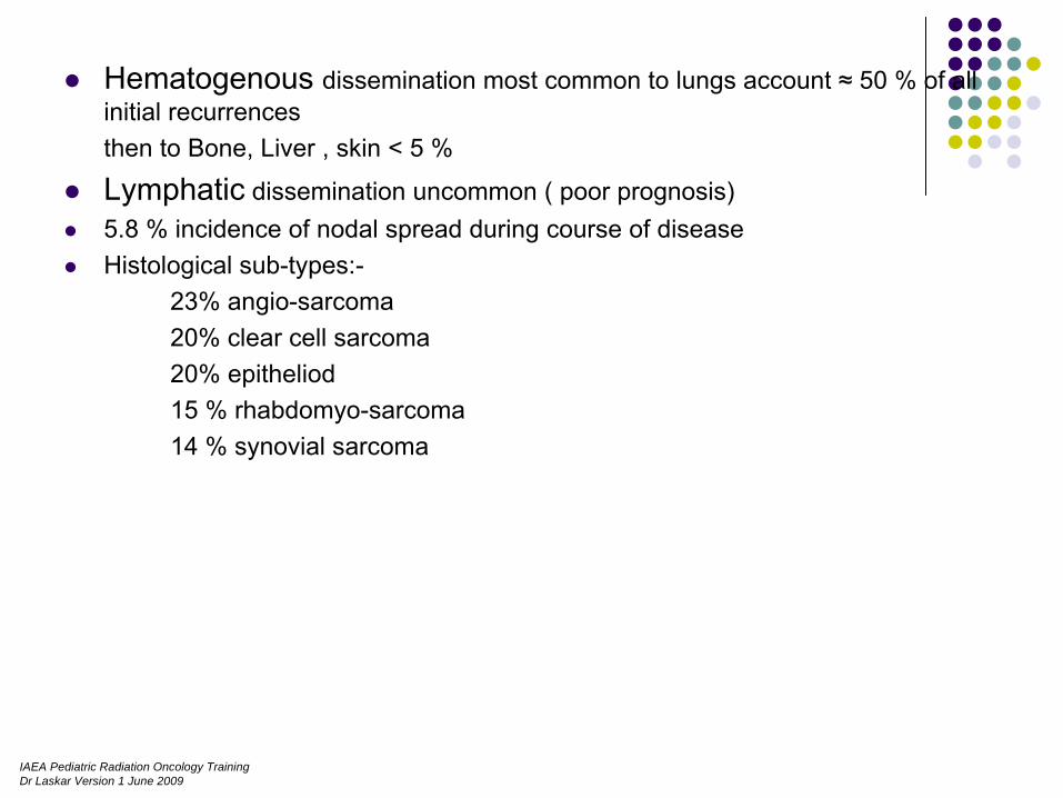
Hematogenous dissemination most common to lungs account ≈ 50 % of all initial recurrencesthen to Bone, Liver , skin < 5 %
Lymphatic dissemination uncommon ( poor prognosis)5.8 % incidence of nodal spread during course of diseaseHistological sub-types:-
23% angio-sarcoma20% clear cell sarcoma20% epitheliod15 % rhabdomyo-sarcoma14 % synovial sarcoma
IAEA Pediatric Radiation Oncology TrainingDr Laskar Version 1 June 2009
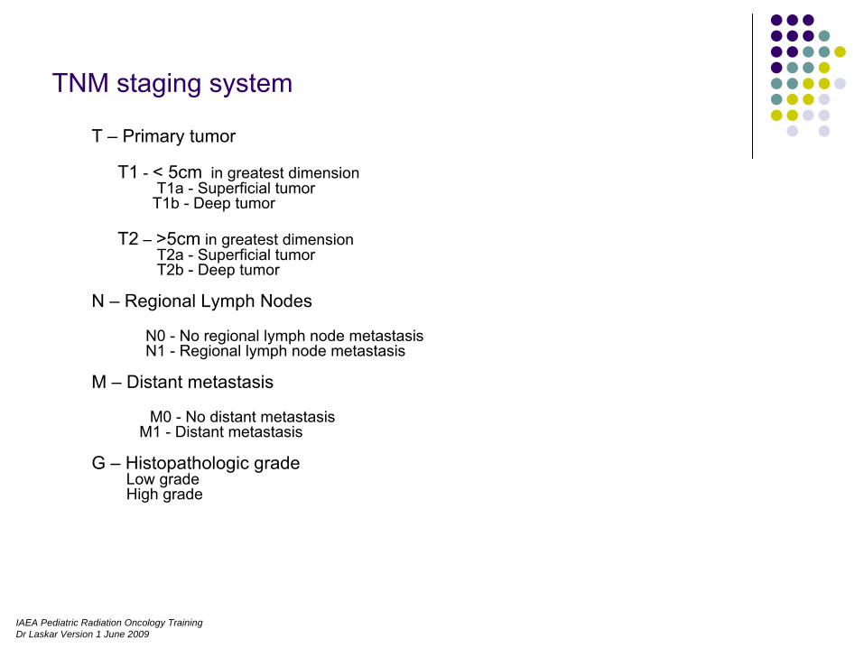
TNM staging system
T –
Primary tumor
T1
-
< 5cm
in greatest dimension
T1a -
Superficial tumor
T1b -
Deep tumor
T2
–
>5cm
in greatest dimension
T2a -
Superficial tumor
T2b -
Deep tumor
N –
Regional Lymph Nodes
N0 -
No regional lymph node metastasis
N1 -
Regional lymph node metastasis
M –
Distant metastasis
M0 -
No distant metastasis
M1 -
Distant metastasis
G –
Histopathologic grade
Low grade High grade
IAEA Pediatric Radiation Oncology TrainingDr Laskar Version 1 June 2009
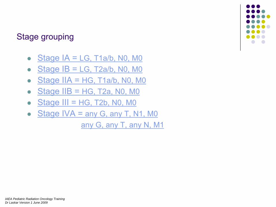
Stage grouping
Stage IA = LG, T1a/b, N0, M0Stage IB = LG, T2a/b, N0, M0Stage IIA = HG, T1a/b, N0, M0Stage IIB = HG, T2a, N0, M0Stage III = HG, T2b, N0, M0Stage IVA = any G, any T, N1, M0
any G, any T, any N, M1
IAEA Pediatric Radiation Oncology TrainingDr Laskar Version 1 June 2009
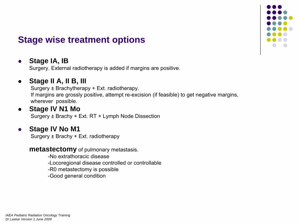
Stage wise treatment options
Stage IA, IBSurgery. External radiotherapy is added if margins are positive.
Stage II A, II B, III Surgery ± Brachytherapy + Ext. radiotherapy.If margins are grossly positive, attempt re-excision (if feasible) to get negative margins, wherever possible. Stage IV N1 MoSurgery ± Brachy
+ Ext. RT + Lymph Node Dissection
Stage IV No M1Surgery ± Brachy
+ Ext. radiotherapy
metastectomy of pulmonary metastasis.-No extrathoracic
disease-Locoregional disease controlled or controllable-R0 metastectomy
is possible-Good general condition
IAEA Pediatric Radiation Oncology TrainingDr Laskar Version 1 June 2009
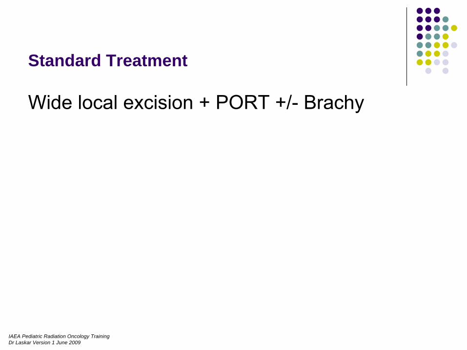
Standard Treatment
Wide local excision + PORT +/-
Brachy
IAEA Pediatric Radiation Oncology TrainingDr Laskar Version 1 June 2009
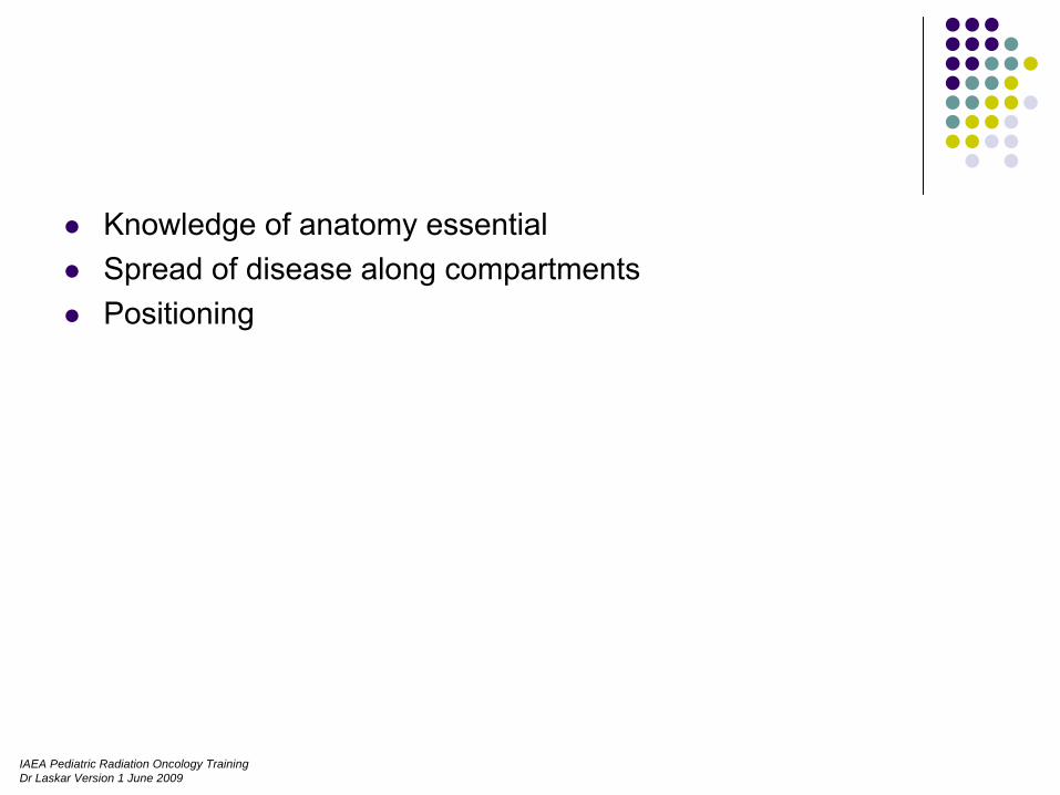
Knowledge of anatomy essentialSpread of disease along compartmentsPositioning
IAEA Pediatric Radiation Oncology TrainingDr Laskar Version 1 June 2009
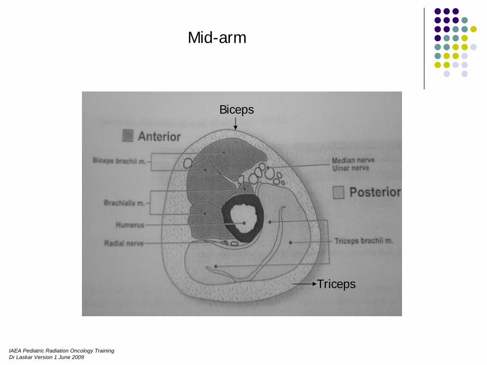
Mid-arm
Biceps
Triceps
IAEA Pediatric Radiation Oncology TrainingDr Laskar Version 1 June 2009
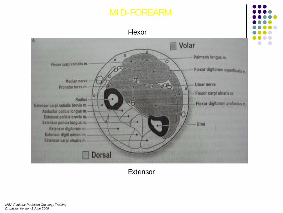
MID-FOREARM
Flexor
Extensor
IAEA Pediatric Radiation Oncology TrainingDr Laskar Version 1 June 2009
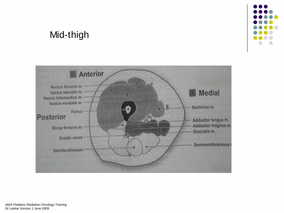
Mid-thigh

IAEA Pediatric Radiation Oncology TrainingDr Laskar Version 1 June 2009
Mid-leg
IAEA Pediatric Radiation Oncology TrainingDr Laskar Version 1 June 2009
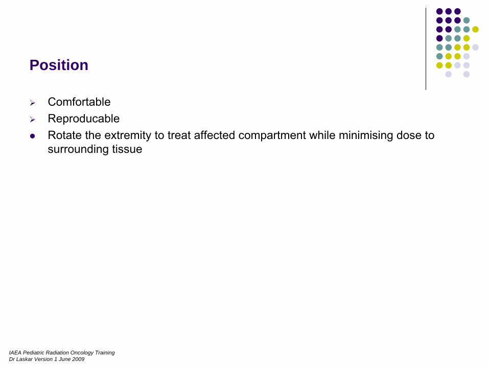
Position
Comfortable ReproducableRotate the extremity to treat affected compartment while minimising dose to surrounding tissue

IAEA Pediatric Radiation Oncology TrainingDr Laskar Version 1 June 2009
FROG-LEG position
Seperates ant. Thigh from post. & medial compartments
IAEA Pediatric Radiation Oncology TrainingDr Laskar Version 1 June 2009
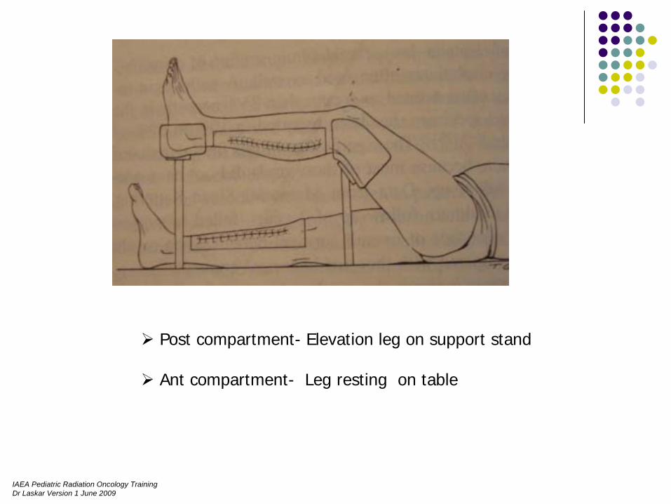
Post compartment- Elevation leg on support stand
Ant compartment- Leg resting on table

IAEA Pediatric Radiation Oncology TrainingDr Laskar Version 1 June 2009
‘ THROWING ’ position
Shoulder 90 abduction & max ext rotation
Adequately separates Biceps compart. From Triceps
ARM
IAEA Pediatric Radiation Oncology TrainingDr Laskar Version 1 June 2009
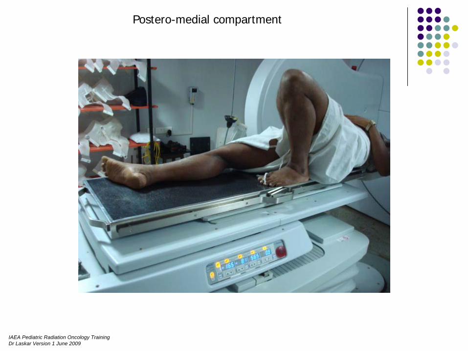
Postero-medial compartment
IAEA Pediatric Radiation Oncology TrainingDr Laskar Version 1 June 2009
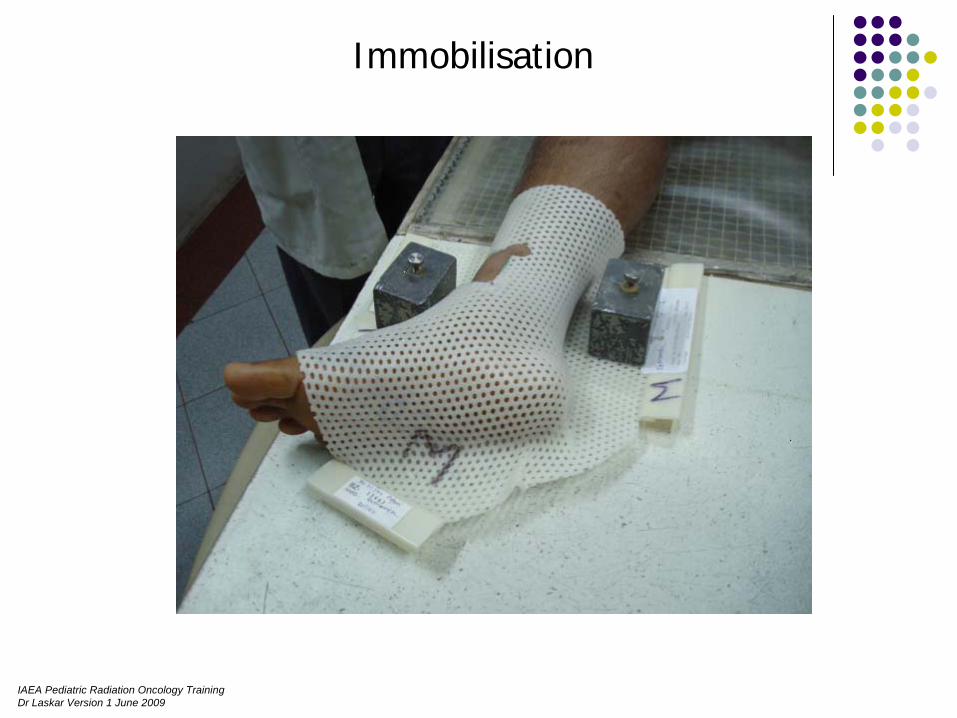
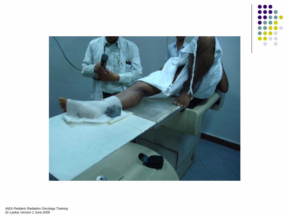
Immobilisation
IAEA Pediatric Radiation Oncology TrainingDr Laskar Version 1 June 2009

IAEA Pediatric Radiation Oncology TrainingDr Laskar Version 1 June 2009

IAEA Pediatric Radiation Oncology TrainingDr Laskar Version 1 June 2009

IAEA Pediatric Radiation Oncology TrainingDr Laskar Version 1 June 2009
Field placements
•• keep uninvolved compartment out of radiation portal keep uninvolved compartment out of radiation portal
•• Avoid joints as far as possibleAvoid joints as far as possible
•• Spare half circumference of uninvolved boneSpare half circumference of uninvolved bone
•• spare at least 1.5 spare at least 1.5 -- 2.0cm of limb circumference from 2.0cm of limb circumference from radiotherapy portal.radiotherapy portal.
•• Margins not extend beyond natural barriers ( Bone, Margins not extend beyond natural barriers ( Bone, fascialfascial planesplanes
•• Cover surgical scar & drain sitesCover surgical scar & drain sites
IAEA Pediatric Radiation Oncology TrainingDr Laskar Version 1 June 2009
Planning Target Volume (PTV):
Phase I - GTV+ Grade I: 4cm margin
Grade II&III: 6 - 8cm margin
Phase II - GTV+ 3cm margin
IAEA Pediatric Radiation Oncology TrainingDr Laskar Version 1 June 2009
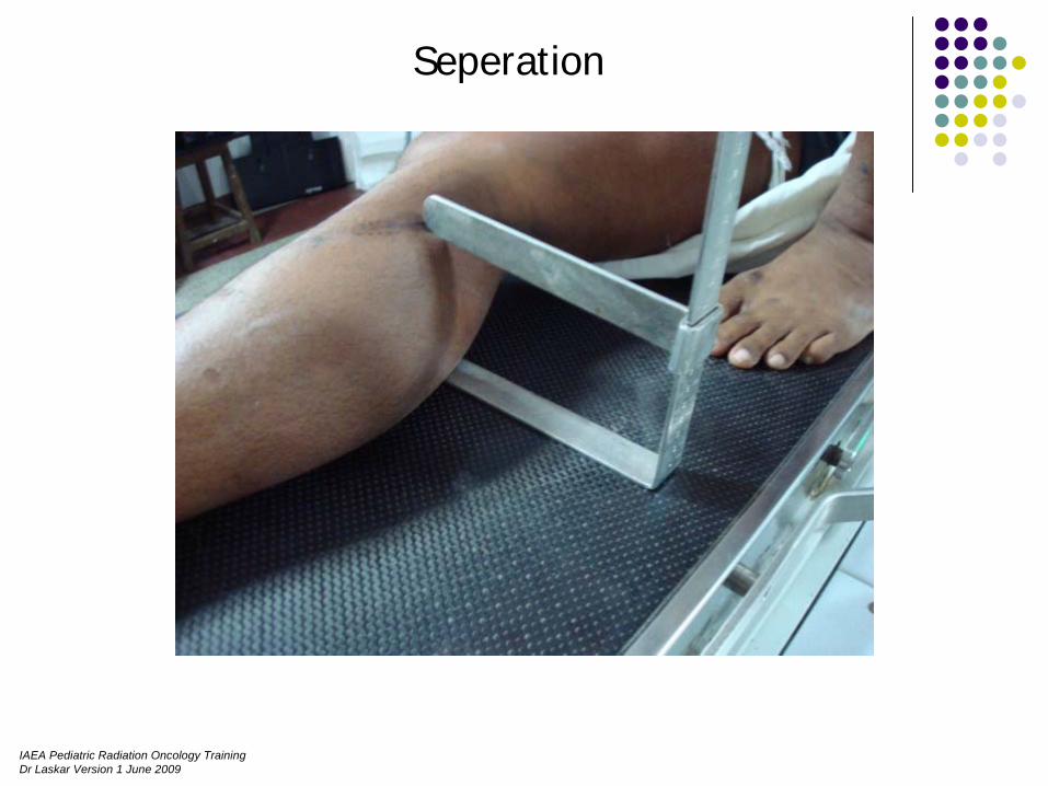
Seperation

IAEA Pediatric Radiation Oncology TrainingDr Laskar Version 1 June 2009
Dose
Phase I -
50Gy / 25# / 5 weeks
Phase II -
R 0: 10 -
12Gy / 5 -
6# / 1 weekR 1: 12 -
16Gy / 6 -
8# / 1 weekR 2: 16 -
20Gy / 8 -
10# / 2 weeks

IAEA Pediatric Radiation Oncology TrainingDr Laskar Version 1 June 2009
During Treatment
Skin carePhysiotherapy

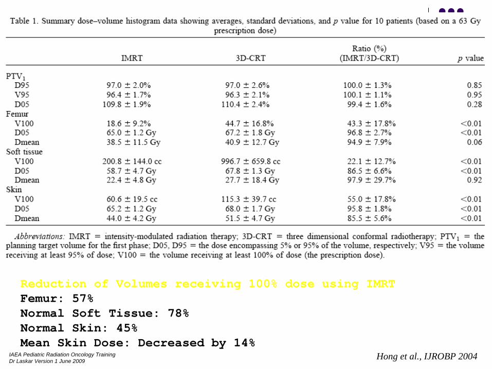
IAEA Pediatric Radiation Oncology TrainingDr Laskar Version 1 June 2009 Hong et al., IJROBP 2004
3D-CRT vs. IMRT FOR EXTREMITY SARCOMAS

IAEA Pediatric Radiation Oncology TrainingDr Laskar Version 1 June 2009
CAX
10cm SUP
3D-CRT IMRT
Hong et al., IJROBP 2004
IAEA Pediatric Radiation Oncology TrainingDr Laskar Version 1 June 2009
Reduction of Volumes receiving 100% dose using IMRTFemur: 57%Normal Soft Tissue: 78%Normal Skin: 45%Mean Skin Dose: Decreased by 14%
Hong et al., IJROBP 2004
IAEA Pediatric Radiation Oncology TrainingDr Laskar Version 1 June 2009
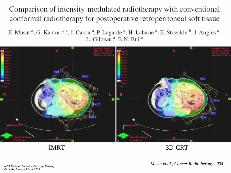
IMRT 3D-CRT
Musat et al., Cancer Radiotherapy 2004

IAEA Pediatric Radiation Oncology TrainingDr Laskar Version 1 June 2009
Musat et al., Cancer Radiotherapy 2004
IAEA Pediatric Radiation Oncology TrainingDr Laskar Version 1 June 2009
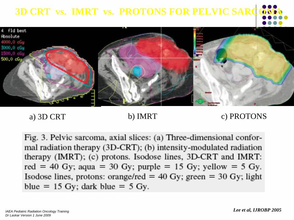
a) 3D CRT b) IMRT c) PROTONS
3D CRT vs. IMRT vs. PROTONS FOR PELVIC SARCOMA
Lee et al, IJROBP 2005

IAEA Pediatric Radiation Oncology TrainingDr Laskar Version 1 June 2009 IJROBP 2002