slid - apps.dtic.mil
TRANSCRIPT

00 DAMD17-86-C-606300
"OIX FILE CGP'If SFt.UHC ON RPmAToNSHIP OF RYDo•LIMDA
00 PTSYNA IC NEUROTOXINS
9~AIWUAL REPORT
Ah"IHON T. TUROGER A. MILLER DTICNOBUHIRO MORI0
SEP 2 9 1871
SliDMAR CH 5, 1987
Supported by
U. S. ARMY MEDICAL RESEARCE AND DEVELOPHM T COMMANDFort Detrics, Frederick, Maryland 21701-5012
Contract No. DAMD17-86-C-6063
Colorado State UniversityDepartment of Biochemistry
Fort Collins, Colorad6 80523.
Approved for public release; distribution unlimited
The findings in this report are not to be construed as an officialDepartment of the Army position unless so designated by otherauthorized documents.

CLASIICTIO O RTISPORT DOCUMENTATION. PAGE
Ia. REPORT SECURITY CLASSIFICATION lb. RESTRILTIVE MARKIONG'
Unclassified _______________________
Za& SAiCUINIM CLASSIFICTION AUTH4ORITY 3. OISTRIOUTION/AVARILITY OF REPORT
2b. flA4~TON/~NGPDI~A ~See. Distribution C1tateunt at end of-xaport.
4. PERFQMING ORGANIZATION RE PORT NUMBER(S S. MONITORIG diGAIZTIONREPORT NUMaER(S)
6a. NAME OF PERFORMING ORGAIZATION 6b. OFFICE SYMBO`L 7&. NAME OF MONITOWNG ORGANIZ ATION
Colorado State University 0I okik
6C. ADDRES S (City, Sftat, andE ZI Co*e) 7b. ADDRESS (CIMp Sftf & ZIP Cod*)
Department of BiochemistryFort Collins, Colorado 80523
B& NAME OF FUNDING /SPONISORING 8bW. OFFICE SYMBOL 9. PROCUREMENT INSTWWMNT IOENTIFIATiON NUMBER
ORGAIZATION. U. S. Army Medica1l ''''-- AD?-6C66Research & Develop. CommandI
SC. ADDRESS (City. SUat. an ZIP Code) 10. SOURCE OF FUNDING NUMBERS
Fort Detrick, Frederick, MD 21701-5012 ELMET NOCNESSI. TKOR N UNITPROGRMN10 NRO. MC61SIO NO. A61102A 10S112 A IA098
11. TITLE (.Incduf Security CudANt16OIFIJ
Structure -Function Relationship of Bydrophiliidae Postsyriaptic, Neurotoxiris (U)
12. PERSONAL AUTHOR(S)Tu, Anthony T., Miller, Roger A., and Mori, Nobuhiro
13s. TYPE OF RI;PORT 113b. TIME COVEAED 114. DATE OF REPORT (Tha.ýAfo&flt Doy) 115. PAGE COUNTAnnual lFRom 3/1/86 T02/2L8/7 1987 March 5
16. SUPPLEMENTARY NOTATION
17.' COSATI CODESý 13. SUBJECT TERMS (Continu on revevin if nuemryw and Weide b10100 number)
FIELD GROUP SUB.o5ROUP Keywords. - sea snake toxins, postsynaptic neurotoxins,chemical modification of toxins, hydrophilicity index oftoxin and acetylcholine receptor, amino acid sequence of
[19. ABST (Continue on Mrn if011 ane~r d 0*entfy by bWmck numbee) neurotoxins.
"".This annual report is comprised of three parts:A.,- The hydrophilicity index of Lapemis toxin from Lapemis hardvickii sea snake and the a-
subunit pf the acetylcholine receptor of Torpedo californc have been determined and
plotted versus the sequence position.
B. Laperis toxin is a competitive inhibitor of cz-bungarotoxin (uaBTX) in bind'-.ng to ACR.
Two artinine residues out of three which are present in Lapemis toxin were -modified
by phenylglyoxal.
C. The major rneurotoxin was isolated from the sea snake venom of Acalyptophis peronii.
captured in the Gulf of Thailand. Partial N-terminal sc.quence of the toxin was
identified up to 37 residues. Ten more residues beyond residue No. 37 were also
identified, but the exact positions of these ten residues have not yet been identified.
20. DISTRIBUTION /AVAILABILITY OF ABSTRACT 21. ABSTRACT SECURITY CLASSIFICATION
03 UNCLASSIFIEDAJNLIMITED X3 SAME AS RPT. C) DTIC USERS Unclassified -
22a., NAME OF RESPONSIBLE INDIVIDUAL 22b. TELEPHONE (Include Area Code) 22c. OFFICE SYMBOL
Mrs. Virginia M. Miller 301/663-7325 SGRD-RMI-S
L"D0 Formi 1473, JUN 86 Pre vious editions are obsolete. SECURITY CWASIFICATION OF THIS PAGqE

2
d. Title Page
Structure-Function Relationship of HydrophiidaePostsynaptic Neurotoxins
The report is comprised of three parts. They are:
A. Hydrophilicity Analysis of Lapemis Toxin and Acetyl-cholin. Receptor *-Subunit
B. Isolation and Chemical Modification of LapemisToxin from Lapemis hardwickii Toxin
C. Isolation and Amino Acid Sequence of Neurotoxinfrom the Venom of Sea Snake Acalytophis peronii
I|

3
0. Summary
A. The hydrophilicity index of Lapemis toxin from Lapemishardwickii sea snake and the a-subunit of the acetyl-c-holine receptor of .Toredo californica have beendetermined and plotted versus the sequence porition.The results show that the sea snake neurotoxin isquite hydrophilic and suggests an interaction at ahydrophilic region of the acetylcholine receptor.The analysis results of the acetylcholine receptorsubunit support the known information about: the intra-membrane subunit. Other studies' information hasbeen considered along with the hydrophilicity resultsand a model of binding interaction is proposed.
D. Lapemis toxin, the major 'toxin, was isolated fromthe venom of Lapemis hardwickii captured in the Gulfof Thailand. Acetylcholine receptor (ACR) was iso-lated from the electric organ tissue of Torpedocalifornica. Lapemis toxin is a competitive inhibi-tor of a-bungarotoxin (aBTX) in binding to ACR. Thearginine residues out of three which are present inLapemis toxin were modified by phenylglyoxal. Themodification experiment will be done on the ACR boundneurotoxin in the future.
C. The major neurotoxin was isolated from the sea snakevenom of Acalyptophis peronii captured in the Gulfof Thailand. The molecular weight was approximately6,500. Acalyptophis toxin bound to acetylcholinereceptor and was a competitive inhibitor of a-bungarotoxin binding. Partial N-terminal sequenceof the toxin was identified up to 37 residues. Tenmore residues beyond residue No. 37 were also identi-fied, but the exact positions of these ten residueshave not yet been identified. The complete sequencewill be identified in the near future.
f. Foreword
A. Hydrophilicity Analysis of Lapemis Toxin and Acetyl-choline Receptor a-Subunit
In order to elucidate the theoretical explana-tion of the neurotoxin (NT) acetylcholine receptor(ACR) interaction, the hydrophilicity index analysiswas employed.
Lapemis toxin is a short-chain postsynapticneurotoxin found in the venom of the sea snakeLapemis hardwickii. Lapemis toxin is a single poly-peptide of 60 amino acids. The isolation, character-ization, and sequence for Lapemis torin have beendetermined by Fox et al., 1977.

4
The acetylcholine receptor (ACR) is a ligand-gated ion-channel transmembrane glycoprotein which,in response to the binding of acetyIcholine, mediatesthe translocation of cations across the plasmamembrane in which the ACR resides.
The ACR has an overall molecular weight ofapproximately 270,000 and is comprised of four non-identical subunits with molecular weights of 40,000(a), 50,000 (0), 60,000 (y). and 65,000 (6), and thestoichiometry of a2A6. The a-subunit of the ACR has437 amino acid residues and one site of glycosylationof aspargine 141 (Martinez-Carrion et al., 1975;Raftery et al., 1980; Noda et al., 1982; Noda et al.,1983; Claudia et al., 1983; Nomoto et al., 1986).
Sea snake neurotoxins-bind to the a-subunit ofthe ACR and inhibit the binding of acetylcholine andinhibit the conformation change in the ACR thus block-ing the depolarization of the myocytes that occurs innormal nerve transmission to muscles (Endo et al., 1986).
It is well known that one of the most importantparameters of protein-protein interaction and proteinfolding and structure is due to hydrophobic-hydrophilicinteractions. To better understand the ACR-NT inter-action, the hydrophilicity index analysis has beendone. Through this analysis the nature of the inter-action has helped narrow the regions of importancebetween these two proteins. The more that is learnedabout this interaction will eventually lead to theexact chemical nature of the ACR-NT binding. Withthis knowledge, the structure-function of the normalfunctioning ACR and pathological conditions involvingthe ACR will be better understood.
3. Isolation and Chemical Modification of Lapemis Toxinfrom Lapermis hardwickii Toxin
Sea snake neurotoxins inhibit nerve transmissionat the neuromuscular junction. Lapemis toxin, themajor toxin isolated from Lapemis hardwickii venom,strongly binds to acetylcholine receptors. The toxinis one of many snake neurotoxins extensively investi-gated. It consists of 60 amino acid residues withmolecular weight of 6,800. Its amino acid sequencewas also established. However, the exact mechanismof binding to acetylcholine receptor has not beenelucidated yet. In order to understand the interac-tion of neurotoxin to acetylcholine receptor, neuro-toxin-ACR complex will be modified. It iz assumedthat the residues involved in the ACR binding shou.dnot be .modified. As control, arginine residues offree neurotox.'n was modified first and the resultwill be compared with that of bound toxin.

5
C. Isolation and Amino Acid Sequence of Neurotox .n fromthe Venom of Sea Snake Acalytophis peronii
Sea snakes (Family: Hydrophiidae) are a bundantin the tropical and subtropical zones of the IndianOcean and the Pacific Ocean. On the coast of theGulf of Thailand there are numerous numbers dvarieties of sea snakes. Lapemis hardwickiit venomwas e.tensively studied. AcalXptophis peroni wasalso a quite common sea snake in the Gulf of ailand,but its venom has not been investigated yet. There-fore, we don't know anything about the nature andstructure of neurotoxins present in A. peroni venom.Three grams of venom were obtained in 19 9. heamount of the venom is large enough to isolat themajor neurotoxin; therefore, the isolation an aminoacid sequence determination were 'made.
In conducting research using animals, the in-'esti atorsadhered to the "Guide for the Care and Use of Laborat ryAnimals," prepared by the Committee on Care and Use oLaboratory Animals of the Institute of Laboratory AnimalResources, National Research Council (NIH Publication No.86-23, Revised 1985).
Accesion For
NTIS CRA&IDTIC TAD Q)Undannou , LVI- 0Justfication..... .... ...
By ..... . ............ .
Distribution IAvailability Codes
Av~,I xa•d I orDist Sxc~
(cev
•---• 2 .... .. I III UIIIII I II __
, ~INI m

6
g. Table of Contents
Ftont CoverDD Form 1473 ............................ 1Title Page .............................. 2
Foreword ................................. 3Table of Contents 6Body of the Report ...................... 7

7
i. Body of the Report
A. Hydrophilicity Analysis of Lapemis Toxin and Acetyl-choline RecePtor a-Subunit by Anthony T. Tu, RogerA. Miller, and Nobuhiro Mori
Methods
Since the sequences of Lapemis toxin and the a-subunitof the acetylcholine receptor are known, the application ofthe hydrophilicity analysis was possible.
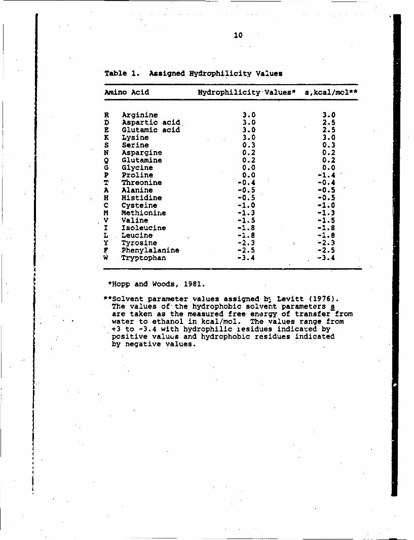
A computer program was developed for the Apple Ile personalcomputer based on the program by Hopp and Woods, 1983. Theprogram takes the given sequence and assigns the hydrophilicityvalue to each amino acid. These values are assigned based onsolvent parameter work by Levitt, 1976. The values of thehydrophobic solvent par&meters s are taken as the measuredfree energy c.A transfer from water to ethanol in kcal/mol.The values assigned to each of the twenty amino acids areshown in Table'l. The values range from +3 to -3.4 with hydro-philic residues indicated by positive values and hydrophobicresidues indicated by negative values. The program then calcu-lates the average hydrophilicity value of a moving hexapaptidewindow through given sequence, and this average value isplotted versus the first amino acid residue of each window.
The sequence information for Lapemis hardwickii Lapemistoxin (NT) was taken from Fox et al., 1977.
The sequence information for the a-subunit of the Torpedocalifornica ACR was taken from Noda et al., 1982. Thesequences for NT and ACR are shown in Fig. 1.
Results and Discussion
One of the most importtnt forces involved in protein in-teractions is called the hydrophobic interaction force. Otherforces which also play a role are electrostatic, van der Waals,and hydrogen bonding. The hydrophobic interaction force ispoorly understood but is known to affect the entropy of thesolvent and the solute (protein). Yet this force is so impor-tant in explaining why some proteins are soluble in water andpolar solvents or lipids and nonpolar solvents. The hydrophil-icity index is a method of quantifying this important force.
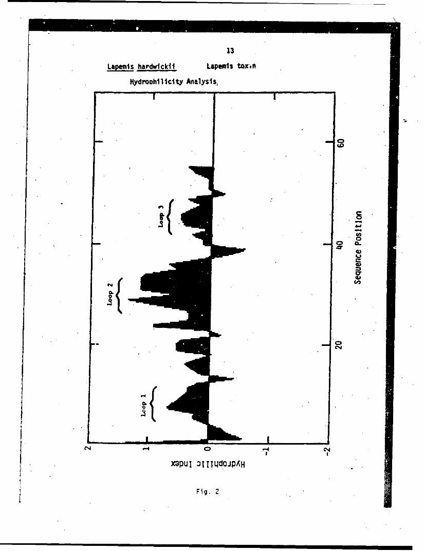
The plot of the hydrophilicity index versus sequenceposition for Lapemis toxin is shown in Fig. 2. The overallmolecule is quite hydrophilic as shown by the large positivearea under the curve. Loop 1 occurs from sequence positions4 through 15. Loop 2 occurs from sequence positions 24through 37. Loop 3 occurs from sequence pcsitions 42 through51. Note that the loops 1, 2, and 3 regions are hydrophilicwith the maximu•n hydrophilic values shown in loop 2.

8
The NT structure proposed is based on X-ray diffractionstudies n toxin a from Laticauda semifasciata venom, which isanother hort-chain neurotoxin with similar sequence homologyto the pemis toxin (Tsernoglou and Petsko, 1976). The proposedstructur is shown in Fig. 3. In this figure the 60 amino acidresidues are shown with the sequence number. The stippledcircles -epresent hydrophilic residues based on the hydrophili-c4ty index analysis. It has been shcr- that the tryptophan 27residue s functionally important in the toxic role of Lapemistoxin (Ti and Hong, 1971). Studies on other short-chain neuro-toxins h ve shownan importance to toxicity of glutamic acid 19(Chang e al., 1971a), lysine 45 (Chang et al., 1971b), histi-dine 30 Huang et al., 1972), and arginine 31 (Yang et al.,1974). rom this information and the following informationabout th ACR, a proposal of the ACR-NT interaction is made.
The ACR is diagrammatically shown in Fig. 4 (Hayashi andNomoto, 1986). The two a-subunits are the sites of acetyl-choline Dr neurotoxin binding to the ACR. The a-subunit isproposed to go in and out of the cell membrane several timeswith the N-terminal portion being found on the extracellularside of the resident cell and the C-terminal portion beingfound in the cytosol cr interior of the cell.
The plot of the hydrophilicity index versus sequenceposition for the a-subunit of the ACR was plotted in foursectio..s with sequences 1 through 110 shown in Fig. 5; sequences110 throigh 220 shown in Fig. 6; sequences 220 through 330 areshown in Fig. 7; and sequences 330 through 437 are shown inFig. 8. The plots show many regions of hydrophilicity andregions f hydrophobicity. Intramembraneous regions M6, M7,Ml, M2, nd M3 are bracketed.
Hyd ophilic regions are most likely found in the cytosolor extra ellular regions due to the water environment. Thehydropho ic regions are most likely found either in the hydro-phobic e vironment of the cell membrane lipid bilayer or areinternalized in globular domains of the protein, folding insuch a way as to minimize contact with the water environmentof the eptracellular region ox cytosol.
Frm the hydrophilicity index analyses and informationabout Lapemis toxin, other short-chain neurotoxins and thea-subunit of the ACR, a model of NT-ACR interaction is madeand shown in Fig. 9. Information about the ACR a-subunit istabulated in Table 2, which is used to make the proposed mode'l.The carbohydrate moiety at asparginine 141 of the a-subunit rfthe ACR was determined by Nomoto (1986). Tryptophan 27 of theNT is important to toxicity and the proposed interaction iswith the first N-acetylglucosamine of the carbohydrate moietyat the asparginine 141 of the ACR a-subunit. This interac-tion is based on the known interaction of the tryptophanresidues of lysozyme in binding the N-acetylglucosamine of thebacterial membrane carbohydrate moieties (Zubay, 1983). The} arginine 31 residue of the NT is important, probably due to
V

9
its chemical and structural similarity to acetylcholine.The lysine 45 and glutamic acid 19 of the NT probably playa role by charge interaction with complementary residues ofthe ACR a-subunit. The histidine 30 of the NT probably playsa role by aiding in stabilizing the interaction of thearginine 31 Of the NT with the binding at or near the disul-fide cystine 192-193 of the ACR a-subunit.
The exact nature of the binding and specificity of thebinding are still unclear and certeinly lies in similar com-plementary regions of the NT and ACR a-subunit. The hydro-philicity index analysis has shown the hydrophilic natureof the NT and suggests the NT would seek out a hydrophilicregion of the a-subunit of the ACR.
The more studies concentrated on the ACR-NT interactionwill lead to the exact chemical nature of binding, givinga clearer picture of the mechanism of toxicity of NT. Thiswork will also aid in the understanding of the struature-function relationships of the a-subunit ACR and the entireintact ACR, which will aid in treatments of pathologicaldisorders of the ACR such as found in snakebites and myas-thenia gravis.
10
Table 1. Assigned Hydrophilicity Values
Amino Acid Hydrophilicity Values* s,kcal/mcl**
R Arginine 3.0 3.0D Aspartic acid 3.0 2.5E Glutamic acid 3.0 2.5K Lysine 3.0 3.0S Serine 0.3 0.3N Aspargine 0.2 0.2Q Glutamine 0.2 0.2G Glycine 0.0 0.0P Proline 0.0 -1.4T Threonine -0.4 -0.4A Alanine -0.5 -0.5H Histidine -0.5 -0.5C Cysteine -1.0 -1.0M Methionine -1.3 -1.3V Valine -1.5 -1.5I Isoleucine -1.8 -1.8L Leucine -1.8 -1.8Y Tyrosine -2.3 -2.3F Phenylalanine -2.5 -2.5W Tryptophan -3.4 -3.4
*Hopp and Woods, 1981.
**Solvent parameter values assigned b, Levitt (1976).The values of the hydrophobic solvent parameters sare taken as the measured free energy of transfer fromwater to ethanol in kcal/mol. The values range from+÷3 to -3.4 with hydrophilic zesidues indicated bypositive valuus and hydrophobic residues indicatedby negative values.

11
Table 2. Assignment of Sequence Information for theAcetylcholine Receptor a-Subunit.
Sequence # Assigivment Reference #
1 - 141 Extraceilular 1141 Site of glycosylation 2,5142 - 151 Intramembraneous M6 1152 158 Cytosolic 1159 - 191 Intramembraneous M7 1182 - 198 Neurotoxin binding region,, 3
also binds antibodies185 - 196 Binds a-bungarotoxin 4192 - 193 ACH binding site 2192 - 210 Extracellular 1211 - 236 Intramenbraneous MI 1237 - *242 Cystolic 1243 - 260 (Intramembraneous) (M2) 1261 - 276 (Extracellular) 1277 - 297 (Intramembraneous) (M3) 1298 - 329 (Cystolic) 1330 - 437 Cystolic 1
Ref. 1 - Ratnam et al., 1986Ref. 2 - Oblas et al., 1986Ref. 3 - Mulac-Jericevic and Atassi, 1986Ref. 4 - Neuman et al., 1986Ref. 5 - Nomoto et al., 1986

12
Laei hardwickii Lapemis Toxin Sequence
10 20 30 40 50 60KTCcMQSs. QPUTTMICE.- SSCYD-1WSD --T=MRC;C .GCPQVKPGIX. LEC WCB1cN. N
Torpedo californica Acetylcholine Receptor(a-subunit) Sequence
10 20 30 40 50 60SEHETRVA . NLLENYNKVI . RPVEUITHTV . DITVGLQLIQ. LISVDEVNQI. VETNVRLRQQ . WIDVRLRWNP
70 80 90 100 110 120 130ADYGGIKKIR. LPSDDVWLPD. LVLYNNADGD. FAIVHMTKLL. LDYTGKIM4WT. PPAIFKSYCE. LIVTHIPPDQ
140 150 160 170 180 190 200QNCNMKLGIW.- TYDGTKVS IS. PESDRPDLST. FMESGEWVM0C. DYRGWKHWVY YTCC.PDTPYL. DITYHFMQR
210 220 230 240. 230 260 270IPLY.FVVNVI. IPCLLPSLT . GLVFYLPTDS. GEKMTLS ISV. LSLTVFLLV. IVELIPSTSS.*AVPLIGKYMfL.
280 290 300 310 320 330 340FThfIFVISSI -IIT^VVVMT. HRSPSTHTMP. q#VRICIPIDT. IPNVMFFSTh. MWAKEKQEN . KIFADDIDIS
350 360 370 380 30400 410DISGKQVTGE .VIPQTPLfc. PDVKSAIEGV.IKYIAEMKMSD. EESSNAAEEW. KYVAMVIDHI .LLCVMLIýCI
420 430 437IGTVSVFAGR .LIELSQEG
The-single letter notation for the twenty amino acidresidues are: A - Alanine, C - Cysteine/Cystine,D - Aspartic acid, E - Glutamic acid, F - Phenylalanine,G - Glycirie, H -Histidine, I -Isoleucine, K - Lysine,L - Leucine, M - flethionine, N -Aspeargine, P - Proline,Q- Glt4tamine, R - Arginine, S -Serine, T - Threonine,V - Vah.'ne, W - Tryptophan, and Y - Tyrosine.
Fig. 1. Lapemiis hardwi,-kii Lapernis Toxin Sequence andTorpedo californica Acetylcholine Receptor (a-( subunit; Sequence.
13
Lapemis hardwlckii Lopemis toxort
IjydroDhil lclty Analysis,
4oC
XaPUI OITTql.dOJPiAH
Fig. 2

14
Proposed Structure of Lapemis ToxinLapemis hardwickil Lapemis Toxin
10 20 30 40 50 60NTCCNQSS.QPKTTTWcAE.SSCYKKTWSD. ERGTP.IERGC.GCPQVKPGIK.LECCHTNECN.N
Fig 3

Diagrammatic Model of theAcetylcholine Receptor
Fig. 4

16
Acetylcholine Receptor Alpha-Subunit Hydrophilicity Analysison Sequence Positions 1 - 110
C
Co
I• I
CDI V-4 C*4
. ,,I
IxPUI 1II loJP4 H
Fig. 5

17
Acetyiclioline Receptor Alpha-Subunit Hydrophilicity Analysis on SequencePositions 110 - 120.
U~I~ 0
l -I
-N
So C-,
I I-
x)pul :tI dq(JOJPAH
Fig. 6

Ac~t~l~ l"~ ,C*Ptf Mp -Subunit H4ydr '*Ph llicitY nlss
onSQll
aptor A ph ositiofis 220o 330.
AC~tlchoine ec,0
00
1Ei4
C-
ee4
Fig. 7

19Acetylcholine Receptor Alpha-Subunit Hydrophilicity Analysis on Sequence
Positions 330 - 437.
40
/CD IC
40)
4.DCNI '-
Fig. 8

20
Model of NT - Alpha ACR Intoraction
X=q
C4
Fig. 9

21
B. Isolation and Chemical Modification of LapemisToxin from Lapemis hardwickii Toxin by Anthony T.Tu and Nobuhiro Mori
Methods
Sea snakes, Lapemis hardwickii, were captured in the Gulfof Thailand near Songkla, Thailand. In order to extract thevenom, the sea snakes were decapitated as soon as the fisher-men brought them to the Songkla harbor, and the venom glandswere excised. After dry venom glands were received in Coloradofrom Thailand by air mail, they were pulverized thoroughlywith an electric grinder and the venom was extracted withdistilled water. After the insoluble tissue debris wasremoved by centrifugation, the supernatant liquid was filteredand lyophilized.
Laperais Toxin: Lapemis toxin was isolated from crude venomessentially by the method of our earlier publication (Fox et al.,1977). Two grams of venom were dissolved in 5 ml of 10 mMpotassium phosphate, pH 6.5 buffer containing 0.1 M NaCl.Upon dissolution, the mixture was loaded onto a Sephadex G-50column (2.5 x 100 ci.) equilibrated with the phosphate buffer.The elution was carried out with the phosphate buffer at 46C;three ml fractions were collected.
The absorbance at 280 nm of each fraction was determinedwith a spectrometer. The toxicity of each pooled peak wasanalyzed according to the method of Litchfield and Wilcoxon(1949). Various quantities of the lyophilized protein peakswere first dissolved in a 0.09% NaCl solution. Swiss Websterwhite mice weighing 18-20 g were then injected with 0.1 ml ofthe toxin solution in the tail vein and segregated for 24 hto monitor the effects. Most of the mice either succumbed tothe tonin injection within 60 min or survived the 24 h interval.
The G-50 peak with the highest specific toxicity wasdissolved in 4 ml of distilled water, and dialyzed (Spectra/Pordialysis membrane, MWCO 2,000) against 10 mM potassium phosphate,pH 7.8, buffer (2-46C). This sample was then applied to acarboxymethyl cellulose (Sigma Chemical) cation-exchange column(2 x 35 cm) equilibrated with the same buffer at 2-46C. Thecolumn was developed with a linear salt gradient from 0 to0.4 NaCl, 1000 ml total volume. The absorbance at 280 nm wasmonitored and the protein peaks pooled and assayed for toxicity.
Isolation of ACR: ACR-rich membrane and pure ACR were isolatedfrom the electric organ tissue of Torpedo californica. ACR-rich membrane is the membrane fragment containing ACR; therefore,it also contains phospholipids. Purified ACR is composed offive subunits of four kinds having stoichiometry a20Y6. ACRwas isolated by the affinity chromotographic method of Froehnerand Rafto (1979). Cobrotoxin affinity resin
m J

22
was prepared as previously described (Brockes & Hall, 1975)using Sepharose 4B. The amount of active a-neurotoxin perml of Sepharose was estimated to be approximately 0.1 mg orabout 20% of the input. ACR-rich membrane was isolated bythe method of Froehner and Rafto (1979).
All procedures were carried out at 40C. Approximately75 g of frozen tissue was sliced into small pieces with a r zorblade and homogenized in 250 mL of 10 mM Tris-Cl, pH 7.4,containing 1 mM EDTA, 1 mM EGTA, 0.1 mM PhCh 2 SO 2 F, and Tras lol(10 units/mL) (buffer A) in a Waring blender. The homogena ewas passed through a double layer of cheesecloth and centrifuged for 45 min at 27,000 g. The pellet was resuspended i25 mL of buffer A with a Dounce homogenizer, and Triton X-1 0was added to a final concentration of 1%. After 1 h, insol lematerial was removed by centrifugation (30 min at 27,000 g)and the supernatant (Triton extract) was retained for affin typurification.
Pure ACR was isolated as follows: The Triton extract wasstirred with 2-3 mL of cobrotoxin-Sepharose 4B (previouslywashed with buffer A containing 1 M NaCl and then equilibratedwith buffer A) for 1.5-2 h. The mixture was then poured intoa 10-mL plastic syringe with a nylon net support to form acolumn. The column was washed sequentially with 5 mL ofbuffer A containing 1.0% Triton X-,100 (buffer B), 10 mL ofbuffer B containing 1 M NaCl, and 5 mL of buffer A containirg0.1% Triton X-100. Approximately 0.8 column volume of 1Mcarbamoylcholine chloride in buffer A containing 0.1% TritorX-100 was run into the column. After 0.5-2 h, the firstelution was collected. A second elution was collected aftexa further incubation of 12-15 h or, alternatively, the columncontents were poured into 20-25 mL of carbamoylcholine solutionand stirred slowly for 12-15 h. Eluted receptor was thencollected after repouring the column. Both procedures forthe second elution give comparable results. The affinity-purified fractions were puoled and then dialyzed against fo rchanges of 500 mL of buffer A containing 0.1% Triton X-100 orat lea3t 2 h each. ror some preparations, 10 mM NEM was ad edto all buffers except the dialysis buffer which contained 1 mMNEM. Purified receptor stored at 40C showed no loss of [12 •I]-•BuTx binding activity and, except for some aggregation, nosubstantial change in its subunit structure over a period oseveral months. Receptor stored at -706C lost some bindingactivity after several freezings and thawings, but no changin subunit structure was noted.
Modification of Arginine Residues: Modification of arginineresidues in Lapemis toxin with phenylglyoxal was performedby a modification of the method described by Yang et al.(Biochim. Biophys. Acta 365, 1-14, 1974).
To a solution of Lapemis toxin (1.6 unoles) in 0.1 ml f
0.2 M N-ethylmorpholine acetate buffer (pH 8.0), a 100-foldmolar excess of phenylglyoxal in 0.3 ml of the same buffer

23
was added, and the reaction was allowed to proceed at 270Cfor 1 h. Reactions at pH values other than 8.0 were carriedout in the same way with appropriate buffers. The mixturewas passed through a column of Bio-Gel P-6 (0.9 x 40 cm, MWcut off 6000 daltons) followed by ion exchange chromatographyon CN-cellulose with a gradient of increasing salt concentrationfrom 0.005 M ammonium acetate, pH 5.8 to 6.8. The fractionsof the main protein peaks were lyophilized and desalted byusing a Bio-Gel P-6 column equilibrated with 1% acetic acid.The protein fractions. were then pooled and lyophilized.
The homogeneity of the modified toxin was checked byHPLC. Samples of phenylglyoxal derivatives were applied onan ODS column (4.6 nmu x 25 cm) equilibrated with .0.1% TFA(buffer A) and eluted with a linear gradient of 40% buffer B(0.1% TFA containing 30% acetonitrile and 70% 2-propanol) for40 min. Flow rate was 0.8 ml/min and the effluent is monitoredat 280 n.
HN 0 M 1
P-HN-C + 2 -C- P-NH-C HCtNH2 0 0-c-, 0
phenylglyoxal 0+
H 0N2
Phenylglyoxal derivatives were very stable even after HCLhydrolyzation.
Results and Discussion
Fractionation - The fractionation patterns of the first andsecond steps are shown in Fig. 10A and B.
Homogeneity - Homogeneity was examined ,)y analytical HPLC,Beckman series 345, and found to be a singlepeak. The toxin was also homogeneous in gelelectrophoresis.
Toxicity - The final purified toxin was lethal 4t thelevel of 0.15 4g/g by intravenous injectionin mice. In order to minimize the use ofmice, the LD50 of crude venom was 1.4 pg/g,while that of purified Lapemis toxin wasfound to be 0.66.

24
Purified ACR and Binding to Lapemis Toxin
Purified ACR is composed of five subunits, of which twoare identical, giving the stoichiometry Q2AY6. Since two ofthe subunits are identical, ACR showed four bands in SDS gelelectrophoresis (Fig. 11). Lapemis toxin bound to ACR andformed a complex .(Fig. 12).
ACR-Lapemis toxin complex dissociated into ACR and Lapemistoxin after reduction by mercapto ethanol (Fig. 12). SDSalone did not dissociate the complex (Fig. 12).
Lapemis toxin replaced a-bungarotoxin 21a -BTx) in bindingto ACR, indicating it is a competitor to 12 •I-a-BTx and bindsto the same site in ACR.
Modification of Arginine Residue: There are three arginine-residues out of 60 total residues. An objective of ourresearch is to find which residues are involved in the ACRbinding. In order to clarify this,' two experiments are needed.One is a control experiment; that is to modify arginine residuesof free Lapemis toxin (not bound to ACR). The other experimentis to modify Lapemis toxin after binding to ACR. The firstexperiment was done and it was found that two arginines outof three were modified for unbound Lapemis toxin (Table 2).Chemical modification of Lapemis toxin bound to ACR has notbeen done yet.
Table 2. Amino Acid Composition of Lapemis Toxin (Unbound toACR) Before and After Modification of Arginine Residue.
Before Modification After ModificationAmino Acids (from Sequence) (Actual Analysis)
Asp 6 3.6 (6)Glu 8 8.5 (9)Ser 5 4.6 (5)Gly 4 4.4 (4)Thr 7 7.3 (7)His 2 1.9 (2)Ala 1 1.3 (2)Pro 3 3.3 )Arg 3 1.4Tyr 1 1.0 (1)Met 1 0.5 (1)Val 1 1.3 (1)Cys 9 8.3 (9)Ile 2leu 1 2.4 (2)Leu ILys 5 4.4 (4)Phe 0 0Trp 1
Total 61

25
LEGEND
Fig. 10 Isolation scheme of the Lapemis toxin from thevenom of Lapemis hardwickii. The isolationprocedure is described under ExperimentalMethod.
Fig. 11 Subunits of reduced acetylcholine receptor shownin SDS polyacrylamide gel electrophoresis.
Fig. 12. SDS-ti;trophoresis of 1, 2, 3, indicating thedissociation of ACR-LT complex to individualsubunits and LT (Lapemis toxin) after 2-me-rcaptoethanol reduction. 4, 5, 6 indicatedthat ACR-L2 complex is not dissociated without2-mercaptoethanol. See very weak LT bandcomparing to the electrophoretic band of 1,2, 3.
I2.I26
Sephadeg 5-50
12.5,
51.0
47.
35.0
2.0
0 50 100 1SO 200TTu nnubr (3 m1/tub,)
€14-Cel1ig.s1
UI!
4.0 . Z
N
M 3.0
U
wC
Z02.0 o0OTu*nubr 3ml.•
0i . i

27
MW
66K .... nm -
45K loom"-
36K ,.
29KFu 11
Figure 11

28
123 456
AchR
MW
66K-. 6
y45K pa36Km
29Km24Km f
u- LT-2
Figure 12
29
C. Isolation and Amino Acid Sequence of Neurotoxinfrom the Venom of Sea Snake Acalyptophis peroniiby Anthony T. Tu and Nobuhiro Mori
Experiments
Acalyptophis peronii was captured in the Gulf of Thailandoff the coast of Songkla in Nov. tmber-December 1979. The seasnake was decapitated and venom glands were removed. Driedvenom glands were sent to Colorado and extracted twice withdistilled water after venom glands were pulverized. Theextracted proteins were designated "crude venom." Lyophilizedcrude venom was stored in a freezer until used.
Isolation of Acalyptophis Toxin
The major toxin, Acalyptophis toxin, was isolated fromcrude venom from AcAlyptophis peronii using two-step columnchromatography of G-50 Sephadex and CM-cellulose.. Thecondition for the isolation was the same as the method ofLapemis toxin isolation published by Tu et al. (1971).
Homogeneity was established by LKB Tachophor model 2127using a cationic system.' Cacodylic acid at pH 7.0 was usedas the leading ion and 0.01 M creatinine as the terminatingion. Ampholine (1%) was used as a spacer electrolyte. Aconstant current of 75 vA and a variable voltage of 3-12 kVwere used.
Amino Acid Sequence Determination
The purified toxin was reduced and alkylated followingthe method of Crestfield as described by Elzinga (1970).Microsequence analysis was performed on approximately 500 picomoles of each peptide using an Applied Biosystems model 4Ai;protein sequencer similar to that described by Hewick et al.(1981). Phenylthiohydantoin amino acids were identified b;,HPLC using A Beckman-Altex Ultrasphere ODS column and thetrifluoroacetic acid/acetonitrile buffer system reported byHawke et al. (1982).
For tryptic digestion, the toxin was first treated withcitraconic anhydride to block the lysine residues. The citra-conylated CM-toxin (CM-CA toxin) was then subjected to trypticdigestion using an enzyme to substrate ratio of 1:30 (w/w).After 16 h at 23°C the digestion was terminated.,by the additionof soybean trypsin inhibitor and glacial acetic acid to 25%.The mixture was then passed through a Sephadex G-50 column equi-librated with 25% acetic acid which served to separate the pro-ducts. Separation was made further by an HPLC reverse phasecolumn.
Different toxin peptide fragments were made using theendopeptidase, arginine C, after carboxymethylation of thereduced toxin.

30
Results and Discussion
Isolation of Neurotoxin
The major torin, Acalyptophis toxin, was isolated by acombination of gel filtration and CM-cellulose ion-exchangechromatography (Fig. lA,B). Homogeneity was established byanalytical capillary isotachophoresis LKB Tachophor model 2127using cationic system. A sharp single peak was obtained forthe toxin, indicating a high degree of homogeneity (Fig. 1C).Toxicity, expressed as LD5 0 , was 0.125 Ug/g in mice by intra-venous injection.
Amino Acid Sequence
Three methods, were used to determine the amino acidsequence of Acalyptophis toxin. First, the sequence analysiswas made directly using carboxymethylated toxin. The secondmethod was to analyze the fragments obtained from trypticdigestion. The third method was to analyze the sequence ofendopeptidase, arginine C treated peptide fragments.
1. The sequence obtained from CM-toxin is shown here:
1 2 10Met-Thr-CMC-CMC-Asn-Gln-Gln-Ser-Ser-Gln-
11 15 20Pro-Lys-Thr-Thr-Thr-Asn-CMC-Ala-Gly-Asn-
21 25 30Ser-CMC-Tyr-Lys-Lys-Thr-Trp-Ser-Asp-Thr-
31 35Arg-Gly-Thr-Ile-Ile-Glu-Arg-
2. The Sequence of Tryptic Digestion Fragments
Toxin after tryptic digestion was subjected to separationby HPLC. Altogether ten fractions were observed in the HPLCchromatogram (Fig. 2). Fractions 3, 4,, 5, 6, and 7 were sequencuCwith the results shown below.
Fraction 3 G-C-G-C-P-Q-V-K-S - - K
Fraction 4 G-T-I-I-E-R and V-K-S-G
Fraction 5 T-T-T-N-A-G-N-S-Y-KFraction 6 M-T-C-C-N-Q-Q-S-S-Q-P-K
Fraction 7 S-Q-H-R
By combining the sequence data of fragments and carobxymethylatedtoxin, the following overlapping patterns can be obtained.
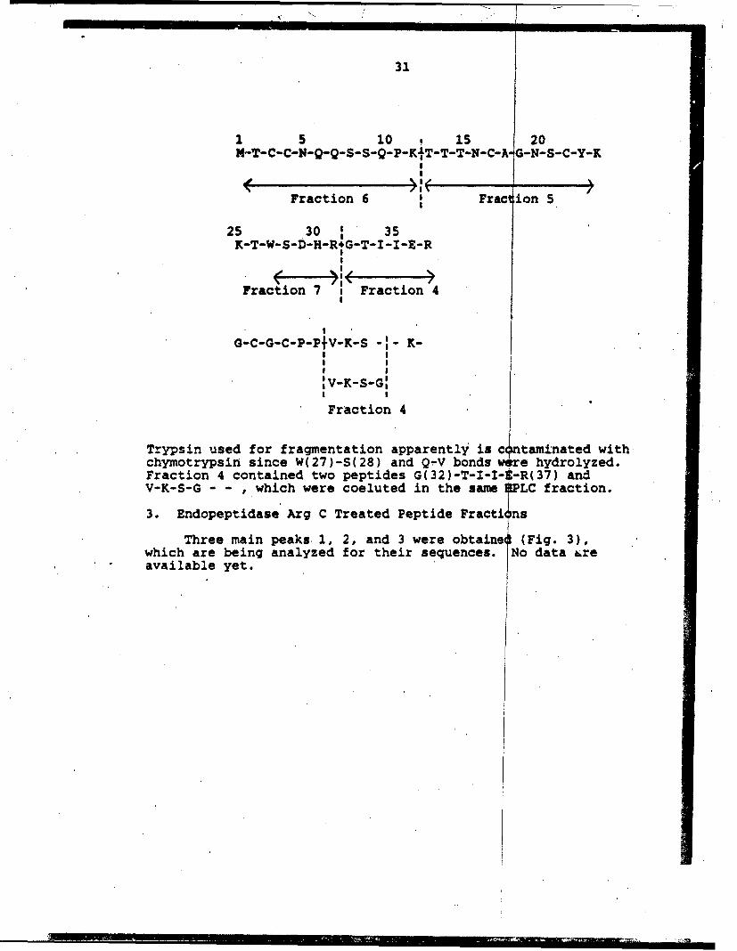
31
1 5 10 is1 20M-T-C-C-N-Q-Q-S-S-Q-P-KtT-T-T-N-C-A G-N-S-C-Y-I(
Fraction 6 SFracl ion 5
25 .30 ' 35K-T-W-S-D-H-R*G-T-I -I-E-R
Fraction 7 sFraction 4
G-C-G-C-P-PfV-K-S -:-K-
:V-K-S-G:
Fraction 4
Trypsin used for fragmentation apparently is c taminated withchymotrypsini since W(27)-S(28) and Qý-V bonds e hydrolyzed.Fraction 4 contained two peptides G(32)-T-I-I- -R(37) andV-K-S-G -- ,which were coeluted in the same LC fraction.
3. Endopeptidase Arg C Treated Peptide Fracti nns
Three main peaks. 1, 2, and 3 were obtaine (Fig. 3),which are being analyzed for their sequences. No data &reavailable yet.

32
LEGEND
Fig. 13 Isolation scheme of the Acalyptophis toxinfrom the venom of Acalyptophis Peronii (A and B).The isolation procedure is described underExperimental Method. Homogeneity was establishedby isotachophoresis using a cationic system (C).
Fig. 14 HPLC profiles of the tryptic digests of citra-conylated CM-toxin..
Fig. 15 HPLC profiles of the endopeptidase Arl C digestsof CM-toxin.

33
G-50
IS
a 6
Tubeb~~. MI 30//OO b.O
B _ _ _ _ _ _ _1C

34
4 51
20.1
TIri (min)
Fig. 14

35
0.51
0a4
0.3-
0.2
0.1
0 115 2 25i 30
Time (min)
Fig. 15

36
References
Brockes, 3. P., and Hall, Z. W. (1975). Acetylcholinereceptors in normal and denervated rat ~japhragm muscle.I. Purification and interaction with [ 1 'I]-J-Bungaro-toxin. Biochemistry 14, 2092-2099.
Chang, C. C., Yang, C. C., Kurobe, M., Nakai, K., and Hayashi,K. (1971a). The identification of the special glutamicacid residue essential for activity of cobrotoxin.Biochem. Biophys. Res. Commun. 43, 429.
Chang, C. C., Yang, C. C., Nakai, K., and Hayashi, K. (1971b).Studies on the status of free amino and carboxyl groups,in cobrotoxin. Biochim. Biophys. Acta 251, 334.
Claudio, T., Ballivet, M., Patrick, J., and Heinemann, S.(1983). Nucleotide and deduced amino acid sequences ofTorpedo californica acetylcholine receptor gamma chain.Proc. Natl. Acad. sci. 80, 1111-1115.
Elzinga, M. (1970). Amino acid sequence studies on rabbitskeleton muscle actin. Cyanogen bromide cleavage ofthe protein and determination of the sequences of sevenof the resulting peptides. Biochemistry 9, 1365-1374.
Endo, T., Nakanishi, M., Furokawa, S., JoUbert, F. J., Tamiya,N., and Hayashi, K. (1986). Stopped-flow fluorescencestudies on binding kinetics of neurotoxins with acetyl-choline receptor. Biochemistry 25, 395-404.
Fox, J. W., Elzinga, M., and Tu, A. T. (1977). Amino acidsequence of a snake neurotoxin from the venom of Lapemishardwickii and the detection of a sulfhydryl group bylaser Raman spectroscopy. FEBS Lett. 80, 217-220.
Froehner, S. C., and Rafto, S. (1979). Comparison of subunitsof Torpedo californica acetylcholine receptor by peptidemapping. Biochemistry,18, 301-307.
Hawke, D., Yuan, P. M., and Shively, J. E. (1982). Micro-sequence analysis of peptides and proteins. Anal.Biochem. 120, 302-311.
Hayashi, K., and Nomoto, H. (1986). Relationship betweenstructure and function of AChR. Med. Immunol. (Japan)11, 425-434.
Hewick, R. M., Hunkapiler, H. W., Hood, L. E., and Dreyer,W. J. (1981). A gas-liquid solid phase peptide andprotein sequenator. J. Biol. Chem. 256, 7990-7997.

37
Hopp, T. P., and Woods, K. R. (1981). Prediction of proteinantigenic determinants from amino acid sequences. Proc.Natl. Acad. Sci. 78, 3824-3828.
Hopp, T. P., and Woods, K. R. (1983). A computer programfor predicting protein antigenic determinants. Mol.Immunol. 20, 483-489.
Huang, J. S., Liu, S. S., Ling, K. H., Chang, C. C., and Yang,C. C. (1972).; Photoxidation of cobrotoxin. J. FormosanMed. Assoc. 71, 383.
Levitt, M. (1976). A simplified representation of proteinconformations for rapid simulation of protein folding.J. Mol. Biol. 104, 59-107.
Litchfield, J. T., and Wilcoxon, F. (1949). A simplifiedmethod of evaluating dose-effect experiments. J. pharmacol.
. Ther. 96, 99-113.
Martinez-Carrion, M., Sator, V., and Raftery, M. A. (1975).The molecular weight of an acetylcholine receptorisolated from Torpedo californica. Biochem. Biophys.Res. Commun. 65, 129.
Mulac-Jericevic, B., and Atassi, M. Z. (1986). Segment a182-198 of Torpedo californica acetylcholine receptor con-tains a second toxin-binding region and binds anti-receptor antibodies. FEBS Lett,. 199, 68-74.
Neumann, D., Barchan, D., Safran, A,, Gershoni, J. M., andFuchs, S. (1986). Mapping of the a-Bvngarotoxinbinding site within the a-subunit of the acetylcholinereceptor. Proc. Natl. Aced. Sci. 83, 3008-3011.
Noda, M., Takohashi, H., Tanabe, T., Tc7osato, M., Furutani,Y., Asai, M., 1nayama, S., Miyata, T., and Numa, S.(1982). Primary structure of a-suaunit precursor ofTorpedo californica acetylcholine receptor deduced fromcDNA sequence, Nature 299, 793-797.
Noda, M., Takashi, H., Tanabe, T., Toyosato, M,' Furutani,Y., Hirose, F., Asai, M., Inayama, S., Miyata, T., andNuma, S. (1983). Primary structure of beta and deltasubunit precursors of Torpedo californica acetylcholinereceptor from cDNA sequences. Nature 301, 251-255.
Nomoto, H., Takahashi, N., Nagaki, Y., Endo, S., Arata, Y.,and Hayashi. K. (1986). Carbohydrate structures ofacetylcholine receptor from Torpedo cal.fornica anddistribution of oligosaccharides among subunits.Eur. J. Biochem. 157, 233-242.
•i .•_~ 'ORII

38
Oblas, B., Singer, R. H., and Boyd, N. D. (1986). Locationof a polypeptide sequence within the a-subunit of theacetylcholine receptor containing the cholinergic bindingsite. Mol. Pharmacol. 29, 649-656.
Raftery, M. A., Hunkapillar, M. W., Strader, C. D., and Hood,L. E. (1980). Acetylcholine receptor: Complex ofhomologous subunits. Science 208, 1454-1457.
Ratnam, M., Nguyen, D. L., Rivier, J., Sargent, P. B., andLindstrom, J. (1986). Transmembrane topography ofnicotinic acetylcholine receptor: Invnunochemical testscontradict theoretical predictions based on hydropho-bicity profiles. Biochemistry 25, 2633-2643.
Tsernoglou, D., and Petsko, G. A. (1976). The crystal struc-ture of a post-synaptic neurotoxin from sea snake at2.2 A resolution. FEBS Lett. 68, 1.
Tu, A. T., and Hong, B. (1971). Purification and chemicalstudies of a toxin from the venom of Lapemis hardwickii(Hardwick's sea snake). J. Biol. Chem. 246, 2772.
Yang, C. C., Chang, C. C., and Lion, I. F. (1974). Studieson the status of arginine residues in cobrotoxin.Biochim. Biophys. Acta 365, 1-14.
Zubay, G. L. (1983). Biochemistry. Addison-Wesley Publish-ing Co., Reading, Mass., 269-174.
39
DISTRIBUTION LIST
5 copies CommanderUS Army Medical Research Institute of
Infectious DiseasesATTN: SGRD-UIZ-MFort Detrick, Frederick, MD 21701-5011
1 copy CommanderUS Army Medical Research and Development CommandATTN: SGRD-RMI-SFort Detrick, Frederick, MD 21701-5012
12 copies Defense Technical Information Center (DTIC)ATTN: DTIC-DDACCameron StationAlexandria, VA 22304-6145
1 copy Dean,School of MedicineUniformed Services University of the
,Health Sciences4301 Jones Bridge RoadBethesda, MD 20814-4799
1 copy CommandantAcademy of Health Sciences, US ArmyATTN: AHS-CDMFort Sam Houston, TX 78234-61GJ