salicylate–urea-based soluble epoxide hydrolase inhibitors ...salicylate–urea-based soluble...
TRANSCRIPT

Bioorganic & Medicinal Chemistry Letters 19 (2009) 1784–1789
Contents lists available at ScienceDirect
Bioorganic & Medicinal Chemistry Letters
journal homepage: www.elsevier .com/ locate/bmcl
Salicylate–urea-based soluble epoxide hydrolase inhibitors with highmetabolic and chemical stabilities
Takeo Kasagami �, In-Hae Kim �,�, Hsing-Ju Tsai, Kosuke Nishi �, Bruce D. Hammock, Christophe Morisseau *
Department of Entomology and University of California Davis Cancer Center, University of California, One Shields Avenue, Davis, CA 95616, USA
a r t i c l e i n f o a b s t r a c t
0
Article history:Received 16 December 2008Revised 20 January 2009Accepted 21 January 2009Available online 27 January 2009Keywords:Soluble epoxide hydrolaseUreaSalicylateIntramolecular hydrogen bondMetabolic and chemical stabilityHypertensionAnti-inflammation
0960-894X/$ - see front matter � 2009 Elsevier Ltd.doi:10.1016/j.bmcl.2009.01.069
* Corresponding author. Tel.: +1 530 752 6571; faxE-mail address: [email protected] (C. Mor
� These authors contributed equally to this work.� Present address: Institute of Molecular Science, C
300 Yongbong-dong, Buk-gu, Gwangju 500-757, South
We investigated N-adamantyl-N -phenyl urea derivatives as simple sEH inhibitors. Salicylate ester deriv-atives have high inhibitory activities against human sEH, while the free benzoic acids are less active. Themethyl salicylate derivative is a potent sEH inhibitor, which also has high metabolic and chemical stabil-ities; suggesting that such inhibitors are potential lead molecule for bioactive compounds acting in vivo.
� 2009 Elsevier Ltd. All rights reserved.
Figure 1. General schematic structure of urea-based human sEH inhibitors.
Epoxide hydrolases (EH, EC 3.3.2.3) catalyze the hydrolysis ofepoxides and arene oxides to their corresponding diols.1 The mam-malian soluble EH (sEH) is involved in maintenance of homeostasisthrough hydration of endogenous lipid epoxides such as epoxyei-cosatrienoic acids (EETs).1,2 EETs, derived from arachidonic acidby cytochrome P450 epoxygenation, have various biological activ-ities. EETs have effect on vascular tone. In addition, it is known thatthe 11,12- and 14,15-regioisomers of EETs have anti-inflammatoryactivity.2 The EETs inhibit activation of nuclear factor kappa B(NF-jB) and its nuclear translocation. This means that, under cer-tain conditions, EETs will reduce activation of a variety of pro-inflammatory peptides and proteins such as cyclooxygenase 2.3
Since sEH converts EETs to the less biological active correspondingdiols (dihydroxyeicosatrienoic acids), sEH inhibition can result inanti-inflammatory and anti-hypertensive effects.
1,3-Disubstituted ureas, and the corresponding amides and car-bamates are strong sEH inhibitors.4,5 Our previous findings sug-gested that ureas with two additional pharmacophores (P2 andP3, Fig. 1) have strong inhibition in vitro as well as good bioactivityin vivo.5 The primary pharmacophore is the urea group bearing abulky and/or hydrophobic substituent such as the adamantyl,
All rights reserved.
: +1 530 752 1537.isseau).
honnam National University,Korea.
cyclohexyl, alkyl or aryl groups (R1, Fig. 1). The secondary pharma-cophore (P2) is a polar group, such as ketone, ester, alcohol, sulfox-ide, sulfonamide or ether located five or six atoms away from thecarbonyl group of the urea. A hydrophobic linker (L) joins the ureaor amide central pharmacophore to the secondary pharmacophore.Unlike straight alkyl chains, the presence of cyclic linker (L), suchas a cyclohexane or benzene ring, between the primary (urea)and secondary (P2) pharmacophores, seemed to increase the bio-availability in a canine model.5 Such cyclic linkers also reduce flex-ibility and are thought to make compounds more ‘‘drug-like”.6 Inaddition, the introduction of polar group(s) on such linkers likelywill improve solubility, ease of formulation, and often their bio-availability. The nature of group R2 situated between P2 and P3 ismore open to variation in term of size and polarity as long as P3
that is a polar group is at least 12 Å away from the urea carbonyl.5
Therefore, in this study, we designed simple adamantyl ureaswith a phenyl linker. These sEH inhibitors focus on the secondarypharmacophore. Usage of a phenyl moiety has several advantagesfor drug design, (1) the benzene ring, which is UV active, enablesone to easily trace the target molecule during the synthesis or later

T. Kasagami et al. / Bioorg. Med. Chem. Lett. 19 (2009) 1784–1789 1785
scale-up and upon HPLC analysis; (2) as mentioned above, a ben-zene ring will play a role as a linker to keep a rigid distance be-tween the primary and secondary pharmacophores; (3) abenzene ring, unlike a cycloalkane ring, has no stereoisomers, thussimplifying synthesis; and finally (4) numerous substituted ani-lines are commercially available simplifying the synthesis of ureaswith functionalized phenyl rings, such as carboxylic acids that willincrease the water solubility of the molecule. Many of these inter-mediates are inexpensive facilitating design of drugs for use indeveloping countries. Further, a carboxylic acid group can be con-nected with other functional group like alcohol or amine to designthe tertiary pharmacophore. Herein, we report the sEH inhibitoryactivity as well as metabolic and chemical stabilities of simple N-adamantyl-N0-phenyl urea derivatives.
The urea compounds (1–4) were synthesized through the di-rect reaction with adamantyl isocyanate and the appropriateamine (Scheme 1). Although the isocyanate functional groupcan react with many nucleophiles such as alcohols, carboxylicacids and amines, the hydroxy group of aminophenol did not re-act significantly under our reaction conditions. However, for thesynthesis of the carboxylic acid group-containing compound(14, 15, 19, 21, 23, 25 and 27), it was necessary to protect theacid function with a methyl ester that was removed followingpurification by alkaline hydrolysis (Scheme 1). The startingmethyl esters (I–VII) were synthesized from the correspondingacids (i–vii) in a conventional manner. After the reaction withthe isocyanate and appropriate amine followed by evaporation,the unreacted starting materials were removed by washing thecrude product with a hexane:ethyl acetate mixture. This opera-tion was simple but effective to remove not only the residualstarting materials but also the N,N0-bisadamantyl urea which isnot only present in the commercially available adamantyl isocya-nate but can be formed as a side product during urea synthesis.This relatively insoluble high melting solid also shows stronginhibition towards sEH.4 After washing, the desired compoundwas purified through silica gel column chromatography andrecrystallization as final purification stage. The compounds 8–11
NCO NH
NH
Oa
H2N
NH
NH
O
COOHn
R3
H2N
COOCH3n
R3 R3
1-7
16, 18, 20, 22, 24, 2
c
a (R1=H, OH or OCH
n=0~3R3=H or OH
i-vii
I-VII
H2NR1
Scheme 1. Syntheses of the urea compounds used in this study. Reagents and conditchloride, 45–50 �C, overnight; (c) methanol, concd H2SO4, overnight; (d) THF, 5% NaO(dimethylamino)propyl] carbodiimide, appropriate amine, room temperature.
were prepared from the corresponding phenol (3 or 4) and appro-priate acyl chloride in a conventional manner.
The inhibitory activity of the ureas with a benzene ring bearinga hydroxyl, ether, ester, carboxylic acid or amide functional groupare given in Table 1. When a hydroxyl group was introduced in themeta (3) or para (4) position on the benzene ring, the inhibitoryactivity against human sEH was almost the same level as thenon-functionalized compound 1. However, the introduction of hy-droxy group as an ortho isomer (2) reduced the inhibitory potencyby roughly 10-fold. It was found that the introduction of methoxygroup in the ortho position (5) reduced the inhibitory activity dra-matically although meta (6) and para (7) isomers possessed stronginhibition against sEH. These data indicated that the meta or paraposition on the benzene ring is a preferable position for the intro-duction of a functional group without adversely affecting sEH inhi-bition. We previously observed that only compounds bearinghydrogen or fluoride atoms on the ortho positions have sEH inhibi-tion activity.4d The compounds bearing ester (8–13) as well asether groups (6 and 7) were around 2- to 6-fold better sEH inhib-itors than 3 and 4, and even better than 1, suggesting that an etheror ester group with hydrophobic properties helps bind to the activesite of the human sEH enzyme. In previous studies,4,5 it was foundthat butyrate and caproate derivatives substituted in the 3 positionof the urea were inactive as inhibitors of the sEH. However, theiresters as secondary pharmacophores were highly active and couldbe used as soft drugs. In contrast, substitution of the 3 position ofthe urea with long chain acids such as dodecyl gave a tertiary phar-macophore as active as the ester, the acid or an acid mimic.5 Inthese cases, the ester is a pro-drug increasing ease of formulationand absorption. In the series of compounds described herein, mostesters were very active (8–13). The dramatic decrease in the activ-ity of the free acids is illustrated by compounds 14 and 15. Espe-cially, compound 15, which had the acid group in the paraposition, is 83-fold less active towards sEH than the un-substitutedphenyl 1 or the corresponding ester 13. Cyclic amides with a mor-pholine (16 and 17) or piperidine (18) were also synthesized.Unfortunately, up to 9-fold reduction in inhibitory potencies re-
R1NH
NH
O O R2
O
b
NH
NH
OCOOCH3n
R3
COOHn
NH
NH
ON
O
XY
12-14
8-11
15, 17, 19, 21, 23, 25, 276, 28
d
e
3)
X=O, C or NY=CH3 or 4-morpholinyl
(R2=CH3 or phenyl)
ions: (a) 1,2-dichloroethane, 45–50 �C, overnight; (b) THF, NaH, acetyl or benzoylH aq, reflux, 2–4 h; (e) dichloromethane, 4-dimethylaminophenol, 1-ethyl-3-[3-
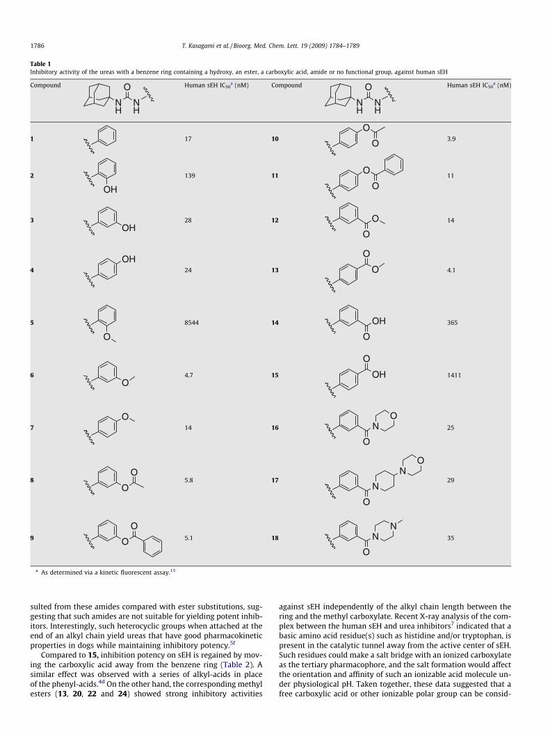
Table 1Inhibitory activity of the ureas with a benzene ring containing a hydroxy, an ester, a carboxylic acid, amide or no functional group, against human sEH
Compound
NH
NH
O Human sEH IC50a (nM) Compound
NH
NH
O Human sEH IC50a (nM)
1 17 10
O
O3.9
2
OH
139 11O
O11
3OH
28 12 O
O
14
4
OH24 13 O
O
4.1
5
O
8544 14 OH
O
365
6O
4.7 15 OH
O
1411
7
O14 16 N
O
O
25
8O
O5.8 17
N
O
NO
29
9 O
O5.1 18 N
N
O
35
a As determined via a kinetic fluorescent assay.13
1786 T. Kasagami et al. / Bioorg. Med. Chem. Lett. 19 (2009) 1784–1789
sulted from these amides compared with ester substitutions, sug-gesting that such amides are not suitable for yielding potent inhib-itors. Interestingly, such heterocyclic groups when attached at theend of an alkyl chain yield ureas that have good pharmacokineticproperties in dogs while maintaining inhibitory potency.5f
Compared to 15, inhibition potency on sEH is regained by mov-ing the carboxylic acid away from the benzene ring (Table 2). Asimilar effect was observed with a series of alkyl-acids in placeof the phenyl-acids.4d On the other hand, the corresponding methylesters (13, 20, 22 and 24) showed strong inhibitory activities
against sEH independently of the alkyl chain length between thering and the methyl carboxylate. Recent X-ray analysis of the com-plex between the human sEH and urea inhibitors7 indicated that abasic amino acid residue(s) such as histidine and/or tryptophan, ispresent in the catalytic tunnel away from the active center of sEH.Such residues could make a salt bridge with an ionized carboxylateas the tertiary pharmacophore, and the salt formation would affectthe orientation and affinity of such an ionizable acid molecule un-der physiological pH. Taken together, these data suggested that afree carboxylic acid or other ionizable polar group can be consid-

Table 2Inhibitory activity of the urea compounds with alkyl chains (C0–C3) between abenzene ring and a carboxylate functional group, against human sEH
NH
NH
O OR
On
R = H R = CH3
Numbers of carbon (n) Compound IC50a (nM) Compound IC50
a (nM)
0 15 1411 13 4.11 19 392 20 4.02 21 126 22 2.23 23 19.9 24 3.1
a As determined via a kinetic fluorescent assay.13
T. Kasagami et al. / Bioorg. Med. Chem. Lett. 19 (2009) 1784–1789 1787
ered distal from the urea carbonyl group as the tertiary but not thesecondary pharmacophore. When plotting the distance betweenthe urea carbonyl and the acid carbonyl as a function of the inhibi-tion potency for both a series of alkyl-acids and phenyl-acids(Fig. 2), we observed that both series of compounds followed asimilar hyperbolic curve. These plots suggested that such acidfunctions as a tertiary pharmacophores should be roughly 12 Åform the urea carbonyl.
Figure 2. Effect of the distance between the urea carbonyl and the acid carbonyl onthe inhibition potency. The distances were measured after minimizing the freeenergy of the molecule in gas phase, using the Chem3D� 8.0 software (Cambridge-Soft, Cambridge, MA).
Table 3Inhibitory activity of the benzoate- or the salicylate-based urea compounds againsthuman sEH
Compound
NH
NH
O
R2
R1Human sEH IC50
a (nM)
R1 (para) R2 (meta)
25 OH COOH 11626 OH COOCH3 1227 COOH OH 7128 COOCH3 OH 2.8
a As determined via a kinetic fluorescent assay.13
The presence of a polar functional group in either the meta orpara position on the phenyl group linker appeared beneficial forinhibition (Table 1). Thus, we designed and synthesized moleculescontaining both acid and hydroxyl functions (Table 3). The methylsalicylates 26 and 28 inhibited sEH strongly, with IC50s similar tothose of 12 and 13, suggesting that for the methyl ester, the adja-cent phenolic group did not negatively influence inhibitor bindingto the enzyme. Surprisingly, the salicylic acids 25 and 27 showed3- and 20-fold better inhibitory activity against sEH than 14 and15, respectively. Infrared analysis of course shows strong internalhydrogen bonds of the salicylates (25 and 27) in contrast to thefree carboxylic acids (14 and 15). Furthermore, the hydroxyl groupby itself placed on meta 3 or para 4 position did not improved theinhibitor potency of 1, suggesting that the hydroxyl group plays animportant role in binding with the active site of sEH only if a car-boxylic function is placed on the adjacent carbon. In such cases, ahydrogen bond between the carboxylate and the hydroxyl is prob-ably formed that certainly reduces the negative effect of the acidfunction on the inhibition potency.
In general, the metabolic stability of a candidate compound willinfluence drug efficacy in vivo. Although all esters synthesized inthis study showed strong inhibitory activities against the recombi-nant human sEH, it was anticipated that they would be hydrolyzedeasily in the body. Thus, six compounds (10–13, 26 and 28) weretested for in vitro stability with human hepatic S9 fraction withor without NADPH, a cofactor necessary for cytochrome P450activity.8 For all the compounds tested the results obtained weresimilar with or without NADPH, suggesting that the principal routeof metabolism of these chemicals did not involve P450s. As ex-pected, for most compounds only a small amount of the parentcompound was left after 60 min of incubation (Table 4), and largeamounts of the corresponding acids were detected. Unexpectedly,90% or more of the methyl esters 13 and 28 remained after thereaction, and the levels of the corresponding acid metabolites (15and 27, respectively) were below the limit of detection, indicatingthat, for this series of compounds, esters present on the para posi-tion are more metabolically stable than on a meta position. Thequasi-absence of ester hydrolysis for the para compounds (13and 28) is probably due to steric interactions that do not permitan optimal binding into the liver esterases for hydrolysis. Com-pared to 12, the presence of a hydroxyl group in para 26 increasedthe metabolic stability of the resulting compound 10-fold. Other-wise, 10 and 11, which give the phenol 4, were also decomposedeasily under the same reaction conditions. The acetate 10 washydrolyzed completely in an hour to give the corresponding phenol
Table 4In vitro stability of the urea compounds containing an ester group on a phenyl ringwith human liver S9 incubation
Compound
NH
NH
O
R2
R1 NADPH Residual parentcompound (%)
R1 (para) R2 (meta) � +
12 H COOCH3 3.5 6.926 OH COOCH3 46 5113 COOCH3 H 97 9528 COOCH3 OH 93 8910 OC(O)CH3 H <0.1 <0.111 OC(O)Ph H 32 31
29a H O(CH2CH2O)2C2H5 N.D.b 5630a O(CH2CH2O)2C2H5 H N.D.b 88
a IC50s on the human sEH are 1.8 and 1.1 nM for 29 and 30, respectively.b Not determined. Since the polyethoxylates are ethers not esters, incubation in
the absence of NADPH were not performed.

1788 T. Kasagami et al. / Bioorg. Med. Chem. Lett. 19 (2009) 1784–1789
4. Compound 11 was hydrolyzed slower than 10 to produce around70% of the phenol 4, while 30% of the starting compound 11 wasfound. Imai et al.9 also reported that a phenyl acetate derivativewas hydrolyzed more rapidly by liver microsomal carboxylesteras-es than methyl salicylate and benzoate derivatives. An earlier ser-ies of ether containing compounds with a 5 or 6 carbon alkylspacer yielded potent inhibitors with excellent physical propertiesbut poor stability.5a Compounds 29 and 30 indicate that the methylethers of 6 and 7 can be replaced successfully with polyethyleneglycol chains.
To investigate the surprising enzymatic stability of the benzoateesters (13 and 28), and because most esterases have a nucleophilicmechanism of action, we compared the chemical stability of thesetwo compounds in alkaline solution (40 mM NaOH; 22 �C) with thepara-phenolic esters (10 and 11) (Fig. 3). As observed with liver S9(Table 4), 10 and 11 were hydrolyzed very fast with complete con-version to the corresponding phenol 4 in less than 15 min (data notshown). While 13 and 28 have similar metabolic stability with liverS9 (Table 4), they were hydrolyzed at a different rate in alkalinesolution (Fig. 3). The methyl benzoate 13 was hydrolyzed graduallyto the corresponding acid 17 with a 2-h half-life. By contrast, themethyl salicylate 28 was still more stable under the same reactionconditions, with �80% remaining after 7 h. These results suggest adifferent mechanism of hydrolysis for both compounds. In effect,the neighboring phenolate was shown to influence salicylate ester
Table 5Physicochemical properties of the benzoate- or the salicylate-based urea compounds
Compound
NH
NH
O
R2
R1Solubility in watera (mg
R1 (para) R2 (meta)
13 COOCH3 H 0.417 COOH H 1027 COOH OH 19928 COOCH3 OH 0.2
a Solubility in 0.1 M sodium phosphate buffer (pH 7.4) at 23 ± 1.5 �C.b Buffer saturated 1-octanol and 0.1 M sodium phosphate buffer (pH 7.4) were used.c logP value was calculated following the equation; logP = log10 ([conc. of compoundd clogP was calculated using ChemDraw� software (CambridgeSoft, Cambridge, MA).e The clogP of both the acid and base forms of the carboxylic acid are given.
Figure 3. Time-dependent hydrolysis of the methyl benzoate- and salicylate-basedurea compounds (13 and 28) under alkaline conditions (40 mM NaOH) at 22 �C.
hydrolysis in alkaline conditions.10 This result suggested that intro-ducing a hydroxyl group on the carbon adjacent to the benzoate es-ter improved not only metabolic but also chemical stability of amolecule by influencing its chemical reactivity. Furthermore, addi-tional metabolic stability is obtained due to steric restrictions if theester is placed on the para position.
In addition to target potency and stability, a compound usuallyneeds good solubility to give good exposure to the biochemical tar-get in vivo. As shown in Table 5, the compounds with a free acid(15 and 27) have higher water solubility than the correspondingmethyl esters (13 and 28). Furthermore, as expected the additionof a hydroxyl group on 27 led to a compound 20-fold more solublein water than 17. Interestingly, no such effect was observed for themethyl esters 13 and 28. On the other hand, the hydroxyl group in-creased solubility in octanol, which is a model of solubility in bio-logical membranes,11 but only for the methyl ester 28 not itscorresponding acid 27. This result suggests that for 28 there isprobably an intramolecular bond between the hydroxyl groupand the ester carbonyl leading to increase miscibility with oil,but not in water. These properties could aid absorption in thegut. Finally, for the methyl esters (13 and 28), the measured logPare very similar to the one calculated ones (clogP); however, forthe acids (17 and 27), the measured logP are between the clogPof the acids and corresponding base forms. It suggests that bothforms of 17 and 27 are present at pH 7.4.
Finally, to test their bioavailability, the methyl salicylate 28 thathad good in vitro metabolic and chemical stability in addition topotent sEH inhibitory activity, and its corresponding acid 27 wereadministered orally to a dog. Blood levels of 28 and 27 were deter-mined by LC/MS–MS (Fig. 4). We used AUDA, which has been uti-lized in various biological studies,3,12 for comparison. As shown inFigure 4, the blood concentration of 28 was higher than that of theclassical sEH inhibitor AUDA, indicating a good bioavailability.While 28 and AUDA have similar IC50s (�3 nM) for the humansEH, 28 is more water soluble and metabolically stable than AU-DA.5d Thus, put together, 28 should be more efficient than AUDAto inhibit sEH in vivo. Surprisingly, no 27 was detected in the bloodat all, indicating poor absorption.
In conclusion, benzoate–urea-based sEH inhibitors used in thisstudy had simple chemical structures and were easily synthesized.The aromatic group made them easy to follow on thin layer chro-matography. Furthermore, among them, the methyl salicylate 28which is quite a potent sEH inhibitor has high metabolic and chem-ical stabilities, as well as a good bioavailability. These data suggestthat such inhibitor is a potential lead molecule for a bioactive com-pound in vivo. Finally, Schmelzer et al. reported recently that co-
/mL) Mp (�C) Experimental parameters and logP
Concentrationb (lM) logPc clogPd
Octanol Buffer
219–220 2445 <0.3 >3.9 4.3>300 2720 17 2.2 3.8/�0.5e
205–206 1641 39 1.6 3.9/�1.1e
212–213 3886 <0.3 >4.1 4.1
See Supplementary information in detail.]octanol/[conc. of compound]buffer).

Figure 4. Blood concentration–time profiles of sEH inhibitors (27, 28 and AUDA) indogs following oral gavage at a dose of 0.3 mg/kg in 6 mL of tri-olein rich oil.Pharmacokinetic parameters are given in Supplementary materials.
T. Kasagami et al. / Bioorg. Med. Chem. Lett. 19 (2009) 1784–1789 1789
administration of non-steroidal anti-inflammatory drugs and sEHinhibitors enhanced anti-inflammatory and anti-nociceptional ef-fects through dramatic suppression of prostaglandin E2 produc-tion.3 Since the salicylate–urea compound in this study is also aprospective cyclooxygenase inhibitor, this or related moleculesmay have additional in vivo benefits with biological activities asdual sEH-COX inhibitors. However, using a fluorescent inhibitorscreening assay kit (#700100, Cayman Chemicals, Ann Arbor, MI),we were not able to show any inhibition of COX-1 and -2 withcompound 28 at 10 lM.
Acknowledgments
The authors thank Dr. Jun-Yan Liu for his assistance on MS anal-yses. This study was supported in part by NIEHS Grant R37ES02710, NIEHS Superfund Grant P42 ES004699, and NIH/NHLBIR01 HL059699. H.J.T. is a recipient of a Howard Hughes fellowship.
Supplementary data
Supplementary data associated with this article can be found, inthe online version, at doi:10.1016/j.bmcl.2009.01.069.
References and notes
1. Morisseau, C.; Hammock, B. D. Annu. Rev. Pharmacol. Toxicol. 2005, 45, 311.2. (a) Harder, D. R.; Campbell, W. B.; Roman, R. J. J. Vasc. Res. 1995, 32, 79; (b)
Campbell, W. B.; Gebremedhin, D.; Pratt, P. F.; Harder, D. R. Circ. Res. 1996, 78,415; (c) Node, K.; Huo, Y.; Ruan, X.; Yang, B.; Spiecker, M.; Ley, K.; Zeldin, D. C.;Liao, J. K. Science 1999, 285, 1276; (d) Newman, J. W.; Morisseau, C.; Hammock,B. D. Prog. Lipid Res. 2005, 44, 1; (e) Spiecker, M.; Liao, J. K. Arch. Biochem.Biophys. 2005, 433, 413.
3. (a) Schmelzer, K. R.; Kubala, L.; Newman, J. W.; Kim, I.-H.; Eiserich, J. P.;Hammock, B. D. Proc. Natl. Acad. Sci. U.S.A. 2005, 102, 9772; (b) Schmelzer, K. R.;Inceoglu, B.; Kubala, L.; Kim, I.-H.; Links, S. L.; Eiserich, J. P.; Hammock, B. D.Proc. Natl. Acad. Sci. U.S.A. 2006, 103, 13646.
4. (a) Morisseau, C.; Goodrow, M. H.; Dowdy, D.; Zheng, J.; Greene, J. F.; Sanborn, J.R.; Hammock, B. D. Proc. Natl. Acad. Sci. U.S.A. 1999, 96, 8849; (b) Nakagawa, Y.;Wheelock, C. E.; Morisseau, C.; Goodrow, M. H.; Hammock, B. G.; Hammock, B.D. Bioorg. Med. Chem. 2000, 8, 2663; (c) Morisseau, C.; Newman, J. W.; Dowdy,D. L.; Goodrow, M. H.; Hammock, B. D. Chem. Res. Toxicol. 2001, 14, 409; (d)Morisseau, C.; Goodrow, M. H.; Newman, J. W.; Wheelock, C. E.; Dowdy, D. L.;Hammock, B. D. Biochem. Pharmacol. 2002, 63, 1599; (e) Morisseau, C.;Newman, J. W.; Tsai, H.-J.; Baecker, P. A.; Hammock, B. D. Bioorg. Med. Chem.Lett. 2006, 16, 5439.
5. (a) Kim, I.-H.; Morisseau, C.; Watanabe, T.; Hammock, B. D. J. Med. Chem. 2004,47, 2110; (b) Kim, I.-H.; Heirtzler, F. R.; Morisseau, C.; Nishi, K.; Tsai, H. J.;Hammock, B. D. J. Med. Chem. 2005, 48, 3621; (c) Jones, P. D.; Tsai, H.-J.; Do, Z.N.; Morisseau, C.; Hammock, B. D. Bioorg. Med. Chem. Lett. 2006, 16, 5216; (d)Kim, I.-H.; Nishi, K.; Tsai, H.-J.; Bradford, T.; Koda, Y.; Watanabe, T.; Morisseau,C.; Blanchfield, J.; Toth, I.; Hammock, B. D. Bioorg. Med. Chem. 2007, 15, 312; (e)Hwang, S. H.; Tsai, H.-J.; Liu, J.-Y.; Morisseau, C.; Hammock, B. D. J. Med. Chem.2007, 50, 3825; (f) Kim, I.-H.; Tsai, H.-J.; Nishi, K.; Kasagami, T.; Morisseau, C.;Hammock, B. D. J. Med. Chem. 2007, 50, 5217.
6. Lipinski, C. A.; Lombardo, F.; Dominy, B. W.; Feeney, P. J. Adv. Drug Delivery Rev.2001, 46, 3.
7. Gomez, G. A.; Morisseau, C.; Hammock, B. D.; Christianson, D. W. Protein Sci.2006, 15, 58.
8. Watanabe, T.; Morisseau, C.; Newman, J. W.; Hammock, B. D. Drug Metab.Dispos. 2003, 31, 846.
9. Imai, T.; Taketani, M.; Shii, M.; Hosokawa, M.; Chiba, K. Drug Metab. Dispos.2006, 34, 1734.
10. Khan, M. N.; Fatope, I. L.; Isaac, K. I.; Zubair, M. O. J. Chem. Soc., Perkin Trans. 21986, 5, 655.
11. Dennis, A. S.; van de Waterbeemd, H.; Walker, D. K.. In Pharmacokinetics andMetabolism in Drug Design; Mannhold, R., Kubinyi, H., Timmerman, H., Eds.;Wiley-VCH Verlag GmbH & Co. KGaA: Weinheim, 2006; Vol. 31, pp 1–15.
12. (a) Jung, O.; Brandes, R. P.; Kim, I.-H.; Schweda, F.; Schmidt, R.; Hammock, B. D.;Busse, R.; Fleming, I. Hypertension 2005, 45, 759; (b) Dorrance, A. M.; Rupp, N.;Pollock, D. M.; Newman, J. W.; Hammock, B. D.; Imig, J. D. J. Cardiovasc.Pharmacol. 2005, 46, 842; (c) Imig, J. D.; Zhao, X.; Zaharis, C. Z.; Olearczyk, J. J.;Pollock, D. M.; Newman, J. W.; Kim, I.-H.; Watanabe, T.; Hammock, B. D.Hypertension 2005, 46, 975; (d) Olearczyk, J. J.; Field, M. B.; Kim, I.-H.;Morisseau, C.; Hammock, B. D.; Imig, J. D. J. Pharmacol. Exp. Ther. 2006, 318,1307; (e) Huang, H.; Morisseau, C.; Wang, J.; Yang, T.; Falck, J. R.; Hammock, B.D.; Wang, M. H. Am. J. Physiol. Renal Physiol. 2007, 293, F342.
13. (a) Jones, P. D.; Wolf, N. M.; Morisseau, C.; Whetstone, P.; Hock, B.; Hammock,B. D. Anal. Biochem. 2005, 343, 66; (b) Morisseau, C.; Hammock, B. D. InTechniques for Analysis of Chemical Biotransformation, Current Protocols inToxicology; Bus, J. S., Costa, L. G., Hodgson, E., Lawrence, D. A., Reed, D. J.,Eds.; John Wiley & Sons: New Jersey, 2007; pp 4.23.1–4.23.18.