the molecular structure of an epoxide hydrolase from
TRANSCRIPT

International Journal of Biological Macromolecules 129 (2019) 653–658
Contents lists available at ScienceDirect
International Journal of Biological Macromolecules
j ourna l homepage: ht tp : / /www.e lsev ie r .com/ locate / i jb iomac
The molecular structure of an epoxide hydrolase from Trichoderma reeseiin complex with urea or amide-based inhibitors
Gabriel S. deOliveira a, Patricia P. Adriani a, João Augusto Ribeiro b, ChristopheMorisseau c, BruceD.Hammock c,Marcio Vinicius B. Dias b, Felipe S. Chambergo a,⁎a Escola de Artes, Ciências e Humanidades, Universidade de São Paulo, 1000 Arlindo Bettio Avenue, 03828-000 São Paulo, Brazilb Departamento de Microbiologia, Instituto de Ciências Biomédicas, Universidade de São Paulo, 1374 Avenida Prof. Lineu Prestes, 05508-900 São Paulo, Brasilc Department of Entomology and Nematology, UC Davis Comprehensive Cancer Center, University of California, One Shields Avenue, Davis, CA, USA
⁎ Corresponding author at: Escola de Artes, Ciências e HBettio, Ermelino Matarazzo, 03828-000, Universidade de
E-mail address: [email protected] (F.S. Chambergo).
https://doi.org/10.1016/j.ijbiomac.2019.02.0700141-8130/© 2019 Elsevier B.V. All rights reserved.
a b s t r a c t
a r t i c l e i n f oArticle history:Received 24 December 2018Received in revised form 1 February 2019Accepted 12 February 2019Available online 13 February 2019
Epoxide hydrolases (EHs) are enzymes involved in themetabolism of endogenous and exogenous epoxides, andthe development of EH inhibitors has important applications in themedicine. In humans, EH inhibitors are beingtested in the treatment of cardiovascular diseases and show potent anti-inflammatory effects. EH inhibitors arealso considerate promising molecules against infectious diseases. EHs are functionally very well studied, butonly a few members have its three-dimensional structures characterized. Recently, a new EH from the filamen-tous fungi Trichoderma reseei (TrEH)was reported, and a series of urea or amide-based inhibitorswere identified.In this study, we describe the crystallographic structures of TrEH in complex with five different urea or amide-based inhibitors with resolutions ranging from 2.6 to 1.7 Å. The analysis of these structures reveals themolecularbasis of the inhibition of these compounds.We could also observe that these inhibitors occupy the whole exten-sion of the active site groove and only a few conformational changes are involved. Understanding the structuralbasis EH interactions with different inhibitors might substantially contribute for the study of fungal metabolismand in the development of novel and more efficient antifungal drugs against pathogenic Trichoderma species.
© 2019 Elsevier B.V. All rights reserved.
Keywords:Trichoderma reeseiEpoxide hydrolaseInhibitorsCrystal structure
1. Introduction
Epoxide hydrolases (EHs) are essential enzymes found in all livingorganisms [1,2] and catalyze the hydrolysis of epoxides, which openthe epoxide groups converting them to the corresponding vicinaltrans-dihydrodiols [1,3]. Epoxide hydrolases are involved in variousprocesses in different organisms. Naturally, these enzymes generallyhave three main functions: detoxification, catabolism, and regulationof signaling molecules [2]. Particularly, fungal EHs have been describedto participate in the process of detoxification of xenobiotics, preventingtheir reaction with proteins or DNA, whichmay reduce the toxic effectsof these molecules [4,5].
Urea or amide-based EH inhibitors are molecules that have a urea oramide groups mimicking the epoxide moiety and bind to the catalyticsite of most EHs, preventing the catalysis [6]. Several of these com-pounds have already been shown to be effective as a therapeutic strat-egy in the treatment of cardiovascular diseases since they reducesignificantly the blood pressure in rats [7]. In addition, these molecules
umanidades, 1000 Av. ArlindoSão Paulo, São Paulo, Brazil.
also have several other important bioactivities since they are effectiveagainst neuropathic diabetic pain in rodent models [8]; inhibit vascularsmoothmuscle cell proliferation [9]; showpotent anti-inflammatory ef-fects [10]; and have positive effects against equine laminitis [11].
EH inhibitors are also considered interesting molecules for control-ling pathogens since several EHs have been described to be essentialfor the microorganisms, including Mycobacterium tuberculosis (thecausative agent of tuberculosis), which has an essential EH involved inthe biosynthesis of mycolic acids, an important lipid class from the my-cobacterial cell wall, which is a key virulence factor [12]. Biswal et al.[13] reported that an EH fromM. tuberculosis are involved in a detoxifi-cation pathway and could be a potential drug target for the develop-ment of antitubercular, however, further studies about in vivo activityof these EH inhibitors are necessary. The study from Spillman et al.[14] showed that two EHs from Plasmodium falciparum play an impor-tant role in the infection process of human cells, suggesting that inhibi-tors for these enzymes could be used against malaria. However, studiesabout the role of EH inhibitors in the process ofmalaria infection have sofar not reported. An EH from Pseudomonas aeruginosa, an opportunisticand nosocomial bacteria, helps themicroorganism to establish itself intothe human lungs cavity [15]. Therefore further studies about the inhibi-tion of this EH are necessary. However, there is a complete lack of stud-ies about the activity of these EH inhibitors against pathogenic fungi.

Table 1IC50 for TrEH inhibitors.
Inhibitor structure N° IC50 (nM)* Reference
1 29,0 ± 8,7Data from this
study
2 99,0 ± 8,5 [17]
3138,8 ±14,4
[17]
4259,3 ±71,3
[17]
5357,9 ±57,6
[17]
*Results are averages of triplicate experiments.
Fig. 1. Structure of molecules identified as TrEH inhibitors. Molecules 2–5 were identified and the IC50 was determined by De Oliveira et al. [17]. All inhibitors have a urea or amide groupthat might mimic an epoxide group in the active site of the enzyme.
654 G.S. de Oliveira et al. / International Journal of Biological Macromolecules 129 (2019) 653–658
Recently, our group reported the 3D-structure of a soluble EH fromTrichoderma reeseiQM9414 (TrEH) [16] and we also have identified po-tent and effective inhibitors against this enzyme (Fig. 1, compounds2–5). In addition, these compounds inhibited the fungal growth inN60% [17]. In this study, we have used T. reesei as a microorganismmodel for the Trichoderma genus, although it is also widely used as anindustrial host organism for protein production [18]. Generally,Trichoderma species are saprophytic filamentous fungi with worldwidedistribution in the soil and organic material, but worryingly,some Trichoderma species are also described as an emerging fatal path-ogen in immunocompromised patients, as T. longibrachiatum [19],T. harzianum [20] and T. pseudokoningii [21]. These opportunistic speciesof Trichoderma are clinically relevant pathogens and contribute to in-crease the morbidity and mortality, mainly in immunocompromisedpatients infected with HIV [22].
Herein,we report the 3D-structures of TrEH in complexwithfive dif-ferent urea or amide-based inhibitors at resolution between 1.7 and 2.6Å, which provide insights into the structural base of the specificity andinhibitory mechanisms that may be used in the development of moreEH specific inhibitors for the treatment of infections caused by patho-genic Trichoderma species.
2. Material and methods
2.1. Cloning, overexpression and purification
The cloning, overexpression and purification of TrEH has been pub-lished previously [23]. Briefly, the ORF corresponding to TrEH hasbeen cloned and inserted into a pPROEX-HTa plasmid (Life Technolo-gies, USA), which was used to transform competent E. coli BL21 cells,and the overexpression was carried out by the induction using IPTG.For the purification, the soluble fraction of the bacterial lysate wasloaded onto a His-Trap Chelating column connected to an ÄKTA FPLCSystem (GEHealthcare, USA). Themolecularmass and purity of the pro-tein were determined by SDS–PAGE under denaturing conditions. Theconcentration of the purified protein was estimated following themethod described by Whitaker and Einar-granum [24].
2.2. Inhibitors identification and IC50 determination
An high-throughput screening assay to identify TrEH inhibitors aswell as the method used to determine the respectively IC50 was de-scribed by de Oliveira et al. [17]. Briefly, we screened almost three thou-sands molecules synthesized as described by Shen and Hammock [6]with scaffolds based on urea, amide or carbamate-based inhibitorsagainst TrEH. The inhibitory constants were measured using the TrEH([Enzyme]final = 112.5 ng/mL), a fluorescent substrate (cyano(6-methoxy-naphthalen-2-yl)methyl oxiran-2-ylmethyl carbonate;PHOME) at [Substrate]final = 22.5 μM and urea or amide-based inhibi-tors (1 nM ≤ [Inhibitor]final ≤ 50 μM). These data were measuredusing a Gemini EM fluorescent plate reader (Molecular Devices, USA),with the excitation wavelength of 330 nm and an emission wavelength
of 465 nm. The structures of the inhibitors are given in Table 1, andbold-face numbers throughout the text refer to these compounds.
2.3. Crystallization
The crystallization of TrEH was performed following the protocolestablished previously [16] with few modifications. Briefly, concen-trated TrEH (13 mg/mL) was subjected to the vapor diffusion crystalli-zation method through hanging drop crystallization technique using acondition constituted by 50 mM 3-morpholinopropane-1-sulfonic acid(MOPS), pH 6.5, 40mMpotassium bromide and 44.6% PEG 4000. Previ-ously in the co-crystallization experiments, TrEH was incubated for30 min on ice in the presence of 10 mM of the compounds 1–5 (Fig. 1).
2.4. Data processing and structure resolution
Crystals of TrEH in complex with different inhibitors were diffractedat PETRA-III/DESY (Hamburg, Germany) or at Laboratório Nacional deLuz Síncrotron (LNLS) (Campinas, Brazil). All the X-ray diffraction datawere processed and analyzed using XDS [25], which were scaled bythe program Aimless [26] from the CCP4i suite [27]. The structures ofthe TrEH in complex with different inhibitors were solved by molecularreplacement using the program Phaser [28] using as a searchmodel thestructure of TrEH in apo form (PDB entry: 5URO). The refinement of thestructures was carried out using the program phenix.refine [29] fromPHENIX suite [30] and the visual inspection and manual building wereperformed by the program COOT [31]. The stereochemistry quality ofthe structures was verified using the programMolProbity [32]. To iden-tify the protein-ligand interactions, we have used the program LIGPLOT[33]. PyMOL Molecular Graphics System, Version 1.8 Schrödinger, LLCwas used to prepare high-quality figures.

Fig. 2. Superposition of TrEH structures in complex with 5 different inhibitors. TrEH incomplex with inhibitor 1 (pink), TrEH in complex with inhibitor 2 (yellow), TrEH incomplex with inhibitor 3 (blue), TrEH in complex with inhibitor 4 (gray), TrEH incomplex with inhibitor 5 (red).
655G.S. de Oliveira et al. / International Journal of Biological Macromolecules 129 (2019) 653–658
3. Results and discussion
3.1. Inhibitors identification and IC50 determination
Recently our research group described the screening of about threethousands compounds to identify the molecules with high inhibitory
Fig. 3. Interaction of inhibitors 1–5 to the active site of TrEH. TrEH in complex with inhibitorsResidues that are hydrogen interacting with inhibitors are represented in yellow and those tha
activity against TrEH [17], and we have identified four effective ureaor amide-based TrEH inhibitors (Fig. 1. molecules 2–5). In this work,we have identified and determined the IC50 for another further urea-based TrEH inhibitor synthesized as described in a review made byShen and Hammock [6]. This compound has a similar inhibitory activityto the previously identified molecules (Table 1). 1, 2, 4 and 5 are urea-based inhibitors have several aromatic groups in their structure and, ad-ditionally, 5 has a further adamantane group at the extremity nearer theend of the urea moiety. The compound 3 is the unique amide-based in-hibitor and it has a long aliphatic chain with a terminal amide function,probably mimicking epoxy fatty acids, which have already been de-scribed as a substrate of TrEH [17].
3.2. TrEH structure in complex with inhibitors
Crystals for TrEH in complexwith inhibitors are generally thin platesbelonging to the space group P21212. These crystals diffracted rangingbetween 1.7 and 2.6 Å resolution with a single monomer in the asym-metric unit. The 3D structures of TrEH in complex with the urea-basedinhibitors 1, 2, 4 and 5 and amide-based 3were determined bymolecu-lar replacement using as a searchmodel the structure of T. reesei solubleepoxide hydrolase in apo form (TrEH; PDB entry 5URO) [16]. The X-raydata, crystallographic statistics and stereochemistry structure analysisare in the Supplementary material Table 1.
TrEH has two domains, an α/β hydrolase domain constituted by acentral β-sheet surrounded by α-helices and a cap domain which hasseven α-helices and long loop regions. The superposition of all struc-tures of TrEH in complexes with inhibitors indicated that the bindingof inhibitors does not cause large conformational changes with anR.M.S.D. of Cα for all structures of about 0.5Å, corroborating thehypoth-esis that this enzyme is not very flexible [16] (Fig. 2).
All inhibitors interact through hydrogen bondswith D116, Y167 andY252 (yellow amino acids from Fig. 3), which are key residues involvedin the catalytic mechanism of TrEH [16]. This observation is in agree-ment with the study by Shen and Hammock [6], which indicates thatthe urea and amide groups could be used in EH inhibition studiessince they bind to the catalytic site of EHs. In addition, all inhibitors
: compound 1 (A), compound 2 (B), compound 3 (C), compound 4 (D), compound 5 (E).t are performing hydrophobic interaction are represented in blue.

Fig. 4. Conformational changes of amino acids caused by the interactionswith inhibitors 3and 5. Conformational changes for tryptophan117, phenylalanine165 and Glutamine168for the apo structure (Gray; PDB:5URO) in comparison to the inhibitors 3 (blue) and 5(yellow). The numbers in blue and yellow show the rotation changes in degrees of theapo structure in comparison with structures in complex with inhibitor 3 and 5,respectively.
656 G.S. de Oliveira et al. / International Journal of Biological Macromolecules 129 (2019) 653–658
also have interactions with residues of the catalytic site groove, includ-ing W46, W117, Q168, M193 and H313 (Fig. 3). Additionally, most ofthe inhibitors (three or four) interact with H144, A288, F205, F165and W314 (Fig. 3). Inhibitors 2 and 5 also perform interactions withL289. Specifically, the Inhibitors 2, 3, 4 and 5 interact with A120, T141,M293 and G195, respectively. The hydrophobic interaction involving
Fig. 5. Active site surface of TrEH in complex with inhibitors. TrEH in complex with inhibitors
tryptophan, phenylalanine and histidine residues, which form π-interactions with different rings of the inhibitors 1–5. Fig. 3 shows theπ-stacking interactions of: H144 with 1,3-difluorobenzene of the com-pound 1, quinoline of the compound 2 and benzene of the compound4; F165 and W117 with benzene of the compound 1 and 4, pyridine ofcompound 2 and adamantane of compound 5; andW314with benzeneof the compound 1 and 5.
Differently of the other compounds, 3 is an aliphatic molecule andconsequently is unable to perform π-interaction, however, this com-pound still performs hydrophobic contacts as observed in the othercompounds. In addition, this compound has a terminal amide groupthat might be mimicking epoxy fatty acids substrates as we have dem-onstrated that this enzyme is active against several fatty acids, such asthose formed by arachidonic acid, linolenic acid, eicosapentaenoic acidand docosahexaenoic acid [17].
Analysis of the interactions of TrEH in complexwith inhibitor 5 indi-cates that the amino acids W117, F165 and Q168 undergo significantconformational changes, including rotations in their side chains (17°,30° and 18° for W117, F165, and Q168, respectively) (Fig. 4) to adjustthe interaction with the large adamantane moiety of this inhibitor incomparison to the apo structure. In contrast, other inhibitors, as 3, didnot cause extensively conformational changes on TrEH and only theside chain of F165 rotates, allowing hydrophobic interactions with theinhibitor. However, as most of these inhibitors did not have bulky moi-eties as observed in 5, the rotation of F165 is only about 18° (Fig. 4).
Interesting, all inhibitors identified to have high affinity for TrEH aresufficiently long in size to occupy the whole active site groove, extend-ing to both entries of the cavity (Fig. 5). Therefore, the size of the mole-cule could be a key characteristic for inhibitory activity, that could bereached at high affinity through the optimization of hydrophobic inter-actions of different groups in both sides of the urea or amide groups.
In addition, urea or amide-based EH inhibitors have also been stud-ied in an EH from M tuberculosis [13]. Interestingly, structures of EHfromM. tuberculosis in complex with the best inhibitors for this enzymehave extensions in both sides of the urea or amide groupsmuch smaller
: compound 1 (A), compound 2 (B), compound 3 (C), compound 4 (D), compound 5 (E).
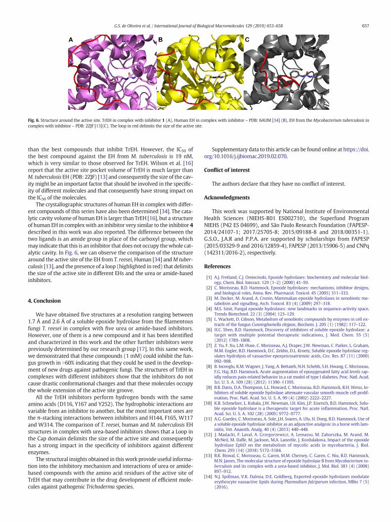
Fig. 6. Structure around the active site. TrEH in complex with inhibitor 1 (A), Human EH in complex with inhibitor – PDB: 6AUM [34] (B), EH from the Mycobacterium tuberculosis incomplex with inhibitor – PDB: 2ZJF [13](C). The loop in red delimits the size of the active site.
657G.S. de Oliveira et al. / International Journal of Biological Macromolecules 129 (2019) 653–658
than the best compounds that inhibit TrEH. However, the IC50 ofthe best compound against the EH from M. tuberculosis is 19 nM,which is very similar to those observed for TrEH. Wilson et al. [16]report that the active site pocket volume of TrEH is much larger thanM. tuberculosis EH (PDB: 2ZJF) [13] and consequently the size of the cav-ity might be an important factor that should be involved in the specific-ity of different molecules and that consequently have strong impact onthe IC50 of the molecules.
The crystallographic structures of human EH in complexwith differ-ent compounds of this series have also been determined [34]. The cata-lytic cavity volume of human EH is larger than TrEH [16], but a structureof human EH in complexwith an inhibitor very similar to the inhibitor 4described in this work was also reported. The difference between thetwo ligands is an amide group in place of the carboxyl group, whichmay indicate that this is an inhibitor that does not occupy thewhole cat-alytic cavity. In Fig. 6, we can observe the comparison of the structurearound the active site of the EH from T. reesei, Human [34] andM tuber-culosis [13], and the presence of a loop (highlighted in red) that delimitsthe size of the active site in different EHs and the urea or amide-basedinhibitors.
4. Conclusion
We have obtained five structures at a resolution ranging between1.7 Å and 2.6 Å of a soluble epoxide hydrolase from the filamentousfungi T. reesei in complex with five urea or amide-based inhibitors.However, one of them is a new compound and it has been identifiedand characterized in this work and the other further inhibitors werepreviously determined by our research group [17]. In this same work,we demonstrated that these compounds (1 mM) could inhibit the fun-gus growth in N60% indicating that they could be used in the develop-ment of new drugs against pathogenic fungi. The structures of TrEH incomplexes with different inhibitors show that the inhibitors do notcause drastic conformational changes and that these molecules occupythe whole extension of the active site groove.
All the TrEH inhibitors perform hydrogen bonds with the sameamino acids (D116, Y167 and Y252). The hydrophobic interactions arevariable from an inhibitor to another, but the most important ones arethe π-stacking interactions between inhibitors and H144, F165, W117and W314. The comparison of T. reesei, human and M. tuberculosis EHstructures in complex with urea-based inhibitors shows that a Loop inthe Cap domain delimits the size of the active site and consequentlyhas a strong impact in the specificity of inhibitors against differentenzymes.
The structural insights obtained in thiswork provide useful informa-tion into the inhibitory mechanism and interactions of urea or amide-based compounds with the amino acid residues of the active site ofTrEH that may contribute in the drug development of efficient mole-cules against pathogenic Trichoderma species.
Supplementary data to this article can be found online at https://doi.org/10.1016/j.ijbiomac.2019.02.070.
Conflict of interest
The authors declare that they have no conflict of interest.
Acknowledgments
This work was supported by National Institute of EnvironmentalHealth Sciences (NIEHS-R01 ES002710), the Superfund ProgramNIEHS (P42 ES 04699), and São Paulo Research Foundation (FAPESP-2014/24107-1; 2017/25705-8; 2015/09188-8 and 2018/00351-1).G.S.O., J.A.R and P.P.A. are supported by scholarships from FAPESP(2015/03329-9 and 2016/12859-4), FAPESP (2013/15906-5) and CNPq(142311/2016-2), respectively.
References
[1] A.J. Fretland, C.J. Omiecinski, Epoxide hydrolases: biochemistry and molecular biol-ogy, Chem. Biol. Interact. 129 (1–2) (2000) 41–59.
[2] C. Morisseau, B.D. Hammock, Epoxide hydrolases: mechanisms, inhibitor designs,and biological roles, Annu. Rev. Pharmacol. Toxicol. 45 (2005) 311–333.
[3] M. Decker, M. Arand, A. Cronin, Mammalian epoxide hydrolases in xenobiotic me-tabolism and signalling, Arch. Toxicol. 83 (4) (2009) 297–318.
[4] M.S. Smit, Fungal epoxide hydrolases: new landmarks in sequence-activity space,Trends Biotechnol. 22 (3) (2004) 123–129.
[5] L. Wackett, D. Gibson, Metabolism of xenobiotic compounds by enzymes in cell ex-tracts of the fungus Cunninghamella elegans, Biochem. J. 205 (1) (1982) 117–122.
[6] H.C. Shen, B.D. Hammock, Discovery of inhibitors of soluble epoxide hydrolase: atarget with multiple potential therapeutic indications, J. Med. Chem. 55 (5)(2012) 1789–1808.
[7] Z. Yu, F. Xu, L.M. Huse, C. Morisseau, A.J. Draper, J.W. Newman, C. Parker, L. Graham,M.M. Engler, B.D. Hammock, D.C. Zeldin, D.L. Kroetz, Soluble epoxide hydrolase reg-ulates hydrolysis of vasoactive epoxyeicosatrienoic acids, Circ. Res. 87 (11) (2000)992–998.
[8] B. Inceoglu, K.M.Wagner, J. Yang, A. Bettaieb, N.H. Schebb, S.H. Hwang, C. Morisseau,F.G. Haj, B.D. Hammock, Acute augmentation of epoxygenated fatty acid levels rap-idly reduces pain-related behavior in a rat model of type I diabetes, Proc. Natl. Acad.Sci. U. S. A. 109 (28) (2012) 11390–11395.
[9] B.B. Davis, D.A. Thompson, L.L. Howard, C. Morisseau, B.D. Hammock, R.H. Weiss, In-hibitors of soluble epoxide hydrolase attenuate vascular smooth muscle cell prolif-eration, Proc. Natl. Acad. Sci. U. S. A. 99 (4) (2002) 2222–2227.
[10] K.R. Schmelzer, L. Kubala, J.W. Newman, I.H. Kim, J.P. Eiserich, B.D. Hammock, Solu-ble epoxide hydrolase is a therapeutic target for acute inflammation, Proc. Natl.Acad. Sci. U. S. A. 102 (28) (2005) 9772–9777.
[11] A.G. Guedes, C. Morisseau, A. Sole, J.H. Soares, A. Ulu, H. Dong, B.D. Hammock, Use ofa soluble epoxide hydrolase inhibitor as an adjunctive analgesic in a horse with lam-initis, Vet. Anaesth. Analg. 40 (4) (2013) 440–448.
[12] J. Madacki, F. Laval, A. Grzegorzewicz, A. Lemassu, M. Zahorszka, M. Arand, M.McNeil, M. Daffe, M. Jackson, M.A. Laneelle, J. Kordulakova, Impact of the epoxidehydrolase EphD on the metabolism of mycolic acids in mycobacteria, J. Biol.Chem. 293 (14) (2018) 5172–5184.
[13] B.K. Biswal, C. Morisseau, G. Garen, M.M. Cherney, C. Garen, C. Niu, B.D. Hammock,M.N. James, Themolecular structure of epoxide hydrolase B fromMycobacterium tu-berculosis and its complex with a urea-based inhibitor, J. Mol. Biol. 381 (4) (2008)897–912.
[14] N.J. Spillman, V.K. Dalmia, D.E. Goldberg, Exported epoxide hydrolases modulateerythrocyte vasoactive lipids during Plasmodium falciparum infection, MBio 7 (5)(2016).

658 G.S. de Oliveira et al. / International Journal of Biological Macromolecules 129 (2019) 653–658
[15] B.A. Flitter, K.L. Hvorecny, E. Ono, T. Eddens, J. Yang, D.H. Kwak, C.D. Bahl, T.H.Hampton, C. Morisseau, B.D. Hammock, X. Liu, J.S. Lee, J.K. Kolls, B.D. Levy, D.R.Madden, J.M. Bomberger, Pseudomonas aeruginosa sabotages the generation ofhost proresolving lipid mediators, Proc. Natl. Acad. Sci. U. S. A. 114 (1) (2017)136–141.
[16] C. Wilson, G.S. De Oliveira, P.P. Adriani, F.S. Chambergo, M.V.B. Dias, Structure of asoluble epoxide hydrolase identified in Trichoderma reesei, Biochim. Biophys. Acta1865 (8) (2017) 1039–1045.
[17] G.S. de Oliveira, P.P. Adriani, H. Wu, C. Morisseau, B.D. Hammock, F.S. Chambergo,Substrate and inhibitor selectivity, and biological activity of an epoxide hydrolasefrom Trichoderma reesei, Mol. Biol. Rep. (2018) 1–9.
[18] G. Zafra, D.V. Cortes-Espinosa, Biodegradation of polycyclic aromatic hydrocarbonsby Trichoderma species: a mini review, Environ. Sci. Pollut. Res. Int. 22 (24) (2015)19426–19433.
[19] Y. Myoken, T. Sugata, Y. Fujita, H. Asaoku, M. Fujihara, Y. Mikami, Fatal necrotizingstomatitis due to Trichoderma longibrachiatum in a neutropenic patient with malig-nant lymphoma: a case report, Int. J. Oral Maxillofac. Surg. 31 (6) (2002) 688–691.
[20] J. Guarro, M.I. Antolin-Ayala, J. Gene, J. Gutierrez-Calzada, C. Nieves-Diez, M.Ortoneda, Fatal case of Trichoderma harzianum infection in a renal transplant recip-ient, J. Clin. Microbiol. 37 (11) (1999) 3751–3755.
[21] A. Gautheret, F. Dromer, J.H. Bourhis, A. Andremont, Trichoderma pseudokoningii as acause of fatal infection in a bonemarrow transplant recipient, Clin. Infect. Dis. 20 (4)(1995) 1063–1064.
[22] T. Walsh, A. Groll, J. Hiemenz, R. Fleming, E. Roilides, E. Anaissie, Infections due toemerging and uncommon medically important fungal pathogens, Clin. Microbiol.Infect. 10 (2004) 48–66.
[23] G.S. de Oliveira, P.P. Adriani, F.G. Borges, A.R. Lopes, P.T. Campana, F.S. Chambergo,Epoxide hydrolase of Trichoderma reesei: biochemical properties and conforma-tional characterization, Int. J. Biol. Macromol. 89 (2016) 569–574.
[24] J.R. Whitaker, P.E. Granum, An absolute method for protein determination based ondifference in absorbance at 235 and 280 nm, Anal. Biochem. 109 (1) (1980)156–159.
[25] W. Kabsch, Xds, Acta Crystallogr. D Biol. Crystallogr. 66 (2) (2010) 125–132.[26] P.R. Evans, G.N. Murshudov, How good are my data and what is the resolution? Acta
Crystallogr. D Biol. Crystallogr. 69 (7) (2013) 1204–1214.[27] M.D. Winn, C.C. Ballard, K.D. Cowtan, E.J. Dodson, P. Emsley, P.R. Evans, R.M. Keegan,
E.B. Krissinel, A.G. Leslie, A. McCoy, Overview of the CCP4 suite and current develop-ments, Acta Crystallogr. Sect. D 67 (4) (2011) 235–242.
[28] A.J. McCoy, R.W. Grosse-Kunstleve, P.D. Adams, M.D. Winn, L.C. Storoni, R.J. Read,Phaser crystallographic software, J. Appl. Crystallogr. 40 (4) (2007) 658–674.
[29] P.V. Afonine, R.W. Grosse-Kunstleve, N. Echols, J.J. Headd, N.W. Moriarty, M.Mustyakimov, T.C. Terwilliger, A. Urzhumtsev, P.H. Zwart, P.D. Adams, Towards au-tomated crystallographic structure refinement with phenix.refine, Acta Crystallogr.D Biol. Crystallogr. 68 (4) (2012) 352–367.
[30] T.C. Terwilliger, P.D. Adams, R.J. Read, A.J. McCoy, N.W. Moriarty, R.W. Grosse-Kunstleve, P.V. Afonine, P.H. Zwart, L.-W. Hung, Decision-making in structure solu-tion using Bayesian estimates of map quality: the PHENIX AutoSol wizard, ActaCrystallogr. D Biol. Crystallogr. 65 (6) (2009) 582–601.
[31] P. Emsley, K. Cowtan, Coot: model-building tools for molecular graphics, ActaCrystallogr. D Biol. Crystallogr. 60 (12) (2004) 2126–2132.
[32] V.B. Chen, W.B. Arendall, J.J. Headd, D.A. Keedy, R.M. Immormino, G.J. Kapral, L.W.Murray, J.S. Richardson, D.C. Richardson, MolProbity: all-atom structure validationfor macromolecular crystallography, Acta Crystallogr. D Biol. Crystallogr. 66 (1)(2010) 12–21.
[33] A.C. Wallace, R.A. Laskowski, J.M. Thornton, LIGPLOT: a program to generate sche-matic diagrams of protein-ligand interactions, Protein Eng. Des. Sel. 8 (2) (1995)127–134.
[34] S.D. Kodani, S. Bhakta, S.H. Hwang, S. Pakhomova, M.E. Newcomer, C. Morisseau,B.D. Hammock, Identification and optimization of soluble epoxide hydrolase inhib-itors with dual potency towards fatty acid amide hydrolase, Bioorg.Med. Chem. Lett.28 (4) (2018) 762–768.