autoantibody response to microsomal epoxide hydrolase in ... · autoantibody response to microsomal...
TRANSCRIPT
Journal of Autoimmunity 28 (2007) 7e18www.elsevier.com/locate/jautimm
Autoantibody response to microsomal epoxide hydrolase inhepatitis C and A
Toshitaka Akatsuka a,*, Nobuharu Kobayashi a, Takashi Ishikawa d,Takafumi Saito e, Michiko Shindo f, Masayoshi Yamauchi g, Kazutaka Kurokohchi h,Hitoshi Miyazawa b, Hongying Duan a, Toshiyuki Matsunaga c, Tsugikazu Komoda c,
Christophe Morisseau i, Bruce D. Hammock i
a Department of Microbiology, Saitama Medical University, Moroyama-cho, Iruma-gun, Saitama 350-0495, Japanb Department of Respiratory Medicine, Saitama Medical University, Saitama, Japan
c Department of Biochemistry, Saitama Medical University, Saitama, Japand Department of Internal Medicine, University of Tokyo, Tokyo, Japan
e Department of Internal Medicine and Molecular Therapeutics, Yamagata University School of Medicine, Yamagata, Japanf Division of Liver Diseases, Akashi Municipal Hospital, Hyogo, Japan
g Yamauchi Medical Clinic, Kanagawa, Japanh Third Department of Internal Medicine, Kagawa University School of Medicine, Kagawa, Japan
i Department of Entomology and Cancer Research Center, University of California, Davis, CA, USA
Received 6 November 2006; revised 17 December 2006; accepted 18 December 2006
Abstract
Autoimmune responses were observed in a large proportion of hepatitis C cases and are suspected to be part of viral pathogenesis. TheAN6520 antigen (AN-Ag) is a normal cellular protein mainly expressed in liver that was found associated with non-A, non-B hepatitis. Toelucidate its pathogenic role in hepatitis C, we developed an IgM capture assay using purified AN-Ag and confirmed that the antibody responseto AN-Ag is associated almost exclusively with hepatitis C cases (29%). Screening of a human liver expression library revealed that AN-Ag ismainly the microsomal epoxide hydrolase (mEH), a drug-metabolizing enzyme that plays an important role in the metabolism of some muta-genic and carcinogenic epoxides. Using the purified recombinant human mEH as an antigen, we now found that antibodies against this proteinare associated with nearly 82% of hepatitis C virus infections and surprisingly with 46% of patients with hepatitis A. The appearance of AN-Ag/mEH in the incubation period of hepatitis C as previously reported and the antibody responses shown here indicate that this enzyme may bea marker for or even a cause of some of the pathology associated with hepatitis C and A.� 2007 Elsevier Ltd. All rights reserved.
Keywords: Baculovirus; Hepatitis A virus; Hepatitis C virus; Purification; Radioimmunoassay
Abbreviations: ELISA, enzyme-linked immunosorbent assay; S/N ratio, signal/noise ratio; ALT, alanine aminotransferase; CsCl, cesium chloride; OPD,
o-phenylenediamine dihydrochloride; FCS, fetal calf serum; BSA, bovine serum albumin; FITC, fluorescein isothiocyanate; TRITC, tetramethyl rhodamine
isothiocyanate.
* Corresponding author. Tel.: þ81 49 276 1165; fax: þ81 49 295 9107.
E-mail addresses: [email protected] (T. Akatsuka), [email protected] (N. Kobayashi), [email protected] (T. Ishikawa),
[email protected] (T. Saito), [email protected] (M. Shindo), [email protected] (M. Yamauchi), [email protected].
jp (K. Kurokohchi), [email protected] (H. Miyazawa), [email protected] (H. Duan), [email protected] (T. Matsunaga), tkalp1lp@
saitama-med.ac.jp (T. Komoda), [email protected] (C. Morisseau), [email protected] (B.D. Hammock).
0896-8411/$ - see front matter � 2007 Elsevier Ltd. All rights reserved.
doi:10.1016/j.jaut.2006.12.005

8 T. Akatsuka et al. / Journal of Autoimmunity 28 (2007) 7e18
1. Introduction
Hepatitis C virus (HCV), a member of the Flaviviridaefamily is thought not to be directly cytopathic, rather it trig-gers an immune-mediated inflammatory response that elimi-nates the virus and/or slowly damages the hepatocytes [1].Although humoral and cellular immune responses duringHCV infection were extensively studied, their pathogenic rolesare still unclear.
Before the isolation of the virus by molecular biologicaltechniques [2], many antigens were claimed to be associatedwith hepatitis C (non-A, non-B hepatitis; NANBH) using im-munological techniques [3e5]. We found an antigen (AN6520antigen; AN-Ag) in the liver of patients with NANBH whichformed a precipitin line with convalescent sera from patientswith NANBH. We purified the antigen and developed passivehemagglutination assay (PHA) using antigen-coated erythro-cytes to detect antibody in patients’ sera. We found that the an-tigen is composed of particles with molecular weight of morethan 1.5� 106 Da and diameter of 29e34 nm. The antibodywas detected in 37.5% in NANBH cases, but not in controlgroups [6]. Then we developed monoclonal antibodies(mAbs) and used one of them, 1F12, to develop radioimmuno-assays (RIAs) for the antigen and for the patient’s antibody[7]. The antigen and antibody were detected in the acute phaseand convalescent phase sera, respectively, of some patientswith NANBH. In sera obtained sequentially from chimpanzeesinfected with NANBH agent (now known as HCV), the ANantigen appears during the incubation time before the eleva-tion of ALT [8]. Based on these results, we initially thoughtthat the AN-Ag is from the viral particles of NANBH agent.However, we later showed that AN-Ag is a normal cellularprotein mainly expressed in the microsomal fraction of liver,however, its concentration varies considerably between indi-viduals [8].
Toward isolating and identifying AN-Ag, we initially triedto use the RIA assays developed previously [7]. However, theinhibition RIA is not specific enough in that it cross reactedwith an unknown protein present in the serum of many people.Thus, we are reporting herein a novel IgM capture RIAmethod that is more immunoglobulin-specific than the previ-ous inhibition assay. Using this assay, we investigated thespecificity of anti-AN antibody response to HCV infection.Further, we identified and cloned the cDNA of AN-Ag. Wealso confirmed the antibody response using the purified anti-gen expressed by cDNA. These results show some insightsabout the role of the AN-antigen in the pathogenesis ofhepatitis.
2. Materials and methods
2.1. Patients
Sera used in the study shown in Table 1 were collected byJikei University Hospital from 1980 to 1981. During this pe-riod, informed consent was not commonly obtained. HepatitisC cases in this study were from the epidemic in Shimizu city
in Japan [9]. They were diagnosed as NANBH and laterproven to be hepatitis C by serological diagnosis (Ortho Diag-nostic, NY) [10]. Sera from patients with various forms ofhepatitis as well as normal blood donors shown in Tables 2and 3 were obtained from 2000 to 2005 under the appropriateapproval guidelines from the following institutions: TokyoUniversity Hospital, Yamagata University Hospital, AkashiMunicipal Hospital, and Kagawa University Hospital, Japan.The diagnosis of viral hepatitis was made on the basis of theresults of virological tests with histopathological findings,and drug-induced hepatitis was defined on the basis of the pa-tient’s medical history to identify any possible hepatotoxinswith clinical and histopathological findings.
2.2. Cell lines
The hepatocellular carcinoma (HCC) cell line, huH-2 [11]was obtained from the Japanese Cancer Research ResourcesBank. THLE-5b, a normal liver cell line which was immortal-ized by transfecting with the plasmid containing SV40 T-Ag[12], was a gift from Dr. C.C. Harris (CCR, NCI, NIH).Both were maintained in RPMI1640 media with 5% heat-inac-tivated FCS. BHK-21 was obtained from the American TypeCulture Collection (Rockville, MD) and maintained inEMEM media with 5% FCS.
2.3. Antibodies
Rabbit anti-AN antibody and mouse anti-AN mAb 1F12have been described [6,7]. Rabbit anti-mEH antibody was
Table 1
Prevalence of anti-AN6520 IgM in sera from patients with acute hepatitis and
normal donors
Type of hepatitis No. of cases Ab positive %
Type A 37 (39) 1 (0) 2.7 (2.6)
Type B 17 (18) 0 (0) 0.0 (0.0)
Type C 31 (38) 9 (13) 29.0* (34.2*)
Normal donors 21 (26) 0 (0) 0.0 (0.0)
Results by IgM capture RIA using purified AN-Ag that were obtained from
1980 to 1981 are shown. Combined data of RIA and ELISA are shown in
parentheses.
*P< 0.01 by Fisher’s exact probability test.
Table 2
Prevalence of anti-mEH IgM in sera from patients with acute and chronic
hepatitis and normal donors
Type of hepatitis No. of cases Ab positive %
Type A (acute) 22 10 45.5*
Type B (acute) 12 0 0.0
Type B (chronic) 27 2 7.4
Type C (acute) 22 18 81.8*
Type C (chronic) 63 16 25.4**
Drug-induced 16 0 0.0
Normal donors 13 0 0.0
Results by IgM capture RIA using the membrane-bound form of mEH are
shown.
*P< 0.01 and **P< 0.05 by Fisher’s exact probability test.
9T. Akatsuka et al. / Journal of Autoimmunity 28 (2007) 7e18
raised by injecting a rabbit (s.c.) with 400 mg of solubilizedand purified mEH three times (first with Freund’s completeadjuvant, second and third immunizations with incompleteadjuvant). Ascites of hybridoma producing human IgM(m-chain)-specific mAb #9 was a kind gift from Dr. M. Arita(National Institute of Infectious Diseases, Japan).
2.4. Transfection, immunofluorescence staining, andimmunoblotting
BHK-21 cells were transfected using Lipofectin (Invitro-gen, Carlsbad, CA) according to the manufacturer’s instruc-tion. After being transferred to a Lab-Tek chamber slide(NUNC, Naperville, IL), they were double-stained with rabbitantiserum diluted (1:200) in supernatant of 1F12 hybridomaculture, followed by the mixture of TRITC-conjugated goatanti-rabbit IgG (Beckman Coulter, France) (1:50) and FITC-conjugated goat anti-mouse IgG (Sigma, St. Louis, MO)(1:100). SDS-PAGE was performed by applying 400 ng and50 ng of protein onto a 10% minigel (Mini-Protean; Bio-Rad,Hercules, CA) for silver staining and immunoblotting, respec-tively. After separation and transfer to an Immobilon-P mem-brane (Millipore, Bedford, MA), the antigen was detectedwith rabbit anti-mEH (1:500) followed by alkaline phospha-tase-labeled goat anti-rabbit IgG (KPL, Gaithersburg, MA)and BCIP/NBT substrate (KPL).
2.5. IgM capture assay
A flat-bottomed eight-well strip plate (COSTAR, Acton,MA) was used as the solid phase and 50 ml of ascites fluidof anti-human IgM hybridoma #9 diluted (1:10,000) in PBSwas added to each well and incubated overnight at 4 �C. Afterblocking with 200 ml of 5% BSA in PBS for 1 h at 37 �C and
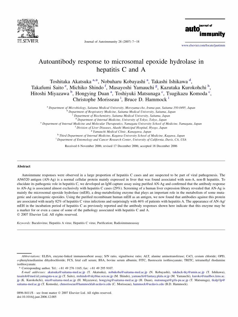
Table 3
Comparison of anti-mEH and anti-AN IgM in sera from patients with acute
and chronic hepatitis and normal donors
Case Type of hepatitis Anti-mEH anti-AN
1 Type A (acute) 9.69 1.18
2 Type A (acute) 1.35 1.25
3 Type A (acute) 15.37 1.13
4 Type B (acute) 1.68 1.12
5 Type B (acute) 1.36 1.11
6 Type B (acute) 0.51 0.86
7 Type B (acute) 0.69 0.98
8 Type B (acute) 0.72 0.93
9 Type C (acute) 0.74 0.76
10 Type C (acute) 2.48 2.63
11 Type C (acute) 1.18 0.96
12 Type C (acute) 2.67 0.82
13 Type C (chronic) 2.59 3.19
14 Type C (chronic) 15.26 7.34
15 Type C (chronic) 4.36 4.52
16 Type C (chronic) 3.51 6.10
17 Normal 1.10 1.63
18 Normal 0.76 1.00
19 Normal 0.55 1.06
20 Normal 1.14 0.94
S/N ratios are shown. Positive results (�2.1) are shown in bold.
washing, 50 ml of serum samples diluted (1:20) in PBS con-taining 1% BSA and 0.1% NaN3 (RIA buffer) was addedand incubated overnight at 4 �C. After washing, 1� 105 cpmof 125I-labeled AN-Ag or membrane-bound form of mEH in50 ml of RIA buffer was added and incubated for 2 h at37 �C. Each well was separated after washing, and the bound ra-dioactivity was counted by a gammacounter. An ELISA was alsodeveloped, in which the 125I-AN-Ag was replaced by non-labeledAN-Ag (500 ng/ml in RIA buffer), and the bound antigen wasdetected by incubation with horseradish peroxidase-labeled1F12 (for 1 h at 37 �C) followed by OPD substrate (SIGMASt Louis, MO). The reaction was stopped by adding H2SO4
and OD492 was read. Labeling with 125I and peroxidase wasperformed by the chloramine-T method [13] and P. Nakane’smethod [14], respectively. Cut-off values in RIA and ELISAwere 2.1 (S/N ratio) and 1.0 (above the mean OD of negativecontrol) using sera from five normal donors as the negativecontrol, respectively. They corresponded to 4 SD above themean values. All the washing steps were done with PBScontaining 0.05% Tween 20. For confirmation of the specificityof the reaction, serum sample was incubated with 50 mg/ml ofanti-human IgG (g-chain) or anti-human IgM (m-chain) anti-body (Pierce, Rockford, IL) overnight at 4 �C in a test tube be-fore being added to the antibody coated plate. Statisticalanalysis was performed by Scheffe’s F-test (Figs. 1 and 7)and Fisher’s exact probability test (Tables 1 and 2).
2.6. cDNA identification
A lgt11 cDNA library from human liver (Clontech, Pal-oAlto, CA) was screened with rabbit anti-AN antibody [6]
Group
S/N
ratio
0
1
2
3
4
5
Type A Type B Type C Normaln = 37 n = 17 n = 31 n = 21
P<.01 P<.01P<.01
Fig. 1. IgM capture RIA for the detection of anti-AN6520 antibodies. IgM was
captured by IgM-specific monoclonal antibody on the solid phase and detected
with 125I-AN-Ag. S/N values of sera from patients with acute hepatitis A (type
A), hepatitis B (type B), and hepatitis C (type C), and normal donors (normal)
are shown. A circle represents a case, and a dotted line represents a cut-off
value (S/N¼ 2.1). The mean value of each group is shown by a horizontal
bar. For statistical analysis, Scheffe’s F-test was used.

10 T. Akatsuka et al. / Journal of Autoimmunity 28 (2007) 7e18
(50 mg/ml), by the standard protocol [15]. After four cycles ofselection, they were further selected by using their phage ly-sates for affinity purification [16] of rabbit anti-AN antibodyfollowed by incubation of the eluted antibody on the immuno-blot of mEH-containing cell lysate. Briefly, Hybond-C filter(Amersham, Buckinghamshire, UK) (1.0� 4.0 cm) that boundplaque protein was soaked with 20% FCS in 10 mM TriseHClpH 7.4-buffered saline (TBS) containing 0.05% Tween 20overnight at 4 �C, then incubated with 0.5 ml of antibody over-night at 4 �C. After washing, the bound antibody was eluted byincubating the filter with 160 ml of 0.2 M glycineeHCl (pH2.5) for 2 min. The eluate was immediately neutralized byadding 80 ml of 1.0 M K2HPO4 (pH 9.0), then 100 ml of50% FCS in TBS was added. The eluted antibody was incu-bated on the western blot of the microsomal fraction [17] ofHCC line huH-2 which expresses mEH. From the finallyselected clone HLC4, the insert DNA was digested withEcoR1 into 3 fragments, and ligated into a pKF3 vector inthe Enforcement Cloning System (Takara, Tokyo, Japan).The insert of each plasmid was sequenced with pKF3 sequenc-ing primers F2 and R3 (Takara).
2.7. Expression and purification of mEH
Total RNA of human liver cell line THLE-5b was extractedwith ISOGEN (Nippon Gene, Tokyo, Japan), and mRNA wasisolated with Dynabeads with oligo dT25 (Dynal, Oslo,Norway). The first strand cDNA was synthesized by reversetranscriptase (Takara), and the mEH cDNA was amplified byPCR with KOD-Plus polymerase (Toyobo, Tokyo, Japan)and 50- and 30-end primers of mEH cDNA accompanyingHindIII and BamHI sequences, respectively; 50-AAGCTTATGTGGCTAGAAATCCTCCTCA-30 and 50-GGATCCTCATTGCCGCTCCAGCACCGACAGG-30. The PCR product wasdigested with HindIII and BamHI, and ligated into the samecloning sites of pcDNA3(þ) (Invitrogen). The sequence ofthe insert mEH cDNA in the ligated plasmid was confirmedwith the appropriate primers synthesized from the determinedsequence. Expression of human mEH in a recombinant bacu-lovirus system has been described [18]. The purification pro-cedure of the solubilized form of mEH was based on themethod of Lacourciere [19] with some modifications: Sf9 cells(1 L culture) were infected with recombinant baculovirus(m.o.i.¼ 10), and after 96 h incubation, the cells were sus-pended in 0.1 M phosphate buffer pH 7.4 containing 1 mMPMSF, 1 mM EDTA and 1 mM DTT. The cells were homog-enized with Polytron homogenizer and centrifuged at100,000� g for 60 min. The pellet was suspended with bufferA (10 mM TriseHCl, pH 7.6) containing 1% Tween 20 bysonication, and centrifuged at 100,000� g for 60 min. Thepellet was suspended with buffer A containing 1% TritonX-100 by sonication, and centrifuged at 100,000� g for60 min. The supernatant was dialyzed overnight against bufferB (10 mM TriseHCl, pH 9.0 containing 0.05% Triton X-100)and applied onto a 10� 2.5 cm Q-Sepharose (Amersham,Uppsala, Sweden) column equilibrated with buffer B. The col-umn was washed with buffer B containing 100 mM NaCl, and
then the enzyme was eluted with buffer B containing 200 mMNaCl. Active fractions were pooled, concentrated using Cen-triplus YM-10 (Millipore, Bedford, MA), and then dialyzedagainst buffer A containing 0.05% Triton X-100. Membrane-bound form of mEH was purified according to the methodfor AN-Ag [6]. The homogenate from 1.2 g of infected SF9cell pellet was applied onto a Sephacryl S-300 (Amersham,Uppsala, Sweden) column (2.5� 90 cm) and eluted withTBS containing 1 mM EDTA (buffer C). Void volume frac-tions were centrifuged at 100,000� g for 120 min. The pelletwas suspended by sonication with 1.5 ml of buffer C contain-ing 1% Tween 20 and then 200 ml of 1 M Tris pH 9.0. wasadded. It was layered onto the gradient of 5e60% (w/v)sucrose in buffer C (30 ml) and after centrifugation at63,000� g for 16 h, 1.2 ml fractions were collected. Anti-gen-positive fractions were pooled, dialyzed against buffer Covernight, and then concentrated with Centriplus YM-100. Itwas next layered on CsCl gradients of 7% to 42% (w/v) inbuffer C (4.5 ml) and centrifuged at 175,000� g for 20 h.One hundred and seventy microliter fractions were collectedand antigen-positive peak was dialyzed against buffer C over-night, and concentrated with Centriplus YM-100. All thepurification procedures were performed at 4 �C. Protein con-centration was measured by micro BCA assay (Pierce), andthe antigen was titrated by antibody sandwich ELISA usinghorseradish peroxidase-labeled 1F12 using purified AN-Agas the standard.
2.8. Analysis of enzyme activity of mEH
The mEH enzyme activity was determined by measuringthe formation rate of dihydrobenzoin produced from hydroly-sis of cis-stilbene oxide by the method of Maekawa et al. [20].HPLC analysis was performed using a Tosoh 8000 system(Tosoh, Tokyo, Japan).
2.9. Chimpanzee experiments
The housing, maintenance, and care of the chimpanzeesused in this study met requirements for the humane use of an-imals in scientific research as defined by the National Insti-tutes of Health. Two chimpanzees (Ch1360 and Ch1365)were inoculated i.v. with w103 infectious doses of theHM175 strain of HAV [21]. Twenty-three weeks after HAVinfection, Ch1365 was inoculated i.v. with w103 infectiousdoses of the H strain of HCV along with the third chimpanzeeCh1397 as described [22]. Serum samples were taken weeklyfor up to 32 weeks.
3. Results
3.1. IgM capture assay using purified AN antigen
Results of the IgM capture assay for antibody to the puri-fied AN-Ag are presented in Table 1. This assay eliminatedthe false positive results observed in the older sandwich type

11T. Akatsuka et al. / Journal of Autoimmunity 28 (2007) 7e18
RIA in which a non-immunoglobulin factor that bound to AN-Ag was detected. Using this method, we found that 29% of thehepatitis C sera had antibody to the AN-Ag compared to 2.7%of hepatitis A patients while hepatitis B and normal donors didnot have the antibody. The S/N ratios were also significantlyhigher in hepatitis C than any other groups (Fig. 1). Specificityof the reaction was confirmed by the blocking experiments inwhich antibody-positive sera were incubated with anti-m chainor anti-g chain antibody before being added onto the solidphase (not shown). We also developed an IgM capture ELISAwhich is more specific than RIA because the bound Ag wasdetected by the enzyme-labeled anti-AN-Ag mAb. By combin-ing the results of the two assays, the prevalence of the antibodyin the hepatitis C group rose to 34.2% (Table 1). Sequentialserum samples from three patients with hepatitis C were testedby ELISA (Fig. 2). It shows that the antibody appeared whileALT was still elevated and declined along with resolving ofhepatitis.
3.2. Isolation of cDNA of AN antigen
To identify the cDNA of AN antigen, a lgt11 expressionlibrary from human liver was used because we previouslyfound that this antigen is a normal cellular protein mainlyexpressed in liver [8]. mAbs to AN-Ag are known to recog-nize only conformational epitopes that are easily disruptedby detergents. Therefore, polyclonal rabbit antibody that re-vealed a band in western blotting was chosen for immunoscre-ening of the library [8]. Out of about one million plaques, weobtained 46 positive plaques in the first screening. After re-peating the screening four times, we selected 12 plaques.Their phage lysates were used for affinity purification [16]of the rabbit antiserum (Fig. 3). Although the antiserumshowed additional bands on the immunoblot of the huH-2cell extract, the antibody eluted from two clones (HLC3and HLC4) out of 12 gave a single band (w49 kDa) corre-sponding to AN-Ag. The inserted cDNA of HLC4 wassubcloned and sequenced. GenBank BLAST search revealedthat the sequence matches perfectly human mEH cDNA[23] encompassing nucleotide 74 to 1460 of the reportedtranscript.
3.3. Confirmation of identity of AN antigen as mEH
The entire coding region of mEH cDNA was cloned froma normal human liver cell line, inserted in an expression vectorpcDNA3(þ) and then transfected into BHK-21 cells. mEHwas also expressed in insect cells by recombinant baculovirusand used for purification as described [18,19]. The purifiedmEH was then used to raise rabbit antiserum. When BHKcell transfectants were double-stained with rabbit anti-mEHand mouse anti-AN mAb 1F12, both antibodies exhibited sim-ilar staining patterns with the former being a little broader thanthe other (Fig. 4AeC). In addition, the rabbit polyclonal anti-AN antibody and 1F12 also gave almost identical staining(Fig. 4DeF). This indicates that the human mEH is responsi-ble for most of the antigenicity of AN-Ag. Purified forms of
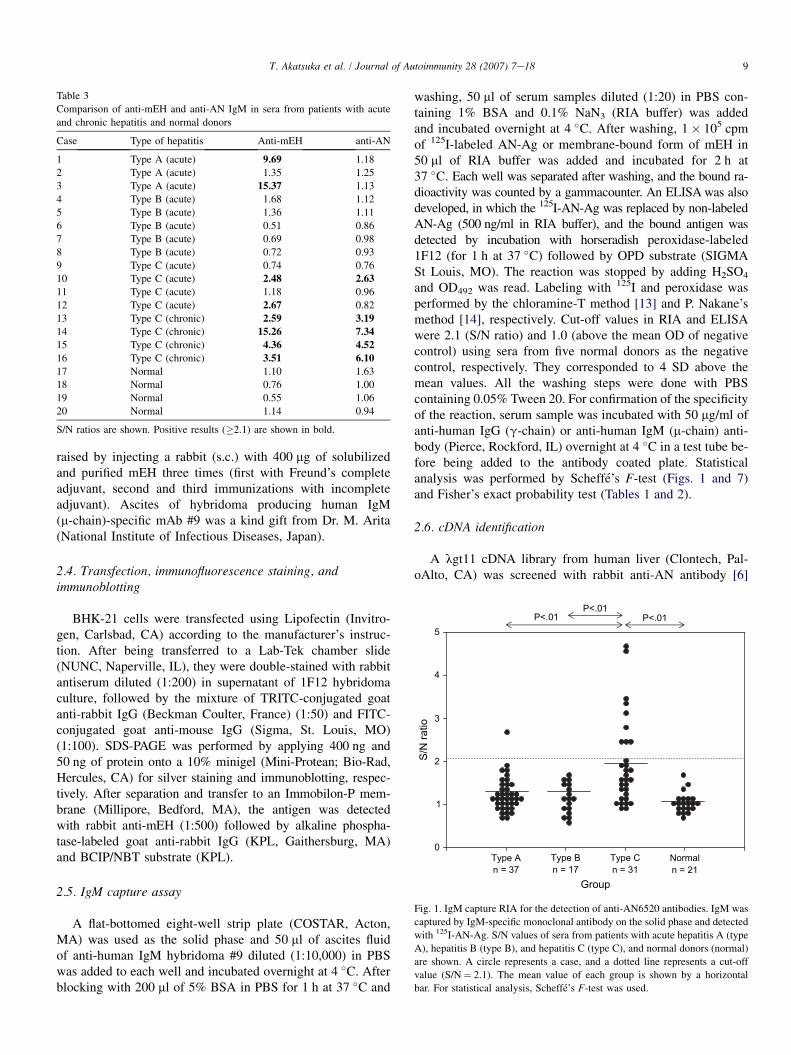
mEH and AN-Ag were compared by SDS-PAGE and westernblotting. Both showed a single band of more than 95% purityas judged by densitometer scanning (Fig. 5A). These bandshad similar size, and were also detected by both the rabbitanti-mEH (Fig. 5B), and the rabbit anti-AN antibody (notshown).
A
B
C
Weeks after onset0 1 2 3 4 5 6 7 8 9 10 11 12
ALT
0
100
200
300
400
500
600
700
O.D
.
0.0
0.2
0.4
0.6
0.8
1.0
1.2
1.4ALTIgM Ab
ALTIgM Ab
ALTIgM Ab
Weeks after onset0 2 4 6 8 10 12 14 16 18 20 22
ALT
100
0
200
300
400
500
600
700
O.D
.
0.0
0.1
0.2
0.3
0.4
0.5
Weeks after onset0 1 2 3 4 5 6 7 8 9 10 11 12 13
ALT
0
50
100
150
200
250
300
O.D
.
0.0
0.2
0.4
0.6
0.8
1.0
1.2
1.4
1.6
1.8
Fig. 2. IgM capture ELISA of serial serum samples from three patients with
acute hepatitis C (AeC). Capturing of IgM was the same as for Fig. 1 but
the bound antibody was detected by non-labeled AN-Ag followed by horserad-
ish peroxidase-labeled monoclonal antibody 1F12. ALT ( ) is expressed by
international units per liter. ELISA data (-B-) are expressed by OD492.

12 T. Akatsuka et al. / Journal of Autoimmunity 28 (2007) 7e18
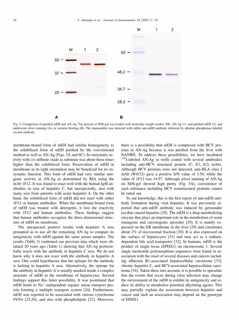
3.4. Purification of membrane-bound form of mEH
We then tested if anti-AN antibodies in the sera of hepatitisC patients recognize mEH. Purified mEH for the experimentsdescribed above was obtained following a conventional meth-odology: solubilization with detergent and ion-exchange chro-matography. However, we previously showed that AN-Ag iscomposed of particles 29e34 nm in diameter containingw49 kDa subunits. If treated with detergents, the particleslose reactivity with human antibodies as well as mouse mono-clonal antibodies [8]. Therefore, we decided to purify mEHwithout disrupting the membrane-bound form, and followedthe method used for purification of AN-Ag from liver [6]. Ahomogenate of recombinant baculovirus-infected Sf9 cellswas first passed through a gel-filtration column. The voidvolume fraction was then subjected to ultracentrifugation ina sucrose-gradient. As shown in Fig. 6A, we obtained twoantigen-positive peaks. As observed in SDS-PAGE, the firstpeak (fractions 3e7) had the highest specific antigenic activityand contained mostly the w49 kDa band with few, very lightadditional protein bands. The first peak fractions were pooled
Fig. 3. Second step selection of lgt11 cDNA library from human liver by af-
finity purification of the antibody. The filter that bound the phage lysate of the
candidate clone was incubated with rabbit anti-AN antibody. After washing,
the bound antibody was reacted with western blot of microsomal fraction of
huH-2 cells that expresses AN-Ag. The eluates from the phage lysates of
HLC1eHLC4 (lanes 1e4), rabbit anti-AN antibody before purification
(lane 5). The arrow denotes the AN-Ag (w49 kDa).
and applied onto a CsCl gradient (Fig. 6B). The main antigen-positive fractions (12e22) were pooled and concentrated.This method yielded 32 mg of purified protein from 1.2 g ofcell pellet. This membrane-bound form and the detergent-solubilized form of human mEH are both at least 95% pureas judged by SDS-PAGE (Fig. 6C). Enzyme activity wasmeasured on both preparations using cis-stilbene oxide assubstrate; the membrane-bound form displayed almost threetimes higher activity than the solubilized form (1550 vs.571 nmol min�1 mg�1). The antigen/protein ratio of themembrane-bound form was comparable to AN-Ag (99.5% ofAN-Ag) whereas the detergent-solubilized form had no reac-tivity with mAb 1F12 as expected (not shown).
3.5. IgM capture assay using mEH as antigen
Antibody to the purified membrane-bound form of mEHwas detected by the IgM capture RIA in serum samplesfrom patients with various forms of hepatitis (Table 2). Theantibody was detected in w82% of patients with acute hepa-titis C and also in w25% of chronic hepatitis C cases. Unex-pectedly, it was also detected in w46% of patients with acutehepatitis A. The incidence in other forms of hepatitis such astype B and drug-induced hepatitis was very low. The S/Nratios were also significantly higher in hepatitis C and hepatitisA than any other groups (Fig. 7). When the radio-labeled mEHwas treated with NP40 or SDS, it lost the reactivity withantibodies in both hepatitis A and C sera (not shown) suggest-ing that the antibodies recognize conformational epitope ofmembrane-bound mEH similar to what we previously ob-served for AN-Ag [8].
Because the results with sera of hepatitis A patients(Table 2) were very different from the results previouslyobtained with AN-Ag (Table 1), the remaining AN-Ag waslabeled and applied to some of the sera used for the experi-ments of Table 2. Table 3 shows that AN-Ag did not reactwith the antibodies in hepatitis A, but did react positivelywith some of anti-mEH positive hepatitis C sera. Therefore,AN-Ag seems to have more selectivity for hepatitis C serathan mEH.
In order to better define the relationship of anti-mEH to dis-ease, we tested serial serum samples from three chimpanzeesexperimentally infected with HAV and/or HCV (Ch1360,HAV; Ch1365, HAV and HCV; Ch1397, HCV). Sera were col-lected first between four and two weeks before inoculation,then regularly for up to at least six weeks after the peak ofenzyme elevation. Although chimpanzee’s IgM was boundto the capturing antibody with the same efficiency as humanIgM, we did not see any positive signals at any time point(S/N¼ 1.06 (mean)� 0.06 (SD)).
4. Discussion
Using IgM capture RIA and ELISA methods, the anti-body to AN-Ag was detected almost exclusively in serafrom patients with hepatitis C (34.2%) with only one excep-tion out of 39 cases of hepatitis A (2.6%) (Table 1).

13T. Akatsuka et al. / Journal of Autoimmunity 28 (2007) 7e18
Fig. 4. Double-staining of BHK transfectant expressing mEH. After transfection, the cells were transferred to a chamber slide and cultured. Then they were fixed
with acetone and double-stained by rabbit anti-mEH and mouse anti-AN mAb 1F12 (A and B), or by rabbit anti-AN antibody and 1F12 (D and E). Rabbit and
mouse antibodies were detected by TRITC- and FITC-labeled second antibodies, respectively. The merged image of A and B and that of D and E are shown in C
and F, respectively.
Because hepatitis C virus (HCV) is thought to not bedirectly cytopathic as are hepatitis A and B viruses, wehypothesized that AN-Ag that is particular to HCV infec-tion, plays a role in the pathogenesis of hepatitis C. Asa first step, we identified the antigen to which these hepati-tis C-specific antibodies are directed. For this purpose, wescreened a human liver expression library, and we isolateda cDNA which codes for a protein that reacts stronglywith AN antibodies. The sequence of this cDNA matchesalmost perfectly the sequence of the human microsomalepoxide hydrolase (mEH) cDNA encompassing 1288 bp ofthe coding region and 98 bp of 30-noncoding region. Therecombinant purified human mEH had the same size asthe purified AN-Ag when analyzed by SDS-PAGE, and
we observed that they have similar antigenicity in immuno-fluorescence staining (Fig. 4) and western blotting (Fig. 5).These findings indicate that the human mEH is responsiblefor most of the antigenicity of AN-Ag.
To confirm these results, we then wished to test that theanti-AN antibody activity in hepatitis C sera was againstmEH. However, human anti-AN antibody, like the mousemonoclonal antibody 1F12, is known to recognize only thethree-dimensional structure of AN-Ag, and lost its recognitionwhen AN-Ag is treated with some detergents [8]. Therefore, toovercome this shortcoming, we expressed the mEH in largeamounts in insect cells using recombinant baculovirus system,and then purified the protein without disrupting its membraneconformation by not using any detergent. The purified
14 T. Akatsuka et al. / Journal of Autoimmunity 28 (2007) 7e18
Fig. 5. Comparison of purified mEH and AN-Ag. Ten percent of SDS-gel was loaded with molecular weight marker (M), AN-Ag (1), and purified mEH (2), and
underwent silver staining (A), or western blotting (B). The immunoblot was detected with rabbit anti-mEH antibody followed by alkaline phosphatase-labeled
second antibody.
membrane-bound form of mEH had similar homogeneity tothe solubilized form of mEH purified by the conventionalmethod as well as AN-Ag (Figs. 5A and 6C). Its enzymatic ac-tivity with cis-stilbene oxide as substrate was about three timeshigher than the solubilized form. Preservation of mEH inmembrane in its right orientation may be beneficial for its en-zymatic function. This form of mEH had very similar anti-genic activity as AN-Ag as determined by RIA using themAb 1F12. It was found to react well with the human IgM an-tibodies in sera of hepatitis C, but unexpectedly, also withmany sera from patients with acute hepatitis A. On the otherhand, the solubilized form of mEH did not react with either1F12 or human antibodies. When the membrane-bound formof mEH was treated with detergents, it lost the reactivitywith 1F12 and human antibodies. These findings suggestthat human antibodies recognize the three-dimensional struc-ture of mEH on membrane.
The unexpected positive results with hepatitis A seraprompted us to use all the remaining AN-Ag to compare itsantigenicity with mEH against the same serum samples. Theresults (Table 3) confirmed our previous data which were ob-tained 20 years ago (Table 1) showing that AN-Ag preferen-tially reacts with the antibody in hepatitis C sera. We do notknow why it does not react with the antibody in hepatitis Asera. One could hypothesize that the epitope for the antibodyis lacking in hepatitis A sera. Alternatively, the epitope forthe antibody in hepatitis A is usually masked inside a complexstructure of mEH in the membrane of hepatocytes. Severalfindings support this latter possibility. It was postulated thatmEH binds to Naþ-independent organic anion transport pro-tein forming a multiple transport system [24]. Furthermore,mEH was reported to be associated with various cytochromeP450s [25,26], and also with phospholipids [27]. Moreover,
there is a possibility that mEH is complexed with HCV pro-teins in AN-Ag because it was purified from the liver withNANBH. To address these possibilities, we have incubated125I-labeled AN-Ag in wells coated with several antibodiesincluding anti-HCV structural protein (C, E1, E2) mAbs.Although HCV proteins were not detected, anti-HLA class ImAb (W6/32) gave a positive S/N value of 3.50, while thevalue of 1F12 was 14.97. Although silver staining of AN-Agon SDS-gel showed high purity (Fig. 5A), coexistence ofsuch substance including HCV nonstructural proteins cannotbe ruled out.
To our knowledge, this is the first report of anti-mEH anti-body formation during viral hepatitis. It was previously re-ported that anti-mEH antibody was induced by germandertea that caused hepatitis [28]. The mEH is a drug-metabolizingenzyme that plays an important role in the metabolism of somemutagenic and carcinogenic epoxides [29]. It is mainly ex-pressed on the ER membrane in the liver [29] and constitutesabout 2% of microsomal fraction [30]. It is also expressed onthe surface of hepatocytes [31] and may act as a sodium-dependent bile acid transporter [32]. In humans, mEH is theproduct of single locus (EPHX1) on chromosome 1. Severalsingle nucleotide polymorphism sequences were found in as-sociation with the onset of several diseases and cancers includ-ing aflatoxin B1-associated hepatocellular carcinoma [33],chronic hepatitis C, and HCV-associated hepatocellular carci-noma [34]. Taken these into account, it is possible to speculatethat the events that occur during virus infection may changethe environment of the mEH to exhibit its antigenicity and re-duce its ability to metabolize potential alkylating agents. Thismay partially explain the association between hepatitis andcancer and such an association may depend on the genotypeof EPHX1.
15T. Akatsuka et al. / Journal of Autoimmunity 28 (2007) 7e18
Fraction0 2 4 6 8 10 12 14 16 18 20 22 24 26 28 30
Prot
ein
(OD
280)
0.0
0.2
0.4
0.6
0.8
1.0
1.2
1.4
Antig
en (O
D49
2)
0.0
0.2
0.4
0.6
0.8
1.0
1.2
1.4
1.6
1.8
2.0
Density (g/cm
3)
1.00
1.05
1.10
1.15
1.20
1.25
ProteinAntigenDensity
#4 #14 #17 #24M
-
--
-
-
-
-
A
Fraction0 5 10 15 20 25 30 35
Prot
ein
(OD
280)
0.00
0.05
0.10
0.15
0.20
Antig
en (O
D49
2)
0.0
0.5
1.0
1.5
2.0
2.5
3.0
Density (g/cm
3)
1.00
1.10
1.20
1.30
1.40
1.50
ProteinAntigen Density
#14 #17#5
B
1 2C
93
203
115
48.2
34.7
28.2
21.1
-
--
-
-
-
-
203
11593
48.2
34.7
28.2
21.1
48.2 -
34.7 -
28.2 -
93.0 -
Fig. 6. Purification of membrane-bound form of mEH by sucrose-gradient ultracentrifugation. After gel-filtration of the homogenate of baculovirus-infected Sf9
cells, void volume fractions were pooled and underwent sucrose-gradient ultracentrifugation (A). Each fraction was assayed for AN-Ag by antibody sandwich
ELISA and the data are shown by OD492 ( ). OD280 (-B-) and density ( ) are also shown. Aliquots of four fractions (#4, #14, #17 and #24) were applied
on a 10% SDS-gel, separated, and detected by silver staining. The first peak fractions (#2 to #6) were pooled, and after dialysis and concentration, underwent
CsCl gradient ultracentrifugation (B). SDS-PAGE and silver staining of the fraction #5, #14, and #17 are shown. Antigen-positive fractions (#12 to #21) were
pooled, dialyzed and concentrated. The membrane-bound form of mEH (2) was compared with the solubilized form (1) by SDS-PAGE and silver staining (C).
Mechanisms of the autoantibody response are still un-known, but several possibilities can be enumerated. The firstone is molecular mimicry. Several other drug-metabolizing en-zymes such as CYP2A6/2A7 and UGT have been recognizedas the targets of autoantibodies in type 2 autoimmune hepatitis(AIH-2) and in a proportion of chronic viral hepatitis [35]. Asthe mechanisms of initiation of those autoimmune response,molecular mimicry between CYP2D6 or CYP2A6 and HCVproteins at B-cell [36] and T-cell [37] levels, respectively,was proposed. We did not find significant sequence homologybetween mEH, HCV and HAV; however, HCV shares proper-ties with picornaviruses which include a similar genome
organization of 50-noncoding region and possibly translationregulation through an internal ribosome entry site [38]. Asthe second possibility, the mEH that leaked into the blood cir-culation during hepatocyte damage can sensitize the immunesystem. However, this is fairly improbable because: (1) anti-mEH antibodies were not found in drug-induced hepatitis orhepatitis B (Table 2); (2) after inducing severe hepatitis inrats and rabbits by injecting D-galactosamine and carbon tetra-chloride, respectively, we detected the AN-Ag in parallel withALT elevation in the sera of those animals but anti-AN/mEHantibodies were never detected (not shown); and (3) the anti-mEH antibodies in patients were found when ALT was still

16 T. Akatsuka et al. / Journal of Autoimmunity 28 (2007) 7e18
high (Fig. 2), which indicates antibody production must havehappened before the elevation of ALT. Otherwise, in theexperimental HCV infection of chimpanzee, the antigen wasdetected during the incubation period before the elevation ofALT [8]. These findings raise the third possibility that replica-tion of such RNA viruses could lead to changes in the environ-ment surrounding mEH or of its structure. Viral replicationmay loosen the association of mEH with the membrane bysome unknown mechanisms. However, these mechanismsneed to be specific to HCV replication because the replicationof other viruses such as hepatitis B virus does not cause theautoimmune response. The released mEH might have a newantigenicity that is different from that of the mEH that leakedduring hepatocyte damage, and has never been recognized bythe immune system. This seems to be somewhat related to thepreneoplastic antigen (PNA). Okita and Farber [39] demon-strated that there is an antigen in preneoplastic foci in liversthat is released into the blood. Following extensive studiesby the Farber lab, Levin et al. [40] demonstrated that thePNA is similar if not identical to the microsomal epoxide hy-drolase. Later a rapid radiochemical assay for the PNA was de-veloped for human blood and shown to be associated withliver cancer, but it was also found to be associated with severalother types of liver damage [41]. Interestingly, another alpha/beta hydrolase fold enzyme e carboxylesterase e also hasbeen shown to be released from hepatic endoplasmic reticulumand appears in the blood following liver injury [42]. These
AH-A AH-B CH-B AH-C
Group
CH-C Drug Normaln = 22 n = 12 n = 27 n = 22 n = 62 n = 16 n = 13
P<.01P<.05
0
5
10
15
20
S/N
ratio
(31.23)
Fig. 7. IgM capture RIA with for the detection of anti-mEH antibodies. Cap-
turing of IgM was the same as for Fig. 1 and the bound antibody was detected
with 125I-mEH. S/N values of sera from patients with acute hepatitis A (AH-
A), acute hepatitis B (AH-B), chronic hepatitis B (CH-B), acute hepatitis C
(AH-C), chronic hepatitis C (CH-C), drug-induced hepatitis (drug), and nor-
mal donors (normal) are shown. A closed circle and an open circle represent
a case and two cases, respectively. A dotted line represents a cut-off value (S/
N¼ 2.1). The mean value of each group is shown by a horizontal bar. For sta-
tistical analysis, Scheffe’s F-test was used.
early data caution of course that the mEH antigen may notbe a perfectly diagnostic marker for hepatitis, but they alsobring up that a small battery of proteins including possible al-pha-fetoprotein [43] could be very useful diagnostic tools andpossibly give hints to the mechanisms by which these virusescause severe liver damage. This report and previous studies in-dicate as well that this antigen as well as the previously dis-cussed PNA have subtle differences from the microsomalepoxide hydrolase. For HCV infection, we showed that themEH needs to be attached to the membrane to generate theautoimmune response, indicating that AN-Ag and PNA aredifferent, but both generated from mEH.
The profile of the antibody response obtained from serialserum samples (Fig. 2) suggests that anti-mEH response haspathogenic role, but larger number of samples are need. It isnot easy to test the effects of anti-mEH antibody on hepato-cytes in vitro. Although mEH is expressed on the surface ofhepatocytes [31], it is rapidly down-regulated in culture.mEH is not expressed on the surface of established cell linesor our BHK transfectants.
Interestingly, the anti-AN antibody was not detected in serafrom three chimpanzees infected with HAV or HCV at anytime points of experiments even at several weeks after thepeak of enzyme elevation. We cloned the chimpanzee’smEH cDNA and found that its protein sequence differs fromthe human one by three amino acids which are unique tothis species (unpublished). We do not know if the antibody re-sponse of infected chimpanzees will react against the purifiedchimpanzee mEH. If this protein still does not react with thesera, it may explain why chimpanzees do not develop severehepatitis in HCV and HAV infection.
HAV can replicate in cell culture. The recent developmentof an in vitro replication system in which the HCV repliconfunctions [44e46] will also enable us to analyze the influ-ence of virus replication to mEH in vitro. We have prelimi-nary data suggesting that HAV and HCV replication causestranslocation of mEH to cell surface and culture fluid in in-fected hepatocyte cell lines and also alterations in enzymeactivity (manuscript in preparation). Taken together, the virusinfection seems to influence the association of mEH with themembrane, and possibly affect its physiological function.This might be related to pathogenesis in HAV and HCVinfections.
Acknowledgements
The authors thank Hiroe Akatsuka and Akira Takagi fortechnical assistance. We also thank Dr Curt C. Harris (NCI,NIH) for providing us with THLE-5b and Dr Stephen M. Fein-stone (CBER, FDA) for helpful discussions and preparing themanuscript. This work was supported by the grant from theMinistry of Science, Culture, Technology, and Sports of Japanto the Research Center for Genomic Medicine, Saitama Med-ical University, and partly supported by NIEHS Grant R37ES02710, NIEHS Superfund Grant P42 ES04699, and NIEHSCenter for Environmental Health Sciences P30 ES05707.

17T. Akatsuka et al. / Journal of Autoimmunity 28 (2007) 7e18
References
[1] Chisari FV. Unscrambling hepatitis C virus-host interactions. Nature
2005;436:930e2.
[2] Choo Q-L, Kuo G, Weiner AJ, Overby LR, Bradley DW, Houghton M.
Isolation of a cDNA clone derived from a blood-borne non-A, non-B
viral hepatitis genome. Science 1989;244:359e62.
[3] Shirachi R, Shiraishi H, Tateda A, Kikuchi K, Ishida N. Hepatitis ‘‘C’’
antigen in non-A, non-B post-transfusion hepatitis. Lancet 1978;2:
853e6.
[4] Vitvitski L, Trepo C, Prince AM, Brotman B. Detection of virus-associ-
ated antigen in serum and liver of patients with non-A non-B hepatitis.
Lancet 1979;2:1263e7.
[5] Tabor E, Mitchell FD, Goudeau AM, Gerety RJ. Detection of an antigen-
antibody system in serum associated with human non-A, non-B hepatitis.
J Med Virol 1979;4:161e9.
[6] Tohmatsu J, Morimoto T, Katsuhara N, Abe K, Shikata T. AN6520 Ag:
an antigen purified from liver with non-A, non-B Hepatitis. J Med Virol
1985;15:357e71.
[7] Akatsuka T, Tohmatsu J, Yoshihara N, Katsuhara N, Okamoto T,
Shikata T, et al. Detection of an antigen (AN6520) possibly related to
non-A, non-B hepatitis, by monoclonal antibodies. I. J Med Virol 1986;
20:33e42.
[8] Akatsuka T, Tohmatsu J, Abe K, Shikata T, Ishikawa T, Nakajima K,
et al. Non-A, non-B hepatitis related AN6520 Ag is a normal cellular
protein mainly expressed in liver. II. J Med Virol 1986;20:43e56.
[9] Yamauchi M, Nakajima H, Kimura K, Fujisawa K, Kameda H,
Nakahara M, et al. An epidemic of non-A, non-B hepatitis in Japan.
Am J Gastroenterol 1983;78:652e5.
[10] Fujisawa K, Yamauchi M, Kameda H, Shikata T, Nishioka K. Detection
of antibodies to hepatitis C virus in patients involved in an outbreak of
non-A, non-B hepatitis in the Okitsu area of Japan. In: Hollinger FB,
Lemon SM, Margolis H, editors. Viral hepatitis and liver disease. Balti-
more: Williams and Wilkins; 1991. p. 415e7.
[11] Huh N, Utakoji T. Production of HBs-antigen by two new human hepa-
toma cell lines and its enhancement by dexamethasone. Gann 1981;72:
178e9.
[12] Lechner JF, Smoot DT, Pfeifer AMA, Cole KH, Weston A,
Groopman JD, et al. A non-tumorigenic human liver epithelial
cell culture model for chemical and biological carcinogenesis
investigations. In: Rhim JS, Dritschilo A, editors. Neoplastic transfor-
mation in human cell systems. New Jersey: Humana Press; 1991. p.
307e21.
[13] McConahey PJ, Dixon FJ. A method of trace iodination of proteins
for immunologic studies. Int Arch Allergy Appl Immunol 1966;29:
185e9.
[14] Nakane PK, Kawaoi A. Peroxidase-labeled antibody. A new method of
conjugation. J Histochem Cytochem 1974;22:1084e91.
[15] Mierendorf RC, Percy C, Young RA. Gene isolation by screening lgt11
libraries with antibodies. In: Berger SL, Kimmel AR, editors. Guide
to molecular cloning techniques. San Diego: Academic Press; 1987. p.
458e69.
[16] Stanley JR, Tanaka T, Mueller S, Klauskovtun V, Roop D. Isolation of
complementary DNA for bullous pemphigoid antigen by use of patients’
autoantibodies. J Clin Invest 1988;82:1864e70.
[17] Ekins S, Maenpaa J, Wrighton A. In vitro metabolism: subcellular frac-
tions. In: Woolf TF, editor. Handbook of drug metabolism. New York:
Marcel Dekker; 1999. p. 363e99.
[18] Hinton AC, Hammock BD. Juvenile hormone esterase (JHE) from Ten-
ebrio molitor: full-length cDNA sequence, in vitro expression, and char-
acterization of the recombinant protein. Insect Biochem Molec Biol
2003;33:477e87.
[19] Lacourciere GM, Vakharia VN, Tan CP, Morris DI, Edwards GH,
Moos M, et al. Interaction of hepatic microsomal epoxide hydrolase
derived from a recombinant baculovirus expression system with an
azarene oxide and an aziridine substrate analogue. Biochemistry 1993;32:
2610e6.
[20] Maekawa K, Itoda M, Hanioka N, Saito Y, Murayama N, Nakajima O,
et al. Non-synonymous single nucleotide alterations in the microsomal
epoxide hydrolase gene and their functional effects. Xenobiotica 2003;
33:277e87.
[21] Cohen JI, Ticehurst JR, Purcell RH, Buckler-White A, Baroudy BM.
Complete nucleotide sequence of wild-type hepatitis A virus: comparison
with different strains of hepatitis A virus and other picornaviruses. J Virol
1987;61:50e9.
[22] Shindo M, Di Bisceglie AM, Biswas R, Mihalik K, Feinstone SM.
Hepatitis C virus replication during acute infection in the chimpanzee.
J Infect Dis 1992;166:424e7.
[23] Jackson MR, Craft JA, Burchell B. Nucleotide and deduced amino acid
sequence of human liver microsomal epoxide hydrolase. Nucleic Acids
Res 1987;15:7188.
[24] von Dippe P, Levy D. Reconstitution of the immunopurified 49-kDa
sodium-dependent bile acid transport protein derived from hepatocyte
sinusoidal plasma membranes. J Biol Chem 1990;265:14812e6.
[25] Holder J, Yagi H, Dansette P, Jerina DM, Levin W, Lu AYH, et al. Effects
of inducers and epoxide hydrolase on the metabolism of benzo[a]pyrene
by liver microsomes and a reconstituted system: analysis by high pres-
sure liquid chromatography. Proc Natl Acad Sci U S A 1974;71:
4356e60.
[26] Ishii Y, Takeda S, Yamada H, Oguri K. Functional proteineprotein inter-
action of drug metabolizing enzymes. Front Biosci 2005;10:887e95.
[27] Griffin MJ. Regulation of rat liver epoxide hydrolase by tightly bound
phosphoinositides. Proc Okla Acad Sci 1999;79:1e6.
[28] De Berardinis V, Moulis C, Maurice M, Beaune P, Pessayre D,
Pompon D, et al. Human microsomal epoxide hydrolase is the target
of germander-induced autoantibodies on the surface of human hepato-
cytes. Mol Pharmacol 2000;58:542e51.
[29] Newman JW, Morisseau C, Hammock BD. Epoxide hydrolases: their
roles and interactions with lipid metabolism. Prog Lipid Res 2005;44:
1e51.
[30] Gill SS, Wie SI, Guenthner TM, Oesch F, Hammock BD. Rapid and sen-
sitive enzyme-linked immunosorbent assay for the microsomal epoxide
hydrolase. Carcinogenesis 1982;3:1307e10.
[31] Zhu QS, von Dippe P, Xing W, Levy D. Membrane topology and cell sur-
face targeting of microsomal epoxide hydrolase. J Biol Chem 1999;274:
27898e904.
[32] von Dippe P, Zhu QS, Levy D. Cell surface expression and bile acid
transport function of one topological form of m-epoxide hydrolase. Bio-
chem Biophys Res Commun 2003;309:804e9.
[33] McGlynn KA, Rosvold EA, Lustbader ED, Hu Y, Clapper ML, Zhou T,
et al. Susceptibility to hepatocellular carcinoma is associated with ge-
netic variation in the enzyme detoxication of aflatoxin B1. Proc Natl
Acad Sci U S A 1995;92:2384e7.
[34] Sonzogni L, Silvestri L, De Silvestri A, Gritti C, Foti L, Zavaglia C, et al.
Polymorphisms of microsomal epoxide hydrolase gene and severity of
HCV-related liver disease. Hepatology 2002;36:195e201.
[35] Bogdanos DP, Mcfarlane IG. Cytochrome P450 2A6 meets P450 2D6:
an enigma of viral infections and autoimmunity. J Hepatol 2003;39:
860e3.
[36] Kerkar N, Choudhuri K, Ma Y, Mahmoud A, Bogdanos DP, Muratori L,
et al. Cytochrome P4502D6193e212: a new immunodominant epitope
and target of virus/self cross-reactivity in liver kidney microsomal
autoantibody type 1-positive liver disease. J Immunol 2003;170:
1481e9.
[37] Kammer AR, van der Burg SH, Grabscheid B, Hunziker IP,
Kwappenberg KMC, Reichen J, et al. Molecular mimicry of human
cytochrome P450 by hepatitis C virus at the level of cytotoxic T cell
recognition. J Exp Med 1999;190:169e76.
[38] Yoo BJ, Spaete RR, Geballe AP, Selby M, Houghton M, Han JH. 5’ end-
dependent translation initiation of hepatitis-C viral RNA and the pres-
ence of putative positive and negative translational control elements
within the 5’ untranslated region. Virology 1992;191:889e99.
[39] Okita K, Farber E. An antigen common to preneoplastic hepatocyte
populations and to liver cancer induced by N-2-fluorenyl-acetamide,

18 T. Akatsuka et al. / Journal of Autoimmunity 28 (2007) 7e18
ethionine, or other hepatocarcinogens. Gann Monogr Canc Res 1975;
17:283e99.
[40] Levin W, Lu AYH, Thomas PE, Ryan D, Kizer DE, Griffin MJ. Identifi-
cation of epoxide hydrolase as the preneoplastic antigen in rat liver
hyperplastic nodules. Proc Natl Acad Sci U S A 1978;75:3240e3.
[41] Hammock BD, Loury DN, Moody DE, Ruebner B, Baselt R, Milam KM,
et al. A methodology for the analysis of the preneoplastic antigen.
Carcinogenesis 1984;5:1467e73.
[42] Talcott RE, Pond SM, Ketterman A, Becker CE. Ethylesterases as
indicators of liver damage. I. Studies on malathion carboxylesterases.
Toxicol Appl Pharmacol 1982;65:69e74.
[43] Ruoslahti E, Seppala M. Alpha-fetoprotein in cancer and fetal develop-
ment. Adv Cancer Res 1979;29:275e346.
[44] Wakita T, Pietschmann T, Kato T, Date T, Miyamoto M, Zhao Z, et al.
Production of infectious hepatitis C virus in tissue culture from a cloned
viral genome. Nature Med 2005;11:791e6.
[45] Lindenbach BD, Evans MJ, Syder AJ, Wolk B, Tellinghuisen TL,
Liu CC, et al. Complete replication of hepatitis C virus in cell culture.
Science 2005;309:623e6.
[46] Zhong J, Gastaminza P, Cheng G, Kapadia S, Kato T, Burton DR, et al.
Robust hepatitis C virus infection in vitro. Proc Natl Acad Sci U S A
2005;102:9294e9.