role of micrornas in regulating vitamin d signalling in...
TRANSCRIPT

DIPLOMARBEIT
Titel der Diplomarbeit
Role of microRNAs in Regulating Vitamin D Signalling in Colorectal Cancer
Verfasser
Lukas Gober
angestrebter akademischer Grad
Magister der Naturwissenschaften (Mag.rer.nat.)
Wien, im September 2013
Studienkennzahl lt. Studienblatt: A 441
Studienrichtung lt. Studienblatt: Diplomstudium Genetik - Mikrobiologie
Betreuerin / Betreuer: Univ. Prof. Dr. Timothy Skern


Acknowledgement
In the first place, I would like to thank my parents and my granddad for their
constant encouragement and financial support throughout my whole studies. Many
thanks to my brother, Niki and my friends, who supported me in different ways
during my studies and this thesis.
I am very thankful to my supervisor Assoc. Prof. Dr. Enikö Kallay for letting me
working in her lab and for her personal words of encouragement when an
experiment did not work as expected.
Many thanks to my co-workers and friends Julia, Doris, Ira, Abhi, Sama and
Charlotte, who helped me in the lab from the first to the last day. Further I would
like to thank Teresa for her constant help and advices.
I am grateful to Doris, Julia, Ira, Niki, my dad and my supervisor for proof-reading
this diploma thesis and providing me with suggestions for improvement.
I would like to thank Glenville Jones, Candace Johnson and Witold Filipowicz for
their permission to use their created figures in this diploma thesis.


1
Table of Contents
1 Introduction .............................................................................................. 9
1.1 Vitamin D and colorectal cancer .............................................................. 9
1.1.1 Introduction ....................................................................................... 9
1.1.2 The history of vitamin D – A story of rickets, cod-liver oil and
sunlight ........................................................................................... 10
1.1.3 Vitamin D pathway .......................................................................... 12
1.1.4 1,25-dihydroxyvitamin D3, vitamin D receptor and gene
expression ...................................................................................... 15
1.1.5 1,25-dihydroxyvitamin D3 – A pleiotropic hormone .......................... 15
1.1.6 Vitamin D and colorectal cancer ..................................................... 16
1.2 MicroRNAs ............................................................................................ 17
1.2.1 Introduction ..................................................................................... 17
1.2.2 History of microRNAs – It all began in a worm ................................ 18
1.2.3 Biogenesis of microRNAs ............................................................... 18
1.2.3.1 Introduction .............................................................................. 18
1.2.3.2 Canonical microRNAs:............................................................. 19
1.2.3.3 Mirtrons ................................................................................... 19
1.2.3.4 Transport into the cytoplasm and processing of precursor
microRNAs to generate mature microRNAs ............................. 21
1.2.3.5 Actions of microRNAs .............................................................. 21
1.2.4 MicroRNAs and cancer ................................................................... 22
1.2.4.1 Introduction .............................................................................. 22
1.2.4.2 MicroRNAs as oncogenes ....................................................... 22
1.2.4.3 MicroRNAs as tumor suppressors ........................................... 23
1.2.5 miR-22 ............................................................................................ 23
1.2.6 miR-125b ........................................................................................ 24
2 Aim of this study .................................................................................... 25

2
3 Materials and Methods .......................................................................... 26
3.1 Cell lines ................................................................................................ 26
3.1.1 Colon cancer cell lines .................................................................... 26
3.1.1.1 HT-29 ...................................................................................... 26
3.1.1.2 COGA-1A ................................................................................ 26
3.1.1.3 COGA-13 ................................................................................ 26
3.1.1.4 Caco-2/15 ................................................................................ 26
3.1.1.5 Caco-2/AQ .............................................................................. 27
3.1.1.6 LT97 ........................................................................................ 27
3.1.2 Prostate cancer cell line ................................................................. 27
3.1.2.1 DU145 ..................................................................................... 27
3.2 Cell culture conditions............................................................................ 27
3.2.1 General .......................................................................................... 27
3.2.2 Colon cancer cell lines .................................................................... 28
3.2.3 Prostate cancer cell line ................................................................. 28
3.2.4 ITS media for 1,25-D3 treatment ..................................................... 28
3.3 Viability assays ...................................................................................... 28
3.4 Colorectal tumour samples .................................................................... 29
3.5 RNA extraction ...................................................................................... 29
3.6 Reverse transcription ............................................................................. 30
3.6.1 MicroRNAs ..................................................................................... 30
3.6.2 Messenger RNAs ........................................................................... 30
3.7 Quantitative reverse transcriptase polymerase chain reaction
(qRT-PCR) ............................................................................................ 31
3.7.1 General .......................................................................................... 31
3.7.2 MicroRNAs ..................................................................................... 32
3.7.3 Messenger RNAs ........................................................................... 32
4 Results .................................................................................................. 33
4.1 Viablity assays ....................................................................................... 33

3
4.2 Basal expression of miR-22 and miR-125b in HT-29, COGA-1A, COGA-
13, Caco-2/15, Caco-2/AQ, LT97 and DU145 cancer cell lines .............. 35
4.3 Basal expression of CYP24A1 in HT-29, DU145 and COGA-13 cancer
cell lines ................................................................................................. 36
4.4 Basal expression of Snail in HT-29, DU145 and COGA-13 cancer cell
lines ....................................................................................................... 36
4.5 Role of 1,25-D3 in regulating miR-22 and miR-125b in cancer cell lines . 37
4.5.1 Effect of 1,25-D3 on the expression of miR-22, miR-125b and
CYP24A1 in COGA-13 colon cancer cells .................................................... 37
4.5.1.1 Effect of 1,25-D3 on the expression of CYP24A1 in COGA-13
colon cancer cells ...................................................................................... 37
4.5.1.2 Effect of 1,25-D3 on the expression of miR-22 and miR-125b in
the COGA-13 colon cancer cells ............................................................... 38
4.5.2 Effect of 1,25-D3 on the expression of miR-22, miR-125b and
CYP24A1 in DU145 prostate cancer cells ..................................................... 40
4.5.2.1 Effect of 1,25-D3 on the expression of CYP24A1 in DU145
prostate cancer cells ................................................................................. 40
4.5.2.2 Effect of 1,25-D3 on the expression of miR-22 and miR-125b in
DU145 prostate cancer cells ..................................................................... 41
4.5.3 Effect of 1,25-D3 on the expression of miR-22, miR-125b and
CYP24A1 in HT-29 colon cancer cells .......................................................... 41
4.5.3.1 Effect of 1,25-D3 on the expression of CYP24A1 in HT-29 colon
cancer cells ............................................................................................... 42
4.5.3.2 Effect of 1,25-D3 on the expression of miR-22 and miR-125b in
HT-29 colon cancer cells ........................................................................... 42
4.6 Role of other factors in regulating miR-22 and miR-125b in Caco-2/AQ
colon cancer cells .................................................................................. 44
4.6.1 The effect of Interleukin-6 (IL-6) on the expression of miR-22 and
miR-125b in 2 weeks confluent Caco-2/AQ colon cancer cells...................... 44
4.6.2 The effect of Tumour necrosis factor alpha (TNF-α) on the
expression of miR-22 and miR-125b in 2 weeks confluent Caco-2/AQ colon
cancer cells................................................................................................... 46

4
4.7 Colorectal tumour samples .................................................................... 47
5 Discussion ............................................................................................. 49
6 References ............................................................................................ 54
7 Abstract ................................................................................................. 67
8 Zusammenfassung ................................................................................ 69
9 Curriculum Vitae .................................................................................... 71

5
List of figures and tables
Figures
Figure 1: Forms of Vitamin D 9
Figure 2: Biosynthesis of 1,25-dihydroxyvitamin D3 13
Figure 3: Vitamin D metabolism 14
Figure 4: Biogenesis of microRNAs 20
Figure 5: Conversion of resazurin to resorufin 29
Figure 6: Effect of 1,25-D3 on the cell viability 35
Figure 7: Basal expression of miR-22 and miR-125b 36
Figure 8: Basal expression of CYP24A1 in HT-29, DU145 and COGA-13 cells 37
Figure 9: Basal expression of Snail in HT-29, DU145 and COGA-13 cells 38
Figure 10: Effect of 1,25-D3 on the expression of CYP24A1 in COGA-13 cells 39
Figure 11: Effect of 1,25-D3 on the expression of miR-22 and miR-125b in COGA-
13 cells 40
Figure 12: Effect of 1,25-D3 on the expression of CYP24A1 in DU145 cells 41
Figure 13: Effect of 1,25-D3 on the expression of miR-22 and miR-125b in DU145
cells 42
Figure 14: Effect of 1,25-D3 on the expression of CYP24A1 in HT-29 cells 43
Figure 15: Long-time and dose-dependent effects of 1,25-D3 on the expression of
miR-22 and miR-125b in HT-29 cells 44
Figure 16: Short-time effects of 1,25-D3 on the expression of miR-22 and miR-
125b in HT-29 cells 45
Figure 17: Effect of IL-6 on the expression of mir-22 and miR-125b in Caco-2/AQ
cells (1 run) 46
6
Figure 18: Effect of IL-6 on the expression of mir-22 and miR-125b in Caco-2/AQ
cells 47
Figure 19: Effect of TNF-α on the expression of miR-22 and miR-125b in Caco-
2/AQ cells (1 run) 47
Figure 20: Effect of TNF-α on the expression of miR-22 and miR-125b in Caco-
2/AQ cells 48
Figure 21: Expression of miR-22 and miR-125b in tissue samples 49
Tables
Table 1: Used RT-Mastermix for miRNAs for one reaction 30
Table 2: Used parameter values to program the thermal cycler for the reverse
transcription of miRNAs 30
Table 3: RT-Mastermix for reverse transcription of mRNAs for one reaction (all
reagents from Thermo Scientific) 31
Table 4: Used parameter values to program the thermal cycler for the reverse
transcription of mRNAs 31
Table 5: Temperature profile for the qRT-PCR 32
Table 6: Used qRT-PCR master mix for miRNAs for one reaction 32
Table 7: Primer pairs used for the qRT-PCR 33

7

8

9
1 Introduction
1.1 Vitamin D and colorectal cancer
1.1.1 Introduction
There are two major forms of vitamin D: (1) vitamin D2 (Ergocalciferol), which is
produced in plants and (2) vitamin D3 (Cholecalciferol), which can be generated in
the skin or ingested (Figure 1). The production of vitamin D3 in the skin was
unknown till 1980 (Holick et al. 1980), therefore it was classified as a vitamin in the
first half of the 19th century because it was believed that ingestion of vitamin D
through diet is its only source.
In this chapter, we focus on the steroid hormone vitamin D3 (1,25-dihydroxyvitamin
D3 or 1,25-D3; the most active metabolite of vitamin D3), more precisely, its
discovery, biosynthesis, influence on gene expression, different functions and
actions in the cellular environment and role in colorectal cancer.
Figure 1: Forms of Vitamin D
Chemical structures of the nutritional forms of vitamin D: Vitamin
D3 (Cholecalciferol) and Vitamin D2 (Ergocalciferol).
(Figure is from (Jones et al. 1998))

10
1.1.2 The history of vitamin D – A story of rickets, cod-liver oil and sunlight
In 1650, the anatomist and physiologist Francis Glisson described the childhood
bone disease called rickets for the first time in a scientific way (Dunn 1998) and
therefore, this essay represents the first characterization of a disease caused by
vitamin D-deficiency. The etiology of rickets was not understood for a long time
because at that point, the existence and importance of vitamins had not been
discovered yet.
It took until 1906 that Frederick Growland Hopkins, who worked on animal feeding
experiments, proposed the existence of essential substances for growth and
survival in the animal’s diet which he called ‘accessory food factors’ (Hopkins
1906). This was the discovery of a new group of substances and these factors
were later called vitamins. In 1929, Hopkins (together with Christiaan Eijkman)
was awarded the Nobel Prize in Physiology or Medicine for this work.
Eight years after the suggestion of the presence of the ‘accessory food factors’ in
the diet, in 1914, Elmer Verner McCollum and Marguerite Davis reported that
laboratory rats were not able to grow when their diets had only lard or olive oil as
source of fat (McCollum and Davis 1914). Interestingly, these rats started to grow
when soluble extracts of butter or eggs were added to the diet. McCollum and
Davis were able to isolate the responsible, fat-soluble substance from butterfat
and named it ‘fat-soluble factor A’ (McCollum and Davis 1914).
In 1919, Sir Edward Mellanby fed dogs, which were kept inside without exposure
to sunlight, with a diet with low-fat milk and bread (Rajakumar 2003). As a result of
this diet, the dogs were diagnosed with rickets. By adding cod-liver oil to the diet,
the outbreak of the disease could be prevented. He wrote in his work (Mellanby
1919):
“Rickets is a deficiency disease which develops in consequence of the
absence of some accessory food factor or factors. It therefore seems
probable that the cause of rickets is a diminished intake of an anti-
rachitic factor, which is either [McCollum’s] fat-soluble factor A, or has
a similar distribution to it.”
In 1922, McCollum and colleagues found out that ‘fat-soluble factor A’ consisted of
2 entities: one of them was later called vitamin A. He called the other entity vitamin

11
D because by that time, vitamin B and C were already discovered and named in a
chronological way (McCollum et al. 1922).
In the meantime, various research groups demonstrated that (1) radiation (from
the sun or artificial sources) of laboratory animals and (2) irradiation of their diets
could cure rickets (Smith and Hume 1923). Although the reason for the cure was
unknown, milk and bread were irradiated with ultraviolet light and this simple
process caused a rapid reduction in the prevalence of rickets in children.
In 1925, Alfred F. Hess, Mildred Weinstock and F. Dorothy Helman isolated
cholesterol from rat brain and hypothesized that it could be activated by exposure
to ultraviolet light (Hess et al. 1925). He wrote:
“ [...] it would seem quite possible that the cholesterol [nowadays it is
known that this is 7-dehydrocholesterol] in the skin is normally
activated by UV-irradiation and rendered anti-rachitic—that the solar
rays and artificial radiations can bring about this conversion. This point
of view regards the superficial skin as an organ, which reacts to
particular light waves rather than as a mere protective covering.”
One year later, in 1926, Hess asked Adolf Windaus for a collaboration to identify
the chemical structure of this anti-rachitic substance, thought to be cholesterol.
Together with Otto Rosenheim, they were able to demonstrate that the precursor
of vitamin D is not cholesterol itself, but a substance which is associated with it
(Moon and Reich 1975). After testing 30 different steroid preparations for anti-
rachitic activity upon irradiation, Hess and Windaus found out that irradiated
ergosterol could cure rickets in rats (Windaus and Hess 1926). At the same time,
Rosenheim together with Thomas Arthur Webster also suggested that ergosterol
was the previtamin D and could be converted to vitamin D by UV irradiation
(Rosenheim and Webster 1927).
In the next years, three research groups worked on the irradiation product of
ergosterol: Webster and his team (Askew et al. 1931), in the Netherlands Engbert
Harmen Reerink and Aart Van Wijk (Wijk and Niekerk 1931) and the group of
Windaus (Windaus 1931). They named the product vitamin D-2 or ergocalciferol.
Ergocalciferol had very strong anti-rachitic effect in rats. However, the question
remained: how animals or humans can obtain their vitamin D by sunlight although
ergosterol does not occur in the organisms.

12
The answer to the question was not solved until the middle 1930s when Windaus
and colleagues isolated the missing link, 7-dehydrocholesterol, from swine skin
and also found it in the skin of rats and humans (Holick 2010). They demonstrated
that this substance is convertible by irradiation and then forms an anti-rachitic
molecule which they called vitamin D3 or cholecalciferol. It was also Windaus and
colleagues who identified its structure.
Leon Velluz and Gaston Amiard clarified the complete photochemical and thermal
reactions that converted ergosterol to calciferol in 1955. 25 years later, Holick and
colleagues were able to elucidate the chronology of chemical reactions leading to
the photoproduction of cholecalciferol in the human skin (Holick et al. 1980).
1.1.3 Vitamin D pathway
Upon exposure to ultraviolet B radiation (wavelength 290-315nm), pre-vitamin D3
(Pre-D3) is generated from 7-dehydrocholesterol in a photochemical reaction in the
skin (Holick et al. 1980). In a time- and temperature-dependent way, pre-vitamin
D3 is isomerized to vitamin D3 (cholecalciferol, D3) (Holick and Tian 1995). Apart
from the synthesis in the skin, there are also foods that are sources of vitamin D3,
like fatty fish (the best source), cheese, liver, egg yolks, mushrooms and milk
(contain smaller amounts). Dietary vitamin D3 is absorbed in the small intestine
(Haddad et al. 1993). Irrespective whether vitamin D3 is synthesized in the skin or
ingested and absorbed, it enters the bloodstream and binds to the multifunctional
protein vitamin D-binding protein (DPB) which transports it to the liver (Cooke et al.
1979).
In the liver, vitamin-D3-25-hydroxylases enzymatically hydroxylate D3 at position
C25 to generate the more stable metabolite 25-hydroxyvitamin D3 (25-D3, calcidiol)
(Jones et al. 1998). Up to date, five vitamin-D3-25-hydroxylases are proposed to
be involved in this reaction: the microsomal CYP2R1, CYP2J2, CYP3A4 and the
mitochondrial CYP27A1 (Hobaus et al. 2013). CYP2R1 is thought to be the most
active enzyme in hydroxylation of D3 at position C25 (Cheng et al. 2004). 25-D3 is
the most abundant vitamin D3 metabolite and its level in the serum is used to
determine the vitamin D status of patients for more than 40 years (Hollis 2005).
DPB binds 25-D3 and transports it in the kidney for further modifications.

13
The enzyme CYP27B1 (25-hydroxyvitamin D3 1-alpha-hydroxylase; a cytochrome
P450 enzyme) is located in the proximal tubule of the kidney. It is expressed also
in other tissues and cell types, like colon, prostate, brain and immune cells
(Townsend et al. 2005). CYP27B1 hydroxylates 25-D3 at position C1 and
generates 1,25-dihydroxyvitamin D3 (Holick et al. 1971), the most active
metabolite of vitamin D3.
The mitochondrial enzyme CYP24A1 (1,25-dihydroxyvitamin D3 24-hydroxylase;
also a cytochrome P450 enzyme) initiates the degradation of 25-dihydroxyvitamin
D3 and 1,25-dihydroxyvitamin D3 via 24-hydroxylation. The reaction products are
the metabolites 24,25-dihydroxyvitamin D3 and 1,24,25-trihydroxyvitamin D3
(Nykjaer et al. 1999) which get excreted (Beckman et al. 1996). The chemical
structures of the different forms of vitamin D are shown in Figure 2 and a
schematic overview of the vitamin D metabolism is illustrated in Figure 3.
Figure 2: Biosynthesis of 1,25-dihydroxyvitamin D3
In the liver, Vitamin D3 (Cholecalciferol) is modulated to 25-hyfrocyvitamin
D3 which is then converted to 1,25-dihydroxycitamin D3 in the kidney.
(Figures adapted from (Jones et al. 1998))
14
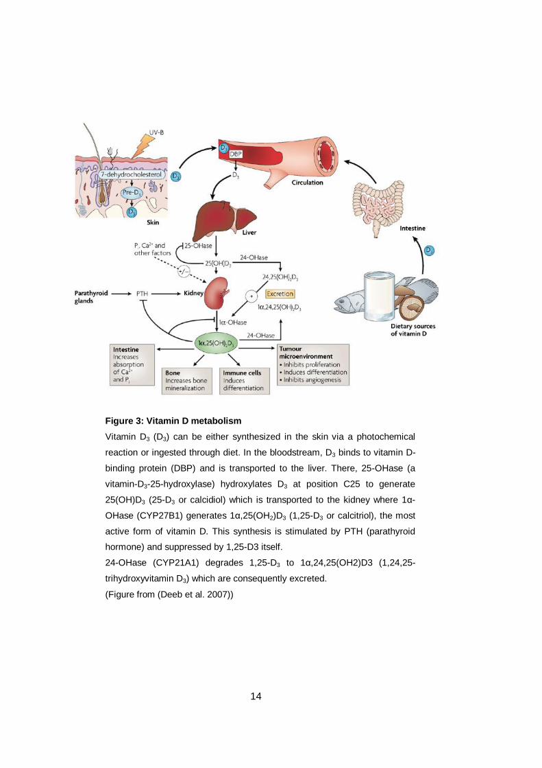
Figure 3: Vitamin D metabolism
Vitamin D3 (D3) can be either synthesized in the skin via a photochemical
reaction or ingested through diet. In the bloodstream, D3 binds to vitamin D-
binding protein (DBP) and is transported to the liver. There, 25-OHase (a
vitamin-D3-25-hydroxylase) hydroxylates D3 at position C25 to generate
25(OH)D3 (25-D3 or calcidiol) which is transported to the kidney where 1α-
OHase (CYP27B1) generates 1α,25(OH2)D3 (1,25-D3 or calcitriol), the most
active form of vitamin D. This synthesis is stimulated by PTH (parathyroid
hormone) and suppressed by 1,25-D3 itself.
24-OHase (CYP21A1) degrades 1,25-D3 to 1α,24,25(OH2)D3 (1,24,25-
trihydroxyvitamin D3) which are consequently excreted.
(Figure from (Deeb et al. 2007))

15
1.1.4 1,25-dihydroxyvitamin D3, vitamin D receptor and gene expression
1,25-D3 associated with DBP (vitamin D-binding protein) enters a target cell and
there, the two molecules dissociate. 1,25-D3 migrates into the cell nucleus and
interacts with the intracellular vitamin D receptor (VDR) (Haussler and Norman
1968). VDR belongs to the nuclear receptor family of transcription factors and is
encoded by the VDR gene (Evans 1998). VDR forms heterodimers with another
intracellular receptor, the retinoid X receptor (RXR). The VDR-RXR-1,25-D3
complex binds to vitamin D response elements (VDRE) in the DNA, which are
specific sequences in the promoter region of the 1,25-D3 target genes, to activate
or suppress their expression (Kimmel-Jehan et al. 1997). Recently, two studies
examined the number of 1,25-D3 stimulated VDR-binding sites in lymphoblastoids
and monocytes via chromatin immunoprecipitation sequencing (Chip-Seq)
technique which indicate the enourmous number of 1820 and 2776 1,25-D3 target
genes (Carlberg et al. 2012).
1.1.5 1,25-dihydroxyvitamin D3 – A pleiotropic hormone
Since the discovery of the connection between rickets and vitamin D, the most
studied role of 1,25-D3 is the regulation of phosphate and calcium homeostasis.
Together with parathyroid hormone (PTH), 1,25-D3 is able to increase the
efficiency of absorption of intestinal calcium and phosphate and plays an important
role in bone mineralisation, bone health and maintenance (Holick 2007). Studies
have demonstrated that VDR is expressed also in colon, brain, breast, bone
marrow and immune cells, and other functions of 1,25-D3 than calcium and bone
homeostasis have been shown (Bikle 2009) which are partly described in the next
paragraphs in this chapter.
Many studies highlight the important role of 1,25-D3 in regulating the immune
system. For instance, Toll-like receptor (TLR) activation of macrophages, which in
turn leads to recognition of microbial infections and initiation of immune responses,
upregulates VDR expression (Liu et al. 2006). Vitamin D supplements enhance the
phagocytic functions of human macrophages (Martineau et al. 2007).
Recent findings have shown that 1,25-D3 induces generation of antimicrobial
peptides, like cathelicidin (Liu et al. 2007), that plays an essential role in the
immune system in host defence against Mycobacterium tuberculosis (Shapira et

16
al. 2010). Vitamin D deficiency has been linked to a fivefold-increased risk for the
development of tuberculosis (Talat et al. 2010). Furthermore, VDR polymorphisms
have been associated with an increased risk for progression of tuberculosis
(Larcombe et al. 2008),(Selvaraj et al. 2008).
Low vitamin D status has been connected with both type 1 and type 2 diabetes
(Scragg et al. 2004),(Chiu et al. 2004). In an interesting study from Finland,
children who received 2000 IU/d vitamin D during their first year of life, had over
80% reduction in risk of incident type 1 diabetes after 31 years, compared with the
control group who did not receive vitamin D supplements (Hyppönen et al. 2001).
Vitamin D deficiency is also associated with multiple sclerosis (MS) (Ascherio et al.
2010), increased mortality (Skaaby et al. 2012), Parkinson’s disease (Vinh Quôc
Luong and Thi Hoàng Nguyên 2012) and some forms of cancer (Krishnan et al.
2010), including colon cancer.
Considering the connection between 1,25-D3 and cancer, it is not surprising that
1,25-D3 and VDR are thought to be involved in the regulation of cell proliferation
and differentiation (Samuel and Sitrin 2008). Studies in VDR knockout mice have
shown an increased epithelial proliferation in the descending colon (Kallay et al.
2001) and an abnormal, reduced epidermal differentiation during the first weeks of
life (Xie et al. 2002). It has also been demonstrated that in kerationcytes, 1,25-D3
downregulates various factors that are involved in cell proliferation, like keratin 16,
c-myc and EGFR (Nagpal et al. 2005).
1.1.6 Vitamin D and colorectal cancer
According to EuropaColon (www.europacolon.com), a colorectal cancer
community founded in 2005, CRC is the second leading cause of cancer-related
deaths in Europe with 230 000 yearly deaths. Several risk factors were suggested
to be important for the outbreak of CRC, like age, family history and life style,
including dietary habits and physical activities (Kang et al. 2011).
In 1980, Frank and Cedric Garland hypothesised for the first time the connection
between colon cancer and low vitamin D status based on ecological studies
(Garland and Garland 2006). They observed a geographic distribution of colon
cancer-related deaths in the United States with lower mortality rates in the south-

17
west compared to the north-west. According to these findings, Garland and
Garland proposed that this inverse relation between CRC incidence and solar
radiation could be caused by the lower vitamin D synthesis in areas with less
sunlight. In 1989, the same authors published a study that results strongly
supported their hypothesis by showing an inverse correlation between CRC and
vitamin D in the USA (Pereira et al. 2012).
CYP24A1 gene encodes the enzyme 1,25-dihydroxyvitamin D3 24-hydroxylase
which degrades 1,25-D3, the most active metabolite of vitamin D3. Several studies
show overexpression of CYP24A1 in different forms of cancer, including the colon
(Bareis et al. 2001),(Anderson et al. 2006), ovarian (Anderson et al. 2006),
cutaneous squamous cell (Reichrath et al. 2004), esophageal (Mimori 2004) and
lung (Parise et al. 2006) carcinomas. These findings indicate a possible important
role of CYP24A1 in the complicated, multistep process of tumorigenesis.
1.2 MicroRNAs
1.2.1 Introduction
MicroRNAs (miRNAs) are a relatively recently discovered group of small non-
coding RNAs which regulate the expression of messenger RNAs (mRNAs) at post-
transcriptional level. miRNAs are found in plants and animals but apart from their
functions, they show differences in their biosynthesis and molecular mechanisms.
The role of miRNAs in various cellular pathways has been investigated for
approximately 20 years. Over 1800 human miRNA species have been identified so
far (according to www.mirbase.org). It is hypothesized that miRNAs are involved in
every major pathway. This chapter describes the history of the discovery of
miRNAs, their biogenesis pathways, their mechanisms to regulate gene
expression, their role in cancer progression, with special focus on microRNA-22
and microRNA-125b.

18
1.2.2 History of microRNAs – It all began in a worm
In 1993, Victor Ambros and his colleagues, Rhonda Feinbaum and Rosalinde Lee,
discovered a small non-coding RNA, called lin-4, in Caenorhabditis elegans (Lee
et al. 1993). They detected that the gene lin-4 which was already known to play an
essential role in the temporal control of the larval development (Horvitz 1980),
does not encode proteins but a pair of small RNAs of approximately 22 and 61
nucleotides in length.
At that time, it was also known that null mutations in the heterochronic gene lin-14
led to an opposite phenotype of the null-lin-4 mutations (Ambros and Horvitz
1984). Ambros’s and Gary Ruvkun’s laboratories recognized that the
approximately 22 nucleotides long lin-4 RNA has antisense complementarity to
many regions in the 3’-UTR (3’-untranslated region) of the lin-14 RNA (Lee et al.
1993), (Wightman 1993) and both working groups described a new regulatory
mechanism of non-protein-coding RNAs, based on the ability of lin-4 to regulate
lin-14 expression by binding to its 3’-UTR.
In the year 2000, a second small non-coding RNA (21nt), let-7, was detected in
Caenorhabditis elegans (Reinhart et al. 2000). let-7 is also involved in regulating
developmental timing and is complementary to the 3’-UTR of the genes lin-14, lin-
28, lin-41, lin-42 and daf-12 but it is not homologous to lin-4. let-7 RNAs were also
detected in samples from different animals, including vertebrates, ascidians,
hemichordate, molluscs, annelids and arthropods (Pasquinelli et al. 2000). This
was the starting signal for a new area of research of a new class of small non
coding RNAs, later called micro RNAs.
Recent findings of miRNAs in Chlamydomonas reinhartii (a unicellular algae)
indicate that this class of RNAs might be evolutionary older than expected
(Schwach et al. 2007),(Allen and Howell 2010) and this leads one to expect more
exciting findings about miRNAs in the future.
1.2.3 Biogenesis of microRNAs
1.2.3.1 Introduction
The biogenesis of miRNAs in animals and plants shows similarities as well as
differences in the major processing steps (Guleria et al. 2011),(Ul Hussain 2012)

19
and in this chapter, the focus will be on the synthesis of animal miRNAs. The
synthesis of miRNAs can start in two different ways which only differ in the first
steps that occur in the nucleus: (1) miRNAs are transcribed either from their own
genes (canonical miRNAs) or (2) they are generated from introns via splicing in a
unique manner (called mirtrons) (see Figure 4).
1.2.3.2 Canonical microRNAs:
RNA polymerase II transcribes a miRNA gene and this transcript is called primary
microRNA (pri-miRNA) with a length of approximately 500-3000 nucleotides. The
pri-miRNA forms a hairpin structure with an imperfectly paired double stranded
stem and a terminal loop. Like a primary transcript from a protein-coding gene, pri-
miRNA is capped and polyadenylated (Cai et al. 2004).
In the nucleus, a multiprotein complex containing the two core components Drosha
and Di George Syndrome critical region gene 8 (DGCR8), called the
microprocessor complex, recognizes the above described secondary structures
(Gregory et al. 2004). Drosha is an RNAse III enzyme and DGCR8 contains an
RNA-binding domain. The microprocessor complex cleaves the pri-miRNA at the
base of the hairpin and generates a precursor miRNA (pre-miRNA) of
approximately 65 nucleotides in length with usually a 2-nt overhang at the 3’-end
(Han et al. 2006).
1.2.3.3 Mirtrons
The alternative miRNA biogenesis, the ‘mirtron’ pathway, combines intron splicing
with dicing in a Drosha-independent manner. The first mirtrons were identified in
Caenorhabditis elegans (Ruby et al. 2007) and Drosophila melanogaster
(Okamura et al. 2007) but comparable loci were later identified in chicken (Glazov
et al. 2008), cows (Glazov et al. 2009), rodents (Rnas et al. 2008) and primates
(Berezikov et al. 2007).

20
Figure 4: Biogenesis of microRNAs
RNA polymerase II (RNAPII) processes MicroRNAs (miRNAs) from genes or
from introns. In the canonical pathway, Drosha and DGCR8 processes a
primary miRNA (pri-miRNA) into a precursor-miRNA (pre-miRNA). In the
mirtron pathway, pre-miRNA are generated via splicing and debranching
from a intron. In both cases, Exportin 5 transports the pre-miRNA into the
cytoplasma where it is cleaved by Dicer, assisted by TRBP, and generates a
miRNA/miRNA* duplex. miRNA* is released and degraded whereas miRNA
incorporated into an miRNA-induced silencing complex (miRISC). The
miRISC–miRNA complex binds partial complementary to the 3’-UTR of a
mRNA and so represses the translation or degrades the mRNA.
(Figure adapted from (Krol et al. 2010)

21
From a transcribed pre-mRNA, the spliceosome removes the mirtron which forms
a lariat that is debranched by the lariat debranching enzyme (Ldbr). Then, the
debranched mirtron forms secondary structures and folds into a pre-miRNA (Ruby
et al. 2007),(Okamura et al. 2007).
1.2.3.4 Transport into the cytoplasm and processing of precursor
microRNAs to generate mature microRNAs
The nuclear export receptor Exportin 5 recognizes the 3’-overhang of the double-
stranded pre-miRNA which is then exported in a Ran-GTP dependent manner into
the cytoplasm (Yi et al. 2003),(Bohnsack et al. 2004). The Dicer, an RNAse III
enzyme, binds the 3’-end of the pre-miRNA and cleaves it near the terminal loop,
approximately 22 nt away from the 3’-end. Through this cleavage which is assisted
by transactivation-responsive (TAR) RNA-binding protein (TRBP), a miRNA-
miRNA* duplex with 2 nt long 3’-overhangs at both ends, is generated (Bernstein
et al. 2001),(Hutvágner et al. 2001). The miRNA*-strand gets often degraded and
the miRNA-strand binds to the ribonucleoprotein miRNA-induced silencing
complex (miRISC).
1.2.3.5 Actions of microRNAs
The miRISC–miRNA complex recognizes and binds partial complementary
messenger RNAs (mRNAs), whereas only 2-8 nucleotides (called the seed region)
match perfectly with the 3’-UTRs of the target mRNA (Brennecke et al.
2005),(Ameres et al. 2007). There are two different ways how the miRISC-miRNA
complex can repress the translation from mRNA into protein: (1) by blocking the
strand for translation into protein or (2) by degrading the mRNA strand (Ul Hussain
2012). Due to the fact that the essential seed region is only few nucleotides long to
which the miRISC-miRNA complex binds, one miRNA regulates multiple mRNAs
and one mRNA can be regulated by multiple miRNAs (Meltzer 2005). This leads to
an enormous number of possible regulatory mechanisms in the process of gene
expression.

22
1.2.4 MicroRNAs and cancer
1.2.4.1 Introduction
Recent findings have shown that miRNAs can be deregulated due to various
genetic and epigenetic alterations, which might affect development of cancer
(Ventura and Jacks 2009). Genetic alterations can occur as a result of
chromosomal abnormalities such as deletion, amplification or translocation of DNA
fragments containing the information for miRNAs (Calin and Croce 2006a).
Various epigenetic events, like altered histone modifications, hypermethylation of
tumour suppressor genes and hypomethylation of oncogenes might influence also
the expression of miRNAs (Melo and Esteller 2011). Approximately fifty percent of
miRNAs are associated with CpG islands, sequences containing high numbers of
cytosine-guanine dinucleotides (Melo and Esteller 2011). Methylated CpG islands
can silence gene expression (Fatemi et al. 2005). Thus, the expression of these
miRNAs can be silenced by DNA hypermethylation.
1.2.4.2 MicroRNAs as oncogenes
miRNAs can act as oncogenes. In the last years, numerous miRNAs with
oncogenic potential have been identified (Medina and Slack 2008). One such
oncogenic miRNA is MicroRNA-21 (miR-21). Many studies have shown that miR-
21 is overexpressed in various types of cancer, like in glioblastoma (Corsten et al.
2007), pancreatic (Roldo et al. 2006) breast (Si et al. 2007), lung (Li et al. 2011),
esophageal squamous cell (Cai et al. 2012), hepatocellular (Meng et al. 2007) and
colorectal (Bullock et al. 2013) cancer. Silencing experiments with antisense miR-
21 showed an increased rate of apoptosis suggesting possible anti-apoptotic
effects of miR-21 (Medina and Slack 2008) (Si et al. 2007).
Further, several tumour suppressors have been shown to be potential miR-21
targets (Medina and Slack 2008), like Phosphatase and Tensin homolog (PTEN)
(Meng et al. 2007) and Tropomyosin 1 (TPM1) (Zhu et al. 2007). Therefore, miR-
21 has been proposed as a possible biomarker for cancer diagnosis (Bowman et
al. 2008).

23
1.2.4.3 MicroRNAs as tumor suppressors
miRNAs can act as tumour suppressors by inhibiting oncogene translation. The
first identified miRNA in human, let-7, belongs to the let-7 microRNA family which
is often downregulated in various forms of cancer (Wang et al. 2012b), such as in
lung (Takamizawa et al. 2004), head and neck squamous cell (Childs et al. 2009),
ovarian (Shell et al. 2007) and prostate (Liu et al. 2012) cancer.
Several studies have shown that let-7 might target and downregulate c-Myc, ras,
high-mobility group A (HMGA), Janus protein tyrosine kinase (JAK), signal
transducer and activator of transcription 3 (STAT3) and Np95/ICBP90-like RING
finger protein (NIRF), which are critical oncogenes in tumour progression due to
their involvement in regulating cell cycle, apoptosis and cell adhesion (Wang et al.
2012).
1.2.5 miR-22
The MicroRNA-22 (miR-22) gene is located on the short arm of chromosome 17 at
position 17p13.3 (www.atlasgeneticsoncology.org). Many studies have shown that
miR-22 is deregulated in various forms of cancer, for example it is downregulated
in breast (Xiong et al. 2010), hepatocellular (Zhang et al. 2010), gastric (Wang et
al. 2013), lung (Ling et al. 2012) and colorectal (Zhang et al. 2012) cancer and
upregulated in advanced non small cell lung cancer (Franchina et al. 2013) and
prostate cancer (Poliseno et al. 2010).
Apart from the possible role of miR-22 in cancer, this miRNA also plays an
important role in multiple pathways that are involved in hematopoiesis and
different cellular functions (Li et al. 2012), such as PTEN and Protein Kinase B
(AKT) (Bar and Dikstein 2010), nuclear factor kappa-light-chain-enhancer of
activated B cells (NF-κB) (Takata et al. 2011) and erstrogen receptor (ESR1)
(Pandey and Picard 2009),(Li et al. 2012).
A recent study indicated a connection between miR-22 and vitamin D in colon
cancer cells (Alvarez-Díaz et al. 2012). Different colon cancer cell lines were
treated with 100nM 1,25-D3 for 48 hours. The treatment increased the expression
of miR-22 more than 2-fold in HT-29 cells. Further, the study showed that 1,25-D3-
induced miR-22 expression may have an inhibitory effect on the proliferation and
migration of the used cells.

24
1.2.6 miR-125b
The MicroRNA-125b (miR-125b) gene is located on the long arm of chromosome
11 at position 11q24.1 (www.atlasgeneticsoncology.org) and is a putative
homologue to lin-4 in Caenorhabditis elegans. As miR-22, many studies have
indicated that miR-125b is often deregulated in different forms of cancer.
On the one hand, miR-125b is downregulated in breast (Iorio et al. 2005), oral
squamous cell (Shiiba et al. 2013), hepatocellular (Jia et al. 2012) and bladder
(Huang et al. 2011) cancer. It has been proposed that miR-125 triggers apoptosis
by downregulating the expression of the pro-survival (anti-apoptotic) proteins Mcl-
1 and Bcl-2 (Gong et al. 2012). Furthermore, it has been suggested that mit-125b
downregulates CYP24A1 post-transcriptionally in breast cancer (Komagata et al.
2009a). This finding might explain why CYP24A1 is often very highly expressed in
some of these tumours.
On the other hand, miR-125b is upregulated in neuroblastoma (Laneve et al.
2007), prostate (Shi et al. 2007) (Ozen et al. 2008), non-small-cell lung (Yuxia et
al. 2012), thyroid (Vriens et al. 2012) and type II endometrial (Jiang et al. 2011)
cancer. In addition, miR-125b might promote proliferation and migration of cancer
cells by targeting the pro-apoptotic protein Tumor protein p53-inducible nuclear
protein 1 (TP53INP1) (Jiang et al. 2011) and induce metastasis by targeting StAR-
related lipid transfer domain protein 13 (STARD13) (Tang et al. 2012).
miR-125b can have pleiotropic effects in several cellular processes. miR-125b
seems to be essential for various forms of cell differentiation. In embryogenesis,
miR-125b is important for the proper differentiation of embryonic stem cells (ESC)
by regulating Dies1 (Differentiation of ESCs 1) (Battista et al. 2013). In human
neuronal differentiation, miR-125b regulates the development and differentiation of
neuronal subtypes (Stappert et al. 2013). Further, miR-125b is a key mediator for
Snail-induced stem cell propagation and chemoresistance (Liu et al. 2013).
In colorectal cancer, miR-125b has been described as tumourigenic. Studies have
reported that miR-125b is upregulated in CRC (Lin et al. 2011), (Nishida et al.
2011). However, the precise role of miR-125b in CRC has to be investigated
further.

25
2 Aim of this study
The two main aims of this diploma thesis were:
To analyze the effect of 1,25-D3 on the expression of miR-22 and miR-
125b in human colorectal and prostate cancer cell lines.
To determine the expression levels of miR-22 and miR-125b in human
colorectal tumour samples and adjacent normal mucosa from the same
patient.
Subaims were:
To investigate the effect of 1,25-D3 on the cell viability of human colorectal
and prostate cancer cell lines.
To analyze the basal expression of miR-22 and miR-125b in human
colorectal and prostate cancer cell lines.
To investigate the effect of Interleukin-6 (IL-6) and tumour necrosis factor
alpha (TNF-α) on the the expression of miR-22 and miR-125b in human
colorecatal cancer cell lines.

26
3 Materials and Methods
3.1 Cell lines
3.1.1 Colon cancer cell lines
3.1.1.1 HT-29
The HT-29 cell line was derived from a 44 years old female Caucasian patient who
suffered from a colorectal adenocarcinoma and it was isolated by Jorgen Fogh in
1964. HT-29 cells are hypertriploid, intestinal epithelial cells growing as adherent
cells.
3.1.1.2 COGA-1A
The COGA-1A cell line is a subclone of the COGA-1 cell line which was isolated
by Ernst Wagner and Alexandra Sinski from a G2 colorectal adenocarcinoma of a
64 years old female patient (Vécsey-Semjén et al. 2002). In cell culture, COGA-1A
cells grow as adherent monolayer.
3.1.1.3 COGA-13
The COGA-13 cell line was established in our laboratory in collaboration with Ernst
Wagner and Alexandra Sinski and derived from a G3 colon tumour. These cells
are undifferentiated. COGA-13 cells express extremely high amounts of CYP24A1
and undetectable CYP27B1 levels (Lechner et al. 2007).
3.1.1.4 Caco-2/15
The Caco-2/15 cell line is a stable clone of the parent Caco-2, which is a
heterogeneous colorectal adenocarcinoma cell line developed under the
leadership of Jorgen Fogh. The Caco-2 cell line was isolated from a primary
colonic tumour of a 72-years old Caucasian male patient.

27
3.1.1.5 Caco-2/AQ
The Caco-2/AQ cell line was established by Andrea Quaroni and is a subclone of
Caco-2/15 cells. Caco-2/AQ cells divide and grow faster than Caco-2/15 cells.
During the logarithmic growth phase, Caco-2/AQ cells have a population doubling
time of approximately 24 hours, whereas Caco-2/15 cells show a cell division time
of around 36 hours (Chirayath et al. 1998).
3.1.1.6 LT97
The human colon adenoma cell line LT97 was isolated from a small polyp of a
patient suffering from familial polyposis coli, under the leadership of Brigitte Marian
in 2002. LT97 cells have a ki-Ras mutation and do not express the tumour
suppressor protein APC (Adenomatous polyposis coli) due to a loss of both alleles
(Richter et al. 2002).
3.1.2 Prostate cancer cell line
3.1.2.1 DU145
The DU145 prostate cancer cell line was derived from a 69 year old Caucasian
patient by Kenneth R. Stone in 1978 and is a hypotriploid cell line. DU145 cells are
epithelial cells growing in as adherent cells. Due to the tumourigenic properties of
DU145, this cell line is frequently used for xenotransplantation studies in
immunodeficient mice.
3.2 Cell culture conditions
3.2.1 General
Cells were routinely cultured at 37°C in a humidified atmosphere of 95% air and
5% CO2. Media was changed every second day and cell lines were routinely
screened for mycoplasma.

28
3.2.2 Colon cancer cell lines
HT-29, COGA-1A, COGA-13, Caco-2/15 and Caco-2/AQ cells were cultured in
Dulbecco’s modified Eagle medium (DMEM; Invitrogen) supplemented with 10%
fetal calf serum (FCS), 10 mM HEPES, 4 mM L-glutamine, 100 U/ml penicillin and
100 µg/ml streptomycin (all from Invitrogen).
LT97 cells were cultured in Ham’s F-12 medium (Sigma) supplemented with 25%
L-15 Leibovitz medium (Sigma), 2 mM L-glutamine (Invitrogen), 1,18 mg/ml
NaHCO3, 44 µg/ml CaCl2xH2O (both Merck), 25 mM HEPES, 2% FCS, 10 µg/ml
insulin, 2,4 U/ml penicillin, 2,4 µg/ml streptomycin, 30 ng/ml EGF (all from
Invitrogen) and 20 µg/ml gentamycin (PAA).
3.2.3 Prostate cancer cell line
DU-145 cells grew in Roswell Park Memorial Institute (RPMI) 1640 medium
(Invitrogen) supplemented with 1mM sodium pyruvate (Sigma), 10% FCS, 4 mM
L-glutamine, 100 U/ml penicillin and 100 µg/ml streptomycin (all from Invitrogen).
3.2.4 ITS media for 1,25-D3 treatment
Due to the presence of substances (like vitamin D-binding protein and albumin) in
FCS, which might influence the effect of 1,25-D3, cells were incubated in FCS-free
ITS media for duration of the treatment. ITS media is composed of DMEM
supplemented with 10 µg/ml insulin, 5,5 µg/ml transferrin, 6,7 ng/ml sodium
selenite (using Insulin-Transferrin-Selenium-Ethanolamine Solution (ITS-X), 100X
from Gibco), 10 mM HEPES, 4 nM L-glutamine, 100 U/ml penicillin and 100 µg/ml
streptomycin (all from Invitrogen).
3.3 Viability assays
CellTiter-Blue® Cell Viability Assay (Promega) was performed according to the
manufacturer’s instructions. In short, the CellTiter-Blue® reagent added to the
media contains the indicator dye resazurin which is converted into the fluorescent
end product resorufin in metabolically active, living cells (Figure 5). The amount of
resorufin can be measured with a fluorometer and indicates the metabolic activity
of the cells.

29
3.4 Colorectal tumour samples
After approval by the ethics committee (EK 258/2010 and EK 06-198-VK) of the
Medical University of Vienna, fresh frozen tissue samples were collected at
Rudolfsstiftung Hospital and the General Hospital of Vienna. After receiving a
written consent from the patients, colorectal tumour tissue and adjacent mucosa
from the same patients were collected by a pathologist who graded and classified
the tumours according to the TNM system (T describes the tumour, N describes
involved lymph nodes and M describes distant metastasis).
3.5 RNA extraction
RNA was extracted with Trizol® reagent (LifeTechnologies) according to the
manufacturer’s protocol and its integrity was determined by staining with Gel Red
(Biotium) on an 1% agarose gel which was scanned and analyzed with a video
camera imaging system under UV light (Herolab). The concentration and the purity
of the isolated RNAs was measured with a full-spectrum spectrophotometer
NanoDrop® ND-1000 (NanoDrop Technologies).
Figure 5: Conversion of resazurin to resorufin
Metabolically active cells convert resazurin to the highly fluorescent end
product resorufin which emits fluorescence at 590nm. The measured
fluorescence is proportional to the number of viable cells.

30
Table 1: Used RT-Mastermix for miRNAs for one reaction
Table 2: Used parameter values to program
the thermal cycler for the reverse transcription
of miRNAs
3.6 Reverse transcription
3.6.1 MicroRNAs
For analyzing the amount of miR-22, miR-125b and RNU6B (U6B small nuclear
RNA; used as housekeeping gene), 10 ng of total RNA (including small RNAs)
were reverse-transcribed into cDNA using TaqMan® MicroRNA Reverse
Transcription Kit and the corresponding RT Primers (Applied Biosystems; assay
000398 for hsa-miR-22-3p, 000449 for hsa-miR-125b-5p and 001093 for RNU6B).
7 µl master mix (Table 1), 10 ng RNA and 3 µl 5X RT primers were added to each
tube. RNAse-free dH2O was added to reach a total volume of 15 µl. After spinning
down and incubating on ice for five minutes, we performed the reverse
transcription in a MyCyler™ Thermal Cycler (BIO-RAD). The parameters used are
shown in Table 2.
Component Volume
100mM dNTPs (with dTTP) 0.15 µL
MultiScribeTM Reverse Transcriptase, 50 U/µL 1.00 µL
10X Reverse Transcription Buffer 1.50 µl
Rnase Inhibitor, 20U/µL 0.19 µl
Nuclease-free water 4.16 µl
3.6.2 Messenger RNAs
Two micrograms of the isolated RNA were reverse-transcribed with RevertAid H
Minus Reverse Transcriptase and random hexamer primers (both Thermo
Step Time Temperature
Hold 30 minutes 16 °C
Hold 30 minutes 42 °C
Hold 5 minutes 85 °C
Hold ∞ 4 °C

31
Table 3: RT-Mastermix for reverse transcription
of mRNAs for one reaction (all reagents from
Thermo Scientific)
Table 4: Used parameter values to program
the thermal cycler for the reverse
transcription of mRNAs
Scientific) into single-stranded cDNA. 1 µl random hexamer primers were mixed
with 2 µg RNA and made up to 13 µl with RNAse-free dH2O. After spinning down,
the mixture was incubated at 70 °C for 10 minutes in a MyCyler™ Thermal Cycler
(BIO-RAD). After 4 minutes on ice, 7 µl of the RT-Mastermix (Table 3) was added
and the mixture was spinned down. Following incubation for 5 minutes at room
temperature, the reverse transcription was performed in the thermal cycler. The
used parameters are shown in Table 4.
Step Time Temperature
Hold 60 minutes 42 °C
Hold 10 minutes 45 °C
Hold 15 minutes 70 °C
Hold ∞ 10 °C
3.7 Quantitative reverse transcriptase polymerase chain reaction (qRT-
PCR)
3.7.1 General
Both quantitative reverse transcriptase polymerase chain reactions (qRT-PCRs)
for miRNAs and mRNAs were performed either in a StepOnePlus™ Real-Time
PCR System or in a 7900HT Fast Real-Time PCR System (both Applied
Biosystems). It has to be mentioned that the analysis of one experiment was
always performed in the same system. The temperature profile for the qRT-PCR is
shown in Table 5.
Reagent Amount
5X Reaction Buffer for RT 4 µl
10 mM dNTPs 2 µl
RevertAid H Minus RT 1 µl
32
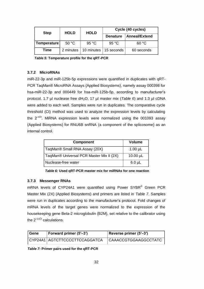
Table 5: Temperature profile for the qRT-PCR
Table 7: Primer pairs used for the qRT-PCR
Step HOLD HOLD Cycle (40 cycles)
Denature Anneal/Extend
Temperature 50 °C 95 °C 95 °C 60 °C
Time 2 minutes 10 minutes 15 seconds 60 seconds
3.7.2 MicroRNAs
miR-22-3p and miR-125b-5p expressions were quantified in duplicates with qRT-
PCR TaqMan® MicroRNA Assays (Applied Biosystems), namely assay 000398 for
hsa-miR-22-3p and 000449 for hsa-miR-125b-5p, according to manufacturer’s
protocol. 1.7 µl nuclease free dH2O, 17 µl master mix (Table 6) and 1.3 µl cDNA
were added to each well. Samples were run in duplicates. The comparative cycle
threshold (Ct) method was used to analyze the expression levels by calculating
the 2-∆Ct. MiRNA expression levels were normalized using the 001093 assay
(Applied Biosystems) for RNU6B snRNA (a component of the splicosome) as an
internal control.
3.7.3 Messenger RNAs
mRNA levels of CYP24A1 were quantified using Power SYBR® Green PCR
Master Mix (2X) (Applied Biosystems) and primers are listed in Table 7. Samples
were run in duplicates according to the manufacturer’s protocol. Fold changes of
mRNA levels of the target genes were normalized to the expression of the
housekeeping gene Beta-2 microglobulin (B2M), set relative to the calibrator using
the 2-∆∆Ct calculations.
Component Volume
TaqMan® Small RNA Assay (20X) 1.00 µL
TaqMan® Universal PCR Master Mix II (2X) 10.00 µL
Nuclease-free water 6.0 µL
Gene Forward primer (5′–3′) Reverse primer (5′–3′)
CYP24A1 AGTCTTCCCCTTCCAGGATCA CAAACCGTGGAAGGCCTATC
Table 6: Used qRT-PCR master mix for miRNAs for one reaction

33
4 Results
4.1 Viablity assays
To assess whether 1,25-D3 influences the viability of the colon cancer cells HT-29,
COGA-13, Caco-2/15, Caco-2/AQ, LT97 and the prostate cancer cell line DU145,
cells were seeded in 96-well plates. All treatments described below were
performed in 8 wells per group.
At 30% confluency, cells were transferred to ITS medium or ITS supplemented
with 1% FCS medum for 24 hours. Then, cells were treated with 10 nM 1,25-D3 for
24 and 48 hours and controls were treated with 0.01% ethanol (corresponding to
the amount of ethanol added in 1,25-D3 treatments).
Cells treated with the highly toxic dose of 5 µM 5-aza-2’-deoxycytidine (AZA) for
24 and 48 hours were used as negative control. We also had a control group
without any treatment.
At the end of the treatments, 20 µl CellTiterBlue® Reagent was added to each well,
the plate was shaken for 10 seconds. Cells were incubated for 4 hours at 37°C in
the incubator. The fluorescence was measured at 560Ex/590Em nm.
1,25-D3 treatment had only minor effect on viability in most cells studied (Figure 6).
In HT-29 colon cancer cells, 1,25-D3 treatment led to a slightly enhanced
metabolic activity. Adding FCS to the ITS media affected only the viability of Caco-
2/AQ cells. A minor inhibitory effect of 1,25-D3 in these cells was seen only if the
cells were grown in ITS. In the DU145 prostate cancer cells, no effect was
detected relating to the treatment or the used media.
All cells showed very low metabolic activity after the treatment with AZA, as
expected.

34
Figure 6: Effect of 1,25-D3 on the cell viability
(A) HT-29, (B) COGA-13, (C) Caco-2/15, (D) Caco-2/AQ, (E) LT97 and (F) DU145
cells were treated with 10 nM 1,25-D3 for 24 and 48 hours in ITS or ITS supplemented
with 1% FCS media (n=8 ± SD).

35
4.2 Basal expression of miR-22 and miR-125b in HT-29, COGA-1A, COGA-
13, Caco-2/15, Caco-2/AQ, LT97 and DU145 cancer cell lines
To determine the basal expression of miR-22 and miR-125b, HT-29, COGA-1A,
COGA-13, Caco-2/15, Caco-2/AQ, LT97 colon and DU145 prostate cancer cells
were cultured in 6-well plates. At 70-80% confluency, the expression levels of miR-
22 and miR-125b were determined using qRT-PCR, and the U6 small nuclear
RNA was used as an internal control (Figure 7).
The expression of miR-22 was considerable higher in the prostate cancer line
DU145 compared with the colon cancer cells. HT-29, COGA-1A, COGA-13, Caco-
2/15 and Caco-2/AQ colon cancer cells expressed miR-22 at similar levels while
the adenoma cell line LT97 showed approximately 2-fold higher expression of
miR-22 compared with the other colon cancer cells.
miR-125b was more than 100-fold higher expressed in COGA-13 cells compared
with all the other examined colon cancer cells in which the expression was very
low. The DU145 prostate cancer cells showed also a much higher expression of
miR-125b compared with the colon cancer cells HT-29, COGA-1A, Caco-2/15,
Caco-2/AQ and LT97.
Figure 7: Basal expression of miR-22 and miR-125b
The relative expression levels of (A) miR-22 and (B) miR-125b in HT-29, COGA-1A,
COGA-13, Caco-2/15, Caco-2/AQ, LT97 colon cancer and DU145 prostate cancer
cells were measured by qRT-PCR, and the U6 small nuclear RNA was used as an
internal control (n=3 ± SEM).

36
4.3 Basal expression of CYP24A1 in HT-29, DU145 and COGA-13 cancer
cell lines
CYP24A1 is the vitamin D degrading enzyme and it is often upregulated in various
forms of cancer, including CRC. Moreover, miR-125b has been suggested to be
involved in the regulation of the expression of this enzyme. We analyzed the basal
expression of CYP24A1 in the HT-29, DU145 and COGA-13 cell line using qRT-
PCR, and B2M was used as an internal control. The expression levels of
CYP24A1 were extremely high in the COGA-13 cells and very low in the HT-29
cells compared with the DU145 cells (Figure 8). All other cell lines studied have
barely detectable basal CYP24A1 levels.
4.4 Basal expression of Snail in HT-29, DU145 and COGA-13 cancer cell
lines
Snail is suggested to play a critical role in cancer progression by inducing
epithelial-mesenchymal transition. Since miR-125b was suggested to be a key
mediator of Snail-induced stem cell propagation and chemoresistance, we
analyzed the basal expression level of Snail in the COGA-13 and DU145 cells
Figure 8: Basal expression of CYP24A1 in HT-29, DU145 and COGA-13
cells
The relative expression levels of CYP24A1 in HT-29 and COGA-13 colon
cancer and DU145 prostate cancer cells were measured by qRT-PCR. B2M
was used as an internal control (n=3 ± SEM).

37
because these cells express high basal levels of miR-125b. Because we also were
interested in the expression levels of both miRNAs in HT-29 cells, we also
measured the amount of Snail in these cells. The expression level of Snail in the
colon cancer cells COGA-13 and HT-29 were very low compared to the prostate
cancer cell line DU145 (Figure 9).
4.5 Role of 1,25-D3 in regulating miR-22 and miR-125b in cancer cell lines
4.5.1 Effect of 1,25-D3 on the expression of miR-22, miR-125b and CYP24A1
in COGA-13 colon cancer cells
4.5.1.1 Effect of 1,25-D3 on the expression of CYP24A1 in COGA-13 colon
cancer cells
CYP24A1 is a classical direct target gene of 1,25-D3. Therefore, first we analyzed
the expression of the vitamin D degrading enzyme CYP24A1 after the treatment
with 10nM 1,25-D3 for 5 hours, since it was suggested that miR-125b is involved in
the regulation of the expression of this enzyme. CYP24A1 expression is extremely
Figure 9: Basal expression of Snail in HT-29, DU145 and COGA-
13 cells
The relative expression levels of Snail in HT-29 and C OGA-13 colon
cancer and DU145 prostate cancer cells were measured by qRT-
PCR. B2M was used as an internal control (n=3 ± SEM).

38
high in these cells, and 1,25-D3 treatment increases its expression only marginally
(Figure 10).
4.5.1.2 Effect of 1,25-D3 on the expression of miR-22 and miR-125b in the
COGA-13 colon cancer cells
COGA-13 cells were seeded in 10 cm Petri dishes (12000 cells/cm²). 10 or 100
nM 1,25-D3 were added for 1, 3, 5, 7, 10 and 24 hours. Controls were treated with
0.01% ethanol (corresponding to the amount of ethanol in the added 1,25-D3). The
expression levels of miR-22 and miR-125b were determined using TaqMan qRT-
PCR.
Both miRNAs showed only minor differences in their expression levels after 1,25-
D3 treatment (Figure 11).
Figure 10: Effect of 1,25-D3 on the expression of CYP24A1
in COGA-13 cells
Expression of CYP24A1 was analyzed after COGA-13 cells
were exposed to 10 nM 1,25-D3 for 5 hours. Expression level
after the treatment was set relative to the vehicle control (n=3
± SEM).

39
Although miR-22 was considered as a target gene for 1,25-D3, we observed no
significant changes in its expression.
Neither 10 nM nor 100 nM 1,25-D3 had changed significantly the expression of
miR-125b in COGA13 cells. There was a tendency of short transient upregulation
after 3 hours followed by a decrease after 5 hours.
Figure 11: Effect of 1,25-D3 on the expression of miR-22 and miR-125b in COGA-13
cells
Expression of miR-22 and miR-125b after treatment with 10 and 100 nM 1,25-D3 for 1, 3, 5,
7, 10 and 24 hours were determined using TaqMan qRT-PCR. Expression levels after the
treatments were set relative to the respective vehicle controls (n=3 ± SEM).

40
Figure 12: Effect of 1,25-D3 on the expression of CYP24A1 in DU145
cells
Expression of CYP24A1 after treatment with 10 nM 1,25-D3 for 1, 3, 5, 7 and
9 hours were determined using qRT-PCR. Expression levels after the
treatments were set relative to the respective vehicle controls.
4.5.2 Effect of 1,25-D3 on the expression of miR-22, miR-125b and CYP24A1
in DU145 prostate cancer cells
DU145 cells were seeded in 12-well plates (5000 cells/cm²). 10 nM 1,25-D3 was
added for 1, 3, 5, 7 and 9 hours. Controls were treated with 0.01% Ethanol
(corresponding to the amount of ethanol in the added 1,25-D3).
4.5.2.1 Effect of 1,25-D3 on the expression of CYP24A1 in DU145 prostate
cancer cells
DU145 cells express detectable levels of CYP24A1, although these have not
reached the level of expression seen in COGA-13 cells. Treatment with 1,25-D3
led to a significant upregulation of CYP24A1 already after 3 hours, peaking after 5
hours, when expression level of CYP24A1 was increased more than 25 times
(Figure 12).

41
Figure 13: Effect of 1,25-D3 on the expression of miR-22 and miR-125b in
DU145 cells
Expression of miR-22 and miR-125b after treatment with 10 nM 1,25-D3 for 1, and 5
hours were determined using TaqMan qRT-PCR. Expression levels after the
treatments were set relative to the respective vehicle controls (n=3 ± SEM).
T-test was used for statistical analysis. * P < 0.05 ,
4.5.2.2 Effect of 1,25-D3 on the expression of miR-22 and miR-125b in
DU145 prostate cancer cells
We decided to analyze the expression of miR-22 and miR-125b after 1 and 5
hours treatment with 1,25-D3. Both miRNAs were downregulated at both
timepoints up to 50 percent (Figure 13). The downregulation of miR-125b after 5
hours and of miR-22 at both timepoints was significant.
4.5.3 Effect of 1,25-D3 on the expression of miR-22, miR-125b and CYP24A1
in HT-29 colon cancer cells
To assess the effect of 1,25-D3 on HT-29 colon cancer cells, cells were seeded in
6-well plates (4210 cells/cm²). For testing the long-time and dose-dependent
effects of 1,25-D3, we treated the cells with 1, 10 and 100 nM 1,25-D3 for 24 and
48 hours. In order to assess whether shorter treatments with 1,25-D3 have already
an effect on the expression of miR-22 and miR-125b in a time-dependent manner,
we treated the cells for 1, 3, 5 and 7 hours with 10 nM 1,25-D3. Controls for each
time point were treated with 0.01% ethanol (corresponding to the amount of
ethanol in the added 1,25-D3).

42
4.5.3.1 Effect of 1,25-D3 on the expression of CYP24A1 in HT-29 colon
cancer cells
We analyzed the expression of the vitamin D degrading enzyme CYP24A1 after
the treatment with 10 and 100 nM 1,25-D3 for 24 and 48 hours. CYP24A1 in the
untreated cells is barely detectable. Treatment with 1,25-D3 increased the
expression dramatically more than 100.000 times. (Figure 14).
4.5.3.2 Effect of 1,25-D3 on the expression of miR-22 and miR-125b in HT-
29 colon cancer cells
Expression levels of miR-22 and miR-125b were determined using TaqMan qRT-
PCR. miR-22 was upregulated up to approximately 100% upon treatment with
1,25-D3 for 24 and 48 hours compared with the vehicle controls. The upregulation
of the expression of miR-22 due to 100 nM 1,25-D3 treatment reached significance
Figure 14: Effect of 1,25-D3 on the expression of CYP24A1 in HT-29 cells
Expression of CYP24A1 was determined after HT-29 cells were treated with 10
and 100 nM 1,25-D3 treatment for 24 and 48 hours. Expression levels after the
treatments were set relative to the respective vehicle control (n=3 ± SEM).
43
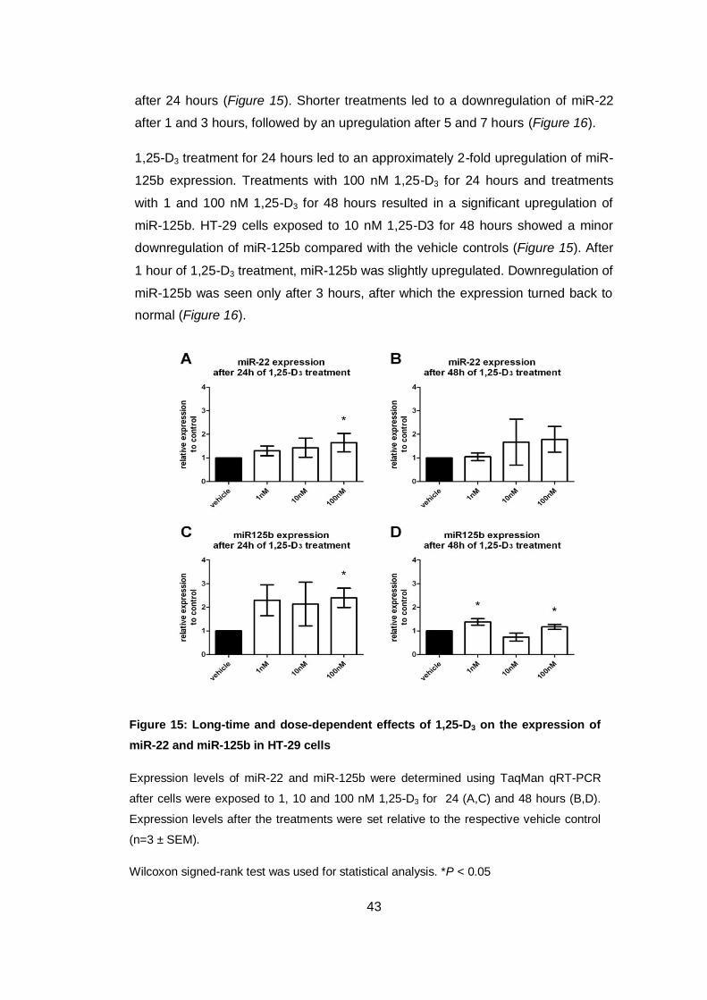
Figure 15: Long-time and dose-dependent effects of 1,25-D3 on the expression of
miR-22 and miR-125b in HT-29 cells
Expression levels of miR-22 and miR-125b were determined using TaqMan qRT-PCR
after cells were exposed to 1, 10 and 100 nM 1,25-D3 for 24 (A,C) and 48 hours (B,D).
Expression levels after the treatments were set relative to the respective vehicle control
(n=3 ± SEM).
Wilcoxon signed-rank test was used for statistical analysis. *P < 0.05
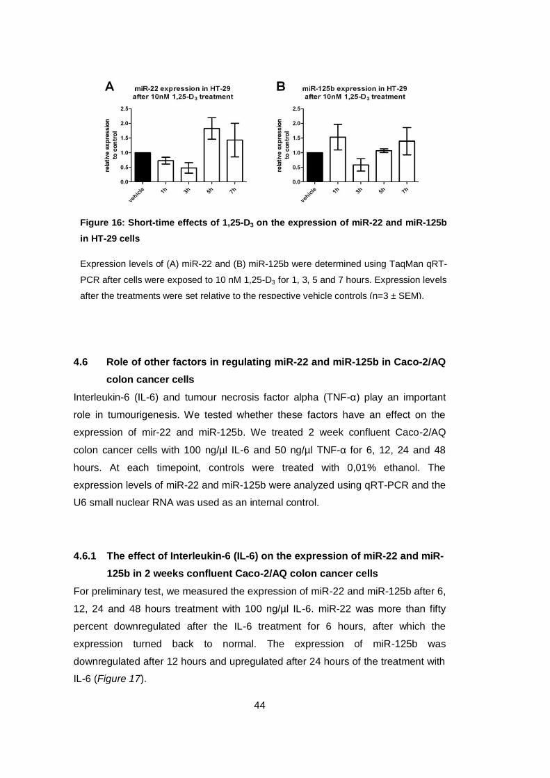
after 24 hours (Figure 15). Shorter treatments led to a downregulation of miR-22
after 1 and 3 hours, followed by an upregulation after 5 and 7 hours (Figure 16).
1,25-D3 treatment for 24 hours led to an approximately 2-fold upregulation of miR-
125b expression. Treatments with 100 nM 1,25-D3 for 24 hours and treatments
with 1 and 100 nM 1,25-D3 for 48 hours resulted in a significant upregulation of
miR-125b. HT-29 cells exposed to 10 nM 1,25-D3 for 48 hours showed a minor
downregulation of miR-125b compared with the vehicle controls (Figure 15). After
1 hour of 1,25-D3 treatment, miR-125b was slightly upregulated. Downregulation of
miR-125b was seen only after 3 hours, after which the expression turned back to
normal (Figure 16).
44
Figure 16: Short-time effects of 1,25-D3 on the expression of miR-22 and miR-125b
in HT-29 cells
Expression levels of (A) miR-22 and (B) miR-125b were determined using TaqMan qRT-
PCR after cells were exposed to 10 nM 1,25-D3 for 1, 3, 5 and 7 hours. Expression levels
after the treatments were set relative to the respective vehicle controls (n=3 ± SEM).
4.6 Role of other factors in regulating miR-22 and miR-125b in Caco-2/AQ
colon cancer cells
Interleukin-6 (IL-6) and tumour necrosis factor alpha (TNF-α) play an important
role in tumourigenesis. We tested whether these factors have an effect on the
expression of mir-22 and miR-125b. We treated 2 week confluent Caco-2/AQ
colon cancer cells with 100 ng/µl IL-6 and 50 ng/µl TNF-α for 6, 12, 24 and 48
hours. At each timepoint, controls were treated with 0,01% ethanol. The
expression levels of miR-22 and miR-125b were analyzed using qRT-PCR and the
U6 small nuclear RNA was used as an internal control.
4.6.1 The effect of Interleukin-6 (IL-6) on the expression of miR-22 and miR-
125b in 2 weeks confluent Caco-2/AQ colon cancer cells
For preliminary test, we measured the expression of miR-22 and miR-125b after 6,
12, 24 and 48 hours treatment with 100 ng/µl IL-6. miR-22 was more than fifty
percent downregulated after the IL-6 treatment for 6 hours, after which the
expression turned back to normal. The expression of miR-125b was
downregulated after 12 hours and upregulated after 24 hours of the treatment with
IL-6 (Figure 17).

45
Figure 17: Effect of IL-6 on the expression of mir-22 and miR-125b in Caco-
2/AQ cells (1 run)
Expression levels of miR-22 and miR-125b were determined after Caco-2/AQ cells
were treated with 100 ng/µl IL-6 for 6, 12, 24 and 48 hours. Expression levels after
the treatments were set relative to the respective vehicle controls.
Figure 18: Effect of IL-6 on the expression of mir-22 and miR-125b in Caco-
2/AQ cells
Expression levels of miR-22 and miR-125b were determined after Caco-2/AQ cells
were treated with 100 ng/µl IL-6 for 6, 12, 24 and 48 hours. Expression levels after
the treatments were set relative to the respective vehicle controls (n=3 ± SEM).
Paired samples test was used for statistical analysis. * P < 0.05
We decided to analyze further the expression of miR-22 after 6, 12 and 24 hours
IL-6 treatment in all 3 runs. miR-125b expression was determined after IL-6
treatment for 12 and 24 hours.

46
We could confirm that IL-6 downregulated the expression of miR-22 significantly
after 6 hours, whereas the expression turned back to normal after 12 hours (Figure
18).
IL-6 slightly downregulated miR-125b expression after 12 hours and doubled after
24 hours (Figure 18).
4.6.2 The effect of Tumour necrosis factor alpha (TNF-α) on the expression
of miR-22 and miR-125b in 2 weeks confluent Caco-2/AQ colon cancer
cells
We observed that miR-22 and miR-125b expression was only upregulated after 24
hours and at the other timpoints, the treatment with TNF-α had no effect (Figure
19).
Thus we decided to analyze further the expression of both miRNAs after
treatment with TNF-α for 24 hours. We could confirm the effect of TNF-α of both
miRNAs. miR-22 level was upregulated more than 1,5-fold and the expression of
miR-125b more than 2-fold (Figure 20).
Figure 19: Effect of TNF-α on the expression of miR-22 and miR-125b in Caco-
2/AQ cells (1 run)
Expression levels of miR-22 and miR-125b were determined after Caco-2/AQ cells
were treated with 50 ng/µl TNF-α for 6, 12, 24 and 48 hours. Expression levels after
the treatments were set relative to the respective vehicle controls.

47
4.7 Colorectal tumour samples
We evaluated further whether miR-22 or miR-125b expression is deregulated in
colorectal cancer by qRT-PCR in 19 samples of human colorectal tumours and the
adjacent mucosa from the same patient.
The expression of both miR-22 and miR-125b was similar in the tumour tissue
compared with the respective adjacent mucosa. The amount of miR-125b was
approximately 10-fold higher compared with the amount of miR-22 (Figure 21).
Figure 20: Effect of TNF-α on the expression of miR-22 and miR-125b in Caco-
2/AQ cells
Expression levels of miR-22 and miR-125b were determined after Caco-2/AQ cells
were treated with 50 ng/µl TNF-α for 24 hours. Expression levels after the
treatments were set relative to the respective vehicle control (n=3 ± SEM).

48
Figure 21: Expression of miR-22 and miR-125b in tissue samples
(A) The mean value of the expression of miR-22 and miR-125b in the
mucosa and tumour samples. (B) Changes in miR-22 and miR-125b
expression between the adjacent mucosa and the tumours in individual
patients (n=19). Patients that have high levels of miR-22 were not the same
patients with high levels of miR-125b.

49
5 Discussion
The two main aims of this study were (1) to investigate the effect of 1,25-D3 on the
expression of miR-22 and miR-125b in cancer cell lines, and (2) to analyze if these
two miRNAs are deregulated in tumour samples from human colon cancer
patients. We focused on these two miRNAs because it has been suggested that
miR-22 expression is induced by 1,25-D3 in colon cancer cells (Alvarez-Díaz et al.
2012) and miR-125b downregulates the vitamin D degrading enzyme CYP24A1
(Komagata et al. 2009b). Furthermore, both miRNAs are often deregulated in
various forms of cancer. Studies have shown that miR-22 is downregulated in
colon (Zhang et al. 2012) and upregulated in prostate (Lin et al. 2011) cancer
patients. Our findings show that 1,25-D3 modulates the expression of both miR-22
and miR-125 in colon as well as in prostate cancer cell lines. In the colon cancer
cells COGA-13 and HT-29 both down- and upregulation of miR-22 and miR-125b
occurred after the 1,25-D3 treatment whereas in the prostate cancer cell line
DU145, both miRNAs were downregulated. In the patients analyzed (n=19), the
expression levels of both miR-22 and miR-125b were similar in the tumour and the
adjacent mucosa of the same patient. Our study found no proof that mir-125b
would regulate CYP24A1 expression, at least not in the cell lines analyzed.
In CRC, miR-22 becomes downregulated, while in prostate cancer it is
upregulated (Poliseno et al. 2010). We could confirm that in the prostate cancer
cells DU145, expression level of miR-22 was significantly higher than in all
investigated colon cancer cells. The prostate cancer cells showed high basal
expression of miR-125b as well, which is in accord with the findings of Shi and
colleagues who reported that miR-125b is upregulated in prostate cancer cell lines
(Shi et al. 2007). Interestingly, the basal expression of miR-125b was very low in
all colon cancer cells analyzed with exception of COGA-13. This was surprising as
we expected a higher expression of miR-125b in most colon cancer cells because
it was suggested to be upregulated in patients suffering from CRC (Nishida et al.
2011). On the other hand, the very high level of miR-125b in COGA-13 cells was
also unexpected, since it was suggested that miR-125b inhibits CYP24A1
expression (Komagata et al. 2009b) and COGA-13 cells express very high
CYP24A1 levels. DU145 cells express CYP24A1 also at reasonably high levels,
considering that in most cell lines CYP24A1 is barely detectable. We expected that

50
high expression level of miR-125b would correlate inversely with CYP24A1 level
but this was not the case in COGA-13 and DU145 cells.
The transcription factor Snail has been shown to be highly expressed in CRC
(Pena et al. 2005). Furthermore, it has been suggested that miR-125b is a key
mediator of Snail-induced stem cell propagation and chemoresistance in breast
cancer cells (Liu et al. 2013). In these cells, high expression levels of miR-125b
correlated with high expression levels of Snail. Due to the very high basal
expression of miR-125b in the COGA-13 and DU145 cells, and the moderate high
expression in HT-29 cells compared with the other investigated cell lines, we also
analyzed the basal expression of Snail in these 3 cell lines. Surprisingly, the
expression level of Snail in the colon cancer cell lines HT-29 and COGA-13 was
very low compared with the prostate cancer cell line DU145. Therefore, in our cells
miR-125b expression seems to be independent of Snail expression. These results
indicate that the high expression of miR-125b in the colon cancer cell line COGA-
13 does not correlate with high expression of Snail. We concluded that the
induction of Snail by miR-125b is tissue dependent and does not occur in our
cancer cell lines.
We showed that the COGA-13 cell line expresses high levels of CYP24A1 and
miR-125b. The 3-fold upregulation of CYP24A1 after 1,25-D3 treatment can be
considered marginal if we compare with other cell lines. 1,25-D3 had only minor
effects on the expression of miR-22 and miR-125b also. We estimated that 1,25-
D3 does not play a major role in the regulation of either miR-22 or miR-125b in the
COGA-13 cell line because 1,25-D3 is degraded quickly due to the high levels of
CYP24A1.
Following the high basal expression of miR-22 and miR-125b in the DU145 cell
line, we analyzed the effect of 1,25-D3 on the expression of both miRNAs. 1,25-D3
had no effect on the expression of CYP24A1 after one hour as expected, but at
later timepoints CYP24A1 was upregulated up to 25-fold. The highest upregulation
was detected after 5 hours. Therefore, we analyzed the expression levels of miR-
22 and miR-125b after 1 and 5 hours. At both timepoints, both miRNAs were
downregulated. The downregulations increased in a time-dependent manner. After
5 hours, both miRNAs were downregulated significantly (P < 0.05). It has been
suggested that miR-125b downregulates CYP24A1 in breast cancer (Komagata et
al. 2009b). Our findings indicate that in the prostate cancer cell line DU145, 1,25-

51
D3-induced upregualtion of CYP24A1 does not result in an upregulation of miR-
125b. We estimate that the regulation of CYP24A1 by miR-125b is tissue
dependent and may not occur in prostate cancer cells. Our results showed that
1,25-D3 has a negative effect on the expression of miR-22 and miR-125b in the
DU145 cell line.
A recent study has shown that in HT-29 colon cancer cells, miR-22 expression is
induced in a dose- and time-dependent manner up to 3-fold due to 1,25-D3
treatment (Alvarez-Díaz et al. 2012). Our results confirmed these findings. In HT-
29 cells, miR-125b was also upregulated more than 2-fold after the treatment with
1,25-D3 for 24 hours but this upregulation was partially lost after 48 hours
treatment. The expression of CYP24A1 increased in a time- and dose-dependent
manner up to 100.000-fold after the treatment with 100 nM 1,25-D3 for 48 hours.
This demonstrates that both CYP24A1 and miR-125b is induced by 1,25-D3
treatment whereby the upregulation of CYP24A1 is 50.000-fold higher than the
upregulation of miR-125b. Since it was suggested that miR-125b downregulates
CYP24A1 (Komagata et al. 2009b), we estimate that miR-125b is upregulated
when CYP24A1 level increases. These results indicate that miR-125b expression
is not stimulated by the amount of CYP24A1. Therefore, we suggest that the
expression of CYP24A1 in HT-29 cells is not regulated by miR-125b.
In a recent study published in April 2013, Padi and colleagues have shown that in
HT-29 cells 1,25-D3 treatment had a significant effect only on the expression of
microRNA-627 (Padi et al. 2013). Interestingly, no expression of miR-22 and miR-
125b were detected in this study. However, we estimate that further studies are
needed to investigate the possible role of 1,25-D3 on the expression of miR-22 and
miR-125b.
Recent studies have shown that the deregulation of miRNA expression plays a
critical role in cancer development (Calin and Croce 2006). Therefore, the analysis
of miRNA expression patterns has become an important field in cancer research.
The deregulation of miR-22 expression in cancer has been shown in many recent
studies. miR-22 is downregulated in breast (Xiong et al. 2010), hepatocellular
(Zhang et al. 2010), gastric (Wang et al. 2013), lung (Ling et al. 2012) and
colorectal (Zhang et al. 2012) cancer and upregulated in advanced non small cell
lung cancer (Franchina et al. 2013) and prostate cancer (Poliseno et al. 2010).
miR-125b is downregulated in breast (Iorio et al. 2005), oral squamous cell (Shiiba

52
et al. 2013), hepatocellular (Jia et al. 2012), bladder (Huang et al. 2011) and
colorectal (Lin et al. 2011), (Nishida et al. 2011) cancer and upregulated in
neuroblastoma (Laneve et al. 2007), prostate (Shi et al. 2007) (Ozen et al. 2008),
non-small-cell lung (Yuxia et al. 2012), thyroid (Vriens et al. 2012) and type II
endometrial (Jiang et al. 2011) cancer. However, in this study we could not show
any significant differences in the expression of miR-22 and miR-125b in colon
tumours compared with the adjacent mucosa of the same patient. Both lower and
higher expression of miR-22 and miR-125b in the tumours were detected
compared with the mucosa. The expression level of miR-125b was around 10-fold
higher than miR-22 in both tumour and mucosa samples. We conclude that
expression of miR-22 and miR-125b were not altered in our patient cohort. All our
patients samples derived from G2 grade tumours and we estimate that possible
deregulations of miR-22 and miR-125b might occur in earlier or later stages of
tumour progression. However, we think that due to our limited samples size, it
would be disproportionate to draw definite conclusions about the expression levels
of these miRNAs.
Interleukin-6 (IL-6) can operate both in a pro-inflammatory and anti-inflammatory
manner. Tumour necrosis factor alpha (TNF-α) is involved in the regulation of cell
proliferation, differentiation and apoptosis. IL-6 and TNF-α play important roles in
tumourigenesis. Studies have shown an increased level of IL-6 in the serum of
CRC patients (Galizia et al. 2002) and the level of IL-6 correlated with the size of
the tumour (Chung and Chang 2003). Our results suggest that inflammatory
cytokines might affect microRNA expression. Short-time treatment with IL-6
decreased the expression of miR-22 significantly by 50% (P < 0.05). The
multifunctional cytokine TNF-α plays a key role in apoptosis, inflammation and
immunity (Van Horssen et al. 2006). Treatment with TNF-α upregulated both miR-
22 and miR-125b but these changes were not significant. These analysis of the
effects of IL-6 and TNF-α on the expression of miR-22 and miR-125b indicated
that IL-6 may play a role in the downregulation of miR-22 but further studies are
needed.
1,25-D3 was reported to show antiproliferative effects in various cancer cells lines
(Kallay et al. 2001), (Xie et al. 2002), (Nagpal et al. 2005). We could not show this
effect in our used cells. Interestingly, Caco-2/AQ cells showed considerable
differences in their metabolic activity with regard to the used media. The presence

53
of 1% FCS in the ITS medium led to a mentionable increase in their viability
compared with the ITS medium.
Based on the results of this study, we conclude that 1,25-D3 modulates the
expression of miR-22 and miR-125b in HT-29 and COGA-13 colon and DU145
prostate cancer cell lines. This effect is time-, concentration- and cell type
dependent. On the one hand, long-time treatment with high concentration of 1,25-
D3 led to a significant upregulation of both miR-22 and miR-125b in the colon
cancer cell line HT-29. On the other hand, short-time treatment with 1,25-D3
decreased the expression of miR-22 and miR-125 in the prostate cancer cell line
DU145. Since it was suggested that miR-125b downregulates the expression of
CYP24A1, we expected that high expression level of CYP24A1 would lead to an
upregulation of miR-125b. Our results show that miR-125b expression is not
stimulated by the amount of CYP24A1 in colon cancer cells. Therefore, we
suggest that the expression of CYP24A1 is not regulated by miR-125b in colon
cancer cell lines HT-29 and COGA-13. Deregulations of miR-22 and miR-125b in
patients suffering from CRC have been reported. However, we did not observe
differences in their expression levels in our patient cohort when we compared their
expression in the tumours with the adjacent mucosa of the same patient. We
analyzed only G2 tumours (n=19) and maybe deregulations of both miRNAs occur
in an earlier or later stage. However, we could show that IL-6 has a significant
effect on the donwregulation of miR-22 in Caco-2/AQ colon cancer cells, indicating
that IL-6 is involved in the regulation of the expression of miR-22.

54
6 References
Allen E, Howell MD (2010) miRNAs in the biogenesis of trans-acting siRNAs in
higher plants. Seminars in Cell and Developmental Biology 21:798–804. doi:
10.1016/j.semcdb.2010.03.008
Alvarez-Díaz S, Valle N, Ferrer-Mayorga G, et al. (2012) MicroRNA-22 is induced
by vitamin D and contributes to its antiproliferative, antimigratory and gene
regulatory effects in colon cancer cells. Human molecular genetics 21:2157–
65. doi: 10.1093/hmg/dds031
Ambros V, Horvitz HR (1984) Heterochronic mutants of the nematode
Caenorhabditis elegans. Science (New York, NY) 226:409–16.
Ameres SL, Martinez J, Schroeder R (2007) Molecular basis for target RNA
recognition and cleavage by human RISC. Cell 130:101–12. doi:
10.1016/j.cell.2007.04.037
Anderson MG, Nakane M, Ruan X, et al. (2006) Expression of VDR and CYP24A1
mRNA in human tumors. Cancer chemotherapy and pharmacology 57:234–
40. doi: 10.1007/s00280-005-0059-7
Ascherio A, Munger KL, Simon KC (2010) Vitamin D and multiple sclerosis. Lancet
neurology 9:599–612. doi: 10.1016/S1474-4422(10)70086-7
Askew FA, Bruce HM, Callow RK, et al. (1931) Crystalline vitamin D. Nature
(Lond) 128:758.
Bar N, Dikstein R (2010) miR-22 forms a regulatory loop in PTEN/AKT pathway
and modulates signaling kinetics. PloS one 5:e10859. doi:
10.1371/journal.pone.0010859
Bareis P, Bises G, Bischof MG, et al. (2001) 25-Hydroxy-Vitamin D Metabolism in
Human Colon Cancer Cells During Tumor Progression. Biochemical and
biophysical research communications 285:1012–7. doi:
10.1006/bbrc.2001.5289
Battista M, Musto A, Navarra A, et al. (2013) miR-125b Regulates the Early Steps
of ESC Differentiation through Dies1 in a TGF-Independent Manner.
International journal of molecular sciences 14:13482–96. doi:
10.3390/ijms140713482
Beckman MJ, Tadikonda P, Werner E, et al. (1996) Human 25-hydroxyvitamin D3-
24-hydroxylase, a multicatalytic enzyme. Biochemistry 35:8465–72. doi:
10.1021/bi960658i

55
Berezikov E, Chung W-J, Willis J, et al. (2007) Mammalian mirtron genes.
Molecular cell 28:328–36. doi: 10.1016/j.molcel.2007.09.028
Bernstein E, Caudy a a, Hammond SM, Hannon GJ (2001) Role for a bidentate
ribonuclease in the initiation step of RNA interference. Nature 409:363–6. doi:
10.1038/35053110
Bikle D (2009) Nonclassic actions of vitamin D. The Journal of clinical
endocrinology and metabolism 94:26–34. doi: 10.1210/jc.2008-1454
Bohnsack MT, Czaplinski K, Görlich D (2004) Exportin 5 is a RanGTP-dependent
dsRNA-binding protein that mediates nuclear export of pre-miRNAs Exportin
5 is a RanGTP-dependent dsRNA-binding protein that mediates nuclear
export of pre-miRNAs. 185–191. doi: 10.1261/rna.5167604.Most
Bowman ED, Yanaihara N, Yuen ST, et al. (2008) MicroRNA Expression Profiles
Associated With Prognosis and Therapeutic Outcome in Colon
Adenocarcinoma. JAMA 299:425–436. doi:
10.1001/jama.299.4.425.MicroRNA
Brennecke J, Stark A, Russell RB, Cohen SM (2005) Principles of microRNA-
target recognition. PLoS biology 3:e85. doi: 10.1371/journal.pbio.0030085
Bullock MD, Pickard KM, Nielsen BS, et al. (2013) Pleiotropic actions of miR-21
highlight the critical role of deregulated stromal microRNAs during colorectal
cancer progression. Cell death & disease 4:e684. doi:
10.1038/cddis.2013.213
Cai E, Gao Y, Wei Z, et al. (2012) Serum miR-21 Expression in Human
Esophageal Squamous Cell Carcinomas Abstract. 13:1563–1567.
Cai X, Hagedorn CH, Cullen BR (2004) Human microRNAs are processed from
capped , polyadenylated transcripts that can also function as mRNAs. 1957–
1966. doi: 10.1261/rna.7135204.miRNAs
Calin G a, Croce CM (2006a) MicroRNAs and chromosomal abnormalities in
cancer cells. Oncogene 25:6202–10. doi: 10.1038/sj.onc.1209910
Calin G a, Croce CM (2006b) MicroRNA signatures in human cancers. Nature
reviews Cancer 6:857–66. doi: 10.1038/nrc1997
Carlberg C, Seuter S, Heikkinen S (2012) The first genome-wide view of vitamin D
receptor locations and their mechanistic implications. Anticancer research
32:271–82.
Cheng JB, Levine M a, Bell NH, et al. (2004) Genetic evidence that the human
CYP2R1 enzyme is a key vitamin D 25-hydroxylase. Proceedings of the

56
National Academy of Sciences of the United States of America 101:7711–5.
doi: 10.1073/pnas.0402490101
Childs G, Fazzari M, Kung G, et al. (2009) Low-level expression of microRNAs let-
7d and miR-205 are prognostic markers of head and neck squamous cell
carcinoma. The American journal of pathology 174:736–45. doi:
10.2353/ajpath.2009.080731
Chirayath MV, Gajdzik L, Hulla W, et al. (1998) Vitamin D increases tight-junction
conductance and paracellular Ca2 + transport in Caco-2 cell cultures Vitamin
D increases tight-junction conductance and paracellular Ca 2ϩ transport in
Caco-2 cell cultures. Am J Physiol Gastrointest Liver Physiol 274:G389–
G396.
Chiu KC, Chu A, Go VLW, Saad MF (2004) Hypovitaminosis D is associated with
insulin resistance and  cell. Am J Clin Nutr 25:820–825.
Chung Y-C, Chang Y-F (2003) Serum interleukin-6 levels reflect the disease
status of colorectal cancer. Journal of surgical oncology 83:222–6. doi:
10.1002/jso.10269
Cooke E, Walgate J, Haddad G (1979) Human serum binding protein for vitamin D
and its metabolites. The Journal of biological chemistry 254:5958–5964.
Corsten MF, Miranda R, Kasmieh R, et al. (2007) MicroRNA-21 knockdown
disrupts glioma growth in vivo and displays synergistic cytotoxicity with neural
precursor cell delivered S-TRAIL in human gliomas. Cancer research
67:8994–9000. doi: 10.1158/0008-5472.CAN-07-1045
Deeb KK, Trump DL, Johnson CS (2007) Vitamin D signalling pathways in cancer:
potential for anticancer therapeutics. Nature reviews Cancer 7:684–700. doi:
10.1038/nrc2196
Dunn PM (1998) Francis Glisson (1597-1677) and the “discovery” of rickets.
Archives of disease in childhood Fetal and neonatal edition 78:F154–5.
Evans RM (1998) The Steroid and Thyroid Hormone Receptor Superfamily -
Evans.pdf. Science 240:889–895.
Fatemi M, Pao MM, Jeong S, et al. (2005) Footprinting of mammalian promoters:
use of a CpG DNA methyltransferase revealing nucleosome positions at a
single molecule level. Nucleic acids research 33:e176. doi:
10.1093/nar/gni180
Franchina T, Amodeo V, Bronte G, et al. (2013) Circulating miR-22, miR-24 and
miR-34a as novel predictive biomarkers to pemetrexed-based chemotherapy

57
in advanced non small cell lung cancer. Journal of cellular physiology 1–11.
doi: 10.1002/jcp.24422
Galizia G, Orditura M, Romano C, et al. (2002) Prognostic significance of
circulating IL-10 and IL-6 serum levels in colon cancer patients undergoing
surgery. Clinical immunology (Orlando, Fla) 102:169–78. doi:
10.1006/clim.2001.5163
Garland CF, Garland FC (2006) Do sunlight and vitamin D reduce the likelihood of
colon cancer? International journal of epidemiology 35:217–20. doi:
10.1093/ije/dyi229
Glazov E a, Kongsuwan K, Assavalapsakul W, et al. (2009) Repertoire of bovine
miRNA and miRNA-like small regulatory RNAs expressed upon viral infection.
PloS one 4:e6349. doi: 10.1371/journal.pone.0006349
Glazov EA, Cottee PA, Barris WC, et al. (2008) A microRNA catalog of the
developing chicken embryo identified by a deep sequencing approach. 957–
964. doi: 10.1101/gr.074740.107.4
Gong J, Zhang J-P, Li B, et al. (2012) MicroRNA-125b promotes apoptosis by
regulating the expression of Mcl-1, Bcl-w and IL-6R. Oncogene 1–9. doi:
10.1038/onc.2012.318
Gregory RI, Yan K-P, Amuthan G, et al. (2004) The Microprocessor complex
mediates the genesis of microRNAs. Nature 432:235–40. doi:
10.1038/nature03120
Guleria P, Mahajan M, Bhardwaj J, Yadav SK (2011) Plant small RNAs:
biogenesis, mode of action and their roles in abiotic stresses. Genomics,
proteomics & bioinformatics 9:183–99. doi: 10.1016/S1672-0229(11)60022-3
Haddad JG, Matsuoka LY, Hollis BW, et al. (1993) Human plasma transport of
vitamin D after its endogenous synthesis. The Journal of clinical investigation
91:2552–5. doi: 10.1172/JCI116492
Han J, Lee Y, Yeom K-H, et al. (2006) Molecular basis for the recognition of
primary microRNAs by the Drosha-DGCR8 complex. Cell 125:887–901. doi:
10.1016/j.cell.2006.03.043
Haussler BYMR, Norman AW (1968) Chromosomal Receptor for Vitamin D
Metabolite. Biochemistry 3:155–162.
Hess AF, Weinstock M, Heelman FD (1925) The antirachitic value of irradiated
phytosterol and cholesterol. J Biol Chem 63:305–309.

58
Hobaus J, Thiem U, Hummel DM, Kallay E (2013) Role of calcium, vitamin d, and
the extrarenal vitamin d hydroxylases in carcinogenesis. Anti-cancer agents
in medicinal chemistry 13:20–35.
Holcik MF, Tian QT (1995) Catalyzed thermal isomerization between previtamin
D3 and Vitamin D3 via b-cyclodextrin complexation - Tian and Holick.pdf. The
Journal of Biological Chemistry 270:8706–8711.
Holick MF (2010) Vitamin D. 25:35–60. doi: 10.1007/978-1-60327-303-9
Holick MF (2007) Vitamin D deficiency. The New England journal of medicine
357:266–81. doi: 10.1056/NEJMra070553
Holick MF, MacLaughlin J a, Clark MB, et al. (1980) Photosynthesis of previtamin
D3 in human skin and the physiologic consequences. Science (New York,
NY) 210:203–5.
Holick MF, Schnoes HK, Deluca HF, et al. (1971) Isolation and identification of
1,25-dihydroxycholecalciferol. A meatbolite of vitamin D active in intestine.
Biochemistry 2799–2804.
Hollis BW (2005) Circulating 25-hydroxyvitamin D levels indicative of vitamin D
sufficiency: Implications for establishing a new effective dietary intake
recommendation for vitamin D. The journal of nutrition 135:317–322.
Hopkins FG (1906) The analyst and the medical man. Analyst 31:385–404.
Van Horssen R, Ten Hagen TLM, Eggermont AMM (2006) TNF-α in Cancer
Treatment - van Horssen.pdf. The Oncologist 11:397–408.
Horvitz HR (1980) Isolation and genetic characterization of cell-lineage mutans of
the nematode caenorhabditis elegans. Genetics 96:435–454.
Huang L, Luo J, Cai Q, et al. (2011) MicroRNA-125b suppresses the development
of bladder cancer by targeting E2F3. International journal of cancer Journal
international du cancer 128:1758–69. doi: 10.1002/ijc.25509
Hutvágner G, McLachlan J, Pasquinelli a E, et al. (2001) A cellular function for the
RNA-interference enzyme Dicer in the maturation of the let-7 small temporal
RNA. Science (New York, NY) 293:834–8. doi: 10.1126/science.1062961
Hyppönen E, Läärä E, Reunanen a, et al. (2001) Intake of vitamin D and risk of
type 1 diabetes: a birth-cohort study. Lancet 358:1500–3. doi:
10.1016/S0140-6736(01)06580-1

59
Iorio MV, Ferracin M, Liu C-G, et al. (2005) MicroRNA gene expression
deregulation in human breast cancer. Cancer research 65:7065–70. doi:
10.1158/0008-5472.CAN-05-1783
Jia H-Y, Wang Y-X, Yan W-T, et al. (2012) MicroRNA-125b Functions as a Tumor
Suppressor in Hepatocellular Carcinoma Cells. International journal of
molecular sciences 13:8762–74. doi: 10.3390/ijms13078762
Jiang F, Liu T, He Y, et al. (2011) MiR-125b promotes proliferation and migration
of type II endometrial carcinoma cells through targeting TP53INP1 tumor
suppressor in vitro and in vivo. BMC cancer 11:425. doi: 10.1186/1471-2407-
11-425
Jones G, Strugnell S a, DeLuca HF (1998) Current understanding of the molecular
actions of vitamin D. Physiological reviews 78:1193–231.
Kallay E, Pietschmann P, Toyokuni S, et al. (2001) Characterization of a vitamin D
receptor knockout mouse as a model of colorectal hyperproliferation and DNA
damage. Carcinogenesis 22:1429–35.
Kang W, Lee S, Jeon E, et al. (2011) Emerging role of vitamin D in colorectal
cancer. World journal of gastrointestinal oncology 3:123–7. doi:
10.4251/wjgo.v3.i8.123
Kimmel-Jehan C, Jehan F, DeLuca HF (1997) Salt concentration determines 1,25-
dihydroxyvitamin D3 dependency of vitamin D receptor-retinoid X receptor--
vitamin D-responsive element complex formation. Archives of biochemistry
and biophysics 341:75–80. doi: 10.1006/abbi.1997.9952
Komagata S, Nakajima M, Takagi S, et al. (2009a) Human CYP24 Catalyzing the
Inactivation of Calcitriol Is Post- Transcriptionally Regulated by miR-125b.
76:702–709. doi: 10.1124/mol.109.056986.in
Komagata S, Nakajima M, Takagi S, et al. (2009b) Human CYP24 Catalyzing the
Inactivation of Calcitriol Is Post- Transcriptionally Regulated by miR-125b.
76:702–709. doi: 10.1124/mol.109.056986.in
Krishnan AV, Trump DL, Johnson CS, Feldman D (2010) The role of vitamin D in
cancer prevention and treatment. Endocrinology and metabolism clinics of
North America 39:401–18, table of contents. doi: 10.1016/j.ecl.2010.02.011
Laneve P, Di Marcotullio L, Gioia U, et al. (2007) The interplay between
microRNAs and the neurotrophin receptor tropomyosin-related kinase C
controls proliferation of human neuroblastoma cells. Proceedings of the
National Academy of Sciences of the United States of America 104:7957–62.
doi: 10.1073/pnas.0700071104

60
Larcombe L a, Orr PH, Lodge AM, et al. (2008) Functional gene polymorphisms in
canadian aboriginal populations with high rates of tuberculosis. The Journal
of infectious diseases 198:1175–9. doi: 10.1086/592049
Lechner D, Kállay E, Cross HS (2007) 1alpha,25-dihydroxyvitamin D3
downregulates CYP27B1 and induces CYP24A1 in colon cells. Molecular and
cellular endocrinology 263:55–64. doi: 10.1016/j.mce.2006.08.009
Lee RC, Feinbaum RL, Ambros V (1993) The C. elegans heterochronic gene lin-4
encodes small RNAs with antisense complementarity to lin-14. Cell 75:843–
54.
Li HS, Greeley N, Sugimoto N, et al. (2012) miR-22 controls Irf8 mRNA
abundance and murine dendritic cell development. PloS one 7:e52341. doi:
10.1371/journal.pone.0052341
Li Y, Li W, Ouyang Q, et al. (2011) Detection of lung cancer with blood microRNA-
21 expression levels in Chinese population. Oncology letters 2:991–994. doi:
10.3892/ol.2011.351
Lin M, Chen W, Huang J, et al. (2011) MicroRNA expression profiles in human
colorectal cancers with liver metastases. Oncology reports 25:739–47. doi:
10.3892/or.2010.1112
Ling B, Wang G-X, Long G, et al. (2012) Tumor suppressor miR-22 suppresses
lung cancer cell progression through post-transcriptional regulation of ErbB3.
Journal of cancer research and clinical oncology 138:1355–61. doi:
10.1007/s00432-012-1194-2
Liu C, Kelnar K, Vlassov AV, et al. (2012) Distinct microRNA expression profiles in
prostate cancer stem/progenitor cells and tumor-suppressive functions of let-
7. Cancer research 72:3393–404. doi: 10.1158/0008-5472.CAN-11-3864
Liu PT, Stenger S, Li H, et al. (2006) Toll-like receptor triggering of a vitamin D-
mediated human antimicrobial response. Science (New York, NY) 311:1770–
3. doi: 10.1126/science.1123933
Liu PT, Stenger S, Tang DH, Modlin RL (2007) Cutting edge: vitamin D-mediated
human antimicrobial activity against Mycobacterium tuberculosis is
dependent on the induction of cathelicidin. Journal of immunology (Baltimore,
Md : 1950) 179:2060–3.
Liu Z, Liu H, Desai S, et al. (2013) miR-125b functions as a key mediator for snail-
induced stem cell propagation and chemoresistance. The Journal of
biological chemistry 288:4334–45. doi: 10.1074/jbc.M112.419168

61
Martineau AR, Wilkinson RJ, Wilkinson K a, et al. (2007) A single dose of vitamin
D enhances immunity to mycobacteria. American journal of respiratory and
critical care medicine 176:208–13. doi: 10.1164/rccm.200701-007OC
McCollum EV, Davis M (1914) Observations on the isolation of the substance in
butter fat which exerts a stimulating effect on growth. J Biol Chem 19:245–
250.
McCollum EV, Simmonds N, Becker JE, Shipley PG (1922) Studies on
experimental rickets. XXI. An demonstration of the existence of a vitamin
which promotes calcium deposition. J Biol Chem 53:293–312.
Medina PP, Slack FJ (2008) microRNAs and cancer - An overview. Cell Cycle
16:2485–2492.
Mellanby E (1919) An experimental investigation on rickets. Lancet 1:407–412.
Melo S a, Esteller M (2011) Dysregulation of microRNAs in cancer: playing with
fire. FEBS letters 585:2087–99. doi: 10.1016/j.febslet.2010.08.009
Meltzer PS (2005) Small RNAs with big impacts. Nature 435:0–1.
Meng F, Henson R, Wehbe-Janek H, et al. (2007) MicroRNA-21 regulates
expression of the PTEN tumor suppressor gene in human hepatocellular
cancer. Gastroenterology 133:647–58. doi: 10.1053/j.gastro.2007.05.022
Mimori K (2004) Clinical significance of the overexpression of the candidate
oncogene CYP24 in esophageal cancer. Annals of Oncology 15:236–241.
doi: 10.1093/annonc/mdh056
Moon JY, Reich CJ (1975) The Vitamin D-Problem An Important Lesson in
Orthomolecular Medicine. Orthomolecular Psychatry 4:123–131.
Nagpal S, Na S, Rathnachalam R (2005) Noncalcemic actions of vitamin D
receptor ligands. Endocrine reviews 26:662–87. doi: 10.1210/er.2004-0002
Nishida N, Yokobori T, Mimori K, et al. (2011) MicroRNA miR-125b is a prognostic
marker in human colorectal cancer. International journal of oncology
38:1437–43. doi: 10.3892/ijo.2011.969
Nykjaer a, Dragun D, Walther D, et al. (1999) An endocytic pathway essential for
renal uptake and activation of the steroid 25-(OH) vitamin D3. Cell 96:507–
15.
Okamura K, Hagen JW, Duan H, et al. (2007) The mirtron pathway generates
microRNA-class regulatory RNAs in Drosophila. Cell 130:89–100. doi:
10.1016/j.cell.2007.06.028.The

62
Ozen M, Creighton CJ, Ozdemir M, Ittmann M (2008) Widespread deregulation of
microRNA expression in human prostate cancer. Oncogene 27:1788–93. doi:
10.1038/sj.onc.1210809
Padi SKR, Zhang Q, Rustum YM, et al. (2013) MicroRNA-627 Mediates the
Epigenetic Mechanisms of Vitamin D to Suppress Proliferation of Human
Colorectal Cancer Cells and Growth of Xenograft Tumors in Mice.
Gastroenterology 145:437–46. doi: 10.1053/j.gastro.2013.04.012
Pandey DP, Picard D (2009) miR-22 Inhibits Estrogen Signaling by Directly
Targeting the Estrogen. 29:3783–3790. doi: 10.1128/MCB.01875-08
Parise R a, Egorin MJ, Kanterewicz B, et al. (2006) CYP24, the enzyme that
catabolizes the antiproliferative agent vitamin D, is increased in lung cancer.
International journal of cancer Journal international du cancer 119:1819–28.
doi: 10.1002/ijc.22058
Pasquinelli a E, Reinhart BJ, Slack F, et al. (2000) Conservation of the sequence
and temporal expression of let-7 heterochronic regulatory RNA. Nature
408:86–9. doi: 10.1038/35040556
Pereira F, Larriba MJ, Muñoz A (2012) Vitamin D and colon cancer. Endocrine-
related cancer 19:R51–71. doi: 10.1530/ERC-11-0388
Poliseno L, Salmena L, Riccardi L, et al. (2010) Idendification of the miR-106b~25
MicroRNA Cluster as a Proto-Oncogenic PTEN-Targeting Intron That
Cooperates with Its Host Gene MCM7 in Transformation. Sci Signal 3:1–27.
doi: 10.1126/scisignal.2000594.Identification
Rajakumar K (2003) Vitamin D, Cod-Liver Oil, Sunlight and Rickets: A historical
Perspective. Official Journal of the American Academy of Pediatrics 112:132–
135.
Reichrath J, Rafi L, Rech M, et al. (2004) Analysis of the vitamin D system in
cutaneous squamous cell carcinomas. Journal of cutaneous pathology
31:224–31.
Reinhart BJ, Slack FJ, Basson M, et al. (2000) The 21-nucleotide let-7 RNA
regulates developmental timing in Caenorhabditis elegans. Nature 403:901–
6. doi: 10.1038/35002607
Richter M, Jurek D, Wrba F, et al. (2002) Cells obtained from colorectal
microadenomas mirror early premalignant growth patterns in vitro. European
journal of cancer (Oxford, England : 1990) 38:1937–45.
Rnas D, Babiarz JE, Ruby JG, et al. (2008) Mouse ES cells express endogenous
shRNAs , siRNAs , and other. 2773–2785. doi: 10.1101/gad.1705308.)

63
Roldo C, Missiaglia E, Hagan JP, et al. (2006) MicroRNA expression abnormalities
in pancreatic endocrine and acinar tumors are associated with distinctive
pathologic features and clinical behavior. Journal of clinical oncology : official
journal of the American Society of Clinical Oncology 24:4677–84. doi:
10.1200/JCO.2005.05.5194
Rosenheim O, Webster T a (1927) The Parent Substance of Vitamin D. The
Biochemical journal 21:389–397.
Ruby JG, Jan CH, Bartel DP (2007) Intronic microRNA precursors that bypass
Drosha processing. Nature 448:83–86.
Samuel S, Sitrin MD (2008) Vitamin D’s role in cell proliferation and differentiation.
Nutrition reviews 66:S116–24. doi: 10.1111/j.1753-4887.2008.00094.x
Schwach F, Studholme DJ, Thuenemann EC, Baulcombe DC (2007) miRNAs
control gene expression in the single-cell alga Chlamydomonas reinhardtii.
447:1126–1130. doi: 10.1038/nature05903
Scragg R, Sowers MF, Bell C (2004) Serum 25-Hydroxyvitamin D, diabetes, and
Ethnicity in the Third National Health and Nutr ition Examination. Diabetes
Care 27:2813–2818.
Selvaraj P, Alagarasu K, Harishankar M, et al. (2008) Regulatory region
polymorphisms of vitamin D receptor gene in pulmonary tuberculosis patients
and normal healthy subjects of south India. International journal of
immunogenetics 35:251–4. doi: 10.1111/j.1744-313X.2008.00764.x
Shapira Y, Agmon-Levin N, Shoenfeld Y (2010) Mycobacterium tuberculosis,
autoimmunity, and vitamin D. Clinical reviews in allergy & immunology
38:169–77. doi: 10.1007/s12016-009-8150-1
Shell S, Park S-M, Radjabi AR, et al. (2007) Let-7 expression defines two
differentiation stages of cancer. Proceedings of the National Academy of
Sciences of the United States of America 104:11400–5. doi:
10.1073/pnas.0704372104
Shi X-B, Xue L, Yang J, et al. (2007) An androgen-regulated miRNA suppresses
Bak1 expression and induces androgen-independent growth of prostate
cancer cells. Proceedings of the National Academy of Sciences of the United
States of America 104:19983–8. doi: 10.1073/pnas.0706641104
Shiiba M, Shinozuka K, Saito K, et al. (2013) MicroRNA-125b regulates
proliferation and radioresistance of oral squamous cell carcinoma. British
journal of cancer 108:1817–21. doi: 10.1038/bjc.2013.175

64
Si M-L, Zhu S, Wu H, et al. (2007) miR-21-mediated tumor growth. Oncogene
26:2799–803. doi: 10.1038/sj.onc.1210083
Skaaby T, Husemoen LLN, Pisinger C, et al. (2012) Vitamin D status and cause-
specific mortality: a general population study. PloS one 7:e52423. doi:
10.1371/journal.pone.0052423
Smith HH, Hume EM (1923) The effect of air, which has been exposed to the
radiations of the mercury-vapour quartz lamp, in promoting the growth of rats,
fed on a diet deficient in fat-soluble vitamins. Biochem J 17:364–372.
Stappert L, Borghese L, Roese-Koerner B, et al. (2013) MicroRNA-based
promotion of human neuronal differentiation and subtype specification. PloS
one 8:e59011. doi: 10.1371/journal.pone.0059011
Takamizawa J, Konishi H, Yanagisawa K, et al. (2004) Reduced Expression of the
let-7 MicroRNAs in Human Lung Cancers in Association with Shortened
Postoperative Survival Advances in Brief Reduced Expression of the let-7
MicroRNAs in Human Lung Cancers in Association with Shortened
Postoperative Survival. 3753–3756.
Takata A, Otsuka M, Kojima K, Yoshikawa T (2011) MicroRNA-22 and microRNA-
140 suppress NF-κB activity by regulating the expression of NF-κB
coactivators. - Takata et al.pdf. Biochemical and biophysical research
communications 411:826–831.
Talat N, Perry S, Parsonnet J, et al. (2010) Vitamin d deficiency and tuberculosis
progression. Emerging infectious diseases 16:853–5. doi:
10.3201/eid1605.091693
Tang F, Zhang R, He Y, et al. (2012) MicroRNA-125b induces metastasis by
targeting STARD13 in MCF-7 and MDA-MB-231 breast cancer cells. PloS
one 7:e35435. doi: 10.1371/journal.pone.0035435
Townsend K, Evans KN, Campbell MJ, et al. (2005) Biological actions of extra-
renal 25-hydroxyvitamin D-1alpha-hydroxylase and implications for
chemoprevention and treatment. The Journal of steroid biochemistry and
molecular biology 97:103–9. doi: 10.1016/j.jsbmb.2005.06.004
Ul Hussain M (2012) Micro-RNAs (miRNAs): genomic organisation, biogenesis
and mode of action. Cell and tissue research 349:405–13. doi:
10.1007/s00441-012-1438-0
Ventura A, Jacks T (2009) MicroRNAs and cancer: short RNAs go a long way. Cell
136:586–91. doi: 10.1016/j.cell.2009.02.005

65
Vinh Quôc Luong K, Thi Hoàng Nguyên L (2012) Vitamin D and Parkinson’s
disease. Journal of neuroscience research 90:2227–36. doi:
10.1002/jnr.23115
Vriens MR, Weng J, Suh I, et al. (2012) MicroRNA expression profiling is a
potential diagnostic tool for thyroid cancer. Cancer 118:3426–32. doi:
10.1002/cncr.26587
Vécsey-Semjén B, Becker K-F, Sinski A, et al. (2002) Novel colon cancer cell lines
leading to better understanding of the diversity of respective primary cancers.
Oncogene 21:4646–62. doi: 10.1038/sj.onc.1205577
Wang W, Li F, Zhang Y, et al. (2013) Reduced expression of miR-22 in gastric
cancer is related to clinicopathologic characteristics or patient prognosis.
Diagnostic pathology 8:102. doi: 10.1186/1746-1596-8-102
Wang X, Cao L, Wang Y, et al. (2012a) Regulation of let-7 and its target
oncogenes (Review). Oncology letters 3:955–960. doi: 10.3892/ol.2012.609
Wang Y, Hu X, Greshock J, et al. (2012b) Genomic DNA copy-number alterations
of the let-7 family in human cancers. PloS one 7:e44399. doi:
10.1371/journal.pone.0044399
Wightman B (1993) Posttranscriptional Regulation of the Heterochronic Gene lin-
14 by W -4 Mediates Temporal Pattern Formation in C . elegans. 75:855–
862.
Wijk V, Niekerk V (1931) The vitamin D problem. Biochem J 25:1001–1009.
Windaus A (1931) The Chemistry of Irradiated Ergosterol. Proceedings of the
Royal Society B: Biological Sciences 108:568–575. doi:
10.1098/rspb.1931.0063
Windaus A, Hess A (1926) Sterine und antirachitisches Vitamin. Nachrichten von
der Gesellschaft der Wissenschaften zu Göttingen 175–184.
Xie Z, Komuves L, Yu Q-C, et al. (2002) Lack of the vitamin D receptor is
associated with reduced epidermal differentiation and hair follicle growth. The
Journal of investigative dermatology 118:11–6. doi: 10.1046/j.1523-
1747.2002.01644.x
Xiong J, Yu D, Wei N, et al. (2010) An estrogen receptor alpha suppressor,
microRNA-22, is downregulated in estrogen receptor alpha-positive human
breast cancer cell lines and clinical samples. The FEBS journal 277:1684–94.
doi: 10.1111/j.1742-4658.2010.07594.x

66
Yi R, Qin Y, Macara IG, Cullen BR (2003) Exportin-5 mediates the nuclear export
of pre-microRNAs and short hairpin RNAs. Genes & development 17:3011–6.
doi: 10.1101/gad.1158803
Yuxia M, Zhennan T, Wei Z (2012) Circulating miR-125b is a novel biomarker for
screening non-small-cell lung cancer and predicts poor prognosis. Journal of
cancer research and clinical oncology 138:2045–50. doi: 10.1007/s00432-
012-1285-0
Zhang G, Xia S, Tian H, et al. (2012) Clinical significance of miR-22 expression in
patients with colorectal cancer. Medical oncology (Northwood, London,
England) 29:3108–12. doi: 10.1007/s12032-012-0233-9
Zhang J, Yang Y, Yang T, et al. (2010) microRNA-22, downregulated in
hepatocellular carcinoma and correlated with prognosis, suppresses cell
proliferation and tumourigenicity. British journal of cancer 103:1215–20. doi:
10.1038/sj.bjc.6605895
Zhu S, Si M-L, Wu H, Mo Y-Y (2007) MicroRNA-21 targets the tumor suppressor
gene tropomyosin 1 (TPM1). The Journal of biological chemistry 282:14328–
36. doi: 10.1074/jbc.M611393200

67
7 Abstract
Colorectal cancer (CRC) is one of the most common cancers in females and
males and is the second leading cause of cancer-related deaths in Europe with
230.000 yearly deaths. Epidemiological studies point to a relationship between low
serum 25 hydroxyvitamin D3 (25-D3) levels (the accepted measure of vitamin D in
the body) and colorectal cancer risk indicating that Vitamin D may prevent tumour
formation. Vitamin D metabolism is a highly regulated, multistep process that
starts with the formation of previtamin D3 in the skin mediated by ultraviolet
radiation as well as intake from the diet. In the liver Vitamin D3 is hydroxylated on
position 25 to form 25-D3. Circulating 25-D3 is converted into 1,25
dihydroxyvitamin D3 (1,25-D3) which is the biologically most active vitamin D
compound and has pleiotropic effects including regulation of cellular proliferation,
differentiation and apoptosis. 1,25-D3 is catabolised by the cytochrome P450 25-
hydroxyvitamin D3 24-hydroxylase which is encoded by the gene CYP24A1.
Unbalanced high CYP24A1 levels were found in a variety of human malignancies,
e.g. colorectal tumours.
MicroRNAs (miRNAs) are short non-coding RNAs with wide range of gene
regulatory activity at the posttranscriptional level. miRNAs associate with several
proteins in the RNA-induced silencing complex leading to mRNA degradation.
Evidence is increasing that miRNAs play a key role in cancer progression. It has
been shown that some become either over-expressed or silenced during tumour
progression and metastasis, indicating a regulatory role in this process. Several
miRNA species have been identified as 1,25-D3 targets in human colon cancer
cells, e.g. microRNA-22 (miR-22) which is induced by 1,25-D3 and contributes to
its antiproliferative, antimigratory and gene regulatory effects. It has been
suggested that CYP24A1 is regulated post-transcriptionally by miR-125b. High
expression levels of miRNA-125b are associated with enhanced malignant
potential.
The aim of this study was to investigate the effect of 1,25-D3 on the expression of
miR-22 and miR-125b in cancer cell lines derived from colon (HT-29 and COGA-
13) and prostate (DU145) tumours by Real-Time PCR and to assess the
expression level of these two miRNAs in human colorectal tumour samples.
Further we tested the effect of Interleukin-6 (IL-6) and tumour necrosis factor alpha

68
(TNF-α), which play an important role in tumourgenesis, on the expression of miR-
22 and miR-125b.
Our results show that 1,25-D3 treatment affects the expression of miR-22 and miR-
125b in HT-29 colon and DU-145 prostate cancer cells in a time- and
concentration-dependent manner. IL-6 and TNF-α also have a time-dependent
effect on the expression of both miRNAs. In addition, we did not observe
significant differences in miR-22 and miR-125b expression levels between human
tumour and mucosa samples, indicating that these microRNAs were similarly
expressed in our patient cohort.

69
8 Zusammenfassung
Kolorektaler Krebs (CRC) ist einer der häufigsten Krebserkrankungen bei Frauen
und Männern und die zweithäufigste zum Tode führende Form von Krebs in
Europa mit 230.000 Sterbefällen jährlich. Epidemiologische Studien zeigen einen
Zusammenhang zwischen niedrigem 25-Hydroxy-Vitamin-D3 (25-D3)
Serumspiegel (der Metabolit zur Messung von Vitamin D im Körper) und
kolorektalem Krebs und deuten darauf hin, dass Vitamin D die Krebsentstehung
verhindern könnte. Der Vitamin-D-Stoffwechsel ist ein hochregulierter,
mehrstufiger Prozess, beginnend mit der Bildung von Prävitamin D3, das in der
Haut durch UV-Strahlung synthetisiert oder als Nahrung aufgenommen wird. In
der Leber wird Vitamin D3 an der Position 25 hydroxyliert und es entsteht 25-D3.
Zirkulierendes 25-D3 wird zu 1,25-D3 konvertiert, welches die biologisch aktivste
Form von Vitamin D ist und pleiotrope Eigenschaften einschließlich der
Regulierung von Zellproliferation, Differentiation und Apoptose besitzt. 1,25-D3
wird durch das Cytochrom P450 24A1 katabolisiert, welches durch das CYP24A1
Gen kodiert ist. Unausgewogen hohe Werte von CYP24A1 werden in
verschiedenen humanen, malignen Tumoren gefunden, wie zum Beispiel in
kolorektalem Krebs.
MicroRNAs (miRNAs) sind kurze, nicht-kodierende RNAs mit großer Bandbreite
von genregulierender Aktivität auf posttranskriptionaler Ebene. miRNAs verbinden
sich mit einigen Proteinen des RNA-induced silencing complex, was zu einer
Degradierung von mRNAs führt. Es gibt immer mehr Hinweise, dass miRNAs eine
Schlüsselrolle bei der Krebsentstehung spielen. Es wurde gezeigt, dass einige
miRNAs während der Entstehung von Krebs und Metastasen entweder
hochreguliert oder stillgelegt werden, was auf eine regulatorische Rolle während
dieser Prozesse hindeutet. Einige miRNAs wurden als Zielgene von 1,25-D3
identifiziert, wie z.B. microRNA-22 (miR-22), welche durch Vitamin D induziert
werden kann und zu dessen antiproliferativen, antimigratorischen und
genregulierenden Eigenschaften beiträgt. Es wurde vorgeschlagen, dass
CYP24A1 posttranskriptionell von miR-125b reguliert wird. Hohe Expression von
miR-125b wird mit erhöhtem malignem Potential assoziiert.
Das Ziel dieser Studie war es, den Effekt von 1,25-D3 auf die Expression von miR-
22 und miR-125b in Krebszelllinien des Kolon (HT-29 und COGA-13) und der
Prostata (DU145) mittels Real-Time PCR zu untersuchen und die

70
Expressionswerte von beiden miRNAs in humanen kolorektalen Tumorproben zu
bestimmen. Weiters haben wir den Effekt von Interleukin-6 (IL-6) und
Tumornekrosefaktor-α (TNF-α), welche eine wichtige Rolle bei der
Tumorentstehung spielen, auf die Expression von miR-22 und miR-125b getestet.
Unsere Resultate zeigen dass 1,25-D3 Behandlung die Expression von miR-22
und miR-125b in HT-29 Kolon- und DU145 Prostatakrebs Zelllinien in einer zeit-
und konzentrationsabhängigen Weise beeinflusst. IL-6 und TNF-α haben auch
einen zeitabhängigen Effekt auf die Expression von beiden miRNAs. Des Weiteren
haben wir keine signifikanten Unterschiede in der miR-22 und miR-125b
Expression zwischen den kolorektalen Tumoren und der Schleimhaut entdeckt,
was darauf hinweist, dass diese microRNAs in unserer Patientenkohorte gleich
expremiert werden.

71
9 Curriculum Vitae
Education
1991-1995 Elementary school
Volksschule in Weissenbach an der Triesting, Austria
1995-2005 High school
Bundesgymnasium und Bundesrealgymnasium Berndorf, Austria
2006-present University of Vienna, Austria
Biology – genetics - microbiology
Extracurricular Activities
June 2010 University of Vienna, Austria – Department of genetics in ecology
Practical course on the topic: Analysis of community composition
and nitrification activity in ammonia-oxidizing archaea enrichment
cultures under different growing conditions
July 2011 Medical University of Vienna, Austria – Department of
Pathophysiology
Practical course on the topic: Prevention of preneoplastic lesions by
dietary vitamin D in a mouse model of colorectal carcinogenesis
Publications
Hummel D., Thiem U., Höbaus J., Mesteri I., Gober L., Stremnizter C., Graca J.,
Obermayer-Pietsch B., Kallay E. (2013) Prevention of Preneoplastic Lesions by
Dietary Vitamin D in a Mouse Model of Colorectal Carcinogenesis. Journal of
Steroid Biochemistry and Molecular Biology 136: 284-288

72
Scientific Symposia – Posters
Gober L., Höbaus J., Fetahu I., Hummel D., Kallay E. Role of microRNAs in
Regulating Vitamin D Signalling in Colorectal Cancer
9th YSA PhD-Symposium, in Vienna, Austria, 19th-20th of June, 2013
Höbaus J., Thiem U., Hummel D., Fetahu I., Gober L., Manhardt T., Kallay E.
Possible Causes and Consequences of the Overexpression of the Vitamin D
Catabolizing Enzyme CYP24A1 in Colorectal Cancer
9th YSA PhD-Symposium, in Vienna, Austria, 19th-20th of June, 2013
Höbaus J., Thiem U., Fetahu I., Hummel D., Gober L., Manhardt T., Kallay E.
Gene Amplification but not Epigenetic Alterations Cause Aberrant Vitamin D 24-
Hydroxylase Expression in Colorectal Cancer
8th YSA PhD-Symposium, in Vienna, Austria, 13th-14th of June, 2012