role of chemokines in airway remodeling and effects on...
TRANSCRIPT
Role of Chemokines in Airway Remodeling and Effects
on Smooth Muscle Proliferation and Survival
Jehan Al Abri
Department of Pathology McGill University Montreal, Canada
April2008
A Thesis submitted to McGill University in partial fulfillment of the requirements of the degree ofMaster of Science
© Jehan Al Abri 2008
1+1 Library and Archives Canada
Bibliothèque et Archives Canada
Published Heritage Bran ch
Direction du Patrimoine de l'édition
395 Wellington Street Ottawa ON K1A ON4 Canada
395, rue Wellington Ottawa ON K1A ON4 Canada
NOTICE: The author has granted a nonexclusive license allowing Library and Archives Canada to reproduce, publish, archive, preserve, conserve, communicate to the public by telecommunication or on the Internet, loan, distribute and sell theses worldwide, for commercial or noncommercial purposes, in microform, paper, electronic and/or any other formats.
The author retains copyright ownership and moral rights in this thesis. Neither the thesis nor substantial extracts from it may be printed or otherwise reproduced without the author's permission.
ln compliance with the Canadian Privacy Act some supporting forms may have been removed from this thesis.
While these forms may be included in the document page count, their removal does not represent any loss of content from the thesis.
• •• Canada
AVIS:
Your file Votre référence ISBN: 978-0-494-51058-2 Our file Notre référence ISBN: 978-0-494-51058-2
L'auteur a accordé une licence non exclusive permettant à la Bibliothèque et Archives Canada de reproduire, publier, archiver, sauvegarder, conserver, transmettre au public par télécommunication ou par l'Internet, prêter, distribuer et vendre des thèses partout dans le monde, à des fins commerciales ou autres, sur support microforme, papier, électronique et/ou autres formats.
L'auteur conserve la propriété du droit d'auteur et des droits moraux qui protège cette thèse. Ni la thèse ni des extraits substantiels de celle-ci ne doivent être imprimés ou autrement reproduits sans son autorisation.
Conformément à la loi canadienne sur la protection de la vie privée, quelques formulaires secondaires ont été enlevés de cette thèse.
Bien que ces formulaires aient inclus dans la pagination, il n'y aura aucun contenu manquant.
2
Acknowledgments
First, I would like to thank my supervisor Dr. Qutayba Ham id for his continuous support, understanding and advice throughout the MSc program. His interest in research and science inspired me through my graduate study experience. He showed me different ways to approach a research problem and the need to be persistent to accomplish any goal.
I also wish to thank my advisors Dr. James Martin, Dr. Richardson and Dr. Zorychta for their guidance, encouragement and the insightful comments. Many thanks to Dr. Andrew Halayko for providing airway smooth muscle cel/s.
I am very grateful to Severine Audusseau, Linda Yahiaoui, Yuki Sumi, Andrea Mogas, David Perfontaine, Severine Leutve, Elsa Schotman, Maria Markoyani, and al/ my other colleagues in the group for the help and support through my time in Meakins. Special thanks to Mrs. Mira Hoffmann for her help with administrative and registration matt ers.
My thanks to al/ the people in Meakins-Christie Laboratories for al/ the great moments we spent together and for their support.
Final/y, ever enough thanks to Mohammed and Faisal for their unconditional love and support.
Abbreviations
ASMC
BrdU
CCL/R
CXCL/R
ECM
ET-1
ERK
FACS
FBS
GM-CSF
GPCR
IFN-"f
IL-
MAPK
MIP-la
RANTES
PDGF
PGE
Thl, 2
TGF
TNF-a
Airway smooth muscle cells
Bromodeoxyuridine
CC chemokine ligand/ receptors
CXC chemokine ligand/ receptors
Extra cellular matrix
Endothelin-1
Extracellular signal-regulated Kinases
Fluorescence-activated cell sorter
F etal bovine serum
Granulocyte-macrophage colony-stimulating factor
G protein-coupled receptors
Interferon-gamma
Interleukin
Mitogen activated protein kinases
Macrophage inflammatory protein- la
Regulated upon activation, normal T cell expressed and secreted
Platelet derived growth factor
Prostaglandin E
T helper cells type 1, 2
Transforming growth factor
Turner necrosis factor-a
3
4
Table of Contents
Abstract ................................................................................................ 7
Chapter 1. Introduction
1.1 Asthma
1.1.1 Epidemiology and Pathophysiology .............................................. 9 1.1.2 Th2 type inflammation, cells and mediators ..................................... 1 0
1.2 Airway Remodeling in Asthma
1.2.1 Introduction .......................................................................... 12 1.2.2 Epithelial Remodeling in Asthma ................................................ 14 1.2.3 Role of Epithelium in Airway Remodeling ..................................... 16 1.2.4 Smooth Muscle Cells ( ASMC) in Asthma ....................................... 17 1.2.5 Interaction between Airway Epithelium and ASMC. ......................... .22
1.3 Role of Chemokines in Asthma
1.3.1 Chemokines in Allergie Inflammation ........................................... 24 1.3.2 Chemokines Receptors ............................................................. 26 1.3.3 Activation ofP42/p44 MAPKinases by Chemokines .......................... .28 1.3.4 Epithelial Derived Chemokines .................................................... 29
1.4 Rationale .................................................................................... 34
1.5 Hypothesis .................................................................................. 35
1.6 Specifie Aims .............................................................................. 35
Chapter 2 Materials and Methods
2.1 Cell Culture and Reagents ............................................................... 36 2.2 3(H)-thymidine Incorporation ............................................................ 37 2.3 BrdU Incorporation and Flow Cytometry Analysis .................................. 38 2.4 Annexin V and Flow Cytometry Analysis ............................................. 39 2.5 Protein Extraction, Immunoprecipitation and Western Blotting ................... .40
5
Chapter 3. Results
3.1 Effects ofRANTES, eotaxin, IL-8, and MIP-la on 3H-thymidine Incorporation by ASMC ....................................................................................... 42
3.2 BrdU Incorporation by Proliferating ASMC and Flow Cytometric Analysis .... .45 3.3 RANTES and Eotaxin Induce p42/p44 MAPKinases Activation ................. .48 3.4 Anti-apoptotic Effects ofChemokines ................................................. 50
Chapter 4. Discussion ........................................................................ 52
Chapter 5. Future Directions ............................................................... 59
Chapter 6. References ........................................................................ 60
6
List of Figures
Figure 1. Schematic pathway for growth factors signaling of cellular proliferation ........................................................................ 29
Figure 2. Effects of eotaxin, RANTES, MIP-1 a and IL-8 on 3H -thymidine incorporation by ASMC ............................................................................ 43
Figure 3. Effects of Chemokines on BrdU Incorporation by ASMC ................. .45
Figure 4. P42/P44 MAPKinases Activation by Chemokines .......................... .4 7
Figure 5. Anti-Apoptotic Effects of Chemokines on ASMC .......................... .49
7
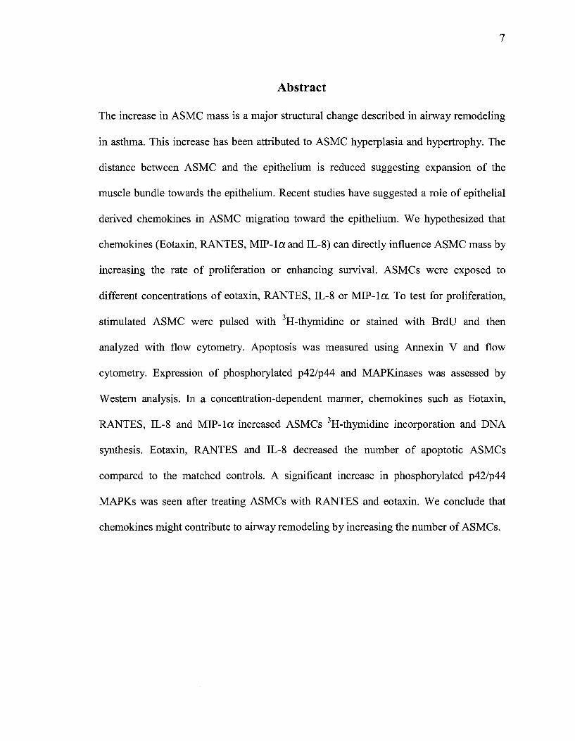
Abstract
The increase in ASMC mass is a major structural change described in airway remodeling
in asthma. This increase has been attributed to ASMC hyperplasia and hypertrophy. The
distance between ASMC and the epithelium is reduced suggesting expansion of the
muscle bundle towards the epithelium. Recent studies have suggested a role of epithelial
derived chemokines in ASMC migration toward the epithelium. We hypothesized that
chemokines (Eotaxin, RANTES, MIP-la and IL-8) can directly influence ASMC mass by
increasing the rate of proliferation or enhancing survival. ASMCs were exposed to
different concentrations of eotaxin, RANTES, IL-8 or MIP-la. To test for proliferation,
stimulated ASMC were pulsed with 3H-thymidine or stained with BrdU and then
analyzed with flow cytometry. Apoptosis was measured using Annexin V and flow
cytometry. Expression of phosphorylated p42/p44 and MAPKinases was assessed by
Western analysis. In a concentration-dependent manner, chemokines such as Eotaxin,
RANTES, IL-8 and MIP-la increased ASMCs 3H-thymidine incorporation and DNA
synthesis. Eotaxin, RANTES and IL-8 decreased the number of apoptotic ASMCs
compared to the matched controls. A significant increase in phosphorylated p42/p44
MAPKs was seen after treating ASMCs with RANTES and eotaxin. We conclude that
chemokines might contribute to airway remodeling by increasing the number of ASMCs.

8
Résumé
L'augmentation de la masse musculaire lisse constitue un des changements
majeurs associés au remodelage des voies respiratoires dans l'asthme. Cette augmentation
est attribuée à l'hyperplasie et à l'hypertrophie des cellules musculaires lisses (ASMC;
airway smooth muscle cells). La distance entre les ASMC et l'épithélium est réduite,
suggérant une expansion des faisceaux musculaires lisses en direction de l'épithélium.
Des études récentes suggèrent le rôle de chimiokines dans la migration des ASMC vers
l'épithélium. Nous supposons que MIP-la, RANTES, Eotaxin, IL-8, peuvent influencer
directement l'augmentation de la masse de ASMC en augmentant leur degré de
prolifération et/ou en diminuant leur degré d'apoptose. Les ASMC furent exposées à des
concentrations croissantes de MIP-la, RANTES, Eotaxin, IL-8. Pour mesurer la
prolifération, des ASMC stimulées ont été exposées à de la thymidine tritiée ou marquées
avec du Bromodeoxyuridine (BrdU), et analysées par cytométrie de flux. L'apoptose a été
mesurée par Annexin V et cytométrie de flux. L'expression de la p42/p44 MAP kinase
phosphorylée a été déterminée par immunoblot 1 buvardage de western. D'une manière
dose-dépendante, MIP-la, RANTES, Eotaxin, IL-8 ont augmenté la prolifération des
ASMC et la synthèse d'ADN. L'IL-8, l'Eotaxine et RANTES ont diminué le nombre de
cellules apoptotiques comparativement aux cellules contrôle. Une augmentation marquée
fut observée au niveau de la phosphorylation des MAP kinases p42/p44 après stimulation
des ASMC avec RANTES et Eotaxin.
Nous concluons que les chimiokines peuvent contribuer au remodelage des voies
respiratoires observé dans l'asthme en augmentant la prolifération des ASMC.

Chapterl. Introduction
1.1 Asthma
9
Asthma is a common chronic inflammatory disease of the airways associated with
branchial hyperresponsiveness and airway remodeling. The Global Initiative for Asthma
(GINA) reports that as many as 300 million people worldwide are affected with asthma.
The prevalence of clinically important asthma is increasing globally and, if the current
trend continues, it is predicted that as many as 400 million people will have asthma by the
year 2025 (GINA/Masoli 2004). Asthma is also the leading cause of childhood morbidity
as measured by hospitalization and school absence (Asher 1995). The importance of
childhood asthma is reflected in the fact that the pattern of asthma establishes during
childhood and persists into adulthood. Many pathological studies support this data by
showing airway remodeling occurring early in childhood (Payne 2003). The chronic
nature of asthma represents a significant economie burden and has an impact on the
patient's life, his family, and the society (Masoli 2004).
Despite intensive research in recent years, the pathogenetic mechanisms and
phenotypical characterization of asthma are still poorly defined. It is widely accepted that
asthma is a heterogeneous disorder and it is considered as a type 2 cytokines-induced
inflammatory response (Robinson 1992).
10
1.1.2 Th2 Type Inflammation, cells and mediators
Type 2 inflammatory responses are characterized by differentiated CD4+ Th2
cells that secrete a specifie panel of cytokines, including IL-4, IL-5, IL-9, IL-13, and
other inflammatory mediators. The cytokines recruit other multiple effector cells
including B cells, mast cells, eosinophils, and basophils (Azzawi 1990). Increased
numbers of these cells within the airway lumen and submucosa have been shown in
asthmatic airways (Bradley 1991). The eosinophils, mast cells, and basophils unique and
separate roles play a major effector function in the perpetuation of asthmatic
inflammation. In addition, Th2 cytokines act on their gene expression and phenotype to
induce and orchestrate the allergie inflammation (Romagnani 2002). For example, mast
cells releasing histamine induce airway hyperresponsiveness and eosinophils are believed
to be responsible for tissue damage and remodeling.
It is widely accepted that Th2 cytokines account for the major pathophysiological
manifestations of allergy and asthma. IL-4 and IL-13 were found to be absolutely
required for allergie inflammation to occur. IL-4 plays a major role in naïve T cell
differentiation into Th2 cells (Geginat 2002) and IgE switching in B-cells (Rush 2002).
IL-4 induces the expression of adhesion molecules by the endothelial cells, facilitating the
trafficking ofinflammatory cells to the targeted tissue (Hirata 1998). IL-13 increases IgE
production but also increases the contractile response of airway smooth muscle cells to
the contractile agonists (Shore 2002). Together, IL-4 and IL-13 are involved in the
development of airway hyperactivity by directly affecting airway smooth muscle cells.
11
Another Th2 cytokine that is involved in eosinophil activation, maturation,
survival (Sanderson 1988), and recruitment (Wang 1989) is IL-5. Moreover, IL-5 is also
implicated in AHR. IL-9 is involved in mast ce11 growth and activation; it induces mast
ce11s to express more FcERI and produce more inflammatory mediators. IL-9 upregulates
mucus production and promotes goblet cell hyperplasia (Louahed 2000). All together,
Th2 cytokines initiate, modulate, and maintain the a11ergic inflammation; therefore,
understanding asthma and other a11ergic diseases requires a comprehensive knowledge of
interactions among cytokines, infiltrating inflammatory ce11s, and resident structural ce11s.
Chemokines are produced by many structural, epithelial ce11s in particular, and by
inflammatory cells especia11y the macrophages. In retum, many inflammatory and
structural ce11s express receptors for different chemokines. Many studies of human
asthma show chemokines in relation to asthmatic inflammation and airway
hyperreactivity (Ste11ato 1997). Chemokines recruit and activate leukocytes and induce
degranulation in mast ce11s, basophils and eosinophils, initiating the early response in
asthma. Careful examination in the animal models of a11ergic airway responses reveals
that the expression and function of these chemokines occur at a specifie stage of the
disease (Lukacs1999). Moreover, the chemokines production is regulated by Th2
cytokines (Marfaing-K 1995).
Th2 ce11s in asthma highly express CC chemokine receptors such as CCR4, CCR8
(Bonecchi 1998), and CCR3 (Sa11usto 1997). These receptors may preferentia11y recruit
lymphocytes that would specifically create the allergie milieu leading to the altered
airway physiology in such diseases. CCR4 and CCR8 are known to be upregulated
selectively upon activation of polarized human Th2 ce11s (D'Ambrosio 1998). The Th2
ce11s will induce selectively the production of chemokines that act on Th2 chemokine
12
receptors. These chemokines act on other leukocytes, recruiting and activating them at the
site of inflammation.
1.2 Airway remodeling in Asthma
Th2 cytokines induce airway structural cells and the infiltrating inflammatory
cells to express inflammatory and remodeling related cytokines, chemokines, growth
factors, and adhesion molecules. Therefore, Th2 cytokines are central in many aspects of
airway remodeling where remodeling can explain part of the disease development.
Animal models continuous exposures to allergen lead to epithelial thickening, goblet cell
hyperplasia, and subepithelial fibrosis (Blyth 1996) and elevated levels ofiL-4, IL-5, and
IL-13 were found in these models (Johnson 2004). IL-4 induces collagen production by
branchial fibroblast (Bergeron 2003) and increases the production of several remodeling
associated factors, such as TGF-,8, PDGF, and others. IL-13 is a mitogen for ASMC
(Shore 2004); therefore, this cytokine might be involved in the increased smooth muscle
mass in asthma. IL-13 and IL-4 cause goblet cell hyperplasia while IL-5 transgenic mice
showed epithelial hypertrophy, goblet cell hyperplasia and focal collagen deposition. IL-5
deficient mice had significantly less peribronchial fibrosis and significantly less
peribronchial smooth muscle compared with wild type mice (Jae Youn Cho 2003). In
addition to mucus cell hyperplasia, IL-9 indirectly increases IL-8 and eotaxin production
(Baraldo 2003) from ASMC, where both chemokines cause ASMC migration
(Govindaraju 2006; Joubert 2006).
Th-2 cytokines promote the production of leukotrienes that are involved in airway
remodeling. Cysteinyl-leukotrienes augment growth factor-induced ASM mitogenesis
through activation of CysLT receptors (Panettieri 1998). Few studies showed Th2 cells
13
adhering to ASMC by CD44 and integrins can induce ASMC to proliferate (Lazaar
1994).
Remodeling refers collectively to a group of structural changes seen in asthmatic
airways. The changes include subepithelial fibrosis which contributes to the thickening of
airway walls due to the deposition of extracellular proteins such as collagens 1 and III,
laminin, and tenascin (Roche 1989), (Robinson 2007). Mucus gland hyperplasia leads to
excessive mucus secretion and can, in severe cases, lead to occlusion of the airways.
Branchial blood vessels increasing in number and size is another remodeling feature
studied in asthma (Li 1997). This change in airway vascularity could be the reason behind
the airway wall edema by increasing the production of VEGF (Hoshino 2001 ). Another
remarkable change is the increase in the branchial smooth muscle mass (Ebina 1990); the
inner border of the smooth muscle bundles is closer to the epithelium, suggesting the new
muscle is preferentially added to the luminal side of the muscle (Benayoun 2003). It
seems these structural changes are accompanied by changes in cellular phenotypes at the
molecular level that are of equal importance (Fixman 2007).
It has been postulated that airway remodeling is responsible for the graduai decline in
lung function; however, remodeling was detected in mild asthma and other studies failed
to relate asthma severity to the degree of airway remodeling (Bousquet 1998).
Many Th2 cytokines, chemokines, and mediators are involved in initiation and
perpetuation of the inflammatory process and remodeling. Consequent! y, treatments that
suppress this Th2 inflammatory response have been developed for therapeutic benefits;
however, sorne individuals are left with residual obstruction after optimal treatment with
corticosteroids and this obstruction is possibly attributed to airway remodeling (J effery
14
2005). Although airway remodeling occurs in response to acute injury or inflammation,
no effective treatment to prevent or to reduce remodeling is available. A few studies have
shown decreased rem ode ling after blocking sorne of the Th2 cytokines.
1.2.1 Epithelial Cells Remodeling in Asthmatic Airways
The epithelium under normal circumstances forms a highly regulated and almost
impermeable barrier. lt is widely reported that the asthmatic epithelium undergoes several
pathological changes and these changes could lead to alterations in host defense and in
the response of the epithelium to exogenous stimuli (Fahy 2001 ). Increased epithelial
shedding or desquamation is a characteristic feature of asthma, although this fact has been
long debated. Areas with desquamated epithelial cells where only basal cells are resting
on the basement membrane are often seen in post partum and branchial biopsies
specimens from asthmatic subjects (Laitinen 1985). Whenever the usually fragile in
appearance epithelium is seen in these specimens it is non-ciliated and vacuolized
(Montefort 1992). lt is common to find epithelial clumps in the sputum and an increased
numbers of epithelial cells in bronchoalveolar lavage of patients with asthma (Kirby
1987). However, epithelial shedding can be due to artifacts of the biopsy procedure (Fahy
2000).
Epithelial damage characterized by an increase in intracellular space and epithelial
shedding have been correlated with branchial hyperreactivity (Jeffrey 1989). Removal of
the epithelium from human isolated branchial smooth muscle appears to enhance the
responsiveness to acetylcholine and histamine (Knight 1990). Enhanced responsiveness
consequent to epithelium loss may prove important with respect to the development of
15
worsening asthma. Epithelial loss also causes exposure of the nerves and mast cells,
increasing the sensitivity of the isolated human airways to methacholine (Jongejan 1991).
The branchial epithelial cells are activated in asthma and these can be activated
through different mechanisms. It has been recently reported that epithelial cells of asthma
patients but not ofhealthy subjects express the FceRI and FceRII functional receptors and
are capable of fixing IgE (Campbell 1998). It is therefore possible that these cells may be
able to interact directly with inhaled allergens. Other studies showed that pollutants such
as N02 and ozone can activate the branchial epithelial cells (Davies 1993); histamine,
Platelet Activating Factor (PAF) (Vignola 1993; Salary 1991), and cytokines were shown
to activate the epithelial cells as weiL
Repair of bron chi al epithelium in asthma is important in maintaining the integrity
of the epithelial barrier. The repair process is a series of complex interactions to re
epithelize the luminal surface (Zahm 1991 ). There is increasing evidence to suggest that
aberrant repair signais in the epithelium occur early in the pathogenesis of asthma. An
aberrant repair process occurring at the mucosal surface may trigger a cascade of events
deeper within the sub-mucosa, leading to direct effects on the amount and behavior of
airway smooth muscle, as well as impacting on sub-mucosal glands and the deposition of
extracellular matrix (Knight 2007).
Asthmatic epithelium exhibits abnormal expression of severa! pro-inflammatory
transcription factors, signal transducers, and activators of transcription (STAT1 and
STAT6) (Mullings 2001). Using epithelial cells obtained by bronchoscopie sampling,
significant intrinsic biochemical and functional differences were reported between
healthy and asthmatic epithelium (Knight 2007). Asthmatic epithelia produce
substantially greater amounts ofiL-6, prostaglandin E2, and EGF (Kicic 2006). Not only
16
the biochemical function of airway epithelium is altered but structurally these cells are
disturbed with separation of these columnar cells from their basal attachments and
increased epithelial permeability (Chetta 1996)
1.2.2 Role of Epithelium in Airway Remodeling
Epithelial shedding and hypertrophy, particularly of goblet cells, are indicative of
remodeled airways (Lloyd 2007). Increasing evidence shows that epithelial cells play a
crucial role in the development of airway remodeling and it has been suggested that these
may act independently of inflammatory events (Holgate 2000). Asthmatic airway
epithelium contributes to the airway remodeling by producing different inflammatory
mediators and growth factors that induce remodeling of other airway tissues. Emerging
theory suggests that not only the intact epithelial cells damage seen in asthma contributes
to rem ode ling but even the damaged cells do so (Holgate 2001 ).
The airway epithelium is a source of a range of proinflammatory molecules,
including those derived from arachidonic acid (15-HETE, PGE2, and lipoxins), nitric
oxide (NO), endothelin-1, a range of cytokines (IL-1,8, IL-5, IL-6, IL-11, GM-CSF, IL-
16, IL-18), chemokines (IL-8, GROa, MCP-1, MCP-3, RANTES, MIPla, MIP2),
eotaxin, and proliferative growth factors (TGF,Bl, TGF,B2), platelet-derived growth factor
(PDGF), insulin like growth factor (IGF-1), and basic fibroblast growth factor (b-FGF)
(Holgate 2002).
Several reports show that epithelial supematant induces a proliferative response in
fibroblasts and myofibroblasts. The supematant contains several growth factors, such as
17
PDGF, bFGF, and TGF-/J and blocking these growth factors partially inhibits the
proliferative response of the myofibroblasts (Zhang 1999).
Recent evidence suggest that epithelial cells are capable of expressing receptors in
a manner that suggests a regulatory role in controlling local inflammatory and remodeling
events (Lukacs 2001 ). lt has been previously shown that, upon appropriate stimulation,
airway epithelial cells have the ability to release a number of cytokines. For example,
TNP-alpha stimulates the epithelial cells to synthesize IL-8 (CXCL8), RANTES
(Regulated upon Activation, Normal T -cell Expressed and Secreted), and MCP-1
(Macrophage chemoattractant Protein) (Cromwell 1992). Furthermore, epithelial cells
produce eotaxin and the production is upregulated by IL-4 and IL-13, which are Th2
cytokines involved in remodeling, as well as by TNP-alpha (Fujisawa 2000). IL-8,
RANTES, eotaxin, and MCPs have been shown to be upregulated in asthmatic airways
thereby highlighting the importance of these molecules in the pathogenesis of asthma
(Holgate 1997).
1.2.3 Smooth Muscle Cells (ASMC) in Asthma
Airway smooth muscle (ASM) is an important tissue involved in the regulation of
bronchomotor tone and exists in the trachea and in the branchial tree up to the terminal
bronchioles. Evidence suggests that ASM undergoes marked phenotypic modulation in
lung development and in disease states such as asthma, chronic bronchitis, and
emphysema; however, the precise function of ASMC in healthy bronchioles remains
unclear (Panettieri 2003).
18
ASMC is a key determinant of aüway hyperresponsiveness and remodeling in
asthma. Aüway myocytes are thought to have capacity to contribute to remodeling due to
their ability for graded and reversible phenotype switching which confers broad
functional capacity (Halayko 2001). Airway myocytes exist in an immature phenotype
that is characterized by a high tendency for proliferation, expression, and secretion of
ECM proteins and by synthesis of inflammatory mediators in response to a number of
environmental eues (Halayko 2003).
ASMC also serve as a rich source for many cytokines, chemokines, growth
factors, adhesion molecules, and even lipid mediators. They express and release eotaxin
in response to Th2 cytokines. Interleukin-13 can act directly on airway smooth muscle
cells leading to changes in the ability of smooth muscle cells to generate chemokines such
as eotaxin and TARC (Shore 2004). ASMC also produce monocyte chemoattractant
protein MCP-1, MCP-2, MCP-3, IL-8, and RANTES when stimulated with
proinflammatory cytokines, such as IL-lb and TNF-a (Hamid 2005). ASMC have been
shown to release a number of extracellular matrix proteins, including collagen I, collagen
III, lumican, and versican. These proteins were shown to be upregulated in the asthmatic
airways (Black JL 2001 ). By the expression of the above mentioned molecules and
others, ASMC regulate and contribute to the ongoing inflammatory process seen in
asthma.
In addition to contraction, ASMC contribute to the remodeling in asthma by
increased proliferation leading to an increase in smooth muscle mass. Such increase in
ASMC could contribute to the exaggerated airway narrowing observed in asthma. Data
obtained from in vivo studies suggest that ASMC can exhibit heterogeneity, i.e., exhibit
proliferative, synthetic, and contractile phenotypes. It has been shown that under
19
stimulation with proinflammatory and inflammatory cytokines, ASMC is capable of
expressing a wide range of molecules (Chung 2000). Sorne reports suggest that the
ASMC immature phenotype is one characterized by high tendency for proliferation and
mediators synthesis (Halayko 2006). In contrast, myocytes of mature phenotype are of
contractile ASMC but are still able to produce sorne ECM, suggesting that sorne ASMC
might exist in intermediate phenotype (Merrilees 1990). Interestingly, ASMC serum
deprivation in vitro can switch or modulate proliferative and synthetic myocytes to a
contractile phenotype (Stephens 1998).
The increase in ASMC content in asthma was first reported more than 78 years
ago (Dunnill 1969). The mechanisms underlying such an increase could be an increased
rate of division or decreased rate of apoptosis. It could also be due to the migration of
ASMC towards the luminal side of the airway through the migration of mesenchymal
cells to the ASMC bundle or by differentiation (Hirst SJ 2004). Proven to a lesser extent,
sorne morphometric studies reported ASMC hypertrophy as an additive explanation of
ASMC mass increase.
Several stimuli have been identified as mitogens for ASMC. Contractile agonists
such as histamine and LTD4 (Panettieri 1998), cytokines such as TNF-a (Amrani 1996)
and IL-1,8 (De S 1993), growth factors, extracellular matrix, and increased stretch
(Hasaneen 2005) were described to influence the proliferation rate of airways smooth
muscle. Fibronectin and collagen I enhance proliferation and encourage expression of the
nuclear proliferation marker Ki67 (Black 2002), whereas cells grown on laminin divide
more slowly but express contractile proteins. This points to the fact that cell-matrix
interactions, in addition to growth factors and cytokines, have important effects on
myocytes phenotype and cell cycle control. Adding fibronectin, collagen I, and laminin
20
to the cultured ASMC provides a strong survival signal (Freyer 2001). Mitogens are
expressed by ASMC and other structural or inflammatory cells, they may modify the
proliferation in a paracrine or autocrine manner. Although a number of mitogens for
ASMC were identified in vitro, it is still not clear which plays the most important role in
vivo (Knox et al2000). It seems that the chronicity of the inflammation in asthma primes
the myocytes by changing the ECM and exposing the cells to many different types of
mediators profiles associated with different types of inflammation.
Asthmatic airway smooth muscle cells grow at approximately twice the rate of
cells from subjects without asthma (Johnson 2001). Accumulated evidence shows that
ERK, one of the MAP Kinases, is the reason behind this difference. ERK activation is
necessary and involved in human ASMC proliferation but activation of ERK is altered in
asthmatic smooth muscle cells. Peak ERK activation in response to a low concentration of
mitogens is greater than what is seen in non asthmatic ASMC (Lee JH 2001 ).
ASMC proliferation is a complex process and is regulated by different cell growth
modulators; sorne modulators are enhancing cell growth and proliferation while others
inhibit it. Activation of G-protein coupled receptors that increase cyclic AMP levels
reduce ASMC proliferation and activation of nuclear hormone receptors by sex hormones
(Hughes 2002), glucocorticoids (Hirst 2004), and prostaglandin E2. In addition, short and
long acting (32- selective agonists, such as salmeterol and formoterol respectively, reduce
proliferative response by reducing cyclin D1levels which are required for the cell entry to
S-phase. Recent identification of other regulators described sorne enzymatic regulation
activity. For example, chymase of the mast cells was shown to modulate ASMC
proliferation and this may indicate the importance and contribution of the mast cells
found in the bundles of smooth muscle (Lazaar 2002). More recently, activated (antigen-
21
specifie) CD4+ T cells were shown to regula te myocyte turnover and proliferation in a
direct cell-cell interaction manner (Ramos-Barbon et al2005).
Data drawn from branchial biopsies has shown that interstitial region beneath the
epithelium is rich in collagen, particularly type I and III that are enhancing ASMC
proliferation. In contrast, ECM in the smooth muscle bundle is rich in the antiproliferative
proteoglycans (Black 2002). Therefore, smooth muscles might migrate from the bundle
toward the epithelium under the influence of epithelial chemoattractants, such as PDGF,
eotaxin, and others and migrate away from the antiproliferative media.
In vitro, ASMC shows a little evidence of undergoing apoptosis. ASMC apoptosis
occurs under extreme conditions such protein synthesis inhibition or prolonged incubation
in protein deficient media (Freyer 2001 ). lt has been suggested that regulation of
apoptosis may be a primary process by which ASM cell number is determined; the
threshold for change in rates of apoptosis may be lower than for proliferation and
reduction in rates of apoptosis may contribute to hypertrophy (Halayko 2006).
Endothelin-1 (ET -1) in duces hypertrophy and reduces apoptosis in ASMC (Halayko
2007). Cardiotrophin (CT-1) enhances airway smooth muscle survival under conditions
of serum deprivation and markedly inhibits TNF-a!Fas-induced apoptosis (Zhou 2003).
The ASMC survival depends strongly on the integrity of the surrounding tissue.
Many proteinases such as neutrophil elastase drive ASMC to apoptosis because they
simply degrade the matrix molecules and cause detachment induced apoptosis (Oltmanns
et al. 2005).
22
1.2.4 Interaction between ASMC and epithelium
Morphometric studies done on biopsies from asthmatic subjects strongly indicate
that the increase in ASM mass is a remarkable phenomenon in asthma, especially in
severe cases of this disease. The increase in ASM area and augmented cell size are
selective structural changes that distinguish severe asthmatic patients from the mild
(Benayoun L 2003). In addition, the distance between the smooth muscle mass and the
epithelium, especially in tissues obtained from asthmatic subjects, is reduced (Pepe 2005).
This phenomenon might reflect the migration of smooth muscle cell toward the
epithelium or it could be the attraction of circulating precursor stem cell populations. The
close proximity could be also due to the ASMC mitogens produced from the epithelial
layer that induce the myocytes growth toward the luminal si de of the airway. Epithelium
is a potent source of different mediators that act as mitogens for ASMC, such PDGF,
EGF, FGF, IGF , TGF-,8, TNF-a and IL-1,8, and this proliferative medium provided by
the epithelium could be also the reason for the ASMC to move away from the
antiproliferative medium found in the smooth muscle bundle it self (Black 2003).
ASMC possess receptors for different epithelial derived products but more
important to the study of epithelial-ASM interaction is that receptors for epithelial derived
CC and CXC chemokine were identified. These chemokines may be important in
inducing airway smooth muscle cells to grow on the epithelial si de of the existing bundle
resulting in close proximity of the ASMC to the epithelium. Sorne of these chemokines
are eotaxin, RANTES, IL-8, and MIP-la and they act through specifie receptors that have
been shown to be expressed on ASMC.
23
ASMC react to IL-8 by expressing CXCRl and CXCR2 constitutively at the
mRNA and protein level but the level of the expression ofboth receptors is relatively low
compared with neutrophils (Govindaraju 2006). Upon stimulation, these receptors
increase the intracellular Ca2+ and cause the smooth muscle to contract and migrate.
Therefore, IL-8 might contribute to the ASM remodeling in asthma.
CCR3 is the most extensively studied receptor among the chemokine receptors. lt
interacts with a number of ligands such as eotaxin and RANTES. In addition, CCR3 is
upregulated in branchial smooth muscle cells of asthmatics. ASMC express functional
CCR3 that upon binding to eotaxin induces smooth muscle migration (Joubert 2005). The
same group and others demonstrated the expression of CCRl (Amin 2005), which binds
RANTES and MIP-la, by ASMC.
24
1.3 Role of Chemokines in Asthma
1.3.1 Chemokines in Allergie Inflammation
The chemokines are a subgroup of chemotactic cytokines (8-1 0 kD) which have
been subdivided into four subfamilies on the basis of the position of either one or two
cysteine residues located near the amino terminus of the protein (CXC, CC, C, and CX3C
subfamilies) (Zlotnik 2000; Table. li Appendix)
A number of chemokines have been identified in relation to asthma and sorne are
correlated to the inflammation severity and airway hyperreactivity. In addition to the
chemokines main function of recruiting leukocytes to the site of the inflammation, sorne
members also have the capacity to direct T helper cells differentiation towards Th1 or
Th2 differentiation (Karpus 1997). MCP-1 can drive undifferentiated T helper cells
toward IL-4 producing lymphocytes while MIP-1a promotes the Th1 differentiation
(Karpus 1997). MCP-1 plays a significant role at the early stages of the diseases
development. This chemokine induces mast cell activation as well and L TC4 release into
the airways, which directly induces AHR (Campbell 1999). Other members of the CC
subfamily recruit eosinophils in the early stage of asthma through the vessels to the lung
interstitium, such as RANTES and MIP-1a (Campbell 1998), whereas eotaxin is needed
at the chronic stage of asthma for eosinophils accumulation (Campbell 1997). Thus the
coordinated expression and ligation of these chemokines appears to be differentially
regulated at specifie stages of asthma (Lukacs 1999).
IL-8 is a member of the CXCL subfamily while RANTES, eotaxin and MIP-1a
belongs to the CC subfamily. A lot of initial work on chemokines has focused on those
that have chemotactic activity for the inflammatory cells in the inflammatory lung
25
diseases. In addition to eotaxin, which is one of the most potent chemokines for
eosinophils, MIP-la and RANTES also lead to recruitment and degranulation of these
cells. Eotaxin and other chemokines have been previously shown to selectively attract
Th2 lymphocytes (Gutierrez-Ramos 2000). Th2 cells are the comerstone of asthma
pathogenesis and the importance of chemokines in the inflammatory process mediated by
lymphocytes is likely to be crucial for the initiation and perpetuation of the disease.
The main stimuli for secretion of chemokines are proinflammatory cytokines such
as IL-l and tumor necrosis factor (TNF-a), bacterial products such as lipopolysaccharide
(LPS), and viral infection. In addition, products of both Thl and Th2 cells, Interferon-)'
(IFN-)') and IL-4, respectively, can also induce the production of these chemokines
independently and in synergywith IL-l and TNF-a.
The regulation of chemokine production and the expression specifically during an
active inflammatory process was demonstrated by several reports. It seems that
chemokine production is controlled by different inflammatory cytokines as well as other
chemokines; IL-4, IL-13, IL-1,8 and TNF-alpha upregulate the expression of the above
mentioned chemokines in asthma models (Rothenberg 2003 and others). More recently,
the potentials of chemokines to regulate the immune response were demonstrated where
dawn-regulation of IL-8 could be achieved by eotaxin introduction in human endothelial
cells (Kunkel2002).

26
1.3.2 Chemokines receptors
Chemokines mediate their actions by binding to specifie receptors. Chemokine
receptors belong to classA of the G protein-coupled receptor (GPCR) superfamily. They
are rhodopsin-like receptors that span the membrane seven times and are coupled to
heterotrimeric Ga/3"( proteins. These receptors are unique among cytokine receptors
because they are seven spanners and coupled to G proteins that in tum activated another
second messenger signais. When the receptors are termed seven spanners it indicates that
they have a serpentine configuration and snake in and out of the cell membrane seven
times. Sorne other cytokines have soluble receptors that represent fragments of the
membrane-bound receptor that have been proteolytically cleaved; with the seven-spanner
chemokine receptors, this is not likely to occur (Kunkel 1991).Chemokines interact with
their receptors at two main sites; one site is the N-terminal region and the other one is
located within an exposed loop that extends between the second and the third cysteine
residue. The N-terminal binding is the part that triggers the receptor (Clark-Lewis 1995).
To date, eight CCR receptors and seven CXCR receptors have been identified.
Upon ligand-receptor interaction, different intracellular signaling pathways are activated,
ultimately leading to cell mobilization and activation. These receptors are expressed by
different inflammatory cells such T cells, B cells and dendritic cells, and by structural
cells such smooth muscle, fibroblasts and endothelial cells. A feature of the chemokine
system is the complexity of the ligand-receptor interactions. Thus, a particular chemokine
(i.e., CCL5) may bind severa! receptors (CCRl, CCR3, and CCR5), and different
chemokines (i.e., CXCL9, CXCLlO, and CXCLll) may bind a single receptor (CXCR3).
27
Evidence suggests that, in sorne cases, ligand redundancy does not mean duplicity of
functions (Bardi 2001).
Severa! studies show that sorne CCR and CXCR receptors are upregulated in
airways of asthma patients (Pillete 2004). During migration, leukocytes, eosinophils, and
neutrophils express a wide range of chemokines receptors on the cell surface. Differences
in the expressed chemokine receptors were seen between cells from the peripheral blood
and the cells at the inflamed tissue (Lukacs 1999). These differences must be necessary to
interact with the inflammatory environment; for example, CCR3 is highly expressed by
Th2 effector cells, eosinophils, mast cells, and basophils in the lung specimens of
asthmatics. Also, the CCR3 ligands include the major chemokines expressed in the
allergie lungs such as RANTES, eotaxin, MCP-4 and MCP-3 (Ying 1999). As well CCR4
and 8 are highly expressed by Th2 in allergie conditions (Panina-Bordignon 2001)
Upon binding to their G-protein coupled receptors, chemokines induce
conformational changes in the a and (3 subunits that can further activate various effector
enzymes such as MAPKinases. The activation includes a rise in the intracellular Ca2+
concentrations, degranulation of the intra-plasmic granules, and increased production of
oxygen radicals and lipid mediators (Lukacs 2001 ).
Chemokine receptors are not only expressed by leukocytes because many airways
structural cells express functional receptors for the major chemokines in asthma. For
example, CCR was detected on epithelial cells, endothelial cells (Ying 1997), and ASMC
(Joubert 2006). The epithelial expression of CCR3 is upregulated by the proinflammatory
cytokines such as TNP-a and INF-')' (Stellato 2001). On the other hand, eotaxin binding to
CCR3 increases the eotaxin and IL-8 production by the structural cells via severa!
MAPKinase pathways (Cui 2002).
28
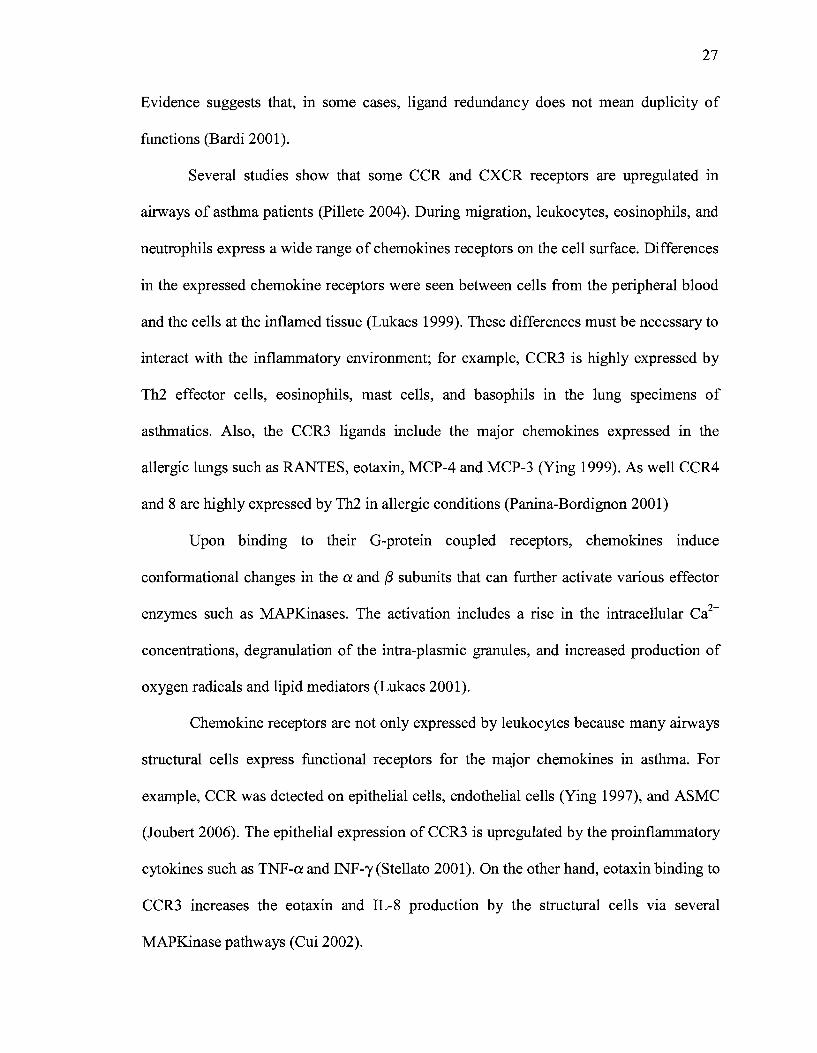
1.3.3 Activation of MAPKinases by Chemokines
It is well established that activation of MAPK by growth factors and G protein
coupled receptors results in the stimulation of DNA synthesis and cell proliferation
(Marshall 1995). Previous studies have shown that PDGF, EGF, and Substance P (SP)
activate MAPK isoforms upon binding to their G-protein coupled receptors in tracheal
smooth muscle cells (Pouyssegur 1992). Once activated, these MAPKs in tum activate
their specifie substrates on downstream targets. It is suggested that the binding of the
growth factor to the GPCR induces the activation of inositol phospholipids hydrolysis
pathway and leads to the release of intracellular Ca2+ and then phosphorylation of Protein
Kinase C (PKC) (Reggoli 1994). PKC is a predominant component in the kinase cascade
that is initiated by ligand attachment to both G-protein coupled receptors and receptors
containing intrinsic tyrosine kinase activity (Otsuka 1993).
Upon activation, PKC mediates intermediate kinase activation. In growth factors
binding it is suggested that Raf-1/MEKinases are subsequently phosphorylated in this
cascade leading to sequential phosphorylation of p42/p44 MAPK which enhances DNA
synthesis and cell proliferation (Yang 2002). Chemokines are expected to follow a similar
signaling pathway to induce cellular proliferation.
29
Fig 1. Schematic pathway for growth factors signaling of cellular proliferation.
Chemokines are expected to follow the same pathway (Adapted from Yang 2002)
1.3.4 Epithelial Derived Chemokines
1.3.4.1 CCLll (eotaxin-1) is a member of CC chemokine subfamily. Eotaxin is widely
recognized to mediate eosinophil chemotaxis and activation in both mice and humans and
is thought to play a central role in eosinophil recruitment to the airways of Ag-challenged
animais (Griffiths 1993). CCL11 is also recognized to induce chemotaxis of basophils
(Y amada 1997), Th2 lymphocytes (Sallusto 1997), mast cells (Oshi 1999), and airway
smooth muscle cells (Joubert 2005). In humans, CCL11 can be detected in the sputum of
patients with moderate and severe asthma and 50% of the total eosinophil chemotactic
activity in such samples is predicted to be due to eotaxin. Recently, an anti- apoptotic
30
effect of eotaxin was reported in the pulmonary artery endothelial cells (Farahi 2007).
Eotaxin is produced by a variety of inflammatory cells including T lymphocytes
(Loetscher 1996), macrophages, eosinophils, and structural cells such branchial epithelial
cells (Ponath 1996), fibroblasts (Teran 1999), smooth muscle cells, and, most recently,
airway parasympathetic neurons (Fryer 2006) and the pulmonary artery endothelial cells
(Farahi 2007). In the lungs, the epithelium is the major producer of eotaxin (Ying 1997).
1.3.4.2 RANTES/ CCLS is a member of the CC chemokines and was identified earl y in
1986 (Yokota 1986). RANTES is recognized as a powerful chemoattractant of
eosinophils, T lymphocytes, neutrophils, monocytes, and basophils (Schall 1990). This
chemokine activates these immune cells and induces the exocytosis of
bronchoconstrictive mediators, such as histamine and cysteinyl leukotrienes, from
basophils and eosinophilic cationic protein from eosinophils (Chung 1999). RANTES is
involved in inflammatory cell recruitment and in the induction of bronchoconstrictive
mediators from cells which result in airflow limitation (Matsunaga 2006). RANTES
initiates severa! other proinflammatory events, such as integrin activation and lipid
mediator biosynthesis (Taub 1996). RANTES is generated predominantly by T cells,
epithelial cells (Matsukura 1995), fibroblasts, and platelets (Kameoshi 1992). RANTES is
constitutively expressed in the lungs of patients with asthma, where increased levels are
detected in the broncoalveolar lavage (BAL) fluid ofthese patients (Folkard 1997).
RANTES also contributes to Th2 type of inflammation by upregulating the
production of IL-5 and IL-6 (Lillard 2001). This particular contribution was detected
shortly after the allergen challenge in the animal models; however, RANTES role shifts
31
toward Th1 augmentation (increases IL-12 and INF--y levels) in the sensitized animais
after several challenges (Koya 2006).
1.3.4.3 Macrophage inflammatory protein (MIP)-la was identified 17 years ago as
the first of four members of the MIP-1 CC chemokine subfamily (Baixeras 1990). These
proteins were termed CCL3 (MIP-la), CCL4 (MIP-1,8), CCL9/10 (MIP-1 -y), and CCL15
(MIP-lô) (Orlofsky 1991). MIP-la has been shown to contribute to
monocyte/macrophage, mast cells (Alam 1992) and neutrophil chemotaxis and activation.
It has been demonstrated that MIP-la plays an important role in eosinophilic
accumulation and activation in humans (Rot 1992) and in vivo after airway challenge
(Lukacs 1996). The epithelial cells are the main source for MIP-la in the lungs (Stellato
1997); MIP-la:'! CCL3 is also produced by macrophages (Wolpe 1989), dendritic cells
(Mohamadzadeh 1996), and lymphocytes (Conlon 1995).
MIP-1 proteins mediate their own biological effects by binding to cell surface CC
chemokine receptors which belong to the G-protein-coupled receptor superfamily (CCL3
binds to CCRl and CCR5) (Gao 1993). Receptor binding involves high affinity
interactions and a subsequent cascade of intracellular events that lead rapidly to a wide
range of target cell functions, including chemotaxis, degranulation, phagocytosis, and
mediator synthesis (Maurer 2004 ).
1.3.4.4 IL-8/ CXCL8 is the major neutrophil chemoattractant and activating factor in
the lungs (Kunke11991). IL-8 induces neutrophils exocytosis of stored proteins as well
as release of hydrogen peroxide and superoxide anions. In addition, this CXC chemokine
evokes eosinophil (Shute 1994) and monocyte migration to the inflamed site (Remick
2005). IL-8 is also involved in a wide variety of physiological and pathological

32
processes, including host defense against bacterial infection, 0 0
ang10 genes1s,
arteriosclerosis, and autoimmune disorders of skin, bones, and joints (Harada 1996).
Elevated concentrations of IL-8 are found in sputum, bronchoalveolar lavage fluid, and
branchial tissues of subjects with pulmonary diseases such as allergie (Smith 1991) and
severe asthma, occupational asthma (Gibson 2001), cystic fibrosis (CF), chronic
obstructive pulmonary disease (COPD) (Nocker 1996), bronchitis, acute respiratory
distress syndrome, and idiopathie pulmonary fibrosis (Richman 1993).
Branchial epithelial cells, neutrophils, macrophages, mast cells, and fibroblasts
are the major producers of CXCL8. IL-8 acts through the specifie receptors CXCR1 and
CXCR2 which are located on the surface of neutrophils, monocytes, airway epithelial
cells, and endothelial cells. Recently it has been shown that airway smooth muscle
constitutively expresses mRNA and protein for both CXCR1 and CXCR2 receptors
(Govindaraju 2006). All of these inflammatory and structural cells produce IL-8 and
express functional receptors of this chemokine; thus, an auto-regulatory pathway for
chemokines within the airways could contribute to the amplification of the allergie
inflammatory response (Eddlestone 2003).
In asthma, airway smooth muscle migration has been postulated as an explanation
for the increased mass of smooth muscle. IL-8 has been shown to be a potent
chemoattractant ofhuman airway smooth muscle cells when compared to PDGF, which is
a strong ASMC chemotactic agent (Govindaraju 2006). The cellular recruitment of
ASMC or inflammatory cells occurs through the development of the chemotactic gradient
so the cell moves toward an area of increased chemokine concentration. In vivo this
gradient could be generated by IL-8 or other chemoattractant molecules, such as PDGF,
binding to basement membrane proteins.
33
The physiological concentration of IL-8 is not known. IL-8 is known to have a
long half life which means it may be accumulated in concentrations high enough to
induce chemoattraction for neutrophils, ASMC, and eosinophils (Remick 2005). Based
on role of IL-8 on airway hyperreactivity and airway remodeling, it is considered as a
potential therapeutic target especially in severe asthma. Strategies designed to target IL-8,
such as anti-IL-8 antibodies or receptor blockade of TLR-4 to limit the release of IL-8,
are still under investigation (Barnes 2007). Another reason to target IL-8 especially in
lung diseases is the link between the excessive release of IL-8 after airway damage and
the damage to the IL-8 receptor on airway neutrophils which might disregulate cytokine
production and lead to excessive neutrophil accumulation (Koehler 2004).
34
1.4 Rationale
lt is not surprising that the altered asthmatic epithelium participates in airway
remodeling in asthma by the production of different mediators that induce remodeling in
other airway tissues. Asthmatic epithelium expresses molecules that have receptors on
inflammatory cells therefore recruiting more cells to the airway. For example, ASMC
express receptors for most of the growth factors produced by the epithelium while smooth
muscle expresses functional receptors for the epithelial-derived chemokines, such as
RANTES, eotaxin, IL-8 and MIP-la. These molecules are released by the ASMC as well
and their effect could be paracrine autocrine.
ASMC mass increase is a well documented phenomenon of asthmatic airways,
especially in severe asthma. The distance between the epithelium and smooth muscle
bundle is reduced and this could be attributed to epithelial-derived migratory and
proliferative signais that induce ASMC remodeling. lt has been shown that IL-8 and
eotaxin cause migration of ASMC; these cells express receptors for both chemokines,
CCR3, CXCRI, and CXCR2 respectively. It has been recently demonstrated that ASMC
migrate towards concentration gradients of eotaxin and IL-8 but the effect of these
chemokines on smooth muscle proliferation and survival is not yet examined. Those two
chemokines and others, such as MIP-la and RANTES, could be mitogens for ASMC. If
so, it could be an additive explanation for why myocytes grow and proliferate toward the
epithelium, the source of these chemokines, and other mitogens.
35
1.5 Hypothesis
W e hypothesize that airway epithelial derived chemokines such as eotaxin,
RANTES, IL-8, and MIP-la, induce proliferation of airway smooth muscle cells and
promote enhanced survival of airway smooth muscle cells by inhibiting apoptosis.
1.6 Specifie Objectives
Firstly, to investigate the effect of the chemokines, IL-8, Eotaxin, RANTES, and
MIP-la, on ASMC remodeling and to examine the effect on ASMC proliferation in vitro.
Secondly, to examine the possible effect of the above mentioned chemokines on ASMC
survival. We set out to examine the possible effect of chemokines on the rate of
apoptosis in ASMC.
Chapter 2. Materials and Methods
2.1 Cell culture and Reagents
36
The ASMC were obtained from two sources. Branchial/tracheal smooth muscle
cells (B/TSMC) were purchased from Cambrex and were positively stained for a-smooth
muscle actin and negatively for factor VIII, CD45, and CD3, as indicated by the
manufacturer. B/TSMC were grown in their optimal medium (SmGM-2; Cambrex)
containing 5% FBS at 37°C in a humidified incubator with 5% C02, as recommended by
the supplier. The second source were primary cultures of human non- asthmatic branchial
ASMC which were obtained from surgical specimens. Segments of lobar or main
branchus measuring 5 x 2 mm were incubated for 90 min at 37°C in 10 ml Hanks'
balanced salt solution buffer (in mM: 5 KCl, 0.3 KH2P04, 138 NaCl, 4 NaHC03, and
5.6 Na2HP04) to which 640 units of collagenase (type IV), 10 mg of soybean trypsin
inhibitor, and 100 units of elastase (type IV) had been added. The digested tissue was
then filtered through a 125- rn Nytex mesh and the resulting cell suspension was
centrifuged. The pellet was then reconstituted in growth medium (Cambrex, SmGm-2)
and plated in 25-cm2 flasks. Complete SmGm2 media were purchased from Cambrex Bio
Science, Walkersville, USA. Confluent cells were detached with 0.025% trypsin and
0.02% EDTA and counted using a hemacytometer. They were identified as smooth
muscle cells by positive immunohistochemical staining for smooth muscle specifie a
actin and positive identification of myosin light chain kinase and calponin by Western
blot analysis. All the chemokines used in the experiments were recombinant human and
purchased from R&D Systems (Minneapolis, Ml). All ASMC used were at passages 2-5.
37
2.2 (3H)-thymidine Incorporation
Metabolic incorporation of tritiated eH)-thymidine into the newly synthesized
DNA is a widely used method to detect the rate of DNA synthesis and monitor celi
proliferation. ASMC were plated in 96 weli plates at a density of 3000-5000 cell/well in
triplicates and grown to 60-70% confluence in complete SmGm2 containing 5% FBS at
37°C in a humidified incubator with 5% C02. Cells were then starved for 48 hours in
0.3% FBS SmGm2 (starving media) containing ali other additives. Following the
starvation period, the starvation medium was replaced with medium supplemented with
the chemokine of interest. Cells were then treated with chemokines or the appropriated
vehicle for 24, 48, or 72 hours. The chemokines of interest were RANTES, eotaxin, IL-8,
and MIP-la. The concentrations for each chemokine tested ranged from O.lng/ml up to
1 OOng/ml, except for IL-8 where concentrations of 50, 100, 500, and 1000 ng/ml were
tested. The vehicle used as a control was 0.1% BSA in PBS; the same vehicle used to
dilute the chemokines. Ali the results were compared to the vehicle and to the positive
control ofPDGF lOng/ml.
Eighteen hours before cell harvesting for thymidine study, cells were pulsed with
1 )JCi of thymidine/ weiL Cells then were washed with PBS and trypsenized prior to
DNA isolation. A Skatron Micro96 cell harvester (Molecular Deviees, Sunnyvale, CA)
was used to isolate DNA foliowing the manufacturer's instructions and then radioactivity
was measured by a liquid scintillation and luminescence counter (Perkin Elmer).
38
2.3 BrdU incorporation and Flow Cytometric Analysis
Cell proliferation was also determined using colorimetrie assay based on the
measurement of a synthetic thymidine analog bromodeoxyuridine (BrdU) incorporation
during DNA synthesis (FITC BrdU Flow Kit, BD Biosciences, CA). The incorporated
BrdU is stained with specifie anti-BrdU fluorescent antibodies and the levels of cell
associated BrdU are then measured with flow cytometry. ASMC were grown in 12 wells
plate at a density of 40,000 cell/well in complete and then medium was switched to
SmGm-2 (supplemented with 0.5% FBS) for 48 hours to induce quiescence and
synchronize the cell cycle. The vehicle or test chemokines were then added to the cells for
24, 48, or 72 hours. The thymidine analog BrdU was added (10f.ll/ml of 1mM BrdU
solution) 18 hours before cells are processed for flow cytometric analysis.
The BrdU flow kit staining protocol provided by the manufacturer was followed
to prepare cells for flow cytometric analysis. After removing the culture media, the
treated cells were trypsenized and washed with PBS. Cells were fixed and permeabilized
using the manufacturer reagents (BD Cytofix/ Cytoperm buffer). The cells were then
treated with DNase to expose the incorporated BrdU for one hour at 37°C. Next, cells
were incubated with 1:50 of diluted anti-BrdU fluorescent antibody for 20 minutes at
room temperature. The stained cells were analyzed immediately with a F ACS Calibur
flow cytometer system using Cell Quest Software (BD Biosciences). Ten thousand events
were collected on each sample.
The experiment controls were as follows: cells treated with PDGF as appositive
control and cells treated only with vehicle as a negative control.
39
The staining controls were the following: cells stained with BrdU only, cells
stained with BrdU and anti-BrdU antibody, cells stained only for anti-BrdU antibody, and
totally unstained cells.
2.4 Annexin V and Flow Cytometric Analysis
One of the earliest indications of apoptosis is the translocation of the membrane
phospholipid phosphatidylserine (PS) from the inner to the outer leaflet of the plasma
membrane. Once exposed to the extracellular environment, binding sites on PS become
available for Annexin V. Annexin V is a 35-36 kD Ca 2+-dependent phospholipid
binding protein with a high affinity for PS. The extemalization of PS precedes other
apoptotic processes, such as loss of plasma membrane integrity, DNA fragmentation, and
chromatin condensation, that come at the end stages of cell death. For these reasons
Annexin V staining is typically used in conjunction with a vital dye such as Propidium
Iodide (PI) to allow the identification of early apoptotic cells (Annexin V-FITC positive,
PI negative). As such, Annexin V can be conjugated to a fluorochrome such as FITC and
used for flow cytometric identification of cells in the earl y phase of apoptosis.
To test ifRANTES, eotaxin, IL-8, and MIP-la have an anti-apoptotic effect, cells
were plated in 12 well plates at a density of 40,000 cell/well in a complete media. The
cells were treated exactly the same as in the BrdU test except for the addition of BrdU to
the treated cells. When the stimulation period was over, Annexin V (Annexin V-FITC
Apoptosis detection kit, BD Biosciences) was used to quantify the percentage of cells
undergoing early apoptosis. The chemokine treated cells were trypsinized and washed
with PBS and then suspended in 100 ml of lX Annexin V binding buffer (lOX is supplied
by the manufacturer). Pive !ll of Annexin V and 5 !ll of PI stains were added to each test
40
tube for a duration of 15 minutes at room temperature and in the dark. Within one hour,
the cells were analyzed using F ACS Calibur flow cytometer by counting ten thousand
events per sample. The following controls were used: totally unstained cells, cells stained
only with Annexin V, and cells stained only with PL
2.5 Protein extraction, Immunoprecipitation and Western Blotting
Western blotting is used to detect the presence of select protein. We used Western
blotting to detect the expression of p42/p44 MAPKinases in a whole cell extract after
stimulation with the chemokines of interest. These MAPKinases are downstream products
expressed upon the stimulation of G-Protein Coupled Receptors (GPCR) which in this
case were the chemokines receptors. P42/p44 activation is essential for subsequent DNA
synthesis and cell proliferation. The specificity of the Western blotting in this case will
depend on the specifie binding of the antibody to the epitope. Anti p42/p44 total and
phosphorylated antibodies were obtained from Santa Cruz Biotechnology, CA.
For the Western blotting studies, ASMC were grown in 6 well plates at a density
of 100,000 cells/well. The cells were starved for 48 hours and then stimulated with the
chemokine of interest for one, five, or ten minutes. Chemokine induced P42/p44 MAPK
activation occurs as early as 1 minute and peaks at 5 minutes and then goes back to the
basallevels.
After the chemokine stimulation, the medium was removed and ASMC were
washed with ice-cold PBS. The cells were then harvested in lysis buffer made of 1%
Triton X-100, 50 mM HEPES (pH 8.0), 150 mM NaCl, 10% glycerol, 2mM EGTA, 1.5
mM MgC12, 10 J.tg/mL aprotinin, 10 J.tg/mL leupeptin, 1 mM phenylmethylsulphonyl
fluoride, and 1 mM sodium orthovanadate. Following lü-minute incubation on ice, the
41
lysates were clarified by centrifugation at 14,000 x g for 10 minutes and supematants
were collected. Protein concentrations were determined using Bradford method. To detect
activation ofp42/p44 MAPK, equal amounts ofwhole celllysates were solubilized into a
boiling Laemmli sample buffer, subjected to a 10% SDS polyacrylamide gel
electrophoresis (PAGE), and transferred to nitrocellulose (Bio-Rad Laboratories Ltd.,
Mississauga, Ontario). For Western immunoblotting, membranes were blocked for one
hour in TBST (10 mM Tris-Cl pH 7.4, 2.5 mM EDTA, 150 mM NaCl, 0.1% Tween-20)
containing 1% BSA at room temperature. The p42/p44 phosphorylation was identified
using phosphor-p42/p44 antibody. The nitrocellulose membranes were incubated
ovemight at 4°C with the anti-phospho-MAPK polyclonal antibody used at a dilution of
1:1000 in TTBS. Membranes were washed with TTBS five times for 5 minutes each and
incubated with a 1:1500 dilution of anti-rabbit horseradish peroxidase antibody
(Amersham Biosciences, Inc., Baie d'Urfe, QC) for one hour. Immunoreactive bands
were detected by enhanced chemiluminescence or ECL (Amersham Biosciences, Inc.,
Baie d'Urfe,QC) and then visualized and quantified on a FluorChem 8000 Imaging
System using AlphaEase software (Alpha Innotech, San Leandro, CA).
2.6 Data Analysis
Data are represented as mean ± SEM. Statistical significance was determined
using a Student's t test. Values ofp<0.05 were considered statistically significant.
42
Chapter 3. Results
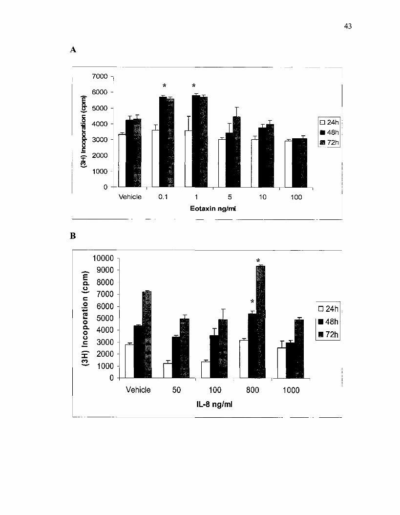
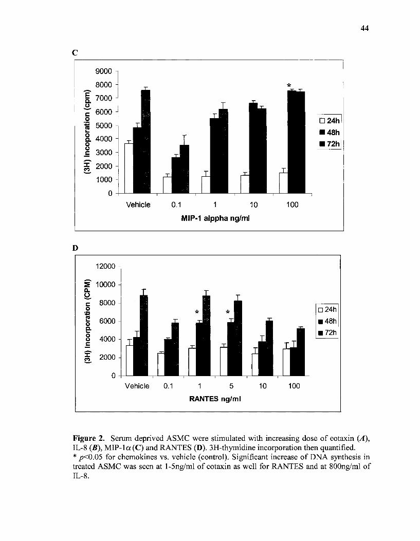
3.1 Effects of RANTES, eotaxin, IL-8 and MIP-la on fH)-thymidine Incorporation by ASMC
To investigate the effect of chemokines on 3H-thymidine incorporation, ASMC
were treated with different concentrations of RANTES, eotaxin, IL-8, and MIP-1a over
different periods of incubation. No increase in 3H-thymidine incorporation was detected
before 12 hours of incubation with any of the chemokines mentioned above. A significant
increase was detected after incubating ASMC with 0.1 and 1 ng/ml of eotaxin compared
to the vehicle as shown in fig 2.A. Incubation of cells with eotaxin for 48 hours induced
more proliferation than 24 hours incubation. There was a 1.2 fold increase in ASMC
numbers after treatment with either 1 or 5ng/ml of RANTES. In contrast, higher
concentrations of IL-8 were tested to see a similar effect; a 25.2% increase in ASMC
number was detected after the cells were stimulated with 800ng/ml of IL-8 with a
significant increase seen after incubation for 48 and 72 hours, see Fig. 2.B. MIP-1a
induced significant 3H-thymidine incorporation after 72 hours of incubation, compared to
the vehicle. The best results were obtained at a concentration of 100ng/ml ofMIP-1a.
PDGF treated smooth muscle cells were used as positive control. PDGF is a
strong mitogen of ASMC. A six to ten fold increase in the number of proliferating cells
was seen after PDGF treatment. These results are consistent with other studies that tested
PDGF mitogenesis. Whenever we used a combination of PDGF and a chemokine, a
synergistic effect could be seen.
43
A
7000
* * 6000 'ê fr - 5000
6 4000 D24h 16 8. .48h
8 3000 Ill 72h .E
2000 -::I: M - 1000
0 Vehicle 0.1 1 5 10 100
Eotaxin ng/ml
B
10000 * - 9000 E 8000 c. (.) - 7000 c:::: 0 - 6000 0 24h ca 5000 ...
•48h 0 c. 4000 0 1172h (.)
c:::: 3000 - 2000 :::I: M
1000 -0
Vehicle 50 100 800 1000
IL-8 ng/ml
c
D
9000
8000
'[ 7000 CJ
~ 6000 0 ~ 5000 ... 8. 4000 8 c 3000 -~ 2000 -
-:E a.. (J -c 0 ~
1! 0 e-8 c -::I:
M -
1000
0
12000
10000
8000
6000
4000
2000
0
44
Vehicle 0.1 1 10 100
MIP-1 alppha ng/ml
Vehicle 0.1 1 5 10 100
RANTES ng/ml
Figure 2. Serum deprived ASMC were stimulated with increasing dose of eotaxin (A), IL-8 (B), MIP-la (C) and RANTES (D). 3H-thymidine incorporation then quantified. * p<0.05 for chemokines vs. vehicle (control). Significant increase of DNA synthesis in treated ASMC was seen at 1-5ng/ml of eotaxin as well for RANTES and at 800ng/ml of IL-8.

45
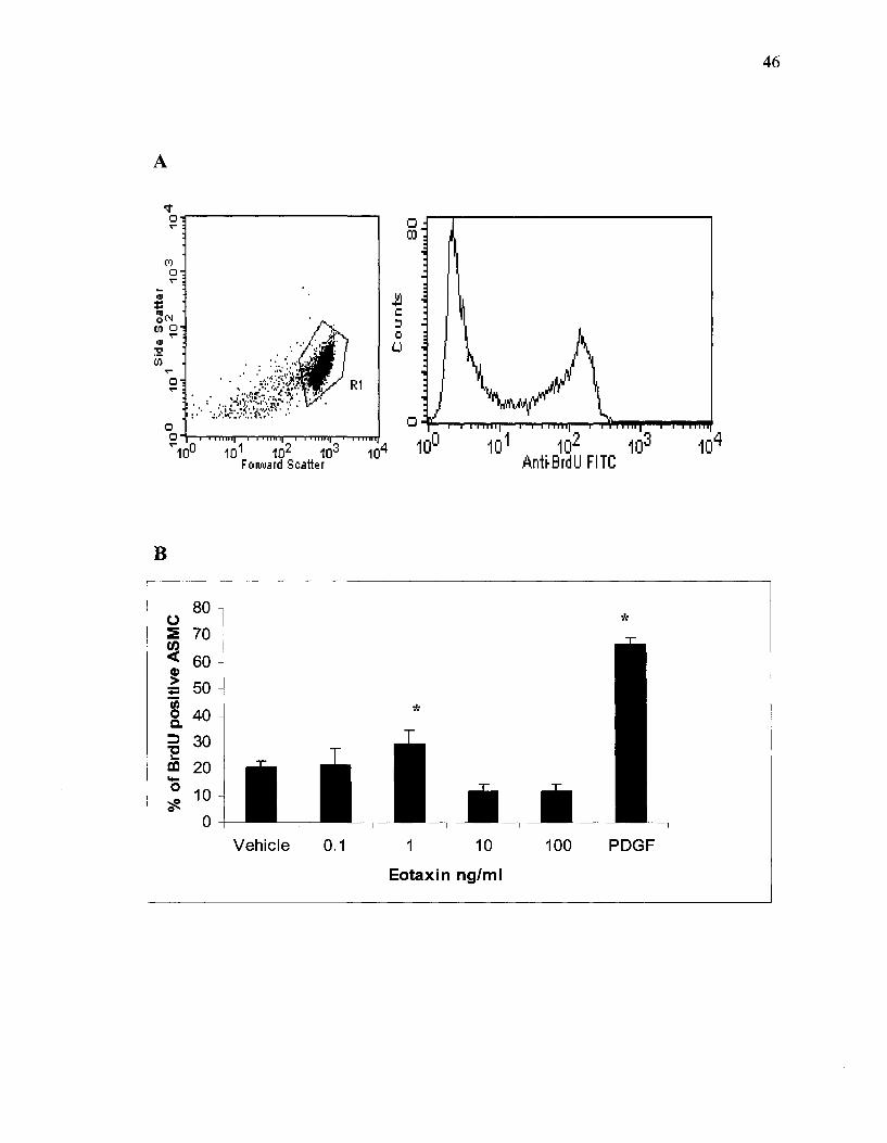
3.2 BrdU Incorporation by Proliferating ASMC
To further examine the effect of chemokines on ASMC proliferation and increase
in mass, the treated cells were stained with BrdU and anti-BrdU antibody. To quantify the
percentage of the proliferating and newly synthesizing DNA ASMC, we analyzed by flow
cytometer to measure the BrdU positive cells in each sample after different chemokines
treatment. The percentage of BrdU positive cells is compared to the vehicle (negative
control). PDGF treated cells were used as a positive control.
Our results show that eotaxin induces ASMC proliferation. ASMC that are treated
with low concentrations (1-5 ng/ml) of eotaxin showed increased BrdU incorporation;
29.2 % of eotaxin treated ASMC incorporated BrdU compared to 19.8% of ASMC
treated only with vehicle (P<0.002). Fig.3 shows the percentage ofBrdU positive ASMC
a:fter treatment with different concentrations of eotaxin over 24 hours. Incubating the
ASMC with eotaxin in excess of 24 hours did not yield significant results. The graph
represents the mean of 4 independent experiments. The error bars represent the standard
error of mean (SEM).
After 24 hours of incubation with RANTES, 24.3% of ASMC started to
synthesize new DNA compared to the vehicle only treated cells. In a concentration
dependent manner, MIP-la increased BrdU incorporation by ASMC a:fter 24 hours of
treatment.
A
~~----------------~ ....
M a ....
B
(.)
:E ~ G,)
> ~
ëii 0 Q. ;:) "0 1.. Ill -0 ~ 0
80
70
60
50
40
30
20
10
0 Vehicle 0.1
0 co
o~~~~~~~~~~~~~~
10°
1 10
Eotaxin ng/ml
102 Ant~BrdU FITC
*
100 PDGF
46

47
c
RANTES
j 50
40 ~ * ~ 30 ;; iii 20 0 Q.
::::1 10 'E al 0 ~ 0
Vehicle RANTES 1 RANTES 5
RANTES ng/ml
D
35
* :!!. 30 Qi u CP 25 >
~ 20 Q.
::::1 15 'C ... ra 10 'ë <fl. 5
0 Vehicle 10 100
M lp-1 alpha ng/m 1
Figure 3.A Chemokines induced DNA synthesis in airways SMC, determination of BrdU positive ASMC using flow cytometry. BrdU incorporation was measured after 24 hours of chemokine treatment. A: Flow cytometry plots of side scatter versus BrdU positive ASMC. B. Quantification of BrdU positive ASMC cells after treatment with different concentrations of eotaxin. At low concentrations, eotaxin induce the ASMC to proliferate. Data represent means ± SEM from four independent experiments. Significant difference for eotaxin treated cells vs. vehicle treated, P<O. 002 by t test. C: BrdU incorporation in RANTES treated airway Sacs. In comparison to the vehicle, RANTES treated cells had significant increase in BrdU positive ASM cells (P<O.OOl) by t test. D: An increase in DNA synthesis with the increase in MIP-1 alpha concentrations added to ASMC. A significant difference seen after stimulation with 10 and 100 ng/ml of MIP-1 alpha compared to the vehicle.
48
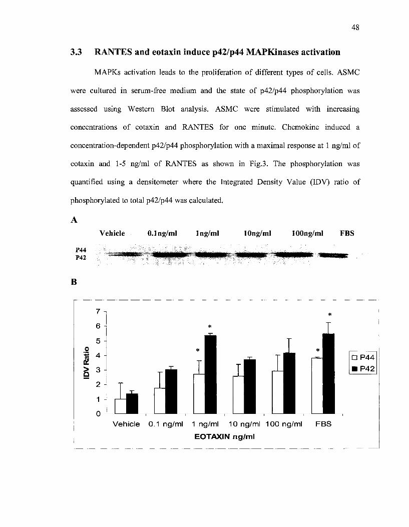
3.3 RANTES and eotaxin induce p42/p44 MAPKinases activation
MAPKs activation leads to the proliferation of different types of cells. ASMC
were cultured in serum-free medium and the state of p42/p44 phosphorylation was
assessed using Western Blot analysis. ASMC were stimulated with increasing
concentrations of eotaxin and RANTES for one minute. Chemokine induced a
concentration-dependent p42/p44 phosphorylation with a maximal response at 1 ng/ml of
eotaxin and 1-5 ng/ml of RANTES as shown in Fig.3. The phosphorylation was
quantified using a densitometer where the Integrated Density Value (IDV) ratio of
phosphorylated to total p42/p44 was calculated.
A
P44 P42
B
Vehicle O.lng/ml lng/ml
7
6
5
lOng/ml lOOng/ml FBS
*
0
~ ~ 4
fl 3 2
2
1
0
Vehicle 0.1 ng/ml 1 ng/ml 10 ng/ml 100 ng/ml FBS
EOTAXIN ng/ml
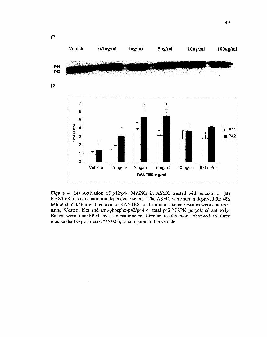
49
c Vehicle O.lng/ml lng/ml 5ng/ml lOng/ml lOOng/ml
P44 P42
D
7
6
5
* *
0
~~ = 4 ~ ~ 3 2
2
0
Vehicle 0.1 ng/ml 1 ng/ml 5 ng/ml 10 ng/ml 100 ng/ml
RANTES ng/ml
Figure 4. (A) Activation of p42/p44 MAPKs in ASMC treated with eotaxin or (B) RANTES in a concentration dependent manner. The ASMC were serum deprived for 48h before stimulation with eotaxin or RANTES for 1 minute. The celllysates were analyzed using Western blot and anti-phospho-p42/p44 or total p42 MAPK polyclonal antibody. Bands were quantified by a densitometer. Similar results were obtained in three independent experiments. * P<0.05, as compared to the vehicle.
50
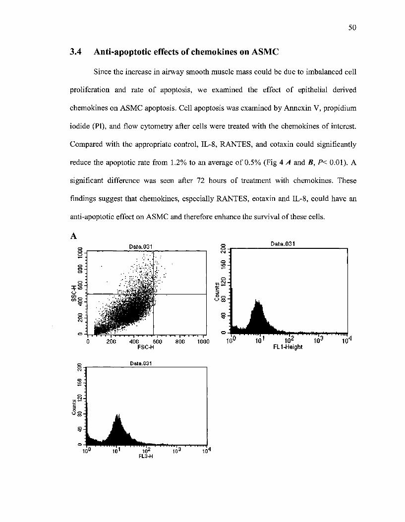
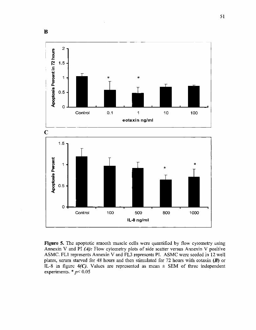
3.4 Anti-apoptotic effects of chemokines on ASMC
Since the increase in airway smooth muscle mass could be due to imbalanced cell
proliferation and rate of apoptosis, we examined the effect of epithelial derived
chemokines on ASMC apoptosis. Cell apoptosis was examined by Annexin V, propidium
iodide (PI), and flow cytometry after cells were treated with the chemokines of interest.
Compared with the appropriate control, IL-8, RANTES, and eotaxin could significantly
reduce the apoptotic rate from 1.2% to an average of 0.5% (Fig 4 A and B, P< 0.01). A
significant difference was seen after 72 hours of treatment with chemokines. These
findings suggest that chemokines, especially RANTES, eotaxin and IL-8, could have an
anti-apoptotic effect on ASMC and therefore enhance the survival ofthese cells.
A 0 0 0
0 0 0:0
0
::ç:~ (.)
(1)0 OOo
v
0 0 ('.J
0
0 0 N
0 ::e 0 N ....,_
;: "' 8~
0 v
0
Data.031
0 200 400 500 FSC-H
Data.031
800 1000
Data.031 g~------------------------~ ('.J
0 ..0
51
B
f 2 ;::, 0
.1:
~ 1.5 c
i 1 Cl) Il. Ill
·~ ë..
0.5
~ 0 Control 0.1 1 10 100
eotaxin ng/ml
c 1.5
.... c ~ 1 Cl) Il. Ill
l 0.5 0
ci"
0 Control 100 500 800 1000
IL-8 ng/ml
Figure 5. The apoptotic smooth muscle cells were quantified by flow cytometry using Annexin V and PI (A): Flow cytometry plots of side scatter versus Annexin V positive ASMC. FLl represents Annexin V and FL3 represents PI. ASMC were seeded in 12 well plates, serum starved for 48 hours and then stimulated for 72 hours with eotaxin (B) or IL-8 in figure 4(C). Values are represented as mean ± SEM of three independent experiments. * p< 0.05
52
Chapter 4. Discussion
The aims of this study were to show a specifie interaction between the airway
epithelium and the airway smooth muscle cells. The epithelium is a potent source of
different mediators and a significant number of them contribute to the rem ode ling seen in
the airway smooth muscle mass. The increase in smooth muscle mass in asthmatic
airways is a well documented structural change, especially in severe asthma. The
imbalance between the ASMC proliferation and the rate of apoptosis could be one
explanation for such increase in mass. Many studies on ASMC challenged the old concept
of treating ASMC as pure structural cells. lt has been shown that ASMC play an
immunoregulatory role in asthmatic airways through their ability to react to many
inflammatory mediators and secrete many more. Part of this interaction could be between
the ASMC and the epithelial derived mediators, specifically the chemokines.
Many recent studies show that chemokines could also have effects on structural
cells in addition to their classical role of leukocyte recruitment and activation. Airway
epithelium secretes RANTES, IL-8, eotaxin, MIP-la and many others. The receptors for
these chemokines have been recently identified on the ASMC. We have demonstrated the
expression of CCRl and CCR3 (Joubert 2005, Joubert 2008), as well as CXCRl and 2
(Govindaraju 2006) by human ASMC. In previous studies we also showed the migration
of ASMC toward eotaxin (Joubert 2005). Another study showed that IL-8 causes the
ASMC to contract and migrate (Govindaraju 2006). These observations, in addition to the
upregualtion of these chemokines in asthmatic airways, suggest that epithelial derived
chemokines could be implicated in the smooth muscle remodeling. Furthermore, they
53
could be implicated in the distance reduction between the airway epithelium and ASMC
mass seen in asthma. Evidence already exists implicating chemokine involvement in
angiogenesis and in cancer cell growth (Vaday 2006, Robinson SC 2003).
This study is not the first to show ASMC proliferation in vitro. ASMC
proliferation is considered as one possible mechanism responsible for the increase in
ASMC mass in asthma. To date, many growth factors, pro-inflammatory cytokines, and
contractile agonists such as histamine and endothelin-1 have been shown to promote
ASMC proliferation. The other possible mechanism is the decreased rates of apoptosis.
To our knowledge, this study is the first show the anti-apoptotic and mitogenic activity of
sorne epithelial derived chemokines on ASMC.
This study describes mrway smooth muscle proliferation in response to four
different chemokines. IL-8, RANTES, eotaxin, and MIP-1a are all up-regulated in
asthmatic airways. ASMC react to these chemokines by expressing functional receptors to
each one of them. RANTES and eotaxin induce ASMC proliferation at low
concentrations while higher doses of IL-8 were needed to demonstrate a similar response.
IL-8, predominantly a neutrophils chemoattractant, also interacts with the structural cells.
IL-8 causes the vascular and airway smooth muscle cells to migrate. Furthermore, it has
been shown that IL-8 is a mitogen for both human and rat aortic smooth muscle cells
(Yue TL 1994).
RANTES/ CCL5 is implicated in tumor growth; high levels ofRANTES correlate
with poor prognosis in breast cancer and small celllung carcinoma. RANTES is a known
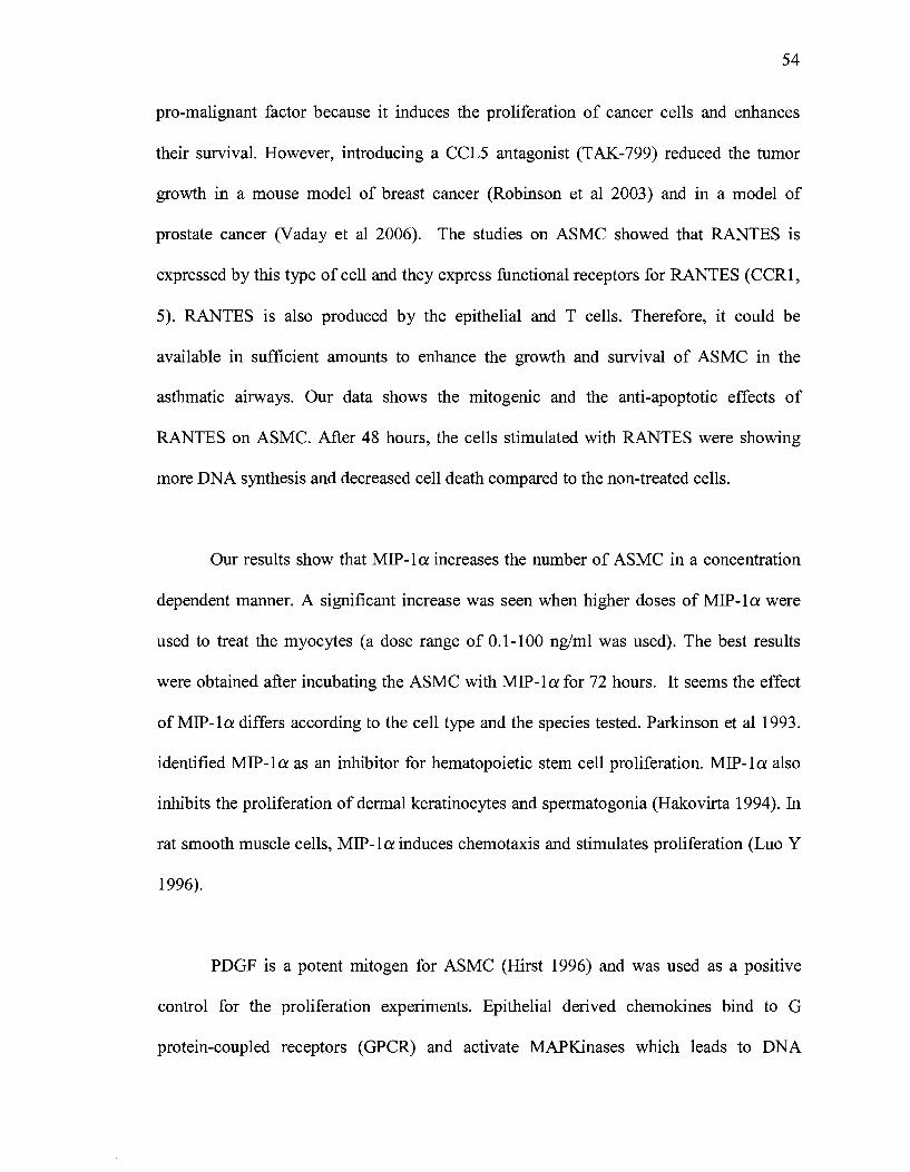
54
pro-malignant factor because it induces the proliferation of cancer cells and enhances
their survival. However, introducing a CCL5 antagonist (TAK-799) reduced the tumor
growth in a mouse model of breast cancer (Robinson et al 2003) and in a model of
prostate cancer (Vaday et al 2006). The studies on ASMC showed that RANTES is
expressed by this type of celland they express functional receptors for RANTES (CCRl,
5). RANTES is also produced by the epithelial and T cells. Therefore, it could be
available in sufficient amounts to enhance the growth and survival of ASMC in the
asthmatic airways. Our data shows the mitogenic and the anti-apoptotic effects of
RANTES on ASMC. After 48 hours, the cells stimulated with RANTES were showing
more DNA synthesis and decreased cell death compared to the non-treated cells.
Our results show that MIP-la increases the number of ASMC in a concentration
dependent manner. A significant increase was seen when higher doses of MIP-la were
used to treat the myocytes (a dose range of 0.1-100 ng/ml was used). The best results
were obtained after incubating the ASMC with MIP-la for 72 hours. It seems the effect
ofMIP-la differs according to the cell type and the species tested. Parkinson et al1993.
identified MIP-la as an inhibitor for hematopoietic stem cell proliferation. MIP-la also
inhibits the proliferation of dermal keratinocytes and spermatogonia (Hakovirta 1994). In
rat smooth muscle cells, MIP-1 a induces chemotaxis and stimula tes proliferation (Luo Y
1996).
PDGF is a potent mitogen for ASMC (Hirst 1996) and was used as a positive
control for the proliferation experiments. Epithelial derived chemokines bind to G
protein-coupled receptors (GPCR) and activate MAPKinases which leads to DNA
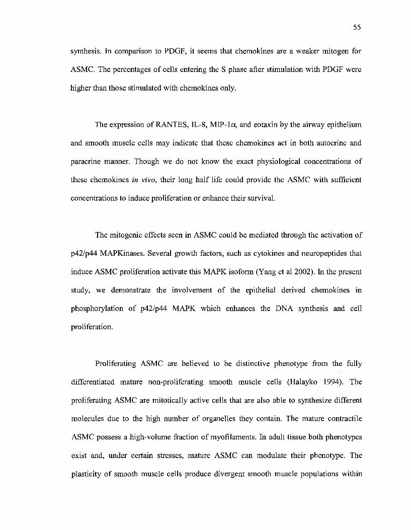
55
synthesis. In comparison to PDGF, it seems that chemokines are a weaker mitogen for
ASMC. The percentages of cells entering the S phase after stimulation with PDGF were
higher than those stimulated with chemokines only.
The expression ofRANTES, IL-8, MIP-la, and eotaxin by the airway epithelium
and smooth muscle cells may indicate that these chemokines act in both autocrine and
paracrine manner. Though we do not know the exact physiological concentrations of
these chemokines in vivo, their long half life could provide the ASMC with sufficient
concentrations to induce proliferation or enhance their survival.
The mitogenic effects seen in ASMC could be mediated through the activation of
p42/p44 MAPKinases. Several growth factors, such as cytokines and neuropeptides that
induce ASMC proliferation activate this MAPK isoform (Yang et al 2002). In the present
study, we demonstrate the involvement of the epithelial derived chemokines in
phosphorylation of p42/p44 MAPK which enhances the DNA synthesis and cell
proliferation.
Proliferating ASMC are believed to be distinctive phenotype from the fully
differentiated mature non-proliferating smooth muscle cells (Halayko 1994). The
proliferating ASMC are mitotically active cells that are also able to synthesize different
molecules due to the high number of organelles they contain. The mature contractile
ASMC possess a high-volume fraction of myofilaments. In adult tissue both phenotypes
exist and, under certain stresses, mature ASMC can modulate their phenotype. The
plasticity of smooth muscle cells produce divergent smooth muscle populations within
56
and between tissues (Halayko 1997). This may explain the small number of ASMC that
undergo proliferation in regard to the total number of cells used in a single test; another
reason could be the use of the primary cell cultures. Although using such cultures has the
advantage of functional and structural similarities to the in vivo environment, these
cultures are heterogeneous in nature and could be one source of variability in response
(Adler 2001). In the current study, both cell lines and primary cell cultures were
examined. The advantage of cell lines lies in the homogeneity of the cell population;
however, sorne phenotypic modulation seems unavoidable and using early passages
seems to matter in the studies of proliferation.
Apoptosis is the programmed cell death that regulates tissue integrity and
development. W e investigated the ability of the epithelial derived chemokines to
decrease the rate of ASMC apoptosis. It had already been reported that IL-8 delays the
spontaneous and TNF-a mediated neutrophils apoptosis in vitro (Kettritz et al 1998).
Blocking the CXCR2 receptor diminishes the effect partially. It is believed that IL-8
effect did not diminish completely because CXCR1 was not blocked (Glynn et al 2002).
Our results show similar effects on ASMC. Treating the cells with IL-8 reduces the
number of Annexin V positive cells compared to the control. After 24 hours of treatment,
no significant difference in the number of apoptotic ASMC was seen but extending the
incubation to 72 hours yielded a difference between the two. Adding to the IL-8 ability to
cause ASMC migration, we speculate that this CXC chemokines does play a significant
role in the ASMC increased mass in asthmatic airways. Keeping in mind that our
experiments were conducted on non-asthmatic ASMC and that smooth muscle cells from

57
asthmatic subjects proliferate at twice the rate seen in non asthmatic cells; then the IL-8
effect on asthmatic cells could be even greater.
The identification of new chemokine functions on the airways will contribute to a
better understanding of their role in immunomodulation and remodeling seen in asthma
and the other chronic inflammatory diseases. Their newly reported effects on ASMC may
involve them in the irreversible airway remodeling by enhancing the number of ASMC,
either by increasing the proliferation and/or delaying the apoptosis. Presently, ASMC are
considered a new target in asthma therapy and even though targeting ASMC may not be
the cure to asthma, it might be useful in relieving the acute symptoms related to airway
hyperreactivity. Smooth muscle contraction is the direct cause of airway narrowing and,
in addition, the increase in smooth muscle mass may cause excess airways narrowing in
asthma by increasing the force generated during airway contraction.
Controlling the airway smooth muscle remodeling has been studied intensively
and shown that glucocorticosteroids inhibit sorne but not all growth factor induced
proliferation of ASMC (Fernandes et al 1999). One study showed that corticosteroids
decrease the IL-8 production by epithelial cells IL-6 and TNF -a. These cytokines are
mitogens for ASMC; therefore, asthma patients might benefit from corticosteroids to treat
inflammation and to a lesser extent remodeling (Bergeron et al 2006). Combining
salbutamol to glucocorticosteroids might result in better reducing airway remodeling
(Kamachi et al 2001). Anti-IgE treatment could be used as an anti inflammatory but no
anti-remodeling benefits were reported. The same case applies for the anti-cytokines
treatments.
58
Our observations showed that RANTES, eotaxin, MIP-la and IL-8 increase the
number of ASMC. Those CC and CXC chemokines are also enhancing the survival of
ASMC. The treated cells are showing a decreased rate of apoptosis compared to the
none treated cells. These effects are mediated in part by MAPK activation. A
significant increase in phosphorylated p42/p44 MAPKs was seen after treating ASMC
with the chemokines of interest. On the basis of these findings, we speculate that
chemokines can influence ASMC mass by direct mechanisms.
59
Chapter 5. Future Directions
This study examined the interaction between the mrway epithelium and the
smooth muscle cells in terms of rem ode ling. The focus was to reveal the role of epithelial
derived chemokines in smooth muscle layer thickness reported in asthmatic airways and
to examine if these chemokines influence the distance shortening between the two layers.
The identification of epithelial derived chemokines', specifically IL-8, RANTES, eotaxin,
and MIP-la, mitogenic and anti-apoptotic effect on ASMC emphasize the involvement of
these small molecules in the remodeling seen in asthma in addition to their major role as
chemoattractant for the inflammatory cells. The study was performed on non-asthmatic
ASM cells; therefore, exploring the role of these chemokines on asthmatic cells will be
crucial to complete the picture and may expand this study by exploring the mechanisms
by which these chemokines induce proliferation and delay apoptosis in ASMC.

60
References
Adler KB, Li Y. Airway epithelium and mucus: intracellular signaling pathways for gene expression and secretion. Am J Respir Cell Mol Biol. 2001 Oct; 25(4):397-400.
Amin K, Janson C, Harvima I, Venge P, Nilsson G. CC chemokine receptors CCR1 and CCR4 are expressed on airway mast cells in allergie asthma. J Allergy Clin Immunol. 2005 Dec; 116(6):1383-6.
Amin K, Ludviksd6ttir D, Janson C, Nettelbladt 0, Bjomsson E, Roomans GM, Boman G, Sevéus L, Venge P. Inflammation and structural changes in the airways of patients with atopic and nonatopic asthma. BHR Group. Am J Respir Crit Care Med. 2000 Dec; 162(6):2295-301.
Amrani Y, Panettieri RA. Airway smooth muscle: contraction and beyond. Int J Biochem Cell Biol. 2003 Mar; 35(3):272-6.
Amrani Y, Panettieri RA Jr, Frossard N, Bronner C. Activation of the TNF alpha-p55 receptor induces myocyte proliferation and modulates agonist-evoked calcium transients in cultured human tracheal smooth muscle cells. Am J Respir Cell Mol Biol. 1996 Jul; 15(1):55-63.
Asher MI, Kiel U, Anderson HR, Beasley R, Crane J, Martinez F, Mitchell EA, Pearce N, Sibbald B, Stewart A W, et al. International Study of Asthma and Allergies in Childhood (ISAAC): rationale and methods. Eur Respir J 1995:8:483--493.
Azzawi M, Bradley B, Jeffery PK, Frew AJ, Wardlaw AJ, Knowles G, Assoufi B, Collins JV, Durham S, Kay AB. Identification of activated T lymphocytes and eosinophils in branchial biopsies in stable atopic asthma. Am Rev Respir Dis. 1990 Dec; 142(6 Pt 1):1407-13.
Baixeras E, Roman-Roman S, Jitsukawa S, Genevee C, Mechiche S, ViegasPequignot E, Hercend T, Triebel F. Cloning and expression of a lymphocyte activation gene (LAG-1). Mol Immunol. 1990 Nov;27(11):1091-102.
Baraldo S, Faffe DS, Moore PE, Whitehead T, McKenna M, Silverman ES, Panettieri RA Jr, Shore SA. Interleukin-9 influences chemokine release in airway smooth muscle: role ofERK. Am J Physiol Lung Cell Mol Physiol. 2003 Jun; 284(6):L1093-102. Jun; 284(6):L1093-102.
Bardi G. et al. 2001. The T cell chemokine receptor CCR7 is intemalized on stimulation with ELC, but not with SLC. Eur. J. Immunol. 31: 3291-3297.
Barnes PJ. New molecular targets for the treatment ofneutrophilic diseases. J Allergy Clin Immunol. 2007 May;119(5):1055-62; quiz 1063-4.
61
Benayoun L, Druilhe A, Dombret MC, Aubier M, Pretolani M. Aüway structural alterations selectively associated with severe asthma. Am J Respir Crit Care Med. 2003 May 15; 167(10):1360-8.
Bergeron C, Boulet LP. Structural changes in airway diseases: characteristics, mechanisms, consequences, and pharmacologie modulation. Chest. 2006 Apr; 129(4):1068-87.
Bergeron C, Pagé N, Joubert P, Barbeau B, Hamid Q, Chakir J. Regulation of procollagen 1 (alpha1) by interleukin-4 in human branchial fibroblasts: a possible role in airway remodeling in asthma. Clin Exp Allergy. 2003 Oct; 33(1 0): 1389-97.
Black JL, Burgess JK, Johnson PR. Airway smooth muscle--its relationship to the extracellular matrix. Respir Physiol Neurobiol. 2003 Sep 16; 137(2-3):339-46.
Black JL, Roth M, Lee J, CarlinS, Johnson PR. Mechanisms of airway remodeling. Airway smooth muscle. Am J Respir Crit Care Med. 2001 Nov 15; 164(10 Pt 2):S63-6.
Blyth DI, Pedrick MS, Savage TJ, Hessel EM, Fattah D. Lung inflammation and epithelial changes in a murine model of atopic asthma. Am J Respir Cell Mol Biol. 1996 May; 14(5):425-38.
Bonecchi R, Bianchi G, Bordignon PP, D'Ambrosio D, Lang R, Borsatti A, Sozzani S, Allavena P, Gray PA, Mantovani A, Sinigaglia F. Differentiai expression of chemokine receptors and chemotactic responsiveness of type 1 T helper cells (Th 1 s) and Th2s. J Exp Med. 1998 Jan 5;187(1):129-34.
Bousquet J, Jeffery PK, Busse WW, Johnson M, Vignola AM. Asthma: from bronchoconstriction to airways inflammation and remodeling. Am J Respir Crit Care Med 2000; 161:1720---1745.
Bradley BL, Azzawi M, Jacobson M, Assoufi B, Collins JV, Irani AM, Schwartz LB, Durham SR, Jeffery PK, Kay AB. Eosinophils, T-lymphocytes, mast cells, neutrophils, and macrophages in branchial biopsy specimens from atopic subjects with asthma: comparison with biopsy specimens from atopic subjects without asthma and normal control subjects and relationship to branchial hyperresponsiveness. J Allergy Clin Immunol. 1991 Oct; 88(4):661-74.
Campbell EM, Charo IF, Kunkel SL, Strieter RM, Boring L, Gosling J, Lukacs NW. Monocyte chemoattractant protein-1 mediates cockroach allergen-induced branchial hyperreactivity in normal but not CCR2-/- mice: the role of mast cells. J Immunol. 1999 Aug 15;163(4):2160-7.
62
Chan V, Burgess JK, Ratoff JC, O'connor BJ, Greenough A, Lee TH, Hirst SJ. Extracellular matrix regulates enhanced eotaxin expression in asthmatic aüway smooth muscle cells. Am J Respir Crit Care Med. 2006 Aug 15; 174(4):379-85.
Chantry D, Burgess LE. Chemokines in allergy. Curr Drug Targets Inflamm Allergy. 2002 Mar; 1(1):109-16.
Cheng SS, Lukacs NW, Kunkel SL. Eotaxin/CCL11 suppresses IL-8/CXCL8 secretion from human dermal microvascular endothelial cells. J Immunol. 2002 Mar 15; 168(6):2887-94.
Chetta, A., A. Foresi, M. Del Donno, G. F. Consigli, G. Bertorelli, A. Pesci, R. A. Barbee, and D. Olivieri. 1996. Branchial responsiveness to distilled water and methacholine and its relationship to inflammation and remodeling of the airways in asthma Am. J. Respir. Crit. Care Med. 53:910-917.
Cho JY, Miller M, Baek KJ, Han JW, Nayar J, Lee SY, McElwain K, McElwain S, Friedman S, Broide DH. Inhibition of airway remodeling in IL-S-deficient mice. J Clin Invest. 2004 Feb; 113(4):551-60
Chung KF, Bames PJ. Cytokines in asthma. Thorax. 1999 Sep; 54(9):825-57.
Clark-Lewis I, Kim KS, Rajarathnam K, Gong JH, Dewald B, Moser B, Baggiolini M, Sykes BD. Structure-activity relationships of chemokines. J Leukoc Biol. 1995 May; 57(5):703-11.
Conlon K, Lloyd A, Chattopadhyay U, Lukacs N, Kunkel S, Schall T, Taub D, Morimoto C, Osborne J, Oppenheim J, et al. CD8+ and CD45RA+ human peripheral blood lymphocytes are potent sources of macrophage inflammatory protein 1 alpha, interleukin-8 and RANTES. Eur J Immunol. 1995 Mar;25(3):751-6.
Conroy DM, Williams TJ. Eotaxin and the attraction of eosinophils to the asthmatic lung. Respir Res. 2001; 2(3):150-6. Epub 2001 Mar 29.
Cromwell 0, Hamid Q, Corrigan CJ, Barkans J, Meng Q, Collins PD, Kay AB: Expression and generation of interleukin-8, IL-6 and granulocyte macrophage colonystimulating factor by branchial epithelial cells and enhancement by IL-l beta and tumour necrosis factor-alpha. Immunology 1992; 77:330-337.
Cui CH, Adachi T, Oyamada H, Kamada Y, Kuwasaki T, Yamada Y, Saito N, Kayaba H, Chihara J. The role of mitogen-activated protein kinases in eotaxininduced cytokine production from branchial epithelial cells. Am J Respir Cell Mol Biol. 2002 Sep; 27(3):329-35.
D'Ambrosio D, Iellem A, Bonecchi R, Mazzeo D, Sozzani S, Mantovani A, Sinigaglia F. Selective up-regulation of chemokine receptors CCR4 and CCR8 upon activation ofpolarized human type 2 Th cells. J Immunol. 1998 Nov 15; 161(10):5111-5.
63
Davies RJ, Devalia JL. Air pollution and airway epithelial cells. Agents Actions Suppl. 1993; 43:87-96.
De S, Zelazny ET, Souhrada JF, Souhrada M. Interleukin-1 beta stimulates the proliferation of cultured airway smooth muscle cells via platelet-derived growth factorAm J Respir Cell Mol Biol. 1993 Dec;9(6):645-51.
Dunnill MS, Massarella GR, Anderson JA. A comparison of the quantitative anatomy of the bronchi in normal subjects, in status asthmaticus, in chronic bronchitis, and in emphysema. Thorax. 1969 Mar;24(2):176-9.
Eddleston J, Christiansen SC, Jenkins GR, Koziol JA, Zuraw BL. Bradykinin increases the in vivo expression of the CXC chemokine receptors CXCRl and CXCR2 in patients with allergie rhinitis. J Allergy Clin Immunol. 2003 Jan; 111(1):106-12.
Fahy JV. Rem ode ling of the airway epithelium in asthma. Am J Respir Crit Care Med. 2001 Nov 15; 164(10 Pt 2):S46-51.
Fahy JV, Corry DB, Boushey HA. Airway inflammation and remodeling in asthma. Curr Opin Pulm Med 2000; 6:15-20.
Farahi N, Cowbum AS, Upton PD, Deighton J, Sobolewski A, Gherardi E, Morrell NW, Chilvers ER. Eotaxin-1/CC Chemokine Ligand 11: A Novel Eosinophil Survival Factor Secreted by Human Pulmonary Artery Endothelial Cells. J Immunol. 2007 Jul 15; 179(2):1264-1273.
Fernandes D, Guida E, Koutsoubos V, Harris T, Vadiveloo P, Wilson JW, Stewart AG. Glucocorticoids inhibit proliferation, cyclin Dl expression, and retinoblastoma protein phosphorylation, but not activity of the extracellular-regulated kinases in human cultured airway smooth muscle. Am. J. Respir. Cell Mol. Biol. 1999; 21:77-88.
Fixman ED, Stewart A, Martin JG. Basic mechanisms of development of airway structural changes in asthma. Eur Respir J. 2007 Feb; 29(2):379-89.
Folkard SG, Westwick J, Millar AB. Production ofinterleukin-8, RANTES and MCP-1 in intrinsic and extrinsic asthmatics. Eur Respir J. 1997 Sep; 10(9):2097-104.
Fryer AD, Stein LH, NieZ, Curtis DE, Evans CM, Hodgson ST, Jose PJ, Belmonte KE, Fitch E, Jacoby DB. Neuronal eotaxin and the effects of CCR3 antagonist on airway hyperreactivity and M2 receptor dysfunction. J Clin Invest. 2006 Jan;116(1):228-36. Epub 2005 Dec 22.
64
Freyer AM, Johnson SR, Hall IP. Effects of growth factors and extracellular matrix on survival of human airway smooth muscle cells. Am J Respir Cell Mol Biol. 2001 Nov; 25(5):569-76.
Fujisawa T, Kato Y, Atsuta J, Terada A, lguchi K, Kamiya H, Yamada H, Nakajima T, Miyamasu M, Rirai K. Chemokine production by the BEAS-2B human branchial epithelial cells: differentiai regulation of eotaxin, IL-8, and RANTES by TH2- and TH1-derived cytokines. J Allergy Clin lmmunology. 2000 Jan; 105(1 Pt 1):126-33.
Gao JL, Kuhns DB, Tiffany HL, McDermott D, Li X, Francke U, Murphy PM. Structure and functional expression of the human macrophage inflammatory protein 1 alpha/RANTES receptor. J Exp Med. 1993 May 1;177(5):1421-7.
Geginat J, Campagnaro S, Sallusto F, Lanzavecchia A. TCR-independent proliferation and differentiation of human CD4+ T cell subsets induced by cytokines. Adv Exp Med Biol. 2002; 512:107-12.
Ghaffar 0, Hamid Q, Renzi PM, Allakhverdi Z, Molet S, Hogg JC, Shore SA, Luster AD, Lamkhioued B. Constitutive and cytokine-stimulated expression of eotaxin by human airway smooth muscle cells. Am J Respir Crit Care Med. 1999 Jun; 159(6):1933-42.
Glynn PC, Henney E, Hall IP. The selective CXCR2 antagonist SB272844 blocks interleukin-8 and growth-related oncogene-alpha-mediated inhibition of spontaneous neutrophil apoptosis. Pulmonary Pharmacology Therapeutics. 2002; 15(2): 103-1 O.
Godessart N. Chemokine receptors: attractive targets for drug discovery. Ann N Y Acad Sei. 2005 Jun; 1051:647-57.
Govindaraju V, Michoud MC, Al-Chalabi M, Ferraro P, Powell WS, Martin JG. Interleukin-8: novel roles in human airway smooth muscle cell contraction and migration. Am J Physiol Cell Physiol. 2006 Nov; 291(5):C957-65. Epub 2006 Ju15.
Griffiths-Johnson DA, Collins PD, Rossi AG, Jose PJ, Williams TJ. The chemokine, eotaxin, activates guinea-pig eosinophils in vitro and causes their accumulation into the lung in vivo. Biochem Biophys Res Commun. 1993 Dec 30;197(3):1167-72. Gutierrez-Ramos JC, Lloyd C, Kapsenberg ML, Gonzalo JA, Coyle AJ. Nonredundant functional groups of chemokines operate in a coordinate manner during the inflammatory response in the lung. lmmunol Rev. 2000 Oct;177:31-42.
Hackett TL, Knight DA. The role of epithelial injury and repair in the origins of asthma. Curr Opin Allergy Clin Immunol. 2007 Feb; 7(1):63-8.
Hakovirta H, Vierula M, Wolpe SD, Parvinen M. MIP-1 alpha is a regulator of mitotic and meiotic DNA synthesis during spermatogenesis. Mol Cell Endocrinol. 1994 Feb;99(1 ): 119-24.
65
Halayko AJ, Amrani Y: Mechanisms of inflammation-mediated airway smooth muscle plasticity and airways remodeling in asthma. Respir Physiol Neurobiol 2003, 137:209-222.
Halayko AJ, Rector E, Stephens NL. Characterization of molecular determinants of smooth muscle cell heterogeneity. Can J Physiol Pharmacology. 1997 Jul; 75(7):917-29.
Halayko AJ, Solway J: Molecular mechanisms of phenotypic plasticity in smooth muscle cells. J Appl Physiol2001, 90:358-368.
Halayko AJ, Stephens NL. Potential role for phenotypic modulation of branchial smooth muscle cells in chronic asthma. Can J Physiol Pharmacology. 1994 Nov; 72(11):1448-57.
Hamann KJ, Vieira JE, Halayko AJ, Dorscheid D, White SR, Forsythe SM, Camoretti-Mercado B, Rabe KF, Solway J. Fas cross-linking induces apoptosis in human airway smooth muscle cells. Am J Physiol Lung Cell Mol Physiol. 2000 Mar; 278(3):L618-24.
Harada A, Mukaida N, and Matsushima K. Interleukin 8 as a novel target for intervention therapy in acute inflammatory diseases. Mol Med Today 2: 482-489, 1996.
Hasaneen NA, Zucker S, Cao J, Chiarelli C, Panettieri RA, Foda HD. Cyclic mechanical strain-induced proliferation and migration of human airway smooth muscle cells: role ofENNPRIN and MMPs. FASEB J 2005; 19: 1507-1509.
Hirata N, Kohrogi H, Iwagoe H, Goto E, Hamamoto J, Fujii K, Yamaguchi T, Kawano 0, Ando M. Allergen exposure induces the expression of endothelial adhesion molecules in passively sensitized human bronchus: time course and the role of cytokines. Am J Respir Cell Mol Biol. 1998 Jan; 18(1):12-20.
Hirst SJ, Bames PJ, Twort CH. PDGF isoform-induced proliferation and receptor expression in human cultured airway smooth muscle cells. Am J Physiol. 1996 Mar; 270(3 Pt 1):L415-28.
Hirst SJ, Martin JG, Bonacci JV, Chan V, Fixman ED, Hamid QA, Herszberg B, Lavoie JP, McVicker CG, Moir LM, Nguyen TT, Peng Q, Ramos-Barb6n D, Stewart AG. Proliferative aspects of airway smooth muscle. J Allergy Clin Immunol. 2004 Aug; 114(2 Suppl):S2-17.
Holgate ST. Airway inflammation and remodeling in asthma: current concepts. Mol Biotechnol. 2002 Oct; 22(2):179-89.
66
Holgate ST, Bodey KS, Janezic A, Frew AJ, Kaplan AP, Teran LM. Release of RANTES, MIP-1 alpha, and MCP-1 into asthmatic airways following endobronchial allergen challenge. Am J Respir Crit Care Med. 1997 Nov; 156(5):1377-83.
Holgate ST, Lackie P, Wilson S, Roche W, Davies D. Bronchial epithelium as a key regulator of airway allergen sensitization and remodeling in asthma. Am J Respir Crit Care Med. 2000 Sep; 162(3 Pt 2):S113-7.
Hoshino M, Takahashi M, Aoike N. Expression of vascular endothelial growth factor, basic fibroblast growth factor, and angiogenin immunoreactivity in asthmatic airway and its relationship to angiogenesis. J. Allergy Clin. Immunol. 2001;107:295-301.
Hughes RA, Harris T, Altmann E, McAllister D, Vlahos R, Robertson A, Cushman M, Wang Z, Stewart AG. 2-Methoxyestradiol and analogs as novel antiproliferative agents: analysis of three-dimensional quantitative structure-activity relationships for DNA synthesis inhibition and estrogen receptor binding. Mol Pharmacol. 2002 May; 61(5):1053-69.
Jeffery P. Anti-inflammatory effects ofinhaled corticosteroids in chronic obstructive pulmonary disease: similarities and differences to asthma. Expert Opin Investig Drugs. 2005 May;14(5):619-32.
Jeffery PK, Wardlaw AJ, Nelson FC, Collins JV, Kay AB. Bronchial biopsies in asthma: an ultrastructural, quantitative study and correlation with hyperreactivity. Am Rev Respir Dis 1989; 140:1745-1753.
Johnson JR, Wiley RE, Fattouh R, Swirski FK, Gajewska BU, Coyle AJ, GutierrezRamos JC, Ellis R, lnman MD, Jordana M. Continuous exposure to house dust mite elicits chronic airway inflammation and structural remodelingAm J Respir Crit Care Med. 2004 Feb 1;169(3):378-85.
Johnson PR, Roth M, Tamm M, Hughes M, Ge Q, King G, Burgess JK, Black JL. Airway smooth muscle cell proliferation is increased in asthma. Am J Respir Crit Care Med. 2001 Aug 1;164(3):474-7.
Johnson Z. et al. 2004. Chemokine inhibition-why, when where, which and how? Biochem. Soc. Trans. 32: 366-377.
Jongejan R, De Jongste J, Raatgeep R, Stijnen T, Bonta I, Kerrebijn K. Effect of epithelial denudation, inflammatory mediators and mast cell activation on the sensitivity ofisolated human airways to methacholine. Eur J Pharmacol. 1991 Oct 15; 203(2): 187-94.
Joubert P, Hamid Q. Role of airway smooth muscle in airway remodeling. J Allergy Clin Immunol. 2005 Sep; 116(3):713-6.
67
Joubert P, Lajoie-Kadoch S, Labonté I, Gounni AS, Maghni K, Wellemans V, Chakir J, Laviolette M, Hamid Q, Lamkhioued B. CCR3 expression and function m asthmatic airway smooth muscle cells. J Immunol. 2005 Aug 15; 175(4):2702-8.
Joubert P, Lajoie-Kadoch S, Welman M, DragonS, Létuvée S, Tolloczko B, Halayko AJ, Gounni AS, Maghni K, Hamid Q.Joubert P, Lajoie-Kadoch S, Welman M, Dragon S, Létuvée S, Tolloczko B, Halayko AJ, Gounni AS, Maghni K, Hamid Q. Expression and regulation of CCR1 by airway smooth muscle cells in asthma. J Immunol. 2008 Jan 15;180(2):1268-75.
Kamachi A, Munakata M, Nasuhara Y et al. Enhancement of goblet cell hyperplasia and airway hyperresponsiveness by salbutamol in a rat model of atopic asthma. Thorax 2001; 56:19-24.
Kameyoshi Y, Dorschner A, Mallet AI, Christophers E, Schroder JM. Cytokine RANTES released by thrombin-stimulated platelets is a potent attractant for human eosinophils. J Exp Med. 1992 Aug 1;176(2):587-92.
Karpus WJ, Lukacs NW, Kennedy KJ, Smith WS, Hurst SD, Barrett TA. Differentiai CC chemokine-induced enhancement of T helper cell cytokine production. J Immunol. 1997 May 1;158(9):4129-36.
Karpus WJ, Kennedy KJ. MIP-1alpha and MCP-1 differentially regulate acute and relapsing autoimmune encephalomyelitis as well as Thl/Th2 lymphocyte differentiation. J Leukoc Biol. 1997 Nov;62(5):681-7.
Kettritz R, Gaido ML, Haller H, Luft FC, Jennette CJ, Falk RJ. Interleukin-8 delays spontaneous and tumor necrosis factor-alpha-mediated apoptosis of human neutrophils. Kidney International. 1998 Jan; 53(1):84-91.
Kicic A, Sutanto EN, Stevens PT, Knight DA, Stick SM. Intrinsic biochemical and functional differences in branchial epithelial cells of children with asthma. Am J Respir Crit Care Med. 2006 Nov 15; 174(10):1110-8.
Kirby JG, Hargreave FE, Gleich GJ, O'Byme PM. Bronchoalveolar cell profiles of asthmatic and non-asthmatic subjects. Am Rev Respir Dis 1987; 136:379-383.
Knight DA, Adcock JA, Phillips MJ, Thompson PJ. The effect of epithelium removal on human branchial smooth muscle responsiveness to acetylcholine and histamine. Pulm Pharmacol. 1990; 3(4):198-202.
Knox AJ, Pang L, Johnson S, HamadA. Airway smooth muscle function in asthma. Clin Exp Allergy. 2000 May; 30(5):606-14.
Koehler DR, Downey GP, Sweezey NB, Tanswell AK, Hu J. Lung inflammation as a therapeutic target in cystic fibrosis. Am J Respir Cell Mol Biol. 2004 Oct; 31(4):377-81.
68
Koya T, Takeda K, Kodama T, Miyahara N, Matsubara S, Balhom A, Joetham A, Dakhama A, Gelfand EW. RANTES (CCL5) regulates aüway responsiveness after repeated allergen challenge. Am J Respir Cell Mol Biol. 2006 Aug; 35(2):147-54.
Kunkel SL, Standiford T, Kasahara K, et al: Interleukin-8 (IL-8): The major neutrophil chemotactic factor in the lung. Exp Lung Res 1991; 17:17-23.
Laitinen LA, Heino M, Laitinen A, Kava T, Haahtela T. Damage of the aüway epithelium and branchial reactivity in patients with asthma. Am Rev Respir Dis 1985; 131:599-606.
Lazaar AL, Albelda SM, Pilewski JM, Brennan B, Puré E, Panettieri RA Jr. T lymphocytes adhere to aüway smooth muscle cells via integrins and CD44 and induce smooth muscle cell DNA synthesis. J Exp Med. 1994 Sep 1; 180(3):807-16.
Lazaar AL, Plotnick MI, Kucich U, Crichton 1, Lotfi S, Das SK, Kane S, Rosenbloom J, Panettieri RA, Schechter NM, PuréE. J Immunol. 2002 Jul15; 169(2):1014-20.
Lee JH, Johnson PRA, Hunt NH, Roth M, Black JL. Extracellular signal regulated kinase (ERK) activation is increased in smooth muscle cells from asthmatic patients. Am J Respir Crit Care Med 2001; 163: A269.
Lee JK, Park C, Kimm K, Rutherford MS. Genome-wide multilocus analysis for immune-mediated complex diseases. Biochem Biophys Res Commun. 2002 Jul 26; 295(4):771-3.
Li L, Xia Y, Nguyen A, Lai YH, Feng L, Mosmann TR, Lo D. Effects of Th2 cytokines on chemokine expression in the lung: IL-13 potently induces eotaxin expression by airway epithelial cells. J Immunol. 1999 Mar 1; 162(5):2477-87.
Li X, Wilson JW. Increased vascularity of the branchial mucosa in mild asthma. Am. J. Respir. Crit. Care Med. 1997; 156:229-233.
Lillard JW Jr, Boyaka PN, Taub DD, McGhee JR. RANTES potentiates antigenspecific mucosal immune responses. J Immunol2001; 166: 162-169.
Lloyd CM, Robinson DS. Allergen-induced airway remodeling. Eur Respir J. 2007 May; 29(5):1020-32.
Loetscher M, Gerber B, Loetscher P, Jones SA, Piali L, Clark-Lewis 1, Baggiolini M, Moser B. Chemokine receptor specifie for IP10 and mig: structure, function, and expression in activated T-lymphocytes. J Exp Med. 1996 Sep 1; 184(3):963-9.
Louahed J, Toda M, Jen J, Hamid Q, Renauld JC, Levitt RC, Nicolaides NC. Interleukin-9 upregulates mucus expression in the Am J Respir Cell Mol Biol. 2000 Jun; 22(6):649-56.

69
Lukacs NW, Oliveira SH, Hogaboam CM. Chemokines and asthma: redundancy of function or a coordinated effort? J Clin Invest. 1999 Oct; 104(8):995-9.
Lukacs NW, Standiford TJ, Chensue SW, Kunkel RG, Strieter RM, Kunkel SL. C-C chemokine-induced eosinophil chemotaxis during allergie airway inflammation. J Leukoc Biol. 1996 Nov; 60(5):573-8.
Ma X, Wang Y, Stephens NL. Serum deprivation induces a unique hypercontractile phenotype of cultured smooth muscle cells. Am J Physiol. 1998 May; 274(5 Pt 1):C1206-14.
Marfaing-Koka A, Devergne 0, Gorgone G, Portier A, Schall TJ, Galanaud P, Emilie D. Regulation of the production of the RANTES chemokine by endothelial cells. Synergistic induction by IFN-gamma plus TNF-alpha and inhibition by IL-4 and IL-13. J lmmunol. 1995 Feb 15; 154(4):1870-8.
Marshall MS. Ras target proteins in eukaryotic cells. FASEB J. 1995 Oct; 9(13):1311-8.
Masoli M, Fabian D, Holt S, Beasley R; Global Initiative for Asthma (GINA) Program. The global burden of asthma: executive summary of the GINA Dissemination Committee report. Allergy. 2004 May; 59(5):469-78.
Matsukura S, Kokubu F, lzumi H, Noda H, Kurokawa M, Tokunaga H, Adachi M. RANTES expression on human branchial epithelial cells. Arerugi. 1995 Jul; 44(7):715-7.
Matsunaga K, Yanagisawa S, Ichikawa T, Ueshima K, Akamatsu K, Hirano T, Nakanishi M, Yamagata T, Minakata Y, Ichinose M. Airway cytokine expression measured by means of protein array in exhaled breath condensate: correlation with physiologie properties in asthmatic patients. J Allergy Clin lmmunol. 2006 Jul; 118(1 ):84-90.
Maurer M, von Stebut E. Macrophage inflammatory protein-1. Int J Biochem Cell Biol. 2004 Oct; 36(1 0): 1882-6.
McWhinnie R, Pechkovsky DV, Zhou D, Lane D, Halayko AJ, Knight DA, Bai TR. Endothelin-1 induces hypertrophy and inhibits apoptosis in human airway smooth muscle cells. Am J Physiol Lung Cell Mol Physiol. 2007 Jan; 292(1):L278-86. Epub 2006 Aug 18.
Merrilees MJ, Campbell JH, Spanidis E, Campbell GR. Glycosaminoglycan synthesis by smooth muscle cells of differing phenotype and their response to endothelial cell conditioned medium. Atherosclerosis. 1990 Apr; 81(3):245-54.

70
Meyer-Hoffert U, Lezcano-Meza D, Bartels J, Montes-Vizuet AR, Schroder JM, Teran LM. Th2- and to a lesser extent Thl-type cytokines upregulate the production ofboth CXC (IL-8 and gro-alpha) and CC (RANTES, eotaxin, eotaxin-2, MCP-3 and MCP-4) chemokines in human airway epithelial cells. Int Arch Allergy Immunol. 2003 Aug; 131(4):264-71.
Mohamadzadeh M, Poltorak AN, Bergstressor PR, Beutler B, Takashima A. Dendritic cells produce macrophage inflammatory protein-1 gamma, a new member of the CC chemokine family. J Immunol. 1996 May 1;156(9):3102-6.
Montefort S, Roberts JA, Beasley R, Holgate ST, Roche WR. The site of disruption of the bronchial epithelium in asthmatic and non-asthmatic subjects. Thorax. 1992 Jul; 47(7):499-503.
Mullings RE, Wilson SJ, Puddicombe SM, Lordan JL, Bucchieri F, Djukanovié R, Howarth PH, Harper S, Holgate ST, Davies DE. Signal transducer and activator of transcription 6 (STA T -6) expression and function in asthmatic bronchial epithelium. J Allergy Clin Immunol. 2001 Nov; 108(5):832-8.
Munakata M. Airway remodeling and airway smooth muscle in asthma. Allergol Int. 2006 Sep; 55(3):235-43.
Naureckas ET, Ndukwu lM, Halayko AJ, Maxwell C, Hershenson MB, Solway J. Bronchoalveolar lavage fluid from asthmatic subjects is mitogenic for human airway smooth muscle. Am J Respir Crit Care Med. 1999 Dec; 160(6):2062-6
Nocker RE, Schoonbrood DF, van de GraafEA, Rack CE, Lutter R, Jansen HM, Out TA. Interleukin-8 in airway inflammation in patients with asthma and chronic obstructive pulmonary disease. Int Arch Allergy Immunol. 1996 Feb; 109(2):183-91.
Ochi H, Hirani WM, Yuan Q, Friend DS, Austen KF, Boyce JA. T helper cell type 2 cytokine-mediated co-mitogenic responses and CCR3 expression during differentiation ofhuman mast cells in vitro. J Exp Med. 1999 Ju119; 190(2):267-80.
Orlofsky A, Berger MS, Prystowsky MB. Novel expression pattern of a new member of the MIP-1 family of cytokine-like genes. Cell Regul. 1991 May;2(5):403-12.
Otsuka M, Y oshioka K. Neurotransmitter functions of mammalian tachykinins. Physiol Rev. 1993 Apr; 73(2):229-308.
Panettieri RA, Tan EM, Ciocca V, et al. Effects of LTD4 on human airway smooth muscle cell proliferation, matrix expression, and contraction in vitro: differentiai sensitivity to cysteinyl leukotriene receptor antagonists. Am J Respir Cell Mol Biol 1998; 19:453-61.
Panina-Bordignon P, Papi A, Mariani M, Di Lucia P, Casoni G, Bellettato C, Buonsanti C, Miotto D, Mapp C, Villa A, Arrigoni G, Fabbri LM, Sinigaglia F. The
71
C-C chemokine receptors CCR4 and CCR8 identify aitway T cells of allergenchallenged atopic asthmatics. J Clin Invest. 2001 Jun; 107(11):1357-64.
Parkinson EK, Graham GJ, Daubersies P, Burns JE, Heufler C, Plumb M, Schuler G, Pragnell lB. Hemopoietic stem cell inhibitor (SCI/MIP-1 alpha) also inhibits clonogenic epidermal keratinocytes proliferation. The Journal of Investigative Dermatology. 1993 Aug;101(2):113-7.
Payne DN, Rogers A V, Adelroth E, Bandi V, Guntupalli KK, Bush A, Jeffery PK. Early thickening of the reticular basement membrane in children with difficult asthma. Am J Respir Crit Care Med. 2003 Jan 1; 167(1):78-82.
Pepe C, Foley S, Shannon J, Lemiere C, Olivenstein R, Ernst P, Ludwig MS, Martin JG, Hamid Q. Differences in airway remodeling between subjects with severe and moderate asthma. J Allergy Clin Immunol. 2005 Sep; 116(3):544-9.
Pilette C, Francis JN, Till SJ, Durham SR. CCR4 ligands are up-regulated in the airways of atopic asthmatics after segmentai allergen challenge. Eur Respir J. 2004 Jun; 23(6):876-84.
Ponath PD, Qin S, Post TW, Wang J, Wu L, Gerard NP, Newman W, Gerard C, Mackay CR. Molecular cloning and characterization of a human eotaxin receptor expressed selectively on eosinophils. J Exp Med. 1996 Jun 1; 183(6):2437-48.
Pouysségur J, Seuwen K. Transmembrane receptors and intracellular pathways that control cell proliferation. Annu Rev Physiol. 1992; 54:195-210.
Regoli D, Boudon A, Fauchére JL. Receptors and antagonists for substance P and related peptides. Pharmacol Rev. 1994 Dec; 46(4):551-99.
Remick DG. Interleukin-8. Crit Care Med 33: S466-S467, 2005.
Reynold A. Panettieri, Jr MD. Airway smooth muscle: An immunomodulatory cell. Journal of Allergy and Clinical Immunology. December 2002; 110(6) Part 2: S269-274.
Richman-Eisenstat JB, Jorens PG, Hebert CA, Ueki 1, and Nadel JA. Interleukin-8: an important chemoattractant in sputum of patients with chronic inflammatory airway diseases. Am J Physiol Lung Cell Mol Physio1264: L413-L418, 1993.
Riffo-Vasquez Y, Spina D. Role of cytokines and chemokines in branchial hyperresponsiveness and airway inflammation. Pharmacol Th er. 2002 Jun; 94(3): 185-211.
Robinson DS, Hamid Q, Ying S, Tsicopoulos A, Barkans J, Bentley AM, Corrigan C, Durham SR, Kay AB. Predominant TH2-like bronchoalveolar T-lymphocyte population in atopic asthma N Engl J Med. 1992 Jan 30;326(5):298-304.
72
Robinson SC, Scott KA, Wilson JL, Thompson RG, Proudfoot AE, Balkwill FR. A chemokine receptor antagonist inhibits experimental breast tumor growth Cancer Res. 2003 Dec 1;63(23):8360-5.
Roche WR, Beasley R, Williams JH, Holgate ST. Subepithelial fibrosis in the bronchi ofasthmatics Lancet. 1989 Mar 11;1(8637):520-4.
Romagnani S. Cytokines and chemoattractant in allergie inflammation. Mol Immunol. 2002 May; 38(12-13):881-5.
Rot A, Krieger M, Brunner T, Bischoff SC, Schall TJ, Dahinden CA. RANTES and macrophage inflammatory protein 1 alpha induce the migration and activation of normal human eosinophil granulocytes. J Exp Med. 1992 Dec 1;176(6):1489-95.
Rush JS, Hasbold J, Hodgkin PD. Cross-linking surface Ig delays CD40 ligand- and IL-4-induced B cell Ig class switching and reveals evidence for independent regulation of B cell proliferation and differentiation. J Immunol. 2002 Mar 15; 168(6):2676-82.
Salary H, Schellenberg R R: Stimulation of human airway epithelial cells by platelet activating factor (PAF) and arachidonic acid produces 15-hydroxyeicosatetraenoic acid (15-HETE) capable of contracting branchial smooth muscle. Pulm Pharmacol 4:1-7, 1991
Sallusto F, Mackay CR, Lanzavecchia A. Selective expression of the eotaxin receptor CCR3 by humanT helper 2 cells. Science. 1997 Sep 26; 277(5334):2005-7.
Sanderson CJ. Interleukin-5: an eosinophil growth and activation factor. Dev Biol Stand. 1988; 69:23-9.
Schall TJ, Bacon K, Toy KJ, Goeddel DV. Selective attraction of monocytes and T lymphocytes of the memory phenotype by cytokine RANTES. Nature 1990; 347:669-671.
Schall TJ. Biology of the RANTES/SIS cytokine family. Cytokine 1991;3: 165-183.
Shore SA. Direct effects of Th2 cytokines on airway smooth muscle. Curr Opin Pharmacol. 2004 Jun; 4(3):235-40.
Shore SA, Moore PE. Effects of cytokines on contractile and dilator responses of airway smooth muscle. Clinical and Experimental Pharmacology and Physiology. 2002 May; 29, 859-866.
Shute J. Interleukin-8 is a potent eosinophil chemo-attractant. Clin Exp Allergy 24: 203-206, 1994.
73
Smith D, Burrows L, Westwick J. Interleukin-8--a mediator of inflammatory lung disease? Adv Exp Med Biol. 1991;305:119-26.
Stellato C, Collins P, Ponath PD, Soler D, Newman W, La Rosa G, Li H, White J, Schwiebert LM, Bickel C, Liu M, Bochner BS, Williams T, Schleimer RP. Production of the novel C-C chemokine MCP-4 by airway cells and comparison of its biological activity to other C-C chemokines. J Clin In v est. 1997 Mar 1; 99( 5):926-36.
Stellato C, Brummet ME, Plitt JR, Shahabuddin S, Baroody FM, Liu MC, Ponath PD, Beek LA. Expression of the C-C chemokine receptor CCR3 in human airway epithelial cells. J Immunol. 2001 Feb 1; 166(3):1457-61.
Stewart AG, Bonacci N, Quan L. Factors controlling airway smooth muscle proliferation in asthma. Curr Allergy Asthma Rep. 2004 Mar; 4(2):109-15.
Taub DD, Ortaldo JR, Turcovski-Corrales SM, Key ML, Longo DL, Murphy WJ. Beta chemokines costimulate lymphocyte cytolysis, proliferation, and lymphokine production. J Leukoc Biol1996; 59:81-89.
Teran LM. CCL chemokines and asthma. Immunology Today. 2000 May; 21(5):235-42.
Teran LM, Davies DE. The chemokines: their potential role in allergie inflammation. Clinicat and Experimental Allergy. 1996 Sep; 26(9):1005-19.
Teran LM, Mochizuki M, Bartels J, Valencia EL, Nakajima T, Rirai K, Schroder JM. Th1- and Th2-type cytokines regulate the expression and production of eotaxin and RANTES by human 1ung fibroblasts. Am J Respir Cell Mol Biol. 1999 Apr; 20(4):777-86.
Tran T, McNeill KD, Gerthoffer WT, Unruh H, Halayko AJ. Endogenous 1aminin is required for human airway smooth muscle cell maturation. Respir Res. 2006 Sep 12; 7:117.
Van der Velden VH, Verheggen MM, Bemasconi S, Sozzani S, Naber BA, van der Linden-van Beurden CA, Hoogsteden HC, Mantovani A, Versnel M. Interleukin-1 beta and interferon-gamma differentially regulate release of monocyte chemotactic protein-1 and interleukin-8 by human bronchial epithelial cells. Eur Cytokine Netw. 1998 Sep; 9(3):269-77.
Vaday GG, Peehl DM, Kadam PA, Lawrence DM. Expression of CCL5 (RANTES) and CCR5 in prostate cancer. Prostate. 2006 Feb 1; 66(2): 124-34.
Vignola AM, Campbell AM, Chanez P, Lacoste P, Michel FB, Godard P, Bousquet J. Activation by histamine ofbronchial epithelial cells from nonasthmatic subjects. Am JRespirCellMo1Bio1.1993 Oct; 9(4):411-7.
74
Wang JM, Rambaldi A, Biondi A, Chen ZG, Sanderson CJ, Mantovani A. Recombinant human interleukin 5 is a selective eosinophil chemoattractant. Eur J Immunol. 1989 Apr; 19(4):701-5.
Wolpe SD, Cerami A. Macrophage inflammatory proteins 1 and 2: members of a novel superfamily of cytokines. FASEB J. 1989 Dec;3(14):2565-73.
www. bdbiosciences.com/features/products/ display __product. php ?keyiD=48
Yahiaoui L, Villeneuve A, Valderrama-Carvajal H, Burke F, Fixman ED. Endothelin-1 regulates proliferative responses, both alone and synergistically with PDGF, in rat tracheal smooth muscle cells. Cell Physiol Biochem. 2006; 17(1-2):37-46.
Yamada H, Rirai K, Miyamasu M, Iikura M, Misak:i Y, Shoji S, Takaishi T, Kasahara T, Morita Y, Ito K. Eotaxin is a potent chemotaxin for human basophils. Biochem Biophys Res Commun. 1997 Feb 13;231(2):365-8.
Yang CM, Hsiao LD, Chien CS, Lin CC, Luo SF, Wang CC. Substance P-induced activation of p42/44 mitogen-activated protein kinase associated with cell proliferation in human tracheal smooth muscle cells. Cell Signal. 2002 Nov; 14(11):913-23.
Yi Luo, Patricia A. D'Amore, and Martin E. Dorf. P-Chemokine TCA3 Binds to and Activates Rat Vascular Smooth Muscle Cells. The journal oflmmunology, 1996, 157: 2143-2148.
Ying S, Meng Q, Zeibecoglou K, Robinson DS, Macfarlane A, Humbert M, Kay AB. Eosinophil chemotactic chemokines (eotaxin, eotaxin-2, RANTES, monocyte chemoattractant protein-3 (MCP-3), and MCP-4), and C-C chemokine receptor 3 expression in branchial biopsies from atopic and nonatopic (Intrinsic) asthmatics. J Immunol. 1999 Dec 1; 163(11):6321-9.
Ying S, Robinson DS, Meng Q, Rottman J, Kennedy R, Ringler DJ, Mackay CR, Daugherty BL, Springer MS, Durham SR, Williams TJ, Kay AB. Enhanced expression of eotaxin and CCR3 mRNA and protein in atopic asthma. Association with airway hyperresponsiveness and predominant co-localization of eotaxin mRNA to branchial epithelial and endothelial cells. Eur J lmmunol. 1997 Dec;27(12):3507-16.
Yokota T, Otsuka T, Mosmann T, Banchereau J, DeFrance T, Blanchard D, De Vries JE, Lee F, Arai K. Isolation and characterization of a human interleukin eDNA clone, homologous to mouse B-cell stimulatory factor 1, that expresses B-cell- and T-cellstimulating activities. Proc Natl Acad Sei US A. 1986 Aug;83(16):5894-8.
Yue TL, Wang X, Sung CP, Oison B, McKenna PJ, Gu JL, Feuerstein GZ. Interleukin-8. A mitogen and chemoattractant for vascular smooth muscle cells. Circulation Research. 1994 Jul; 75(1):1-7.
75
Zahm JM, Chevillard M, Puch elle E. W ound repair of human surface respiratory epithelium. Am J Respir Cell Mol Biol. 1991 Sep;5(3):242-8.
Zimmermann N, Hershey GK, Foster PS, Rothenberg ME. Chemokines in asthma: cooperative interaction between chemokines and IL-13. J Allergy and Clinical Immunology. 2003 Feb; 111(2):227-42.
Zlotnik A, Y oshie 0: Chemokines: A new classification system and their role in immunity. Immunity 2000; 12:121-127.
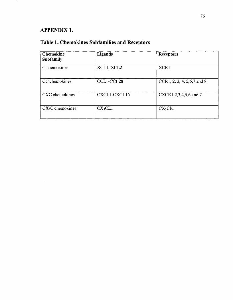
76
APPENDIX 1.
Table 1. Chemokines Subfamilies and Receptors
Chemokine Ligands Receptors Subfamily
C chemokines XCLl, XCL2 XCRl
CC chemokines CCL1-CCL28 CCRl, 2, 3, 4, 5,6,7 and 8
CXC chemokines CXCL1-CXCL16 CXCR1,2,3,4,5,6 and 7
cx3c chemokines CX3CLl CX3CR1

77
APPENDIX2.
Ethical Approval for the use ofhuman cells in the project. Documents attached.