relation between microstructure, destabilization phenomena
TRANSCRIPT

Food Structure Food Structure
Volume 4 Number 2 Article 6
1985
Relation Between Microstructure, Destabilization Phenomena and Relation Between Microstructure, Destabilization Phenomena and
Rheological Properties of Whippable Emulsions Rheological Properties of Whippable Emulsions
W. Buchheim
N. M. Barfod
N. Krog
Follow this and additional works at: https://digitalcommons.usu.edu/foodmicrostructure
Part of the Food Science Commons
Recommended Citation Recommended Citation Buchheim, W.; Barfod, N. M.; and Krog, N. (1985) "Relation Between Microstructure, Destabilization Phenomena and Rheological Properties of Whippable Emulsions," Food Structure: Vol. 4 : No. 2 , Article 6. Available at: https://digitalcommons.usu.edu/foodmicrostructure/vol4/iss2/6
This Article is brought to you for free and open access by the Western Dairy Center at DigitalCommons@USU. It has been accepted for inclusion in Food Structure by an authorized administrator of DigitalCommons@USU. For more information, please contact [email protected].

FOOD MICROSTRUCfURE, Vol. 4 (1985) , pp. 221-232 SEM Inc. , AMF O'Hare (Chicago) , IL 60666-0507 U.S.A.
07 30-5419/ 85$ 1.00+.05
RELATION BETWEEN MICROSTRUCTURE, DESTABILIZATION PHENOMENA AND RHEOLOGICAL PROPERTIES OF WHIPPABLE EMULSIONS
W. Buchheim , N .M. Barfod ,* and N . Krog*
Bundesanstalt fUr Milchforschung, D-2300 Kiel , West Germany *Grindsted Products, DK-8220 Brabrand. Denmark
Abstract
The structure of spray-dried whippable emulsions (toppings) containing different types of lipid surfactants, was investigated by electron microscopy using the freeze-fracture technique. The size distribution of the lipid particles within the powders varied with the type of the surfactant used. After reconstitution of the topping powders in water. a strong destabilization phenomenon took place to an extent depending on the type of the surfactant. Simultaneously a crystall ization of coalesced lipid particles occurred along with an increase in viscosity of the emulsions. The degree of crystallization was measured by p-NMR . It has been concluded that these phenomena are closely related to whippability and foam firmness.
The structure of whipped topping emulsions (foam) is characterized by the presence of large lipid crystals at the surface of air bubbles . This structure is different from the structure of whipped liquid (imitation) cream or dairy cream , where the air bubbles are predominantly stabilized by agglomerated fat globules from which the surface membrane has been partly removed during the whipping process .
Initial paper received February 13 1985 Manuscript received August 04 1985 Direct inquiries to N. Krog Telephone number: 45-6-25 33 66
Key Words: Transmission electron microscopy, freeze-fracture technique, pulsed-nuclear magnetic resonance , X-ray diffraction, viscosity, destabilization of emulsions, imitation cream , topping emulsions, whippable emulsions , surfactants .
227
Introduction
The structure of whipped dairy cream has been investigated by Buchheim (2) who demonstrated by electron microscopy that the air bubbles in the foam are surrounded by a layer of fat globules which partially protrude into the air bubbles. Furthermore it was observed that the adsorbed fat globules had lost their protective membrane layers and that liquid fat was present in open spaces between solid fat globules.
Schmidt and van Hooydonk (5) used scanning electron microscopy to investigate the effect of homogenization on dairy cream and confirmed the structure described by Buchheim (2).
Whereas Transmission Electron Microscopy (TEM) and Scanning Electron Microscopy (SEM) studies on whipped dairy cream demonstrate the microstructure of dairy cream , no such investigations have yet been carried out on imitation cream or powdered toppings. It is well known that reconstituted topping emulsions show an increase in viscosity related to desired whippability, and that only certain types of surfactants give good foam properties (I) . It has so far been anticipated that the whipping properties of topping emulsions are partially due to an agglomeration of the lipid droplets in the emulsions.
Since spray-dried , vegetable-fat based , whippable emulsions are being widely accepted in resemblance to dairy cream and offer advantages in cost , transportation and long term storage, it has been decided to study the structural characteristics of these systems in detail.
Materials and Methods
Preparations of topping powders and liquid imitation creams The topping powders were made by spray-drying an emulsion
containing 24-26 % hydrogenated coconut oil with a final melting point (m.p.) of 3l °C, 4-6 % surfactant , 16% maltodextrin , 4% sodium caseinate, and 50 % water. The emulsions were prepared by melting coconut fat and surfactant together at 80°C and separately dissolving the caseinate and maltodextrin in the water phase at 90°C. The two phases were then mixed together and homogenized on a one-stage high-pressure piston homogenizer with a Rannie liquid whirling type valve at a pressure of 100 kg/cm2 at 80°C. The emulsion was then spray-dried, using a rotating atomizer (16,000 rpm) , with an air inlet temperature of 150°C and an outlet temperature of 90°C. The spray-dried topping powder was cooled to soc for one hour and then stored at 20°C or at a lower temperature.

W. Buchhei m , N.M. Barfod and N. Krog
The follow ing sur fac tants were used: Di still ed propylene glyco l monostea ratc (PG MS), di still ed monoglycerides based on parti ally hyd rogenated soy bean o il (GM O), and di stilled monoglycerides based on full y hydrogenated soy bean o il (GMS). The surfac tants conta ined a minimum of 90 % monoeste rs and are commerc ial prod ucts manufactured by G rindsted Products A/S, Denmark hav ing the fo llow ing trade names: Promodan SP (PGMS), Dimodan 0 (GMO), and Dimodan PV (GMS). A topping powder based on coconut fa t , sodium caseinate, and maltadextrin and conta ining no surfactant was used as a refe rence.
For compari son an ultra- high temperature (UHT) treated li quid , vegetable-fat based cream (imitati on cream), was included in the experiments. It contained 29.4 % hydrogenated palm kernel o il (m .p. 36°C), 0.6 % lac tylated monoglycerides (Lactodan P 22, manu fac tured by G rindsted Products A/S, Denmark), O.OS % sodium alg inate, I% suc rose and 68.9S % sk im milk . The fa t and surfacta nt we re melted togethe r at 7S °C, and added to the skim milk in which sodium a lg inate and suc rose had been di ssolved with ag itating during heating to 7S 0 C. The mi xture was first homogenized at a low pressure (IS kg/cm2) to make a fairl y stable emulsion. then UHT-treated (144°C, 4 s) fo llowed by a second homogeni zati on at ISO kg/cm2, and finall y cooled to soc. Viscosity mea surements
The topping powders we re reconstituted in water at IS°C by mi xing I pa rt of powder with 3 pa rts of water. The emuls ion was then transferred to the cup of a Haake Rotov isco Viscometer with sensor system MVII. The temperature in the senso r cup was thermostatica lly controlled at IS0
, 18° or 20°C. The viscos ity of the emul s ion was measured continuously at a shea r rate of 1.76 s - I as a functi on of time up to two hours. Whipping test and foam texture m ea surements
One pa rt of the topp ing powder. was reconstituted in three parts of water at l0 °C and IS°C and whipped w ith a Kenwood Chief Mi xe r at max imum speed fo r 2 o r 3 min depending on the type of emuls ifie r used. The whipped c ream was then tested with a Voland Stevens Texture Analyzer using a cy lindrical probe (TA S) 12.7 mm in di amete r, adjusted to a penetration depth of 10 mm at a speed of 2 mm s - I and using a load range position 100 g. Crystallization measurements
The ex tent of c rysta llizati on of the fat phase was analyzed by measuring the solid fa t content (SFC) by a pulsed-w ide- line proton NMR technique (6) in reconstituted emuls ions using deute rated wate r (D20 ) instead of normal wate r.
Fat crystal modifications were dete rmined at 22 oc by X-ray diffracti on ana lys is us ing an Enraf- Nonius Guinie r-De Wolff Camera and a Philips X- ray gene rato r w ith a Cu-anode as Xray source. (Wavelength = O. IS4 nm). Transmission electron microscopy
The topping powders, the reconstituted systems, the liquid imitation cream , and the whipped systems were prepared for electron microscopy by the freeze-fracture technique us ing a BALZERS BA 360 M unit. Prior to cryofi xation the va rious types of samples were treated as follows: I) The topping powders were suspended in anhydrous glycerol as described earlie r (3). 2) The powders were reconstituted in wate r (1:3) at lS 0 C. These emul sions as well as the liquid imitation c ream were cryoprotected by mi xing with glycerol to a fina l concentration of 30 % (v/v). 3) The whipped systems were not c ryoprotected , but the freezefracture spec imen holders we re cove red with a small droplet
222
of g lyce ro l in o rder to achieve a better adhes io n of the foam. Sma ll volumes (1 - 2 mm3) of the various samples were trans
fe rred onto no rmal freeze- fracture spec ime n holders (Ba lzers), cryofi xed by immers ion into melting Freon 22 ( - 160°C) and stored under liquid nit rogen . Freeze-fracturing was carried out at - 120°C, fo llowed immediately by replication (ca 1.7 nm plat inum/ca rbon under an ang le of 4S degrees fo llowed by ca 20 nm of pure ca rbo n) us ing an electron gun .
T he replicas were c leaned fo r I h with a S% sodium hypochlo rite solution followed by acetone. Distilled water baths were used as intermediate steps. Electron mic rog raphs were ta ken with a Philips EM 301 e lectron mic roscope at 80 kY.
Results
E lectron microscopy Topping powders - By freeze-fracturing suspensions of spray
dr ied powders in non-aqueous media deta il ed in fo rmation can be obtained about the surface as well as about the inte rnal structure of powder particles (3). In the fo llowing text , results a re reported exclus ively on the inte rnal structure, i.e. , the size and appea rance of the lipid droplets because these aspects are of re levance fo r the phys ical phenomena desc ribed in thi s pape r. Characte ristic views of the fine structure of the powder matrices are g iven for the 4 types of topping powders studied .
F ig. Ia shows a powder which conta ined PGMS (Promodan SP) as the surfactant. The lipid droplets a re mo re o r less globular, and the ir diamete rs vary between ca 0.1 and I.S p.m. They appea r to be ve ry densely packed in the powder matri x, therema ining space between them is represented by maltodextrin and sod ium caseinate. The lipid droplets are sometimes cleaved near the ir pe riphe ry show ing irregularly shaped c rysta lline plate lets, but more often they are cleaved inte rnall y (Fig. lb) . The very smooth appea rance of the fracture planes can be desc ribed as cleaved crystalline plate lets running like di scs through the entire d roplets.
The topping powder conta ining GMS (Dimodan PV) is shown in Fig. 2a. The lipid d roplets are again of mostly globular shape but of di stinctl y mo re uniform s ize of approx imate ly O.S p.m .
Fig. 1. Topping powder with PGMS (Promodan SP). (a) Internal structure of a cleaved powder particle showing the dense packaging of lipid droplets (L). Bar = lp.m. (b) Detailed view demonstrating the occurrence of peripheral (A) and internal (B) cleavages of individual lipid droplets. C: caseinate/maltodextrin matrix. Bar = 0.25 p.m.
Fig. 2. Topping powder with GMS (Dimodan PV). (a) Internal structure of a cleaved powder particle. The lipid droplets are smaller and of distinctly more uniform size as compared to the topping powder with PGMS (Fig. 1). Bar = 1 p.m. (b) Detailed view. Bar = 0.25 p.m.
Fig. 3. Topping powder with GMO (Dimodan 0). (a) Internal structure of a cleaved powder particle. The sizes and shapes of the lipid droplets resemble those in the topping powder with GMS (Fig. 2). Bar = lp.m. (b) Detailed view. Internally cleaved lipid droplets (A) exhibit very often a smooth structure originating from a disc-like crystallization of the lipid phase. Bar = 0.25 p.m.

Microstructure of whippable emulsions
223

W. Buchheim , N .M . Barfod and N . Krog
'2'24
Microstructure of whippable emulsions
The individual droplets are either cleaved nea r their periphery or internally, showing again- similar to the PGMS sample the smooth disc-like platelets (Fig. 2b).
When using GMO (Dimodan 0) as the surfactant the s izes and the shapes of lipid droplets are similar to the GMS sample as demonstrated in Fig. 3a. The peculiar, disc-like crystallization within individual droplets appears, however, distinctly more pronounced as compared to the samples described above (Fig. 3b).
The spray-dried emulsion without surfactant shows lipid droplets on an average of larger sizes, which have been cleaved preferentially along crystal layers near their periphery (Fig. 4a). Internally fractured droplets sometimes show a fine structure of the oil which has to be interpreted as being amorphously solidified , i.e. , having been liquid before cryofixation (Fig. 4b). Other droplets , although considerably less in numbers as compared to the other samples, also show regularly arranged crystal layers running like discs through them (Fig. 4c).
Fig. 4. Topping powder without surfactant. (a) Internal structure of a cleaved powder particle. The sizes of the lipid droplets are on an average larger and less uniform as compared to the topping powders with GMS (Fig. 2) and GMO (Fig. 3). Note that comparatively more lipid droplets have been cleaved near their periphery. Bar = 1 ~-tm. (b) Detailed view. Internally cleaved lipid droplets (A) exhibit sometimes a fine structure which has to be interpreted as being amorphously solidified, i.e., having been liquid before cryofixation. Bar = 0.25 ~-tm. (c) Detailed view of a cleaved lipid droplet with regularly arranged, planar crystal layers. Bar = 0.25 ~-tm.
Fig. 5. Reconstituted topping emulsion with PGMS (after 5 min). The lipid phase (L) forms large, irregularly shaped aggregates of agglomerated, but strongly deformed lipid particles. The aqueous phase (A) is penetrating into the agglomerated lipid phase. Bar = 1 ~-tm.
Fig. 6. Reconstituted topping emulsion with PGMS (after 24 h). (a) As compared to a period of 5 min (Fig. 5) the structural transformation process of the lipid phase (L) has considerably proceeded. It appears to represent a three.dimensional network which is penetrated by the aqueous phase (A). Bar = 1 f.tnl· (b) Detailed view of the characteristic crystalline platelets which have formed from the lipid droplets within the powder particle (compare to Fig. 1). s: surface views of cleaved crystalline lipid aggregates; c: cross-fractured platelets; Na: sodium caseinate particles in the aqueous phase. Bar = 0.25 ~-tm.
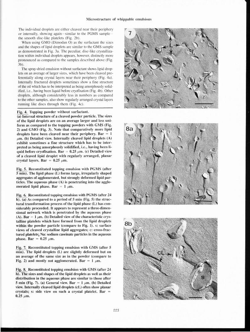
Fig. 7. Reconstituted topping emulsion with GMS (after 5 min). The lipid droplets (L) are slightly deformed but on an average of the same size as in the powder (compare to Fig. 2) and mostly not agglomerated. Bar = 1 ~-tm.
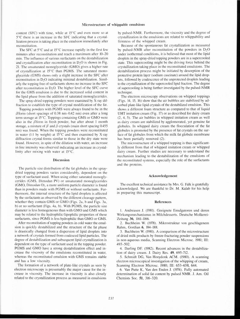
Fig. 8. Reconstituted topping emulsion with GMS (after 24 h). The sizes and shapes of the lipid droplets as well as their distribution in the aqueous phase are similar to those after 5 min (Fig. 7). (a) General view. Bar = 1 ~-tm. (b) Detailed view. Internally cleaved lipid droplets (cL) often show planar crystals; s: side view on such a crystal platelet. Bar = 0.25 ~-tm.
225

W. Buchheim , N.M . Barfod and N . Krog
226
Fig. 12. Reconstituted topping emulsion without emulsifier (after 24 h). There are no structural changes detectable as compared to a period of 5 min (Fig. 11). Bar = 1 lim.

Microstructure of whippable emulsions
Fig. 9. Reconstituted topping emulsion with GMO (after 5 min). A strong structural transformation of the original lipid droplets (see Fig. 3) into elongated, plate-like particles has taken place. These platelets appear to be more or less separated from each other. Bar = 1 11m.
Fig. 10. Reconstituted topping emulsion with GMO (after 24 h). (a) The structural transformation of the lipid particles into large thin platelets has proceeded. These crystal platelets appear either cross-fractured (cC) or surface-fractured (sC) according to their orientation in the specimen. Only rarely globular lipid particles can be detected (L). Bar = 1 11m. (b) Detailed view of a platelet exposing the smooth surface of individual crystal layers. Bar = 0.25 11m. (c) Detailed view of cross-fractured platelets (c) which consist of stacks of crystalline monolayers. L: lipid droplet (Note the amorphous fine structure of previously liquid fat!) from which a crystal platelet (P) has grown. Bar = 0.25 11m.
Fig. 11. Reconstituted topping emulsion without emulsifier (after 5 min). The lipid droplets are on an average of the same size and shape as in the powder (compare to Fig. 4). Bar = 1 11m.
Reconstituted powders-The 4 different topping powders were reconstituted with water at l5 °C (3 parts water added to 1 part of powder) and subsequently stored at 4°C for up to 24 h before further treatment. The structural changes occurring within the various emulsions can be demonstrated by comparing the state immediately after reconstitution with that after a longer period (24 h) .
The topping system containing PGMS as a surfactant shows a very uneven distribution of the lipid phase following only a very short period of a few minutes in the aqueous environment (Fig. 5). Although a few particles still appear to retain their original sizes and shapes, i.e., the sizes and shapes which are typical for the dried system , the vast majority forms compact aggregates within which the globular shapes of lipid droplets can hardly be detected. Crystallization phenomena which resulted in a partial destruction of the lipid droplets and a peculiar agglomeration of crystalline platelets appear to have taken place. The aqueous phase in which the caseinate particles (10-20 nrn in diameter) are clearly detectable, is penetrating into the agglomerated lipid phase.
The longer the influence of the aqueous environment on the lipid phase lasts the further the structural transformation process proceeds. Fig. 6a gives a typical view after 24 h of storage which is not too different from the view after the storage period of 1 h. The lipid phase shows pronounced crystalline structures (i.e., stacks of crystal monolayers) which are embedded in amorphous (i.e., previously liquid) fat. Lipid droplets are only rarely present , similar to the powder, and the lipid phase appears to be transformed into a three-dimensional network of interconnected , irregularly shaped aggregates , many of which are several 11m
in diameter. This final state of the lipid phase is also shown at a higher magnification (Fig. 6b) .
After reconstituting the powder containing GMS, comparable structural changes do not occur (Figs. 7-8a) . The state of the particles a few minutes after the dispersion of the powder in
227
water is shown in Fig. 7. Most lipid droplets are float ing separately in the aqueous phase, and agglomeration of the droplets is mostl y absent. Most droplets are of the sizes found in the dried state; their shapes are not as globular as in the powder but the degree of deformation is moderate. The same situation occurs also after longer storage periods (Fig. 8a), i. e., the emulsion remains in a stable state. At a higher magnification, the extent and type of crystal formation within the lipid droplets can be well recognized (Fig. 8b).
When using GMO as surfactant , strong structural changes again occur in the reconstituted system but in quite a different manner as compared to the system containing PGMS. Fig. 9 demonstrates the state of an emulsion shortly after reconstitution . Globular lipid particles as in the powder are rare whereas elongated , plate-like particles are very frequent . It can be seen that they develop from more globular ones, are mostly up to I J1l11 long and often 10-50 nm thick. These strongly deformed particles appear to be more or less separated from each other, i. e., they do not coalesce into larger :1ggregates as in the system with PGMS (compare Fig. 9 with Fig. 5). After 24 h the lipid particles in the reconstituted GMO system show a st ill more pronounced structural transformation into large thin platelets or lamellae which increase distinctly in size (Fig. lOa). It can be seen at higher magnification that these particles consist mai nly of stacks of crystalline mono layers (Figs. lOb, c).
In the reconstituted topping made from the powder which contains no surfactant only lipid particles of globular shapes are found. The sizes of the lipid droplets are identical to those seen in the dry powder particles. Similar images were obtained irrespective of the time elapsed after reconstitution (Figs . II , 12). Neither agglomeration nor stronger deformation of particles can be seen.
UHT imitation cream-The UHT-treated imitation cream has a fine structure which is very different from the reconstituted topping systems. As Fig. 13a demonstrates , the lipid particles are of an almost perfectly globular shape, not agglomerated and up to ca I J1lTl in diameter. The most characteristic feature , however, is the type of crysta lli zation within the lipid droplets. Concentric crystal monolayers are present within nearly each droplet and therefore cause preferentially a periphe ral cleavage (Fig. 13b). When the globules have been cross-:hctured, a rather thi ck shell of these concentric crysta l layers becomes visible (Fig. 13c).
Whipped toppings-From the four different topping powders only those containing PGMS (Promodan SP) and GMO (Dimodan 0) as surfactants formed stable foams when the reconstituted systems were whipped . The topping powder with GMS (Dimodan PV) gave a very soft , unstable foam.
For comparison, only one typica l small area of the foam of each type of sample is shown ; thi s area includes the air-water interface of a cross-fractured air bubble.
Fig. 14 shows the appearance of the air-water interface in the topping system with PGMS. The inner surface of the air bubbles is completely covered with crystalline lipid material , which appears to be adsorbed to the interface in the form of a continuous layer ca. 0.1 J1l11 thick. In the foam lamella , i.e. , in the serum phase between the air bubbles there are mostly irregularly shaped lipid aggregates, but sometimes also some more globular particles are present. This is to some extent similar to the situation in the reconstituted systems (compare to Fig. 5 or 6a) .

W. Buchheim , N. M . Barfod and N. Krog
228
Fig. 13. UHT-treated imitation cream. (a) General view of the emulsion. The lipid droplets are of an almost perfectly globular shape and not agglomerated. Most droplets have been peripherally cleaved due to the presence of concentric crystal monolayers. Bar = 1~-tm. (b) Detailed view of a peripherally cleaved lipid droplet showing individual concentric monolayers (cL). Bar = 0.25 J..tm. (c) Detailed view of a cross-fractured lipid droplet showing these concentric monolayers (cL) from a direction perpendicular to that of Fig. 13b. Bar = 0.25 J..tm.

Microstructure of whippable emulsions
Table 1. Whipping tests and foam firmness of toppings at 10°C and l5°C.
Surfactant used Overrun (%) 10°C l5°C
Promodan SP (PGMS) 290 192
Dimodan 0 (GMO) 50 65
Dimodan PV (GMS) 270 50
No surfactant 80 12
The whipped topping containing GMS as surfactant is shown in Fig. 15. The inner surface of the air bubble is not as completely covered with crystalline material as in the PGMS system (Fig. 14); frequently there are globular lipid particles protruding into the air. Within the serum phase of the foam, the lipid particles resemble those within the reconstituted system (compare Fig. 15 to Fig. 7 or 8a) but directly at the air-water interface they appear to be in a more agglomerated state.
When GMO is used as a surfactant, the inner surface of the air bubbles is nearly completely covered with flat crystal layers (Fig. 16) , more similar to the PGMS system than to the GMS system. The dimensions of the crystal platelets are larger than in the PGMS system. Within the foam lamella , i.e. , in the serum phase, the characteristic lamellae of platelet-like structures prevail; they have already been typical for the reconstituted system.
The structure of the whipped, liquid (UHT treated) imitation
Fig. 14. Whipped topping emulsion with PGMS. This micrograph (and also Figs. 15-17) represents a small but characteristic area of the boundary between an air bubble (A) and the serum phase (s) of the foam. aL: crystalline lipid material on the inner surface of the air bubble; sL: lipid aggregates in the serum phase. Arrows point to the air-water interface. Bar = 1 ,urn.
Fig. 15. Whipped topping emulsion with GMS. A: air bubble; S: serum phase; aL: globular lipid particle protruding into the air; sL: agglomerated lipid particles adhering to the air-water interface (arrows). In the serum phase also many lipid particles (L) are present in a non-agglomerated state similar to the reconstituted topping emulsion (compare to Figs. 7 and 8). Bar = 1 ,urn.
Fig. 16. Whipped topping emulsion with GMO. The inner surface of air bubbles (A) is nearly completely covered with flat crystalline lipid layers (aL). In the serum phase (S) lipid platelets (sL) in an aggregated or non-aggregated state prevail. Arrows point to the air-water interface. Bar = 1 ,urn.
Fig. 17. Whipped UHT-treated imitation cream. The air bubbles (A) appear to be covered with a monolayer of nearly undeformed lipid globules (sL) which protrude with a part of their volume into the bubble (aL). Occasionally crystal platelets (c) are present on the inner surface of the air bubble. S: serum phase. Arrows point to the air-water interface. Bar = 1 ,urn.
229
Foam firmness (g) General remarks on the 10°C l5°C whipped toppings
16 9 Soft, stable
26 24 Stiff, very stable
5 <I Very soft and unstable , syneresis
<I <I Extremely soft and unstable, syneresis
cream is completely different from the other whipped systems studied. Fig. 17 shows that the air bubbles appear to be covered with a monolayer of lipid globules which are rarely deformed and which protude with a substantial part of their volume into the bubble. Only rarely plate-like crystal layers are present at the interface. Emulsion viscosity, whippability and foam texture
Viscosity-When a topping powder is reconstituted into a cream by mixing l part of powder with 3 parts of water and the emulsion is kept below room temperature, an increase in viscosity will occur. The viscosity is dependent on temperature as shown in Fig. 18 for a topping emulsion with PGMS. At a temperature higher than 20°C, the increase in viscosity is negligible.
The rate of the viscosity increase is also dependent on the type of surfactant used as shown in Fig. 19. The emulsion viscosity is higher with GMO than with PGMS at l5°C; the topping emulsions with GMS or free of surfactants at all show no increase in viscosity on aging.
Whippability and foam firmness-The results of the whipping tests and the foam firmness measurements are shown in Table I. The whipping test was done at two temperatures, 10° and l5 °C. In general, a lower whipping temperature gave a higher overrun and firmness of the foam. Toppings with PGMS gave a fairly soft decoration foam, while the foam made from the GMO topping was firmer with good foam stability which was ideal for cake decoration.
However the GMO topping powder is not stable for extended storage periods. It loses its whipping property, probably due to a change in the crystalline properties of the fat phase.
Toppings with GMS or free of surfactants gave very light foams with large air bubbles. These foams were very unstable and separated quickly into a water phase with a collapsed cream layer at the top.
Crystallization measurements-The TEM studies described above indicate that crystallization of the lipid phase in the topping powders took place after they had been reconstituted in water. This crystallization process was evaluated using a pulsedNMR technique with deuterated water as solvent in order to eliminate the proton signal from water. The topping emulsions were reconstituted, stored, and analyzed under isothermal conditions.
Fig. 20 shows the results of pulsed-NMR measurements of a topping emulsion with 8% of PGMS as surfactant. Since both the maltodextrin and the sodium caseinate are dissolved in D20, the signal from protons in the solid state can only arrive from the crystallized fraction of the lipid phase. From Fig. 20 it appears that at 20°C and 35 °C there is no change in the solid fat

W. Buchheim, N.M. Barfod and N. Krog
20~----------------------------------~
-(/)
co a.. -
15
~ 10 ·u; 0 (.) (/)
> 5
0.5 1.0 1.5 2.0
Time (hours)
Fig. 18. Viscosity of the reconstituted topping emulsion (1:3) with PGMS (Promodan SP) at different temperatures as a function of time. Shear rate 1.76 sec - 1.
50C
60
-'#. 40 -20
• • • • • 0 ~----~--~----~-----T-----r----~ 0 10 20 30 40 50 60
Time (min.)
Fig. 20. Crystallization of supercooled lipid fractions in the topping emulsion with PGMS, reconstituted (1:3) in deuterated water (D20), measured by pulsed-NMR at different temperatures. SFC: solid fat content.
Z30
20~----------------------------------~
-(/)
co a..
15
~ 10 (/)
0 () (/)
> 5
0~==~==~====:3=.,~4·~======~======~ 0 0.5 1.0 1.5 2.0
Time (hours) at 15°C
Fig. 19. Viscosity of reconstituted topping emulsions (1:3) with different surfactants as a function of time at 15°C. Shear rate 1.76 sec - 1. 1: GMO (Dimodan 0), 2: PGMS (Promodan SP), 3: GMS (Dimodan PV), 4: no surfactant added.
40
-'#. -cj 30 u.: u)
20 ...
... ~r-~~--... ~4~· --~·~--------------1
10~--------~----------~--------~~ 0 10 20 30
Time (min.) at 15°C
Fig. 21. Crystallization of supercooled lipid fractions in topping emulsions with different surfactants, reconstituted (1:3) in deuterated water (D20), measured by pulsed-NMR at 15°C. SFC: solid fat content. 1: GMO (Dimodan 0), 2: PGMS (Promodan SP), 3: GMS (Dimodan PV), 4: no surfactant added.
Microstructure of whippable emulsions
content (SFC) with time, while at IS °C and even more so at soc there is an increase in the SFC indicating that a crysta llization process is taking place in the emulsion immediately after reconstitution .
The SFC at soc and at IS °C increase rapidly in the first few minutes after reconstitution and reach a maximum after 10-20 min . The influence of various surfactants on the destabilization and crystallization after reconstitution in DzO is shown in Fig. 21. The unsaturated monoglyceride (GMO) gives a higher rate of crystallization at lS °C than PGMS. The saturated monoglyceride (GMS) shows only a slight increase in the SFC after reconstitution in D20 indicating minimal destabilization. Similarly the topping free of surfactants shows no increase in the SFC after reconstitution in D20 . The higher level of the SFC curve for the GMS emulsion is due to the increased solid content in the lipid phase from the addition of saturated monoglycerides.
The spray-dried topping powders were examined by X-ray diffraction to establi sh the type of crystal modification of the fat.
Topping powders with PGMS were always found to be in the ~form (short spacings of 0.38 nm- 0.42 nm) even after a long term storage at IS 0 C. Toppings containing GMS or GMO were also in the ~form in fresh powder, but after about 1 month storage, a mixture of {32 and {)-crystals (main short spac ing 0.4SS nm) was found. When the topping powders were reconstituted in water (1 :1 by weight) at 1S °C and then examined by X-ray diffraction crystal-forms s imilar to those in the dry state were found . However, in spite of the dilution with water, an increase in line intensity was observed indicating an increase in crysta llinity afte r mixing with water at 1S 0 C.
Discussion
The particle size distribution of the fat globules in the spraydried topping powders varies considerably, dependent on the type of surfactant used . When usi ng e ither saturated monoglycerides (GMS, Dimodan PV) or unsaturated monoglycerides (GMO, Dimodan 0) , a more uniform particle diameter is found than in powders made with PGMS or without surfactants. Furthermore, the internal structure of the lipid droplets is affected by the surfactants as observed by the different cleavage pattern , whether they contain GMS or GMO (Figs. 2a, band Figs. 3a, b) or no surfactant (Figs. 4a , b). With PGMS, the particle size diameter is less homogeneous than with GMO and GMS which may be re lated to the hydrophilic/ lipophilic properties of these surfactants, since PGMS is less hydrophilic than GMO or GMS.
After reconstitution of topping powders in cold water the emulsion is quickly destabilized and the structure of the fat phase is drastically changed from a dispersion of lipid droplets into a network of crystals formed from coalesced lipid particles. The degree of destabilization and subsequent lipid crystallization is dependent on the type of surfactant used in the topping powder. PGMS and GMO have a strong destabilization effect and increase the viscosity of the emulsions reconstituted in water, whereas the reconstituted emulsion with GMS remains stable and has a low viscosity.
The formation of a network of plate-like crystals as seen by electron microscopy is presumably the major cause for the increase in viscosity. The increase in viscosity is also closely related to the crystallization process as measured in emulsions
231
by pul sed-NMR. Furthermore, the viscosity and the degree of crystallization in the emulsions are related to whippability and firmness of the whipped cream .
Because of the spontaneous fat crystallization as measured by pul sed-NMR after reconstitution of the powders in DzO under isothermal conditions , it is be lieved that most of the lipid droplets in the spray-dried topping powders are in a supercooled state. This supercooling might be the driving force behind the crystallization taking place in the reconstituted emulsions. The destabilization process might be initiated by desorption of the protective protein layer (sodium caseinate) around the lipid droplets, followed by coalescence of the unprotected droplets leading to the crystallization of the supercooled lipid fraction. The degree of supercooling is being further investigated by the pulsed-NMR technique.
The electron microscopy observations on whipped toppings (Figs . 14, lS, 16) show that the air bubbles are stabilized by adsorbed plate-like lipid crystals of the destabilized emulsion. This shows a different foam structure as compared to that of liquid UHT imitation cream (Fig. 17) or as described for dairy cream (2, 4, S). The air bubbles in whipped imitation cream as well as dairy cream are stabilized by agglomerated, yet genuine fat g lobules. In whipped dairy cream the flocculation of the fat globules is promoted by the presence of fat crystals on the surface of fat globules from which the milk fat globule membrane has been partially removed (2).
The microstructure of a whipped topping is thus significantly different from that of whipped imitation cream or whipped dairy c ream . Further studies are necessa ry to understand the mechanism leading to the destabilization of the emulsions of the reconstituted systems, espec ially the role of the surfactants and the proteins.
Acknowledgement
The excellent technical assistance by Mrs. G. Falk is gratefully acknowledged. We are thankful to Dr. M . Kalab for his help in preparing this manusc ript.
References
I. Andreasen J. (1981). Geeignete Emulgatoren und deren Wirkungsmechanismus in Milchdesserts. Deutsche MolkereiZeitung 36, l16l-l166.
2. Buchheim W. (1978) . Mikrostruktur von geschlagenem Rahm , Gordian 6, 184-188.
3. Buchheim W. (1981). A comparison of the microstructure of dried milk products by freeze-fracturing powder suspensions in non-aqueous media , Scanning Electron Microsc. 1981 ; III : 493-S02.
4. Darling DF. (1982). Recent advances in the destabilization of dairy cream. J. Dairy Res. 49, 69S-712.
S. Schmidt DG, Van Hooydonk ACM . (1980) . A scanning electron microscopical investigation of the whipping of cream, Scanning Electron Microsc. 1980; Ill: 653-6S8, 644.
6. Van Putte K, Van den Enden J. (1974) . Fully automated determination of solid fat content by pulsed NMR. J. Am. Oil Chemists Soc. 51, 316-320.

W. Buchheim , N.M . Barfod and N. Krog
Discussion with Reviewers
J.M. deMan: The cryofixation technique involves sample preparation at low temperature . How would the structural features of solidified fat relate to the actual conditions prevailing at the temperature of use of these products? What is meant by the expression "amorphously solidified" (e.g .. in the legend to Fig. 4b)? Authors: According to our experience the cooling rates during the cryofixation procedure used are generally high enough in order to avoid the formation of fat crystals. Liquid fat solidifies in a manner which results in a largely amorphous fine structure on freeze-fracture electron micrographs. For this reason visible fat crystals actually existed in the specimen before cryofixation. It appears , however, necessary to apply more advanced cryofixation procedures with much higher cooling rates (e.g. , the propane-jet freezing method) in order to obtain further experience.
P. Walstra: In my view it is highly probable that in the emulsions with PGMS and GMO fat crystals stick partly out of the droplets, which makes the latter vulnerable to partial coalescence, particularly during shear (see e.g., M.A.J.S. van Boeke! & P. Walstra , Colloids and Surfaces, 3 (1981), 109, and your reference 4) . During dissolving, shear is inevitable and partial coalescence or clumping occurs. This may then trigger further crystal growth , as some droplets must have been devoid of crystals due to supercooling (see e.g., P. Walstra & E.C. H. van Beresteyn , Neth . Milk Dairy 1. 29 (1975), 35). The size and shape of the crystals and particularly their contact angle with oil and water may depend on the surfactant , and these factors strongly affect the partial coalescence, which, in turn , causes the increase in viscosity and affects whipping properties . Authors: In general, our TEM observations of reconstituted emulsions, containing GMO or PGMS, show that the lipid phase is present in the form of a network of crystal -plates rather than globular lipid droplets. The mechanism suggested by you may well be the initial step in the structural change from gloublar lipid droplets to a crystal matrix .
We think , however, that the crystallization taking place in toppings goes far beyond of what takes place at the surface of the globules , but rather involves the whole lipid phase. Here we also must consider the difference in composition of our topping emulsions compared to dairy emulsions referred to in the references given by you. The concentration of surfactants (i.e . , PGMS) in toppings is as high as 12-20 % based on the total lipid phase, while it is much lower in dairy emulsions. Furthermore the TEM observations of whipped toppings with GMO or PGMS show that the foam is stabilized by adsorbed lipid crystals oriented at the air-water interface, and not by globular lipid droplets as found with dairy cream according to ref. 2 , 4 and 5, and as found here with imitation cream (see Fig. 17).
2 32
P. Walstra: Due to the presence of surfactant , the contact angle as measured in water may even become small enough for the crystals to become easily dislodged from the oil droplets (or their remnants) . Perhaps they flocculate as such (although on the other hand the aggregates may just be due to partial coalescence). The crystals can easily become adsorbed into the airwater interface, thereby stabilizing the air bubbles aginst disproportionation and coalescence. Authors: We think your comment is very relevant to our findings, especially in the light of the high surfactant concentration needed in topping system in order to obtain the necessary destabilization and to obtain satisfactory whipping properties.