dna destabilization, cancer, and inflammation - beljanski
TRANSCRIPT
DNA Destabilization, Cancer, and InflammationJohn L. Hall, Ph.D.
Senior Scientific Advisor, The Beljanski Foundation, Inc.
First published as:“Chapter 6. DNA Destabilization, Cancer, and Inflammation.”
John L. Hall, Ph.D.Anti-Aging Therapeutics Volume XIII, 53-64.
(c) American Academy of Anti-Aging Medicine; Chicago, IL USA; 2011. .
Reprint prepared for The Beljanski Foundation Inc. (New York, NY USA) by A4M. The A4M does not endorse, in either direct or implied manner, the Author(s),
their Affiliation(s), or the content of their work.
53
Chapter 6 DNA Destabilization, Cancer, and Inflammation
John L. Hall, Ph.D.
ABSTRACT For many years the central paradigm of cancer research has been that specific mutations in a critical set of regulatory genes cause the disease. The mutational theory of cancer has been such a gripping paradigm that alternative explanations for carcinogenesis have been slow to develop and gain attention. This paper will introduce an alternative theory of cancer – the theory of DNA destabilization. Keywords: DNA destabilization, DNA disorder, carcinogens, cancer DNA, inflammation, benign prostatic hyperplasia INTRODUCTION From Cancer Genes to Cancer DNA For more than two generations, the central paradigm of cancer research has been that specific mutations in a critical set of regulatory genes cause the disease.1,2 The success of this paradigm – known as the mutational theory of cancer – has been limited. Numerous oncogenes and tumor suppressors have been characterized and mutations in these genes have been associated with a variety of cancers, but in only a few cases have these mutations been shown to cause cancer. For example, mutations in both alleles of the tumor suppressor gene Rb1 lead to retinal cancer in the absence of any known secondary event.3 On the other hand, mutations in the tumor suppressor gene p53, have been found in many cancers, but recent studies suggest that these mutations, like those of many other tumor suppressor genes, appear to accumulate after carcinogenesis and hence may contribute to the development of cancer, but are rarely, if ever, the cause.4-6 All paradigms have momentum, but with its origins founded on the centrality of the genetic code, the corruption of genes and their corresponding proteins by mutagens, and indeed its modest claims to success, the mutational theory of cancer has had a very special lease on life. Oncogenes and tumor suppressors have enjoyed a celebrity status – their discovery and characterization has been exhilarating for molecular biologists –and their popularity as the major players in carcinogenesis remains largely intact. Moreover, the paradigm is inherently open-ended: there might always be a few more novel oncogenes with the critical point mutations that complete the story line and justify the paradigm.7 Recent advances in DNA sequencing technology are now enabling the mutational theory to be fully vetted as the complete genomes of patients with various cancers are sequenced and the data are analyzed for sets of specific mutations that can be associated with specific cancers.8,9 This process will elucidate the complex genetic basis of cancer just as it is becoming apparent that cancer does not begin as a genetic disease. The mutational theory of cancer has been such a gripping paradigm that alternative explanations for carcinogenesis have been slow to develop and gain attention. For many scientists there has been a sense of disbelief that there could be alternative models that really matter. The mutational theory has always had its skeptics outside the mainstream, however recently it has engendered growing criticism and the paradigm has been attacked by independent sources in the scientific literature and in the popular press.10-14 When the research of one proponent of the mutational theory was questioned – pertaining to whether an adduct of cigarette smoke induced a specific p53 mutation critical to the development of lung cancer – he replied: “The idea that cancer is more than just a mutation has been around a long time.” Just what is this idea? The answer is that the structure of the DNA molecule itself suffers physical damage, referred to as destabilization or disorder that is directly caused by carcinogens and oxidative reactions. The appearance of destabilized or disordered DNA correlates with the earliest stages of cancer (precancer) and is apparently the primary event in carcinogenesis. The irony is that the focus is still on DNA, but DNA as a physical duplex not DNA as the genetic code and cancer is associated with damage to DNA structure, not with mutations that alter the sequence and function of proteins. It is the stability of the hydrogen bonds in the double helix and the chemical integrity of the nitrogenous bases that are
54
compromised at the onset of most cancers. Destabilized DNA, because its double helix is relatively open, is more accessible to enzymes and so inherently more likely to undergo the aberrant duplication and transcription that distinguish cancer cells. Of course, these physical changes that are the primary cause of cancer are also likely to enable mutations that, in this paradigm, appear as secondary. DNA DESTABILIZATION Mirko Beljanski and the Discovery of DNA Destabilization Mirko Beljanski was born in Yugoslavia, educated in Paris, and after a fellowship in Dr. Severo Ochoa’s laboratory at New York University, he returned to Paris where he worked for most of his career at the Pasteur Institute. His early work focused on antibiotic resistance in bacteria and respiratory enzymes, but his focus shifted to DNA and RNA. He became interested in how the fundamental mechanisms of DNA duplication and transcription – how information is copied and retrieved from DNA. He focused on the regulation of the opening and closing of the DNA duplex that must occur every time a gene is transcribed or DNA is duplicated. For DNA replication, the enzyme DNA polymerase must access and traverse a single strand of the DNA duplex; for RNA transcription, the enzyme RNA polymerase must access and traverse a single strand of the DNA duplex. Both processes require a local opening of the DNA – a strand separation – that enables enzyme function followed by a closing of the strands and reformation of a stable double helix. The opening and closing of the DNA double helix is regulated at multiple levels in the cell including higher order packaging/unpackaging, nucleosomes and histones, enzymes, cofactors, hormones, and ions. At the level of the duplex itself, for DNA strand separation to occur the hydrogen bonds that hold the complementary chains of DNA together must be broken. These weak chemical bonds that stabilize the double helix exist between the complementary nitrogenous bases that align inside the duplex. When a gene is transcribed or DNA is duplicated, these hydrogen bonds are systematically broken by enzymes called DNA helicases that unravel the DNA duplex into single strands. While the polymerases do their jobs, the single strands are maintained by the presence of single stranded DNA binding proteins that prevent the tendency of the separated strands to reanneal and reestablish hydrogen bonding. After transcription or DNA synthesis and the removal of the binding proteins, the duplex is reformed as the hydrogen bonds restabilize. Beljanski was the first scientist to appreciate and to develop the idea that unregulated DNA strand separation – which he referred to as DNA destabilization – could be the primary basis of pathologies like cancer that are normally assumed to be genetic diseases.15 The process of DNA destabilization in the service of gene expression and DNA duplication is normally under the control of endogenous factors as well as (expected) environmental signals, but it is also susceptible to external factors and environmental contaminants (including carcinogens) that interact directly with DNA and induce a destabilizing effect. Note that these external factors can include endogenous biomolecules (e.g. hormones) that are administered exogenously. As Beljanski stated:
“The observation that carcinogens or steroid hormones may induce the appearance of cancer cells and/or the differentiation of particular cell lines suggested that, under certain conditions, the secondary structure of DNA chains may be destabilized by such agents. This view was strengthened by theoretical considerations, indicating that the appearance of cancer cells requires the persistent and cumulative action of carcinogens or estrogens in the systems.”15
This model of the destabilizing effects of carcinogens on the DNA duplex is illustrated in Figure 1. Carcinogens with affinity for DNA may bind at various sites along the outside of the duplex and in the major groove. This binding exerts a destabilizing effect that may entail the breaking of hydrogen bonds. When hydrogen bonds are broken across segments of the duplex, openings or loops of single stranded DNA form as they do under the influence of DNA helicase. The loops are the physical manifestation of DNA destabilization and if they persist may provide entry points for polymerases that are active on the exposed single strands.

55
Figure 1. Illustration of the destabilization of DNA caused by binding of carcinogens. Binding of
carcinogens disrupts the hydrogen bonds that maintain the DNA duplex and the strands of the duplex are separated. Enzymes for DNA replication have increased access to the initiation sites on the exposed
strands and DNA duplication may be abnormally accelerated. Assays for DNA Stability I The absorption of light at specific wavelengths is a characteristic of many biomolecules. In the case of ultraviolet (UV) light and DNA, the level of absorption depends on whether the DNA is in a double stranded duplex or in single stranded form. The absorption of UV by DNA that has been completely separated into single strands by chemical treatment is significantly higher than the UV absorption of double stranded DNA under physiological conditions (%40). So UV absorption is a measure of how tightly the two strands of DNA are held together in the double helix: the greater the strand separation the higher the absorption of UV.15 Beljanski used this method to compare the stability of DNAs that were purified from normal and cancerous cells. The absorption of DNA from cancer cells was consistently higher (by 1-5%) than the results found for normal DNA. This difference is minor, but it proved to be reproducible through hundreds of experiments using DNA purified from a variety of tumors from humans and animals. Beljanski concluded that the chemical bonds that hold the double helix together are disrupted in cancer DNA which results in more openings or loops than are present in normal DNA.15 Beljanski went on to develop a second, more sensitive assay for DNA destabilization based on the normal physiological role of strand separation in DNA duplication.16 The relatively open segments that are the hallmark of cancer DNA should serve as a more active template for DNA polymerase than the more tightly wound duplexes characteristic of normal DNA. This prediction was confirmed in vitro: purified cancer DNA replicated more rapidly than normal DNA as measured by the incorporation of specially labeled nucleotides into new DNA strands. Given the exact same amount of DNA template, nucleotide building blocks, and the enzyme DNA polymerase the reaction containing cancer DNA consistently synthesized more new DNA. Thus DNA destabilization was correlated with enhanced DNA synthesis.17 The Oncotest: Carcinogens Destabilize DNA Both the UV absorption and DNA synthesis assays were subsequently used as the basis for a new assay, the Oncotest, which reveals whether a given compound has an effect on DNA stability.16 In both cases, various compounds, naturally-occurring or manmade, were added to the assay to determine their effect on DNA structure and template activity. The first priority was measuring the effects of known carcinogens and the results for dimethylbenz(a)anthracene (DMBA) are shown in Figure 2. In Figure 2A,

56
the impact of various concentrations of DMBA on UV absorption by cancer and normal DNA are compared. The addition of 2 micrograms per milliliter of DMBA results in a significant increase in UV absorption by the cancer DNA, whereas the effect of the DMBA on UV absorption of the normal DNA is minor. Increasing the concentration of DMBA increases the UV absorption of the cancer DNA; the effect on normal DNA is relatively slight. Similar results are shown in Figure 2B for the DNA synthesis Oncotest. The presence of DMBA in the reaction triggers a boost in newly synthesized DNA whereas the compound has little or no effect on the DNA synthesis from the normal template.
Figure 2. Differential effects of dimethylbenz-anthracene (DMBA) on purified DNA from cancer and normal cells. (A) UV absorption of cancer DNA is significantly greater than normal DNA in the presence of DMBA; the increase in UV absorption is plotted against increasing amounts of DMBA per assay. (B) DNA
synthesis from the cancer DNA template is significantly greater than synthesis from the normal DNA template in the presence of DMBA; the quantity of DNA synthesized is plotted against increasing amounts
of DMBA per assay.
57
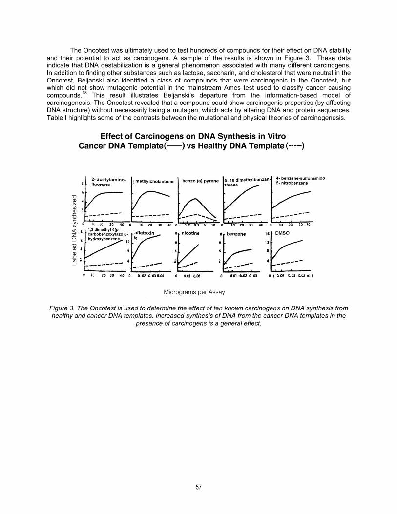
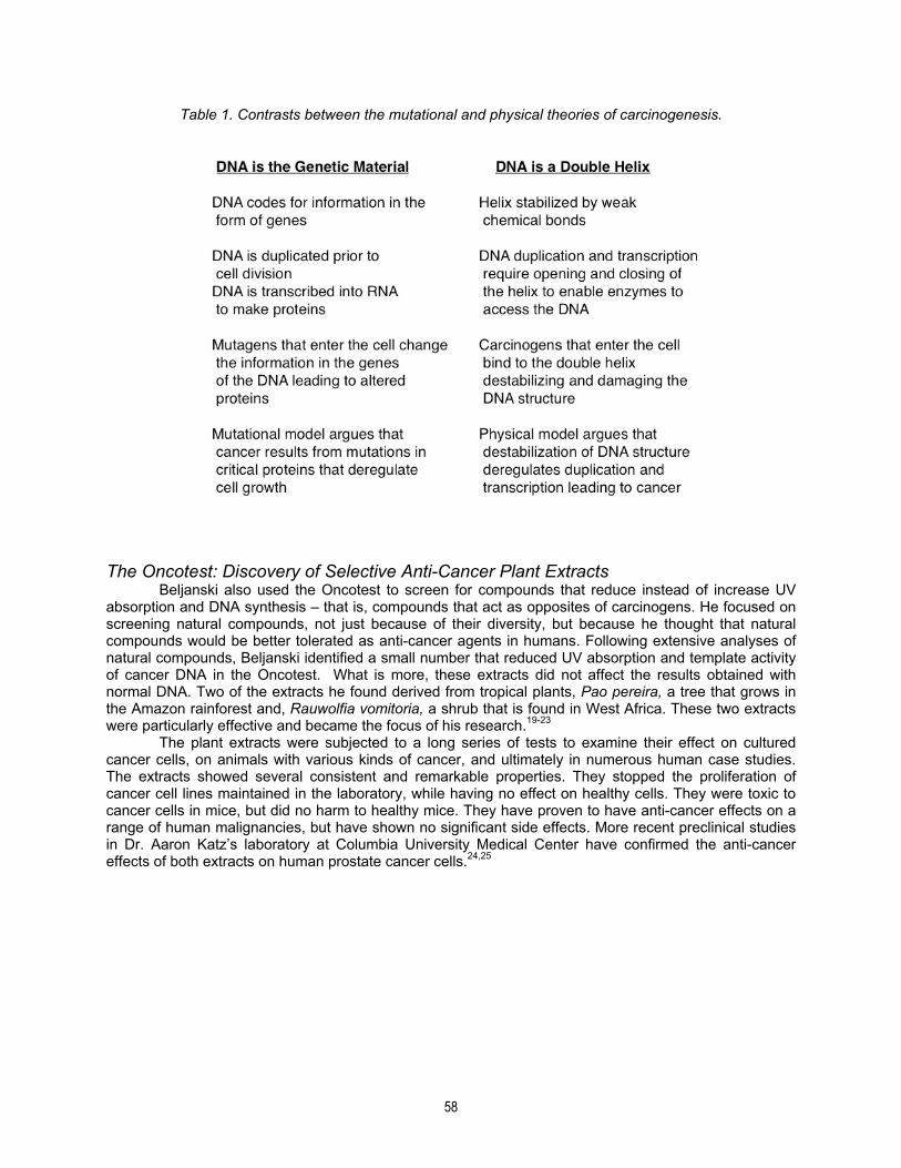
The Oncotest was ultimately used to test hundreds of compounds for their effect on DNA stability and their potential to act as carcinogens. A sample of the results is shown in Figure 3. These data indicate that DNA destabilization is a general phenomenon associated with many different carcinogens. In addition to finding other substances such as lactose, saccharin, and cholesterol that were neutral in the Oncotest, Beljanski also identified a class of compounds that were carcinogenic in the Oncotest, but which did not show mutagenic potential in the mainstream Ames test used to classify cancer causing compounds.18 This result illustrates Beljanski’s departure from the information-based model of carcinogenesis. The Oncotest revealed that a compound could show carcinogenic properties (by affecting DNA structure) without necessarily being a mutagen, which acts by altering DNA and protein sequences. Table I highlights some of the contrasts between the mutational and physical theories of carcinogenesis.
Figure 3. The Oncotest is used to determine the effect of ten known carcinogens on DNA synthesis from healthy and cancer DNA templates. Increased synthesis of DNA from the cancer DNA templates in the
presence of carcinogens is a general effect.
58
Table 1. Contrasts between the mutational and physical theories of carcinogenesis.
The Oncotest: Discovery of Selective Anti-Cancer Plant Extracts Beljanski also used the Oncotest to screen for compounds that reduce instead of increase UV absorption and DNA synthesis – that is, compounds that act as opposites of carcinogens. He focused on screening natural compounds, not just because of their diversity, but because he thought that natural compounds would be better tolerated as anti-cancer agents in humans. Following extensive analyses of natural compounds, Beljanski identified a small number that reduced UV absorption and template activity of cancer DNA in the Oncotest. What is more, these extracts did not affect the results obtained with normal DNA. Two of the extracts he found derived from tropical plants, Pao pereira, a tree that grows in the Amazon rainforest and, Rauwolfia vomitoria, a shrub that is found in West Africa. These two extracts were particularly effective and became the focus of his research.19-23 The plant extracts were subjected to a long series of tests to examine their effect on cultured cancer cells, on animals with various kinds of cancer, and ultimately in numerous human case studies. The extracts showed several consistent and remarkable properties. They stopped the proliferation of cancer cell lines maintained in the laboratory, while having no effect on healthy cells. They were toxic to cancer cells in mice, but did no harm to healthy mice. They have proven to have anti-cancer effects on a range of human malignancies, but have shown no significant side effects. More recent preclinical studies in Dr. Aaron Katz’s laboratory at Columbia University Medical Center have confirmed the anti-cancer effects of both extracts on human prostate cancer cells.24,25
59
DNA Destabilization and Carcinogens: Reversible and Chronic Effects Beljanski’s model for carcinogenesis was that: 1) DNA is destabilized by the binding of carcinogens; 2) binding and release of carcinogens is dynamic and reversible, so DNA destabilization is reversible; 3) but the effect of persistent interaction with carcinogens is cumulative; 4) leading to segments of DNA that are permanently destabilized. Cells with destabilized DNA are susceptible to unregulated DNA duplication, gene expression and cell division. For some carcinogens, when present at significant and persistent levels, the induction of hyperplasia is direct. Beljanski wrote:
“Thus, for example, 2-fluorene acetamide (2-FAA), a carcinogen, administered to rats or mice, results in the appearance of neoplastic nodules in the liver. The DNA of the cells in these nodules is in a ‘destabilized’ form. Cells possessing such a DNA divide very rapidly compared to cells outside the nodules. Nucleus enlargement observed in the presence of a carcinogen is also accompanied by accelerated cell division and indicates that the agent used has set in motion the various steps of cell division.”
And in summary:
“Many experimental data have shown that carcinogenesis and differentiation of cultured cells in vitro, i.e., the two opposite poles of cellular physiology, may be induced and controlled by various endogenous or exogenous substances. This happens through local, limited DNA strand opening or closing. The fact that a cancer cell may differentiate in the presence of dimethyl sulfoxide (DMSO), tumor promoters or carcinogens into a normal cell strongly indicates that the cancerous state of a cell is not caused by mutations, i.e., changes in the structure of the genetic DNA itself.”
D.C. Malins and the Discovery of DNA Disorder In a series of elegant studies published in the Proceedings of the National Academy of Sciences, D.C. Malins and his colleagues at the Pacific Northwest Research Foundation in Seattle made a detailed characterization of carcinogen-induced structural changes in DNA. This work was performed completely independently – and apparently without knowledge of Beljanski’s research and yet there is a profound connection with Beljanksi’s observations and theories. Assays for DNA Stability II Malins used absorption spectroscopy with infrared (IR) light (rather than UV) to analyze purified DNA samples from two different sources.26 The first samples were taken from the livers of fish that had been exposed to environmental chemicals and the second samples were taken from the livers of fish grown in healthy waters. The raw absorption data taken over a range of IR wavelengths was processed through a mathematical algorithm called a Fourier transform to generate a graph of the absorbance of the sample at each wavelength (Fig 4). Comparison of the spectral profiles for the two DNA samples showed several statistically significant differences at various wavelengths. These differences reflect changes in the structure of the DNA in the sample from fish exposed to carcinogens. At several of the wavelengths these structural changes have been correlated with specific chemical damage to the DNA molecule. For example, one of the differences (1410 cm-1) signifies the deformation of carbon-hydrogen and nitrogen-hydrogen bonds of the nucleotide bases of the DNA. A second difference (1262 cm-1) is attributed to deformation of the phosphodiester backbone of the DNA. Malins stated that “Fourier-transform infrared (FTIR) spectroscopy of the control DNA and that of the exposed group provided direct evidence in a living system for profound chemically induced damage to the infrastructure of this biopolymer (the DNA)” and concluded that the DNA in the exposed fish “underwent an unprecedented degree of structural modification suggesting that it is considerably more responsive to environmental stresses than previously believed possible.”

60
Figure 4. Fourier Transform-Infrared spectra and P-value profiles for comparison of DNA from control group (unexposed to chemicals, solid line) and experimental group (exposed to chemicals, dashed line). P-value indicates statistical significance of the differences between the two spectral profiles. Reprinted
by permission. DNA Disorder and Cancer The structural damage that Malins detected in the liver DNA of fish growing in contaminated waters was not directly associated with cancer – approximately 20% of these fish went on to develop liver cancer – rather, it is associated with a precancerous state associated with new forms of structurally diverse DNA that are “likely to play an important role in carcinogenesis.”27 Malins made the connection between DNA structural damage and cancer in studies of purified DNA from human cancers (ovary, breast and prostate) and he used the term ‘disorder’ to describe the damaged or modified DNAs found in these cancer cells.28-31 Malins used the differences in spectral profiles he obtained for DNA from normal, precancerous, cancerous, and metastatic cells to generate statistical models that showed that DNA disorder is detectable in four reproducible and discrete stages that correspond directly to the histology of the tissues from which the DNAs were prepared (healthy, precancerous, cancerous, and metastatic). These data indicate that relative to the DNA structure found in normal cells, the DNA structure in precancerous cells is disordered, meaning that there is a change in DNA phenotype prior to the actual formation of a tumor. DNA from cells in a tumor also exhibits its own phenotype of disorder distinct from both normal and precancerous phenotypes. Finally, DNA from metastatic cells also has its own distinct disordered model. A recurrent theme in these studies is that the progression of DNA disorder through these four stages is, in fact, a causal factor in the transformation of normal tissues into primary and metastatic tumors. Malins performed several additional spectroscopic studies that support this view. For example, his group analyzed the purified DNA from histologically normal prostates of men ages 16 to 80 years and found that there are progressive changes in the structure of the DNA with increasing age.32 The DNA from older men exhibited a cancer-like phenotype that resembled the phenotype seen in men with primary prostate cancer. This cancer-like phenotype was not seen in the DNA from younger men, indicating that it results from age-related structural damage to the prostate DNA. This result is consistent with the fact that cancer is a disease of age so the accumulation of structural damage would be expected to associate with the onset of cancer in older men. In fact, the majority of prostate cancers are diagnosed
61
in men who are over 65 years of age. As noted above, Malins also found a distinct DNA damage profile for metastatic prostate cancer DNA and his technology offers the basis for an important diagnostic tool for determining which cancers will metastasize. The Chemistry of Structural DNA Damage Malins provides evidence that his spectroscopic analyses are detecting chemical modifications to the DNA polymer. For example, he found that 8-hydroxy purine lesions in prostate DNA increased 3-fold in men between the ages of 16 and 83 and that 8-hydroxy guanine lesions increase significantly in the livers of animals as they age. These chemical changes in the purine bases (that together with pyrimidine bases) comprise the DNA are thought to arise from what is called free radical damage. Free radicals are highly reactive small molecules that arise for a number of reasons in our bodies, and that catalyze chemical reactions that damage our DNA. Free radical induced damage is also thought to be an important factor in cancer and in the aging process. The reaction of the hydroxyl radical with DNA can lead to the modification of diverse structural groups in addition to 8-hydroxy guanine and these reactions can promote mutations and even cause the loss of phosphoric acid from the DNA backbone and breakage of the DNA strands. Hydroxy purine lesions are one of the major forms of free radical induced DNA damage that contribute to DNA disorder, but there are others and the reader is referred to Malins’ papers for more details. INFLAMMATION Inflammation has a DNA Phenotype: Benign Prostatic Hyperplasia As mentioned above, Malins detected distinct forms of damaged DNA that were purified from tissues that had been characterized histologically, including tissues that were not classified as cancerous. One striking example is the finding of altered forms of DNA in the prostates of men suffering from BPH (benign prostatic hyperplasia).31 BPH is associated with urinary problems caused by an enlarged prostate and it is common among older men. BPH is explicitly not cancer – it is benign – rather it is a form of inflammation caused by irritation or infection that leads to swelling of the prostate.33 The urinary symptoms caused by BPH are a problem, especially when they are severe, but this condition is also a serious concern because of evidence linking chronic inflammation to cancer.34-37 Malins data indicate that significant and specific structural modifications in DNA occur in the progression of normal tissue to BPH and normal tissue to prostate cancer, and that the modifications are unique for each of the two progressions. He concludes: “the progressive alteration of DNA structure in response to factors in the microenvironment, notably the hydroxyl radical, are likely etiologically related to the development of cellular lesions – breast tumors, prostate tumors, and BPH. It is suggested that intervention to forestall or correct the genetic instability of these tissues and likely increase in cancer risk should focus on controlling the cellular redox status and hydroxyl radical concentration.”31 Beljanski’s Plant Extracts: Anti-Inflammatory Effect on BPH The anti-cancer effects of the Pao periera and Rauwolfia vomitoria plant extracts are well documented, but their anti-inflammatory effects are just beginning to be appreciated. Beljanski published one study showing that the Pao extract suppresses the activity of the cytokine, interleukin (IL)-6, which is a major inflammatory pathway.38 More recently, a combination of the Pao and Rauwolfia extracts was tested in a clinical trial at Columbia University Medical Center conducted by Dr. Aaron Katz. This Phase I trial was designed primarily to assess the safety and tolerability of the extract combination for men with elevated PSA levels but a negative biopsy for prostate cancer. As often happens in scientific research, the results of the trial contained an important surprising result: the men with BPH reported significant improvement in their symptoms as measured by the standard AUA questionnaire. Experiments are now underway to discover the full extent of the anti-inflammatory effects of the Pao and Rauwolfia extracts. It will be interesting to see the effects of the extracts on the specific inflammatory pathways that are activated in men with BPH.39,40
62
The anti-cancer effects of Beljanski’s plant extracts are thought to be based on the binding of active molecules to the destabilized DNA in cancer cells meaning that destabilized DNA itself is the target of action. Their anti-inflammatory properties makes these extracts even more interesting and raises the question whether these molecules also recognize the partially destabilized or disordered DNA found in prostate cells with BPH. CONCLUDING REMARKS Beljanski employed UV spectroscopy to detect strand separation of the DNA from cancer cells whereas Malins used FTIR spectroscopy to detect diverse forms of physical damage in the DNA from cancer cells. How are DNA destabilization and DNA disorder related? The answer: like the partners in a bad marriage that enable each other’s negative behavior. DNA destabilization caused by binding of carcinogens, may be transient and reversible, but may nevertheless expose the molecules of the DNA to attack by free radicals, which, as we’ve seen, can cause serious chemical damage. Most, if not all, of the diverse forms of this damage, whether the deformation of chemicals bonds, the addition of chemical groups, or the opening of ring structures that are normally closed, will further weaken and strain the hydrogen bonds that stabilize the duplex. If the carcinogens are released from the DNA, the duplex can restabilize, assuming that the chemical damage allows for it. The chemical damage can be repaired if the DNA repair system of the cell remains competent and further damage may be prevented in the presence of antioxidants. Yet another wave of insult by carcinogens and free radicals is inevitable and with age, the effects of destabilization and disorder accumulate and persist. Through progressive loss of regulation of duplication and transcription a cell makes the transitions from normal to precancer and from precancer to cancer. Both destabilization and disorder enable mutations. REFERENCES 1. Nordling C. A new theory on cancer-inducing mechanism. Br J Cancer. 1953;7:68-72. 2. Cairns J. Cancer: Science and Society. W. H. Freeman and Company;1978. 3. Scheffer H, Imhof SM, Moll AC. From gene to disease; retinoblastoma and the RB1 gene. Ned
Tijdschr Geneeskd. 2001;145:1245-1247. 4. Ueda H, Ullrich SJ, Gangemi JD, Kappel CA, Ngo L, Feitelson MA, Jay G. Functional inactivation but
not structural mutation of p53 causes liver cancer. Nat Genet. 1995;9:41-47. 5. Offner S, Schmaus W, Witter K, et al. p53 gene mutations are not required for early dissemination of
cancer cells. Proc Natl Acad Sci. 1999;96:6942-6946. 6. Rodin SN,Rodin AS. Human lung cancer and p53: The interplay between mutagenesis and selection.
Proc Natl Acad Sci. 2000;97:12244-12249. 7. Greenman C, Stephens P, Smith R, Dalgliesh GL, Hunter C, Bignell G, et al. Patterns of somatic
mutation in human cancer genomes. Nature. 2007: 446;153-158. 8. Forbes SA, Bindal N, Bamford S, et al. COSMIC: mining complete cancer genomes in the Catalogue
of Somatic Mutations in Cancer. Nucleic Acids Res. 2011;39:D945-950. 9. International Cancer Genome Consortium, Hudson TJ, Anderson W, Artez A, et al. International
network of cancer genome projects. Nature. 2010; 464:993-998. 10. Soto AM, Sonnenschein C. The somatic mutation theory of cancer: growing problems with the
paradigm? Bioessays. 2004;26:1097-1107. 11. Edwards AM, Isserlin R, Bader GD, Frye SV, Willson TM, Yu FH. Too many roads not taken. Nature
2011;470:163-165. 12. Wayt GW. The roots of cancer. Scientific American Special Edition. 2004;14 (3):60-69. 13. Strohman R. Toward a new paradigm for life. Beyond genetic determinism. 22 March, 2001. Available
at: http://www.psrast.org/strohmnewgen.htm. Accessed February 18, 2011. 14. Leaf C, Burke D. Why we’re losing the war On cancer (and how to win it). Fortune Magazine. 2004;
March 22. 15. Beljanski M. The regulation of DNA replication and transcription. The role of trigger molecules in
normal and malignant gene expression. Experimental Biology and Medicine. 1983;8:1-190. 16. Beljanski M. Oncotest: A DNA assay system for the screening of carcinogenic substances. IRCS
Medical Science. 1979;7:476. 17. Beljanski M, Bourgarel P, Beljanski MS. Correlation between in vitro DNA synthesis, DNA strand
separation and in vivo multiplication of cancer cells. Expl Cell Biol. 1981;49 :220-231.
63
18. Ames B, Lee F, Durston W. An improved bacterial test system for the detection and classification of mutagens and carcinogens. Proc Natl Acad Sci. 1973;70:782-786.
19. Beljanski M, Beljanski MS. Three alkaloids as selective destroyers of the proliferative capacity of cancer cells. IRCS Med Sci. 1984;12:587-588.
20. Beljanski M, Beljanski MS. Three alkaloids as selective destroyers of cancer cells in mice. Synergy with classic anticancer drugs. Oncology.1986;43:198-203.
21. Beljanski M, Crochet S, Beljanski MS. A potent and selective inhibitor of human BCNU resistant glioma cell multiplication. Anticancer Research. 1993;13:2301-2308.
22. Beljanski M, Crochet S. Selective inhibitor (PB-100) of human glioblastoma cell multiplication. Journal of Neuro-Oncology.1994;21:62.
23. Beljanski M, Crochet S. The selective anticancer agent PB-100 and BG-8 are active against human melanoma cells, but do not affect non malignant fibroblasts. International Journal of Oncology. 1996;8:1143-1148.
24. Bemis DL, Capodice JL, Gorroochurn P, Katz AE, Uttyan R. Anti-prostate cancer activity of a ß-carboline alkaloid enriched extract from Rauwolfia vomitoria. International Journal of Oncology. 2006;29:1065-1073.
25. Bemis DL, Capodice J, Desai M, Katz AE, Buttyan R. ß-carboline alkaloid-enriched extract from the Amazonian rainforest tree Pao pereira suppresses prostate cancer cells. Journal of the Society for Integrative Oncology. 2009;7:59-65.
26. Malins DC, Gunselman SJ. Fourier-transform infrared spectroscopy and gas chromatography-mass spectrometry reveal a remarkable degree of structural damage in the DNA pf wild fish exposed to toxic chemicals. Proc Natl Acad Sci. USA. 1994;91:13038-13041.
27. Malins DC, Polissar NL, Gunselman SJ. Infrared spectral models demonstrate that exposure to environmental chemicals leads to new forms of DNA Proc Natl Acad Sci. 1997;94:3611-3615.
28. Malins DC, Polissar NL, Schaefer S, Su Y, Vinson M. A unified theory of carcinogenesis based on order–disorder transitions in DNA structure as studied in the human ovary and breast. Proc Natl Acad Sci. 1998;95:7637-7642.
29. Malins DC, Polissar NL, Gunselman SJ. Tumor progression to the metastatic state involves structural modifications in DNA markedly different from those associated with primary tumor formation. Proc Natl Acad Sci. 1996;93:14047-14052.
30. Malins DC, Polissar NL, Gunselman SJ. Progression of human breast cancers to the metastatic state is linked to hydroxyl radical-induced DNA damage Proc Natl Acad Sci. 1996:93:2557-2563.
31. Malins DC, Polissar NL, Gunselman SJ. Models of DNA structure achieve almost perfect discrimination between normal prostate, benign prostatic hyperplasia (BPH), and adenocarcinoma and have a high potential for predicting BPH and prostate cancer. Proc Natl Acad Sci. 1997;94:259-264.
32. Malins DC, Johnson PM, Barker EA, Polissar NL, Wheeler TM, Anderson KM. Cancer-related changes in prostate DNA as men age and early identification of metastasis in primary prostate tumors. Proc Natl Acad Sci. 2003;100:5401-5406.
33. Fibbi B, Penna G, Morelli A, Adorini L, Maggi M. Chronic inflammation in the pathogenesis of benign prostatic hyperplasia. Int J Androl. 2010;33:475-488.
34. Meira LB, Bugni JM, Green SL, et al. DNA damage induced by chronic inflammation contributes to colon carcinogenesis in mice. Journal of Clinical Investigation 2008;118:2516-2525.
35. Allvena P, Garlanda C, Borello MG, et al. Pathways connecting inflammation and cancer. Curr Opin Genet Dev. 2008;18:3-10.
36. Coussens LM, Werb Z. Inflammation and cancer. Nature. 2002;420:860-867. 37. De Marzo AM, Platz EA, Sutcliffe S, Xu J, Grönberg H, Drake CG, Nakai Y, Isaacs WB, Nelson WG.
Inflammation in prostate carcinogenesis. Nature 2007;7:256-269. 38. Beljanski M, Crochet S. The selective anticancer agent PB-100 inhibits interleukin-6 induced
enhancement of glioblastoma cell proliferation in vitro. International Journal of Oncology. 1994;5:873-879.
39. Liu L, Li Q, Han P, Li X, Zeng H, Zhu Y, Wei Q. Evaluation of interleukin-8 in expressed prostatic secretion as a reliable biomarker of inflammation in benign prostatic hyperplasia. Urology. 2009;74:340-344.

64
40. Penna G, Fibbi B, Amuchastegui S, Cossetti C, Aquilano F, Laverny G, Gacci M, Crescioli C, Maggi M, Adorini L. Human Benign Prostatic Hyperplasia Stromal Cells As Inducers and Targets of Chronic Immuno-Mediated Inflammation. The Journal of Immunology 2009;182:4056-4064.
ABOUT THE AUTHOR
John L. Hall, Ph.D. completed graduate work in biochemistry at Princeton University (New Jersey, USA), and New York University (New York, USA). As a postdoctoral fellow and then as a professor at Rockefeller University, he focused on the fundamental mechanisms relating cell motility to cell division and cancer. Dr. Hall is currently Director of Research at Natural Source International, Ltd. and Senior Scientific Advisor at the Beljanski Foundation, Inc.
John L. Hall, Ph.D.
John Hall did his graduate work in biochemistry at PrincetonUniversity and New York University. As a post-doctoral fellow and thenas a professor at Rockefeller University, his research focused on thefundamental mechanisms relating cell motility to cell division and cancer. He has served as an executive and as a consultant in thebiotechnology industry. Dr. Hall is Senior Scientific Advisor to the boardof the Beljanski Foundation. The Beljanski Foundation is a nonprofitorganization led by Monique Beljanski in New York City. Its aim is toencourage the continuation of research undertaken by molecular biologist Mirko Beljanski, who worked for many years at the PasteurInstitute in Paris. Dr. Beljanski's research was rich in discovery. The Beljanski Foundation is dedicated to expanding the scientific legacy of this research scientist and his achievements in the scientific and medical fields. (www.beljanski.com)