bilateral vestibular loss leads to active destabilization of … · bilateral vestibular loss leads...
TRANSCRIPT

95:3783-3797, 2006. First published Mar 22, 2006; doi:10.1152/jn.00034.2006 J NeurophysiolMacpherson Paul J. Stapley, Lena H. Ting, Chen Kuifu, Dirk G. Everaert and Jane M.Turns in the Standing Cat Destabilization of Balance During Voluntary Head Bilateral Vestibular Loss Leads to Active
You might find this additional information useful...
29 articles, 13 of which you can access free at: This article cites http://jn.physiology.org/cgi/content/full/95/6/3783#BIBL
including high-resolution figures, can be found at: Updated information and services http://jn.physiology.org/cgi/content/full/95/6/3783
can be found at: Journal of Neurophysiologyabout Additional material and information http://www.the-aps.org/publications/jn
This information is current as of June 11, 2006 .
http://www.the-aps.org/.American Physiological Society. ISSN: 0022-3077, ESSN: 1522-1598. Visit our website at (monthly) by the American Physiological Society, 9650 Rockville Pike, Bethesda MD 20814-3991. Copyright © 2005 by the
publishes original articles on the function of the nervous system. It is published 12 times a yearJournal of Neurophysiology
on June 11, 2006 jn.physiology.org
Dow
nloaded from

Bilateral Vestibular Loss Leads to Active Destabilization of Balance DuringVoluntary Head Turns in the Standing Cat
Paul J. Stapley,1 Lena H. Ting,3 Chen Kuifu,4 Dirk G. Everaert,2 and Jane M. Macpherson5
1Department of Kinesiology and Physical Education, McGill University, Montreal, Quebec; 2Department of Physiology, University ofAlberta, Centre for Neuroscience, Edmonton, Alberta, Canada; 3W. H. Coulter Department of Biomedical Engineering, Emory Universityand Georgia Institute of Technology, Atlanta, Georgia; 4College of Science, China Agricultural University, Beijing, China; and5Neurological Sciences Institute, Oregon Health and Science University, Beaverton, Oregon
Submitted 11 January 2006; accepted in final form 9 March 2006
Stapley, Paul J., Lena H. Ting, Chen Kuifu, Dirk G. Everaert, andJane M. Macpherson. Bilateral vestibular loss leads to active desta-bilization of balance during voluntary head turns in the standing cat.J Neurophysiol 95: 3783–3797, 2006. First published March 23, 2006;doi:10.1152/jn.00034.2006. The purpose of this study was to deter-mine the source of postural instability in labyrinthectomized catsduring lateral head turns. Cats were trained to maintain the head in aforward orientation and then perform a rapid, large-amplitude headturn to left or right in yaw, while standing freely on a force platform.Head turns were biomechanically complex with the primary move-ment in the yaw plane accompanied by an ipsilateral ear-down rolland nose-down pitch. Cats used a strategy of pushing off by activatingextensors of the contralateral forelimb while using all four limbs toproduce a rotational moment of force about the vertical axis. Afterbilateral labyrinthectomy, the initial components of the head turn andaccompanying postural responses were hypermetric, but otherwisesimilar to those produced before the lesion. However, near the time ofpeak yaw velocity, the lesioned cats produced an unexpected burst inextensors of the contralateral limbs that thrust the body to theipsilateral side, leading to falls. This postural error was in the frontal(roll) plane, even though the primary movement was a rotation in thehorizontal (yaw) plane. The response error decreased in amplitudewith compensation but did not disappear. We conclude that lack ofvestibular input results in active destabilization of balance duringvoluntary head movement. We postulate that the postural imbalancearises from the misperception that the trunk was rolling contralater-ally, based on signals from neck proprioceptors in the absence ofvestibular inputs.
I N T R O D U C T I O N
The precise role of vestibular afferent input in posture andbalance control is unclear, especially for voluntary movement.We previously showed that cats with bilateral vestibular lossrespond normally to unexpected disturbances of standing bal-ance in the horizontal plane (Inglis and Macpherson 1995;Macpherson and Inglis 1993). Yet, these cats are quite unstablewhen performing voluntary head turns during stance and loco-motion, often losing their balance entirely (Thomson et al.1991). Similarly, humans with bilateral vestibular loss shownormal responses to translation during stance (Horak 1990;Horak et al. 1990). However, even well-compensated patientswith bilateral vestibular loss exhibit ataxic gait when asked toturn their head while walking forward, and often adopt a
strategy of “fixing” the head to the trunk (Herdman 1994).Humans and animals lacking vestibular input demonstratebroad-based stance and ataxic gait and often show difficultybalancing in a variety of simple tasks (Lacour and Borel 1993;Marchand et al. 1988). Exactly why the loss of vestibular inputhas such a profound effect on standing balance under someconditions and not others is not known.
It is widely regarded that the postural control system inte-grates vestibular, somatosensory (cutaneous and propriocep-tive), and visual sensory inputs for balance, and weights eachinput according to the context and prior experience of the task(Horak and Macpherson 1996). Together these inputs providea complete picture of body orientation and dynamics within aparticular environmental context, on which the responses re-quired for maintaining balance during different postural controltasks are generated. It is believed that multiple sources ofsensory input are required to resolve ambiguities regarding theposition and motion of the body within the gravitational forcefield. We do not know how the loss of one input system, suchas vestibular, affects the computation of body posture andmovement.
One explanation may be that a loss of vestibular input leadsto a miscalculation of trunk-in-space while the head is moving.Trunk-in-space refers to the orientation and motion of the trunkrelative to Earth-based coordinates (such as line of gravity). Itis widely accepted that inputs from vestibular receptors (semi-circular canals and otoliths) combine with neck afferent inputs(muscle spindles, joint receptors) by vestibulospinal and cer-vicospinal reflex pathways, respectively, to maintain stanceindependent of head orientation (Pompeiano 1984; Roberts1978; Wilson and Peterson 1981). It has also been suggested,from psychophysical studies in humans, that vestibular andneck afferent inputs are used for perception of the position andmotion of trunk-in-space (Mergner et al. 1997). That is, trunk-in-space may be computed by the combination of the head-in-space signal (vestibular) and the head-on-trunk signal (neckproprioceptive). If so, the absence of either vestibular or neckproprioceptive information would lead to an erroneous esti-mate of trunk-in-space.
This study was designed to investigate the effect of absenceof vestibular afferent input on balance control in the standingcat, during voluntary head movements to the left and right.Specifically, we sought to determine the underlying cause for
Address for reprint requests and other correspondence: P. J. Stapley,Balance and Voluntary Movement Laboratory, Department of Kinesiology andPhysical Education, McGill University, 475 Pine Ave West, Montreal, QuebecH2W 1S4, Canada (E-mail: [email protected]).
The costs of publication of this article were defrayed in part by the paymentof page charges. The article must therefore be hereby marked “advertisement”in accordance with 18 U.S.C. Section 1734 solely to indicate this fact.
J Neurophysiol 95: 3783–3797, 2006.First published March 23, 2006; doi:10.1152/jn.00034.2006.
37830022-3077/06 $8.00 Copyright © 2006 The American Physiological Societywww.jn.org
on June 11, 2006 jn.physiology.org
Dow
nloaded from

the dramatic loss of balance during head turns immediatelyafter bilateral labyrinthectomy. We began with the premisethat, during head turns in the intact cat, vestibular and neckafferent inputs combine, to inform the nervous system that thetrunk remains stable in space, that is, vestibular and necksignals effectively cancel one another. After labyrinthectomy,motion of the head-in-space is no longer sensed by vestibularafferents yet motion of the head-on-trunk continues to besensed by neck afferents. Thus the unopposed neck afferentsignal results in a large apparent error signal during movementsof the head. This error signal represents a misperception oftrunk-in-space, thus leading to an erroneous postural responseand imbalance. We hypothesized that, if this schema is correct,then labyrinthectomized cats should produce inappropriatepostural adjustments during active head turns. Our results showthat balance was actively destabilized after inappropriate elec-tromyographic (EMG) activity that was initiated after the headbegan turning. This finding supports the idea that vestibularinput is necessary for accurately computing motion of thetrunk-in-space while the head is moving relative to the trunk.
M E T H O D S
Subjects and implantation
Experiments were conducted with the approval of the local Insti-tutional Animal Care and Use Committee and conformed to theguidelines established by the National Institutes for Health regardingthe care and treatment of animals. A total of six adult cats, all females(Br, Kn, So, St, Ti, and Ve), ranging in weight from 3.4 to 4.9 kg,were used in this study. After a training period, animals were im-planted with pairs of multistranded stainless steel wires with Tefloninsulation into 16 selected fore- and hindlimb, axial, and neck musclesfor the measurement of EMG activity. After control experiments,three of the six cats (St, Ti, and Ve) underwent a complete bilaterallabyrinthectomy (see details of the procedure below). For both pro-cedures, animals were prepared for surgery in aseptic conditions andunder general anesthesia.
Experimental protocol
Cats stood unrestrained on four force plates mounted on a movableplatform, with their weight equally distributed between left and rightsides. Training procedures were previously described in detail(Macpherson et al. 1987). Each animal stood at a constant fore–hindpaw distance during experiments, which corresponded to itspreferred stance distance as determined during free stance on thelaboratory floor. Each animal was trained using food reward andpositive reinforcement to maintain quiet stance with its head orientedforward and level. They were then trained to make rapid voluntaryhead movements followed by a hold period, to their left and right sides(schematic in Fig. 1A). An experimenter stood in front of the animaland threw an object (such as a Frisbee or cat toy) to the animal’s rightor left side, to elicit large, rapid head movements. We found itnecessary, especially for the postlesion condition, to use large objectsof interest to the subjects, to maintain their attention and elicit thenumbers of head movements required within and across days. Laby-rinthectomized cats have great difficulty fixating and tracking smallobjects such as the lights that are typically used for visual-trackingexperiments.
Each 5-s trial consisted of �1 s of quiet stance during which theanimal’s head was oriented forward, followed by a voluntary move-ment of the head in the direction of the cue, and the maintenance ofa leftward or a rightward final head position. A food reward followedeach correctly executed trial. Trials were eliminated if the head moved
before the cue or if a limb was lifted off the force plate before orduring the movement. During control sessions, �20 trials for eachdirection of head movement were collected (total 40 trials per ses-sion); �10 sessions of normal head movements were recorded fromeach animal.
Postural adjustments of the animals were quantified in terms ofthree-dimensional (3D) forces exerted by each paw against the sup-
FIG. 1. A: top-down schematic view of a leftward head turn in yaw. B:coordinate systems. Data Presentation system is the primary reference frame,including body kinematics and ground reaction forces. Head angle data areshown according to the following convention: pitch was defined as the rotationof the head around the mediolateral (X) axis (positive � nose down), roll aboutthe anteroposterior (Y) axis (positive � left ear down), and yaw about thevertical (Z) axis (positive � nose left). Fastrak reference frame shows thecoordinate system of the Fastrak device during data collection. Note that thex-axis is opposite to and collinear with the z-axis of the Data Presentationframe; the z-axis is collinear with the x-axis of the Data Presentation frame; they-axes are identical. C: formula for calculating total body moment of forceabout the z-axis, Mz, from force components Fx and Fy of the left and rightforelimbs (LF, RF) and hindlimbs (LH, RH). l � center–center distancebetween front and rear force plates; w � center–center distance between leftand right force plates; CCW, counterclockwise. D: frequency distribution ofpeak yaw velocity (absolute values) from control trials (above) and postlesiontrials (below) of left and right turns for cat St. Of the 51 control trials, 17 (1/3)fell below 300°/s and 17 (1/3) fell above 410°/s. Averages were generated fromtrials falling within each third of the control data. Same cutoffs (300 and410°/s) were used for the postlesion data, even though the frequency distribu-tion was not identical to that of the control.
3784 STAPLEY ET AL.
J Neurophysiol • VOL 95 • JUNE 2006 • www.jn.org
on June 11, 2006 jn.physiology.org
Dow
nloaded from
port surface, EMG activity, head linear and angular position in space,and 3D positions of the body segments, bilaterally for all cats. Headposition was recorded using a 6-df magnetic tracking device (Fastrakby Polhemus, Colchester, VT) and body kinematics were recordedusing an optoelectronic system (Vicon, Lake Forest, CA). The Amlabsystem (Amlab Technologies, Lewisham, NSW, Australia) was usedto collect force, raw EMG (1,000 sa/s), and Fastrak (100 sa/s) data andto trigger the Vicon collection (100 sa/s). The onset of data collectionfor each trial was timed to the video sync signal of the Vicon system.
Using a custom-built frame, the coordinate system of the Vicon wastransformed before each recording session to be collinear with thegravity vector, Earth horizontal, and the long axis of the platform andthus collinear with the force recording system (Fig. 1B, Data Presen-tation). Passive reflective markers (7 mm in diameter) were placed atthe following anatomical landmarks on both sides of the body:metacarpophalangeal (MCP); wrist, elbow, and shoulder (glenohu-meral) joints; scapula tip at the top of the spine; metatarsophalangeal(MTP); ankle, knee, and hip joints; and the iliac crest of the pelvis. Forsites over which the skin was particularly loose (e.g., knee, shoulder,scapula), the markers were placed using palpation of the joint whilethe cat was standing in position on the platform.
The Fastrak transmitter was mounted about 20 cm above the headof the cat and the receiver attached to the EMG connector, which wascemented to the skull. Using a custom-built device, the position andorientation of the transmitter were measured relative to the Earth-referenced system of the Vicon device before the series of experi-ments. The angular offsets were then programmed back into theFastrak system at the beginning of each data collection session, toalign the Fastrak coordinate system with Earth vertical and platformhorizontal (Fig. 1B, Fastrak). Although aligned, the Fastrak coordinatesystem was rotated 90° about the y-axis relative to the Vicon and forcesystem. The Fastrak data consisted of three linear positions (x, y, z inmm) and the quaternion (four components). We found the quaternionrepresentation of angular position to be the most efficient and reliablemethod for on-line data acquisition. The position and orientation ofthe receiver relative to each cat’s head were measured in stereotaxiccoordinates post mortem.
Once the control data were collected, the vestibular system of eachof three animals was lesioned bilaterally in one surgery using thetechnique described by Money and Scott (1962). An opening wasmade in the vestibule, and mechanical disruption was used to destroythe hair cells in the vestibule and neighboring ampullae. The horizon-tal and anterior canals were typically visualized and disrupted whiledrilling through the temporal bone. After surgery, animals wereplaced in a padded cage and allowed to recover from the anesthetic.Behavioral techniques used to evaluate the completeness of the lesionwere described previously (Thomson et al. 1991). None of the threeanimals tested in our study showed signs of nystagmus or postrotatorynystagmus at any time during the postlesion monitoring period (�1mo). All animals were unable to right themselves when droppedwithout vision from a supine or upside-down position onto a thickfoam surface (Money and Scott 1962; Watt 1976).
Data analysis
The various types of data were all imported into one file for eachtrial and subsequent analysis was performed using MATLAB (TheMathWorks, Natick, MA). Head linear displacement data were ex-pressed in the Vicon coordinate system based on the measuredposition of the electrical center of the Fastrak transmitter. Headangular displacement was expressed as Euler angles (computed fromthe quaternion), of pitch, roll, and yaw rotations about the Fastrak z-,y-, and x-axes, respectively. The angular data were then transformedrelative to stereotaxic zero based on the post mortem measures of theFastrak receiver position for each cat. For simplicity, the head angulardata are shown in the figures according to the main coordinate systemillustrated in Fig. 1B, Data Presentation, to conform to the force and
Vicon coordinate system. Note the only difference with the Fastraksystem is the reversal in sign for the yaw rotation (nose-left turn isexpressed as positive). However, it is important to note that all datatransformations used a rotation matrix in the original recordingcoordinate system because of the noncommutative nature of rotationaxes.
Data were filtered and processed off-line using custom MATLABroutines, applying a fourth-order Butterworth filter with zero phaseshift: force data were filtered at 100 Hz, kinematic data at 7 Hz, andEMG data were high-pass filtered at 35 Hz, demeaned, rectified, andlow-pass filtered at 30 Hz. Head angular velocity was computed fromthe position data. Force plate data were used to compute groundreaction force (GRF) vectors in three planes and the position of thecenter of pressure (CoP) in the horizontal plane (resultant of thevertical forces). In addition, the horizontal plane forces from the fourlimbs were used to calculate whole body moment of force (Mz)around a vertical axis passing through a point central to the four forceplates (Fig. 1C).
For each trial, the onset and end of head movements were deter-mined from the yaw angular velocity. From the 5-s acquisition period,data were analyzed for a 2.5-s period, 1 s before and 1.5 s after theonset of head movements, and averaged both to the onset and peak ofangular velocity of the yaw head movement. Trials were selected foranalysis based on the following criteria: 1) the initial head positionwas within ��20 deg of zero in the yaw plane; 2) yaw angularvelocity had a relatively uniform, monophasic initial peak (althoughsome smaller peaks on the trailing end were allowed, as shown in Fig.2A); and 3) after the initial peak, yaw velocity did not cross zero orincrease significantly in the opposite direction. In other words, thehead either remained steady at the final position or rotated in the samedirection in a second movement, but never reversed in movementtoward the initial position. The criteria had to be relaxed for thepostlesion trials because the head movements were more irregular andataxic and the velocity profiles were often segmented. However,movements with a reversal in yaw position were excluded.
R E S U L T S
In summary, head turns before and after labyrinthectomywere characterized by simultaneous rotation of the head andscapular girdle in yaw and lateral bending of the anterior trunk,accompanied by a rotational moment of force (Mz) at theground around a vertical axis. Postural adjustments for thehead turns were similar before and after lesion until near thetime of peak angular velocity, after which the lesioned animalsbecame unstable and leaned or fell, most often to the ipsilateralside with respect to the direction of head movement.
Postural strategies accompanying head movements in theintact cat
Leftward yaw movements were accompanied by left ear-down roll rotations, as illustrated in Fig. 2A for the represen-tative single trial from subject So. Similarly, rightward yawturns were accompanied by right ear-down roll rotations. Inboth left and right turns, the head pitched in a nose-downdirection. Left and right head turns were reasonably symmetricbut, for clarity, only leftward turns will be shown in illustra-tions. Some of the EMGs in the figures are from right turns andhave therefore been reflected to the opposite limb, for illustra-tion purposes.
The motion of the body over time is shown in the series ofstick figures in Fig. 2B, with the same trial shown from twodifferent viewpoints. The initial position (Fig. 2B, black sticks)
3785DESTABILIZATION DURING HEAD TURNS AFTER LABYRINTHECTOMY
J Neurophysiol • VOL 95 • JUNE 2006 • www.jn.org
on June 11, 2006 jn.physiology.org
Dow
nloaded from

Right
0
150° 750°/s
-1000 -500 0 500 1000 1500 ms
5 N
0.1
Mz (Nm)
yaw (+CCW)
pitch (+nose down)
roll (+L ear down)
yaw veloc
EMG scale
REFM 250µV
FDL 2mV
Fz
VMED 50µV
-1.0 0 1.5 s
TRLO 250µV
TRLA 250µV
ACRD 1000µV
GLUT 25µV
CLTR 100µV
-1.0 0 1.5 s
Fz
FzFz
A B Kinematics
25303540
1020
3040
50
15
20
25
30
35
40
2530
3540
10
15
20
25
30
35
40
45
50
55
20
30
40
50
1
2C Forces and EMGsLeft Fore Right Fore
Left Hind Right Hind
1 N
1 N
AntAnt
Left
endendpkVpkVonset
+CCW
Fx vs Fy
Fx vs Fy
St
St
St
St
St
St
St
St
St
So
So
So
St
So
So
So
D Center of Pressure (CoP)
cm
-1 0 1
5
6
7
anterior
rightonset
4
cm
Head kinematics
FIG. 2. Characteristics of a left head turn in an intact cat (one representative trial, cat So). A: head angular position in yaw (red trace), pitch (blue trace), androll (green trace) from 1 s before to 1.5 s after the onset of the head movement (vertical gray line). Head yaw angular velocity is shown in black. Vertical redline marks the time of peak yaw angular velocity. Whole body moment of force around the vertical axis (Mz) is shown in the bottom panel. B: 3D kinematicsof head and body motion over time from 2 different perspectives (top and side views). Black sticks show the initial position of each segment 200 ms before theonset of head movement; red sticks, at the peak of yaw angular velocity; cyan sticks, when the forequarters are maximally displaced to the contralateral (right)side; and green sticks, the final position of the body at the end of head movement. For each time point, the head is represented by a circle showing the computedposition of the skull–C1 joint and a line indicating the orientation of the head in space, relative to stereotaxic zero. Circled inset: enlargement of the 2 forelimbsticks (from paws to shoulder joints) and the head symbol at 3 time points. Left and right shoulder markers are joined by dotted lines of the corresponding color,to highlight the rotation of the shoulder girdle. Note how the 2 shoulder joints traverse arcs in opposite directions (gray dotted lines). Skull–C1 joint moveslaterally as the head rotates in yaw. Axis units in centimeters. C: forces and EMGs. Representative EMGs and vertical ground reaction force (Fz) are plotted foreach limb over time with onset, peak yaw velocity, and end of movement indicated by the gray, red, and green lines, respectively. EMGs are a composite from2 subjects, St and So. At the top of the traces for each limb are shown horizontal plane force vectors (Fx vs. Fy) plotted at the time of onset (black) and peakangular velocity (red) of the yaw head movement. Thin lines indicate trajectories of the vector endpoints over time. Vectors are plotted from a single origin foreach paw and represent the cat-generated forces that are equal and opposite to the ground reaction force (GRF). Arrows indicate the CW rotation of the vectorsin the horizontal plane. CLTR, cleidotrapezius; ACRD, acromiodeltoideus; TRLO, long head of triceps brachii; TRLA, lateral head of triceps brachii; GLUT,gluteus medius; REFM, rectus femoris; VMED, vastus medialis; and FDL, flexor digitorum longus. D: trajectory of center of pressure (CoP) in the horizontalplane. Dots are plotted every 5 ms, with red before peak yaw velocity and black after. Black, red, and green squares represent the times of onset, peak yawvelocity, and end of the head movement, respectively. Arrows indicate direction of motion across various time points.
3786 STAPLEY ET AL.
J Neurophysiol • VOL 95 • JUNE 2006 • www.jn.org
on June 11, 2006 jn.physiology.org
Dow
nloaded from

shows the head oriented forward. As the head turns to the left,the anterior trunk moves rightward (Fig. 2B, arrow 1, red, cyansticks). The shoulder girdle then simultaneously moves for-ward and rotates counterclockwise (Fig. 2B, arrow 2, greensticks) as the trunk flexes laterally (trunk flexion was notcaptured by the kinematic recordings but was visually obviousduring data collection). At the end of head movement, theanterior trunk moves back toward the left while the shouldergirdle remains rotated and the trunk laterally flexed. Thehindlimbs and pelvis show little motion, except for a slightextension toward the end of the head turn. This sequence ofhead and trunk motion varied somewhat across subjects but thebasic characteristics were robust and repeatable.
The contralateral forelimb exhibits an increase in verticalforce (Fz) and the ipsilateral forelimb a decrease, simultaneouswith movement onset (Fig. 2C). The hindlimbs typically showlittle change in vertical force until after the peak in yaw angularvelocity. The net effect of changes in Fz is shown in the motionof the CoP (Fig. 2D). The CoP initially moves forward thenrightward, reflecting the push-off of the contralateral forelimbat the initiation of the leftward head turn. After peak yawvelocity, the CoP moves backward as the hindlimbs in-crease Fz.
Three of the four limbs produce a clockwise (CW) rotationof the horizontal plane force vector (Fx vs. Fy) at the supportsurface (Fig. 2C, shown as cat generated forces that are equaland opposite to the GRF). The horizontal plane force compo-nents give rise to the ground reaction moment, Mz, whichrotates in a counterclockwise (CCW) direction and reflects theangular acceleration of the body about a central vertical axis, asthe animal performs the head movement (Fig. 2A). All but theslowest head turns are accompanied by a reaction moment, Mz,in the same direction as the head turn.
Figure 2C illustrates the basic muscle activation patternaccompanying a leftward head turn. The onset of the headmovement is preceded by activation of extensors of the rightforelimb [acromiodeltoideus (ACRD), long head of tricepsbrachii (TRLO), lateral head of triceps brachii (TRLA)], whichcontribute not only to support of this limb (increased Fz), butalso to the rotation of the horizontal plane force vector (andthus Mz). The unloading of the left forelimb is accompanied byreduced activity in TRLA. The activation of cleidotrapezius(CLTR) in this limb may contribute to the head turn and/or theincrease in horizontal plane force vector amplitude. In theipsilateral (left) hindlimb, the abductor, gluteus medius(GLUT), and anterior muscles, rectus femoris (REFM), vastusmedialis (VMED), and flexor digitorum longus (FDL), likelycontribute primarily to rotation of the horizontal plane forcevector because there is little change in the support force (Fz)before peak yaw velocity. None of the recorded muscles of thecontralateral, right hindlimb showed a significant increase inactivity, so it is not clear whether the contribution of that limbto Mz was passive or active. Because the force vector rotatedin the adduction direction, the relevant muscles may not havebeen sampled.
Head turns from all subjects exhibited a wide range of peakamplitude and peak yaw velocity. Trials were averaged in threegroups based on peak velocity. Left and right head turns forcontrol data were combined to generate a frequency distribu-tion of absolute values of peak yaw velocity within eachsubject (e.g., Fig. 1D, cat St). The distribution was divided into
thirds to generate the cutoff velocity values for each groupaverage. These same cutoffs were used for the postlesion datawithin a subject, regardless of the distribution of that data set.This method was found to produce the best matching ofaverage peak velocity before and after lesion for each of slow,medium, and fast head turns. Angular displacement of the headin roll was consistently toward the side of the head turn (i.e.,left yaw and left-ear-down roll) but the peak amplitude andvelocity of the roll motion were considerably smaller than inyaw, as expected. Peak roll angular velocity was linearlyrelated to peak yaw velocity (Fig. 4, middle), but the slopevaried across subjects (0.14, 0.18, 0.25, 0.27, 0.29, and 0.41deg/s of roll per deg/s of yaw across the six subjects). Mzincreased with peak yaw velocity across all subjects as illus-trated in Fig. 3A (bottom traces). The initial peak of Mzincreased linearly with peak yaw velocity (Fig. 4, left) with acharacteristic slope for each subject. On average, CoP moveda small distance contralaterally along the x-dimension until thetime of peak yaw velocity, consistent with the vertical forcechanges in the forelimbs (Fig. 3B). After peak velocity, theCoP reversed direction along the x-dimension and ended in aposition ipsilateral to the starting point, consistent with theshift of the head and shoulder girdle to the side of the headturn. The anteroposterior motion of the CoP was more variable(Fig. 3B).
Postural strategies accompanying head movements in thelabyrinthectomized cat
For the first several days to 1 wk after labyrinthectomy, catswere ataxic and exhibited the behaviors previously described indetail (Thomson et al. 1991). Initially, all three cats requiredlight touch for balance while standing on the platform, andfrequently lost balance or stepped during head turns (Table 1).Actual falls were prevented by the experimenter. Only thosetrials in which the cat stood without support before the headturn were accepted for further analysis. Trials classified as“supported” could not be analyzed because the ground reactionforces were contaminated by the force generated by the exper-imenter.
In the postlesion animal, the initial phase of the headmovement was similar to that of the control trials. However,around the time of peak yaw velocity, the cat initiated a thrust,which propelled the body to the side of the head turn, oftenwith enough force to cause falling. As the animals compen-sated over time for the loss of vestibular inputs, the amplitudeof this thrust decreased and balance was more easily main-tained.
Figure 5 illustrates these features in a side-by-side compar-ison of two single trials from cat St, one on the 4th day afterlesion and the other from the control data set. Trials werematched by peak yaw velocity. The trajectory of head move-ment during the leftward turn was similar across the two trials(Fig. 5A), but two characteristic features stand out in thepostlesion case: 1) a rhythmic tremor is obvious in the rollangular position signal; and 2) the head turn shows a largeovershoot and return in the yaw signal as well as Mz, charac-teristic of vestibular hypermetria. The contralateral (right)forelimb extensors, TRLA and TRLO, show an initial burstrelated to the thrust of the limb, which contributes to theinitiation of the head turn. The slightly larger amplitude in the
3787DESTABILIZATION DURING HEAD TURNS AFTER LABYRINTHECTOMY
J Neurophysiol • VOL 95 • JUNE 2006 • www.jn.org
on June 11, 2006 jn.physiology.org
Dow
nloaded from

postlesion trial is consistent with the hypermetria. The mostmarked effect of vestibular loss is seen near the time of peakyaw velocity (Fig. 5A, gray bars) when there is an additionalburst of EMG activity in the postlesion trial but not the control.This abnormal EMG is closely followed by a second peak inthe force traces that is not present in the control trial.
Like the EMGs, the initial period of force trajectories inhorizontal and frontal planes is similar in control and postle-sion trials (Fig. 5B). The initial direction of the horizontal planeforce vectors is more laterally directed in the postlesion trial,suggesting a strategy of bracing to stabilize stance. Neverthe-less, the vectors rotate in similar manner to the control trial up
to the time of peak yaw velocity (Fig. 5B, top, gray arrows).Near the time of peak yaw velocity, the postlesion trial exhibitsa large lateral and downward thrust from both fore- andhindlimb on the contralateral (right) side. The black arrows inthe top left of Fig. 5B show the direction of thrust for the rightlimbs in the horizontal plane. Figure 5B, bottom left shows thenet frontal plane vector from both fore- and hindlimb on theright side of the cat. Note that the initial rightward anddownward force trajectory (red dots) shows a return toward theorigin by the end of peak yaw velocity. This first force patternis similar to the control and is part of the normal initiation ofthe head turn. The trajectory then abruptly changes direction
-0.02
0
0.02
0.04 Nm(+CCW)M
z
-5
0
5
10
15
20
25
-50
0
50
100
150
200
250
500
0
100
200
300
400
deg/s
roll
roll vel
deg
0
20
40
60
80
100
yaw
pitch
yaw vel
deg deg/s
252 (34)°/s 64 (31)° n=31
424 (163)°/s 95 (28)°
n=24
-1.0 -0.5 0 0.5 1.0 1.5 -1.0 -0.5 0 0.5 1.0 1.5-1.0 -0.5 0 0.5 1.0 1.5
Yaw veloc: 129 (44)°/s Yaw ampl: 31 (13)°
n = 31
Hea
d an
gula
r po
sitio
n, v
eloc
ity
Time (s) with respect to peak yaw velocity
Averages for Left Head Turn Across 3 Velocity RangesA
B
0 0.51
1.5
0 0.51.0
1.5
CoP
0 0.50.5
1.0
cm
cm
anterior
right
FIG. 3. Left head turn data averaged across trials by velocity,for cat Ti. Trials were grouped according to peak yaw velocityand averaged for slow, medium, and fast head turns [mean (SD)velocity and amplitude for the 3 groups is shown at the top ofeach column of figures]. Velocity cutoffs were based on com-bined left and right turns and so these averages for left turns onlydo not have equal numbers of trials. A: time traces of headangular position and velocity, and moment about the z-axis.Note that Mz increases with peak yaw velocity (conventions asin Fig. 2A). B: average horizontal plane CoP displacements foreach velocity (conventions as in Fig. 2D).
FIG. 4. Linear regressions among dy-namic variables, for individual trials of bothleft and right turns, from cat Ve. Left: peakyaw velocity vs. peak Mz. Middle: peak yawvelocity vs. peak roll velocity. Right: peakyaw amplitude vs. peak roll amplitude. Bydefinition, left turns have positive amplitudeand velocity values, and right turns, negativevalues.
3788 STAPLEY ET AL.
J Neurophysiol • VOL 95 • JUNE 2006 • www.jn.org
on June 11, 2006 jn.physiology.org
Dow
nloaded from

and points downward and outward a second time (see blackdots, black arrow). The outcome of this second thrust down andto the right is a fall to the left, as illustrated in the kinematictrajectories. This response was consistent across all threelabyrinthectomized cats (Fig. 7).
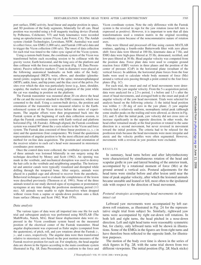
The stick figures of Fig. 5C show the similarities in theinitial phase of the head movement in the control and postle-sion trials. In both, the anterior trunk moves slightly to the right(Fig. 5C, arrow 1) as the head begins rotating to the left. Then,the shoulder girdle rotates CCW in yaw and returns to the leftas the head movement is completing (Fig. 5C, arrow 2).However, as a result of the abnormal rightward thrust in thepostlesion trial, the trunk then accelerates to the left and the catfalls to that side (Fig. 5C, bottom stick figures; note the rightforepaw has stepped to the left while the right hind lifts off theforce plate).
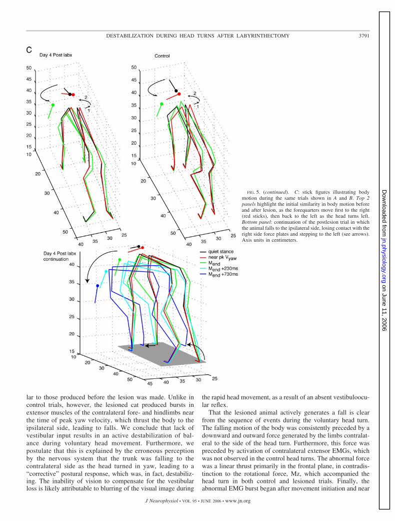
Figure 6 compares control and postlesion data for cat Stacross three velocity ranges, averaged with respect to peak yawvelocity. Trials from the 1st wk only were included in thesepostlesion data, to eliminate any significant effects of compen-sation. On average, the control and postlesion data were wellmatched for yaw velocity and amplitude profiles (Fig. 6A, toptraces). Peak Mz was larger in the postlesion case across allvelocities, and followed by a reversal, especially at the highestvelocity. This was reflected in the increase in slope in therelationship between peak yaw velocity and peak Mz for allthree lesioned cats (Fig. 9). The frontal plane force trajectoriesof the right side illustrate the abnormal thrust that follows peakyaw velocity in the postlesion averages (Fig. 6B). The down-ward and rightward force after peak yaw velocity increasedwith velocity of the head turn.
Figure 6C shows a composite from all three cats of averagedEMGs, comparing pre- and postlesion activity across the threevelocity ranges. Extensors of the contralateral fore- and hind-limbs show the large activation just before peak yaw velocitythat characterizes the postlesion data (Fig. 6C; see, e.g., graybars in traces of velocity range 3). Also of interest is thereciprocal inhibition in some extensors of the ipsilateral fore-and hindlimb (Fig. 6C, gray bars in TRLA, GLUT). The lateextensor activation in the ipsilateral limbs is a reaction to
increased loading of those limbs as the body is thrust to theipsilateral side. The EMG, kinetic, and kinematic data suggestthat the lesioned animal actively destabilizes its balance afterthe head movement is under way, by applying force against theground to drive the body toward the ipsilateral side.
One might suppose that overbalancing to the ipsilateral sideafter vestibular lesion merely arises from hypermetria duringthe latter part of the head turn, when the body normally followsthe head as in the control condition. The postlesion hyperme-tria is exemplified by the increase in peak Mz, which indicatesthat the net acceleration of the body around a central verticalaxis was higher than that in the control case, for a given peakvelocity of head turn. Was the excessive rotational accelerationsufficient to cause instability and falling? To explore thispossibility, head turns were studied in two cats, which dem-onstrated similar hypermetria during head turns. These cats hadsomatosensory loss but normal vestibular function. Somatosen-sory loss was induced by pyridoxine intoxication, which causesloss of peripheral afferent fibers in the diameter range of �7–9�m, affecting primarily group I muscle and large cutaneousafferents of the limbs (Stapley et al. 2002). Both subjects (Brand Kn) showed hypermetria as evidenced by the higher-peakMz in the postlesion head turn compared with control (Fig. 7).Nevertheless, neither somatosensory loss animal showed anyevidence of abnormal thrust in the frontal plane force trajec-tories from the contralateral limbs (Fig. 7, far right column).Instead, the force trajectories are disorganized and irregularcompared with control.
Change in voluntary head turns with timevestibular compensation
Head turns were recorded up to about 40 days after laby-rinthectomy. As previously reported by Thomson et al. (1991)animals showed the greatest ataxia and instability during thefirst 3 days after labyrinthectomy. Some improvement wasobserved by the end of the first week. By the end of the month,the animals were able to run in the lab and even negotiatecornering without falling. The head turn data were divided intothree to four periods and averaged by velocity to examine theextent of recovery. Only trials in which the animals were ableto stand unaided on the platform were included. Cats Ti and Stwere able to stand independently for a portion of trials from thefirst day. Cat Ve, although able to stand independently on thefloor, could not stand on the platform without light supportuntil day 3 and would not produce head turns until day 4.Figure 8 shows plots of frontal plane force trajectories for thecontralateral limbs before lesion and during recovery for allthree subjects for the highest velocity average. During theacute phase (days 1–3) animals were highly ataxic and at highrisk for falls as mentioned above (Table 1). Frontal plane forcegenerated at the time of peak head velocity was very large andresulted in falling or stepping to the ipsilateral side with respectto the head turn. Over time, all animals showed considerableimprovement in balance and were able to remain standing, feetin place, after head turns. Even though the amplitude of thefrontal plane force decreased over time, the abnormal thrustdownward and contralateral remained and the profile of theforce vector trajectory never returned to the control pattern.Thus vestibular compensation was accompanied by a reductionin amplitude of the active destabilization but no modification of
TABLE 1. Numbers of head-turn trials classified by behavior afterlabyrinthectomy
SubjectDays
Postlesion Supported Balanced Falls Step/Lift
St 1–3 34 42 13 164–7 1 121 5 9
11–19 0 143 1 1143 0 58 0 0
Ve 4–8 0 229 1 1811–31 0 242 0 237–43 0 84 0 0
Ti 1–3 2 79 14 54–8 0 133 0 39–38 0 203 0 2
Values are means � SD. For supported trials, the experimenter providedlight touch before and during head turn. For the remainder of the trials, catswere freely standing before head turn and ended the trial in one of threeconditions: 1) balanced � remained standing, feet in place; 2) falls � fallingduring head turn, which required a catch by the experimenter; and 3) step-lift � head turn induced stepping off force plate by one or more paws, or liftingand replacing of one or more paws.
3789DESTABILIZATION DURING HEAD TURNS AFTER LABYRINTHECTOMY
J Neurophysiol • VOL 95 • JUNE 2006 • www.jn.org
on June 11, 2006 jn.physiology.org
Dow
nloaded from

direction of the erroneous response. The relationship betweenpeak yaw velocity and peak Mz showed a dramatic increase inslope during the initial period after lesion, compared withcontrol (Fig. 9). The slope decreased over time but did notreturn to control levels, suggesting that the hypermetria wasreduced but not completely compensated.
D I S C U S S I O N
Cats that underwent a complete bilateral labyrinthectomyshowed frequent instability and loss of balance during volun-tary head turns. The initial components of the head turn andaccompanying postural responses were hypermetric, but simi-
FIG. 5. Representative trials of a left head turn in cat St,before and 4 days after labyrinthectomy. A: head angularkinematics, ground reaction force components (GRFz,GRFx), moment (Mz), and right forelimb extensor EMGs(TRLO, TRLA) plotted against time relative to onset ofhead movement at time 0 (vertical gray line). Trials werematched by similar peak yaw velocity and segmentation ofthe yaw velocity curve. Note the similarity between lesionand control trials from onset of head movement to peakvelocity. Vertical gray bars highlight the period from peakyaw velocity to the end of head movement. Note during thisperiod in the postlesion trial, the increase in vertical andlateral forces of the right limbs (green traces) and the extraburst of activity in the forelimb extensors compared with thecontrol trial, suggesting an active push-off by the right sidelimbs. Pink arrow beneath the time axis indicates the mo-ment at which the right limbs lose contact with the forceplates as the animal falls to the ipsilateral (left) side. B, top:horizontal plane vector trajectories of cat-generated force(conventions as in Fig. 2C). Bottom: cat-generated force inthe frontal plane. Dots represent the change in net forcefrom background (Fx vs. Fz) for the right side fore- andhindlimbs combined. Red dots are values from onset to peakvelocity of the head movement; black dots, from peakvelocity to stable posture after the end of head movement(control) or to the time the right limbs lost contact with theforce plates (postlesion). Time between each dot is 5 ms.Forces at the end of head movement are represented by thegreen lines in the horizontal plane plots and the green squarein the frontal plane plots. Gray arrows indicate the rotationof the force vectors in the horizontal plane from onset topeak yaw velocity, which generate the moment about thez-axis. Black arrows highlight the differences betweenpostlesion and control trials for the period after peak yawvelocity. Note the large rightward thrust in the postlesiontrial. This thrust is also seen in the frontal plane (bottomplots) where the combined force of the right fore- andhindlimbs is downward and to the right, beginning at thetime of peak yaw velocity. LF, RF left, right forelimb; LH,RH left, right hindlimb.
3790 STAPLEY ET AL.
J Neurophysiol • VOL 95 • JUNE 2006 • www.jn.org
on June 11, 2006 jn.physiology.org
Dow
nloaded from
lar to those produced before the lesion was made. Unlike incontrol trials, however, the lesioned cat produced bursts inextensor muscles of the contralateral fore- and hindlimbs nearthe time of peak yaw velocity, which thrust the body to theipsilateral side, leading to falls. We conclude that lack ofvestibular input results in an active destabilization of bal-ance during voluntary head movement. Furthermore, wepostulate that this is explained by the erroneous perceptionby the nervous system that the trunk was falling to thecontralateral side as the head turned in yaw, leading to a“corrective” postural response, which was, in fact, destabiliz-ing. The inability of vision to compensate for the vestibularloss is likely attributable to blurring of the visual image during
the rapid head movement, as a result of an absent vestibuloocu-lar reflex.
That the lesioned animal actively generates a fall is clearfrom the sequence of events during the voluntary head turn.The falling motion of the body was consistently preceded by adownward and outward force generated by the limbs contralat-eral to the side of the head turn. Furthermore, this force waspreceded by activation of contralateral extensor EMGs, whichwas not observed in the control head turns. The abnormal forcewas a linear thrust primarily in the frontal plane, in contradis-tinction to the rotational force, Mz, which accompanied thehead turn in both control and lesioned trials. Finally, theabnormal EMG burst began after movement initiation and near
FIG. 5. (continued). C: stick figures illustrating bodymotion during the same trials shown in A and B. Top 2panels highlight the initial similarity in body motion beforeand after lesion, as the forequarters move first to the right(red sticks), then back to the left as the head turns left.Bottom panel: continuation of the postlesion trial in whichthe animal falls to the ipsilateral side, losing contact with theright side force plates and stepping to the left (see arrows).Axis units in centimeters.
3791DESTABILIZATION DURING HEAD TURNS AFTER LABYRINTHECTOMY
J Neurophysiol • VOL 95 • JUNE 2006 • www.jn.org
on June 11, 2006 jn.physiology.org
Dow
nloaded from
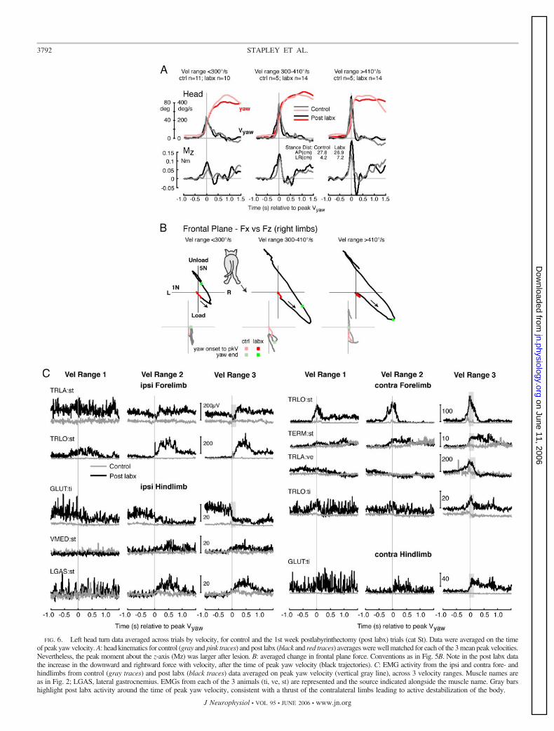
FIG. 6. Left head turn data averaged across trials by velocity, for control and the 1st week postlabyrinthectomy (post labx) trials (cat St). Data were averaged on the timeof peak yaw velocity. A: head kinematics for control (gray and pink traces) and post labx (black and red traces) averages were well matched for each of the 3 mean peak velocities.Nevertheless, the peak moment about the z-axis (Mz) was larger after lesion. B: averaged change in frontal plane force. Conventions as in Fig. 5B. Note in the post labx datathe increase in the downward and rightward force with velocity, after the time of peak yaw velocity (black trajectories). C: EMG activity from the ipsi and contra fore- andhindlimbs from control (gray traces) and post labx (black traces) data averaged on peak yaw velocity (vertical gray line), across 3 velocity ranges. Muscle names areas in Fig. 2; LGAS, lateral gastrocnemius. EMGs from each of the 3 animals (ti, ve, st) are represented and the source indicated alongside the muscle name. Gray barshighlight post labx activity around the time of peak yaw velocity, consistent with a thrust of the contralateral limbs leading to active destabilization of the body.
3792 STAPLEY ET AL.
J Neurophysiol • VOL 95 • JUNE 2006 • www.jn.org
on June 11, 2006 jn.physiology.org
Dow
nloaded from

the time of peak yaw velocity, suggesting that the imbalancewas linked to some feature of the head movement.
The question arises, then, as to the origin of the activedestabilization characteristic of the labyrinthectomized animal.Because the abnormal EMG bursts started after the head was inmotion, it is likely they were initiated by sensory feedbackfrom the ongoing head movement itself. The relevant sensorysignals would likely be those occurring �40 ms before theonset of the abnormal EMG bursts, to allow time for thepostural system to process the inputs and generate the re-sponse. The fall typically occurred in the frontal plane, leadingus to suggest that the critical afferent signal that triggered thefall was linked to motion of the head in the frontal (roll) plane.As shown in Figs. 2, 3, and 5, a voluntary head turn isaccompanied by a stereotypical roll rotation of the head in thedirection of ipsilateral ear down. During the head turn, theintact cat receives both vestibular signals encoding velocity ofhead roll in space and neck proprioceptive signals of head rollwith respect to the trunk. When the head-on-trunk is subtractedfrom head-in-space, the result is zero, which indicates that thetrunk is not moving and therefore no postural correction isnecessary. In contrast, the lesioned cat lacks the signal of headroll in space; thus we suggest that the neck proprioceptive inputof head-on-trunk, in the absence of an accompanying head-in-
space input, is interpreted as the body rolling under a stablehead. In other words, the lesioned animal perceives that theirtrunk is falling in the frontal or roll plane, rather than the headrolling on a stable trunk. Figure 10 illustrates how a leftear-down roll of the head, which accompanies a left turn, couldbe misinterpreted as a rightward fall of the body in the absenceof vestibular input. Such a misperception would trigger anerroneous postural response to thrust the body to the ipsilateralor left side, consistent with our observations.
The absence of otolith inputs may also contribute to anillusory body motion to the contralateral side. The head turns inour study were characterized by a significant translation com-ponent, ipsilateral and backward (e.g., Figs. 2B and 5C), whichwould normally stimulate the otolith organs. After labyrinthec-tomy, a leftward head turn could be misinterpreted as a right-ward linear translation of the body in space, thus reinforcingthe illusion in the roll plane.
Concerning the yaw rotation, there is no evidence that thelesioned cat misperceived the rotation of the body about thevertical axis. Perhaps the combination of the motor command(efference copy), visual feedback, and limb proprioceptiveinputs relating to Mz were sufficient to override the lack of ayaw vestibular signal and indicate the successful completion ofthe primary goal of a gaze shift. The nose-down pitch rotation
FIG. 7. Head kinematics, Mz, and change in frontalplane force averaged with respect to peak yaw velocityfor the highest-velocity group within each cat. Pre- andpostlesion data are shown for the 3 vestibular cats(above), and 2 somatosensory-loss cats (below). Note thehypermetria in the Mz traces for all cats. Only the ves-tibular-loss animals displayed the downward and right-ward force thrust beginning around the time of peak yawvelocity. Force trajectories for the somatosensory-lossanimals were more disorganized and erratic. Arrows in-dicate the direction of the frontal plane force vectortrajectory immediately after peak yaw velocity.
3793DESTABILIZATION DURING HEAD TURNS AFTER LABYRINTHECTOMY
J Neurophysiol • VOL 95 • JUNE 2006 • www.jn.org
on June 11, 2006 jn.physiology.org
Dow
nloaded from
that accompanied the head turn may have generated a misper-ception that the trunk was rotating tail down in pitch. If so, theeffects were too subtle to be detected in our analysis, probablybecause of the inherent stability of the cat in pitch arising fromthe long base of support.
Our proposal lends support to the classical hypothesis that acombination of vestibular and neck afferent information con-tributes directly to trunk stability in space (Lindsay et al. 1976;Roberts 1978; Von Holst and Mittelstaedt 1950). Variousauthors have proposed that to maintain balance as the headturns, vestibulospinal and cervicospinal reflexes sum togetherto conserve a stable trunk position (Mergner et al. 1983;Pompeiano 1984; Roberts 1978; Wilson and Peterson 1981). Inthe decerebrate preparation, Lindsay et al. (1976) demonstratedthat vestibular and neck reflexes are cancelled out when thehead is rotated on a stable trunk. The reflex studies aresupported by the finding that some neurons in the vestibularnuclei are modulated by opposing directions of vestibular andneck stimuli for yaw rotations (Anastasopoulos and Mergner1982) and pitch and roll rotations (Boyle and Pompeiano 1980;Kasper et al. 1988a,b); activity in these neurons is cancelledout in a frequency-dependent manner, during head rotation ona stationary body (i.e., vestibular and neck stimulation com-bined). Furthermore, some neurons responded to head rotationin a manner consistent with the encoding of head position in
space (Kasper et al. 1988b). More recent studies in awake,behaving animals (reviewed in Cullen and Roy 2004) haveshown that the responses of vestibular neurons depend on thecurrent behavior and may reflect signals not only from vestibularafferents but also from proprioceptive afferents and efferencecopy signals of the motor command during active movements.
Mergner and colleagues proposed a model for the perceptionof trunk-in-space using proprioceptive and vestibular afferents(Mergner and Rosemeier 1998). This model was originallybased on data from perception studies in humans subjected topassive movements, but has more recently been expanded(Peterka 2002) to balance control in the pitch plane (Mergneret al. 2003). In this schema, trunk-in-space is derived from twodirections of sensory “chaining” (or integration): 1) a top-down(i.e., head to trunk/center of mass) combination of vestibularinputs, and neck and trunk proprioceptive inputs (Mergner etal. 1997) and 2) a bottom-up (i.e., feet to trunk) proprioceptivechaining from limb proprioceptors to trunk (Mergner andRosemeier 1998). Both directions of sensory integration arepurported to be necessary, to resolve sensory ambiguitiesregarding self-motion versus support surface motion. Ourstudy provides experimental support for the top-down conceptby inferring an erroneous perception of trunk-in-space whenone of the receptor types in this chain (vestibular) is notproviding accurate information. Our data also suggest that the
FIG. 8. Change in frontal plane forces before le-sion and during the vestibular compensation period.Frontal plane force trajectories were averaged for theperiods indicated for each cat after lesion, for thehighest-velocity trials and with respect to peak yawvelocity. Arrows indicate direction of the frontal planeforce trajectory after peak yaw velocity. Note thechange in orientation of the trajectories for controlcompared with postlesion data. Note further that thebasic shape of the trajectories does not change overdays postlesion, whereas the extent, or amplitude,becomes smaller.
3794 STAPLEY ET AL.
J Neurophysiol • VOL 95 • JUNE 2006 • www.jn.org
on June 11, 2006 jn.physiology.org
Dow
nloaded from

bottom-up proprioceptive chaining, which remains intact in thelabyrinthectomized cat, is likely overridden by the top-downsystem during a voluntary head turn. In other words, eventhough the limb proprioceptive input should accurately reportthat the support surface is stable, and foot cutaneous inputshould provide veridical information about acceleration of thebody relative to the support surface (Ting and Macpherson2004), these signals are not sufficient to overcome the apparentperception that the body is falling. As compensation proceeds,a greater reliance on somatosensation may underlie the le-sioned animal’s ability to reduce the amplitude of the inappro-priate postural imbalance.
An alternative, but less likely, explanation for the loss ofbalance during head turns is overbalancing that results fromhypermetria. Hypermetria is an abnormal scaling (increase) ofmotor behavior that follows various lesions, and has previouslybeen reported to occur after bilateral vestibular loss, duringgaze shifts in monkeys (Dichgans et al. 1973) and humans(Kasai and Zee 1978), and during support surface translationsin the cat (Inglis and Macpherson 1992, 1995). Inglis andMacpherson (1995) provided a discussion of the underlyingbasis of postural hypermetria with bilateral vestibular loss. Inthe present study, hypermetria was manifest by overshoot ofthe head angular position in yaw, and higher peak rotationalmoment of force, Mz, for a given peak yaw velocity of the head(i.e., increase in slope in the relationship between peak Vyawand peak Mz). Despite the hypermetria demonstrated by these
animals, however, it is unlikely to be the cause of the desta-bilization for the following reasons. Hypermetria occurred inthe horizontal (yaw) plane (rotation about the vertical axis),which was the primary plane of the voluntary movement. Thehypermetric Mz is interpreted as an excessive acceleration ofthe body around the central vertical axis and was followed bya rapid reversal in Mz, to decelerate and stop the movement. Incontrast, the postural instability occurred in the frontal (roll)plane. The destabilizing thrust was exerted laterally and down-ward at the contralateral limbs, resulting in a motion of thebody to the ipsilateral side. It is unlikely that the abnormalforce in the frontal plane arose from an excessive rotationalforce in the orthogonal horizontal plane. Moreover, this abnor-mal behavior was characterized by EMG bursts and forcepeaks that were distinct from the initial components of the headturn and absent in the control, as best seen in single trials (e.g.,Fig. 5A) rather than averages where the variation across trialstends to blend the first and second peaks, especially in the forces.
Other important evidence that the destabilization did notresult from hypermetria came from the two animals withperipheral somatosensory loss induced by high-dose pyridox-ine (vitamin B6). We previously showed that somatosensoryloss induces ataxia and hypermetric responses to support sur-face translation manifest by frequent overshoots and delayedreversals of the position of the center of mass (Stapley et al.2002). In the present study, the two animals with somatosen-sory loss also showed hypermetria during voluntary head turns,but they did not show the lateral destabilizing thrust charac-teristic of the vestibular-loss animals. Therefore we concludethat active destabilization of balance during voluntary headturns is a specific result of the loss of accurate vestibularinformation regarding the acceleration of the head-in-space.
Over time, the labyrinthectomized animals compensated byreducing the amplitude of both the primary moment of force,Mz, and the subsequent destabilizing force in the frontal plane.
FIG. 10. Schema illustrating the proposed misinterpretation of trunk posi-tion in space after bilateral labyrinthectomy. A: in the intact cat, vestibularinputs encode the angular velocity of the head-in-space as it rolls to the leftwhile proprioceptive input from neck muscles encodes the leftward movementof the head with respect to the trunk, leading to the correct perception that thehead is moving left ear down on a stable trunk. B: in the labyrinthectomizedanimal, vestibular inputs send the erroneous signal that the head is not movingin space while proprioceptive inputs from neck muscles indicate a leftwardmovement of the head with respect to the trunk, just as in A. Combination ofvestibular and neck information leads to the erroneous perception that the trunkis rolling right side down, while the head remains upright. (Figure adaptedfrom Melvill Jones 2000.)
FIG. 9. Relationship between peak yaw velocity and peak Mz before lesionand during the vestibular compensation period. Units of slopes are Nm � deg�1
� s�1 � 10�4.
3795DESTABILIZATION DURING HEAD TURNS AFTER LABYRINTHECTOMY
J Neurophysiol • VOL 95 • JUNE 2006 • www.jn.org
on June 11, 2006 jn.physiology.org
Dow
nloaded from

By the end of the first week animals could consistently producehead movements without falling. Even though the amplitude ofthe destabilizing force decreased, this response persisted, rightup until the animals were killed. We may conclude that thecompensation process allowed the animals to turn down move-ment amplitude but did not modify the errors in computation oftrunk-in-space by the postural control system. Vestibular com-pensation of postural and motor deficits after labyrinthectomyis known to involve the restoration of normal, symmetricallevels of spontaneous discharge in the deafferented vestibularnuclei (Galiana et al. 1984; Gernandt and Thulin 1952;Markham et al. 1977; Precht et al. 1966; Xerri et al. 1983).Restoration of this discharge would lead to increased drive ofcerebellar Purkinje cells. Thus it is likely that after bilaterallabyrinthectomy, the toning down of the hypermetria seen inthe present study could have resulted from a restoration ofinhibitory tone from the cerebellum, perhaps through tonicdescending drive in vestibulospinal and/or reticulospinal path-ways. This mechanism could be viewed as a nonspecificreduction in gain of the motor output, turning down bothdesired as well as inappropriate actions. In part, the restorationof tonic activity in the vestibular nuclei results from increasedsynaptic drive from somatosensory spinal afferents (Jensen1979). One might speculate that an increased somatosensorydrive would mediate an increase in reliance on somatosensoryinputs for balance control. This might manifest as an increasein weighting of the bottom-up chaining of proprioceptiveinputs (Mergner and Rosemeier 1998) for accurately comput-ing trunk-in-space. That this does not seem to be the case isevident by the persistence of the inappropriate destabilizingthrust during voluntary head turns. It is unknown whether theanimals might have achieved total suppression of the abnormalresponse with a longer recovery period.
To conclude, the results of this study provide the firstevidence that animals with vestibular loss actively generate adestabilizing force that, in the early phase after lesion, can leadto falls. These results suggest that absence of vestibular inputsleads to misinterpretation of the position and motion of thetrunk-in-space during active head movements. Therefore ves-tibular information is critically important for calculating posi-tion of the trunk-in-space for the balance control system whenactive head turns are made. This mechanism may underlie thepostural instability vestibular patients experience while turningtheir heads during stance or locomotion (Herdman 1994).
A C K N O W L E D G M E N T S
We thank Dr. Charles Russell, N. Schuff, and I. Albrecht for expert technicalassistance.
G R A N T S
This study was funded by National Institute of Deafness and Other Com-munication Disorders and the American Hearing Research Foundationawarded to J. M. Macpherson.
R E F E R E N C E S
Anastasopoulos D and Mergner T. Canal–neck interaction in vestibularnuclear neurons of the cat. Exp Brain Res 46: 269–280, 1982.
Boyle R and Pompeiano O. Reciprocal responses to sinusoidal tilt of neuronsin Deiters’ nucleus and their dynamic characteristics. Arch Ital Biol 118:1–32, 1980.
Cullen KE and Roy JE. Signal processing in the vestibular system duringactive versus passive head movements. J Neurophysiol 91: 1919–1933,2004.
Dichgans J, Bizzi E, Morasso P, and Tagliasco V. Mechanisms underlyingrecovery of eye-head coordination following bilateral labyrinthectomy inmonkeys. Exp Brain Res 18: 548–562, 1973.
Galiana HL, Flohr H, and Melvill Jones G. A reevaluation of intervestibularnuclear coupling: its role in vestibular compensation. J Neurophysiol 51:242–259, 1984.
Gernandt BE and Thulin LH. Vestibular connections to the brainstem. Am JPhysiol 171: 121–127, 1952.
Herdman SJ. Vestibular rehabilitation. In: Contemporary Perspectives inRehabilitation, edited by Wolf SL. Philadelphia, PA: F.A. Davis, 1994, p.392.
Horak FB. Comparison of cerebellar and vestibular loss on scaling of posturalresponses. In: Disorders of Posture and Gait, edited by Brandt T, Paulus W,Bles W, Dieterich M, Krafczyk S, and Straube A. Stuttgart, Germany: GeorgThieme Verlag, 1990, p. 370–373.
Horak FB and Macpherson JM. Postural orientation and equilibrium. In:Handbook of Physiology. Exercise: Regulation and Integration of MultipleSystems. Bethesda, MD: Am. Physiol. Soc., 1996, sect. 12, p. 255–292.
Horak FB, Nashner LM, and Diener HC. Postural strategies associated withsomatosensory and vestibular loss. Exp Brain Res 82: 167–177, 1990.
Inglis JT and Macpherson JM. Postural responses following bilateral laby-rinthectomy. In: Posture and Gait: Control Mechanisms, edited by Wool-lacott MH and Horak FB. Eugene, OR: Univ. of Oregon Press, 1992, p.268–271.
Inglis JT and Macpherson JM. Bilateral labyrinthectomy in the cat: effectson the postural response to translation. J Neurophysiol 73: 1181–1191, 1995.
Jensen DW. Vestibular compensation: tonic spinal influence upon spontane-ous descending vestibular nuclear activity. Neuroscience 4: 1075–1084,1979.
Kasai T and Zee DS. Eye-head coordination in labyrinthine-defective humanbeings. Brain Res 144: 123–141, 1978.
Kasper J, Schor RH, and Wilson VJ. Response of vestibular neurons to headrotations in vertical planes. I. Response to vestibular stimulation. J Neuro-physiol 60: 1753–1764, 1988a.
Kasper J, Schor RH, and Wilson VJ. Response of vestibular neurons to headrotations in vertical planes. II. Response to neck stimulation and vestibular-neck interaction. J Neurophysiol 60: 1765–1778, 1988b.
Lacour M and Borel L. Vestibular control of posture and gait. Arch Ital Biol131: 81–104, 1993.
Lindsay KW, Roberts TDM, and Rosenberg JR. Asymmetric tonic laby-rinth reflexes and their interaction with neck reflexes in the decerebrate cat.J Physiol 261: 583–601, 1976.
Macpherson JM and Inglis JT. Stance and balance following bilaterallabyrinthectomy. In: Natural and Artificial Control of Hearing and Balance,edited by Allum JHJ, Allum-Mecklenburg D, Harris F, and Probst R. NewYork: Elsevier, 1993, p. 219–228.
Macpherson JM, Lywood DW, and van Eyken A. A system for the analysisof posture and stance in quadrupeds. J Neurosci Methods 20: 73–82, 1987.
Marchand AR, Amblard B, and Cremieux J. Visual and vestibular controlof locomotion in early and late sensory-deprived cats. In: VestibulospinalControl of Posture and Locomotion, edited by Pompeiano O and Allum J.New York: Elsevier, 1988, p. 229–238.
Markham CH, Yagi T, and Curthoys IS. The contribution of the contralat-eral labyrinth to second-order neuronal activity in the cat. Brain Res 138:99–109, 1977.
Melvill Jones G. Posture. In: Principles of Neural Science, edited by KandelER, Schwartz JH, and Jessell TM. New York: McGraw-Hill, 2000, p. 823.
Mergner T, Huber W, and Becker W. Vestibular–neck interaction andtransformation of sensory coordinates. J Vestib Res 7: 347–367, 1997.
Mergner T, Maurer C, and Peterka RJ. A multisensory posture controlmodel of human upright stance. Prog Brain Res 142: 189–201, 2003.
Mergner T, Nardi GL, Becker W, and Deecke L. The role of canal–neckinteraction for the perception of horizontal trunk and head rotation. ExpBrain Res 49: 198–208, 1983.
Mergner T and Rosemeier T. Interaction of vestibular, somatosensory andvisual signals for postural control and motion perception under terrestrialand microgravity conditions—a conceptual model. Brain Res Rev 28:118–135, 1998.
Money KE and Scott JW. Functions of separate sensory receptors ofnonauditory labyrinth of the cat. Am J Physiol 202: 1211–1220, 1962.
Peterka RJ. Sensorimotor integration in human postural control. J Neuro-physiol 88: 1097–1118, 2002.
3796 STAPLEY ET AL.
J Neurophysiol • VOL 95 • JUNE 2006 • www.jn.org
on June 11, 2006 jn.physiology.org
Dow
nloaded from
Pompeiano O. Excitatory and inhibitory influences on the spinal cord duringvestibular and neck reflexes. In: The Vestibular System: Fundamental andClinical Observations, edited by Stahle J. Acta Otolaryngol Suppl, 1984, p.5–9.
Precht W, Shimazu H, and Markham CH. A mechanism of central com-pensation of vestibular function following hemilabyrinthectomy. J Neuro-physiol 29: 996–1010, 1966.
Roberts TDM. Neurophysiology of Postural Mechanisms (2nd ed.). London:Butterworth, 1978, p. 354.
Stapley PJ, Ting LH, Hulliger M, and Macpherson JM. Automatic posturalresponses are delayed by pyridoxine-induced somatosensory loss. Neuro-science 22: 5803–5807, 2002.
Thomson DB, Inglis JT, Schor RH, and Macpherson JM. Bilateral laby-rinthectomy in the cat: motor behavior and quiet stance parameters. ExpBrain Res 85: 364–372, 1991.
Ting LH and Macpherson JM. Ratio of shear to load ground reaction forcemay underlie the directional tuning of the automatic postural response torotation and translation. J Neurophysiol 92: 808–823, 2004.
Von Holst E and Mittelstaedt H. Das Reafferenzprinzip (Wechselwirkungenzwischen Zentralvervensystem und Peripherie. J Naturwissenschaften 37:464–476, 1950.
Watt DGD. Responses of cats to sudden falls: an otolith-originating reflexassisting landing. J Neurophysiol 39: 257–265, 1976.
Wilson VJ and Peterson BW. Vestibulospinal and reticulospinal systems. In:Handbook of Physiology. The Nervous System. Motor Control. Bethesda,MD: Am. Physiol. Soc., 1981, sect. 1, vol. II, pt. 2, p. 667–702.
Xerri C, Gianni S, Manzoni D, and Pompeiano O. Central compensation ofvestibular deficits. I. Response characteristics of lateral vestibular neurons toroll tilt after ipsilateral labyrinth deafferentation. J Neurophysiol 50: 428–448, 1983.
3797DESTABILIZATION DURING HEAD TURNS AFTER LABYRINTHECTOMY
J Neurophysiol • VOL 95 • JUNE 2006 • www.jn.org
on June 11, 2006 jn.physiology.org
Dow
nloaded from