diabetes mellitus–induced microvascular destabilization in the
TRANSCRIPT
Listen to this manuscript’s
audio summary by
JACC Editor-in-Chief
Dr. Valentin Fuster.
J O U R N A L O F T H E AM E R I C A N C O L L E G E O F C A R D I O L O G Y V O L . 6 9 , N O . 2 , 2 0 1 7
ª 2 0 1 7 B Y T H E AM E R I C A N C O L L E G E O F C A R D I O L O G Y F O U N D A T I O N
P U B L I S H E D B Y E L S E V I E R
I S S N 0 7 3 5 - 1 0 9 7 / $ 3 6 . 0 0
h t t p : / / d x . d o i . o r g / 1 0 . 1 0 1 6 / j . j a c c . 2 0 1 6 . 1 0 . 0 5 8
Diabetes Mellitus–Induced MicrovascularDestabilization in the Myocardium
Rabea Hinkel, DVM,a,b,c Andrea Howe, DVM,a Simone Renner, DVM,d,e Judy Ng, PHD,a Seungmin Lee, PHD,aKatharina Klett, DVM,a,c Veronika Kaczmarek, MD,a,c Alessandra Moretti, PHD,a,b Karl-Ludwig Laugwitz, MD,a,b
Philipp Skroblin, PHD,f Manuel Mayr, MD,f Hendrik Milting, MD,g Andreas Dendorfer, MD,h Bruno Reichart, MD,h
Eckhard Wolf, DVM,d,e Christian Kupatt, MDa,b,h
ABSTRACT
Fro
Ge
for
Ve
Ne
Kle
Ce
Ge
Re
01G
rep
Ma
BACKGROUND Diabetes mellitus causes microcirculatory rarefaction and may impair the responsiveness of ischemic
myocardium to proangiogenic factors.
OBJECTIVES This study sought to determine whether microvascular destabilization affects organ function and ther-
apeutic neovascularization in diabetes mellitus.
METHODS The authors obtainedmyocardial samples from patients with end-stage heart failure at time of transplant, with
or without diabetes mellitus. Diabetic (db) and wild-type (wt) pigs were used to analyze myocardial vascularization and
function. Chronic ischemia was induced percutaneously (day 0) in the circumflex artery. At day 28, recombinant adeno-
associated virus (rAAV) (5� 1012 viral particles encoding vascular endothelial growth factor-A [VEGF-A] or thymosin beta 4
[Tb4]) was applied regionally. CD31þ capillaries per high power field (c/hpf) and NG2þ pericyte coverage were analyzed.
Globalmyocardial function (ejection fraction [EF] and left ventricular end-diastolic pressure)was assessed at days 28 and 56.
RESULTS Diabetic human myocardial explants revealed capillary rarefaction and pericyte loss compared to nondiabetic
explants. Hyperglycemia in db pigs, even without ischemia, induced capillary rarefaction in themyocardium (163� 14 c/hpf
in db vs. 234 � 8 c/hpf in wt hearts; p < 0.005), concomitant with a distinct loss of EF (44.9% vs. 53.4% in nondiabetic
controls; p<0.05). Capillary density further decreased in chronic ischemic hearts, as did EF (both p<0.05). Treatmentwith
rAAV.Tb4 enhanced capillary density and maturation in db hearts less efficiently than in wt hearts, similar to collateral
growth. rAAV.VEGF-A, though stimulating angiogenesis, induced neither pericyte recruitment nor collateral growth. As a
result, rAAV.Tb4 but not rAAV.VEGF-A improved EF in db hearts (34.5� 1.4%), but less so than in wt hearts (44.8� 1.5%).
CONCLUSIONS Diabetes mellitus destabilized microvascular vessels of the heart, affecting the amplitude of
therapeutic neovascularization via rAAV.Tb4 in a translational large animal model of hibernating myocardium.
(J Am Coll Cardiol 2017;69:131–43) © 2017 by the American College of Cardiology Foundation.
D iabetes mellitus (DM) is one of the mostimportant risk factors for developing car-diovascular disease (1,2). Moreover, DM in-
duces additional major adverse coronary events after
m the aI. Medizinische Klinik und Poliklinik, University Clinic Rechts
rmany; bDZHK (German Center for Cardiovascular Research), partner site
Cardiovascular Prevention, Klinikum der Universität München, Munich,
terinary Sciences, Ludwig Maximilian University of Munich, Munich, Ger
uherberg, Germany; fKing’s College London British Heart Foundation
ssmann Institute, Heart and Diabetes Center North Rhine-Westphalia, Ba
ntre for Experimental Medicine, Ludwig Maximilian University of Munic
rman Research Foundation (DFG, TRR 127 to Drs. Hinkel, Reichart, Wolf,
search (DZHK), the German Center for Diabetes Research (DZD), and the G
U1105 to Dr. Kupatt). Dr. Mayr has filed and licensed patent application
orted that they have no relationships relevant to the contents of this pap
nuscript received July 27, 2016; revised manuscript received October 10,
percutaneous coronary interventions (3–6) andbypass grafting, particularly if poorly controlled (7).This comparative disadvantage is also evident wheninterventions are performed for acute coronary
der Isar, Technical University of Munich, Munich,
Munich Heart Alliance, Munich, Germany; cInstitute
Munich, Germany; dGene Center and Department of
many; eGerman Center for Diabetes Research (DZD),
Centre, London, United Kingdom; gErich & Hanna
d Oeynhausen, Germany; and the hWalter-Brendel-
h, Munich, Germany. This work was funded by the
and Kupatt), the German Center for Cardiovascular
erman Ministry for Education and Research (BMBF
s on microRNA biomarkers. All other authors have
er to disclose.
2016, accepted October 12, 2016.

ABBR EV I A T I ON S
AND ACRONYMS
Ang = angiopoietin
c/hpf = capillaries per high
power field
db = diabetic
LV = left ventricular
miR = microribonucleic acid
p/hpf = pericytes per high
power field
rAAV = recombinant adeno-
associated virus
RCx = ramus circumflex
Tb4 = thymosin beta 4
VEGF-A = vascular endothelial
growth factor A
wt = wild type
Hinkel et al. J A C C V O L . 6 9 , N O . 2 , 2 0 1 7
Diabetes in CVD J A N U A R Y 1 7 , 2 0 1 7 : 1 3 1 – 4 3
132
syndromes (8) and for chronic coronarylesions, aimed at resolving contractiledysfunction in viable myocardium (i.e.,hibernating myocardium) (9,10).
Apparently, macrovascular treatment op-tions for coronary obstructions are antago-nized by additional factors beyond the reachof conventional recanalization strategies (11).A continuous inflammatory disposition ofmicrovessels has been attributed to vesselregression in most organs (12), except forreactive inflammatory vessel growth in theeye (13). Both rarefaction and capillarysprouting imply vessel destabilization as acommon denominator. In this concept, peri-cyte detachment is caused by inflammatoryendothelial activation (13). The diabetic in-flammatory process might be aggravated by
exogenous vessel-destabilizing factors such asvascular endothelial growth factor A (VEGF-A) (14),potentially blunting its efficacy in therapeutic neo-vascularization. Enhancing microvascular stability(e.g., by providing platelet-derived growth factor Bfor pericyte attraction) has been demonstrated to in-crease blood flow into ischemic myocardium, whenadded to VEGF-A (15). One vascular growth factor,
SEE PAGE 144
which provides both capillary sprouting and pericyteinvestment, is thymosin beta 4 (Tb4) (16), which in-duces lasting and functional microvascular networksin wild-type (wt) animals, and stabilizes microvesselsin the instance of inflammation (17).
To determine whether microvascular destabiliza-tion affects organ function and therapeutic neo-vascularization in a clinically relevant large animalmodel, we studied ischemic cardiomyopathy inINSC94Y transgenic pigs, a model of permanentneonatal DM (18). In this model, fasting glucose levelsincreased to 300 to 400 mg/dl after birth. Weanalyzed hearts of 5-month-old db and wt pig heartswith or without hibernating myocardium, and appliedregional adeno-associated virus (AAV)–based vasculargene therapy in the latter. Our results indicated thatmolecular treatment aiming at balanced vasculargrowth and maturation can improve hibernatingmyocardium in individuals with DM, although to alesser extent than in age-matched non-DM controls.
METHODS
Tissue samples of the nonischemic and ischemic an-imals (ramus circumflex [RCx] perfused area, wt anddb) and patient samples (5 in the non-DM and 4 in the
DM group, left ventricle [LV], ischemic area) wereanalyzed for capillary density (platelet endothelialcell adhesion molecule-1–positive cells) and pericyteinvestment (NG2-positive cells). More informationabout tissue staining is in the Online Appendix.
Myocardial tissue specimens were procured frompatients undergoing heart transplantation (OnlineTable 1). Patients provided informed consent for thescientific use of the explanted tissue. The study wasapproved by the institutional ethics boards of theclinical and experimental study contributors (H.M.,A.D.). Specimens of LV myocardium (4 in the non-DMand 5 in the DM group) were obtained as 2 � 2 cm2
transmural biopsies from explanted failing hearts atthe Heart and Diabetes Center of North Rhine-Westphalia, and were prepared as described in theOnline Appendix.
Isometric contraction force was measured at apreload of 1 mN under continuous field stimulation(rate 0.5 Hz, pulse duration 3 ms) at 1.5-fold excita-tion threshold. Strain- and rate-related alterations ofcontractility were determined in the presence of 1 mMisoprenaline. Maximum twitch force was assessed atoptimum preload. Tissue elastance was calculatedfrom the increase in diastolic tension provoked by a1 mm extension beyond relaxed length.
The preparation of cells for direct Matrigel assays(BD Biosciences, Heidelberg, Germany), pericytecoculture experiments, and shear stress experimentsis described in the Online Appendix.
Recombinant AAV (rAAV) 2.9 vectors encodingb-galactosidase lacZ, Tb4, or VEGF-A were producedusing the triple transfection method as describedearlier (19) and in the Online Appendix.
Transgenic pigs presenting with permanentneonatal DM were generated as described previously(18). The INSC94Y mutation disrupts 1 of the 2 disulfidebonds between the A and B chains of the insulinmolecule, resulting in misfolded insulin, impaired in-sulin secretion, endoplasmic reticulum stress, andapoptosis of the pancreatic beta cells. Consequently,these transgenic pigs present with permanentneonatal DM, which was treated with insulin until7 days before experiment onset. Healthy nontrans-genic littermates served as controls (Online Figure 1A).
German landrace pigs were anesthetized and instru-mented as previously described (20) and in the OnlineAppendix. Of 37 animals initiated by stent placement,5 (3 wt and 2 db) were lost due to sudden cardiac deathduring the first 28 days, whereas no animal was lostbetween days 28 and 56. The remaining 32 animals(14 wt, 18 db) were treated by mock transduction(5� 1012 rAAV containing no transgene; n¼ 7wt, n¼ 6 db),rAAV.VEGF-A transduction (5 � 1012 particles;

FIGURE 1 DM Alters Cardiac Function
nondiabetic
**
**
*
12
10
8
6
4
2
0
1.2
1
0.8
ΔΔ C
T
987654321
0
6
5
4
3
2
1
0
0.6
0.4
0.2
8
7
6
5
4
3
2
1
0
0
diabetic
nondiabetic
NG2
PECA
M-1
diabetic
nondiabeticdiabetic
p=0.1
p=0.38
Tβ4 Ang1/Ang2
nondiabetic diabeticnondiabetic diabetic
nondiabetic
NG2
+ Ce
lls /
hpf
Elas
tanc
e [m
N/m
m]
Twitc
h at
1 m
N Pr
eloa
d [m
N]
PECA
M-1
+ C
ells
/ hp
f
diabetic
A B
C D
E F
(A) Tissue samples of patients with diabetes mellitus (DM) undergoing heart transplantation display capillary rarefaction. (B) Besides a
reduced capillary density (platelet endothelial cell adhesion molecule-1 positive [PECAM-1þ]), (C) these patients demonstrated a loss of
pericytes (NG2þ). (D) DM status did not change expression of thymosin beta 4 (Tb4) in the human cardiac tissue of end-stage heart failure
patients. The ratio of angiopoietin (Ang) 1 and 2 expression was reduced in patients with DM. (E) Force development of human cardiac tissue
slides revealed a reduced contractile function in DM patients undergoing heart transplantation. (F) Ventricular stiffness is impaired in DM
heart slices compared to non-DM samples. Mean � SEM; n ¼ 4; *p < 0.05. hpf ¼ high power field.
J A C C V O L . 6 9 , N O . 2 , 2 0 1 7 Hinkel et al.J A N U A R Y 1 7 , 2 0 1 7 : 1 3 1 – 4 3 Diabetes in CVD
133
n ¼ 5 db), or rAAV.Tb4 transduction (5 � 1012 particles;n ¼ 7 wt and db) via continuous pressure-regulatedretroinfusion into the lateral vein (20).
Twenty-eight days after implantation of a reductionstent, the myocardium was transduced with Tb4 orVEGF-A by selective pressure regulated retroinfusion
of 5 � 1012 rAAV.Tb4 or 1 � 1013 rAAV.VEGF-A particlesinto the lateral vein, which anatomically drains theRCx-perfused myocardium (15) (Online Figure 1B).Four weeks later (day 56), we assessed collateralmorphology and myocardial function as well as per-formed tissue harvesting for histochemical analysis.

FIGURE 2 High Glucose Levels Alter Proangiogenic Effects
Normo-glucosecontrol
40
30
*
*
**
***
** **
**
****
** **
**
**
**
20
10
0
40
50
30
20
10
0
10987654321
0
40
50
30
20
10
0
Tβ4 control Tβ4 VEGF-A
control Tβ4 control Tβ4 VEGF-A
High-glucose
Normo-glucosecontrol Tβ4
control Tβ4 control Tβ4
control Tβ4 VEGF-AHigh-glucose
Normo-Glucose
Ring
s / h
pf
Unco
vere
d Ar
ea [%
]Pe
ricyt
es/R
ing
mer
geTH
P-1 C
ells
peric
ytes
tube
s
High-Glucosecontrol Tβ4 control Tβ4 VEGF-ANormo-Glucose High-Glucose
control Tβ4 control Tβ4 VEGF-ANormo-Glucose High-Glucose
control Tβ4control Tβ4Normo-Glucose High-Glucose
Normo-glucosecontrol Tβ4 control Tβ4 VEGF-A
High-glucoseA B
C
E
D
(A) Tube formation capacity of murine endothelial cells is altered under high glucose levels; Tb4 and vascular endothelial growth factor-A (VEGF-A) transfection
enhanced tube formation under normal and high glucose levels (scale bar ¼ 200 mm). (B) Similarly, high glucose reduced endothelial cell migration in vitro
in a wound-scratch assay (red area ¼ uncovered area; scale bar ¼ 200 mm) whereas Tb4 or VEGF-A overexpression enhanced migration capacity in normal- and
high-glucose conditions. (C and D) High glucose levels reduced tube maturation, assessed as pericyte recruitment (green fluorescence) to endothelial rings
(red fluorescence; scale bar ¼ 100 mm); transfection of Tb4, but not of VEGF-A, enhanced maturation at normal and high glucose levels. (E) High glucose levels
increased adhesion of human monocytic THP-1 cells under venular shear stress (4 dyn/s; scale bar ¼ 100 mm); Tb4 transfection reduced this endothelium-leukocyte
interaction in normal and high glucose levels. Mean � SEM; n ¼ 3. *p < 0.05; **p < 0.001. Abbreviations as in Figure 1.
Hinkel et al. J A C C V O L . 6 9 , N O . 2 , 2 0 1 7
Diabetes in CVD J A N U A R Y 1 7 , 2 0 1 7 : 1 3 1 – 4 3
134

FIGURE 3 Capillary Rarefaction
400 ****
**
**
**
**
350
300
250
200
150
100
50
wild type diabetic
wild type diabetic
wild type diabetic
0
300
250
200
150
100
80
70
60
50
40
30
20
10
0
50
0
wild type diabetic
wild type diabetic
wild type diabeticBaseline 120 150 bpm
wild type diabetic
300
250
200
150
100
50
0
18
wt
wt
Mer
geNG
2PE
CAM
-1
db
db
1614121086420
14
*
25
20
15
10
5
0
12
10
8
6
4
2
0
Gluc
ose
[mg/
dl]
NG2
+ Ce
lls/h
pfEF
[%]
SES
[%]
Fibr
osis
[%]
PECA
M-1
+ C
ells
/ hp
fLV
EDP
[mm
Hg]
A B C
G H I
D E F
(A) Fasted blood glucose levels are enhanced in the transgenic diabetic (db) pigs (C94Y mutation in the porcine insulin gene) compared to the wild-type
(wt) littermates. In cardiac tissue, (B and C) capillary density (PECAM-1þ) and (B and D) vessel integrity (NG2þ) were reduced in the transgenic db versus
wt pigs. (E and F) Fibrosis (scale bar ¼ 100 mm) was significantly increased in db hearts. Transgenic db pigs also demonstrated (G) reduced ejection
fraction (EF) and (H) diminished contractile functional reserve assessed by subendothelial sonomicrometry at rest and rapid atrial pacing (120 and
150 beats/min [bpm]). (I) Left ventricular end-diastolic pressure (LVEDP) increased under high-glucose conditions in db pigs. Mean � SEM; n ¼ 4.
*p < 0.05; **p < 0.001. SES ¼ subendocardial segment shortening; other abbreviations as in Figures 1 and 2.
J A C C V O L . 6 9 , N O . 2 , 2 0 1 7 Hinkel et al.J A N U A R Y 1 7 , 2 0 1 7 : 1 3 1 – 4 3 Diabetes in CVD
135
Global myocardial function was assessed at days28 and 56 by means of a pressure-tip catheter placedin the left ventricle (for LV end-diastolic and sys-tolic pressures, dP/dtmax [contraction velocity], and
dP/dtmin [relaxation velocity]), whereas LV angiog-raphy was performed in anterior-posterior position forthe analysis of ejection fraction, yielding slightlysmaller control values than a right anterior oblique
FIGURE 4 DM Relevance for Vascularization in Chronic Myocardial Ischemia
wt control
cont
rol
dbw
t
cont
rol
PECAM-1 NG2 Merge
db controlwt rAAV.Tβ4 db rAAV.Tβ4 db rAAV.VEGF-A
rAAV
.Tβ4
rAAV
.Tβ4
B
E
wtcontrol
350
300
250
200
150
100
50
0
500
600
400
300
200
100
0
C
A
wtrAAV.Tβ4
**
***
**
**
dbcontrol
dbrAAV.Tβ4
dbrAAV.VEGF-A
**
day 0
**
day 28
**
day 56
wt control wt rAAV.Tβ4 db controldb rAAV.Tβ4 db rAAV.VEGF-A
wtcontrol
350
300
250
200
150
100
50
0
D
wtrAAV.Tβ4
***
**
**
dbcontrol
dbrAAV.Tβ4
dbrAAV.VEGF-A
**
wtcontrol
8
7
6
4
5
3
2
1
0
F
wtrAAV.Tβ4
*
***
dbcontrol
dbrAAV.Tβ4
dbrAAV.VEGF-A
**
wtcontrol
3
2
1
0
G
wtrAAV.Tβ4
**
*
dbcontrol
dbrAAV.Tβ4
dbrAAV.VEGF-A
p=0.064
PECA
M-1
+ C
ells
/ hp
fGl
ucos
e [m
g/dl
]
NG2
+ Ce
lls /
hpf
Colla
tera
ls
Rent
rop
Scor
e
Continued on the next page
Hinkel et al. J A C C V O L . 6 9 , N O . 2 , 2 0 1 7
Diabetes in CVD J A N U A R Y 1 7 , 2 0 1 7 : 1 3 1 – 4 3
136

J A C C V O L . 6 9 , N O . 2 , 2 0 1 7 Hinkel et al.J A N U A R Y 1 7 , 2 0 1 7 : 1 3 1 – 4 3 Diabetes in CVD
137
view. At day 56, subendocardial segment shorteningwas performed after placing sonomicrometry crystalpairs into the ischemic (circumflex-perfused) regionand normalized to measurements in the control region(left anterior descending artery perfused area).Though 1 animal of the treatment group died duringsubendocardial segment shortening assessment, itprovided all other data.
Post-mortem angiograms were taken for visuali-zation of collateral formation and Rentrop scoreanalysis: 0 ¼ no filling; 1¼ side branch filling; 2 ¼partial main vessel filling; and 3 ¼ complete mainvessel filling. Tissue from nonischemic and ischemicLV myocardium was utilized for determining capil-lary density.
STATISTICAL METHODS. The results are given asmean � SEM. Statistical analysis of results betweenmore than 2 experimental groups was performed with1-way analysis of variance; 2-way analysis of variancewas applied in all other figures that compared morethan 2 groups and more than 1 condition. Whenever asignificant effect was obtained with analysis of vari-ance, we performed multiple comparison tests be-tween the groups using the Student Newman-Keulsprocedure. Two experimental groups were comparedby Student t test. All procedures were performed withan SPSS Version 19.0.2 (IBM Corporation, Armonk,New York). Differences between groups wereconsidered significant at p < 0.05.
RESULTS
Although db microvascular alterations are well knownin peripheral or retinal tissue of patients, data arescarce that describe a structure-function correlation indb hearts. Therefore, we first compared myocardiumof end-stage hearts from patients with and withoutDM undergoing heart transplantation, utilizing spec-imens of freshly explanted hearts for histological andfunctional analyses. We found a distinct capillaryrarefaction and pericyte loss in hearts derived frompatients with DM (Figures 1A to 1C), accompanied by alowered angiopoietin (Ang) 1/Ang2 ratio in the DMgroup (Figure 1D). This microvascular alteration coin-
FIGURE 4 Continued
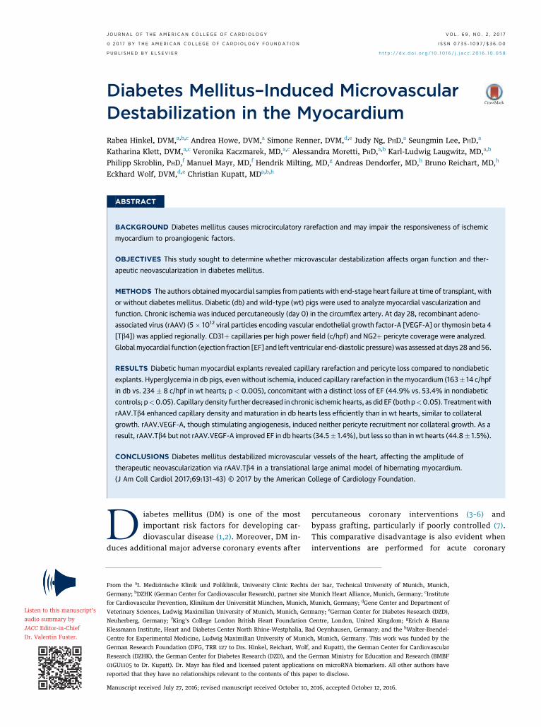
(A) Fasted blood glucose levels are enhanced in db pigs at days 0, 28, an
capillary rarefaction (PECAM-1þ) in both wt and db pig hearts. The redu
tissue in both control groups. This effect was at least partially reversed
application. (E) Representative pictures and (F) quantification of collatera
application showed no cardioprotective effect. (G) Perfusion of the vess
and db control animals. Tb4, but not VEGF-A overexpression, was capab
rAAV ¼ recombinant adeno-associated virus; other abbreviations as in F
cided with a loss of contractility in the papillarymuscle ex vivo (Figure 1E). Moreover, wall stiffness, aparameter for impaired ventricular filling indicatingdiastolic heart failure, was found increased in thesame hearts (Figure 1F), all confirming a correlationbetween microvascular disturbance and dysfunctionin hearts from DM patients.
Next, we investigated the impact of increasedglucose levels on sprouting and maturation of endo-thelial cells in vitro. We used 2 well-known proan-giogenic stimuli, VEGF-A and Tb4 (16), to induce tubeformation of microvascular endothelial cells (bEnd3and human umbilical venous endothelial cells). Un-der normal glucose conditions, cells displayed a highspontaneous tube formation rate, which was almost2-fold increased upon Tb4 stimulation (Figure 2A).Tube formation was reduced at high glucose con-centration (61% of cells at normal glucose), butincreased to 174% upon Tb4 and 162% upon VEGF-Astimulation (Figure 2A). Consistently, Tb4 enhancedthe migratory capacity of endothelial cells in vitro inboth normo- and high-glucose conditions, althoughto a lesser extent in the high-glucose culture condi-tion (Figure 2B). Of note, although Tb4 increasedpericyte recruitment to the newly formed capillaryrings at normo- and high-glucose conditions, addingVEGF-A did not alter pericyte recruitment and high-glucose conditions impaired the capability of endo-thelial cells to attract pericytic cells to newly formedcapillary rings (Figures 2C and 2D).
The proangiogenic endothelial activation of Tb4under normo- and high-glucose conditions did notinduce proinflammatory events, because adhesion ofmonocytic THP-1 cells to endothelial cells decreasedin both conditions upon Tb4 treatment (Figure 2E).Nevertheless, adhesion of THP-1 cells on endothelialcells was found increased at high compared to normalglucose levels, pointing to a chronic inflammatorystate of db endothelium (Figure 2E).
Next, we investigated the impact of high glucoselevels on the microcirculatory status and myocardialfunction in otherwise unchallenged hearts in vivo.Noteworthy, at 5 months of age with blood glucoselevels >300 mg/dl (Figure 3A), a distinct capillary
d 56 versus their age-matched wt littermates. Chronic myocardial ischemia leads to (B and C)
ced capillary number is associated with (B and D) a loss of pericytes (NG2þ) in the ischemic
after rAAV.Tb4 transduction, less so in db animals, an effect not observed after rAAV.VEGF-A
l growth, measured at day 56, which was enhanced after rAAV.Tb4 application; rAAV.VEGF-A
el distal to the occlusion site, displayed as Rentrop score, revealed a reduced filling in wt
le of enhancing vessel perfusion. Mean � SEM; n ¼ 6 to 8. *p < 0.05; **p < 0.001.
igures 1 to 3.

FIGURE 5 Tb4-Induced Cardioprotection
wtcontrol
**
**
* **
****
** ** **
**
****
* ** ***
***
**
*** *
** *
*50
45
40
35
30
25
20
15
10
5
0
25
20
15
10
5
0
25
30
20
15
10
5
0
25
30
20
15
10
5
0
A
C D
E F
B
EF [%
]LV
EDP
[mm
Hg]
SES
Non-
Isch
emic
Reg
ion
[%]
SES
Isch
emic
Reg
ion
[%]
4
2
3
0
1
-5
-4
-3
-2
-1
Delta
LVE
DP [m
m H
g]
0
-5
-10
5
10
15
20
25
Delta
EF
[%]
wtrAAV.Tβ4
dbcontrol
dbrAAV.Tβ4
dbrAAV.VEGF-A
wtcontrol
wtrAAV.Tβ4
Baseline Pacing 120 Pacing 150 bpm Baseline Pacing 120 Pacing 150 bpm
dbcontrol
dbrAAV.Tβ4
dbrAAV.VEGF-A
wtcontrol
wtrAAV.Tβ4
dbcontrol
dbrAAV.Tβ4
dbrAAV.VEGF-A
wtcontrol
wtrAAV.Tβ4
dbcontrol
dbrAAV.Tβ4
dbrAAV.VEGF-A
wt control wt rAAV.Tβ4 db control db rAAV.Tβ4 db rAAV.VEGF-A
(A) Systolic myocardial function, measured by EF, improved in rAAV.Tb4-transduced but not in rAAV.VEGF-A treated animals at day 56. (B) Change from day 28 to 56
(DEF) showed comparable changes in wt and db rAAV.Tb4 animals, although at different levels. (C) LVEDP increased in wt and db ischemic hearts days 28 to 56, unless
Tb4 was overexpressed. VEGF-A transduction did not reduce the enhanced LVEDP in the ischemic db hearts. (D) Change from days 28 to 56 (DLVEDP) showed similar
effects in wt and db rAAV.Tb4 animals, but at different levels. (E) Functional reserve in the nonischemic (left anterior descending region) tissue is diminished in db
hearts compared to normoglycemic animals. (F) In the ischemic (ramus circumflexus region), rAAV.Tb4 application enhanced myocardial contractile function in both
wt and db animals, whereas rAAV.VEGF-A showed no protective effect. Mean � SEM; n ¼ 6 to 8. *p < 0.05; ** p < 0.001. Abbreviations as in Figures 1 to 4.
Hinkel et al. J A C C V O L . 6 9 , N O . 2 , 2 0 1 7
Diabetes in CVD J A N U A R Y 1 7 , 2 0 1 7 : 1 3 1 – 4 3
138

FIGURE 6 DM-Induced Molecular Alterations
Ang1
/Ang
2 Ex
pres
sion
Fibr
osis
[%]
dbrAAV.VEGF-A
dbrAAV.Tβ4
wtrAAV.Tβ4
wtrAAV.Tβ4
wtcontrol
3
2.5
2
1.5
1
0.5
0db
controldb
rAAV.Tβ4db
rAAV.VEGF-A
**
*
*
****
*
rAAV.Tβ4control
rAAV.Tβ4control
wt
db
rAAV.VEGF-A
dbcontrol
wtcontrol
25
20
15
10
5
0
A B
C
(A) Ang1/Ang2 expression improved after Tb4 but not VEGF-A transduction in both wt and db animals. (B and C) Chronic myocardial ischemia-induced
fibrosis (scale bar ¼ 100 mm) is accelerated under diabetic conditions (wt control vs. db control). Overexpression of Tb4 or VEGF-A significantly reduced
fibrosis in the ischemic tissue in both wt and db hearts. Mean � SEM; n ¼ 6 to 8 for fibrosis. *p < 0.05; **p < 0.001. Abbreviations as in Figures 1, 3, and 4.
J A C C V O L . 6 9 , N O . 2 , 2 0 1 7 Hinkel et al.J A N U A R Y 1 7 , 2 0 1 7 : 1 3 1 – 4 3 Diabetes in CVD
139
rarefaction was detectable in normoxic db pig hearts(163 � 14 capillaries per high power field [c/hpf] in dbvs. 234 � 8 c/hpf in wt) (Figures 3B and 3C). Moreover,pericyte investment of the remaining capillaries wasdecreased, rendering the microcirculatory networksin nonischemic hearts impaired (144 � 6 vs. 219 � 4pericytes per hpf [p/hpf]) (Figures 3B and 3D). Coin-cidentally, an increase in interstitial fibrosis wasnoted in db hearts (15.6 � 0.6% vs. 2.1 � 0.5%)(Figures 3E and 3F). This structural alteration wasassociated with an impairment of systolic parameterssuch as ejection fraction (44.9 � 1.9% in db vs. 53.4 �1.2% in wt hearts) (Figure 3G). Consistently, dbhearts demonstrated a decrease in functional reserve
(6.6 � 1.3% vs. 15.4 � 2.3% at 150 beats/min)(Figure 3H) as well as an increase of LV end-diastolicpressure (12.7 � 1.0 mm Hg vs. 8.2 � 0.7 mm Hg)(Figure 3I).
In a second set of experiments, we analyzedthe response of db porcine hearts (blood glucose>300 mg/dl) (Figure 4A) and age-matched controlhearts subjected to regional chronic ischemia inflictedby gradual occlusion of the circumflex artery. Wefound a higher degree of microcirculatory rarefactionin db than in normoglycemic hibernating myocardium(129 � 7 c/hpf vs. 150 � 4 c/hpf) (Figures 4B and 4C).Pericyte investment of capillaries was also decreasedin db hearts (106 � 4 p/hpf vs. 136 � 5 p/hpf)

Hinkel et al. J A C C V O L . 6 9 , N O . 2 , 2 0 1 7
Diabetes in CVD J A N U A R Y 1 7 , 2 0 1 7 : 1 3 1 – 4 3
140
(Figures 4B to 4D). Of note, rAAV.Tb4 transductionsignificantly induced capillary growth and matura-tion in db pigs (183 � 9 c/hpf, 163 � 14 p/hpf),although it was less pronounced than in wt hearts(294 � 6 c/hpf, 286 � 7 p/hpf) (Figures 4B to D).Treatment of db hearts with rAAV.VEGF-A treatmentyielded an increase in capillaries (192 � 4 c/hpf)(Figures 4B and 4C). However, the pericyte invest-ment did not concomitantly increase (87 � 4 p/hpf)(Figures 4B and 4D).
The number of collaterals, which may be formed tocompensate for chronic total RCx occlusion, was lowin both db and wt groups (2.1 � 0.5 visible collateralsper heart vs. 1.9 � 0.2 visible collaterals per heart)(Figures 4E and 4F). Upon rAAV.Tb4 transduction,however, collateral formation increased to a largerextent in wt hearts (6.7 � 0.5 collaterals/heart) than indb hearts (4.7 � 0.3 collaterals/heart) (Figures 4Eand 4F). Consistently, Rentrop score indicatingdistal filling of the occluded vessel reached a higherlevel in wt hearts than in db hearts (2.6 � 0.2 vs.2.1 � 0.1) (Figures 4E and 4G). Notably, rAAV.VEGF-Atreatment did not increase arteriogenesis in theischemic region of db hearts (2.2 collaterals/heart),with a Rentrop score at control level (rAAV.VEGF-A1.2 � 0.2 vs. control 1.4 � 0.2) (Figures 4E to G).
The structural impairment of db microvasculatureblunted the functional response of therapeutic neo-vascularization: the gain of ejection fraction inducedby rAAV.Tb4 treatment was less pronounced in the dbcompared to the wt group (Figures 5A and 5B, OnlineFigure 2A). Consistently, the rise in LV end-diastolicpressure observed in untreated ischemic animals(Figure 5C, Online Figure 2B) was reversed afterrAAV.Tb4 treatment in either background (Figures 5Cand 5D). Moreover, the level of regional myocardialfunction in the nonischemic area under rapid pacingwas significantly reduced in db hearts (db 8.8 � 1.2%vs. wt 16.7 � 1.0%) (Figure 5E). In the ischemic region,recovery upon rAAV.Tb4 treatment was present in dbhearts (8.5 � 1.4% in treated db hearts vs. 1.9 � 0.8%in untreated db hearts), but significantly less pro-nounced than in wt hearts (rAAV.Tb4 16.0 � 1.2%)(Figure 5F). In the ischemic region of db hearts,rAAV.VEGF-A did not alter functional reserve (2.9 �0.7%) (Online Figure 2C).
Because the complex myovascular interaction in dbhearts destabilized microvessels and inhibited thera-peutic vessel growth, we investigated the Ang1/Ang2ratio in the ischemic hearts. The reduced Ang1/Ang2ratio in ischemic tissue was enhanced upon Tb4 over-expression in wt as well as db hearts (Figure 6A, OnlineFigure 1C). However, rAAV.VEGF-A application did not
improve the Ang1/Ang2 ratio, pointing to the lack ofmature microvascular growth. In addition, we inves-tigated the alterations in microribonucleic acid (miR),which might account for the pleiotropic actionsinduced by the hyperglycemic state. Three vasoactivemiRs were found up-regulated in the hyperglycemichearts (Online Figure 3): miR 26a, which is implicatedin impaired wound healing angiogenesis in DM (21);miR 92a, a known inhibitor of cardiac angiogenesis(22,23); and miR 133a, whose level was previously re-ported to be elevated in db hearts (24). We found these3 miRs significantly up-regulated in the porcine dbhearts (Online Figure 3), suggesting them as potentialnovel mediators of microvascular rarefaction in DM.Furthermore, fibrosis was induced in the ischemictissue (db 22.5 � 1.0% vs. wt 19.0 � 0.8%) (Figures 6Band 6C). rAAV.Tb4 application significantly reducedfibrosis in db as well as wt hearts (13.1 � 0.6% and11.0 � 0.4%, respectively) (Figures 6B and 6C).
DISCUSSION
In the current study, we assessed the impact of car-diovascular risk factors on the induction of vasculargrowth in the microvascular and macrovascularcompartment. We found that db human heartsdisplayed capillary rarefaction and pericyte loss,accompanied by decreased contractility and increasedstiffness (Figure 1). Moreover, hyperglycemia attenu-ated tube formation, migration, and pericyte attrac-tion upon proangiogenic stimulation in vitro(Figure 2). In vivo, untreated DM induced microcir-culatory rarefaction (Figure 3) and a distinct func-tional cardiac impairment in a transgenic INSC94Y pigmodel. Induction of regional chronic ischemiaimpaired microvascular density in both hyperglyce-mic and normoglycemic hearts, without functionalcollateralization of the ischemic region (Figure 4).Forced angiogenesis via rAAV.VEGF-A, withoutconcomitant vessel maturation had no significanteffect on regional and global function at rest and un-der rapid pacing (Figures 4 and 5). However, balancedvascular growth was induced via rAAV.Tb4 in db pighearts, though to a significantly lesser extent than inwt hearts (Figure 4). Functional impairment of hiber-nating myocardium was improved in db hearts uponTb4 treatment, albeit not to the same extent as innormoglycemic hearts (Figure 5). MiR analysis of dbhearts revealed increased concentrations of anti-angiogenic miR 26a, miR 92a, and miR 133a. Addi-tionally, a higher degree of fibrosis was observed,which was partially reversed by Tb4 and VEGF-A(Figure 6, Online Figure 3). The Central Illustration

CENTRAL ILLUSTRATION Tb4 Gene Therapy and Therapeutic Neovascularization
Hinkel, R. et al. J Am Coll Cardiol. 2017;69(2):131–43.
Diabetes mellitus impairs vascular density in the heart. Proangiogenic gene therapy via thymosin beta 4 (Tb4) or vascular endothelial growth factor-A (VEGF-A)
induces capillary growth in the diabetic heart. However, only Tb4 is capable of inducing therapeutic neovascularization via vessel growth and maturation, thereby
improving perfusion and myocardial function in ischemic normal as well as diabetic hearts. rAAV ¼ recombinant adeno-associated virus.
J A C C V O L . 6 9 , N O . 2 , 2 0 1 7 Hinkel et al.J A N U A R Y 1 7 , 2 0 1 7 : 1 3 1 – 4 3 Diabetes in CVD
141
depicts the specific microcirculatory alterations of DMand the effects of Tb4 and VEGF-A.
Notably, uncontrolled diabetes with continuouslyelevated glucose levels (Figure 3A) itself suffices torarefy microcirculatory density of cardiac muscle.Capillary loss and pericyte dropout have been welldescribed in the retina (25), but are revealed for thefirst time in transgenic db pig hearts and in hearts
from patients with diabetes. Previously, microvesseldestabilization has been attributed to Ang2 over-expression (26). A partial inhibitor of the tyrosinekinase with immunoglobulin-like and EGF-likedomains 2 (Tie2) receptor, Ang2 might destabilizemicrovessels by antagonizing the Ang1/Tie2 interac-tion, which provides a quiescent and maturevessel state. This feature has been attributed to

PERSPECTIVES
COMPETENCY IN MEDICAL KNOWLEDGE: In a
transgenic, porcine model of DM (InsC94Y),
microcirculatory instability compromises myocardial
function and angiogenesis.
TRANSLATIONAL OUTLOOK: Future researchers
should seek ways to promote capillary stability
in patients with diabetes, as this may help
preserve myocardial function and reduce myocardial
ischemia.
Hinkel et al. J A C C V O L . 6 9 , N O . 2 , 2 0 1 7
Diabetes in CVD J A N U A R Y 1 7 , 2 0 1 7 : 1 3 1 – 4 3
142
inflammatory destabilization of microvessels insepsis (27), but may also occur in chronic inflamma-tory endothelial activation (25) or in streptozotocin-induced experimental DM (28). Notably, increasedAng2 expression has been found in aged db/db micecharacterized by capillary rarefaction and fibrosis(29). In our study, the ratio of Ang1/Ang2 was slightly,but not significantly, decreased in db pig and humanhearts compared to normoglycemic hearts (Figures 1Dand 6A, Online Figure 1C), but increased uponrAAV.Tb4 treatment. In the human tissue, Tb4expression was unaltered even though DM humancorneas display a distinct Tb4 reduction (30). How-ever, the vascular growth induction by Tb4 was pre-viously found to be sensitive to Ang2 (16). Thus, animproved Ang1/Ang2 ratio appears crucial for thebalanced vascular growth provided by rAAV.Tb4.
Interestingly, rAAV.VEGF-A had no effect on theAng1/Ang2 ratio (Figure 6A, Online Figure 1C). Thiswas consistent with our previous observationin normoglycemic chronic ischemic pig hearts (15),where rAAV.VEGF-A did not yield sufficientmicrovessel maturation nor macrovessel growth(arteriogenesis) for functional improvement inour hibernating myocardium model. Lack of micro-vessel maturation was found critical, because additionof placenta-derived growth factor B to VEGF-Asufficed to induce augmentation of micro- and mac-rovessels as well as functional improvement (15).
With rAAV.Tb4 treatment, we found a decrease ofCD14þ monocytes, representing the proinflammatoryM1 phenotype, in normoglycemic and hyperglycemicpig hearts (Online Figure 1D). One potential signaltriggering continuous inflammation is increasedbinding of advanced glycation endproducts to theirreceptor (14). Besides its intracellular pro-inflammatory signaling and high mobility groupbox 1- and nuclear facto kB-dependent up-regulationof adhesion molecules (31), cytokines, and chemo-kines, advanced glycation endproducts to their re-ceptor itself interact with Mac-1 on circulatingleukocytes, mediating firm adhesion (32,33) andfurther inflammatory stimulation. Moreover, inmetabolic stress (e.g., DM) endosomal damage-associated molecular patterns may activate theinflammasome for a perpetual inflammatory state(34). Finally, miR alterations may take a toll ofmicrovascular preservation. In this respect, miR 92a,miR 26a, and miR133b have shown a distinct in-crease in hyperglycemia (Online Figure 3), whichmay indicate their involvement in db vascular rare-faction and suggest them as potential therapeutictargets for miR inhibitors (23).
STUDY LIMITATIONS. In this study, we used atransgenic pig model of insulin-dependent DM toinvestigate the efficacy of vascular gene therapyusing rAAV.Tb4 and rAAV.VEGF-A as therapeuticagents. Thus, we analyzed an untreated cardiac riskfactor, representing a worst-case scenario for apotential loss of treatment function. We assumethat most patients of the target population receivecurrent medical treatment schemes, blunting thedescribed loss of efficacy. Nevertheless, comorbid-ities such as DM must be taken into accountwhen calculating the size of a potential treatmenteffect.
CONCLUSIONS
We used a transgenic db pig model to quantifyvessel growth and functional improvement in hi-bernating myocardium subjected to rAAV.Tb4 ther-apy to improve neovascularization. Our resultsindicated an attenuated, but still significant, in-crease of microvessels and macrovessels thatimproved myocardial function. Thus, we concludedthat in the presence of DM, balanced vascular genetherapy, targeting microvascular maturation, andmacrovascular growth in addition to angiogenesis iseffective, although not to the same extent as inhealthy wt animals.
ACKNOWLEDGMENT The authors thank Anja Wolffor expert technical assistance.
REPRINT REQUESTS AND CORRESPONDENCE: Dr.Christian Kupatt, 1. Med. Klinik, University ClinicRechts der Isar, Technical University Munich,Ismaninger Strasse 22, 81675 Munich, Germany.E-mail: [email protected].

J A C C V O L . 6 9 , N O . 2 , 2 0 1 7 Hinkel et al.J A N U A R Y 1 7 , 2 0 1 7 : 1 3 1 – 4 3 Diabetes in CVD
143
RE F E RENCE S
1. Kannel WB. The Framingham Study: ITS 50-yearlegacy and future promise. J Atheroscler Thromb2000;6:60–6.
2. Kim J-J, Hwang B-H, Choi IJ, et al. Impact ofdiabetes duration on the extent and severity ofcoronary atheroma burden and long-term clinicaloutcome in asymptomatic type 2 diabetic patients:evaluation by coronary CT angiography. Eur HeartJ Cardiovasc Imaging 2015;16:1065–73.
3. Bittner V, Bertolet M, Barraza FR, et al.Comprehensive cardiovascular risk factor controlimproves survival: the BARI 2D trial. J Am CollCardiol 2015;66:765–73.
4. Everett BM, Brooks MM, Vlachos HE,Chaitman BR, Frye RL, Bhatt DL. Troponin andcardiac events in stable ischemic heart disease anddiabetes. N Engl J Med 2015;373:610–20.
5. Baber U, Mehran R, Giustino G, et al. Coronarythrombosis and major bleeding after PCI withdrug-eluting stents: risk scores from PARIS. J AmColl Cardiol 2016;67:2224–34.
6. Schoos MM, Dangas GD, Mehran R, et al. Impactof hemoglobin A1c levels on residual plateletreactivity and outcomes after insertion of coronarydrug-eluting stents (from the ADAPT-DES Study).Am J Cardiol 2016;117:192–200.
7. Nyström T, Holzmann MJ, Eliasson B, Kuhl J,Sartipy U. Glycemic control in type 1 diabetes andlong-term risk of cardiovascular events or deathafter coronary artery bypass grafting. J Am CollCardiol 2015;66:535–43.
8. Klempfner R, Elis A, Matezky S, et al. Temporaltrends in management and outcome of diabeticand non-diabetic patients with acute coronarysyndrome (ACS): residual risk of long-term mor-tality persists: Insights from the ACS Israeli Survey(ACSIS) 2000–2010. Int J Cardiol 2015;179:546–51.
9. Schuster A, Morton G, Chiribiri A, Perera D,Vanoverschelde JL, Nagel E. Imaging in the man-agement of ischemic cardiomyopathy: specialfocus on magnetic resonance. J Am Coll Cardiol2012;59:359–70.
10. BARI Investigators. The final 10-year follow-up results from the BARI randomized trial. J AmColl Cardiol 2007;49:1600–6.
11. Rosenson RS, Fioretto P, Dodson PM. Doesmicrovascular disease predict macrovascularevents in type 2 diabetes? Atherosclerosis 2011;218:13–8.
12. Orasanu G, Plutzky J. The pathologic contin-uum of diabetic vascular disease. J Am Coll Cardiol2009;53:S35–42.
13. Hammes H-P, Feng Y, Pfister F, Brownlee M.Diabetic retinopathy: targeting vasoregression.Diabetes 2011;60:9–16.
14. Shoji T, Koyama H, Morioka T, et al. Receptorfor advanced glycation end products is involved inimpaired angiogenic response in diabetes. Dia-betes 2006;55:2245–55.
15. Kupatt C, Hinkel R, Pfosser A, et al. Cotrans-fection of vascular endothelial growth factor-Aand platelet-derived growth factor-B via recom-binant adeno-associated virus resolves chronicischemic malperfusion: role of vessel maturation.J Am Coll Cardiol 2010;56:414–22.
16. Hinkel R, Trenkwalder T, Petersen B, et al.MRTF-A controls vessel growth and maturation byincreasing the expression of CCN1 and CCN2. NatCommun 2014;5:3970.
17. Bongiovanni D, Ziegler T, D’Almeida S, et al.Thymosin beta4 attenuates microcirculatory andhemodynamic destabilization in sepsis. ExpertOpin Biol Ther 2015;15 Suppl 1:S203–10.
18. Renner S, Braun-Reichhart C, Blutke A, et al.Permanent neonatal diabetes in INSC94Y trans-genic pigs. Diabetes 2013;62:1505–11.
19. Bish LT, Morine K, Sleeper MM, et al. AAV9provides global cardiac gene transfer superior toAAV1, AAV6, AAV7, and AAV8 in the mouse andrat. Hum Gene Ther 2008;19:1359–68.
20. Kupatt C, Hinkel R, von Bruhl ML, et al.Endothelial nitric oxide synthase overexpressionprovides a functionally relevant angiogenic switchin hibernating pig myocardium. J Am Coll Cardiol2007;49:1575–84.
21. Icli B, Nabzdyk CS, Lujan-Hernandez J, et al.Regulation of impaired angiogenesis in diabeticdermal wound healing by microRNA-26a. J MolCell Cardiol 2016;91:151–9.
22. Bonauer A, Carmona G, Iwasaki M, et al.MicroRNA-92a controls angiogenesis and func-tional recovery of ischemic tissues in mice. Science2009;324:1710–3.
23. Hinkel R, Penzkofer D, Zuehlke S, et al. Inhi-bition of microRNA-92a protects against ischemia-reperfusion injury in a large animal model.Circulation 2013;128:1066–75.
24. Fichtlscherer S, Zeiher AM, Dimmeler S.Circulating microRNAs: biomarkers or mediators ofcardiovascular diseases? Arterioscler Thromb VascBiol 2011;31:2383–90.
25. Hammes HP, Du X, Edelstein D, et al. Benfo-tiamine blocks three major pathways of hyper-glycemic damage and prevents experimentaldiabetic retinopathy. Nat Med 2003;9:294–9.
26. Hammes HP, Lin J, Wagner P, et al.Angiopoietin-2 causes pericyte dropout in thenormal retina: evidence for involvement in dia-betic retinopathy. Diabetes 2004;53:1104–10.
27. Ziegler T, Horstkotte J, Schwab C, et al.Angiopoietin 2 mediates microvascular and he-modynamic alterations in sepsis. J Clin Invest2013;123:3436–45.
28. Tuo QH, Zeng H, Stinnett A, et al. Critical roleof angiopoietins/Tie-2 in hyperglycemic exacer-bation of myocardial infarction and impairedangiogenesis. Am J Physiol Heart Circ Physiol2008;294:H2547–57.
29. Gonzalez-Quesada C, Cavalera M, Biernacka A,et al. Thrombospondin-1 induction in the diabeticmyocardium stabilizes the cardiac matrix in addi-tion to promoting vascular rarefaction throughangiopoietin-2 upregulation. Circ Res 2013;113:1331–44.
30. Saghizadeh M, Kramerov AA, Tajbakhsh J,et al. Proteinase and growth factor alterationsrevealed by gene microarray analysis of humandiabetic corneas. Invest Ophthalmol Vis Sci 2005;46:3604–15.
31. Schmidt AM, Hori O, Chen JX, et al. Advancedglycation endproducts interacting with theirendothelial receptor induce expression of vascularcell adhesion molecule-1 (VCAM-1) in culturedhuman endothelial cells and in mice. A potentialmechanism for the accelerated vasculopathy ofdiabetes. J Clin Invest 1995;96:1395–403.
32. Frommhold D, Kamphues A, Hepper I, et al.RAGE and ICAM-1 cooperate in mediating leuko-cyte recruitment during acute inflammationin vivo. Blood 2010;116:841–9.
33. Ziegler T, Horstkotte M, Lange P, et al.Endothelial RAGE exacerbates acute pos-tischaemic cardiac inflammation. J Thromb Hae-most 2016;116:300–8.
34. Chibber R, Ben-Mahmud BM, Mann GE,Zhang JJ, Kohner EM. Protein kinase C b2-dependent phosphorylation of core 2 GlcNAc-Tpromotes leukocyte-endothelial cell adhesion: amechanism underlying capillary occlusion in dia-betic retinopathy. Diabetes 2003;52:1519–27.
KEY WORDS angiogenesis, chronicmyocardial ischemia, gene therapy,thymosin b4
APPENDIX For an expanded Methods sectionas well as supplemental figures and a table,please see the online version of this article.