regulation of microglia and macrophage polarization via ... · induced by nqdi-1 drug, decreased...
TRANSCRIPT

fnmol-10-00261 August 10, 2017 Time: 16:9 # 1
ORIGINAL RESEARCHpublished: 14 August 2017
doi: 10.3389/fnmol.2017.00261
Edited by:Clevio Nobrega,
University of Algarve, Portugal
Reviewed by:Tobias Engel,
Royal College of Surgeons in Ireland,Ireland
Albert Quintana,Universitat Autònoma de Barcelona,
SpainLiliana Simões Mendonça,
Centro de Neurociências e BiologiaCelular (CNC), Portugal
*Correspondence:Bon-Nyeo Koo
Received: 31 May 2017Accepted: 31 July 2017
Published: 14 August 2017
Citation:Cheon SY, Kim EJ, Kim JM,
Kam EH, Ko BW and Koo B-N(2017) Regulation of Microglia
and Macrophage Polarization viaApoptosis Signal-Regulating Kinase 1
Silencing after Ischemic/HypoxicInjury. Front. Mol. Neurosci. 10:261.
doi: 10.3389/fnmol.2017.00261
Regulation of Microglia andMacrophage Polarization viaApoptosis Signal-Regulating Kinase1 Silencing after Ischemic/HypoxicInjurySo Yeong Cheon1,2, Eun Jung Kim1,2, Jeong Min Kim1,2, Eun Hee Kam1,2,Byung Woong Ko1 and Bon-Nyeo Koo1,2*
1 Department of Anesthesiology and Pain Medicine, Yonsei University College of Medicine, Seoul, South Korea, 2 Anesthesiaand Pain Research Institute, Yonsei University College of Medicine, Seoul, South Korea
Inflammation is implicated in ischemic stroke and is involved in abnormal homeostasis.Activation of the immune system leads to breakdown of the blood–brain barrier and,thereby, infiltration of immune cells into the brain. Upon cerebral ischemia, infiltratedmacrophages and microglia (resident CNS immune cell) are activated, change theirphenotype to M1 or M2 based on the microenvironment, migrate toward damagedtissue, and are involved in repair or damage. Those of M1 phenotype releasepro-inflammatory mediators, which are associated with tissue damage, while thoseof M2 phenotype release anti-inflammatory mediators, which are related to tissuerecovery. Moreover, late inflammation continually stimulates immune cell infiltration andleads to brain infarction. Therefore, regulation of M1/M2 phenotypes under persistentinflammatory conditions after cerebral ischemia is important for brain repair. Herein, wefocus on apoptosis signal-regulating kinase 1 (ASK1), which is involved in apoptoticcell death, brain infarction, and production of inflammatory mediators after cerebralischemia. We hypothesized that ASK1 is involved in the polarization of M1/M2phenotype and the function of microglia and macrophage during the late stage ofischemia/hypoxia. We investigated the effects of ASK1 in mice subjected to middlecerebral artery occlusion and on BV2 microglia and RAW264.7 macrophage cell linessubjected to oxygen-glucose deprivation. Our results showed that ASK1 silencingeffectively reduced Iba-1 or CD11b-positive cells in ischemic areas, suppressedpro-inflammatory cytokines, and increased anti-inflammatory mediator levels at 7 daysafter cerebral ischemia. In cultured microglia and macrophages, ASK1 inhibition,induced by NQDI-1 drug, decreased the expression and release of M1-associatedfactors and increased those of M2-associated factors after hypoxia/reperfusion (H/R).At the gene level, ASK1 inhibition suppressed M1-associated genes and augmentedM2-associated genes. In gap closure assay, ASK1 inhibition reduced the migrationrate of microglia and macrophages after H/R. Taken together, our results provide new
Frontiers in Molecular Neuroscience | www.frontiersin.org 1 August 2017 | Volume 10 | Article 261
fnmol-10-00261 August 10, 2017 Time: 16:9 # 2
Cheon et al. Effects of ASK1 on Microglia/Macrophage
information that suggests ASK1 controls the polarization of M1/M2 and the functionof microglia and macrophage under sustained-inflammatory conditions. Regulation ofpersistent inflammation via M1/M2 polarization by ASK1 is a novel strategy for repairafter ischemic stroke.
Keywords: apoptosis signal-regulating kinase 1 (ASK1), M1/M2 polarization, BV2 microglia cell line, RAW264.7macrophage cell line, cerebral ischemia, hypoxia, late inflammation, ischemic stroke
INTRODUCTION
Inflammation is a key contributor to the pathophysiology ofischemic stroke and is responsible for abnormal homeostasis(Amantea et al., 2009; Denes et al., 2010; Iadecola andAnrather, 2011). After ischemic insults, activation of theimmune system causes leakiness of the blood–brain barrier(BBB), and immune cells such as macrophages, neutrophilsand leukocytes, infiltrate the lesion area via the disruptedBBB (Amantea et al., 2009; Denes et al., 2010; Iadecola andAnrather, 2011). Recruitment of immune cells results in initiationof inflammatory processes in the brain parenchyma, whichinclude both tissue damage and repair (Fu et al., 2015).Although inflammation is involved in all phases of ischemiccascade (Iadecola and Anrather, 2011), late inflammationin the brain, particularly after ischemic stroke, provokespersistent immune cell recruitment and eventually aggravatescerebral infarcts (Mena et al., 2004; Vogelgesang et al., 2014).Therefore, regulation of late inflammation in ischemic strokeis considered important to prevent development of subsequentbrain damage.
Once activated, microglia and infiltrated macrophages are ableto modify their morphology and function by switching betweenM1 and M2 phenotypes (Tugal et al., 2013; Sica et al., 2015).A classical (M1) polarized phenotype secretes Th1 cytokines andpro-inflammatory mediators, such as interleukin (IL)-6, IL-1β,and tumor necrosis factor (TNF)-α, which are involved in tissuedamage and prolong the neuro-inflammatory response (Tugalet al., 2013; Sica et al., 2015; Qin et al., 2016). The alternative(M2) phenotype releases Th2 cytokines and anti-inflammatorymediators including transforming growth factor (TGF)-β, IL-4,IL-10, and arginase (arg-1), which are associated with preventionof inflammation, tissue repair, and neuro-protection (Tugal et al.,2013; Sica et al., 2015; Qin et al., 2016). Although little isknown about the relationship between the chronic phase ofcerebral ischemia and immune cell phenotype modulating theexaggerated inflammatory environment after ischemic strokevia regulation of M1/M2 polarization could be a therapeuticaim.
After I/R injury, the excessive accumulation of reactiveoxygen species (ROS) in cells induces oxidative stress andsubsequent damage (Cheon et al., 2013). ROS is a key player inthe pathophysiology of cerebral ischemia and ASK1 is knownas the initial responder of ROS (Kim et al., 2011; Cheonet al., 2016b). Also, ASK1 is activated by various stimuli, suchas oxidative stress, calcium overload, and receptor-mediatedinflammatory signals (Hayakawa et al., 2006; Takeda et al.,2008). After ischemic insult, ASK1 immediately promotesintracellular signaling and induces apoptotic cell death, and
down-regulation of ASK1 diminishes cell death and cerebralinfarct. Previous studies demonstrated that ASK1 is associatedwith brain edema (Kim et al., 2011; Song et al., 2015). In addition,in mammals, ASK1 has been shown to modify innate immunityand to be required for inflammatory response (Matsukawaet al., 2004; Hayakawa et al., 2006; Takeda et al., 2008).ASK1 deficiency leads to suppression of lipopolysaccharide(LPS)-induced inflammatory cytokine production, such as IL-6,IL-1β, and TNF-α in splenocytes and bone marrow-deriveddendritic cells (BMDCs). Also, in an LPS-induced sepsismodel, ASK1-deficient mice shows resistance against LPS injury(Matsukawa et al., 2004; Takeda et al., 2008). Further, in primarymicroglia cell culture, ASK1 is found to be closely related to TNF-α and iNOS production (Katome et al., 2013). Class A scavengerreceptor suppresses M1 macrophages, thereby reducing IL-6, IL-1β, and TNF-α in accordance with the dampening ofASK1/p38/NF-κB pathway (Hu et al., 2011). In this study, wefocused on ASK1 and we hypothesized that ASK1 is likelyto affect inflammation via modulation of M1/M2 polarizationafter I/R injury. Therefore, we examined whether ASK1 couldmodulate polarization of microglia and macrophage in mice afterischemic injury and in cultured BV2 microglia and RAW264.7macrophage cell lines after hypoxic injury.
MATERIALS AND METHODS
Mouse Focal Cerebral Ischemia ModelAll animal experiments in this study were conducted inaccordance by the Guide for the Care and Use of LaboratoryAnimal Care and were approved by Institutional Animal Careand Use Committee (IACUC) in Yonsei University. Mice wereprovided free access to chow and water under a 12-h darkand light cycle. A total of 52, adult male C57BL/6 miceaged 8–12 weeks (Orient, Seongnam, Korea) were used inthis study. To mimic ischemic stroke, we utilized a middlecerebral occlusion (MCAO) model. Anesthesia was induced with5% isoflurane and maintained with 2% isoflurane in mixedgas. Mice were placed onto a homeothermic blanket, and alongitudinal incision was made along the midline of the neck.MCA was occluded with a 6-0 blue nylon suture for 1 hand mice were reperfused for 7 days. At 7 days after MCAO,Zoletil mixture (30 mg/kg; active ingredient: Zolezepam andTiletamin; Virbac Laboratories, Carros, France) was injectedintraperitoneally. Mice were cardiac-perfused with saline, andbrains were isolated for immunohistochemistry and ELISA assay.The mice were divided into three groups: normal group (control),ischemia/reperfusion (I/R) group, and ischemia/reperfusion+ si-ASK1 treated (I/R+si-ASK1) group.
Frontiers in Molecular Neuroscience | www.frontiersin.org 2 August 2017 | Volume 10 | Article 261

fnmol-10-00261 August 10, 2017 Time: 16:9 # 3
Cheon et al. Effects of ASK1 on Microglia/Macrophage
siRNA ExperimentASK1-siRNA was purchased from Ambion [Austin, TX,United States; sense, 5′-GCUGGUAAUUUAUACACuGtt-3′;antisense, 5′-CAGUGUAUAAAUUACGAGCtt–3′; conc, 5 µM(178.5–192.3 nM/g)] (Cheon et al., 2016b; Cho et al., 2016). Basedon a previous study, a 100-µl solution mixed with siPORTNeoFX(Ambion) and ASK1-siRNA was administrated into the leftventricle of the mice by an osmotic pump with a brain infusionkit (Alzet, Cupertino, CA, United States) for 3 days beforeMCAO. The osmotic pump was planted subcutaneously on thedorsal side, and brain infusion cannula connected to the osmoticpump was placed on the left ventricle (mediolateral 1.0 mm,anteroposterior 0.2 mm, dorsoventral 3.0 mm). The hole in theskull was made by using a drill (Cheon et al., 2016b; Cho et al.,2016).
BV2 Microglia and RAW 264.7Macrophage Cell CulturesMurine brain microglia (BV2 cell line) were cultured withRPMI 1640 (HycloneTM, GE Healthcare Life Sciences, Logan,UT, United States) containing 10% fetal bovine serum (FBS,GE Healthcare Life Sciences) and 1% penicillin-streptomycinsolution (Thermo Scientific, Waltham, MA, United States).Murine macrophages (RAW 264.7cell line) were cultured withDMEM high glucose cultured media (HycloneTM, GE HealthcareLife Sciences) containing 10% FBS (GE Healthcare Life Sciences)and 1% penicillin-streptomycin solution (Thermo Scientific) at37◦C. The cultured cells were incubated in a humid atmosphereunder the presence of 5% CO2 at 37◦C.
Oxygen/Glucose DeprivationTo induce oxygen and glucose deprivation, microglia andmacrophage cultures were transferred to an anaerobicchamber after being washed with phosphate buffer saline(PBS). Transferred cells were then cultured in deoxygenatedglucose-free balanced solution (BSS0) containing 5.36 mM KCl,0.81 mM NaH2PO4, 0.81 mM MgSO4, and116 mM NaCl, andincubated for 4 h in a 37◦C anaerobic chamber. After 4 h, thecells were washed with PBS, the cultured media was changed,and cells were incubated for 24 h under the presence of 5% CO2at 37◦C. To inhibit ASK1, the ASK1 inhibitor NQDI-1 drug(600 nM, Tocris Bioscience, Bristol, United Kingdom) was usedin this study and was applied 1 h before hypoxia and 4 h duringhypoxia.
Immunofluorescence StainingMice were cardiac-perfused with 4% formaldehyde, and brainsspecimens were fixed with 4% formaldehyde for 24 h. Fixedbrains were immersed in 30% sucrose for 2 days and thenfrozen with OCT compound (Sakura Finetek Japan Co., Ltd.,Tokyo, Japan) at −70◦C in a deep freezer. Frozen braintissues were sectioned coronally with a cryotome with 20-µmthickness and onto coated slide glass. After drying the slideat room temperature, sections were treated with Trion-X 100(0.3%) for permeability over 1 h and treated blocking solution[5% bovine serum albumin (BSA)] at room temperature for
1 h. After washing with PBS, primary antibodies such asanti-Iba-1 (Abcam, Cambridge, United Kingdom), anti-CD11b(Millipore, Bedford, MA, United States), anti-CD206 (Abcam),and anti-ASK1 (Santa Cruz Biotechnology, Santa Cruz, CA,United States) were incubated, respectively, overnight at 4◦C.Secondary antibody conjugated FITC or Rhodamine (JacksonImmunoResearch Laboratories, West Grove, PA, United States)was used and incubated for 1 h at room temperature afterwashing with PBS. Slides were mounted with Vectashield withDAPI (Vector Laboratories Inc., Burlingame, CA, United States).The specimens were observed by using an LSM 700 confocalmicroscope (Carl Zeiss, Thornwood, NY, United States) andmicroscope (Olympus, Tokyo, Japan). Immunoreactivities forCD11b, Iba-1, CD206, and ASK1 were measured using ImageJ(ImageJ, Bethesda, MD, United States) and Zen 2010 software(Carl Zeiss) by analyzing the mean intensities, which apply amanual threshold above the background intensity. The meanintensities were analyzed by average intensity, obtained fromthree slice of each mouse. Three fields of the cortex, striatum, andhippocampus were randomly chosen from each specimen.
Cresyl Violet StainingBrains of mice were isolated and fixed with 4% formaldehyde andsectioned coronally with a cryotome at a thickness of 20-µm.Sectioned brains were stained with filtered cresyl violet acetate(0.5%) (Sigma–Aldrich, St. Louis, MO, United States), which wasdissolved in 300 mL distilled water with 10% glacial acetic acid.Slides were incubated with cresyl violet solution for 3 min atroom temperature and coverslipped with permanent mountingmedium (Vector Laboratories), and then, samples were observedunder a microscope (Olympus).
Enzyme-Linked Immunosorbent Assay(ELISA)To evaluated cytokine levels from tissue, BV2 microglia cellline, BV2 microglial supernatants, RAW 264.7 macrophage cellline and RAW264.7 macrophage supernatants, ELISA assayswere performed according to commercial protocols. Levels ofpro-inflammatory cytokines, such as IL-1β, IL-6, and TNF-α, and anti-inflammatory cytokines including IL-10, weremeasured. Samples were analyzed by High Sensitivity mouse IL-1beta/IL-1F2, IL-6, TNF-alpha, and IL-10 Quantikine ELISA kits(R&D systems, Minneapolis, MN, United States). Following themanufacturer’s instructions, reagents and standards in the ELISAkits were prepared, and prepared sample was added to each wellwith assay diluents and incubated. After washing with wash bufferfor three times, samples were incubated with mouse IL-1β, IL-6,TNF-α, or IL-10 conjugates for 2 h at room temperature. Afterreactions completed, samples were read at a 450-nm wavelengthby an ELISA reader.
Real-Time-PCRTo evaluate M1 and M2 phenotypes, BV2 microglia and RAW264.7 macrophage cell lines were collected from which totalRNA was extracted by using RNeasy R© Mini Kit (QIAGEN,Austin, TX, United States). RNA concentration was assessed
Frontiers in Molecular Neuroscience | www.frontiersin.org 3 August 2017 | Volume 10 | Article 261

fnmol-10-00261 August 10, 2017 Time: 16:9 # 4
Cheon et al. Effects of ASK1 on Microglia/Macrophage
using NanoDrop R© ND-1000 (Thermo Scientific, NanoDropTechnologies, Wilmington, DE, United States). Real-timePCR was performed using the one-step SYBR PrimeScriptRT-PCR Kit II (Perfect Real Time) (Takara Bio Inc., JAPAN)on an ABI StepOne Plus. PCR was performed in a totalreaction mixture volume of 20 µl, composed of One stepSYBR RT-PCR Buffer, PrimeScript 1 step Enzyme Mix 2,ROX Reference Dye, each forward primer (100 nM), eachreverse primer (100 nM), and sample RNA diluted in RNaseFree dH2O. The RNA samples were used by 10-fold serialdilution. The primers were as follows: TNF-α: Forward (F)5′-ACGGCATGGATCTCAAAGAC-3′, Reverse (R) 5′-AGATAGCAAATCGGCTGACG-3′, IL-1β: (F) 5′-TGTCTTGGCCGAGGACTAAGG-3′, (R) 5′-TGGGCTGGACTGTTTCTAATGC-3′,IL-6: (F) 5′-TCCAGTTGCCTTCTTGGGAC-3′, (R) 5′-GTGTAATTAAGCCTCCGACTTG-3′, iNOS: (F) 5′- CAGCTGGGCTGTACAAACCTT-3′, (R) 5′-CATTGGAAGTGAAGCGTTTCG-3′, Cxcl10: (F) 5′-GGATGGCTGTCCTAGCTCTG-3′, (R)5′-TGAGCTAGGGAGGACAAGGA-3′, IL-10: (F) 5′-GCTCTTACTGACTGGCATGAG-3′, (R) 5′-CGCAGCTCTAGGAGCATGTG-3′, Ym-1: (F) 5′-GGGCATACCTTTATCCTGAG-3′, (R)5′-CCACTGAAGTCATCCATGTC-3′, ASK1: (F) 5′-AGGACGGAGACTGTGAGGGT-3′, (R) 5′-GTCCTGCATAGACGATCCCAT-3′, GAPDH: (F) 5′-CCATTTGCAGTGGCAAAG-3′, (R)5′-CACCCCATTTGATGTTAGTG-3′. The standard curve andmelt curve analysis were used and cycling threshold (Ct) valueswere normalized to Ct values of housekeeping gene. The valueswere presented by relative quantity (RQ). The results wereanalyzed by StepOnesoftware v2.3.
Gap Closure AssayMicroglia or macrophages were seeded in 60-mm dishes 1 dayprior to experimentation. A gap was made with a sterile, 1000-µl,blue pipette tip, and dishes were washed with DPBS to remove cellculture media and cell debris. Cultured cells underwent oxygenand glucose deprivation for 4 h and were transferred to normalculture media in a 37◦C anaerobic chamber. Cells were incubatedfor 24 h in a 37◦C anaerobic chamber, and images were obtainedusing inverted microscope with a digital camera (Leica, MC179,Wetzlar, Germany).
Flow Cytometry Analysis of BV2Microglia and RAW 264.7 MacrophageCell LinesBV2 microglia and RAW264.7 macrophage cell line werecollected and washed with DPBS. FACS buffer [Dulbecco’sPhosphate-Buffered Saline (PBS) with 2% Fetal Bovine Serum,0.09% Sodium Azide] (BD PharmingenTM, BD Biosciences, SanHose, CA, United States) were added to cells and incubatedfor 10 min. Next, cells were centrifuged at 250 g for 3 min,and the pellet was resuspended with 500 µl of FACS buffer.Cells were blocked with mouse Fc blocking solution (1:50,BD Biosciences) for 10 min on ice and centrifuged at 250 gfor 3 min at 4◦C. Cells were then stained with the followingantibodies: mouse anti-CD40-FITC (1:50 MACS Miltenyi Biotec,Bergisch Gladbach, Germany) and mouse anti-CD206-PE (1:50,
BD PharmingenTM, BD biosciences) for 30 min on ice. Cellsuspensions were then filtered through a cell strainer witha 40-µm nylon mesh. Cell fluorescence was acquired usingflow cytometry with a LSR II analyzer (BD PharmingenTM,BD biosciences). Data were analyzed using FlowJo software,version10 (FLOWJO, LLC, Ashland, OR, United States).
TUNEL AssayTerminal deoxynucleotidyl transferase dUTP nick end labeling(TUNEL) assay was performed to evaluate DNA fragmentation.We used TUNEL assay kit, purchased from Roche Diagnostics(Indianapolis, IN, United States), and performed according tothe manufacturer’s manual. Counterstaining was performed withpropidium iodide (PI) (Sigma–Aldrich) and the number ofTUNEL-positive cells was counted in three 100 µm × 100 µmsquares of each specimen and expressed as the number ofTUNEL-positive cells square millimeter.
Statistical AnalysisData are presented as the mean ± standard deviation (SD).Statistical comparisons between the groups were assessedwith non-parametric Mann–Whitney test (Prism version 5.0software, GraphPad Software, San Diego, CA, United States).Statistical significance among groups was assigned to p-values lessthan 0.05.
RESULTS
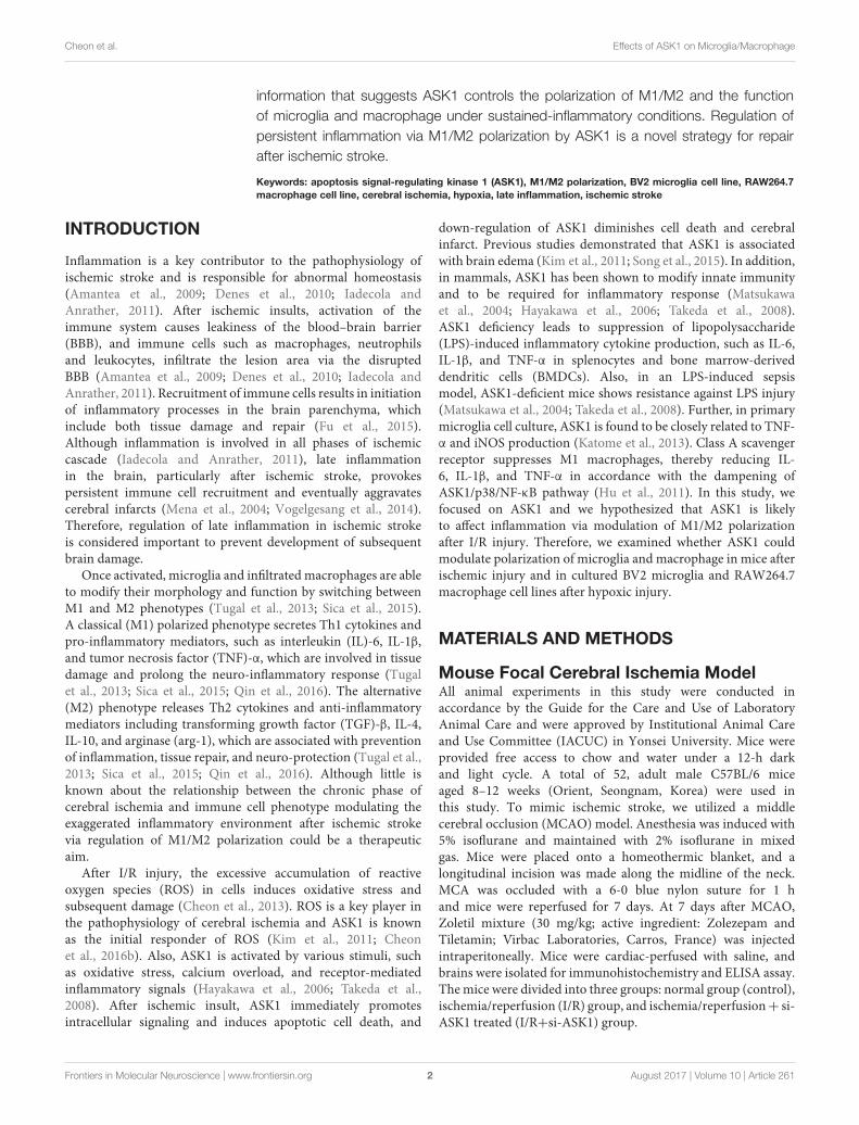
Increase in Microglia/InfiltratedMacrophages Reduced in the IschemicCortex and Striatum after ASK1 SilencingThe previous study showed that siRNA for ASK1 significantlyreduces ASK1 level (Kim et al., 2011; Cheon et al., 2016b).In this study, we followed this method and confirmedimmunohistochemistry for ASK1 with/without administration ofsiRNA for ASK1 at 7 days after I/R. We observed that ischemia-induced ASK1 expression was efficiently reduced after ASK1silencing (Supplementary Figure 1A). In addition, at 3 days afterI/R, we performed real-time PCR for ASK1. Upregulated ASK1transcripts after I/R significantly diminished after ASK1 silencing(Supplementary Figure 1B). Ischemic injury induces a functionalchange in the brain and leads to cell death, the morphologicalchanges of which were observed. To observe cell morphologyafter cerebral ischemia, we performed cresyl violet staining 7 daysafter (I/R) (Figure 1A). As shown in Figure 1A, cell bodiesobserved in the cortex and striatum in the I/R group were smallerand thinner than those in the control group. After silencingASK1, we observed comparatively rounder and healthier cellbodies, compared with those of the I/R group, at 7 days afterI/R. To examine microglia and infiltrated macrophages, we usedmicroglia and macrophage markers, such as Iba-1 and CD11b,respectively (Figure 1B). In the control group, Iba-1-positiveand CD11b-positive cells were rarely observed in the cortex andstriatum. However, Iba-1-positive and CD11b-positive cells were
Frontiers in Molecular Neuroscience | www.frontiersin.org 4 August 2017 | Volume 10 | Article 261
fnmol-10-00261 August 10, 2017 Time: 16:9 # 5
Cheon et al. Effects of ASK1 on Microglia/Macrophage
FIGURE 1 | Decreased microglia and macrophage markers in the striatum and cortex, respectively, upon ASK1 silencing at 7 days after cerebral ischemia. (A) Cresylviolet staining for cell histology in the cortex and striatum shows that I/R-induced cell loss was not detected at 7 days in the I/R+si-ASK1 group.(B) Immunofluorescent staining for Iba-1 (green) and CD11b (red) in the cortex and striatum reveals that I/R-induced increase in Iba-1 or CD11b-positive cells werereduced in the I/R+si-ASK1 group at 7 days. (C) The graph shows the relative fluorescent intensities for Iba-1 and CD11b in the striatum (n = 4). (D) The graphindicates the relative fluorescent intensities for Iba-1 and CD11b in the cortex (n = 4). Values are means ± SD. Statistical comparisons between the groups wereassessed with non-parametric Mann–Whitney test (∗p < 0.05, ∗∗p < 0.01, ∗∗∗p < 0.001 vs. I/R group). Statistical parameter (Supplementary Table 1). I/R,ischemia/reperfusion.
densely expressed in the I/R group. After silencing ASK1, Iba-1-positive and CD11b-positive cells decreased in the cortex andstriatum, respectively, despite ischemic injury. The graphs forrelative fluorescent intensity showed increased intensities for Iba-1 and CD11b were efficiently decreased after silencing ASK1,compared with the I/R group (Figures 1C,D). Therefore, ourdata showed that ASK1 silencing results in reduced microglia andmacrophages in the ischemic cortex and striatum, respectively.Statistical parameters were shown in Supplementary Table 1.
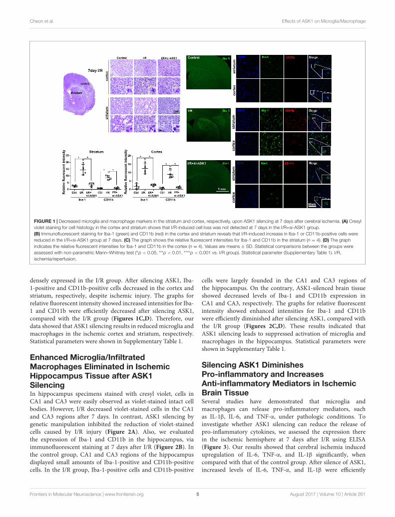
Enhanced Microglia/InfiltratedMacrophages Eliminated in IschemicHippocampus Tissue after ASK1SilencingIn hippocampus specimens stained with cresyl violet, cells inCA1 and CA3 were easily observed as violet-stained intact cellbodies. However, I/R decreased violet-stained cells in the CA1and CA3 regions after 7 days. In contrast, ASK1 silencing bygenetic manipulation inhibited the reduction of violet-stainedcells caused by I/R injury (Figure 2A). Also, we evaluatedthe expression of Iba-1 and CD11b in the hippocampus, viaimmunofluorescent staining at 7 days after I/R (Figure 2B). Inthe control group, CA1 and CA3 regions of the hippocampusdisplayed small amounts of Iba-1-positive and CD11b-positivecells. In the I/R group, Iba-1-positive cells and CD11b-positive
cells were largely founded in the CA1 and CA3 regions ofthe hippocampus. On the contrary, ASK1-silenced brain tissueshowed decreased levels of Iba-1 and CD11b expression inCA1 and CA3, respectively. The graphs for relative fluorescentintensity showed enhanced intensities for Iba-1 and CD11bwere efficiently diminished after silencing ASK1, compared withthe I/R group (Figures 2C,D). These results indicated thatASK1 silencing leads to suppressed activation of microglia andmacrophages in the hippocampus. Statistical parameters wereshown in Supplementary Table 1.
Silencing ASK1 DiminishesPro-inflammatory and IncreasesAnti-inflammatory Mediators in IschemicBrain TissueSeveral studies have demonstrated that microglia andmacrophages can release pro-inflammatory mediators, suchas IL-1β, IL-6, and TNF-α, under pathologic conditions. Toinvestigate whether ASK1 silencing can reduce the release ofpro-inflammatory cytokines, we assessed the expression therein the ischemic hemisphere at 7 days after I/R using ELISA(Figure 3). Our results showed that cerebral ischemia inducedupregulation of IL-6, TNF-α, and IL-1β significantly, whencompared with that of the control group. After silence of ASK1,increased levels of IL-6, TNF-α, and IL-1β were efficiently
Frontiers in Molecular Neuroscience | www.frontiersin.org 5 August 2017 | Volume 10 | Article 261
fnmol-10-00261 August 10, 2017 Time: 16:9 # 6
Cheon et al. Effects of ASK1 on Microglia/Macrophage
FIGURE 2 | Reduced microglia and macrophage markers in the hippocampus upon silence of ASK1 at 7 days after cerebral ischemia. (A) Cresyl violet staining forcell histology in the hippocampus shows that I/R-induced cell loss was not detected at 7 days in the I/R+si-ASK1 group. (B) Immunofluorescent staining for Iba-1(green) and CD11b (red) in the hippocampus revealed that I/R-induced augmentation of Iba-1 or CD11b-positive cells was reduced in the I/R+si-ASK1 group at7 days. (C) The graph shows the relative fluorescent intensities for Iba-1 and CD11b in the hippocampal CA1 region (n = 4). (D) The graph exhibits the relativefluorescent intensities for Iba-1 and CD11b in the hippocampal CA3 region (n = 4). Values are mean ± SD. Statistical comparisons between the groups wereassessed with non-parametric Mann–Whitney test (∗p < 0.05, ∗∗p < 0.01, ∗∗∗p < 0.001 vs. I/R group). Statistical parameter (Supplementary Table 1). I/R,ischemia/reperfusion.
decreased in the ischemic hemisphere. Our ELISA data showedthat ASK1 silencing reduces the expression of pro-inflammatorycytokines in the ischemic hemisphere (Figures 3A–C).
In addition, to examine whether ASK1 silencing can increaseanti-inflammatory mediators, we performed immunofluorescentstaining for CD206 (Figure 3D). Our results showed thatCD206-positive cells in the control group were not significantlydifferent between those in the I/R group; however, ASK1 silencingincreased CD206-positive cells in the striatum and cortex(Figures 3D–G). To determine the effect of ASK1 on the cellfate, we performed TUNEL assay to detect DNA fragmentationat 7 days after I/R (Supplementary Figure 1C). The images andgraphs showed that increased the number of TUNEL-positivecells in the striatum and cortex after I/R was efficiently reducedafter ASK1 silencing. Therefore, our data showed that ASK1silencing suppresses M1 phenotype and increases M2 phenotypein ischemic brain and reduces apoptotic cell death. Statisticalparameters were shown in Supplementary Table 1.
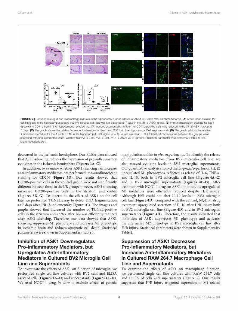
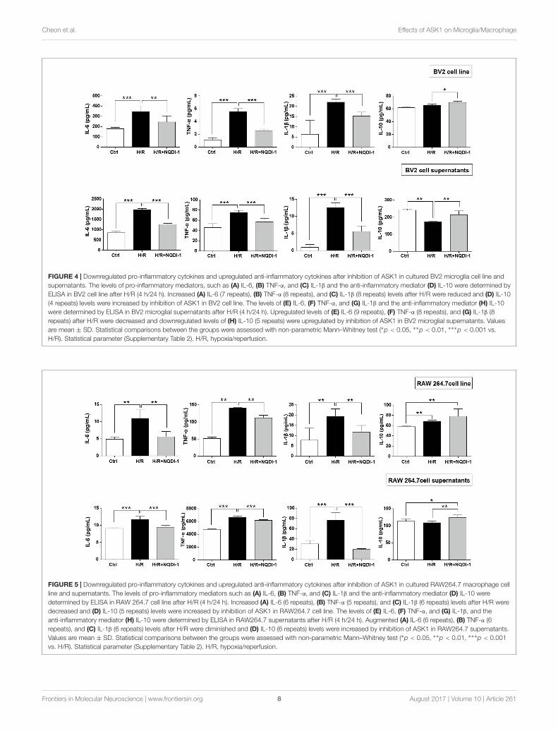
Inhibition of ASK1 DownregulatesPro-inflammatory Mediators, butUpregulates Anti-InflammatoryMediators in Cultured BV2 Microglia CellLine and SupernatantsTo investigate the effects of ASK1 on function of microglia, weperformed single cell line cultures with BV2 cells and ELISAassay of cells (Figures 4A–D) and supernatants (Figures 4E–H).We used NQDI-1 drug in vitro to exclude effects of genetic
manipulation unlike in vivo experiments. To identify the releaseof inflammatory mediators from BV2 microglia cell line, wealso assayed cytokine levels in BV2 microglial supernatants.Our quantitative analysis showed that hypoxia/reperfusion (H/R)upregulated M1 phenotypes, reflected as release of IL-6, TNF-α,and IL-1β, both in BV2 microglia cell line (Figures 4A–C)and in BV2 microglial supernatants (Figures 4E–G). Aftertreatment with NQDI-1 drug, an ASK1 inhibitor, the upregulatedM1 mediators were efficiently reduced despite H/R injury.Although H/R could not alter IL-10 levels in BV2 microgliacell line (Figure 4D), compared with the control, NQDI-1 drugtreatment upregulated secretion of IL-10 after H/R injury bothin BV2 microglia cell line (Figure 4D) and in BV2 microglialsupernatants (Figure 4H). Therefore, the results indicated thatinhibition of ASK1 suppresses M1 phenotype and activatesthe alternative M2 phenotype in BV2 microglia cell line afterH/R injury. Statistical parameters were shown in SupplementaryTable 2.
Suppression of ASK1 DecreasesPro-inflammatory Mediators, butIncreases Anti-Inflammatory Mediatorsin Cultured RAW 264.7 Macrophage CellLine and SupernatantsTo examine the effects of ASK1 on macrophage function,we performed single cell line cultures with RAW 264.7 cellsand ELISA of cells and supernatants (Figure 5). Our resultssuggested that H/R injury triggered expression of M1-related
Frontiers in Molecular Neuroscience | www.frontiersin.org 6 August 2017 | Volume 10 | Article 261

fnmol-10-00261 August 10, 2017 Time: 16:9 # 7
Cheon et al. Effects of ASK1 on Microglia/Macrophage
FIGURE 3 | Decreased pro-inflammatory and increased anti-inflammatory mediators in the ischemic brain by silencing of ASK1 at 7 days after cerebral ischemia.The levels of (A) IL-6, (B) TNF-α, and (C) IL-1β in the hemisphere were assessed by ELISA at 7 days after I/R. I/R-induced upregulation of (A) IL-6, (B) TNF-α, and(C) IL-1β was decreased in the I/R+si-ASK1 group at 7 days (n = 8). Immunofluorescent staining for CD206 was performed at 7 days after I/R (D,F). (D) The relativeintensity graph indicates CD206-positive cells were increased in the striatum (E) and cortex (G), compared with other groups (n = 4–5). Values are mean ± SD.Statistical comparisons between the groups were assessed with non-parametric Mann–Whitney test (∗p < 0.05, ∗∗p < 0.01, ∗∗∗p < 0.001 vs. I/R group). Statisticalparameter (Supplementary Table 1). I/R, ischemia/reperfusion.
mediators, such as IL-6, TNF-α, and IL-1β. However, suppressionof ASK1 significantly decreased M1 phenotypes in RAW 264.7cell line (Figures 5A–C) and in supernatants (Figures 5E–G).In addition, IL-10 levels were also increased after inhibition ofASK1 in spite of H/R injury, as compared with control and H/Rinjured macrophages, both in RAW 264.7 cell line (Figure 5D)and in supernatants (Figure 5H). Therefore, our data showedthat inhibition of ASK1 reduces M1 phenotype and increasesM2 phenotype in RAW 264.7 macrophage cell line. Statisticalparameters were shown in Supplementary Table 2.
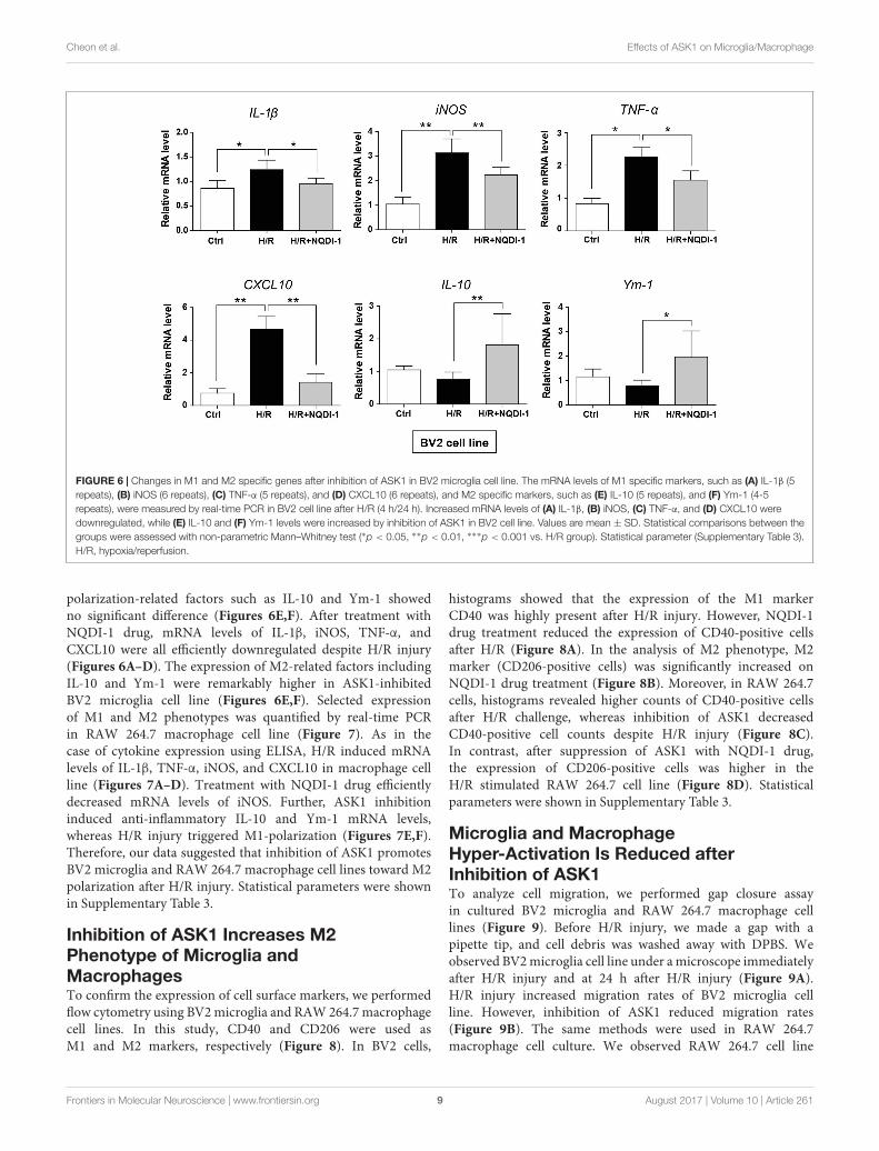
Inhibition of ASK1 Induces Transcriptionof M2 Polarization in BV2 Microglia andRAW 264.7 Macrophage Cell LinesGenetic expression of M1 and M2 markers was quantified byreal-time PCR in BV2 microglia (Figure 6) and in RAW 264.7macrophage cell lines (Figure 7). Similar to cytokine levelsevaluated by ELISA, the mRNA levels of M1 polarization-relatedfactors such as IL-1β, TNF-α, inducible nitric oxide (iNOS),and CXCL10 were all significantly increased in BV2 microgliacell line after H/R injury (Figures 6A–D), and those of M2
Frontiers in Molecular Neuroscience | www.frontiersin.org 7 August 2017 | Volume 10 | Article 261
fnmol-10-00261 August 10, 2017 Time: 16:9 # 8
Cheon et al. Effects of ASK1 on Microglia/Macrophage
FIGURE 4 | Downregulated pro-inflammatory cytokines and upregulated anti-inflammatory cytokines after inhibition of ASK1 in cultured BV2 microglia cell line andsupernatants. The levels of pro-inflammatory mediators, such as (A) IL-6, (B) TNF-α, and (C) IL-1β and the anti-inflammatory mediator (D) IL-10 were determined byELISA in BV2 cell line after H/R (4 h/24 h). Increased (A) IL-6 (7 repeats), (B) TNF-α (8 repeats), and (C) IL-1β (8 repeats) levels after H/R were reduced and (D) IL-10(4 repeats) levels were increased by inhibition of ASK1 in BV2 cell line. The levels of (E) IL-6, (F) TNF-α, and (G) IL-1β and the anti-inflammatory mediator (H) IL-10were determined by ELISA in BV2 microglial supernatants after H/R (4 h/24 h). Upregulated levels of (E) IL-6 (9 repeats), (F) TNF-α (8 repeats), and (G) IL-1β (8repeats) after H/R were decreased and downregulated levels of (H) IL-10 (5 repeats) were upregulated by inhibition of ASK1 in BV2 microglial supernatants. Valuesare mean ± SD. Statistical comparisons between the groups were assessed with non-parametric Mann–Whitney test (∗p < 0.05, ∗∗p < 0.01, ∗∗∗p < 0.001 vs.H/R). Statistical parameter (Supplementary Table 2). H/R, hypoxia/reperfusion.
FIGURE 5 | Downregulated pro-inflammatory cytokines and upregulated anti-inflammatory cytokines after inhibition of ASK1 in cultured RAW264.7 macrophage cellline and supernatants. The levels of pro-inflammatory mediators such as (A) IL-6, (B) TNF-α, and (C) IL-1β and the anti-inflammatory mediator (D) IL-10 weredetermined by ELISA in RAW 264.7 cell line after H/R (4 h/24 h). Increased (A) IL-6 (6 repeats), (B) TNF-α (5 repeats), and (C) IL-1β (6 repeats) levels after H/R weredecreased and (D) IL-10 (5 repeats) levels were increased by inhibition of ASK1 in RAW264.7 cell line. The levels of (E) IL-6, (F) TNF-α, and (G) IL-1β, and theanti-inflammatory mediator (H) IL-10 were determined by ELISA in RAW264.7 supernatants after H/R (4 h/24 h). Augmented (A) IL-6 (6 repeats), (B) TNF-α (6repeats), and (C) IL-1β (6 repeats) levels after H/R were diminished and (D) IL-10 (6 repeats) levels were increased by inhibition of ASK1 in RAW264.7 supernatants.Values are mean ± SD. Statistical comparisons between the groups were assessed with non-parametric Mann–Whitney test (∗p < 0.05, ∗∗p < 0.01, ∗∗∗p < 0.001vs. H/R). Statistical parameter (Supplementary Table 2). H/R, hypoxia/reperfusion.
Frontiers in Molecular Neuroscience | www.frontiersin.org 8 August 2017 | Volume 10 | Article 261
fnmol-10-00261 August 10, 2017 Time: 16:9 # 9
Cheon et al. Effects of ASK1 on Microglia/Macrophage
FIGURE 6 | Changes in M1 and M2 specific genes after inhibition of ASK1 in BV2 microglia cell line. The mRNA levels of M1 specific markers, such as (A) IL-1β (5repeats), (B) iNOS (6 repeats), (C) TNF-α (5 repeats), and (D) CXCL10 (6 repeats), and M2 specific markers, such as (E) IL-10 (5 repeats), and (F) Ym-1 (4-5repeats), were measured by real-time PCR in BV2 cell line after H/R (4 h/24 h). Increased mRNA levels of (A) IL-1β, (B) iNOS, (C) TNF-α, and (D) CXCL10 weredownregulated, while (E) IL-10 and (F) Ym-1 levels were increased by inhibition of ASK1 in BV2 cell line. Values are mean ± SD. Statistical comparisons between thegroups were assessed with non-parametric Mann–Whitney test (∗p < 0.05, ∗∗p < 0.01, ∗∗∗p < 0.001 vs. H/R group). Statistical parameter (Supplementary Table 3).H/R, hypoxia/reperfusion.
polarization-related factors such as IL-10 and Ym-1 showedno significant difference (Figures 6E,F). After treatment withNQDI-1 drug, mRNA levels of IL-1β, iNOS, TNF-α, andCXCL10 were all efficiently downregulated despite H/R injury(Figures 6A–D). The expression of M2-related factors includingIL-10 and Ym-1 were remarkably higher in ASK1-inhibitedBV2 microglia cell line (Figures 6E,F). Selected expressionof M1 and M2 phenotypes was quantified by real-time PCRin RAW 264.7 macrophage cell line (Figure 7). As in thecase of cytokine expression using ELISA, H/R induced mRNAlevels of IL-1β, TNF-α, iNOS, and CXCL10 in macrophage cellline (Figures 7A–D). Treatment with NQDI-1 drug efficientlydecreased mRNA levels of iNOS. Further, ASK1 inhibitioninduced anti-inflammatory IL-10 and Ym-1 mRNA levels,whereas H/R injury triggered M1-polarization (Figures 7E,F).Therefore, our data suggested that inhibition of ASK1 promotesBV2 microglia and RAW 264.7 macrophage cell lines toward M2polarization after H/R injury. Statistical parameters were shownin Supplementary Table 3.
Inhibition of ASK1 Increases M2Phenotype of Microglia andMacrophagesTo confirm the expression of cell surface markers, we performedflow cytometry using BV2 microglia and RAW 264.7 macrophagecell lines. In this study, CD40 and CD206 were used asM1 and M2 markers, respectively (Figure 8). In BV2 cells,
histograms showed that the expression of the M1 markerCD40 was highly present after H/R injury. However, NQDI-1drug treatment reduced the expression of CD40-positive cellsafter H/R (Figure 8A). In the analysis of M2 phenotype, M2marker (CD206-positive cells) was significantly increased onNQDI-1 drug treatment (Figure 8B). Moreover, in RAW 264.7cells, histograms revealed higher counts of CD40-positive cellsafter H/R challenge, whereas inhibition of ASK1 decreasedCD40-positive cell counts despite H/R injury (Figure 8C).In contrast, after suppression of ASK1 with NQDI-1 drug,the expression of CD206-positive cells was higher in theH/R stimulated RAW 264.7 cell line (Figure 8D). Statisticalparameters were shown in Supplementary Table 3.
Microglia and MacrophageHyper-Activation Is Reduced afterInhibition of ASK1To analyze cell migration, we performed gap closure assayin cultured BV2 microglia and RAW 264.7 macrophage celllines (Figure 9). Before H/R injury, we made a gap with apipette tip, and cell debris was washed away with DPBS. Weobserved BV2 microglia cell line under a microscope immediatelyafter H/R injury and at 24 h after H/R injury (Figure 9A).H/R injury increased migration rates of BV2 microglia cellline. However, inhibition of ASK1 reduced migration rates(Figure 9B). The same methods were used in RAW 264.7macrophage cell culture. We observed RAW 264.7 cell line
Frontiers in Molecular Neuroscience | www.frontiersin.org 9 August 2017 | Volume 10 | Article 261

fnmol-10-00261 August 10, 2017 Time: 16:9 # 10
Cheon et al. Effects of ASK1 on Microglia/Macrophage
FIGURE 7 | Changes in M1 and M2 specific genes after inhibition of ASK1 in RAW264.7 macrophage cell line. The mRNA levels of M1 specific markers, such as(A) IL-1β (6 repeats), (B) iNOS (4–5 repeats), (C) TNF-α (4 repeats), and (D) CXCL10 (6 repeats), and M2 specific markers, such as (E) IL-10 (4 repeats) and(F) Ym-1 (5–6 repeats), were measured by real-time PCR in RAW264.7 cell line after H/R (4 h/24 h). The mRNA levels of (A) IL-1β, (B) iNOS, (C) TNF-α, and(D) CXCL10 were upregulated after H/R. The levels of iNOS were reduced, while (E) IL-10 and (F) Ym-1 levels were upregulated by inhibition of ASK1 in RAW264.7cell line. Values are mean ± SD. Statistical comparisons between the groups were assessed with non-parametric Mann–Whitney test (∗p < 0.05, ∗∗p < 0.01,∗∗∗p < 0.001 vs. H/R). Statistical parameter (Supplementary Table 3). H/R, hypoxia/reperfusion.
under a microscope right after H/R injury and at 24 h afterH/R injury (Figure 9C). A high rate of migration was exhibitedin H/R-injured macrophages; however, NQDI-1 drug treatmentretarded the migration rate. Therefore, ASK1 is associatedwith microglia and macrophage migration capacity and ASK1inhibition delayed migration rate. Statistical parameters wereshown in Supplementary Table 3.
DISCUSSION
In this study, we performed in vivo and in vitro ischemicstroke model to examine whether ASK1 modulatesmicroglia/macrophage polarization and function. Weproved that ASK1 controls polarization and function ofmicroglia/macrophage after ischemic/hypoxic injury. Inprevious reports, ASK1 is one of the early responders to differenttypes of stress in the intracellular system, including oxidativestress, calcium overload, and immune response (Hayakawaet al., 2006; Takeda et al., 2008). ASK1 governs activationof mitogen-activated protein kinase (MAP kinase), which isessential for cellular function (Shiizaki et al., 2013), and severalstudies support that ASK1 is closely related to cerebral ischemia(Kim et al., 2011; Cheon et al., 2016b). Under inflammatoryconditions, ASK1 signaling is necessary for TLRs, whichrecognize LPS (Soga et al., 2012). ASK1 deficiency represents LPS
resistance, and genetic deletion of ASK1 involves inhibition ofpro-inflammatory mediator production (Matsukawa et al., 2004;Sumbayev, 2008; Takeda et al., 2008; Mnich et al., 2010). TheASK1/p38 signaling pathway via TLRs is critical for chemokineproduction and promotes inflammation and neurotoxicityafter multiple sclerosis (Guo et al., 2010). In line with studiesthat have demonstrated that ASK1 inhibition attenuatespro-inflammatory cytokines in microglia, ASK1 was found tobe associated with TNF-α and iNOS production in primarymicroglial cell culture, and M1 macrophage suppression by aClass A scavenger receptor resulted in reductions of IL-6, IL-1β,and TNF-α, decreasing ASK1/p38/NF-κB pathway signaling,under pathologic conditions (Hu et al., 2011; Katome et al.,2013; Song and Lee, 2015). Previous reports have shown thatinhibition of ASK1 reduces M1 phenotype after injury andASK1 also played a role in our results. The main findings ofthis study are (1) in the ipsilateral hemisphere, ASK1 silencingresulted in a reduction of ischemic-induced activation ofmicroglia/infiltrated macrophages and increased M2 phenotypein the late phase of cerebral ischemia and (2) ASK1 inhibitionpolarized BV2 microglia and RAW 264.7 macrophage cell linestoward M2 phenotype after hypoxia/reperfusion injury. Ourin vivo study revealed that cerebral ischemia promotes activationof microglia and infiltration of macrophages in brain regions.In the ipsilateral hemisphere, upregulation of pro-inflammatorycytokines, such as IL-6, TNF-α, and IL-1β, was observed.
Frontiers in Molecular Neuroscience | www.frontiersin.org 10 August 2017 | Volume 10 | Article 261

fnmol-10-00261 August 10, 2017 Time: 16:9 # 11
Cheon et al. Effects of ASK1 on Microglia/Macrophage
FIGURE 8 | Alteration of M1 and M2 surface markers after inhibition of ASK1 in BV2 microglia and RAW264.7 macrophages. Flow cytometry was performed fordiscrimination of (A) M1 (CD40) or (B) M2 (CD206) positive cell populations of BV2 cell line after H/R (4 h/24 h). The histogram of flow cytometry shows (C) M1(CD40) and (D) M2 (CD206) positive cells in RAW 264.7 cell line after H/R (4 h/24 h). H/R, hypoxia/reperfusion.
However, ASK1 silenced by siRNA downregulated activationof microglia/macrophage and pro-inflammatory cytokinelevels and upregulated the anti-inflammatory mediators. Ourin vitro study for M1/M2 polarization of microglia/macrophagesrevealed that hypoxic injury stimulates M1-related factors,while ASK1 inhibition by NQDI-1 drug suppressed M1-relatedfactors and promoted M2-related factors in microglia andmacrophage cell lines. In addition, inhibition of ASK1 retardedthe migration rate of both microglia and macrophages in gapclosure assay.
Inflammation has emerged as the key factor in progressionof ischemic stroke, and the inflammatory mediators play animportant role in enlargement of brain damage and neurologicaldysfunction (Matsukawa et al., 2004; Quan and Banks, 2007;Quan, 2008; Banks and Erickson, 2010; Fu et al., 2015;Anrather and Iadecola, 2016). At the onset of ischemic stroke,stagnant blood flow initiates an aberrant immune response thatpromotes infiltration of immune cells such as leukocytes andmacrophages, via a disrupted BBB (Matsukawa et al., 2004;Quan and Banks, 2007; Quan, 2008; Banks and Erickson,2010; Fu et al., 2015; Anrather and Iadecola, 2016). Underthe altered environment, microglia are activated. Meanwhile,the morphology and activation of peripherally infiltratedmacrophages in the brain changes depending on the extracellularstimuli (Durafourt et al., 2012; Hu et al., 2015; Orihuelaet al., 2016), after which activated microglia migrate quicklytoward lesion sites, and lead to cell accumulation by releasing
inflammation-associated mediators (Danton and Dietrich, 2003).Based on the microenvironment, microglia/macrophage canswitch between phenotypes M1 (classically activated) andM2 (alternative activated). Under M1 conditions stimulatedby LPS or IFN-γ, M1 microglia/macrophage express pro-inflammatory mediators of IL-1β, IL-6, TNF-α, iNOS, andCXCL10 and the cell surface markers CD40, CD80, andCD86. Also, cytotoxic effects of the M1 phenotype canexacerbate tissue damage (Durafourt et al., 2012; Hu et al.,2015; Kapellos and Iqbal, 2016; Orihuela et al., 2016; Plastiraet al., 2016). On the contrary, M2 microglia/macrophages exertanti-inflammatory responses by upregulating IL-10, arginase-1,Ym-1 (heparin-binding lectin), and mannose receptor CD206,which modulate tissue repair, regeneration, and remodeling(Durafourt et al., 2012; Barakat and Redzic, 2016; Orihuelaet al., 2016). During the early stage after ischemic insult,expression of the pro-inflammatory cytokine TNF-α is increasedin neurons. However, during the late phase of ischemic insult,it is augmented in microglia/macrophage and other immunecells, with microglia/macrophage being major sources of pro-inflammatory cytokine in ischemic lesions after ischemic stroke(Gregersen et al., 2000; Clausen et al., 2008). Late inflammationin the brain after ischemic stroke is represented by persistentimmune cell infiltration and cerebral infarctions (Mena et al.,2004; Brea et al., 2009; Vogelgesang et al., 2014; Walter et al.,2015; Anrather and Iadecola, 2016). Several studies have reportedthat microglia/macrophage shift their phenotype from M2
Frontiers in Molecular Neuroscience | www.frontiersin.org 11 August 2017 | Volume 10 | Article 261

fnmol-10-00261 August 10, 2017 Time: 16:9 # 12
Cheon et al. Effects of ASK1 on Microglia/Macrophage
FIGURE 9 | Function of BV2 microglia and RAW264.7 macrophages after inhibition of ASK1. (A,B) Gap closure assay was performed for BV2 and RAW264.7 cells(A) Images at 0 h after hypoxia and 24 h after hypoxia/reperfusion in BV2 cell line. (B) The graph represents that H/R-induced increases in migration rate weredecreased by inhibition of ASK1 in BV2 cell line (4 repeats). (C) Images at 0 h after hypoxia and 24 h after hypoxia/reperfusion in RAW264.7 cell line. (D) The graphrepresents that H/R-induced increases in migration rate were reduced by inhibition of ASK1 in RAW264.7 cell line (4 repeats). Values are mean ± SD. Statisticalcomparisons between the groups were assessed with non-parametric Mann–Whitney test (∗p < 0.05, ∗∗p < 0.01, ∗∗∗p < 0.001 vs. H/R). Statistical parameter(Supplementary Table 3). H/R, hypoxia/reperfusion.
to M1 after cerebral ischemia and M1 microglia/macrophagedominate the ischemic lesion in the brain, which exacerbatebrain injury (Frieler et al., 2011; Hu et al., 2012). M1polarized microglia/macrophage during chronic inflammationaugment neuronal damage and block reestablishment ofneuronal network, thereby retarding brain recovery (Hu et al.,2012). However, M2 polarized microglia/macrophage showimproved phagocytic activity for clearance of necrotic debrisand ameliorate production of inflammatory mediators (Huet al., 2012). M2 phenotype modifies the extracellular matrixand promotes axonal regeneration and angiogenesis (Kigerlet al., 2009). In addition, after ischemic/hypoxic insult, M2microglia/macrophage mediates clearance of ischemic tissueand blockade of brain injury, leading to neuronal survival(Hu et al., 2012). Therefore, anti-inflammatory M2 may havebeneficial effects on ameliorating the development of braindamage. In the current study, cerebral ischemia augmentedthe recruitment of macrophage and microglia activationand migration, and led the brain toward M1 environment.
However, ASK1 silencing by si-RNA/ASK1 inhibition byNQDI-1 drug diminished M1 phenotype-specific markers(secretion of pro-inflammatory mediators and downregulationof pro-inflammatory mediator genes) of macrophage/microgliaand upregulated M2 specific markers (expression of anti-inflammatory mannose receptor and upregulation of anti-inflammatory mediator genes) in the later stages of the recoveryperiod after ischemic/hypoxic injuries. To our knowledge, this isthe first investigations of ASK1 in regards to M1/M2 polarizationof microglia and macrophages in the late stage of ischemicstroke.
However, our limitations are as follows: first, it is notclarified which ASK1-downstream or -related signaling pathwayaffects M1/M2 phenotype. Although several molecules (JNK,p38, and Akt) are known to be involved in polarization(Zhang and Zhang, 2015; Vergadi et al., 2017; Zhong et al.,2017), we only proceeded to examine changes of M1/M2phenotype by silencing/inhibiting ASK1 levels. Second,we focused on “classical” M1/M2 polarization in this
Frontiers in Molecular Neuroscience | www.frontiersin.org 12 August 2017 | Volume 10 | Article 261

fnmol-10-00261 August 10, 2017 Time: 16:9 # 13
Cheon et al. Effects of ASK1 on Microglia/Macrophage
study. However, Martinez et al. (2008) and Mantovani et al.(2004) review the subpopulation of polarization, such as M1,M2a, M2b, and M2c. Ransohoff (2016) raises some concernson existence of M1/M2 polarization (Mantovani et al., 2004;Martinez et al., 2008). Third, the previous studies proved thatASK1 inhibitor NQDI-1 shows a possible therapeutic applicationas a protective drug in ischemic stroke (Song et al., 2015;Cheon et al., 2016a). However, it is not fully demonstratedits safety and application. If this limitation is resolved, itwill be one of the candidates for use in ischemic strokepatients.
CONCLUSION
We showed that ASK1 mediates M1/M2 polarization andthe functions of microglia and macrophages. Modulation ofthe late inflammatory environment by M1/M2 regulation ofmicroglia and macrophage via ASK1 silencing after cerebralischemia may be an attractive strategy for recovery fromstroke.
AUTHOR CONTRIBUTIONS
B-NK designed this study, supervised the project, interpreted alldata, and wrote the manuscript. SC participated in the collectionof data, interpretation of data, and writing of the first draft ofthe manuscript. EJK, JK, EHK, and BK participated in the datacollection and interpretation.
FUNDING
This study was supported by a National Research Foundation ofKorea (NRF) grant funded by the Korea government (MSIP) (No.2017R1A2B4009) awarded to B-NK.
SUPPLEMENTARY MATERIAL
The Supplementary Material for this article can be foundonline at: http://journal.frontiersin.org/article/10.3389/fnmol.2017.00261/full#supplementary-material
REFERENCESAmantea, D., Nappi, G., Bernardi, G., Bagetta, G., and Corasaniti, M. T.
(2009). Post-ischemic brain damage: pathophysiology and role of inflammatorymediators. FEBS J. 276, 13–26. doi: 10.1111/j.1742-4658.2008.06766.x
Anrather, J., and Iadecola, C. (2016). Inflammation and stroke: an overview.Neurotherapeutics 13, 661–670. doi: 10.1007/s13311-016-0483-x
Banks, W. A., and Erickson, M. A. (2010). The blood-brain barrier and immunefunction and dysfunction. Neurobiol. Dis. 37, 26–32. doi: 10.1016/j.nbd.2009.07.031
Barakat, R., and Redzic, Z. (2016). The role of activated microglia and residentmacrophages in the neurovascular unit during cerebral ischemia: Is the jury stillout? Med. Princ. Pract. 25(Suppl. 1), 3–14. doi: 10.1159/000435858
Brea, D., Sobrino, T., Ramos-Cabrer, P., and Castillo, J. (2009). Inflammatory andneuroimmunomodulatory changes in acute cerebral ischemia. Cerebrovasc. Dis.27(Suppl. 1), 48–64. doi: 10.1159/000200441
Cheon, S. Y., Cho, K. J., Kim, S. Y., Kam, E. H., Lee, J. E., and Koo, B. N.(2016a). Blockade of apoptosis signal-regulating kinase 1 attenuates matrixmetalloproteinase 9 activity in brain endothelial cells and the subsequentapoptosis in neurons after ischemic injury. Front. Cell Neurosci. 10:213.doi: 10.3389/fncel.2016.00213
Cheon, S. Y., Cho, K. J., Lee, J. E., Kim, H. W., Lee, S. K., Kim, H. J., et al.(2013). Cerebroprotective effects of red ginseng extract pretreatment againstischemia-induced oxidative stress and apoptosis. Int. J. Neurosci. 123, 269–277.doi: 10.3109/00207454.2012.758120
Cheon, S. Y., Cho, K. J., Song, J., and Kim, G. W. (2016b). Knockdown of apoptosissignal-regulating kinase 1 affects ischaemia-induced astrocyte activationand glial scar formation. Eur. J. Neurosci. 43, 912–922. doi: 10.1111/ejn.13175
Cho, K. J., Cheon, S. Y., and Kim, G. W. (2016). Apoptosis signal-regulating kinase1 mediates striatal degeneration via the regulation of C1q. Sci. Rep. 6:18840.doi: 10.1038/srep18840
Clausen, B. H., Lambertsen, K. L., Babcock, A. A., Holm, T. H., Dagnaes-Hansen, F., and Finsen, B. (2008). Interleukin-1beta and tumor necrosisfactor-alpha are expressed by different subsets of microglia and macrophagesafter ischemic stroke in mice. J. Neuroinflammation 5:46. doi: 10.1186/1742-2094-5-46
Danton, G. H., and Dietrich, W. D. (2003). Inflammatory mechanisms afterischemia and stroke. J. Neuropathol. Exp. Neurol. 62, 127–136.
Denes, A., Thornton, P., Rothwell, N. J., and Allan, S. M. (2010). Inflammationand brain injury: acute cerebral ischaemia, peripheral and central
inflammation. Brain Behav. Immun. 24, 708–723. doi: 10.1016/j.bbi.2009.09.010
Durafourt, B. A., Moore, C. S., Zammit, D. A., Johnson, T. A., Zaguia, F., Guiot,M. C., et al. (2012). Comparison of polarization properties of human adultmicroglia and blood-derived macrophages. Glia 60, 717–727. doi: 10.1002/glia.22298
Frieler, R. A., Meng, H., Duan, S. Z., Berger, S., Schutz, G., He, Y., et al. (2011).Myeloid-specific deletion of the mineralocorticoid receptor reduces infarctvolume and alters inflammation during cerebral ischemia. Stroke 42, 179–185.doi: 10.1161/STROKEAHA.110.598441
Fu, Y., Liu, Q., Anrather, J., and Shi, F. D. (2015). Immune interventions in stroke.Nat. Rev. Neurol. 11, 524–535. doi: 10.1038/nrneurol.2015.144
Gregersen, R., Lambertsen, K., and Finsen, B. (2000). Microglia and macrophagesare the major source of tumor necrosis factor in permanent middle cerebralartery occlusion in mice. J. Cereb. Blood Flow Metab. 20, 53–65. doi: 10.1097/00004647-200001000-00009
Guo, X., Harada, C., Namekata, K., Matsuzawa, A., Camps, M., Ji, H., et al.(2010). Regulation of the severity of neuroinflammation and demyelination byTLR-ASK1-p38 pathway. EMBO Mol. Med. 2, 504–515. doi: 10.1002/emmm.201000103
Hayakawa, T., Matsuzawa, A., Noguchi, T., Takeda, K., and Ichijo, H. (2006). TheASK1-MAP kinase pathways in immune and stress responses. Microbes Infect.8, 1098–1107. doi: 10.1016/j.micinf.2005.12.001
Hu, X., Leak, R. K., Shi, Y., Suenaga, J., Gao, Y., Zheng, P., et al. (2015). Microglialand macrophage polarization-new prospects for brain repair. Nat. Rev. Neurol.11, 56–64. doi: 10.1038/nrneurol.2014.207
Hu, X., Li, P., Guo, Y., Wang, H., Leak, R. K., Chen, S., et al. (2012).Microglia/macrophage polarization dynamics reveal novel mechanism of injuryexpansion after focal cerebral ischemia. Stroke 43, 3063–3070. doi: 10.1161/STROKEAHA.112.659656
Hu, Y., Zhang, H., Lu, Y., Bai, H., Xu, Y., Zhu, X., et al. (2011). Class Ascavenger receptor attenuates myocardial infarction-induced cardiomyocytenecrosis through suppressing M1 macrophage subset polarization. Basic Res.Cardiol. 106, 1311–1328. doi: 10.1007/s00395-011-0204-x
Iadecola, C., and Anrather, J. (2011). The immunology of stroke: from mechanismsto translation. Nat. Med. 17, 796–808. doi: 10.1038/nm.2399
Kapellos, T. S., and Iqbal, A. J. (2016). Epigenetic control of macrophagepolarisation and soluble mediator gene expression during inflammation.Mediators Inflamm. 2016:6591703. doi: 10.1155/2016/6591703
Katome, T., Namekata, K., Guo, X., Semba, K., Kittaka, D., Kawamura, K.,et al. (2013). Inhibition of ASK1-p38 pathway prevents neural cell death
Frontiers in Molecular Neuroscience | www.frontiersin.org 13 August 2017 | Volume 10 | Article 261
fnmol-10-00261 August 10, 2017 Time: 16:9 # 14
Cheon et al. Effects of ASK1 on Microglia/Macrophage
following optic nerve injury. Cell Death Differ. 20, 270–280. doi: 10.1038/cdd.2012.122
Kigerl, K. A., Gensel, J. C., Ankeny, D. P., Alexander, J. K., Donnelly, D. J., andPopovich, P. G. (2009). Identification of two distinct macrophage subsets withdivergent effects causing either neurotoxicity or regeneration in the injuredmouse spinal cord. J. Neurosci. 29, 13435–13444. doi: 10.1523/JNEUROSCI.3257-09.2009
Kim, H. W., Cho, K. J., Lee, S. K., and Kim, G. W. (2011). Apoptosis signal-regulating kinase 1 (Ask1) targeted small interfering RNA on ischemicneuronal cell death. Brain Res. 1412, 73–78. doi: 10.1016/j.brainres.2011.07.018
Mantovani, A., Sica, A., Sozzani, S., Allavena, P., Vecchi, A., and Locati, M.(2004). The chemokine system in diverse forms of macrophage activationand polarization. Trends Immunol. 25, 677–686. doi: 10.1016/j.it.2004.09.015
Martinez, F. O., Sica, A., Mantovani, A., and Locati, M. (2008). Macrophageactivation and polarization. Front. Biosci. 13:453–461.
Matsukawa, J., Matsuzawa, A., Takeda, K., and Ichijo, H. (2004). The ASK1-MAP kinase cascades in mammalian stress response. J. Biochem. 136, 261–265.doi: 10.1093/jb/mvh134
Mena, H., Cadavid, D., and Rushing, E. J. (2004). Human cerebral infarct: aproposed histopathologic classification based on 137 cases. Acta Neuropathol.108, 524–530. doi: 10.1007/s00401-004-0918-z
Mnich, S. J., Blanner, P. M., Hu, L. G., Shaffer, A. F., Happa, F. A., O’Neil, S., et al.(2010). Critical role for apoptosis signal-regulating kinase 1 in the developmentof inflammatory K/BxN serum-induced arthritis. Int. Immunopharmacol. 10,1170–1176. doi: 10.1016/j.intimp.2010.06.023
Orihuela, R., McPherson, C. A., and Harry, G. J. (2016). Microglial M1/M2polarization and metabolic states. Br. J. Pharmacol. 173, 649–665. doi: 10.1111/bph.13139
Plastira, I., Bernhart, E., Goeritzer, M., Reicher, H., Kumble, V. B.,Kogelnik, N., et al. (2016). 1-Oleyl-lysophosphatidic acid (LPA) promotespolarization of BV-2 and primary murine microglia towards an M1-like phenotype. J. Neuroinflammation 13:205. doi: 10.1186/s12974-016-0701-9
Qin, Y., Sun, X., Shao, X., Hu, M. X., Feng, J., Chen, Z., et al. (2016).Lipopolysaccharide preconditioning induces an anti-inflammatory phenotypein BV2 microglia. Cell Mol. Neurobiol. 36, 1269–1277. doi: 10.1007/s10571-015-0324-1
Quan, N. (2008). Immune-to-brain signaling: How important are the blood-brain barrier-independent pathways? Mol. Neurobiol. 37, 142–152. doi: 10.1007/s12035-008-8026-z
Quan, N., and Banks, W. A. (2007). Brain-immune communication pathways.Brain Behav. Immun. 21, 727–735. doi: 10.1016/j.bbi.2007.05.005
Ransohoff, R. M. (2016). A polarizing question: Do M1 and M2 microglia exist?Nat. Neurosci. 19, 987–991. doi: 10.1038/nn.4338
Shiizaki, S., Naguro, I., and Ichijo, H. (2013). Activation mechanisms of ASK1 inresponse to various stresses and its significance in intracellular signaling. Adv.Biol. Regul. 53, 135–144. doi: 10.1016/j.jbior.2012.09.006
Sica, A., Erreni, M., Allavena, P., and Porta, C. (2015). Macrophage polarization inpathology. Cell Mol. Life Sci. 72, 4111–4126. doi: 10.1007/s00018-015-1995-y
Soga, M., Matsuzawa, A., and Ichijo, H. (2012). Oxidative stress-induced diseasesvia the ASK1 signaling pathway. Int. J. Cell Biol. 2012:439587. doi: 10.1155/2012/439587
Song, J., Cheon, S. Y., Lee, W. T., Park, K. A., and Lee, J. E. (2015). The effectof ASK1 on vascular permeability and edema formation in cerebral ischemia.Brain Res. 1595, 143–155. doi: 10.1016/j.brainres.2014.11.024
Song, J., and Lee, J. E. (2015). ASK1 modulates the expression of microRNA Let7Ain microglia under high glucose in vitro condition. Front. Cell Neurosci. 9:198.doi: 10.3389/fncel.2015.00198
Sumbayev, V. V. (2008). LPS-induced Toll-like receptor 4 signalling triggers cross-talk of apoptosis signal-regulating kinase 1 (ASK1) and HIF-1alpha protein.FEBS Lett. 582, 319–326. doi: 10.1016/j.febslet.2007.12.024
Takeda, K., Noguchi, T., Naguro, I., and Ichijo, H. (2008). Apoptosis signal-regulating kinase 1 in stress and immune response. Annu. Rev. Pharmacol.Toxicol. 48, 199–225. doi: 10.1146/annurev.pharmtox.48.113006.094606
Tugal, D., Liao, X., and Jain, M. K. (2013). Transcriptional control of macrophagepolarization. Arterioscler. Thromb. Vasc. Biol. 33, 1135–1144. doi: 10.1161/ATVBAHA.113.301453
Vergadi, E., Ieronymaki, E., Lyroni, K., Vaporidi, K., and Tsatsanis, C. (2017).Akt signaling pathway in macrophage activation and M1/M2 Polarization.J. Immunol. 198, 1006–1014. doi: 10.4049/jimmunol.1601515
Vogelgesang, A., Becker, K. J., and Dressel, A. (2014). Immunological consequencesof ischemic stroke. Acta Neurol. Scand. 129, 1–12. doi: 10.1111/ane.12165
Walter, H. L., Walberer, M., Rueger, M. A., Backes, H., Wiedermann, D.,Hoehn, M., et al. (2015). In vivo analysis of neuroinflammation in the latechronic phase after experimental stroke. Neuroscience 292, 71–80. doi: 10.1016/j.neuroscience.2015.02.024
Zhang, O., and Zhang, J. (2015). Atorvastatin promotes human monocytedifferentiation toward alternative M2 macrophages through p38 mitogen-activated protein kinase-dependent peroxisome proliferator-activated receptorgamma activation. Int. Immunopharmacol. 26, 58–64. doi: 10.1016/j.intimp.2015.03.005
Zhong, J., Wang, H., Chen, W., Sun, Z., Chen, J., Xu, Y., et al. (2017).Ubiquitylation of MFHAS1 by the ubiquitin ligase praja2 promotes M1macrophage polarization by activating JNK and p38 pathways. Cell Death Dis.8:e2763. doi: 10.1038/cddis.2017.102
Conflict of Interest Statement: The authors declare that the research wasconducted in the absence of any commercial or financial relationships that couldbe construed as a potential conflict of interest.
Copyright © 2017 Cheon, Kim, Kim, Kam, Ko and Koo. This is an open-access articledistributed under the terms of the Creative Commons Attribution License (CC BY).The use, distribution or reproduction in other forums is permitted, provided theoriginal author(s) or licensor are credited and that the original publication in thisjournal is cited, in accordance with accepted academic practice. No use, distributionor reproduction is permitted which does not comply with these terms.
Frontiers in Molecular Neuroscience | www.frontiersin.org 14 August 2017 | Volume 10 | Article 261