microglia development in the quail cerebellum - ugrhera.ugr.es/doi/15024854.pdf · microglia...
TRANSCRIPT

Microglia Developmentin the Quail Cerebellum
MIGUEL A. CUADROS, JUAN RODRIGUEZ-RUIZ, RUTH CALVENTE,
ANTONIO ALMENDROS, JOSE L. MARIN-TEVA, AND JULIO NAVASCUES*
Departamento de Biologıa Celular, Facultad de Ciencias, Universidad de Granada,E-18071 Granada, Spain
ABSTRACTWe used the QH1 antibody to study changes in the morphological features and
distribution of microglial cells throughout development in the quail cerebellum. Fewmicroglial precursors were present in the cerebellar anlage before the ninth incubation day(E9), whereas many precursors apparently entered the cerebellum from the meninges in thebasal region of the cerebellar peduncles between E9 and E16. From this point of entry into thenervous parenchyma, they spread through the cerebellar white matter, forming a ‘stream’ oflabeled cells that could be seen until hatching (E16). The number of microglial cells in thecerebellar cortex increased during the last days of embryonic life and first posthatching week,whereas microglial density within the white matter decreased after hatching. As a conse-quence, the differences in microglial cell density observed in the cerebellar cortex and thewhite matter during embryonic life diminished after hatching, and microglia showed a nearlyhomogeneous pattern of distribution in adult cerebella. Ameboid and poorly ramifiedmicroglial cells were found in developing stages, whereas only mature microglia appeared inadult cerebella. Our observations suggest that microglial precursors enter the cerebellaranlage mainly by traversing the pial surface at the basal region of the peduncles, then migratealong the white matter, and finally move radially to the different cortical layers. Differentia-tion occurs after the microglial cells have reached their final position. In other brain regionsthe development of microglia follows similar stages, suggesting that these steps are generalrules of microglial development in the central nervous system. J. Comp. Neurol. 389:390–401,1997. r 1997 Wiley-Liss, Inc.
Indexing terms: QH1 antibody; CNS invasion; ameboid microglia; cell migration; differentiation
Knowledge about microglia did not advance significantlyfrom the earliest descriptions by Rio-Hortega (reviewed inRio-Hortega, 1932) until new tools to study these cellswere developed. Of the techniques now used to labelmicroglia, histochemical staining of nucleoside diphospha-tase and thiamine pyrophosphatase activities have re-vealed the distribution and morphology of microglial cellsin the developing and adult brain of rodents (Murabe andSano, 1982; Vela et al., 1995), humans (Fujimoto et al.,1989), birds (Fujimoto et al., 1987), and lizards (Castel-lano, 1991). Different lectins also label microglial cells insome species (Streit and Kreutzberg, 1987; Hutchins et al.,1992; Acarin et al., 1994). In addition, antibodies recogniz-ing microglial cells have been found in a number of species,including fishes (Dowding et al., 1991), amphibians (Good-brand and Gaze, 1991), rodents (Perry et al., 1985;Imamura et al., 1990; Ling et al., 1990; Gehrmann andKreutzberg, 1991; Perry and Gordon, 1991), and humans(Penfold et al., 1991).
The QH1 antibody, which recognizes endothelial andhemopoietic cells in the quail (Pardanaud et al., 1987),labels all stages of development of quail microglia (Cuadroset al., 1992). Taking advantage of this labeling, we studiedthe development of microglia in the quail optic tectum(Cuadros et al., 1994) and retina (Navascues et al., 1995).These studies revealed that microglia develop from precur-sors that invade the nervous tissue in a highly stereotypi-cal manner.Although nonendothelial cells of hemangioblas-tic lineage appear in the avian nervous system at earlydevelopmental stages (Cuadros et al., 1993), many of themicroglial precursors enter the nervous tissue at later
Grant sponsor: DGICYT of the Spanish Ministry of Education andCulture; Grant number: PB94-0789.
*Correspondence to: Prof. Julio Navascues, Departamento de BiologıaCelular. Facultad de Ciencias, Universidad de Granada. E-18071 Granada,Spain. E-mail: [email protected]
Received 6 February 1997; Revised 14 July 1997; Accepted 16 July 1997
THE JOURNAL OF COMPARATIVE NEUROLOGY 389:390–401 (1997)
r 1997 WILEY-LISS, INC.

stages through specific points in the pial surface. So, largenumbers of microglial precursors concentrate at both sidesof the pial surface in the ventrolateral region of the quailoptic lobes during embryonic life (Cuadros et al., 1994) andcould enter the quail retina from the pecten (Navascues etal., 1995).
Once they have entered the nervous parenchyma, micro-glial precursors migrate to their final location (Cuadros etal., 1994; Navascues et al., 1995), where they change fromameboid cells (also called brain macrophages) to ramifiedmature microglia (Ling and Wong, 1993). This transforma-tion has been carefully analyzed in the corpus callosumand cortex of rats (Wu et al., 1992, 1993), and has also beendescribed in the developing central nervous system (CNS)of humans (Fujimoto et al., 1989) and birds (Fujimoto etal., 1987; Cuadros et al., 1994). Microglial precursorsbecome ramified microglia only after reaching their finalposition in the quail optic tectum (Cuadros et al., 1994)and retina (Navascues et al., 1995). Therefore, microgliaseem to modify their morphological features as theirsurrounding environment changes, supporting the viewthat their phenotype depends on the place where the cellsare located (Lawson et al., 1990; Perry and Gordon, 1991).
In this paper, we analyze the pattern of microgliadevelopment in the cerebellum, and compare it with thatobserved in the optic tectum and retina. Our observationsreveal that during embryonic development a large propor-tion of microglial cells enter the cerebellum from themeninges of the basal region of the cerebellar pedunclesand afterward distribute through the developing whitematter and cortical layers.
MATERIALS AND METHODS
Animals and histology
Embryonic and hatched quails (Coturnix coturnix ja-ponica) used in this study ranged from day 6 of incubation(E6) to E16, when quails normally hatch, and from the firstpostnatal day (P0) to adulthood (P45; Table 1). Embryosyounger than E8 were decapitated and the heads im-mersed in Bouin’s fixative for 1 to 3 days without furtherdissection. Older embryonic brains and those of hatchedquails were dissected and fixed for 3 to 5 days in the samefixative. Quails older than P3 were anesthetized withether and perfused through the heart before decapitation.Procedures for death of the animals and material retrievalwere in accordance with specifications from the unit ofLaboratory Animals of the University of Granada. Thebrains were embedded in paraffin, and serial sections 7- to10-µm-thick, were obtained on a rotatory microtome. Inaddition, brains of E8, E12, E16, P3, and adult quails(Table 1) were fixed in 4% paraformaldehyde in phosphatebuffered saline (PBS) for 4 to 6 hours, and sagittal sections50- to 60-µm-thick were obtained by using a Vibratome.
Immunocytochemistry
Both paraffin and Vibratome sections were stained withQH1 antibody (Pardanaud et al., 1987). The antibody wasobtained as culture supernatant from the DevelopmentalStudies Hybridoma Bank maintained by the Departmentof Pharmacology and Molecular Sciences at Johns HopkinsUniversity School of Medicine (Baltimore, MD) and theDepartment of Biology at the University of Iowa (IowaCity, IA), under contract number NO1-HD-2-3144 from the
National Institute of Child Health and Human Develop-ment (NICHD).
Immunocytochemical processing of paraffin sections wasperformed as previously reported (Cuadros et al., 1992,1993). After removing endogenous peroxidase activity bytreating the sections with 0.3% hydrogen peroxide in PBS,the sections were incubated overnight in QH1 supernatantdiluted 1:2, washed extensively in PBS, and successivelyincubated for 30 to 45 minutes in the secondary antibody(biotinylated goat anti-mouse IgG, Sigma, St. Louis, MO)and streptavidin-biotin-peroxidase complex (Extravidin,Sigma). Peroxidase activity was revealed with a PBSsolution containing 0.025% diaminobenzidine and 0.0003%hydrogen peroxide. Sections were lightly counterstainedwith hematoxylin and mounted in DPX (BDH, Poole, UK).
For Vibratome sections, the immunocytochemical proce-dure was similar to that described above, with somemodifications. Treatment with hydrogen peroxide wasomitted, 1% Triton X-100 (Sigma) was added to the PBSused for washing, and incubation times were longer: theVibratome sections were left for 30 to 40 hours in QH1antibody, 10 to 12 hours in the secondary antibody and 3 to4 hours in the streptavidin-biotin-peroxidase complex.Peroxidase activity was revealed in the same solution as inparaffin sections. The Vibratome sections were then dehy-drated and mounted without counterstaining in DPX.
Analysis of cell density and distribution
The bodies of nonendothelial labeled cells were countedin ten 10-µm-thick parasagittal sections from one cerebel-lum per stage. The area of each section used was measuredon drawings made with a camera lucida. After E9, theborder of the developing cortex was clearly distinguish-able, making it possible to determine the area occupied bythe cortex and by the rest of the cerebellum (white matterand cerebellar nuclei). These measurements were used toestimate the density of labeled cells at each age. Additionalmeasurements and counts in other specimens at E9, E16,and P2 gave analogous results. Sections selected for count-ing were always located close the midline, so that we couldcompare data from similar levels at different ages; inE6–E8 embryos, however, sections selected were morelateral as the cerebellar anlage is very thin near themidline and contains concentrations of labeled cells, whichmight have skewed the results. The QH1-labeled cellsshowed heterogeneous size and shape, frequently bore cellprocesses, and had general features that changed withage; these facts made the use of precise stereologicalmethods difficult. In any case, although the approach used
TABLE 1. Number of Embryos Analyzed at Each Developmental Stage1
Age ParaffinTS Paraffin SS Vibratome
E6 2 1E7 1 2E8 1 1 2E9 2 2E10 2 1E11 1 1E12 2 1 2E14 2 2E16 2 2 2P1 1 2P2 1 2 2P3 3P10 1 1P45 1 1 2
1E, embryonic day; P, posthatching day; TS, transverse sections; SS, sagittal sections.
MICROGLIA IN DEVELOPING CEREBELLUM 391

here did not reveal the precise number of labeled cells, itprovided general information about their number anddistribution in the cerebellum, allowing us to comparethem throughout development.
Drawings of representative sections were made by usinga camera lucida with a 36.3 objective. In these sections theprecise locations of macrophages and microglial cells werenoted and subsequently checked with a 316 objective toeliminate cross-sections of blood vessels incorrectly markedas macrophages or microglial cells at lower magnification.
RESULTS
Previous work in our laboratory (Cuadros et al., 1992,1994) showed that the QH1 antibody recognizes in thedeveloping and mature quail brain all the microglial celltypes described in mammals (Fujimoto et al., 1989; Milli-gan et al., 1991; Perry and Gordon, 1991; Ling and Wong,1993). Although blood vessels show QH1 immunoreactiv-ity in both developing and adult brains, we will use theterms ‘QH11 cells’ and ‘labeled cells’ to refer only tononendothelial immunoreactive cells, i.e., presumptivemicroglial cells.
QH11 cell density during cerebellardevelopment
We estimated the density of QH11 cells in the midsagit-tal region of the developing and mature cerebella as anindication of the number of microglial cells in each develop-mental stage. Figure 1A,B show the variations in labeledcell density observed during development. Little variationin labeled cell density was observed between E6 and E9;because the cerebellar anlage increased in size during thisperiod (Fig. 1C) new QH11 cells would appear within it ata rate sufficient to maintain a constant density. Thedensity of labeled cells increased at E10 and E11 anddecreased at later embryonic stages. Although increasingdensity necessarily implies that new QH11 cells appear inthe cerebellar anlage, decreasing density does not implythat the absolute number of labeled cells diminishes. Infact, new labeled cells may continue to appear within thecerebellar anlage, but the rate of addition of such cellswould be lower than the rate of growth of the cerebellum,and, therefore their density decreases. The density oflabeled cells varied little after hatching, suggesting thatnew cells once again entered the cerebellum at a rate thatmaintained a constant cell density despite the growth ofthe cerebellum.
Presumptive cortical regions and regions that will be-come white matter and cerebellar nuclei could be clearlydistinguished from E10 onward, making it possible todetermine labeled cell densities in each of these zones.Figure 1B shows that the density of QH1-labeled cells inthe white matter and cerebellar nuclei increased untilhatching and decreased during the first posthatching days.In cortical layers QH11 cell density was lower duringembryonic and early posthatching development, and it didnot show variations, in contrast with the white matter andcerebellar nuclei. In the adult, QH11 cell densities weresimilar in both regions, confirming that the labeled cellswere distributed almost homogeneously throughout thecerebellum.
QH11 cell distribution during cerebellardevelopment
Previous work showed that QH11 nonendothelial cellswere present in presumptive regions of the cerebellum inE2–E3 quail embryos (Cuadros et al., 1993), well beforethe youngest developmental stage considered here (E6). AtE6 the cerebellar anlage was constituted by a thickneuroepithelial layer lining the ventricle, and a nonhomo-geneous mantle layer in which fiber-rich and cell-richregions could be distinguished. Round or ameboid cellsappeared in both the neuroepithelial and the mantle layer(Figs. 2A, 3). Many QH11 cells were seen along the verythin dorsal midline (Fig. 3A).
The pattern of distribution of labeled cells in E7 and E8embryos was very similar to that in E6. At E9, QH1-labeled cells located around the ventricles were more
Fig. 1. Plots of the density of QH1-labeled nonendothelial cells inthe developing cerebellum from E6 to adulthood. A: Labeled celldensity in the entire cerebellum. B: Densities recorded in the whitematter and cerebellar nuclei (squares) and cortex (triangles). Notethat this plot begins at E10, when the anlage of the cortical layers canbe clearly determined. Each point in A and B is the mean valueobtained in a single specimen, and bars represent standard errors. C:Surface area of the sections used to estimate the QH1-labeled cellsdensity at each stage. The size of the entire cerebellar area (circles),that of the white matter and cerebellar nuclei (squares), and thatoccupied by the cortical layers (triangles) are represented.
392 M. A. CUADROS ET AL.

numerous than in previous stages, and fewer labeled cellswere present in other regions (Figs. 2B, 4). Many of thesecells were dendritic in shape, with processes that fre-quently ended in flat expansions (Fig. 4).
The definitive laminar organization of the cerebellumemerged during subsequent development, when the inter-nal granule layer and the Purkinje cell layer becamediscernible (Fig. 5A). At E10–E12 numerous labeled cellswere seen both in and around the neuroepithelial layer,and in the developing white matter (Figs. 2C, 5B). Thesecells were usually round with large cytoplasmic vacuolesand occasionally cell debris. Labeled cells that appeared inthe presumptive cerebellar nuclei and cortex were fewer innumber and more ramified than those in the white matter(Fig. 5C). Labeled cells in the molecular layer were fre-quently elongated and oriented parallel to the boundariesbetween the molecular layer and the external granulelayer (Fig. 5D). Some of the labeled cells in the molecularlayer had processes that entered the external granulelayer, but no soma of labeled nonendothelial cells werefound within this layer.
At later stages of embryonic development, labeled cellswere still more numerous within the cerebellar whitematter than in cortical layers (Fig. 2D). The proportion ofameboid cells in the white matter decreased between E12and E16, and a larger proportion of the cells were elon-gated or ramified. Elongated cells were oriented parallel tothe fibers running toward the apex of cerebellar folia (Fig.6A). QH11 cells within the cerebellar nuclei had moreramified morphology than those in the surrounding whitematter (Fig. 6B). In addition, QH1-labeled cells were morenumerous within cortical layers, where they showed rami-fied shapes that increased in complexity with age (Fig. 6C).
Many labeled cells appeared within the internal granulelayer, were less numerous in the molecular layer, and werenot detected within the external granule layer.
In hatched quails, ramified QH1 labeled cells becamemore frequent in the cortical layers, whereas labeled cellsin the white matter were stained more weakly. Because ofthe increase in labeled cells in the cortex, clear differencesin QH11 cell density were no longer observable in thecerebellum after hatching, and microglial cells were distrib-uted homogeneously in the different regions of the maturecerebellum (Figs. 1B, 2E,F). Few, if any, labeled ameboidcells were found in the cerebellum 1 week after hatching(Fig. 7), and all labeled cells observed in the adult cerebellawere ramified (Fig. 8). Microglial cells in the adult cerebel-lum showed differences in staining intensity and morphol-ogy. In the molecular layer, microglial cells were morestrongly labeled and had thicker processes than in otherlayers (Fig. 8A). In the white matter QH1 labeling wasweak, and most cell processes were oriented parallel to thefiber bundles (Fig. 8C,D).
Migration of labeled cells in the cerebellum
No clear routes of entry were detected in E6–E8 em-bryos. From E9 on, numerous labeled cells were seen in thebasal portion of the cerebellar peduncles, from which acontinuous stream of labeled cells originated and pen-etrated the white matter (Fig. 9A). Labeled cells in thebasal peduncles appeared on both sides of the pial surface,i.e., in the meninges and in the subpial nervous paren-chyma (Fig. 9B). The number of cells within the streamincreased during embryonic life, reaching more and moredistant regions within the white matter, so that by E12,cells of the stream had reached portions of white matter inthe core of cerebellar folia. Before E12 many of the labeledcells constituting the stream were round or ameboid (Fig.9C), but subsequently the proportion of elongated cellsincreased; elongated cells were frequently oriented paral-lel to axon bundles in the white matter (Fig. 9D). Thestream was still present at E16, but no traces of it wererecognizable in cerebella from hatched quails. Therefore,new labeled cells apparently enter the cerebellum bytraversing the pial surface in the basal region of thecerebellar peduncles and moving through the white mat-ter. The external granule layer, the most external layer ofthe developing cerebellum, was always completely devoidof nonendothelial labeled cells.
Many round, ameboid, and ramified labeled cells madecontact with blood vessels either at their soma or via cellprocesses (Fig. 9E). Sometimes clusters of small roundQH11 cells were observed around a vessel (Fig. 9F).Labeled cells were also frequently found close to theventricular surface, sometimes within the width of theneuroepithelial layer. We also saw labeled cells within theventricular lumen, where some of them were apparently inthe act of traversing the ventricular layer (Fig. 9G,H).
DISCUSSION
The specificity of the labeling of microglia with the QH1antibody has been discussed previously (Cuadros et al.,1992, 1994; Navascues et al., 1994, 1996). Like in theretina, where QH1 labeled some Muller cells (Navascueset al., 1994), this antibody also marked some Bergmannglia of the cerebellar cortex (Fig. 7C). This labeling wasgenerally weaker than that of microglial cells, affected
Fig. 2. Camera lucida drawings from parasagittal paraffin sectionsof the cerebellum, in which QH1-labeled nonendothelial cells areshown. A: E6; B: E9; C: E12; D: E16; E: P2; F: P45. In all the drawings,anterior is on the left and dorsal on the top. Scale bar 5 850 µm.
MICROGLIA IN DEVELOPING CEREBELLUM 393

only a small proportion of Bergmann cells, which hadmorphological features clearly different from those ofmicroglial cells, and occurred only during the last part ofdevelopment. Therefore, it did not interfere with theidentification of microglial cells, and we conclude that theQH1 antibody remains a useful tool to analyze this celltype during cerebellar development.
That quail microglial cells are labeled by an antibodyrecognizing hematopoietic cells suggests that they belongto the blood cell line. However, this is not a definitive proof,because some nonhematogenous cells also show QH1immunoreactivity in the retina (Navascues et al., 1995)and cerebellum (this article).
Our observations provide new insights about the waysmicroglial precursors enter the developing cerebellum, thedistribution of microglial cells during development, andthe morphological differentiation of microglial cells. Eachone of these topics will be discussed.
Entry of microglial cells into the CNS
If microglial precursors originate outside the neuroepi-thelium, they must invade the nervous parenchyma. Threemain routes of invasion of the CNS by microglial precur-sors have been proposed: (i) from the meninges, by travers-ing the pial surface, (ii) from the ventricles, by traversingthe ventricular (or ependymal) layer, and (iii) from theblood circulation, by traversing the endothelial wall. Micro-glial precursors probably use all three routes (Jordan andThomas, 1988; Cuadros et al., 1994; Navascues et al.,1996); the role of each may vary in different regions of thecentral nervous system, and in different periods of develop-ment. Some labeled mitotic cells were observed within thecerebellum during embryonic stages, but they were scarce,so that proliferation did not appear to be the main mecha-nism accounting for the increase in number during devel-opment.
Fig. 3. QH1-labeled cells in E6 cerebella. A: Transverse sectionshowing labeled cells near the ventricular layer (arrows) and scatteredthroughout the mantle layer (arrowheads). Note the thin roof of thecerebellar anlage (open arrow), where some labeled cells appear. V,
ventricle. Paraffin section. B: Labeled cells near the ventricular layer(arrows) and in the mantle layer (arrowhead). Blood vessels are alsolabeled by QH1. Transverse paraffin section. Scale bar 5 150 µm in A,50 µm in B.
Fig. 4. QH11 cells in a E9 cerebellum. The ventricular layer, cuttangentially in this parasagittal section, shows ameboid cells (arrows)and cells with dendritic morphology (arrowheads). Paraffin section.Scale bar 5 35 µm.
394 M. A. CUADROS ET AL.

Our observations strongly suggest that a large propor-tion of microglial precursors enter the cerebellar anlage bytraversing the pial surface at the basal region of thecerebellar peduncles, where concentrations of labeled cellsappear during embryonic stages. Similar concentrations ofmicroglial cells have been described during postnataldevelopment in the mouse and rat (Ashwell, 1990; Boya etal., 1991; Wolswijk, 1995). We cannot rule out that some ofthese cells reach this region by migrating beneath the pialsurface of the brainstem, following the axon bundles of thelemniscus spinalis (identified according to Youngren andPhillips, 1978) and enter the cerebellum at these points. Itis not clear whether Rio-Hortega (1932) alluded to thesame region when he wrote that ‘the penetration of
microglia in the cerebellum takes place chiefly through theangles formed by the medullary veils.’ In any case, theregions of the cerebellar peduncles crowded by largenumbers of microglial cells probably correspond to the‘sources of microglia’ described by Rio-Hortega (1932) inthe developing brain.
In addition to the massive invasion of microglia precur-sors at certain points, individual microglial cells may enterthe nervous parenchyma by traversing the pial surface inother regions (Boya et al., 1979; Cuadros et al., 1994;unpublished observations). However, this diffuse entry ofprecursors through the pial surface does not occur in thequail (this study) or rat (Boya et al., 1991) cerebellum.During development, the most external layer of the cerebel-
Fig. 5. QH11 cells in E10–E12 cerebella. A: Parasagittal section ofan E11 cerebellum. Concentrations of labeled cells are present in thewhite matter, constituting the stream. Few of such cells are present inother regions. Some individual labeled cells cannot be distinguishedfrom blood vessel profiles at this magnification. Asterisk, presumptivecerebellar nuclei. Paraffin section. B: Higher magnification of aparasagittal section of an E12 cerebellum. Round and ameboid cellsgather in the developing white matter near the ventricle (on the left),while smaller numbers of such cells appear in the white matter of thedeveloping folia. Ramified cells located in the cortex are marked byarrowheads. Open arrows, blood vessels. Paraffin section. C: Trans-verse section of an E11 cerebellum. Many ameboid cells are present in
the white matter, constituting the stream. Some ramified cells (arrow-heads) can be seen in the presumptive cortex (bottom of the figure) andcerebellar nuclei (top of the figure). Paraffin section. D: Transversesection of an E10 cerebellum. This section cuts tangentially the borderbetween the external granule layer (in the background) and themolecular layer, and reveals the morphological features of cells locatedat this border. Elongated labeled cells (arrows) are apparently in theact of migrating parallel to the border of the external granule layer.Some processes of these cells penetrate the external granule layer, buttheir body is always located outside this layer. Blood vessels areindicated by open arrows. Scale bar 5 590 µm in A, 175 µm in B, 80 µmin C, 50 µm in D.
MICROGLIA IN DEVELOPING CEREBELLUM 395

lar anlage is the external granule layer, which is consis-tently devoid of nonendothelial QH1 labeled cells. Thislayer may be a barrier to microglial invasion, precludingthe diffuse migration observed in other regions of thedeveloping CNS.
Ashwell (1990) proposed that microglial precursors en-tered the cerebellum from the ventricle. This possibility issupported by the observation of clusters of QH11 cellsclose the ventricular layer, and the presence of labeledcells in the ventricle and within the ventricular layer.Clusters of QH11 cells were sometimes seen around avessel, suggesting that these cells had entered the nervousparenchyma from the blood. However, our results shed nolight on this possible route of migration, as the QH1antibody also labels the endothelial cells lining the vessel.
In brief, microglial precursors invade the developingquail cerebellum through the pial and ventricular surfacesmainly during embryonic life. In hatched quails the concen-trations of labeled cells in the basal region of the pedunclesand close the ventricle have disappeared. Thus, massivemigration of microglial precursors apparently ends aroundthe time of hatching. Nevertheless, new microglial cellsare probably added after this time, as microglial densitydid not decrease during growth of the cerebellum untiladulthood.
Distribution of microglial cells duringdevelopment
As in the cerebellum of other species (Ashwell, 1990;Boya et al., 1991; Chugani et al., 1991; Milligan et al.,1991; Wolswijk, 1995), mostly ameboid microglial cellsappear within the cerebellar white matter during quaildevelopment. Clusters of ameboid cells have been de-scribed in the white matter of several areas of the develop-ing brain in a number of species (Murabe and Sano, 1982;Lent et al., 1985; Perry et al., 1985; Ashwell, 1991;Chugani et al., 1991; Milligan et al., 1991; Cuadros et al.,1994). The presence of such clusters of ameboid cells hasbeen related to the removal of transient or exuberantaxons (Ling, 1981; Innocenti et al., 1983; Milligan et al.,1991), but in the avian optic tectum they have beeninterpreted as groups of microglial precursors spreadingwithin this region of the central nervous system (Cuadroset al., 1994). In the cerebellum, ameboid microglial cellsare arranged in a continuous stream that originates in thebasal region of the cerebellar peduncles and extendsthrough the white matter. This stream may be a ‘highway’in which axon bundles provide an oriented substrate fortangential migration of microglial precursors. This migra-tion probably ends around the time of hatching, becausethe stream is no longer recognizable in hatched quails.After reaching particular points within the stream, indi-vidual microglia precursors appear to migrate radiallyalong different ‘secondary roads’ to invade the corticallayers. Radial migration occurs during embryonic life andthe first posthatching days, and eventually causes thatmicroglial cells had similar density in cortical layers andwhite matter in the cerebellum of adult quails. Thissequence of events explain the differences in cell densityobserved during development.
In both tangential and radial migration of microglialprecursors, structures oriented in the direction of migra-tion are available to precursors, and may guide theirmigration (Navascues et al., 1996). The white mattercontains large axon bundles that enter the cerebellar
Fig. 6. QH1-labeled cells in E14–E16 cerebella. A: Elongatedmicroglial cells (arrows) in the white matter of an E16 cerebellum. C,cerebellar cortex. Parasagittal paraffin section. B: Ramified microglialcells (arrowheads) in the presumptive cerebellar nuclei of an E14cerebellum. Note the presence of large neurons in this area. Trans-verse paraffin section. C: Ramified cells in the cortex of an E16cerebellum. One of these cells (arrow) is associated to a blood vesselthat traverse the molecular layer. Other cells (arrowheads) lie near thePurkinje cell layer. Note that the external granule layer lacks oflabeled nonendothelial cells. The open arrow marks a blood vessel inthe external granule layer. Transverse paraffin section. Scale bar 5 70µm in A, 60 µm in B, 50 µm in C.
396 M. A. CUADROS ET AL.

anlage at the cerebellar peduncles and then spread to theapex of each folia. These bundles may guide the migrationof precursors within the stream. Radially oriented ele-ments such as radial glia and axons coursing toward themolecular layer may play a similar role in the radialmigration of microglia precursors within the cerebellum.Axons and glia have been implicated in guiding cellmigration within the developing cerebellum (Hatten, 1990;Mason et al., 1990).
In addition to contact-guidance factors, diffusible factorsmay also affect microglial precursor migration. Studies invitro have shown that microglial cells respond to chemotac-tic stimuli (Yao et al., 1990; Gilad and Gilad, 1995), butthus far no factors operating in vivo have been identified.Some of the factors may be produced by dying cells.
Ashwell (1990) reported that concentrations of microgliain the developing cerebellum largely match the distribu-tion of pyknotic figures during postnatal development inthe mouse, except in the external granule layer, andWolswijk (1995) found large numbers of ameboid cells inregions with high levels of cell death in the rat (Krueger etal., 1995). So, factors released during death may thereforeattract microglial precursors. However, the correlationbetween the distribution of cell death and the presence ofmicroglial precursors is not exact; as already indicated, nomicroglial cells appear within the external granule layer,despite that cell death is frequent in this layer. Moreover,the distribution of microglial precursors does not matchthat of cell death in the developing optic tectum (Cuadroset al., 1994) or retina (unpublished results). Therefore,
Fig. 7. QH1-labeled cells in P2–P10 cerebella. A: The molecularlayer has been cut tangentially in this transverse section of a P2cerebellum. Some ramified labeled cells (arrowheads) send out branchesthat lie parallel to the border of this layer. P, Purkinje cells. Paraffinsection. B: Vibratome section of a P2 cerebellum. Ramified microglia(arrowheads) are seen in both the internal granule layer (IGL) and thewhite matter (WM). C: Cerebellar cortex of a P10 cerebellum. Micro-
glial cells (arrowheads) are seen in the internal granule layer (IGL)and molecular layer (ML). The arrow points out a QH1-labeledBergmann cell. Transverse paraffin section. D: Ramified microglia inthe white matter (arrows) and internal granule layer (arrowheads) ina parasagittal section of a P10 cerebellum. Paraffin section. Scalebar 5 50 µm in A, 35 µm in B, 45 µm in C, 40 µm in D.
MICROGLIA IN DEVELOPING CEREBELLUM 397

other stimuli, in addition to those produced during cell andaxonal degeneration, must be involved in the migration ofmicroglial precursors.
Although our quantitative data do not allow to setdefinitive conclusions, it is clear that the density of micro-glial precursors during development is greater in thecerebellar anlage than in the optic tectum (compare Fig. 1with Fig. 3 in Cuadros et al., 1994). Analysis in thedeveloping cerebellum and tectum were done with similarprocedures, and the discrepancies observed cannot beattributed to methodological factors. Microglial cells maybe more numerous in the cerebellum during developmentbecause they play a role in organogenetic processes in thispart of the central nervous system, and which are morecomplex than in the optic tectum. In any case, the densityof microglial cells in the adult cerebellum is similar to thatobserved in the adult optic tectum of the quail (Cuadros etal., 1994) and is comparable to the density in the adult
mouse cerebellum reported by Lawson et al. (1990) but ismuch lower than the surface density values inferred fromthe data reported in Vela et al. (1995).
Differentiation of microglial cells
Ramified microglia are thought to originate from ame-boid cells (Ling and Wong, 1993) through intermediateforms with progressively more complex ramification pat-terns. This concept has received support from observationsin the developing quail optic tectum (Cuadros et al., 1994),retina (Navascues et al., 1995), and cerebellum (thisarticle). Microglial cell differentiation does not take placesimultaneously in different parts of the quail cerebellum;more elaborate ramification patterns were observed ateach developmental stage in the cortical layers than in thewhite matter. Thus, it seems that microglial cells differen-tiate after having reached their definitive location outsidethe highway (the white matter) on which they migrate,
Fig. 8. QH1-labeled cells in Vibratome sections of adult cerebella.A: Ramified microglia (arrowheads) in the molecular layer. Manymicroglial cell processes oriented perpendicular or parallel to thecerebellar surface. B: Labeled mature microglia (arrowheads) in theinternal granule layer. WM, white matter. C: A similar field in
the cerebellum of another animal. In addition to the microglia in theinternal granule layer (arrows), two microglial cells are seen in thewhite matter (arrowheads). D: Ramified microglia (arrowheads) inthe white matter near the cerebellar nuclei. Scale bar 5 45 µm in A,C,50 µm in B, 40 µm in D.
398 M. A. CUADROS ET AL.
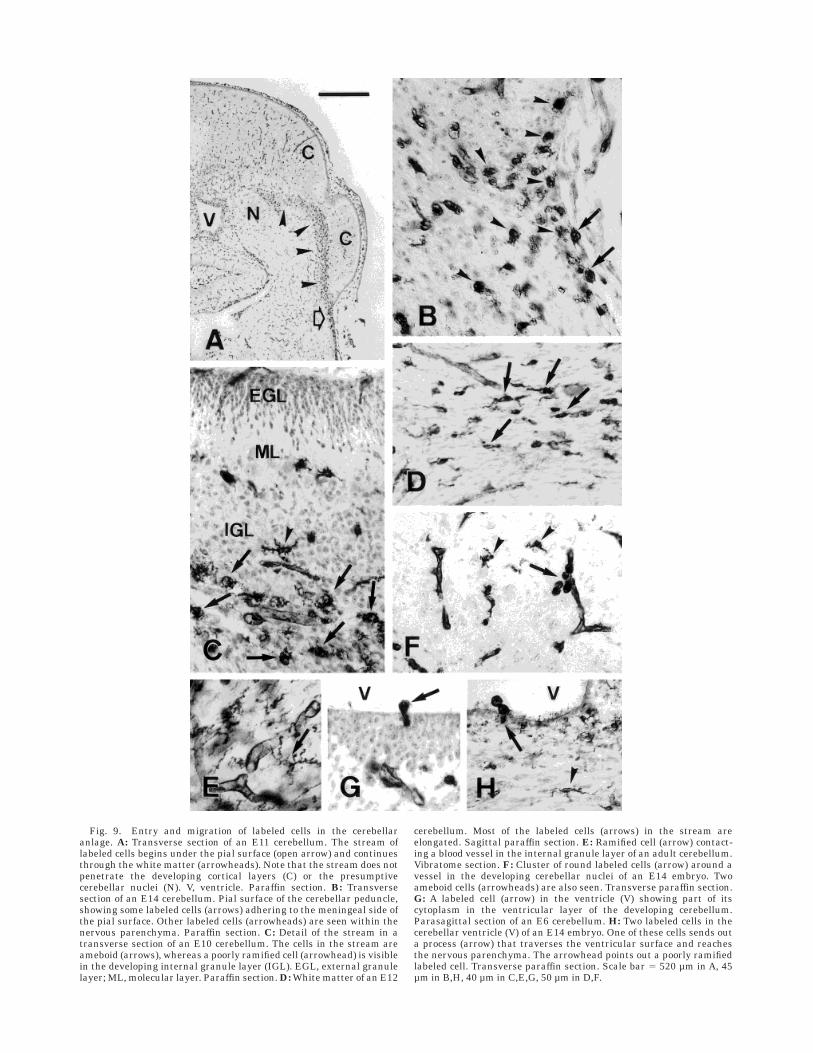
Fig. 9. Entry and migration of labeled cells in the cerebellaranlage. A: Transverse section of an E11 cerebellum. The stream oflabeled cells begins under the pial surface (open arrow) and continuesthrough the white matter (arrowheads). Note that the stream does notpenetrate the developing cortical layers (C) or the presumptivecerebellar nuclei (N). V, ventricle. Paraffin section. B: Transversesection of an E14 cerebellum. Pial surface of the cerebellar peduncle,showing some labeled cells (arrows) adhering to the meningeal side ofthe pial surface. Other labeled cells (arrowheads) are seen within thenervous parenchyma. Paraffin section. C: Detail of the stream in atransverse section of an E10 cerebellum. The cells in the stream areameboid (arrows), whereas a poorly ramified cell (arrowhead) is visiblein the developing internal granule layer (IGL). EGL, external granulelayer; ML, molecular layer. Paraffin section. D: White matter of an E12
cerebellum. Most of the labeled cells (arrows) in the stream areelongated. Sagittal paraffin section. E: Ramified cell (arrow) contact-ing a blood vessel in the internal granule layer of an adult cerebellum.Vibratome section. F: Cluster of round labeled cells (arrow) around avessel in the developing cerebellar nuclei of an E14 embryo. Twoameboid cells (arrowheads) are also seen. Transverse paraffin section.G: A labeled cell (arrow) in the ventricle (V) showing part of itscytoplasm in the ventricular layer of the developing cerebellum.Parasagittal section of an E6 cerebellum. H: Two labeled cells in thecerebellar ventricle (V) of an E14 embryo. One of these cells sends outa process (arrow) that traverses the ventricular surface and reachesthe nervous parenchyma. The arrowhead points out a poorly ramifiedlabeled cell. Transverse paraffin section. Scale bar 5 520 µm in A, 45µm in B,H, 40 µm in C,E,G, 50 µm in D,F.

and that the differentiation program of the microgliavaries depending on the environmental factors present ineach region. These factors account for the differences inmorphological features of microglia during development(Cuadros et al., 1992, 1994; Wu et al., 1993) and indifferent regions of the adult brain (Lawson et al., 1990;Vela et al., 1995).
CONCLUSIONS
Although definitive conclusions cannot be drawn with-out experimental work, our observations suggest a pictureof microglia development summarized in Figure 10. Themain route of entry of microglial precursors in the cerebel-
lar anlage is across the pial surface at the basal region ofthe cerebellar peduncles. This invasion apparently takesplace from E8–E9 to E16. Microglial precursors may alsoinvade the nervous parenchyma of the cerebellum by otherroutes, i.e., traversing the wall of blood vessels and/or theventricular layer, but these routes are used by muchsmaller numbers of cells than those that cross the pialsurface near the cerebellar peduncles. Microglial cellsfrom the peduncles migrate tangentially along the cerebel-lar white matter between E9 and hatching, and constitutea stream of labeled cells. Individual microglial cells leavethe stream at some points and migrate radially to theirfinal locations within the developing cortex. Radial migra-tion into the cerebellar cortex probably begins shortly aftertangential migration within the stream has started, andlasts until the second posthatching week. Finally, cellsdifferentiate once they have reached their final position inthe cerebellar nuclei, the cortical layers or the whitematter.
The development of microglia in the quail cerebellumproceeds in several steps: invasion of the nervous paren-chyma at specific points, long-distance tangential migra-tion along an oriented substrate, radial migration, anddifferentiation. This sequence is the same as that foundduring microglial development in the optic tectum (Cuadroset al., 1994) and retina (Navascues et al., 1995); wetherefore suggest that it reflects a set of general rules inmicroglial development in the quail brain.
ACKNOWLEDGMENTS
We thank Karen Shashok for editing the manuscript.
LITERATURE CITED
Acarin, L., J.M. Vela, B. Gonzalez, and B. Castellano (1994) Demonstrationof poly-N-acetyl lactosamine residues in ameboid and ramified microg-lia cells in rat brain by tomato lectin binding. J. Histochem. Cytochem.42:1033–1041.
Ashwell, K. (1990) Microglia and cell death in the developing mousecerebellum. Dev. Brain Res. 55:219–230.
Ashwell, K. (1991) The distribution of microglia and cell death in the fetalrat forebrain. Dev. Brain Res. 58:1–12.
Boya, J., J. Calvo, and A. Prado (1979) The origin of microglial cells. J. Anat.129:177–186.
Boya, J., J.L. Calvo, A.L. Carbonell, and A. Borregon (1991) A lectinhistochemistry study on the development of rat microglial cells. J. Anat.175:229–236.
Castellano, B., B. Gonzalez, I. Dalmau, and J.M. Vela (1991) Identificationand distribution of microglial cells in the cerebral cortex of the lizard: Ahistochemical study. J. Comp. Neurol. 311:434–444.
Chugani, D.C., N.L. Kedersha, and L.H. Rome (1991) Vault immunofluores-cence in the brain: New insights regarding the origin of microglia. J.Neurosci. 11:256–287.
Cuadros, M.A., A. Moujahid, G. Martın-Partido, and J. Navascues (1992)Microglia in the mature and developing quail brain as revealed by amonoclonal antibody recognizing hemopoietic cells. Neurosci. Lett.148:11–14.
Cuadros, M.A., C. Martin, P. Coltey, A. Almendros, and J. Navascues (1993)First appearance, distribution, and origin of macrophages in the earlydevelopment of the avian central nervous system. J. Comp. Neurol.330:113–129.
Cuadros, M.A., A. Moujahid, A. Quesada, and J. Navascues (1994) Develop-ment of microglia in the quail optic tectum. J. Comp. Neurol. 348:207–224.
Dowding, A.J., A. Maggs, and J. Scholes (1991) Diversity amongst themicroglia in growing and regenerating fish CNS: Immunohistochemicalcharacterization using FL.1, an anti-macrophage monoclonal antibody.Glia 4:345–364.
Fig. 10. Schematic drawings showing the proposed migration ofmicroglial precursors in the cerebellum. A: Three-dimensional draw-ing of an E11–E12 cerebellum sectioned along a parasagittal plane. B:Transverse section at the level marked by the vertical dotted line in A.Many of the microglial precursors enter the cerebellar anlage from themeninges at the base of the cerebellar peduncles (CP) at the pointsmarked by open arrows in B. The microglial precursors migratethrough the white matter constituting the stream described in the text(dotted region in A and B). Individual microglial precursors migrateradially from this region (arrowheads in A and B) to invade the cortex(C in A and B). In addition, other microglial precursors may enter bytraversing the ventricular layer (small arrows in A and B) or endothe-lial wall (not shown). BS, brain stem; N, cerebellar nuclei; V, ventricle.
400 M. A. CUADROS ET AL.

Fujimoto, E., A. Miki, and H. Mizoguti (1987) Histochemical studies of thedifferentiation of microglial cells in the cerebral hemispheres of chickembryos and chicks. Histochemistry 87:209–216.
Fujimoto, E., A. Miki, and H. Mizoguti (1989) Histochemical study of thedifferentiation of microglial cells in the developing human cerebralhemispheres. J. Anat. 166:253–264.
Gehrmann, J., and G.W. Kreutzberg (1991) Characterisation of two newmonoclonal antibodies directed against rat microglia. J. Comp. Neurol.313:409–430.
Gilad, G.M., and V.H. Gilad (1995) Chemotaxis and accumulation of nervegrowth factor by microglia and macrophages. J. Neurosci. Res. 41:594–602.
Goodbrand, I.A., and R.M. Gaze (1991) Microglia in tadpoles of Xenopuslaevis: Normal distribution and the response to optic nerve injury. Anat.Embryol. 184:71–82.
Hatten, M.E. (1990) Riding the glial monorail: A common mechanism forglial-guided neuronal migration in different regions of the developingmammalian brain. Trends Neurosci. 13:179–184.
Hutchins, K.D., D.W. Dickson, W.K. Rashbaum, and W.D. Lyman (1992)Localization of morphologically distinct microglial populations in thedeveloping human fetal brain: Implications for ontogeny. Dev. BrainRes. 55:95–102.
Imamura, K., M. Ito, A. Suzumura, J. Asai, and A. Takahashi (1990)Generation and characterization of monoclonal antibodies against ratmicroglia and ontogenic distribution of positive cells. Lab. Invest.63:853–861.
Innocenti, G.M., S. Clarke, and H. Koppel (1983) Transitory macrophagesin the white matter of the developing visual cortex: II. Development andrelations with axonal pathways. Dev. Brain Res. 11:55–66.
Jordan, F.L., and W.E. Thomas (1988) Brain macrophages: Questions oforigin and interrelationship. Brain Res. Rev. 13:165–178.
Krueger, B.K., J.F. Burne, and M.C. Raff (1995) Evidence for large-scaleastrocyte death in the developing cerebellum. J. Neurosci. 15:3366–3374.
Lawson, L.J., V.H. Perry, P. Dri, and S. Gordon (1990) Heterogeneity in thedistribution and morphology of microglia in the normal adult mousebrain. Neuroscience 39:151–170.
Lent, R., R. Linden, and L. Cavalcante (1985) Transient populations ofpresumptive macrophages in the brain of the developing hamster, asindicated by endocytosis of blood-borne horseradish peroxidase. Neuro-science 15:1203–1215.
Ling, E.A. (1981) The origin and nature of microglia. In S. Fedoroff and L.Hertz (eds): Advances in Cell Neurobiology, Vol 2. London: AcademicPress, pp. 33–82.
Ling, E.A., C. Kaur, T.Y. Yick, and W.C. Wong (1990) Immunocytochemicallocalization of CR3 complement receptors with OX-42 in ameboidmicroglia in postnatal rats. Anat. Embryol. 182:481–486.
Ling, E.A., and W.C. Wong (1993) The origin and nature of ramified andameboid microglia: A historical review and current concepts. Glia7:9–18.
Mason, C.A., S. Christakos, and S. Catalano (1990) Early climbing fiberinteractions with Purkinje cells in the postnatal mouse cerebellum. J.Comp. Neurol. 297:77–90.
Milligan, C.E., T.J. Cunningham, and P. Levitt (1991) Differential immuno-chemical markers reveal the normal distribution of brain macrophagesand microglia in the developing rat brain. J. Comp. Neurol. 314:125–135.
Murabe, Y., and Y. Sano (1982) Morphological studies on neuroglia. VI.Postnatal development of microglial cells. Cell Tissue Res. 225:469–485.
Navascues, J., A. Moujahid, A. Quesada, and M.A. Cuadros (1994) Microg-lia in the avian retina: Immunocytochemical demonstration in the adultquail. J. Comp. Neurol. 350:171–186.
Navascues, J., A. Moujahid, A. Almendros, J.L. Marın-Teva, and M.A.Cuadros (1995) Origin of microglia in the quail retina: Central-to-peripheral and vitreal-to-scleral migration of microglial precursorsduring development. J. Comp. Neurol. 354:209–228.
Navascues, J., M.A. Cuadros, and A. Almendros (1996) Development ofmicroglia: Evidence from studies in the avian central nervous system.In E.A. Ling, C.K. Tan, and C.B.C. Tan (eds): Topical Issues inMicroglial Research. Singapore: Singapore Neuroscience Association,pp. 43–64.
Pardanaud, L., C. Altmann, P. Kitos, F. Dieterlen-Lievre, and C.A. Buck(1987) Vasculogenesis in the early quail blastodisc as studied with amonoclonal antibody recognizing endothelial cells. Development 100:339–349.
Penfold, P.L., M.C. Madigan, and J.M. Provis (1991) Antibodies to humanleucocyte antigens indicate subpopulations of microglia in humanretina. Vis. Neurosci. 7:383–388.
Perry, V.H., D.A. Hume, and S. Gordon (1985) Immunohistochemicallocalization of macrophages and microglia in the adult and developingmouse brain. Neuroscience 15:313–326.
Perry, V.H., and S. Gordon (1991) Macrophages and the nervous system.Int. Rev. Cytol. 125:203–244.
Rio-Hortega, P. (1932) Microglia. In W. Penfield (ed): Cytology and CellularPathology of the Nervous System, Vol 2. New York: Paul B. Hoeber, pp.482–534.
Streit, W.J., and G.W. Kreutzberg (1987) Lectin binding by resting andreactive microglia. J. Neurocytol. 16:249–260.
Vela, J.M., I. Dalmau, B. Gonzalez, and B. Castellano (1995) Morphologyand distribution of microglial cells in the young and adult mousecerebellum. J. Comp. Neurol. 361:602–616.
Wolswijk, G. (1995) Strongly GD31 cells in the developing and adult ratcerebellum belong to the microglial lineage rather than to the oligoden-drocyte lineage. Glia 13:13–26.
Wu, C.H., C.Y. Wen, J.Y. Shieh, and E.A. Ling (1992) A quantitative andmorphometric study of the transformation of amoeboid microglia intoramified microglia in the developing corpus callosum in rats. J. Anat.181:423–430.
Wu, C.H., C.Y. Wen, J.Y. Shieh, and E.A. Ling (1993) A quantitative study ofthe differentiation of microglial cells in the developing cerebral cortex inrats. J. Anat. 182:403–413.
Yao, J., L. Harvath, D.L. Gilbert, and C.A. Colton (1990) Chemotaxis by aCNS macrophage, the microglia. J. Neurosci. Res. 27:36–42.
Youngren, O.M., and R.E. Phillips (1978) A stereotaxic atlas of the brain ofthe three-day-old domestic chick. J. Comp. Neurol. 181:567–600.
MICROGLIA IN DEVELOPING CEREBELLUM 401