redefinitionofthecarbohydratebindingspecificityof ......
TRANSCRIPT
Redefinition of the Carbohydrate Binding Specificity ofHelicobacter pylori BabA Adhesin*□S
Received for publication, June 3, 2012, and in revised form, July 6, 2012 Published, JBC Papers in Press, July 20, 2012, DOI 10.1074/jbc.M112.387654
John Benktander‡, Jonas Ångstrom‡, Michael E. Breimer§, and Susann Teneberg‡1
From the ‡Institute of Biomedicine, Department of Medical Biochemistry and Cell Biology, P. O. Box 440, University of Gothenburg,S-405 30 Goteborg, Sweden and §Department of Surgery, Sahlgrenska University Hospital, S-41 345 Goteborg, Sweden
Background: The BabA adhesin mediates binding of Helicobacter pylori to the gastric epithelium.Results: Binding of BabA to blood group O and A determinants on type 4 core chains was demonstrated.Conclusion: The BabA binds to blood group determinants on both type 1 and type 4 core chains.Significance: Characterization of the binding specificities of BabA is important for understanding the interactions betweenH.pylori and target cells.
Certain Helicobacter pylori strains adhere to the human gas-tric epithelium using the blood group antigen-binding adhesin(BabA). All BabA-expressingH. pylori strains bind to the bloodgroup O determinants on type 1 core chains, i.e. to the Lewis bantigen (Fuc�2Gal�3(Fuc�4)GlcNAc; Leb) and the H type 1determinant (Fuc�2Gal�3GlcNAc). Recently, BabA strainshave been categorized into those recognizing only Leb and Htype 1 determinants (designated specialist strains) and thosethat also bind to A and B type 1 determinants (designated gen-eralist strains). Here, the structural requirements for carbohy-drate recognition by generalist and specialist BabAwere furtherexplored by binding of these types of strains to a panel of differ-ent glycosphingolipids. Three glycosphingolipids recognized byboth specialist and generalist BabAwere isolated from the smallintestine of a blood groupOpig and characterized bymass spec-trometry and proton NMR as H type 1 pentaglycosylceramide(Fuc�2Gal�3GlcNAc�3Gal�4Glc�1Cer), Globo H hexaglyco-sylceramide (Fuc�2Gal�3GalNAc�3Gal�4Gal�4Glc�1Cer),and a mixture of three complex glycosphingolipids (Fuc�-2Gal�4GlcNAc�6(Fuc�2Gal�3GlcNAc�3)Gal�3GlcNAc�-3Gal�4Glc�1Cer, Fuc�2Gal�3GlcNAc�6(Fuc�2Gal�3Glc-NAc�3)Gal�3GlcNAc�3Gal�4Glc�1Cer, and Fuc�2Gal�4-(Fuc�3)GlcNAc�6(Fuc�2Gal�3GlcNAc�3)Gal�3GlcNAc�-3Gal�4Glc�1Cer). In addition to the binding of both strains tothe Globo H hexaglycosylceramide, i.e. a blood group O deter-minant on a type 4 core chain, the generalist strain bound to theGlobo A heptaglycosylceramide (GalNAc�3(Fuc�2)Gal�3-GalNAc�3Gal�4Gal�4Glc�1Cer), i.e. a blood groupAdetermi-nant on a type 4 core chain. The binding of BabA to the two setsof isoreceptors is due to conformational similarities of the ter-minal disaccharides ofH type 1 andGloboH and of the terminaltrisaccharides of A type 1 and Globo A.
Attachment of microbes to cell surface receptors on the tar-get tissue is considered an essential step in the initiation, estab-lishment, and maintenance of infection. In recent years, a largenumber of studies have aimed at the identification of potentialmicrobial host receptors, the majority of which appear to beglycoconjugates (1–3). Glycoconjugates exhibit a characteristicand specific pattern of expression, which is dependent on theanimal species, age, individual, and cell type (4). Thus, the rec-ognition of a specific carbohydrate receptor on the host cellsurface determines at least in part the host, tissue, and age spec-ificities of microbial infections.Adherence of the gastric pathogen Helicobacter pylori to
human gastric epithelial cells is required for prolonged persist-ence in the stomach. Initial studies of potential target cell recep-tors for H. pylori demonstrated the binding of certain strainsof this bacterium to the Lewis b blood group antigen(Fuc�2Gal�3(Fuc�4)GlcNAc;2 Leb)3 (5), and subsequently theH. pylori Leb-binding adhesin, blood group antigen-bindingadhesin (BabA) was identified (6). H. pylori strains expressingBabA together with the vacuolating cytotoxin VacA and thecytotoxin-associated antigen CagA (triple positive strains) areassociated with severe gastric diseases such as peptic ulcer andgastric adenocarcinoma (7, 8).Subsequent studies demonstrated that the BabA adhesin has
adapted to the fucosylated blood group antigensmost prevalentin the local population (9). In Europe and the United Stateswhere blood groupA, B, andO phenotypes all are common, theH. pylori strains (designated generalist strains) bind to bloodgroup A, B, and O type 1 determinants. However, in popula-tions such as the indigenous South American native popula-tion, which only has the blood groupOphenotype, theH. pylori
* This study was supported by Swedish Research Council Grant 12628, theSwedish Cancer Foundation, and governmental grants (to the SahlgrenskaUniversity Hospital).Author’s Choice—Final version full access.
□S This article contains supplemental Figs. S1 and S2.1 To whom correspondence should be addressed. Tel.: 46-31-786-34-92; Fax:
46-31-413-190; E-mail: [email protected].
2 The glycosphingolipid nomenclature follows the recommendations by theIUPAC-IUB Commission on Biochemical Nomenclature (Chester, M. A.(1998) IUPAC-IUB Joint Commission on Biochemical Nomenclature (JCBN).Nomenclature of glycolipids—recommendations 1997. Eur. J. Biochem.257, 293–298). It is assumed that Gal, Glc, GlcNAc, GalNAc, NeuAc, andNeuGc are of the D configuration; Fuc is of the L configuration; and allsugars are present in the pyranose form.
3 The abbreviations used are: Leb, Lewis b antigen; BabA, blood group anti-gen-binding adhesin; ESI, electrospray ionization; Hex, hexose; HexNAc,N-acetylhexosamine; Cer, ceramide; Ley, Lewis y antigen; Lea, Lewis a anti-gen; NeuGc, N-glycolylneuraminic acid.
THE JOURNAL OF BIOLOGICAL CHEMISTRY VOL. 287, NO. 38, pp. 31712–31724, September 14, 2012Author’s Choice © 2012 by The American Society for Biochemistry and Molecular Biology, Inc. Published in the U.S.A.
31712 JOURNAL OF BIOLOGICAL CHEMISTRY VOLUME 287 • NUMBER 38 • SEPTEMBER 14, 2012
by guest on April 22, 2019
http://ww
w.jbc.org/
Dow
nloaded from

strains (designated specialist strains) bind only to the bloodgroup O type 1 determinants (Leb and the H type 1). Thus, thecarbohydrate binding site of BabA of generalist strains canaccommodate an extension of the blood group O determinantwith an �3-linked GalNAc or Gal (creating the blood group Aand B determinants, respectively), whereas this extension is nottolerated by the BabA of specialist strains. Consequently, theBabA adhesins from these strains have differences in the archi-tecture of their carbohydrate binding sites.In the present study, the structural requirements for carbo-
hydrate recognition by BabA of generalist and specialist H.pylori strains were further explored. Radiolabeled H. pyloristrains were examined for binding to a panel of different glyco-sphingolipids from various sources separated on thin-layerplates, and glycosphingolipids recognized by wild type special-ist and/or generalist H. pylori, but not by a deletion mutantstrain lacking the BabA adhesin, were isolated and character-ized by mass spectrometry and proton NMR. Comparativebinding studies demonstrated that the BabA adhesin in addi-tion to blood group determinants on type 1 core chains recog-nizes blood group O and A determinants on type 4 core chainswith binding to Globo H (i.e. H type 4) by both strains andGlobo A (i.e. A type 4) by the generalist strain. Inspection ofminimum energy models revealed topographical similarities inthe spatial orientation of the terminal disaccharide(Fuc�2Gal�3) of theGloboHandH5 type 1 glycosphingolipids,accounting for the BabA cross-reactivity.
EXPERIMENTAL PROCEDURES
H. pylori Strains, Culture Conditions, and Labeling—Thegeneralist H. pylori strain J99 and the construction of the J99/BabA� mutant babA::cam were described by Mahdavi et al.(10). The specialist H. pylori strain S831 was described (9).
For chromatogram binding experiments, the bacteria weregrown in a microaerophilic atmosphere at 37 °C for 48 h onBrucella medium (Difco) containing 10% fetal calf serum (Har-lan Sera-Lab, Loughborough, UK) inactivated at 56 °C and BBLIsoVitaleX Enrichment (BD Biosciences). The mutant strainJ99/BabA� was cultured on the same medium supplementedwith chloramphenicol (20 �g/ml). Bacteria were radiolabeledby the addition of 50 �Ci [35S]methionine (Amersham Biosci-ences) diluted in 0.5 ml of phosphate-buffered saline (PBS), pH7.3 to the culture plates. After incubation for 12–72 h at 37 °Cunder microaerophilic conditions, the bacteria were harvested,centrifuged three times, and thereafter suspended to 1 � 108cfu/ml in PBS. The specific activities of the suspensions were�1 cpm/100 H. pylori organisms.Chromatogram Binding Assays—Reference glycosphingolip-
ids were isolated and characterized by mass spectrometry andproton NMR as described (11).Thin-layer chromatography was performed on glass- or alu-
minum-backed silica gel 60HPTLCplates (Merck).Mixtures ofglycosphingolipids (40 �g) or pure compounds (40 ng–4 �g)were separated using chloroform/methanol/water (60:35:8 byvolume) as the solvent system. Chemical detection was accom-plished by anisaldehyde (12).Binding of 35S-labeled H. pylori to glycosphingolipids on
thin-layer chromatograms was done as reported previously
(13). Dried chromatograms were dipped for 1 min in diethylether/n-hexane (1:5 by volume) containing 0.5% (w/v) poly-isobutylmethacrylate (Aldrich). After drying, the chromato-gramswere soaked in PBS containing 2%bovine serumalbumin(w/v), 0.1% NaN3 (w/v), and 0.1% Tween 20 (by volume) for 2 hat room temperature. The chromatograms were subsequentlycovered with radiolabeled bacteria diluted in PBS (2–5 � 106cpm/ml). Incubation was done for 2 h at room temperaturefollowed by repeated washings with PBS. The chromatogramswere thereafter exposed to XAR-5 x-ray films (Eastman KodakCo.) for 12 h.Chromatogram binding assays withmousemonoclonal anti-
bodies directed against the Globo H determinant (MBr1, EnzoLife Sciences), the Leb determinant (BG-6/T218, Signet/Co-vance), the H type 1 determinant (17-206, Abcam), and the Htype 2 determinant (A583, DakoCytomationNorden A/S) weredone as described (13) using 125I-labeled monoclonal anti-mouse antibodies (Z0259, DakoCytomation Norden A/S) fordetection.Isolation of H. pylori-binding Glycosphingolipids—Total acid
and non-acid glycosphingolipid fractions were isolated bystandardmethods (11). Briefly, thematerial was lyophilized andthen extracted in two steps in a Soxhlet apparatus with chloro-form and methanol (2:1 and 1:9 by volume, respectively). Thematerial obtained was subjected tomild alkaline hydrolysis anddialysis followed by separation on a silicic acid column. Acidand non-acid glycosphingolipid fractions were obtained bychromatography on a DEAE-cellulose column. To separate thenon-acid glycolipids from alkali-stable phospholipids, this frac-tion was acetylated and separated on a second silicic acid col-umn followed by deacetylation and dialysis. Final purificationswere done by chromatographies on DEAE-cellulose and silicicacid columns.The non-acid glycosphingolipid fractions were separated by
repeated silicic acid chromatography, and final separation wasachieved by HPLC or by chromatography on Iatrobead(Iatrobeads 6RS-8060, Iatron Laboratories, Tokyo, Japan) col-umns and elution with chloroform/methanol/water (65:25:4 byvolume) followed by chloroform/methanol/water (60:35:8 byvolume) and finally chloroform/methanol/water (40:40:12by volume). Throughout the separation procedures, aliquots ofthe fractions obtained were analyzed by thin-layer chromatog-raphy, and fractions that were colored green by anisaldehydewere tested for binding of H. pylori using the chromatogrambinding assay. The fractions were pooled according to themobility on thin-layer chromatograms and theirH. pylori bind-ing activity.Endoglycoceramidase Digestion and LC-ESI/MS—Endogly-
coceramidase II from Rhodococcus spp. (14) (Takara BioEurope S.A., Gennevilliers, France) was used for hydrolysis ofglycosphingolipids. Briefly, 50 mg of glycosphingolipids weresuspended in 100 ml of 0.05 M sodium acetate buffer, pH 5.0containing 120 mg of sodium cholate and sonicated briefly.Thereafter, 1 milliunit of endoglycoceramidase II was added,and the mixture was incubated at 37 °C for 48 h. The reactionwas stopped by addition of chloroform/methanol/water to thefinal proportions 8:4:3 (by volume). The oligosaccharide-con-taining upper phase thus obtained was separated from deter-
H. pylori BabA-binding Glycosphingolipids
SEPTEMBER 14, 2012 • VOLUME 287 • NUMBER 38 JOURNAL OF BIOLOGICAL CHEMISTRY 31713
by guest on April 22, 2019
http://ww
w.jbc.org/
Dow
nloaded from

gent on a Sep-Pak QMA cartridge (Waters, Milford, MA). Theeluant containing the oligosaccharides was dried under nitro-gen and under vacuum.The glycosphingolipid-derived oligosaccharides were ana-
lyzed by LC/MS and MS/MS as described (15). In brief, theoligosaccharides were separated on a column (200 � 0.180mm) packed in house with 5-mm porous graphite particles(Hypercarb, Thermo Scientific) and eluted with an acetonitrilegradient (A, 10 mM ammonium bicarbonate; B, 10 mM ammo-nium bicarbonate in 80% acetonitrile). The saccharides wereanalyzed in the negative ionmode on anLTQ linear quadrupoleion trap mass spectrometer (Thermo Electron, San Jose, CA).LC-ESI/MS and ESI/MS/MS of Native Glycosphingolipids—
The glycosphingolipids (dissolved in methanol/acetonitrile,75:25 by volume) were separated on a 200� 0.150-mm columnpacked in house with 5-mm polyamine II particles (YMCEurope GmbH, Dinslaken, Germany) and eluted with a watergradient (A, 100% acetonitrile; B, 10 mM ammonium bicarbon-ate). Samples were analyzed on an LTQ linear quadrupole iontrap mass spectrometer by LC-ESI/MS at �3.5 kV. A full scan(m/z 500–1800; two microscans; maximum time, 100 ms; tar-get value, 30,000) was performed followed by data-dependentMS2 scans (two microscans; maximum time, 100 ms; targetvalue, 10,000) with a normalized collision energy of 35%, anisolation window of 2.5 units, an activation q of 0.25, and anactivation time of 30 ms.Proton NMR Spectroscopy—1H NMR spectra were acquired
on a Varian 600-MHz spectrometer at 30 °C. Samples were dis-solved in dimethyl sulfoxide/D2O (98:2 by volume) after deute-rium exchange. Two-dimensional double quantum-filteredcorrelated spectroscopy (COSY) spectra were recorded usingthe standard pulse sequence (16).Molecular Modeling—Minimum energy models of different
glycosphingolipids were constructed using the CHARMmforce field within the Discovery Studio molecular modelingpackage (Accelrys, Inc., San Diego, CA) and literature values asstarting points for the glycosidic torsion angles (17, 18).
RESULTS
Binding of H. pylori to Glycosphingolipid Mixtures—Screen-ing for BabA-mediated binding ofH. pyloriwas done by bindingof the generalist H. pylori strain J99, the specialist strain S831,and the deletion mutant strain J99/BabA� to non-acid glyco-sphingolipid fractions from various sources to expose the bac-teria to a large number of potentially binding-active carbohy-drate structures. Thus, the binding of the bacteria to non-acidglycosphingolipid mixtures isolated from the small intestine ofdifferent species (human, rat, cat, and pig (19–23)), erythro-cytes of different species (human, cat, rabbit, dog, horse,chicken, and sheep (24)), human cancers (lung, kidney, colon,liver, and gastric cancers (25)), and human stomach (26) wastested. Thereby, three glycosphingolipids recognized by boththe generalist and specialistH. pylori strainwere detected in thenon-acid glycosphingolipid fraction from the small intestinalepithelium of a blood group O pig (Fig. 1, B and C, lane 1). Thebinding-active compounds migrated in the penta-, hexa-, andocta-/nonaglycosylceramide regions, respectively. No bindingof the deletion mutant strain J99/BabA� to the porcine intes-
tinal glycosphingolipids was obtained (data not shown), indi-cating that the binding of the wild type bacteria to these com-pounds was mediated by BabA.Isolation of the H. pylori-binding Glycosphingolipids from
Porcine Intestine—A total non-acid fraction from blood groupO porcine small intestinal epithelium (160 mg) was separatedby repeated silica gel chromatography and Iatrobead columnchromatography, and the subfractions obtained were tested forH. pylori binding activity. After pooling of binding-active frac-tions, three subfractions containing H. pylori-binding glyco-sphingolipids were obtained. One of these fraction (designatedfraction P-I (0.2 mg)) migrated in the pentaglycosylceramideregion, whereas the fraction designated fraction P-II (0.2 mg)migrated in the hexaglycosylceramide region (Fig. 2, lanes 1 and2). LC-ESI/MS of the third fraction containing the slowestmigratingH. pylori-binding compounds showed that this was amixture of several glycosphingolipids. This fraction was there-fore further separated on an Iatrobead column, and after pool-ing of theH. pylori-binding fractions, 0.3mg of the slowmigrat-ingH. pylori-binding compound (designated fraction P-III) wasobtained (Fig. 2, lane 3).Characterization of the H. pylori-binding Fraction P-I from
Porcine Intestine—LC-ESI/MS, proton NMR, and antibodybinding demonstrated that fraction P-I was a mixture of the Htype 1 pentaglycosylceramide (Fuc�2Gal�3GlcNAc�3Gal-�4Glc�1Cer) and the B5 pentaglycosylceramide (Gal�3-Gal�4GlcNAc�3Gal�4Glc�1Cer) (data not shown).Characterization of the H. pylori-binding Fraction P-II from
Porcine Intestine—Characterization of the BabA binding frac-tion P-II demonstrated the Globo H hexaglycosylceramide(Fuc�2Gal�3GalNAc�3Gal�4Gal�4Glc�1Cer) as the majorcompound. This conclusion was based on the following prop-erties. (i) ESI/MS of the native fraction P-II gave a major [M �2H�]2� ion atm/z 784, corresponding to amolecular ion atm/z1568, demonstrating a glycosphingolipid with one Fuc, oneHexNAc, and four Hex residues and phytosphingosine withhydroxy 16:0 fatty acid (data not shown). The series of C, Y, andZ ions obtained by MS2 of the [M � 2H�]2� ion at m/z 784
FIGURE 1. Binding of a generalist and a specialist H. pylori strain to non-acid glycosphingolipids of the small intestinal epithelium of a bloodgroup O pig. The glycosphingolipids were separated on aluminum-backedsilica gel plates using chloroform/methanol/water (60:35:8 by volume) as thesolvent system. The chromatogram in A was stained with anisaldehyde.Duplicate chromatograms were incubated with the 35S-labeled H. pylori gen-eralist strain J99 (B) and the H. pylori specialist strain S831 (C) followed byautoradiography for 12 h as described under “Experimental Procedures.”Lane 1, non-acid glycosphingolipids of the intestinal epithelium of a bloodgroup O pig, 40 �g; lane 2, reference H type 2 pentaglycosylceramide(Fuc�2Gal�4GlcNAc�3Gal�4Glc�1Cer), 4 �g; lane 3, reference Lea pentagly-cosylceramide (Gal�3(Fuc�4)GlcNAc�3Gal�4Glc�1Cer), 2 �g; lane 4, refer-ence Leb hexaglycosylceramide (Fuc�2Gal�3(Fuc�4)GlcNAc�3Gal�4Glc�1Cer),4 �g; lane 5, reference B type 1 heptaglycosylceramide (Gal�3(Fuc�2)-Gal�3(Fuc�4)GlcNAc�3Gal�4Glc�1Cer), 2 �g.
H. pylori BabA-binding Glycosphingolipids
31714 JOURNAL OF BIOLOGICAL CHEMISTRY VOLUME 287 • NUMBER 38 • SEPTEMBER 14, 2012
by guest on April 22, 2019
http://ww
w.jbc.org/
Dow
nloaded from

demonstrated a Fuc-Hex-HexNAc-Hex-Hex-Hex sequence(supplemental Fig. S1).(ii) LC-ESI/MS of oligosaccharides gives the resolution of
isomeric saccharides, and the carbohydrate sequence can bededuced from series of C type fragment ions obtained by MS2
(15). In addition, diagnostic cross-ring 0,2A type fragment ionsare present in MS2 spectra of oligosaccharides with a Hex orHexNAc substituted at C-4 and thus allow differentiation oflinkage positions (15, 27, 28).LC-ESI/MS of the oligosaccharides obtained by hydrolysis of
fraction P-II with Rhodococcus endoglycoceramidase II gavetwo late eluting molecular ions (Fig. 3, A–C). These ions werefound at m/z 852 (retention time, 26.8–27.2 min) and at m/z1014 (retention time, 25.2–25.7 min) and demonstrated oneoligosaccharide with one Fuc, one HexNAc and three Hex res-idues and one oligosaccharide with one Fuc, one HexNAc, andfour Hex residues, respectively.The MS2 spectrum of the molecular ion atm/z 852 (Fig. 3D)
had a C type fragment ion series (C2 atm/z 325, C3 atm/z 528,and C4 at m/z 690), demonstrating a Fuc-Hex-HexNAc-Hex-Hex sequence. The features of this MS2 spectrum were verysimilar to the MS2 spectrum of reference H type 1 pentaglyco-sylceramide (15).
MS2 of the molecular ion at m/z 1014 (Fig. 3E) also gave aseries of C type fragment ionswithC2 atm/z 325, C3 atm/z 528,and C4 atm/z 690 along with a C5 ion atm/z 852, identifying aFuc-Hex-HexNAc-Hex-Hex-Hex sequence. The 0,2A5 frag-ment ion at m/z 792 and the 0,2A6 fragment ion at m/z 954indicated that the two hexoses at the reducing end were sub-stituted at C-4, i.e. a Fuc-Hex-HexNAc-Hex-4Hex-4Hexsequence.(iii) The anomeric region of the proton NMR spectrum of
fraction P-II (Fig. 3F) revealed a single dominating species withsix carbohydrate residues that is identical to the previously pub-lishedGloboHglycosphingolipid (29) as evidenced by signals at4.949 (Fuc�2), 4.802 (Gal�4), 4.468 (GalNAc�3), 4.456 (Gal�3),4.247 (Gal�4), and 4.208 ppm (Glc�1), thus yielding thesequence Fuc�2Gal�3GalNAc�3Gal�4Gal�4Glc�1Cer in ac-cordance with the mass spectrometry data above.Thus, by mass spectrometry and proton NMR, the BabA-
binding hexaglycosylceramide of blood group O pig intestinewas identified as the Globo H glycosphingolipid. In the basepeak chromatogram from LC-ESI/MS of the oligosaccharidesobtained by hydrolysis of fraction P-II with Rhodococcusendoglycoceramidase (Fig. 3A), the major molecular ion wasfound atm/z 852, corresponding to the H type 1 pentaglycosyl-ceramide. Still, proton NMR demonstrated that fraction P-IIwas a relatively pure Globo H glycosphingolipid. This discrep-ancy is due to the restricted hydrolytic capacity of the Rhodo-coccus endoglycoceraminidase II, which has a relative resist-ance of hydrolysis for globo series glycosphingolipids (14, 30).The ideal enzyme would have been the ceramide glycanasefrom Macrobdella decora that has a more universal hydrolyticactivity toward glycosphingolipids (31). However, the M.decora enzyme is no longer available commercially.Characterization of the Slow Migrating H. pylori-binding
Fraction P-III from Porcine Intestine—Antibody binding, massspectrometry, and proton NMR demonstrated that fractionP-III was a mixture of two branched decaglycosylceramideswith terminal H type 1 epitopes (Fuc�2Gal�3GlcNAc�6-(Fuc�2Gal�3GlcNAc�3)Gal�3GlcNAc�3Gal�4Glc�1Cer andFuc�2Gal�4GlcNAc�6(Fuc�2Gal�3GlcNAc�3)Gal�3GlcNA-c�3Gal�4Glc�1Cer) and a related undecaglycosylceramidewith a Fuc�3 substitution of the GlcNAc of the 6-branch, yield-ing an Ley determinant (Fuc�2Gal�4(Fuc�3)GlcNAc�6-(Fuc�2Gal�3GlcNAc�3)Gal�3GlcNAc�3Gal�4Glc�1Cer). Thisconclusion is based on the following observations. (i) The glyco-sphingolipid fraction P-III was stained by both the anti-H type 1antibody and the anti-H type 2 antibody (Fig. 2,D and E, lane 3).(ii) ESI/MS of the native fraction P-III gave a major [M �
2H�]2� ion at m/z 1132, corresponding to a molecular ion atm/z 2264, indicating a decasaccharide with two Fuc, threeHexNAc, and fiveHex residues combinedwith sphingosine andnon-hydroxy 16:0 fatty acid (data not shown). In addition, therewas an [M � 2H�]2� ion at m/z 1205, corresponding to amolecular ion at m/z 2410, suggesting an undecasaccharidewith three Fuc, three HexNAc, and five Hex residues combinedwith sphingosine and non-hydroxy 16:0 fatty acid.(iii) LC-ESI/MS of the oligosaccharides obtained by hydrol-
ysis of fraction P-III with Rhodococcus endoglycoceramidase IIhad two [M� 2H�]2� ions atm/z 864, corresponding tomolec-
FIGURE 2. H. pylori-binding glycosphingolipids isolated from the smallintestinal epithelium of a blood group O pig. The glycosphingolipids wereseparated on aluminum-backed silica gel plates using chloroform/methanol/water (60:35:8 by volume) as the solvent system. The chromatogram in A wasstained with anisaldehyde. Duplicate chromatograms were incubated with the35S-labeled H. pylori generalist strain J99 (B), the H. pylori specialist strain S831 (C),the monoclonal anti-H type 1 antibody 17-206 (D), and the monoclonal anti-Htype 2 antibody 92FR-A2 (E) followed by autoradiography for 12 h as describedunder “Experimental Procedures.” Lane 1, fraction P-I isolated from pig intestine,2 �g; lane 2, fraction P-II from pig intestine, 2 �g; lane 3, fraction P-III from pigintestine, 2 �g; lane 4, reference Leb hexaglycosylceramide (Fuc�2-Gal�3(Fuc�4)GlcNAc�3Gal�4Glc�1Cer), 2 �g; lane 5, reference H type 2 penta-glycosylceramide (Fuc�2Gal�4GlcNAc�3Gal�4Glc�1Cer), 2 �g.
H. pylori BabA-binding Glycosphingolipids
SEPTEMBER 14, 2012 • VOLUME 287 • NUMBER 38 JOURNAL OF BIOLOGICAL CHEMISTRY 31715
by guest on April 22, 2019
http://ww
w.jbc.org/
Dow
nloaded from

ular ions atm/z 1728, demonstrating two decasaccharides, bothwith two Fuc, three HexNAc, and five Hex residues (supple-mental Fig. S2). The minor [M � 2H�]2� ion eluted at 23.4–24.5 min, and the major [M � 2H�]2� ion eluted at 25.8–26.1min. The MS2 spectra of the minor and major [M � 2H�]2�
ions both had weak lower mass regions, but in both cases, aterminal Fuc-Hex-HexNAc sequence was indicated by C2 ions
atm/z 325 and/or C3 ions atm/z 528 or B3 ions atm/z 510 (Fig.4, A and B). In addition, there were intense C type ions at m/z1201, 1404, and 1566.MS3 of the ion atm/z 1201 at retention time 23.4 min gave a
C3 ion at m/z 528, again demonstrating a terminal Fuc-Hex-HexNAc sequence (Fig. 4C). In contrast, the MS3 spectrum ofthe ion at m/z 1201 at retention time 26.3 min was dominated
FIGURE 3. Characterization of the H. pylori BabA-binding fraction P-II from the small intestinal epithelium of a blood group O pig. A, base peakchromatogram from LC-ESI/MS of the oligosaccharides obtained by digestion of the H. pylori BabA-binding fraction P-II with Rhodococcus endoglycocerami-dase II. B, mass chromatogram of m/z 852. C, mass chromatogram of m/z 1014. D, MS2 spectrum of the [M � H�]� ion at m/z 852 (retention time (RT), 26.8 min).The interpretation formula shows the deduced oligosaccharide sequence. E, MS2 spectrum of the [M � H�]� ion at m/z 1014 (retention time, 25.3 min). Theinterpretation formula shows the deduced oligosaccharide sequence. F, anomeric region of the 600-MHz proton NMR spectrum of fraction P-II (30 °C). Thesample was dissolved in dimethyl sulfoxide/D2O (98:2 by volume) after deuterium exchange.
H. pylori BabA-binding Glycosphingolipids
31716 JOURNAL OF BIOLOGICAL CHEMISTRY VOLUME 287 • NUMBER 38 • SEPTEMBER 14, 2012
by guest on April 22, 2019
http://ww
w.jbc.org/
Dow
nloaded from
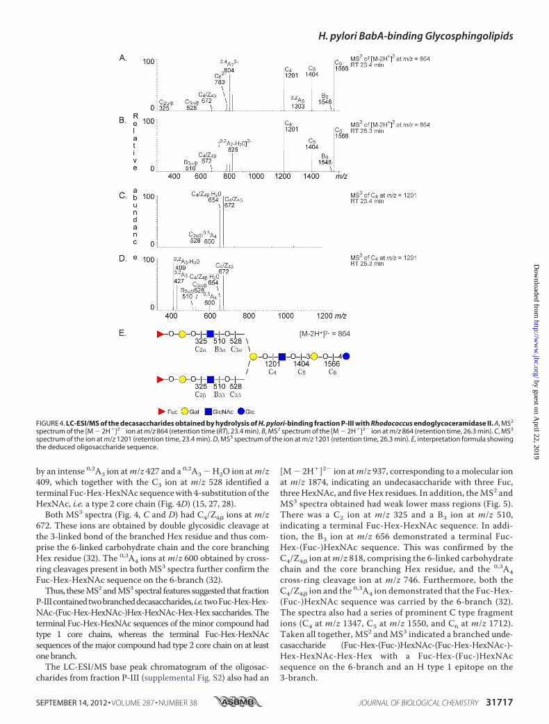
by an intense 0,2A3 ion atm/z 427 and a 0,2A3 � H2O ion atm/z409, which together with the C3 ion at m/z 528 identified aterminal Fuc-Hex-HexNAc sequencewith 4-substitution of theHexNAc, i.e. a type 2 core chain (Fig. 4D) (15, 27, 28).Both MS3 spectra (Fig. 4, C and D) had C4/Z4� ions at m/z
672. These ions are obtained by double glycosidic cleavage atthe 3-linked bond of the branched Hex residue and thus com-prise the 6-linked carbohydrate chain and the core branchingHex residue (32). The 0,3A4 ions at m/z 600 obtained by cross-ring cleavages present in both MS3 spectra further confirm theFuc-Hex-HexNAc sequence on the 6-branch (32).Thus, theseMS2 andMS3 spectral features suggested that fraction
P-IIIcontainedtwobrancheddecasaccharides, i.e.twoFuc-Hex-Hex-NAc-(Fuc-Hex-HexNAc-)Hex-HexNAc-Hex-Hex saccharides. Theterminal Fuc-Hex-HexNAc sequences of the minor compound hadtype 1 core chains, whereas the terminal Fuc-Hex-HexNAcsequences of the major compound had type 2 core chain on at leastone branch.The LC-ESI/MS base peak chromatogram of the oligosac-
charides from fraction P-III (supplemental Fig. S2) also had an
[M� 2H�]2� ion atm/z 937, corresponding to amolecular ionat m/z 1874, indicating an undecasaccharide with three Fuc,threeHexNAc, and fiveHex residues. In addition, theMS2 andMS3 spectra obtained had weak lower mass regions (Fig. 5).There was a C2 ion at m/z 325 and a B3 ion at m/z 510,indicating a terminal Fuc-Hex-HexNAc sequence. In addi-tion, the B3 ion at m/z 656 demonstrated a terminal Fuc-Hex-(Fuc-)HexNAc sequence. This was confirmed by theC4/Z4� ion atm/z 818, comprising the 6-linked carbohydratechain and the core branching Hex residue, and the 0,3A4cross-ring cleavage ion at m/z 746. Furthermore, both theC4/Z4� ion and the 0,3A4 ion demonstrated that the Fuc-Hex-(Fuc-)HexNAc sequence was carried by the 6-branch (32).The spectra also had a series of prominent C type fragmentions (C4 at m/z 1347, C5 at m/z 1550, and C6 at m/z 1712).Taken all together, MS2 and MS3 indicated a branched unde-casaccharide (Fuc-Hex-(Fuc-)HexNAc-(Fuc-Hex-HexNAc-)-Hex-HexNAc-Hex-Hex with a Fuc-Hex-(Fuc-)HexNAcsequence on the 6-branch and an H type 1 epitope on the3-branch.
FIGURE 4. LC-ESI/MS of the decasaccharides obtained by hydrolysis of H. pylori-binding fraction P-III with Rhodococcus endoglycoceramidase II. A, MS2
spectrum of the [M � 2H�]2� ion at m/z 864 (retention time (RT), 23.4 min). B, MS2 spectrum of the [M � 2H�]2� ion at m/z 864 (retention time, 26.3 min). C, MS3
spectrum of the ion at m/z 1201 (retention time, 23.4 min). D, MS3 spectrum of the ion at m/z 1201 (retention time, 26.3 min). E, interpretation formula showingthe deduced oligosaccharide sequence.
H. pylori BabA-binding Glycosphingolipids
SEPTEMBER 14, 2012 • VOLUME 287 • NUMBER 38 JOURNAL OF BIOLOGICAL CHEMISTRY 31717
by guest on April 22, 2019
http://ww
w.jbc.org/
Dow
nloaded from

(iv) The anomeric region of the proton NMR spectrum offraction P-III is shown in Fig. 6. Fraction P-III contains twodecaglycosylceramides (Fuc�2Gal�4GlcNAc�6(Fuc�2-Gal�3GlcNAc�3)Gal�3GlcNAc�3Gal�4Glc�1Cer andFuc�2Gal�3GlcNAc�6(Fuc�2Gal�3GlcNAc�3)Gal�3GlcNA-c�3Gal�4Glc�1Cer) that have been isolated previously fromrat (33) and pig intestine (23) and characterized in detail byNMR (using DMSO/D2O (98:2) as solvent). In fraction P-III,the glycosphingolipid with mixed type 1/type 2 branches(Fuc�2Gal�4GlcNAc�6(Fuc�2Gal�3GlcNAc�3)Gal�3GlcN-Ac�3Gal�4Glc�1Cer) is the major compound as evidencedby the relative intensities of the Fuc�2 signals. The chemicalshift data are summarized in Table 1. In addition, a novelglycosphingolipid structure with an Ley determinant on the 6-branch and an H type 1 determinant on the 3-branch (Fuc�2-Gal�4(Fuc�3)GlcNAc�6(Fuc�2Gal�3GlcNAc�3)Gal�3-GlcNAc�3Gal�4Glc�1Cer) could be characterized as shownin Fig. 6 and Table 1.Comparative Glycosphingolipid Binding Assays—Thereafter,
the binding of the specialist H. pylori strain S831 and the gen-eralist strain J99 to a number of reference glycosphingolipidswas evaluated. The results are summarized in Table 2. Whenusing this set of reference glycosphingolipids, only the Leb
hexaglycosylceramide was recognized by the specialist strainS831 (Fig. 7C, lane 1), whereas the generalistH. pylori strain J99in addition to the Leb hexaglycosylceramide bound to the Atype 1 hexaglycosylceramide (GalNAc�3(Fuc�2)Gal�3-GlcNAc�3Gal�4Glc�1Cer; Fig. 7B, lane 4), the B type 1hexaglycosylceramide (Gal�3(Fuc�2)Gal�3GlcNAc�3Gal�4-Glc�1Cer; Table 2, Number 9), the A type 1 heptaglycosylcer-amide (GalNAc�3(Fuc�2)Gal�3(Fuc�4)GlcNAc�3Gal�4-Glc�1Cer; Fig. 7B, lane 3), the B type 1 heptaglycosylceramide(Gal�3(Fuc�2)Gal�3(Fuc�4)GlcNAc�3Gal�4Glc�1Cer; Fig. 7B,lane 2), the A type 1 octaglycosylceramide (GalNAc�3-(Fuc�2)Gal�3GlcNAc�3Gal�3GlcNAc�3Gal�4Glc�1Cer; Table2, Number 17), and the repetitive A type 1 nonaglycosylceramide(GalNAc�3(Fuc�2)Gal�3GalNAc�3(Fuc�2)Gal�3GlcNAc�3-Gal�4Glc�1Cer; Fig. 7B, lane 6). Furthermore, the chromatogrambinding assay revealed that the A type 4 heptaglycosylceramide(Globo A; GalNAc�3(Fuc�2)Gal�3GalNAc�3Gal�4Gal�4Glc-�1Cer; Fig. 7B, lane 5) was also recognized by the generalist strain.
However, no type 2 core counterparts of these compoundswere recognized such as e.g. the H type 2 pentaglycosylcer-amide (Fig. 1, lane 2; Table 2, Number 5), the Ley hexaglycosyl-ceramide (Number 8), the A type 2 hexaglycosylceramide (Num-ber 12), the B type 2 hexaglycosylceramide (Number 10), the A
FIGURE 5. LC-ESI/MS of the undecasaccharide obtained by hydrolysis of H. pylori-binding fraction P-III with Rhodococcus endoglycoceramidase II.A, MS2 spectrum of the [M � 2H�]2� ion at m/z 937 (retention time (RT), 25.1 min). B, MS3 spectrum of the ion at m/z 1712 (retention time, 25.1 min).C, interpretation formula showing the deduced oligosaccharide sequence.
H. pylori BabA-binding Glycosphingolipids
31718 JOURNAL OF BIOLOGICAL CHEMISTRY VOLUME 287 • NUMBER 38 • SEPTEMBER 14, 2012
by guest on April 22, 2019
http://ww
w.jbc.org/
Dow
nloaded from

type2heptaglycosylceramide (Fig. 8B, lane7;Number15), and theA type 2 nonaglycosylceramide (Number 19). Furthermore, the Atetraglycosylceramide (Number 1) and theA type 3 nonaglycosyl-ceramide (Number 20) were also non-binding.When the generalist and specialist H. pylori strains were
comparedwith respect to their ability to bind to dilutions of thebinding-active glycosphingolipids on thin-layer chromato-grams, the Leb hexaglycosylceramide was the preferred ligandof both strains, and two strains bound to this compound withsimilar detection limits (Fig. 8, lanes 1–3). In addition, the gen-eralist strain J99 bound to the GalNAc�3-substituted Leb (i.e.the A type 1 heptaglycosylceramide), the Globo A heptaglyco-sylceramide, and the nonaglycosylceramidewith repetitive type1 blood group A determinants in all cases with detection limitsat �40 ng (Fig. 8A).Molecular Modeling—Inspection of the minimum energy
models of the H type 1 pentaglycosylceramide and the Globo H
hexaglycosylceramide revealed a substantial topographical simi-larity, which makes it reasonable that these two compounds maybe accommodated within the same carbohydrate binding site ofBabA (Fig. 9). In contrast, the terminal disaccharide of the non-binding H type 2 pentaglycosylceramide (right) is rotated relativeto the same disaccharide in the H type 1 pentaglycosylceramide(left) and the Globo H hexaglycosylceramide (center) by �90°,explaining why this compound is non-binding.Binding of Anti-Globo H to Glycosphingolipids from Human
Stomach—Having established thatH. pylori recognizes the GloboH glycosphingolipid, we next examined whether this glycosphin-golipid is present in the target tissue of H. pylori by binding ofmonoclonal antibodies directed against the GloboH determinantto non-acid glycosphingolipid fractions from human stomach.Thereby, binding in the hexaglycosylceramide region wasobserved in the non-acid fractions from the stomach of the twoindividuals tested (Fig. 10B, lanes 1 and 2). Both human stomach
FIGURE 6. Anomeric region of the 600-MHz proton NMR spectrum of the H. pylori-binding fraction P-III from porcine small intestinal epithelium(30 °C). The sample was dissolved in dimethyl sulfoxide/D2O (98:2 by volume) after deuterium exchange.
TABLE 1Chemical shifts (ppm) of anomeric resonances of glycosphingolipids in fraction P-III from porcine small intestine dissolved in dimethyl sulfoxide/D2O (98:2 by volume) identified by 600-MHz NMR at 30 °C
Structure XI X IX VIII VII VI V IV III II I
A Fuc�2 Gal�3 GlcNAc�6 (Fuc�2) Gal�3 (GlcNAc�3) Gal�3 GlcNA�3 Gal�4 Glc�1 Cer4.962 4.41 4.36 4.982 4.427 4.547 4.20 4.779 4.26 4.19
B Fuc�2 Gal�4 GlcNAc�6 (Fuc�2) Gal�3 (GlcNAc�3) Gal�3 GlcNA�3 Gal�4 Glc�1 Cer5.028 4.33 4.33 4.982 4.427 4.547 4.20 4.779 4.26 4.19
C (Fuc�2) Gal�4 (Fuc�3) GlcNAc�6) (Fuc�2) Gal�3 (GlcNAc�3) Gal�3 GlcNA�3 Gal�4 Glc�1 Cer4.939 4.39 4.798 4.41 4.982 4.427 4.547 4.20 4.779 4.26 4.19
D Fuc�2 Gal�4 (Fuc�3) GlcNA�3 Gal�4 Glc�1 Cera4.962 4.398 4.852 4.680 4.261 4.218
a For comparative purposes the chemical shift values are given for the Ley hexaglycosylceramide from human erythrocytes characterized by Clausen et al. (41) at 30 °C. It isnoteworthy that although the Fuc�2 signals in the Ley determinants are only separated by 0.020 ppm for structures C and D the corresponding Fuc�3 signals are separatedby as much as 0.054 ppm due to the fact that the determinant is located on a 6-branch. Ley determinants on a 6-branch have not been described previously. This conclu-sion is confirmed, however, by examining the H5/H6 correlations in the double quantum-filtered COSY spectrum, which clearly reveals cross-peaks at 4.00/1.08 and 4.68/1.03 ppm originating from the Fuc�2 and Fuc�3 residues, respectively, of a Ley determinant.
H. pylori BabA-binding Glycosphingolipids
SEPTEMBER 14, 2012 • VOLUME 287 • NUMBER 38 JOURNAL OF BIOLOGICAL CHEMISTRY 31719
by guest on April 22, 2019
http://ww
w.jbc.org/
Dow
nloaded from

TA
BLE
2C
om
par
iso
no
fgly
cosp
hin
go
lipid
bin
din
gp
refe
ren
ces
ofa
gen
eral
ist
H.p
ylo
rist
rain
(J99
),a
spec
ialis
tH
.pyl
ori
stra
in(S
831)
,an
da
Bab
Ad
elet
ion
mu
tan
tH
.pyl
ori
stra
in(J
99/B
abA
�)
No.
Abb
reviation
Structure
H.p
yloriJ99
H.p
yloriS
831
H.p
yloriJ99
/Bab
A�
Source
(Ref.)
1A-4
GalNAc�
3(Fu
c�2)Gal
�4G
lc�1C
erRa
tintestin
e(20)
2Le
a -5
Gal
�3(Fu
c�4)GlcNAc�
3Gal
�4G
lc�1C
er�
a�
�Hum
anintestine(42)
3Le
x -5
Gal
�4(Fu
c�3)GlcNAc�
3Gal
�4G
lc�1C
er�
��
Dog
intestine(42)
4H5type
1Fu
c�2G
al�3G
lcNAc�
3Gal
�4G
lc�1C
er�
��
Porcineintestine(22)
5H5type
2Fu
c�2G
al�4G
lcNAc�
3Gal
�4G
lc�1C
er�
��
Hum
anerythrocytes
(43)
6H6type
4(G
lobo
H)
Fuc�
2Gal
�3G
alNAc�
3Gal
�4G
al�4G
lc�1C
er�
��
Porcineintestineb
7Le
b -6
Fuc �
2Gal
�3(Fu
c�4)GlcNAc�
3Gal
�4G
lc�1C
er�
��
Hum
anintestine(42)
8Le
y -6
Fuc �
2Gal
�4(Fu
c�3)GlcNAc�
3Gal
�4G
lc�1C
er�
��
Dog
intestine(42)
9B6
type
1Gal
�3(Fu
c�2)Gal
�3G
lcNAc�
3Gal
�4G
lc�1C
er�
��
Hum
anintestine(19)
10B6
type
2Gal
�3(Fu
c�2)Gal
�4G
lcNAc�
3Gal
�4G
lc�1C
er�
��
Hum
anerythrocytes
(44)
11A6type
1GalNAc�
3(Fu
c�2)Gal
�3G
lcNAc�
3Gal
�4G
lc�1C
er�
��
Hum
anintestine(19)
12A6type
2GalNAc�
3(Fu
c�2)Gal
�4G
lcNAc�
3Gal
�4G
lc�1C
er�
��
Hum
anerythrocytes
(45)
13B7
type
1Gal
�3(Fu
c�2)Gal
�3(Fu
c�4)GlcNAc�
3Gal
�4G
lc�1C
er�
��
Hum
anintestine(19)
14A7type
1GalNAc�
3(Fu
c�2)Gal
�3(Fu
c�4)GlcNAc�
3Gal
�4G
lc�1C
er�
��
Hum
anintestine(19)
15A7type
2GalNAc�
3(Fu
c�2)Gal
�4(Fu
c�3)GlcNAc�
3Gal
�4G
lc�1C
er�
��
Hum
anerythrocytes
(45)
16A7type
4(G
lobo
A)
GalNAc�
3(Fu
c�2)Gal
�3G
alNAc�
3Gal
�4G
al�4G
lc�1C
er�
��
Porcineintestine(22)
17A8type
1GalNAc�
3(Fu
c�2)Gal
�3G
lcNAc�
Gal
�3G
lcNAc�
3Gal
�4G
lc�1C
er�
��
Porcineintestine(22)
18A9type
1GalNAc�
3(Fu
c�2)Gal
�3G
alNAc�
3(Fu
c�2)Gal
�3G
lcNAc�
3Gal
�4G
lc�1C
er�
��
Porcineintestine(22)
19A9type
2GalNAc�
3(Fu
c�2)Gal
�4(Fu
c�3)GlcNAc�
3Gal
�4G
lcNAc�
3Gal
�4G
lc�1C
er�
��
Cat
intestine(21)
20A9type
3GalNAc�
3(Fu
c�2)Gal
�3G
alNAc�
3(Fu
c�2)Gal
�4G
lcNAc�
3Gal
�4G
lc�1C
er�
��
Hum
anerythrocytes
(46)
21Dim
ericLe
aGal
�3(Fu
c�4)GlcNAc�
3Gal
�3(Fu
c�4)GlcNAc�
3Gal
�4G
lc�1C
er�
��
Hum
anintestinec
22Dim
ericLe
xGal
�4(Fu
c�3)GlcNAc�
3Gal
�4(Fu
c�3)GlcNAc�
3Gal
�4G
lc�1C
er�
��
d
23Fu
c�2G
al�3G
lcNAc�
6(Fu
c�2G
al�3G
lcNAc�
3)Gal
�3G
lcNAc�
3Gal
�4G
lc�1C
er�
e�
�Pigintestineb
24Fu
c�2G
al�4G
lcNAc�
6(Fu
c�2G
al�3G
lcNAc�
3)Gal
�3G
lcNAc�
3Gal
�4G
lc�1C
er�
e�
�Pigintestineb
25Fu
c�2G
al�4(Fu
c�3)GlcNAc�
6(Fu
c�2G
al�3G
lcNAc�
3)Gal
�3G
lcNAc�
3Gal
�4G
lc�1C
er�
e�
�Pigintestineb
26NeuGc-nL
6NeuGc�
3Gal
�4G
lcNAc�
3Gal
�4G
lcNAc�
3Gal
�4G
lc�1C
er�
��
Rabb
itthym
us(47)
aBind
ingisdefin
edas
follo
ws:
�deno
tesa
bind
ingwhe
n1
�gof
theglycosph
ingo
lipid
was
appliedon
thethin-la
yerc
hrom
atog
ram,w
hereas
�deno
tesn
obind
ingeven
at4
�g.
bPresen
tstudy.
cJ.Be
nktand
er,J.Å
ngstrom,H
.Karlss
on,M
.Leben
s,an
dS.Ten
eberg,subm
itted
man
uscript.
dGlycosphing
olipid
Num
ber2
2was
prepared
from
sialyl-dim
ericLe
x(10)
bymild
acid
hydrolysis.
eH.pylorib
inding
toglycosph
ingo
lipidsN
umbers
23–2
5was
determ
ined
usingamixture
ofthethreecompo
unds.
H. pylori BabA-binding Glycosphingolipids
31720 JOURNAL OF BIOLOGICAL CHEMISTRY VOLUME 287 • NUMBER 38 • SEPTEMBER 14, 2012
by guest on April 22, 2019
http://ww
w.jbc.org/
Dow
nloaded from

samples also contained the Leb hexaglycosylceramide as indicatedby the binding of the anti-Leb antibody (Fig. 10C). The anti-H type1 antibody cross-reacted with the Globo H glycosphingolipid tosomeextent.However, nobindingof the anti-H type 1 antibody tothe non-acid glycosphingolipid fractions from human stomachwas obtained, although it bound intensely to the pentaglycosyl-ceramide and to the slow migrating glycosphingolipid of bloodgroupO pig intestine (Fig. 10D).
DISCUSSION
The binding of microbes to host target cells is crucial to thedelivery of virulence factors, and in the case of H. pylori, it wasrecently shown that BabA-mediated binding of the bacteria toLeb on the epithelium leads to an increased type IV secretion
system activity, resulting in the production of proinflammatorycytokines and precancer-related factors (34).The initial observation that the fucosylated blood group anti-
gens H type 1 and Leb are mediators of H. pylori adhesion tohuman gastric epithelial cells (5) was followed by a division ofBabA-producing H. pylori strains into specialist and generaliststrains, depending on their mode of binding to Leb and related
FIGURE 7. Comparison of binding of a generalist and a specialist H. pyloristrain to reference glycosphingolipids. The glycosphingolipids were sepa-rated on aluminum-backed silica gel plates using chloroform/methanol/wa-ter (60:35:8 by volume) as the solvent system. The chromatogram in A wasstained with anisaldehyde. Duplicate chromatograms were incubated withthe 35S-labeled H. pylori generalist strain J99 (B) and the H. pylori specialiststrain S831 (C) followed by autoradiography for 12 h as described under“Experimental Procedures.” Lane 1, Leb hexaglycosylceramide (Fuc�2Gal�3-(Fuc�4)GlcNAc�3Gal�4Glc�1Cer), 2 �g; lane 2, B type 1 heptaglycosylcer-amide (Gal�3(Fuc�2)Gal�3(Fuc�4)GlcNAc�3Gal�4Glc�1Cer), 2 �g; lane 3, Atype 1 heptaglycosylceramide (GalNAc�3(Fuc�2)Gal�3(Fuc�4)Glc-NAc�3Gal�4Glc�1Cer), 2 �g; lane 4, A type 1 hexaglycosylceramide(GalNAc�3(Fuc�2)Gal�3GlcNAc�3Gal�4Glc�1Cer), 2 �g; lane 5, Globo A/Atype 4 heptaglycosylceramide (GalNAc�3(Fuc�2)Gal�3GalNAc�3Gal�4Gal�4Glc�1Cer), 2 �g; lane 6, A nonaglycosylceramide (GalNAc�3-(Fuc�2)Gal�3GalNAc�3(Fuc�2)Gal�3GlcNAc�3Gal�4Glc�1Cer), 2 �g; lane 7,A type 2 heptaglycosylceramide (GalNAc�3(Fuc�2)Gal�4(Fuc�3)-GlcNAc�3Gal�4Glc�1Cer), 2 �g.
FIGURE 8. Comparison of binding of a generalist and a specialist H. pyloristrain to dilutions of pure glycosphingolipids on thin-layer chromato-grams. The glycosphingolipids were separated on aluminum-backed silicagel plates using chloroform/methanol/water (60:35:8 by volume) as the sol-vent system. The chromatograms were incubated with the 35S-labeled H.pylori generalist strain J99 (A) and the H. pylori specialist strain S831 (B) fol-lowed by autoradiography for 12 h as described under “Experimental Proce-dures.” Lane 1, Leb hexaglycosylceramide (Leb-6; Fuc�2Gal�3-(Fuc�4)GlcNAc�3Gal�4Glc�1Cer), 200 ng, and A nonaglycosylceramide (A9-1;GalNAc�3(Fuc�2)Gal�3GalNAc�3(Fuc�2)Gal�3GlcNAc�3Gal�4Glc�1Cer), 200ng; lane 2, Leb hexaglycosylceramide, 80 ng, and A nonaglycosylceramide, 80 ng;lane 3, Leb hexaglycosylceramide, 40 ng, and A nonaglycosylceramide, 40 ng;lane 4, Globo A/A type 4 heptaglycosylceramide (Globo A; GalNAc�3(Fuc�2)-Gal�3GalNAc�3Gal�4Gal�4Glc�1Cer), 200 ng; lane 5, Globo A/A type 4 hepta-glycosylceramide, 80 ng; lane 6, Globo A/A type 4 heptaglycosylceramide, 40 ng;lane 7, A type 1 heptaglycosylceramide (A7-1; GalNAc�3(Fuc�2)-Gal�3(Fuc�4)GlcNAc�3Gal�4Glc�1Cer), 200 ng; lane 8, A type 1 heptaglycosyl-ceramide, 80 ng; lane 9, A type 1 heptaglycosylceramide, 40 ng.
FIGURE 9. Minimum energy models of the H type 1 pentaglycosylcer-amide (Fuc�2Gal�3GlcNAc�3Gal�4Glc�1Cer) (left), the Globo Hhexaglycosylceramide (Fuc�2Gal�3GalNAc�3Gal�4Gal�4Glc�1Cer)(center), and the H type 2 pentaglycosylceramide (Fuc�2Gal�-4GlcNAc�3Gal�4Glc�1Cer) (right). The terminal disaccharides are coloredblue (Fuc�2) and purple (Gal�3/4). The terminal part of the BabA-binding Htype 1 and Globo H glycosphingolipids can be aligned by assuming differentGlc�1Cer torsion angles, whereas the H type 2 terminal disaccharide isrotated �90° in comparison, rendering this glycosphingolipid non-bindingwith respect to BabA.
FIGURE 10. Binding of monoclonal antibodies to human gastric glyco-sphingolipids. Thin-layer chromatogram after detection with anisaldehyde(A) and autoradiograms obtained by binding of the monoclonal anti-Globo Hantibody MBr1 (B), the monoclonal anti-Leb antibody BG-6/T218 (C), and themonoclonal anti-H type 1 antibody 17-206 (D) are shown. The chromato-grams were eluted with chloroform/methanol/water (60:35:8 by volume),and the binding assays were done as described under “Experimental Pro-cedures” followed by autoradiography for 12 h. Lane 1, non-acid glycosph-ingolipids of human stomach (Individual I; blood group O), 40 �g; lane 2,non-acid glycosphingolipids of human stomach (Individual II; bloodgroup A), 40 �g; lane 3, reference Leb hexaglycosylceramide(Fuc�2Gal�3(Fuc�4)GlcNAc�3Gal�4Glc�1Cer), 2 �g; lane 4, non-acid glyco-sphingolipids of the intestinal epithelium of a blood group O pig, 40 �g; lane5, non-acid glycosphingolipids of the intestinal epithelium of a blood group Apig, 40 �g; lane 6, reference H type 1 pentaglycosylceramide(Fuc�2Gal�3GlcNAc�3Gal�4Glc�1Cer), 2 �g; lane 7, reference Globo H hexa-glycosylceramide (Fuc�2Gal�3GalNAc�3Gal�4Gal�4Glc�1Cer), 1 �g; lane 8,reference Globo A heptaglycosylceramide (GalNAc�3(Fuc�2)Gal�3GalNAc-�3Gal�4Gal�4Glc�1Cer), 4 �g.
H. pylori BabA-binding Glycosphingolipids
SEPTEMBER 14, 2012 • VOLUME 287 • NUMBER 38 JOURNAL OF BIOLOGICAL CHEMISTRY 31721
by guest on April 22, 2019
http://ww
w.jbc.org/
Dow
nloaded from

carbohydrate sequences (9). The BabA of specialist strainsbinds only to glycoconjugates with an unsubstituted terminalFuc�2Gal sequence as in the H type 1 and Leb determinants,whereas the generalist BabA tolerates a substitution at 3-posi-tion of the Gal with an �Gal or �GalNAc as in the A or B type 1and ALeb or BLeb determinants.
Here, we further explored the structural requirements forcarbohydrate recognition by BabA of generalist and specialistH. pylori by isolating and characterizing glycosphingolipidsrecognized by wild type specialist and/or generalist H. pyloribut not by the deletionmutant strain lacking the BabA adhesin.The Leb epitope has only been found in humans, but weinitially thought that we had found a porcine Leb glyco-sphingolipid when an H. pylori BabA-binding glycosphingo-lipid co-migrating with the Leb hexaglycosylceramide wasdetected in the non-acid fraction of blood group O pig intes-tine. However, after isolation, this BabA-binding glyco-sphingolipid was characterized as the Globo H hexaglycosyl-ceramide. Further comparative binding studies using ourglycosphingolipid collection confirmed that the BabA adhe-sin in addition to blood group determinants on type 1 corechains recognizes blood group O and A determinants ontype 4 core chains with binding to Globo H by both strainsand Globo A by the generalist strain. The terminal disaccha-rides (Fuc�2Gal�3) of the H type 1 pentaglycosylceramideand the Globo H hexaglycosylceramide adopt conformationsvery similar to each other, and this is also the case for theterminal trisaccharides (GalNAc�3(Fuc�2)Gal�3) of the Atype 1 and the Globo A heptaglycosylceramides (18). Theseconformational similarities thus explain the binding of BabAto the two sets of isoreceptors.The enzymatic machinery involved in the biosynthesis of
Globo H has not yet been fully elucidated. In humans, there aretwo functional fucosyltransferases, designated FUT1 andFUT2, that catalyze addition of an �2-linked fucose to a termi-nal galactose to form the blood group H epitope (for a review,see Ref. 35). These two fucosyltransferases are encoded by twodistinct genes, FUT1 and FUT2. FUT1 acts preferentially ontype 2 chains, whereas type 1 and type 3 chains and to someextent type 2 chains are acceptors for FUT2. Using siRNAstargeting FUT1 and FUT2 in breast cancer stem cells, Chang etal. (36) showed that GloboHmay be synthesized by both FUT1and FUT2.Non-secretor individuals have an increased risk of peptic
ulcer disease (37). In these individuals, the precursor of the Lebsequence, i.e. theH type 1 sequence, is not formed due to lack ofa functional FUT2 enzyme. Consequently, non-secretors havelow amounts of or no Leb antigens on their epithelial surfaces.However, the Globo H sequence can still be formed by FUT1and might thus function as an adhesion factor for BabA-ex-pressing H. pylori in non-secretor individuals.
The slow migrating BabA-binding fraction P-III was charac-terized as a mixture of three complex glycosphingolipids(Fuc�2Gal�4GlcNAc�6(Fuc�2Gal�3GlcNAc�3)Gal�3Glc-NAc�3Gal�4Glc�1Cer, Fuc�2Gal�3GlcNAc�6(Fuc�2Gal-�3GlcNAc�3)Gal�3GlcNAc�3Gal�4Glc�1Cer, and Fuc�2-Gal�4(Fuc�3)GlcNAc�6(Fuc�2Gal�3GlcNAc�3)Gal�3Glc-NAc�3Gal�4Glc�1Cer). The undecaglycosylceramide with an
Ley epitope on the 6-branch and an H type 1 epitope on the3-branch are to our knowledge novel glycosphingolipid struc-tures. The three compounds in fraction P-III all had anH type 1determinant on at least one branch, and thus, all three could berecognized by both specialist and generalist BabA.The binding of the generalistH. pylori strain to the nonagly-
cosylceramidewith a repetitive blood groupAdeterminant andan internal type 1 core chain (GalNAc�3(Fuc�2)Gal�3-GalNAc�3(Fuc�2)Gal�3GlcNAc�3Gal�4Glc�1Cer) is a wildcard.We have previously found that generalistH. pylori strainsbind to the ganglio-Leb hexaglycosylceramide (Fuc�2Gal�3(Fuc�4)GalNAc�4Gal�4Glc�1Cer) (13). Thus, the bindingto this nonaglycosylceramide is most likely due to recognitionof the terminal A determinant on the ganglio core by the gen-eralist BabA.Characterization of the binding specificities of theBabAvari-
ants is important for understanding the molecular interactionsbetweenH. pylori and the target host cells. The presence of theBabA-binding Globo H glycosphingolipid in the human stom-achwas here indicated by the binding ofmonoclonal antibodiesdirected against the Globo H determinant to human gastricglycosphingolipids. Thus,GloboHmayhave a role in theBabA-mediated target tissue adherence of H. pylori.Expression of BabA by H. pylori is associated with severe
gastric inflammation and an increased risk of developing pepticulcer or gastric cancer (38, 39).H. pylori infects more than halfof the world’s population, and although the prevalence of infec-tion is decreasing in developed countries, the infection rate isstill high in developing countries (40). Furthermore, the treat-ment options in developing countries are currently inadequate.Targeting BabA might be important for the development ofnovel treatment strategies against H. pylori.
Acknowledgment—The use of the LTQ linear quadrupole ion trapmass spectrometer (obtained by Swedish Research Council Grant342-2004-4434 to Gunnar Hansson) and the Varian 600-MHzmachine at the Swedish NMR Centre, Hasselblad Laboratory, Uni-versity of Gothenburg, is gratefully acknowledged.
REFERENCES1. Karlsson, K. A. (1989) Animal glycosphingolipids as membrane attach-
ment sites for bacteria. Annu. Rev. Biochem. 58, 309–3502. Esko, J. D. (1999) Essentials in Glycobiology (Varki, A., Cummings, R.,
Esko, J., Freeze, H., Hart, G., andMarth, J., eds) pp. 429–440, Cold SpringHarbor Laboratory, Cold Spring Harbor, NY
3. Pieters, R. J. (2011) Carbohydrate mediated bacterial adhesion. Adv. Exp.Med. Biol. 715, 227–240
4. Stults, C. L., Sweeley, C. C., and Macher, B. A. (1989) Glycosphingolipids:structure, biological source and properties. Methods Enzymol. 179,167–214
5. Boren, T., Falk, P., Roth, K. A., Larson, G., and Normark, S. (1993) Attach-ment of Helicobacter pylori to human gastric epithelium mediated byblood group antigens. Science 262, 1892–1895
6. Ilver, D., Arnqvist, A., Ogren, J., Frick, I. M., Kersulyte, D., Incecik, E. T.,Berg, D. E., Covacci, A., Engstrand, L., and Boren, T. (1998) Helicobacterpylori adhesin binding fucosylated histo-blood group antigens revealed byretagging. Science 279, 373–377
7. Gerhard, M., Lehn, N., Neumayer, N., Boren, T., Rad, R., Schepp, W.,Miehlke, S., Classen, M., and Prinz, C. (1999) Clinical relevance of theHelicobacter pylori gene for blood-group antigen-binding adhesin. Proc.
H. pylori BabA-binding Glycosphingolipids
31722 JOURNAL OF BIOLOGICAL CHEMISTRY VOLUME 287 • NUMBER 38 • SEPTEMBER 14, 2012
by guest on April 22, 2019
http://ww
w.jbc.org/
Dow
nloaded from
Natl. Acad. Sci. U.S.A. 96, 12778–127838. Rad, R., Gerhard, M., Lang, R., Schoniger, M., Rosch, T., Schepp, W.,
Becker, I., Wagner, H., and Prinz, C. (2002) TheHelicobacter pylori bloodgroup antigen-binding adhesin facilitates bacterial colonization and aug-ments a non-specific immune response. J. Immunol. 168, 3033–3041
9. Aspholm-Hurtig, M., Dailide, G., Lahmann,M., Kalia, A., Ilver, D., Roche,N., Vikstrom, S., Sjostrom, R., Linden, S., Backstrom, A., Lundberg, C.,Arnqvist, A., Mahdavi, J., Nilsson, U. J., Velapatino, B., Gilman, R. H.,Gerhard, M., Alarcon, T., Lopez-Brea, M., Nakazawa, T., Fox, J. G., Cor-rea, P., Dominguez-Bello,M. G., Perez-Perez, G. I., Blaser,M. J., Normark,S., Carlstedt, I., Oscarson, S., Teneberg, S., Berg, D. E., and Boren, T. (2004)Functional adaptation of BabA, the Helicobacter pylori blood-group anti-gen binding adhesin. Science 305, 519–522
10. Mahdavi, J., Sonden, B., Hurtig, M., Olfat, F. O., Forsberg, L., Roche, N.,Angstrom, J., Larsson, T., Teneberg, S., Karlsson, K. A., Altraja, S., Wad-strom, T., Kersulyte, D., Berg, D. E., Dubois, A., Petersson, C., Magnusson,K. E., Norberg, T., Lindh, F., Lundskog, B. B., Arnqvist, A., Hammarstrom,L., and Boren, T. (2002) Helicobacter pylori SabA adhesin in persistentinfection and chronic inflammation. Science 297, 573–578
11. Karlsson, K. A. (1987) Preparation of total non-acid glycolipids for overlayanalysis of receptors for bacteria and viruses and for other studies.Meth-ods Enzymol. 138, 212–220
12. Waldi, D. (1962) in Thin-layer Chromatography (Stahl, E., ed) pp.496–515, Springer-Verlag, Berlin
13. Fagerberg, D., Angstrom J., Halim, A., Hultberg, A., Rakhimova, L., Ham-marstrom, L., Boren, T., and Teneberg, S. (2009) Novel Leb-like Helico-bacter pylori-binding glycosphingolipid created by expression of human�-1,3/4-fucosyltransferase in FVB/N mouse stomach. Glycobiology 19,182–191
14. Ito, M., and Yamagata, T. (1989) Purification and characterization of gly-cosphingolipid-specific endoglycosidases (endoglycoceramidases) from amutant strain of Rhodococcus sp. Evidence for three molecular species ofendoglycoceramidase with different specificities. J. Biol. Chem. 264,9510–9519
15. Karlsson, H., Halim, A., and Teneberg, S. (2010) Differentiation of glyco-sphingolipid-derived glycan structural isomers by liquid chromatogra-phy-mass spectrometry. Glycobiology 20, 1103–1116
16. Marion, D., and Wuthrich, K. (1983) Application of phase sensitive two-dimensional correlated spectroscopy (COSY) formeasurements of 1H-1Hspin-spin coupling constants in proteins.Biochem. Biophys. Res. Commun.113, 967–974
17. Imberty, A., Mikros, E., Koca, J., Mollicone, R., Oriol, R., and Perez, S.(1995) Computer simulation of histo-blood group oligosaccharides: en-ergy maps of all constituting disaccharides and potential energy surfacesof 14 ABH and Lewis carbohydrate antigens. Glycoconj. J. 12, 331–349
18. Nyholm, P. G., Samuelsson, B. E., Breimer, M., and Pascher, I. (1989)Conformational analysis of blood groupA-active glycosphingolipids usingHSEA-calculations. The possible significance of the core oligosaccharidechain for the presentation and recognition of the A-determinant. J. Mol.Recognit. 2, 103–113
19. Bjork, S., Breimer, M. E., Hansson, G. C., Karlsson, K. A., and Leffler, H.(1987) Structures of blood group glycosphingolipids of human small in-testine. A relation between the expression of fucolipids of epithelial cellsand the ABO, Le and Se phenotype of the donor. J. Biol. Chem. 262,6758–6765
20. Breimer, M. E., Hansson, G. C., Karlsson, K. A., and Leffler, H. (1982)Isolation and partial characterization of blood group A and H active gly-cosphingolipids of rat small intestine. J. Biol. Chem. 257, 906–912
21. Angstrom, J., Backstrom, M., Berntsson, A., Karlsson, N., Holmgren, J.,Karlsson, K. A., Lebens, M., and Teneberg, S. (2000) Novel carbohydratebinding site recognizing blood group A and B determinants in a choleratoxin/heat-labile enterotoxin B-subunit hybrid. J. Biol. Chem. 275,3231–3238
22. Coddens, A., Diswall,M., Angstrom, J., Breimer,M. E., Goddeeris, B., Cox,E., and Teneberg, S. (2009) Recognition of blood group ABH type 1 deter-minants by the FedF adhesin of F18-fimbriated Escherichia coli. J. Biol.Chem. 284, 9713–9726
23. Diswall, M., Angstrom, J., Schuurman, H. J., Dor, F. J., Rydberg, L., and
Breimer,M. E. (2008) Glycolipid studies in small intestine and pancreas of�1,3-galactosyltransferase knockout miniature swine: �1,3GALT-KO an-imals lack �GAL antigens and contain novel blood group H compounds.Transplant. Proc. 40, 543–546
24. Kundu, S. K. (1993)Glycoconjugates. Composition, Structure and Function(Allen, H. J., andKisailus, E. E., eds) pp. 203–262,Marcel Dekker Inc., NewYork
25. Hakomori, S. (2001) Tumor-associated carbohydrate antigens definingtumor malignancy: basis for development of anti-cancer vaccines. Adv.Exp. Med. Biol. 491, 369–402
26. Teneberg, S., Leonardsson, I., Karlsson, H., Jovall, P. A., Angstrom, J.,Danielsson, D., Naslund, I., Ljungh, A., Wadstrom, T., and Karlsson, K. A.(2002) Lactotetraosylceramide, a novel glycosphingolipid receptor forHe-licobacter pylori, present in human gastric epithelium. J. Biol. Chem. 277,19709–19719
27. Chai, W., Piskarev, V., and Lawson, A. M. (2001) Negative-ion elec-trospray mass spectrometry of neutral underivatized oligosaccharides.Anal. Chem. 73, 651–657
28. Robbe, C., Capon, C., Coddeville, B., andMichalski, J. C. (2004) Diagnosticions for the rapid analysis by nano-electrospray ionization quadrupoletime-of-flight mass spectrometry of O-glycans from human mucins.Rapid Commun. Mass Spectrom. 18, 412–420
29. Holgersson, J., Jovall, P. A., Samuelsson, B. E., and Breimer, M. E. (1990)Structural characterization of non-acid glycosphingolipids in kidneys ofsingle blood group O and A pigs. J. Biochem. 108, 766–777
30. Li, Y. T., Chou, C. W., Li, S. C., Kobayashi, U., Ishibashi, Y. H., and Ito, M.(2009) Preparation of homogenous oligosaccharide chains from glyco-sphingolipids. Glycoconj. J. 26, 929–933
31. Zhou, B., Li, S. C., Laine, R. A., Huang, R. T., and Li, Y. T. (1989) Isolationand characterization of ceramide glycanase from the leech, Macrobdelladecora. J. Biol. Chem. 264, 12272–12277
32. Chai, W., Piskarev, V., and Lawson, A. M. (2002) Branching pattern andsequence analysis of underivatized oligosaccharides by combinedMS/MSof singly and doubly charged molecular ions in negative-ion electrospraymass spectrometry. J. Am. Soc. Mass Spectrom. 13, 670–679
33. Angstrom, J., Larsson, T., Hansson, G. C., Karlsson, K. A., and Henry, S.(2004)Default biosynthesis pathway for blood group-related glycolipids inhuman small intestine as defined by structural identification of linear andbranched glycosylceramides in a group O Le(a�b�) nonsecretor. Glyco-biology 14, 1–12
34. Ishijima, N., Suzuki, M., Ashida, H., Ichikawa, Y., Kanegae, Y., Saito, I.,Boren, T., Haas, R., Sasakawa, C., and Mimuro, H. (2011) BabA-mediatedadherence is a potentiator of the Helicobacter pylori type IV secretionsystem activity. J. Biol. Chem. 286, 25256–25264
35. Marionneau, S., Cailleau-Thomas, A., Rocher, J., Le Moullac-Vaidye, B.,Ruvoen, N., Clement, M., and Le Pendu, J. (2001) ABH and Lewis histo-blood group antigens, amodel for themeaning of oligosaccharide diversityin the face of a changing world. Biochimie 83, 565–573
36. Chang, W. W., Lee, C. H., Lee, P., Lin, J., Hsu, C. W., Hung, J. T., Lin, J. J.,Yu, J. C., Shao, L. E., Yu, J.,Wong, C. H., and Yu, A. L. (2008) Expression ofGlobo H and SSEA3 in breast cancer stem cells and the involvement offucosyl transferases 1 and 2 in Globo H synthesis. Proc. Natl. Acad. Sci.U.S.A. 105, 11667–11672
37. Cederberg, A., Varis, K., Salmi, H. A., Sipponen, P., Harkonen, M., andSarna, S. (1991) Young onset peptic ulcer disease and non-ulcer dyspepsiaare separate entities. Scand. J. Gastroenterol. Suppl. 186, 33–44
38. Fujimoto, S., Olaniyi Ojo, O., Arnqvist, A., Wu, J. Y., Odenbreit, S., Haas,R., Graham, D. Y., and Yamaoka, Y. (2007) Helicobacter pylori BabA ex-pression, gastricmucosal injury, and clinical outcome.Clin. Gastroenterol.Hepatol. 5, 49–58
39. Yamaoka, Y., Ojo, O., Fujimoto, S., Odenbreit, S., Haas, R., Gutierrez, O.,El-Zimaity, H. M., Reddy, R., Arnqvist, A., and Graham, D. Y. (2006) He-licobacter pylori outer membrane proteins and gastroduodenal disease.Gut. 55, 775–781
40. Brown, L.M. (2000)Helicobacter pylori: epidemiology and routes of trans-mission. Epidemiol. Rev. 22, 283–297
41. Clausen, H., Levery, S. B., McKibbin, J. M., and Hakomori, S. (1985) Bloodgroup A determinants with mono- and difucosyl type 1 chain in human
H. pylori BabA-binding Glycosphingolipids
SEPTEMBER 14, 2012 • VOLUME 287 • NUMBER 38 JOURNAL OF BIOLOGICAL CHEMISTRY 31723
by guest on April 22, 2019
http://ww
w.jbc.org/
Dow
nloaded from

erythrocyte membranes. Biochemistry 24, 3578–358642. McKibbin, J. M., Spencer, W. A., Smith, E. L., Mansson, J. E., Karlsson,
K. A., Samuelsson, B. E., Li, Y. T., and Li, S. C. (1982) Lewis blood groupfucolipids and their isomers from human and canine intestine. J. Biol.Chem. 257, 755–760
43. Stellner, K., Watanabe, K., and Hakomori, S. (1973) Isolation and charac-terization of glycosphingolipids with blood group H specificity frommembranes of human erythrocytes. Biochemistry 12, 656–661
44. Koscielak, J., Plasek, A., Gorniak, H., Gardas, A., and Gregor, A. (1973)Structures of fucose-containing glycolipids with H and B blood-group
activity and of sialic acid and glucosamine-containing glycolipid of hu-man-erythrocyte membrane. Eur. J. Biochem. 37, 214–225
45. Koscielak, J., Piasek, A., and Gorniak, H. (1970) in Blood and Tissue Anti-gens (Aminoff, D., ed) pp. 163–176, Academic Press, New York
46. Clausen, H., Levery, S. B., Nudelman, E., Tsuchiya, S., and Hakomori, S.(1985) Repetitive A epitope (type 3 chain A) defined by blood group A1-specific monoclonal antibody TH-1: chemical basis of qualitative A1 andA2 distinction. Proc. Natl. Acad. Sci. U.S.A. 82, 1199–1203
47. Iwamori, M., and Nagai, Y. (1981) Ganglioside composition of rabbit thy-mus. Biochim. Biophys. Acta 665, 205–213
H. pylori BabA-binding Glycosphingolipids
31724 JOURNAL OF BIOLOGICAL CHEMISTRY VOLUME 287 • NUMBER 38 • SEPTEMBER 14, 2012
by guest on April 22, 2019
http://ww
w.jbc.org/
Dow
nloaded from

John Benktander, Jonas Ångström, Michael E. Breimer and Susann TenebergAdhesin
BabAHelicobacter pyloriRedefinition of the Carbohydrate Binding Specificity of
doi: 10.1074/jbc.M112.387654 originally published online July 20, 20122012, 287:31712-31724.J. Biol. Chem.
10.1074/jbc.M112.387654Access the most updated version of this article at doi:
Alerts:
When a correction for this article is posted•
When this article is cited•
to choose from all of JBC's e-mail alertsClick here
Supplemental material:
http://www.jbc.org/content/suppl/2012/07/20/M112.387654.DC1
http://www.jbc.org/content/287/38/31712.full.html#ref-list-1
This article cites 43 references, 18 of which can be accessed free at
by guest on April 22, 2019
http://ww
w.jbc.org/
Dow
nloaded from