drosophila mfap1isrequiredforpre … · drosophilamfap1isrequiredforpre-mrnaprocessingand g 2...
TRANSCRIPT

Drosophila MFAP1 Is Required for Pre-mRNA Processing andG2/M Progression*□S
Received for publication, May 8, 2008, and in revised form, September 2, 1008 Published, JBC Papers in Press, September 2, 2008, DOI 10.1074/jbc.M803512200
Ditte S. Andersen and Nicolas Tapon1
From the Apoptosis and Proliferation Control Laboratory, Cancer Research UK, London Research Institute, 44 Lincoln’s Inn Fields,London WC2A 3PX, United Kingdom
The mammalian spliceosome has mainly been studied usingproteomics. The isolation and comparison of different splicingintermediates has revealed the dynamic association of morethan 200 splicing factors with the spliceosome, relatively few ofwhich have been studied in detail. Here, we report the charac-terization of the Drosophila homologue of microfibril-associ-ated protein 1 (dMFAP1), a previously uncharacterized proteinfound in some human spliceosomal fractions (Jurica, M. S., andMoore, M. J. (2003)Mol. Cell 12, 5–14). We show that dMFAP1binds directly to theDrosophilahomologue of Prp38p (dPrp38),a tri-small nuclear ribonucleoprotein component (Xie, J., Beick-man, K., Otte, E., and Rymond, B. C. (1998) EMBO J. 17, 2938–2946), and is required for pre-mRNA processing. dMFAP1, likedPrp38, is essential for viability, and our in vivo data show thatcells with reduced levels of dMFAP1 or dPrp38 proliferatemoreslowly than normal cells and undergo apoptosis. Consistentwith this, double-stranded RNA-mediated depletion of dPrp38or dMFAP1 causes cells to arrest in G2/M, and this is paralleledby a reduction inmRNA levels of themitotic phosphatase string/cdc25. Interestingly double-stranded RNA-mediated depletionof a wide range of core splicing factors elicits a similar pheno-type, suggesting that the observed G2/M arrest might be a gen-eral consequence of interfering with spliceosome function.
Splicing of pre-mRNAs is a catalytic reaction that involvestwo successive trans-esterification steps. This process is carriedout by a highly conserved ribonucleoprotein complex, calledthe spliceosome. The spliceosome consists of five snRNAs (U1,U2, U4, U5, and U6) andmore than 200 proteins, making it oneof the largest and most complex molecular machines studied(1). Spliceosome assembly is a sequential process that is initi-ated by the recruitment of the U1 small nuclear ribonucleopro-tein (snRNP)2 to the 5�-splice donor site to form a “commit-
ment complex” (“E complex” inmammals) (3, 4). Subsequently,recruitment of the U2 snRNP to the branch site by the U2 aux-iliary factor of 65 kDa (U2AF65) generates the “pre-spliceo-some” (“A complex” in mammals) (5–8). A preformedU4/U6.U5 tri-snRNP unit then joins the U1-U2-pre-mRNAcomplex to form the “complete spliceosome” (“B complex” inmammals). To activate the complete spliceosome several con-formational rearrangements must take place (reviewed in Ref.9). These include unwinding of base pairings between U1 andthe 5�-splice site and between U6 and U4, as well as the forma-tion of a new base pair interaction between U5 and U6 snRNPsand the 5�-splice site and U6 and U2. As a result, U1 and U4snRNPs are released and an active spliceosome is formed (10,11). The yeast RNA helicases Prp28p and Brr2p are both impli-cated in the structural rearrangements that take place duringthe activation step of the spliceosome (11). Prp28p is thought tobe important for the unwinding of base pairings between U1and the 5�-splice site (12) and Brr2p is implicated in unwindingthe U4/U6 duplex, which is essential for the release of U4snRNP (13). Moreover, a pre-assembled Prp19p (pre-mRNAprocessing factor 19 protein)-associated protein complexnamed the 19 complex (NTc) in yeast and the Cdc5-Prp19complex in humans is required for maturation of the spliceo-some (14, 15).Much of the mechanistic insight into the spliceosome has
been derived from yeast genetics. The ease of generating tem-perature-sensitive mutant alleles has allowed a detailed func-tional characterization of individual splicing factors. In con-trast, the function of the mammalian spliceosome has mostlybeen studied by proteomic analysis of spliceosome intermedi-ates (15–21). The isolation and comparison of different spliceo-some intermediates has given insights into the dynamics of thespliceosome and revealed that excision of an intron from apre-mRNA occurs largely by the same sequential path in yeastand higher eukaryotes (15, 16, 19, 20). Importantly, purifica-tions of mammalian spliceosomes have added a vast array ofelements to the pre-existing list of conserved core splicing fac-tors.Many of these have no previous connection to splicing andrequire functional validation (1).Yeast Prp38p is aU4/U6.U5 tri-snRNP component that plays
an important role in thematuration of the spliceosome. Prp38pactivity is dispensable for the initial assembly of the spliceo-some, but is required in a later step for the release of U4 snRNPand activation of the spliceosome (2). It has been speculated
* This work was supported by Cancer Research UK. The costs of publication ofthis article were defrayed in part by the payment of page charges. Thisarticle must therefore be hereby marked “advertisement” in accordancewith 18 U.S.C. Section 1734 solely to indicate this fact.
□S The on-line version of this article (available at http://www.jbc.org) containssupplemental Figs. S1–S3.
1 To whom correspondence should be addressed: P. O. Box 123, 44 Lincoln’sInn Fields, London WC2A 3PX United Kingdom. Tel.: 0044-207-269-3635;Fax: 0044-207-269-3638; E-mail: [email protected].
2 The abbreviations used are: snRNP, small nuclear ribonucleoprotein; qPCR,quantitative PCRs; MFAP1, microfibrillar-associated protein 1; NTc, 19 com-plex; RNAi, RNA interference; PBS, phosphate-buffered saline; GST, gluta-thione S-transferase; BisTris, 2-[bis(2-hydroxyethyl)amino]-2-(hydroxym-ethyl)propane-1,3-diol; MALDI-TOF, matrix-assisted laser desorptionionization time-of-flight; dsRNA, double-stranded RNA; BrdUrd, bromode-
oxyuridine; RT, reverse transcriptase; GFP, green fluorescent protein; PH3,anti-phospho-histone 3.
THE JOURNAL OF BIOLOGICAL CHEMISTRY VOL. 283, NO. 45, pp. 31256 –31267, November 7, 2008© 2008 by The American Society for Biochemistry and Molecular Biology, Inc. Printed in the U.S.A.
31256 JOURNAL OF BIOLOGICAL CHEMISTRY VOLUME 283 • NUMBER 45 • NOVEMBER 7, 2008
by guest on July 4, 2018http://w
ww
.jbc.org/D
ownloaded from

that Prp38p might recruit (or activate) an RNA unwindingactivity necessary for the release of U4 snRNA and the integra-tion ofU6 snRNA into the active site of the spliceosome (2). Theidentity of this RNA unwinding activity remains unknown, butBrr2p is a possible candidate. The mammalian Prp38p homo-logue does not appear to be stably associated with the tri-snRNPs and has been recovered in surprisingly few spliceoso-mal purifications (1). This apparent discrepancy between yeastand higher eukaryotes prompted us to study the DrosophilaPrp38 protein in more detail.We initially used a proteomics approach to identify proteins
associated with dPrp38. In agreement with its function in yeast,dPrp38 associates with several homologues of splicing factorsthat are required for activation of the spliceosome. In addition,we identified dMFAP1 (microfibrillar-associated protein 1),which binds directly to dPrp38. We show that dMFAP1, likedPrp38, associates with spliceosome components and isrequired for pre-mRNA processing. Finally, we find thatdPrp38 and dMFAP1 are required for normal string/cdc25mRNA levels and G2/M progression in cultured cells and forcell survival and growth in vivo.
EXPERIMENTAL PROCEDURES
Generation of dprp38 Mutant and Transgenic Flies—TheG19491 P-element (Genexel Inc.) was inserted 798 bp into theopen reading frame of dprp38 (dprp38G19491). The dprp38E1mutant was generated by mobilization of the G19491 P-ele-ment using standard genetic techniques and identified by PCRusing the following primers: dPrp38-5.03, CTTTGCTCTCAC-TGCGGAGCT; dPrp38-3.3: AGCGAGCAAGTAGGAACGG-AACGGAACGGC. In dprp38E1, the region between bp 428and 798 of the open reading frame has been deleted. Thedmfap1 D2-1 RNAi line was generated by cloning two400-bp inverted repeats of dmfap1 into the pMF3 vectorusing the unique EcoRI and XbaI restriction sites. The400-bp repeats were amplified from genomic DNA using aforward primer containing a BglII site at the 5� end andreverse primers containing an EcoRI or a XbaI site at the 3�end: dMFAP1ForwBglII, GAGAGATCTCGACGAGGTGGA-ATACGAGG; dMFAP1RevEco, GGAATTCCGAACTTGGT-GGTGTCCTGG; dMFAP1RevXba, GTCTAGACGAAC-TTGGTGGTGTCCTGG.The two PCR products were digestedwith BglII and EcoRI or
BglII and XbaI and cloned into the same pMF3 vector digestedwith EcoRI and XbaI. The final construct was introduced intothe germline by injections in the presence of transposase aspreviously described (22, 23). The dMFAP1 (15610) anddPrp38(21136) RNAi lines were obtained from the Vienna DrosophilaRNAi Center.Genotypes—The following genotypes were used: wiso (Fig. 1,
C and D). hs-FLP; FRT42D, dprp38G19491/FRT42D Ubi-GFP(Fig. 1, F and G). hs-FLP; FRT42D, Ubi-GFP, dprp38E1/FRT42D, M(2)531 (Fig. 1, H–K). hs-FLP; Act �cd2 � Gal4,UAS-GFP (Fig. 5, A–C). hs-FLP; UAS-RNAi-dmfap1/Act �cd2 � Gal4, UAS-GFP (Fig. 5, D–F). hs-FLP; UAS-RNAi-dprp38/Act � cd2 � Gal4, UAS-GFP (Fig. 5, G–I).Immunohistochemistry—Mosaic tissues were obtained using
the hs-Flp/FRT system (24). Salivary glands and wing and eye
imaginal discs were dissected from L3 larvae (120 h after egglaying) in 1� PBS. S2 cells were seeded at a density of 5 �106/ml on chamber slides (Nunc, Fig. 6, J–M). Tissues and cellswere fixed in 4% formaldehyde in PBS for 20 min at room tem-perature, washed four times in PBS containing 0.1% TritonX-100 (PBS-T), blocked for 2 h in PBS-T containing 10% goatserum (PBS-TG), and incubated with primary antibodies inPBS-TG overnight at 4 °C. Rabbit anti-cleaved caspase-3(Asp175) (Cell Signaling), rabbit anti-dPrp38_C, mouse anti-�-tubulin (Sigma), and rabbit anti-phospho-histone H3 (PH3)(Upstate) were used at 1:500. The next day, cells and tissueswere washed, blocked in PBS-TG, and incubated with second-ary antibodies at 1:500 (rhodamine red X donkey anti-rabbit,anti-mouse, anti-guinea pig, and fluorescein (isothiocyanate)donkey anti-rabbit from Jackson ImmunoResearch) for 2 h atroom temperature. Hoechst (Sigma) was added to the secondaryantibodymixture during the last 30min of the incubation to stainDNA (Fig. 1D). After washes, cells and tissues were mounted inVectashield. Fluorescence images were acquired using a ZeissLSM510Confocal Laser ScanningMicroscope (�25and40objec-tives) and processed using Adobe photoshop CS2.Cell Culture—Drosophila S2 cells were grown in Schneider’s
medium (Invitrogen) supplemented with 10% heat-inactivatedfetal bovine serum (Sigma), 50 units/ml penicillin, and 50�g/ml streptomycin (Invitrogen) at 25 °C. Transfections weredone using Effectene (Qiagen).Antibodies—The anti-dPrp38_N and anti-dPrp38_C rabbit
antibodies and the anti-dMFAP1 guinea pig antibodywere gen-erated and affinity purified by Eurogentec SA (Seraing, Bel-gium) against peptides corresponding to amino acids 1–15 and315–330 of dPrp38 and 43–57 of dMFAP1.Plasmids—dPrp38�, dPrp38G19491, dMFAP1�N, and
dMFAP1�C were PCR amplified and cloned into thepENTRTM/D-TOPO vector using gene-specific primers: fordPrp38�, sense primer, CACCATGGCCAACCGCACGGTG-AAGG, antisense primer, GATTTCGTTGTTTTCCTCGAG;for dPrp38G19491, sense primer, CACCATGGCCAACCGCA-CGGTGAAGG, antisense primer, TCCTCGTTGTGGGTGT-CCCG; for dMFAP1�N, sense primer, CACCATGGACAAC-GAACCCCGCCTGAAG, antisense primer, TTACTCCAT-CTTTTTTCGCTTCG; and for dMFAP1�C, sense primer,CAC-CATGAGTGCAGCCACCGCCGCCG, antisense primer, CTC-CTCGCTTTCGGTCTCCTC. To generate HA-dPrp38�,HA-dPrp38G19491, HA-dMFAP1�N, and HA-dMFAP1�C,dPrp38�, dPrp38G19491, dMFAP1�N, and dMFAP1�C werecloned into the Gateway pAHW vector (Drosophila GatewayVector Collection). To generate GST-dMFAP1, dMFAP1 wasPCR amplified using gene-specific sense and antisense primerscontaining EcoRI and NotI restriction sites, respectively, andsubcloned into the pGEX4T-1 vector (Amersham Biosciences)using the EcoRI and NotI restriction sites. To generate His-dPrp38, dPrp38 was PCR amplified using gene-specific senseand antisense primers containing KpnI and HindIII restrictionsites, respectively, and subcloned into the pRSET-A vector(Invitrogen) using KpnI and HindIII restriction sites.Immunoprecipitation and GST Pull-down—Immunopre-
cipitations of dPrp38 and dMFAP1 were performed from 1 �109 (Figs. 3B and 4B) or 1� 107 (Fig. 3,D andG–J) S2 cells. Cells
Drosophila Splicing Factors
NOVEMBER 7, 2008 • VOLUME 283 • NUMBER 45 JOURNAL OF BIOLOGICAL CHEMISTRY 31257
by guest on July 4, 2018http://w
ww
.jbc.org/D
ownloaded from
were lysed in 10 ml (Figs. 3B and 4B) or 200 �l (Fig. 3, D andG–J) of Buffer A (50 mM Tris-HCl, pH 8, 150 mM NaCl, 0.5%Nonidet P-40, 1mMEGTA, 0.5 M sodium fluoride, phosphataseinhibitor mixture 2 (Sigma), Complete protease inhibitor mix-ture (Roche)), and cell extracts were cleared of membranousmaterial by centrifugation at 10,000 � g for 15 min. Extractswere incubated with protein A-Sepharose 4B beads (Amer-sham Biosciences) for 1 h to reduce nonspecific binding of pro-teins to the beads in the subsequent purifications. Next, thepre-cleared extracts were incubated with 800 (Figs. 3B and 4B)or 80�l (Fig. 3,D andG–J) of proteinA-Sepharose beads and 10(Figs. 3B and 4B) or 1 �l (Fig. 3, D and G–J) of the relevantantibody for 2 h. A rabbit anti-GFP antibody was used in thecontrol purifications. Subsequently, beads were washed 3 timesin Buffer A, boiled in sample buffer (Invitrogen), and resolvedby SDS-PAGE on 8–16% gradient gels (Bio-Rad, Fig. 4B) or4–12% NuPage BisTris gels (Invitrogen, Fig. 3, B, D, and G–J).Individual protein bands were visualized by Brilliant BlueG-colloidal concentrate (Sigma) staining, cut out, and identi-fied by MALDI-TOF mass spectrometry at the Taplin Biologi-cal Mass Spectrometry Facility (Figs. 3B and 4B).GST pull-downs were performed using 1 �g of bacterially
produced GST or GST-dMFAP1 protein with 50 ng of bac-terially produced His-dPrp38 protein (Fig. 3E). His-dPrp38pull-downs were performed using 500 ng of bacterial-pro-duced His-dPrp38 or His-Peptide (control) and 800 ng ofcleaved GST-dMFAP1 (Fig. 3F).Western Blotting—Proteins were resolved by SDS-PAGE
using 4–12% gradient gels (Invitrogen) and transferred electro-phoretically to polyvinylidene difluoride membranes (Amer-sham Biosciences). The membranes were incubated for 1 h inBlocking Buffer (PBS (137 mM NaCl, 2.7 mM KCl, 4.3 mMNa2HPO4, 1.47 mM KH2PO4, pH 8), 5% milk), and incubatedovernight at 4 °C in the same buffer containing primary anti-bodies at the following dilutions: anti-dPrp38_N, anti-dPrp38_C, anti-dMFAP1, 1:1000; anti-�-tubulin (Develop-mental Studies Hybridoma Bank), 1:2000; anti-GST (CellSignaling Technology), 1:2000. Membranes were washed threetimes in PBS-T, blocked for 1 h, and probed with secondaryantibodies diluted 1:5000 in Blocking Buffer for 1 h at roomtemperature. After three washes in PBS-T, chemiluminescencewas observed using the ECL Plus Western blotting detectionsystem (Amersham Biosciences).dsRNA—dsRNAs were synthesized with aMegascript T7 kit
(Ambion). DNA templates for dsRNA synthesis were PCRamplified from fly genomic DNA or plasmids using primersthat contained 5�T7RNApolymerase-binding sites followed bysense or antisense sequences. The primers were designed usingthe E-RNAi at the DKFZ, Heidelberg (www.dkfz-heidelberg.de/signaling/ernai.html). For eGFP, the following primerswere used: sense primer, GGTGGTGCCCATCCTGGT, anti-sense primer TCGCGCTTCTCGTTGGGG; for CG6049/cus2,sense primer, CTCCTCCTTTCTCTTGGCCT, antisenseprimer, CAAAACGGACGAAACTCCAT; for CG6905/cef1,sense primer, TCTCTAGCTCTCGCTTTCGG, antisenseprimer, AGCAGGTAGTCAAGCTGGGA; for CG8877/prp8, sense primer, TTGCTCCTTGGTCTGCTTTT, anti-sense primer, CATTCACACCTCTGTGTGGG; for CG32604/
prp16, senseprimer,TGTTCAGCAAGAACACCTGC, antisenseprimer, GCCGGAATCGATAACGTAGA; for CG6015/prp17,sense primer, CATTGATGTGGGCCTTCTCT, antisenseprimer, CACGCACCATCCCTAGTTTT; for CG6011/prp18,sense primer, GATGTCTCGCAGACTGTCCA, antisenseprimer, CTGAACGCCAAGAACACAGA; for CG5519/prp19,sense primer, AGCCAGATAGGTTCCGCTTT, antisenseprimer, ACAAACACTGGGCATTCTCC; for CG8241/prp22,sense primer, GCAGTCGGCTTTGTCTAAGG, antisenseprimer, ATGGTGTAGCCAACCTCCTG; for CG30342/dprp38, sense primer, AGCGTGTCTGCGACATTATACTG-CCCC, antisense primer, AGCCTCGCGAGTCCCGTTCCC;and for CG1017/dmfap1, sense primer, AGGGAGCACAGG-GAGCGATTCAGCGG, antisense primer, AGCATTCGCT-TGAGTTCACGCAGCTTC.DNA Profiles—Cells were seeded in 35-mmwells at a density
of 7 � 105 cells/ml in a total of 3 ml of complete medium/welland treated with dsRNA (20 �g/well) targeting genes encodingthe indicated gene products for 3 days. Subsequently, cells wereharvested, collected by centrifugation, washed two times inPBS, fixed in cold 70% ethanol, and stored at 4 °C. Subsequentsteps were performed at room temperature. Fixed cells werewashed twice in PBSA, treatedwith 50�l of 100�g/ml RNaseA(Sigma) for 15min and 250�l of 40�g/ml propidium iodide fora further 30 min, and then analyzed by flow cytometry.BrdUrd Pulse-Chase—Cells were seeded in 35-mmwells at a
density of 7 � 105 cells/ml in a total of 3 ml of complete medi-um/well and treated with dsRNA (20 �g/well) targeting eGFPor dprp38 for 4 days. 15 �M BrdUrd (Sigma) was added to themedium for 15 min, then cells were washed three times withPBS, and BrdUrd-freemediumwas added. Cells were harvestedat the indicated time points, collected by centrifugation,washed two times in PBS, fixed in cold 70% ethanol, and storedat 4 °C. Fixed cells were washed twice in PBS and once inPBS-BT (PBS� 0.1% bovine serum albumin� 0.2%Tween 20).2 �l of monoclonal mouse anti-BrdUrd (BD Biosciences) wereadded directly to the cell pellets, incubated for 20 min in thedark, then cells were washed twice in PBS-BT and incubated in50 �l of fluorescein isothiocyanate-conjugated rabbit anti-mouse F(ab�)2 fragments (DAKO) diluted 1:10 in PBS-BT for 20min. Cells were washed twice in PBSA, treatedwith 50�l of 100�g/ml RNase A (Sigma) for 15 min and 250 �l of 40 �g/mlpropidium iodide for a further 30 min and then analyzed byflow cytometry.Quantitative RT-PCRs—S2 were treated with dsRNA (20
�g/well) targeting genes encoding the indicated gene productsfor 3 days. Total RNA was isolated from the cells using theRNeasy kit (Qiagen) and treated with RQ1 DNase (Promega).Total RNA (1.5 �g) was used for first-strand cDNA synthesiswith avianmyeloblastosis virus reverse transcriptase and oligo-p(dT)15 primer (mRNA) or oligo-p(dN)6 (total RNA) (Roche).Tomeasure pre-mRNA levels, quantitative PCRs (qPCRs) wereperformed on reverse-transcribed total RNA using one intron-and one exon-specific primer. To measure mRNA levels,qPCRs were carried out on reverse-transcribed total mRNAusing exon-specific primers. For stgmRNA, sense primer, GCA-GTTCTCCTTCTCAACGG, antisense primer, GGAGGAGC-TGTCGTTCTACG; for �-tubulin(23C) pre-mRNA, sense
Drosophila Splicing Factors
31258 JOURNAL OF BIOLOGICAL CHEMISTRY VOLUME 283 • NUMBER 45 • NOVEMBER 7, 2008
by guest on July 4, 2018http://w
ww
.jbc.org/D
ownloaded from

primer, GCGCCAAACCTACTATTAACTC, antisense primer,CTACATCACTGATCTCGTCCTG; for �-tubulin(23C)mRNA, sense primer, GGCGGACGACGACCACTAC, anti-sense primer, GGATAGCGGTCCGCCAGGCGC; for eIF3-S10 pre-mRNA, sense primer, GCGGTGTCTGAAGAGA-AAC, antisense primer, CCGCGGATTACATTTTCCAG; foreIF3-S10 mRNA, sense primer, GGCCCGCTATACGCAAC-GTC, antisense primer, CGGCCATTTTCAGGTAGCCG; forgrt pre-mRNA, sense primer, GGAGGCATCAAGAATAA-CCG, antisense primer, GTTTCGATGCAAAAGGAGCTG;for grt mRNA, sense primer, GCAGCTGCGAGCACACTA-ATC, antisense primer, CTGGATAATTCTGGGAGGTGG;for hpo pre-mRNA, sense primer, GGAAAACGGAATGCAA-CAAC, antisense primer, CAATAACAAATGGCCAGCCCT-TTC; for hpomRNA, sense primer, CGGTGAATACCAACA-GAGCTC, antisense primer, CGCCACGGCCATCTCCCGC;and for his3, sense primer, GTGAAGTAGTGAACGTGAAC,antisense primer, CCGCCGAGCTCTGGAATCGC. Real-timeqPCR was performed with Platinum� SYBR� Green qPCRSuperMix-UDG (Invitrogen). PCR was carried out in 96-wellplates using the Chromo 4 Real-time qPCR Detection System(MJ Research). All reactions were performed in four replicates.The relative amount of specific mRNAs and pre-mRNAs undereach condition was calculated after normalization to theintronless histone 3 (his3) transcript.
RESULTS
dPrp38 is a nuclear protein required for developmentalgrowth and proliferation. Budding yeast Prp38p belongs to afamily of proteins defined by the presence of a conserved 180-amino acid Prp38 homology domain of unknown function (25).Homology searches of theDrosophila proteome revealed a pro-tein encoded by the CG30342 gene, which contains a Prp38homology domain at its N terminus (Fig. 1A). Overall, theDrosophila protein shares 75% identity with the humanPrp38 protein (Prpf38a) and 22% identity with yeast Prp38p(17) (supplemental Fig. S1). We will therefore refer to theCG30342-encoded protein as dPrp38.To characterize dPrp38 in vitro and in vivo, two antibodies
were raised against the N and C termini of dPrp38. The anti-bodies were tested by Western blotting on extracts from cul-tured Drosophila S2 cells treated with dsRNAs targeting eGFP(control) or dprp38. In control-treated cells, both antibodiesrecognized a band of the expected size (35 kDa), which wasgreatly reduced uponRNAi treatment (Fig. 1B). As expected fora splicing factor, staining of cells in the salivary glands of awild-type animal revealed that dPrp38 is localized exclusively inthe nucleus (Fig. 1, C and D).To study dprp38 loss-of-function, we obtained a stock bear-
ing a transposon (P-element) insertion in the open readingframe of dprp38 (dprp38G19491) (Fig. 1A). This insertion is pre-dicted to give rise to a deletion of the last 65 amino acids ofdPrp38, and a replacement of this region with two amino acidsencoded by the P-element. Indeed, in Western blots of flyextracts from dprp38G19491 animals, we did not detect a prod-uct with our C-terminal antibody (Fig. 1E, middle panel),whereas the N-terminal antibody detected a product slightlyabove the wild-type band (Fig. 1E, top panel). The fact that the
mutant band migrates higher than expected may be due toaberrant folding.Next, we used the FLP/FRT system (24) to induce mitotic
clones of dprp38G19491 mutant tissue in the eye-imaginal discs(the larval precursor of the adult eye) of heterozygous animals(Fig. 1, F and G). By inducing FLP/FRT-mediated recombina-tion in early eye-imaginal discs, it is possible to generatehomozygousmutant clones (noGFP), corresponding wild-typetwin-spots (two copies of GFP), whereas heterozygous tissuehas one copy of GFP (Fig. 1F). Gene dosage affects dPrp38expression levels because the anti-dPrp38_C staining isbrighter in homozygous wild-type than heterozygous tissue,whereas mutant tissue has little detectable staining (Fig. 1G).However, the dprp38G19491 insertion has no detectable effecton clone growth (compare the size of bright green and blackareas in Fig. 1F).Although the N-terminal half of dPrp38 contains the Prp38
homology domain, the C-terminal half of dPrp38 is much lessconserved. Homozygous dprp38G19491 mutant flies are viable,
FIGURE 1. dPrp38 is a nuclear protein required for developmental growthand proliferation. A, schematic of the dprp38 locus. dPrp38 is encoded by asingle exon. The N-terminal part encodes a region with homology to Prp38(red). The position of the P-element inserted in dprp38G19491 and the impreciseexcision deletion mutant (dprp38E1) are indicated. B, immunoblots on cellextracts prepared from S2 cells treated with dsRNA corresponding to GFP(control) or dPrp38 probed with antibodies recognizing the C- (dPrp38_C) andN-terminal (dPrp38_N) part of dPrp38. C and D, dPrp38 is localized in thenucleus. Wild-type salivary gland tissues from third instar larvae stained withthe anti-dPrp38_C antibody in red (C) or a nuclear dye (D). E, whole extractsfrom control adult flies or flies homozygous mutant for dprp38G19491 wereused for immunoblotting with dPrp38_N (top panel), dPrp38_C (middlepanel), and anti-�-tubulin (bottom panel) antibodies. F–K, eye imaginal discsfrom third instar larvae. Posterior is to the left. F and H, the scale bar is at 100�M. Mitotic clones of dprp38G19491 (F and G) or dprp38E1 (H–K) mutant tissuemarked by the absence of GFP (F) or two copies of GFP (H and K) and stainedwith the dPrp38_C antibody in red (G, I, and K). Inset in H is a blow-up of aregion of the image, with dprp38 mutant areas circled in red and homozygousMinute clones in white. J, a schematic representation of the eye-antennal discwith the area in H, I, and K boxed.
Drosophila Splicing Factors
NOVEMBER 7, 2008 • VOLUME 283 • NUMBER 45 JOURNAL OF BIOLOGICAL CHEMISTRY 31259
by guest on July 4, 2018http://w
ww
.jbc.org/D
ownloaded from

indicating that either dprp38 is not an essential gene or theC-terminal region of dPrp38 is not essential for its function. Toresolve this issue, we generated a new dprp38 mutant allele(dprp38E1) by imprecise excision of the P-element indprp38G19491 (see “Experimental Procedures”). In dprp38E1, aregion of 370 bp is deleted from the dprp38 locus (Fig. 1A). Thisdeletion removes part of the Prp38 homology domain. Flieshomozygous for dprp38E1 die as first instar larvae, but can berescued to viability by GAL4/UAS-driven ubiquitous expres-sion of dPrp38 (26). This demonstrates that dprp38 is indeed anessential gene. Due to the early stage of lethality, we were notable to determinewhether dprp38E1 is a true protein nullmuta-tion, but the severity of the phenotype suggests that dprp38E1 isa functional null or a strong hypomorph.Attempts to recover dprp38E1 mutant clones in heterozy-
gous animals were unsuccessful, indicating that dPrp38 isrequired for normal growth and/or proliferation. In developingDrosophila imaginal discs, slow-growing cells are generallyeliminated by a process known as cell competition, wherebyfast-growing cells actively kill their slower neighbors (27).Minute mutations, which are dominantly acting mutations inribosomal components, are widely used to alleviate the effectsof cell competition (27). By creating mutant clones in aMinutebackground, the proliferation rate of the surrounding heterozy-gous tissue is slowed down, allowing unhealthy mutant cells tosurvive. Thus, GFP-labeled homozygous dprp38E1 mutantclones were generated in eye imaginal discs that are heterozy-gous for a mutation in aMinute gene (Fig. 1,H–K, and “Exper-imental Procedures”). Even in this context, only small homozy-gous dprp38E1 mutant clones could be recovered posterior tothe mitotic furrow where cell proliferation has ceased (Fig. 1H,mutant cells are labeled by two copies ofGFP, red circles). Nota-bly, the mutant clones were similar in size to clones homozy-gous for the Minute mutation (marked by absence of GFP,white circles). Because homozygousMinute clones are severelygrowth defective due to lack of functional ribosomes, this dem-onstrates that dPrp38 function is indeed essential for cellgrowth and proliferation.dPrp38 Is Required for Normal Rates of G2/M Progression—
To further investigate the growth/proliferation defect observedin dprp38mutant tissue (Fig. 1,H, I, and K), DNA profiles wererecorded from Drosophila S2 cells treated with dsRNA target-ing eGFP (control) or dprp38 for the indicated number of days(Fig. 2, A–D�). In parallel, aliquots were removed to calculatethe total number of cells (Fig. 2E) and to analyze dPrp38 proteinlevels (Fig. 2F). The DNA profiles show that cells treated withdsRNA targeting dprp38 start to accumulate in G2/M after 3–4days (Fig. 2,A andB�). After 5–6 days, an increase in the sub-G1(SG1) population is evident, indicating that cells are becomingapoptotic (Fig. 2, C and D�). Consistent with this and our invivo results (Fig. 1, H, I, and K), dsRNA-mediated depletionof dPrp38 slows down and eventually blocks proliferationafter about 4 days (Fig. 2E). Immunoblotting confirms thatthis correlates with a gradual decrease in dPrp38 proteinlevels (Fig. 2F).A BrdUrd pulse can be used to specifically label S-phase cells
within an asynchronously growing population of S2 cells. Tostudy the kinetics with which dPrp38-depleted cells progress
through the cell cycle, aliquots of cells treated with dsRNA tar-geting eGFP (control) or dPrp38 for 4 days (Fig. 2, B and B�)were BrdUrd pulse-labeled for 15 min. Cells were collected atthe indicated time points after the BrdUrd pulse, and BrdUrd-positive cells were recorded by flow cytometry (Fig. 2, G–L�).After 3 h, all dPrp38-depleted cells andmost of the control cellshave left S phase (Fig. 2,H andH�), indicating that cells depletedof dPrp38 undergoDNA replicationwith normal kinetics. After6 h, �50% of control cells have undergone mitosis and enteredG1, whereas the majority of dPrp38-depleted cells are still inG2/M (Fig. 2, I and I�). At the 12-h time point, the majority ofcontrol cells are in G1 (Fig. 2K), and after 15 h some start tore-enter the S phase (Fig. 2L). In contrast, the majority ofdPrp38-depleted cells remain in G2/M even at the 15-h timepoint (Fig. 2L�). A small proportion of dPrp38-depleted cellsenter G1 after 9–12 h, but with severely delayed kinetics (Fig. 2,J�, K�, and L�). Our results therefore show that dPrp38 isrequired for normal rates of entry into and/or progressionthrough mitosis.dPrp38 Associates with Multiple Splicing Factor Homo-
logues—Although yeast Prp38p is an integral component of thetri-snRNP, its mammalian homologue remains poorly charac-terized and has been isolated in comparatively few spliceosomepreparations (1). We therefore wished to identify dPrp38 bind-ing partners to shed light on its function in higher eukaryotes.We performed two large-scale affinity purifications of endoge-nous dPrp38 fromDrosophila S2 cells using the N and C termi-nus antibodies described above (see “Experimental Proce-dures”). The samples from the purification procedures weresubjected to SDS-PAGE, and individual protein bands werevisualized by blue G-colloidal concentrate staining (Fig. 3B).Bands that were present in one or both of the dPrp38 samples,but absent in the control sample, were carefully excised andsubjected to mass spectrometry to identify the proteins (bandsmarked with asterisks in Fig. 3B).Consistentwith a role of dPrp38 in splicing,more than half of
the proteins identified by mass spectrometry represent homo-logues of yeast splicing factors (Fig. 3A). In agreement with thereported function of yeast Prp38p (2), several of the proteinsidentified as dPrp38-associated proteins are homologues ofproteins required for late maturation of the spliceosome inyeast. These include the two U5 snRNP proteins CG8877/U5–220kD/Prp8p and CG10333/U5–100kD/Prp28p and the threeNTc/Cdc5-Prp19 components CG6905/Cdc5/Cef1p, CG5519/hPrp19/Prp19p, and CG6197/XAB2/Syf1p (Drosophila/hu-mans/yeast). Additional splicing factor homologues purified incomplex with dPrp38 included CG9983, and CG12749.CG9983 and CG12749 are the two closest homologues ofHrp1p/heterogeneous nuclear ribonucleoprotein A1 (yeast/humans) in the Drosophila genome. Proteins belonging to thisfamily have been reported to function as negative regulators ofpre-mRNA splicing (28).In addition to splicing factors, we also identified a number of
proteins involved in fatty acid synthesis. Interestingly, mousePrp19p was recently found to be associated with lipid dropletsand to have a putative role in lipid droplet biogenesis (29). It ispossible that other splicing factors could have a similar addi-tional function in lipid biogenesis. However, we did not further
Drosophila Splicing Factors
31260 JOURNAL OF BIOLOGICAL CHEMISTRY VOLUME 283 • NUMBER 45 • NOVEMBER 7, 2008
by guest on July 4, 2018http://w
ww
.jbc.org/D
ownloaded from

investigate a function of dPrp38 inlipid biogenesis and/or trafficking.CG1017/dMFAP1 Encodes a
dPrp38-binding Protein—As well ashomologues of proteins with wellcharacterized functions, our massspectrometry analysis identified theproduct of the predicted geneCG1017, which has not previouslybeen studied in any detail (Fig. 3A).This protein is 52% identical to thehuman MFAP1 (supplemental Fig.S2, alignment). MFAP1 and itshomologues contain no recogniz-able domain; the name refers to thefact that chicken MFAP1 (alsoknown as AMP for associatedmicrofibril protein) was initiallyidentified through a screen for pro-teins detected by an antiserumraised against a crude microfibrilpreparation (30). MFAP1 wasrecovered in a number of purifica-tions of various spliceosomal com-plexes (15, 16, 18, 20, 21) althoughits supposed function as an extracel-lular matrix component initially ledto its classification as a purificationartifact (21, 31). However, ourobservation that dMFAP1 can beisolated in complex with dPrp38prompted us to further characterizethe function of dMFAP1.To verify the interaction between
dPrp38 and dMFAP1, we generatedan antibody against dMFAP1. Theantibody was tested on extractsfrom cells treated with dsRNA tar-geting eGFP (control) or dmfap1(Fig. 3C). The antibody detects aband in control extracts thatmigrates at the expected size of 75kDa. The intensity of this band isstrongly reduced in extracts fromcells depleted of dMFAP1, confirm-ing the specificity of the antibody(Fig. 3C). This antibody and ananti-dPrp38 antibody were used toimmunoprecipitate endogenousdMFAP1 and dPrp38. Westernblotting revealed the presence ofdPrp38 in the dMFAP1 immuno-precipitate and vice versa (Fig. 3D).These data confirm that dMFAP1forms a complex with dPrp38.To examine whether the interac-
tion between dPrp38 and dMFAP1is direct, we performed pull-down
FIGURE 2. dPrp38 is required for proliferation and G2/M progression. A–D�, cells depleted of dPrp38 grad-ually accumulate in G2/M and eventually become apoptotic. S2 cells were treated with dsRNA targeting eGFP(control) or dprp38 for the indicated number of days. Cells were fixed, stained with propidium iodide, andanalyzed by flow cytometry. The ratio of cells in the G2/M relative to G1 phase is indicated for each treatment(G1:G2/M). The peaks corresponding to the sub-G1 (SG1), G1 (2N), and G2/M (4N) populations are indicated onthe x axis. E and F, cells were counted and dPrp38 protein levels measured in parallel. E, control treated (blue) ordPrp38-depleted (red) cells were counted once a day on days 2–7 (D2–D7). F, immunoblotting confirms thatdPrp38 protein levels are progressively reduced in cells depleted of dPrp38. Cell extracts were prepared fromS2 cells treated with dsRNA corresponding to eGFP (control) or dprp38 on D3–D6 and immunoblotted fordPrp38. Anti-�-tubulin is used as a loading control. G–L�, dPrp38-depleted cells progress through G2/M withsevere delays. G–L�, S2 cells were treated with dsRNA targeting eGFP (control) or dprp38 for 4 days andpulse-labeled with BrdUrd for 15 min. Cells were fixed, stained with an anti-BrdUrd antibody and pro-pidium iodide, and analyzed by flow cytometry at the indicated time points after the BrdUrd pulse. Only asmall proportion of dPrp38-depleted cells undergo mitosis and enter G1 compared with control cells(compare J�, K�, and L� with J, K, and L).
Drosophila Splicing Factors
NOVEMBER 7, 2008 • VOLUME 283 • NUMBER 45 JOURNAL OF BIOLOGICAL CHEMISTRY 31261
by guest on July 4, 2018http://w
ww
.jbc.org/D
ownloaded from

experiments using bacterially produced GST (control), GST-dMFAP1 and His-dPrp38 (Fig. 3, E and F). We found that His-dPrp38 can be recovered in theGST-dMFAP1, but not theGSTprecipitate (Fig. 3E). Similarly, cleaved GST-dMFAP1 co-puri-fies with His-dPrp38 (Fig. 3F). Thus, the interaction betweendPrp38 and dMFAP1 is direct.Next, we wished to determine the domain in dMFAP1,
which mediates its interaction with dPrp38 and vice versa.dMFAP1 does not contain any recognizable functional
domains, although the C-terminalhalf of dMFAP1 is highly con-served in humans. Thus, we testedthe ability of N-terminal (aminoacids 1–229, HA-dMFAP1�C)and C-terminal (amino acid 229–478, HA-dMFAP1�N) domains ofdMFAP1 to interact with endoge-nous dPrp38 (Fig. 3, G and H).HA-dMFAP1�N, but not HA-dMFAP1�C, co-immunoprecipi-tates with dPrp38, showing thatthe dMFAP1 interacts withdPrp38 via its conserved C-termi-nal part. As previously discussed,the dprp38G19491 mutation, whichis predicted to cause a deletion ofthe last 65 amino acids of dPrp38,has no effect on growth and prolif-eration in vivo (Fig. 1, F and G). Ifthe ability of dPrp38 to interactwith dMFAP1 is linked with itsessential function, deletion of theC-terminal part of dPrp38 wouldnot be predicted to disrupt its inter-action with dMFAP1. Indeed, wefind that dPrp38 lacking the last 65amino acids (amino acids 1–266,HA-dPrp38P) retains its ability tointeract with dMFAP1 (Fig. 3I). Wealso tested whether the Prp38homology domain of dPrp38(amino acids 1–180, HA-dPrp38�)was sufficient tomediate its interac-tion with dMFAP1. Although smallamounts of dMFAP1 are recoveredin the HA-dPrp38� precipitate, noHA-dPrp38� protein can be de-tected in the dMFAP1 precipitate(Fig. 3J). Thus, whereas the Prp38homology domain of dPrp38 mightbe sufficient for its interactionwith dMFAP1, the presence of anadditional 86 amino acids inHA-dPrp38P appears to be requiredfor full binding ability. In conclu-sion, dMFAP1 binds to the N termi-nus of dPrp38 via its C terminus.dMFAP1 Associates with Several
Splice Factor Homologues—Given the direct interactionbetween dMFAP1 and dPrp38 and the fact thatMFAP1 is pres-ent in several human spliceosomal fractions, we wanted toestablish whether dMFAP1 associates with other componentsof the spliceosome. We performed a large-scale purification ofendogenous dMFAP1 from Drosophila S2 cells using our anti-MFAP1 antibody. Proteins isolated in complex with dMFAP1were subjected to SDS-PAGE, visualized by G-colloidal con-centrate staining, and identified bymass spectrometry (Fig. 4B).
FIGURE 3. dPrp38 associates with several splicing factors and a previously uncharacterized proteindMFAP1. A, table summarizing proteins isolated in complex with endogenous dPrp38. 1, when a protein doesnot have an obvious homologue in yeast, the fly and human IDs are indicated. B, to identify dPrp38-associatedproteins, purifications were performed from S2 cells using anti-dPrp38_N, anti-dPrp38_C, or anti-GFP (control)antibodies. The final eluates were resolved on a 4 –12% NuPage BisTris gel, and stained with Brilliant BlueG-colloidal concentrate. Visible bands (asterisks) were excised and identified by MALDI-TOF mass spectrome-try. The band corresponding to dPrp38 is indicated by two asterisks. C, the specificity of the anti-dMFAP1antibody was confirmed by immunoblotting on cell extracts prepared from S2 cells treated with dsRNA corre-sponding to eGFP (control) or dmfap1 for 3 days. Anti-�-tubulin was used as a loading control. D, dMFAP1co-immunoprecipitates with dPrp38. dPrp38, dMFAP1, and control (anti-GFP) immunoprecipitates from S2cells were blotted for dMFAP1 (top panel) and dPrp38 (bottom panel). E and F, dMFAP1 interacts directly withdPrp38. E, bacterially expressed GST-dMFAP1 or GST alone was incubated with bacterially expressed His-dPrp38, and GST pull-downs were probed for the presence of His-dPrp38 using one of our dPrp38 antibodies(middle panel) or GST-dMFAP1 (top panel) and GST (bottom panel) using an anti-GST antibody. F, bacteriallyexpressed His-dPrp38 or an unrelated His-tagged peptide (control) was incubated with cleaved GST-dMFAP1,and His pull-downs were probed for the presence of dMFAP1 using our anti-dMFAP1 antibody (top panel) orHis-dPrp38 using one of our anti-dPrp38 antibodies (bottom panel). G and H, dPrp38 interacts with the C-ter-minal part of dMFAP1. dPrp38 (G and H), HA-dMFAP1�N (G), HA-dMFAP1�C (H), and control (G and H, anti-V5)immunoprecipitates from S2 cells were blotted for dMFAP1 (top panel) and dPrp38 (bottom panel). I and J,dMFAP1 interacts with the N-terminal part of dPrp38. dMFAP1 (I and J), HA-dPrp38P (I), HA-dPrp38� (J), andcontrol (I and J, anti-V5) immunoprecipitates from S2 cells were blotted for dMFAP1 (top panel) and dPrp38(bottom panel). HA, hemagglutinin.
Drosophila Splicing Factors
31262 JOURNAL OF BIOLOGICAL CHEMISTRY VOLUME 283 • NUMBER 45 • NOVEMBER 7, 2008
by guest on July 4, 2018http://w
ww
.jbc.org/D
ownloaded from

The majority of proteins isolated in complex with dMFAP1are predicted or known to function in splicing (Fig. 4A). Theseinclude dPrp38/Prpf38a/Prp38p, three U5 snRNP proteinsCG8877/U5–220kD/Prp8p, CG5931/U5–200kD/Brr2p, andCG3436/U5–40kD/Rsa4p, one component of the NTc/Cdc5-Prp19 complex, CG6905/Cdc5/Cef1p, and CG2173/DDX27/Drs1p (Drosophila/humans/yeast), which are conserved inyeast, andHfp/Puf60, CG4602/Srp54, andCG6995/Safb2 (Dro-sophila/humans) that are conserved among higher eukaryotes.Interestingly, dPrp38 and dMFAP1 are both associated withmultiple U5 snRNP proteins and one or several components ofthe NTc/Cdc5-Prp19 complex, suggesting that they mightbridge an interaction between the two complexes.dMFAP1 Is Required for Pre-mRNA Processing—The obser-
vations that dMFAP1 interacts directly with dPrp38 (Fig. 3, Eand F) and can be isolated in complex with several splicingfactor homologues (Fig. 4,A andB) suggest that dMFAP1mightfunction in pre-mRNA processing. To further investigate this,we used the �-tubulin transcript as a read-out to study therequirement for dMFAP1 in pre-mRNA processing. Usingquantitative RT-PCR to measure �-tubulin pre-mRNA andmRNA levels, we observed a 70–84% decrease in �-tubulinmRNA levels accompanied by a 3.4–7.3-fold increase in �-tu-bulin pre-mRNA levels in cells depleted of dMFAP1, dPrp38, orthe well characterized splicing factor CG8877/Prp8p (Drosoph-ila/yeast) compared with control cells (Fig. 4C). In addition, wefound that dMFAP1 is required for splicing of several othertested transcripts (Fig. S3). Thus, dMFAP1 is indeed a splicingfactor.
In Vivo Characterization ofdMFAP1—To investigate the requi-rement for dMFAP1 in vivo, we useda transgenic RNAi line from theVienna Drosophila RNAi Center toreduce dMFAP1 levels in varioustissues. Ubiquitous expression ofthe dmfap1 RNAi construct causeslethality at the larval stage. Thelethality is likely to be a conse-quence of the severe growth defectobserved in larvae depleted ofMFAP1 (data not shown). To con-firm that the observed phenotypewas not due to an off-target effect,we generated an independent RNAiline that targets a distinct region ofthe dmfap1 transcript (D2-1, see“Experimental Procedures”). Con-sistent with the phenotype weobserved with the Vienna Drosoph-ila RNAi Center RNAi line, ubiqui-tous expression of the D2-1 dmfap1RNAi construct results in larvallethality (data not shown). Thus,dmfap1, like dPrp38, is an essentialgene.To further characterize the
growth defect observed in larvaewith reduced levels of dMFAP1, we generated GFP-markeddmfap1 RNAi-expressing clones in wing imaginal discs usingthe Flipout/GAL4 technique (Fig. 5,D–F) (32). For comparison,wild-type clones and clones depleted of dPrp38 were generatedin parallel (Fig. 5,A–C andG–I). Consistent with dMFAP1 anddPrp38 being required for normal growth and proliferation,clones with reduced levels of dMFAP1 or dPrp38 (marked byGFP in Fig. 5,D and I) aremarkedly smaller than control clones(marked by GFP in Fig. 5A) generated at the same time. Fur-thermore, depletion of dMFAP1 or dPrp38 in the highly prolif-erating region of the wing pouch results in apoptosis, as meas-ured by increased levels of cleaved-caspase 3 staining in theclones (Fig. 5, E and H). In comparison, no apoptosis could bedetected in wild-type clones (Fig. 5B). Thus, normal levels ofdMFAP1 and dPrp38 are required for clonal growth and sur-vival in proliferating tissue.dMFAP1, Like dPrp38, Is Required for G2/MProgression—To
further investigate the growth/proliferation defect observeddmfap1-mutant tissue, we analyzed the DNA profile from cul-turedDrosophila S2 cells depleted of dMFAP1. Consistent withour in vivo results, dsRNA-mediated depletion of dMFAP1slows down proliferation (data not shown) and cells eventuallyarrest in G2/M (Fig. 5, J and K). Thus, in cells depleted ofdMFAP1 the ratio of cells in G1 and G2/M is 0.42 (Fig. 5K)comparedwith 1.19 for control-treated cells (Fig. 5J). Using ourantibody, we verified the depletion of dMFAP1 (Fig. 5L). Thus,dMFAP1 and dPrp38 are required for G2/M progression andfor proliferation and growth during development.
FIGURE 4. dMFAP1 forms a complex with several splicing factor homologues and is required forpre-mRNA processing. A, table summarizing proteins that were isolated in complex with endogenousdMFAP1. 1, when a protein does not have an obvious homologue in yeast, the fly and human IDs are indicated.B, to identify proteins that form a complex with dMFAP1, purifications were performed from S2 cells usinganti-dMFAP1 or anti-GFP (control) antibodies. Eluates were resolved on a SDS-PAGE gel, and stained withBrilliant Blue G-colloidal concentrate. Visible bands (marked by asterisks) were excised and identified byMALDI-TOF mass spectrometry. The band corresponding to dMFAP1 is indicated by two asterisks. C, dMFAP1 isrequired for pre-mRNA processing. Total RNA was extracted from the S2 cells, and �-tubulin mRNA (black) andpre-mRNA (gray) levels were measured after 1st strand cDNA synthesis by qPCR (see “Experimental Proce-dures”). mRNA and pre-mRNA levels were compared between the different samples by normalization to levelsof the intronless his3 transcript.
Drosophila Splicing Factors
NOVEMBER 7, 2008 • VOLUME 283 • NUMBER 45 JOURNAL OF BIOLOGICAL CHEMISTRY 31263
by guest on July 4, 2018http://w
ww
.jbc.org/D
ownloaded from

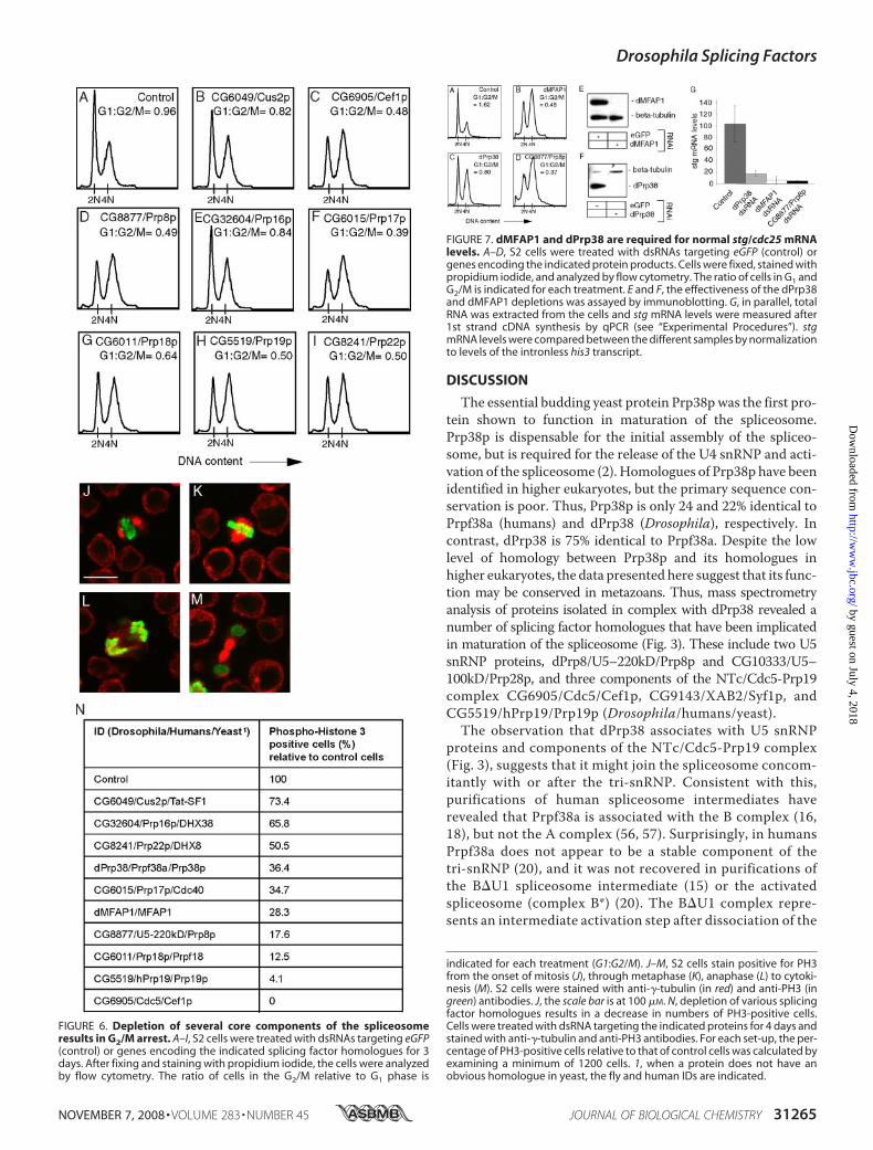
A General Requirement for Splicing Factors in G2/MProgression—Studies carried out with temperature-sensitivemutants in yeast suggest that several splicing factors are impor-tant for cell cycle progression (33–40). The cell cycle arrestphenotypes observed with different splicing factor mutantsseem to vary (41). This could reflect a difference in the severityof the splicing defects among those mutants or differentialrequirements for individual splicing factors. Our data (Figs. 2and 5, J and K) and some evidence from mammalian cells sug-gest that, in higher eukaryotes, at least certain splicing factorsare required for the G2/M transition (42–44). However, it isunclear whether this cell cycle arrest phenotype is general orwhether inactivation of different core components of themeta-zoan spliceosome would give rise to the same variability in cellcycle arrest phenotypes reported in yeast. To address this, weanalyzed the DNA profile from S2 cells treated with dsRNAstargeting a range ofDrosophila splicing factor homologues (Fig.6, B–I). These include Cus2p, which functions in the recruit-ment of U2 to the branch region (45), Cef1p and Prp19p, which
are required formaturation of the spliceosome (46–48), Prp8p,Prp16p, Prp17p, and Prp18p, which act in the second step of thesplicing reaction (49–52), and Prp22p, which is required forrelease of the spliced mRNA (53). Depletion of any of theseputative splicing factors led to an accumulation of cells inG2/M(Fig. 6, B–I). The degree to which cells arrested in G2/M variedbetween the different treatments and might reflect the effi-ciency of the dsRNA-mediated depletion. These data suggestthat, in higher eukaryotes, the G2/M arrest phenotype is a gen-eral outcome of interfering with the basic splicing machinery.However, it is not clear whether this phenotype is due to thedepletion of a factor that is rate-limiting for G2/M progressionor to the activation of a cell cycle checkpoint.To explore the nature of theG2/Marrest inmore details, cells
depleted of dPrp38, dMFAP1, or the splicing factor homo-logues indicated in Fig. 6, B–I, were stained with anti-�-tubulinand anti-phospho-histone 3 (PH3) antibodies. The anti-PH3antibody labels the condensed chromatin from the onset ofmitosis (Fig. 6J) through to cytokinesis (Fig. 6M). To examinewhether cells depleted of dPrp38, dMFAP1, or other splicingfactors arrest in G2 or during mitosis, a minimum of 1200 cellswere examined for each treatment, and the percentage of PH3positive cells was scored. Depletion of either of the tested splic-ing factors results in a 26–100% reduction in the number ofPH3 positive cells relative to that of control cells (Fig. 6N). Wedid not note the accumulation of cells at any stage of mitosis.This suggests that these splicing factor-deficient cells arearrested in G2, prior to chromosome condensation, rather thanat a later stage during mitosis.Depletion of dPrp38 or dMFAP1 Causes a Reduction in stg/
cdc25 mRNA Levels—In Drosophila, mitosis is triggered by atemporally controlled burst of string/Cdc25 (stg) transcription(54). Stg is a phosphatase, which activates the mitotic kinaseCdk1 and is essential for G2/M progression. In addition to thetranscriptional regulation of stg mRNA levels, the presence ofan intron in the stg transcript implies that stgmRNA levels arecritically dependent on a functional spliceosome. Thus, theobservation that depletion of dPrp38, dMFAP1, and a numberof core splicing factor homologues cause cells to arrest in G2/M(Figs. 2, 5K, and 6, B–I) suggests that stgmRNA levels might beaffected under those conditions. To investigate this, we meas-ured stgmRNA levels in cells depleted of dMFAP1, dPrp38, orCG8877/Prp8p (flies/yeast). As expected, treating cells withdsRNA targetingdmfap1,dprp38, orCG8877/prp8 caused cellsto arrest in G2/M (Fig. 7, A–F). Using quantitative RT-PCR tomeasure stgmRNA levels, we observed an 83–96% decrease instg mRNA in cells depleted of dMFAP1, dPrp38, or CG8877/Prp8p compared with control cells (Fig. 7G). The observeddecrease in mRNA levels could be due to a defect in processingof the stg pre-mRNA or could be an indirect effect caused byinterference of a factor required for regulation of stg transcrip-tion. Attempts to measure stg pre-mRNA levels by quantitativeRT-PCR were unsuccessful. This is most likely due to the lowlevels and instability of stg pre-mRNA. Consistent with thisidea, it has previously been reported that many pre-mRNAs donot accumulate in response to a defective spliceosome, but aredegraded by the exosome complex (55).
FIGURE 5. dPrp38 and dMFAP1 are required for G2/M progression anddevelopmental growth and proliferation. A–I, wing imaginal dics fromthird instar larvae. Posterior is to the right. Wild-type clones (A–C) or clonesexpressing dprp38 and dmfap1 RNAi constructs (D–I) were generated usingthe FLP-out technique (marked with GFP in A, D, and G). Clones with reducedlevels of dMFAP1 (D–F) or dPrp38 (G–I) are smaller than control clones (A–C)generated at the same time and undergo apoptosis as measured byincreased levels of cleaved caspase 3 staining in the clones (red in E and H).J–K, cells depleted of dMFAP1 arrest in G2/M. S2 cells were treated with dsRNAtargeting eGFP (control) or dmfap1 for 3 days. Cells were fixed, stained withpropidium iodide, and analyzed by flow cytometry. The ratio of cells in theG2/M relative to G1 phase is indicated for each treatment (G1:G2/M). L, immu-noblotting confirms that dMFAP1 protein levels are reduced in cells treatedwith dsRNA targeting dmfap1 compared with control cells. Anti-�-tubulin isused as a loading control.
Drosophila Splicing Factors
31264 JOURNAL OF BIOLOGICAL CHEMISTRY VOLUME 283 • NUMBER 45 • NOVEMBER 7, 2008
by guest on July 4, 2018http://w
ww
.jbc.org/D
ownloaded from
DISCUSSION
The essential budding yeast protein Prp38pwas the first pro-tein shown to function in maturation of the spliceosome.Prp38p is dispensable for the initial assembly of the spliceo-some, but is required for the release of the U4 snRNP and acti-vation of the spliceosome (2). Homologues of Prp38p have beenidentified in higher eukaryotes, but the primary sequence con-servation is poor. Thus, Prp38p is only 24 and 22% identical toPrpf38a (humans) and dPrp38 (Drosophila), respectively. Incontrast, dPrp38 is 75% identical to Prpf38a. Despite the lowlevel of homology between Prp38p and its homologues inhigher eukaryotes, the data presented here suggest that its func-tion may be conserved in metazoans. Thus, mass spectrometryanalysis of proteins isolated in complex with dPrp38 revealed anumber of splicing factor homologues that have been implicatedin maturation of the spliceosome (Fig. 3). These include two U5snRNP proteins, dPrp8/U5–220kD/Prp8p and CG10333/U5–100kD/Prp28p, and three components of the NTc/Cdc5-Prp19complex CG6905/Cdc5/Cef1p, CG9143/XAB2/Syf1p, andCG5519/hPrp19/Prp19p (Drosophila/humans/yeast).The observation that dPrp38 associates with U5 snRNP
proteins and components of the NTc/Cdc5-Prp19 complex(Fig. 3), suggests that it might join the spliceosome concom-itantly with or after the tri-snRNP. Consistent with this,purifications of human spliceosome intermediates haverevealed that Prpf38a is associated with the B complex (16,18), but not the A complex (56, 57). Surprisingly, in humansPrpf38a does not appear to be a stable component of thetri-snRNP (20), and it was not recovered in purifications ofthe B�U1 spliceosome intermediate (15) or the activatedspliceosome (complex B*) (20). The B�U1 complex repre-sents an intermediate activation step after dissociation of the
FIGURE 6. Depletion of several core components of the spliceosomeresults in G2/M arrest. A–I, S2 cells were treated with dsRNAs targeting eGFP(control) or genes encoding the indicated splicing factor homologues for 3days. After fixing and staining with propidium iodide, the cells were analyzedby flow cytometry. The ratio of cells in the G2/M relative to G1 phase is
indicated for each treatment (G1:G2/M). J–M, S2 cells stain positive for PH3from the onset of mitosis (J), through metaphase (K), anaphase (L) to cytoki-nesis (M). S2 cells were stained with anti-�-tubulin (in red) and anti-PH3 (ingreen) antibodies. J, the scale bar is at 100 �M. N, depletion of various splicingfactor homologues results in a decrease in numbers of PH3-positive cells.Cells were treated with dsRNA targeting the indicated proteins for 4 days andstained with anti-�-tubulin and anti-PH3 antibodies. For each set-up, the per-centage of PH3-positive cells relative to that of control cells was calculated byexamining a minimum of 1200 cells. 1, when a protein does not have anobvious homologue in yeast, the fly and human IDs are indicated.
FIGURE 7. dMFAP1 and dPrp38 are required for normal stg/cdc25 mRNAlevels. A–D, S2 cells were treated with dsRNAs targeting eGFP (control) orgenes encoding the indicated protein products. Cells were fixed, stained withpropidium iodide, and analyzed by flow cytometry. The ratio of cells in G1 andG2/M is indicated for each treatment. E and F, the effectiveness of the dPrp38and dMFAP1 depletions was assayed by immunoblotting. G, in parallel, totalRNA was extracted from the cells and stg mRNA levels were measured after1st strand cDNA synthesis by qPCR (see “Experimental Procedures”). stgmRNA levels were compared between the different samples by normalizationto levels of the intronless his3 transcript.
Drosophila Splicing Factors
NOVEMBER 7, 2008 • VOLUME 283 • NUMBER 45 JOURNAL OF BIOLOGICAL CHEMISTRY 31265
by guest on July 4, 2018http://w
ww
.jbc.org/D
ownloaded from

U1 snRNP, but before unwinding of the U4/U6 duplex (15).This could argue that, in contrast to its yeast counterpart,Prpf38a is not implicated in the activation step or is veryloosely associated with the spliceosome and lost during thepurification procedures. Consistent with the latter possibil-ity, yeast Prp38p association with the spliceosome appears tobe salt-sensitive (2). Our data also favors a role for dPrp38during the activation step of the spliceosome. Thus, dPrp38associates with several components of the Cdc5-Prp19 com-plex, which in humans appear to be stably associated withthe B�1U and B* complexes (15, 20), but not the B complex(15). The data presented here suggests that, like yeastPrp38p, dPrp38 joins the spliceosome concomitantly withthe tri-snRNP and stays associated with the spliceosomeduring at least part of the activation step.In addition to homologues of known splicing factors, we
identified a previously uncharacterized protein, dMFAP1,which interacts directly with dPrp38 (Fig. 3, E and F). HumanMFAP1 has been recovered in a number of spliceosome puri-fications, but was initially discounted as a contaminant dueto its proposed extracellular localization (21, 31). Thechicken MFAP1 protein was originally identified as a puta-tive component of the extracellular matrix in a screen forproteins detected by an antiserum raised against a crudemicrofibril preparation (30). Although we cannot rule out afunction for MFAP1 in the extracellular matrix, our dataargue that either it has dual functions or its identification asan extracellular protein was based on cross-reactivity of theantiserum with a different protein. Here we show thatdMFAP1, like dPrp38, functions in pre-mRNA processing(Fig. 4C and supplemental Fig. S3). Consistent with this,dMFAP1 can be purified in complex with several conservedsplicing factors including dPrp38, the U5 snRNP proteinsCG8877/U5–220kD/Prp8p, CG3436/U5–40kD/Rsa4p, andCG5931/U5–200kD/Brr2p, and the NTc/Cdc5-Prp19 corecomponent CG6905/Cdc5/Cef1p. The association ofdMFAP1 with U5 snRNP proteins and a component of theNTC/Cdc5-Prp19 complex is in agreement with thereported recovery of human MFAP1 in purifications of B,B�1U, and B* complexes (15, 16, 18, 20), but not A com-plexes (56). Thus, dMFAP1/MFAP1 might join the spliceo-some simultaneously with dPrp38/Prpf38a, but seems to bemore tightly associated with the spliceosome during the acti-vation step. Moreover, MFAP1 was not recovered in spliceo-some complexes after the first catalytic step, suggesting thatit functions during the activation step (16, 19).In addition to highly conserved spliceosome components,
dMFAP1 also co-purifies with a number of splicing factorsthat have evolved more recently in metazoans. Unsurpris-ingly, most of these function in the regulation of alternativesplicing. Thus, CG4602/srp54, CG6995/saf-b, and hfp/puf60were all recovered in an RNAi screen for regulators of alter-native splicing, which included 70% of all genes encodingknown Drosophila RNA-binding proteins (58). This is con-sistent with the idea that additional splicing factors haveevolved in metazoans to accommodate more complex splicepatterns. As expected, very few core splicing componentswere found to regulate alternative splicing. An interesting
exception is CG5931/Brr2p, which we isolated in ourdMFAP1 affinity purification (58). dMFAP1 is highly con-served among higher eukaryotes, but not in yeast, indicatingthat it has evolvedmore recently. Notably, dMFAP1 does notcontain a conserved RNA-binding motif and was notincluded in the screen carried out by Park and co-workers(58). Whether, dMFAP1 might regulate alternative splicingin higher eukaryotes therefore remains to be determined.Our in vivo studies of dPrp38 and dMFAP1 revealed that
both are essential proteins required for normal growth duringdevelopment. Consistentwith this, clones ofmutant tissuewithreduced levels of dPrp38 or dMFAP1 exhibit proliferation andgrowth defects and undergo apoptosis (Fig. 5). A more detailedanalysis of the proliferation defects in cell culture shows thatcells depleted of dPrp38 or dMFAP1 arrest in G2/M (Figs. 2 and5K). Moreover, this is paralleled by a substantial reduction inmRNA levels of the mitotic stg/cdc25 phosphatase (Fig. 7G).Thus, the observed G2/M arrest in cells depleted of dPrp38 ordMFAP1 might be the consequence of a defect in stg/cdc25pre-mRNA processing.In yeast, a subset of genes with known functions in splicing
has also been implicated in cell cycle progression. Theseinclude prp3, prp17, prp8, cef1, prp22 (Saccharomyces cerevi-siae) (33, 34, 36, 40, 59–61) and prp1, prp2, prp5, prp6, cdc28/prp8, cdc5, and prp12 (Schizosaccharomyces pombe) (35, 37, 39,62–65). Whereas mutations in some genes encoding splicingfactors cause cells to arrest heterogeneously throughout the cellcycle, cef1-13, prp17�, and prp22-1 mutants arrest homog-enously in G2/M (41). Amodel has been put forward to explainthe variability of the timing of cell cycle arrest, which suggeststhat some splicing factors are differentially required for effi-cient splicing of single transcripts or a subset of transcripts (41).Consistent with this idea, removal of a single intron from the�-tubulin encoding the TUB1 gene alleviates both the G2/Marrest and cell division defect observed in the cef1-13 mutant,but only partially rescues the cell division defect observed inprp17� and prp22-1 (41, 66). Instead, the G2/M arrest andgrowth defect in the prp17� mutant can be rescued by removalof an intron from the ANC1 gene (67). The molecular basis forthis specificity remains unclear.The range of cell cycle phenotypes observed with different
splicing factor mutants in yeast prompted us to study therequirement for various Drosophila splicing factors in cellcycle progression. We analyzed the effect of individualdepletions of splicing factors that are predicted to functionat various stages of the spliceosome cycle. Depletion of any ofthose splicing factors caused cells to arrest in G2/M, suggest-ing that it is a general consequence of interfering with spli-ceosome activity (Fig. 6). Consistent with this, a genome-wide RNA interference screen in HeLa cells for factorsrequired for mitosis identified a number of genes encodingsplicing factors (68). However, we cannot conclude whetherthe observed G2/M arrest results from of a failure to splice apre-mRNA encoding a factor that is rate-limiting for entryinto mitosis (e.g. stg/cdc25), or whether a defect in splicingactivates a mitotic checkpoint that causes cells to arrest inG2. Future research might resolve this issue.
Drosophila Splicing Factors
31266 JOURNAL OF BIOLOGICAL CHEMISTRY VOLUME 283 • NUMBER 45 • NOVEMBER 7, 2008
by guest on July 4, 2018http://w
ww
.jbc.org/D
ownloaded from

Acknowledgments—We are grateful to the Bloomington DrosophilaStock Center and the Vienna Drosophila RNAi Center for fly stocks,and the Developmental Studies Hybridoma Bank for antibodies. Wethank Terrence Gilbank, Steve Murray, and Frances Earl for trans-genic injections, the LRI FACS lab for help with FACS analysis, RuthBrain for advice on hairpin cloning, and the Taplin Biological MassSpectrometry Facility for protein identification. We are grateful toJulien Colombani, Sally Leevers, and Filipe Josue for comments on themanuscript.
REFERENCES1. Jurica, M. S., and Moore, M. J. (2003)Mol. Cell 12, 5–142. Xie, J., Beickman, K., Otte, E., and Rymond, B. C. (1998) EMBO J. 17,
2938–29463. Rosbash, M., and Seraphin, B. (1991) Trends Biochem. Sci. 16, 187–1904. Ruby, S. W., and Abelson, J. (1988) Science 242, 1028–10355. Zorio, D. A., and Blumenthal, T. (1999) Nature 402, 835–8386. Wu, S., Romfo, C. M., Nilsen, T.W., and Green, M. R. (1999)Nature 402,
832–8357. Merendino, L., Guth, S., Bilbao, D., Martinez, C., and Valcarcel, J. (1999)
Nature 402, 838–8418. Singh, R., Banerjee, H., andGreen,M. R. (2000)RNA (Cold SpringHarbor)
6, 901–9119. Brow, D. A. (2002) Annu. Rev. Genet. 36, 333–36010. Madhani, H. D., and Guthrie, C. (1994) Annu. Rev. Genet. 28, 1–2611. Staley, J. P., and Guthrie, C. (1998) Cell 92, 315–32612. Chen, J. Y., Stands, L., Staley, J. P., Jackups, R. R., Jr., Latus, L. J., andChang,
T. H. (2001)Mol. Cell 7, 227–23213. Raghunathan, P. L., and Guthrie, C. (1998) Curr. Biol. 8, 847–85514. Chen, C. H., Tsai,W. Y., Chen, H. R.,Wang, C. H., and Cheng, S. C. (2001)
J. Biol. Chem. 276, 488–49415. Makarova, O. V., Makarov, E. M., Urlaub, H., Will, C. L., Gentzel, M.,
Wilm, M., and Luhrmann, R. (2004) EMBO J. 23, 2381–239116. Bessonov, S., Anokhina, M., Will, C. L., Urlaub, H., and Luhrmann, R.
(2008) Nature 452, 846–85017. Chen, Y. I., Maika, S. D., and Stevens, S. W. (2006) J. Mol. Biol. 361,
412–41918. Deckert, J., Hartmuth, K., Boehringer, D., Behzadnia, N., Will, C. L., Kast-
ner, B., Stark, H., Urlaub, H., and Luhrmann, R. (2006)Mol. Cell. Biol. 26,5528–5543
19. Jurica, M. S., Licklider, L. J., Gygi, S. R., Grigorieff, N., and Moore, M. J.(2002) RNA (Cold Spring Harbor) 8, 426–439
20. Makarov, E. M., Makarova, O. V., Urlaub, H., Gentzel, M., Will, C. L.,Wilm, M., and Luhrmann, R. (2002) Science 298, 2205–2208
21. Neubauer, G., King, A., Rappsilber, J., Calvio, C., Watson, M., Ajuh, P.,Sleeman, J., Lamond, A., and Mann, M. (1998) Nat. Genet. 20, 46–50
22. Rubin, G. M., and Spradling, A. C. (1982) Science 218, 348–35323. Brand, A. H., and Perrimon, N. (1993) Development 118, 401–41524. Xu, T., and Rubin, G. M. (1993) Development 117, 1223–123725. Blanton, S., Srinivasan, A., and Rymond, B. (1992) Mol. Cell. Biol. 12,
3939–394726. Brand, A. H., Manoukian, A. S., and Perrimon, N. (1994) Methods Cell
Biol. 44, 635–65427. Diaz, B., and Moreno, E. (2005) Exp. Cell Res. 306, 317–32228. Zhu, J., Mayeda, A., and Krainer, A. R. (2001)Mol. Cell 8, 1351–136129. Cho, S. Y., Shin, E. S., Park, P. J., Shin, D. W., Chang, H. K., Kim, D., Lee,
H.H., Lee, J. H., Kim, S.H., Song,M. J., Chang, I. S., Lee,O. S., andLee, T. R.(2007) J. Biol. Chem. 282, 2456–2465
30. Horrigan, S. K., Rich, C. B., Streeten, B.W., Li, Z. Y., and Foster, J. A. (1992)J. Biol. Chem. 267, 10087–10095
31. Neubauer, G. (2005)Methods Enzymol. 405, 236–26332. Pignoni, F., Hu, B., andZipursky, S. (1997)Proc.Natl. Acad. Sci. U. S. A.94,
9220–922533. Boger-Nadjar, E., Vaisman, N., Ben-Yehuda, S., Kassir, Y., and Kupiec, M.
(1998)Mol. Gen. Genet. 260, 232–24134. Hwang, L. H., and Murray, A. W. (1997)Mol. Biol. Cell 8, 1877–188735. Lundgren, K., Allan, S., Urushiyama, S., Tani, T., Ohshima, Y., Frendewey,
D., and Beach, D. (1996)Mol. Biol. Cell 7, 1083–109436. Ohi, R., Feoktistova, A., McCann, S., Valentine, V., Look, A. T., Lipsick,
J. S., and Gould, K. L. (1998)Mol. Cell. Biol. 18, 4097–410837. Potashkin, J., Kim, D., Fons, M., Humphrey, T., and Frendewey, D. (1998)
Curr. Genet. 34, 153–16338. Stevens, S. W., and Abelson, J. (1999) Proc. Natl. Acad. Sci. U. S. A. 96,
7226–723139. Urushiyama, S., Tani, T., and Ohshima, Y. (1997) Genetics 147, 101–11540. Vaisman, N., Tsouladze, A., Robzyk, K., Ben-Yehuda, S., Kupiec, M., and
Kassir, Y. (1995)Mol. Gen. Genet. 247, 123–13641. Burns, C., Ohi, R., Mehta, S., O’Toole, E., Winey, M., Clark, T., Sugnet, C.,
Ares, M., and Gould, K. (2002)Mol. Cell. Biol. 22, 801–81542. Bernstein, H. S., and Coughlin, S. R. (1998) J. Biol. Chem. 273, 4666–467143. Li, X., Wang, J., and Manley, J. L. (2005) Genes Dev. 19, 2705–271444. Pacheco, T. R., Moita, L. F., Gomes, A. Q., Hacohen, N., and Carmo-
Fonseca, M. (2006)Mol. Biol. Cell 17, 4187–419945. Perriman, R., and Ares, M. (2000) Genes Dev. 14, 97–10746. Chan, S. P., and Cheng, S. C. (2005) J. Biol. Chem. 280, 31190–3119947. Chan, S. P., Kao, D. I., Tsai, W. Y., and Cheng, S. C. (2003) Science 302,
279–28248. Tsai, W., Chow, Y., Chen, H., Huang, K., Hong, R., Jan, S., Kuo, N., Tsao,
T., Chen, C., and Cheng, S. (1999) J. Biol. Chem. 274, 9455–946249. James, S., Turner,W., and Schwer, B. (2002) RNA (Cold Spring Harbor) 8,
1068–107750. Schneider, S., Hotz, H., and Schwer, B. (2002) J. Biol. Chem. 277,
15452–1545851. Umen, J., and Guthrie, C. (1995) RNA (Cold Spring Harbor) 1, 584–59752. Wang, Y., andGuthrie, C. (1998)RNA (Cold SpringHarbor) 4, 1216–122953. Schneider, S., Campodonico, E., and Schwer, B. (2004) J. Biol. Chem. 279,
8617–862654. Edgar, B. A., and O’Farrell, P. H. (1989) Cell 57, 177–18755. Bousquet-Antonelli, C., Presutti, C., and Tollervey, D. (2000) Cell 102,
765–77556. Hartmuth, K., Urlaub, H., Vornlocher, H. P., Will, C. L., Gentzel, M.,
Wilm, M., and Luhrmann, R. (2002) Proc. Natl. Acad. Sci. U. S. A. 99,16719–16724
57. Urlaub, H., Hartmuth, K., and Luhrmann, R. (2002)Methods 26, 170–18158. Park, J. W., Parisky, K., Celotto, A. M., Reenan, R. A., and Graveley, B. R.
(2004) Proc. Natl. Acad. Sci. U. S. A. 101, 15974–1597959. Hartwell, L. H.,Mortimer, R. K., Culotti, J., andCulotti,M. (1973)Genetics
74, 267–28660. Johnston, L. H., and Thomas, A. P. (1982)Mol. Gen. Genet. 186, 439–44461. Shea, J. E., Toyn, J. H., and Johnston, L. H. (1994) Nucleic Acids Res. 22,
5555–556462. Habara, Y., Urushiyama, S., Shibuya, T., Ohshima, Y., and Tani, T. (2001)
RNA (Cold Spring Harbor) 7, 671–68163. Nurse, P., Thuriaux, P., and Nasmyth, K. (1976) Mol. Gen. Genet. 146,
167–17864. Ohi, R., McCollum, D., Hirani, B., DenHaese, G. J., Zhang, X., Burke, J. D.,
Turner, K., and Gould, K. L. (1994) EMBO J. 13, 471–48365. Takahashi, K., Yamada, H., and Yanagida, M. (1994) Mol. Biol. Cell 5,
1145–115866. Chawla, G., Sapra, A., Surana, U., and Vijayraghavan, U. (2003) Nucleic
Acids Res. 31, 2333–234367. Dahan, O., and Kupiec, M. (2004) Nucleic Acids Res. 32, 2529–254068. Kittler, R., Putz, G., Pelletier, L., Poser, I., Heninger, A. K., Drechsel, D.,
Fischer, S., Konstantinova, I., Habermann, B., Grabner, H., Yaspo, M. L.,Himmelbauer, H., Korn, B., Neugebauer, K., Pisabarro, M. T., and Buch-holz, F. (2004) Nature 432, 1036–1040
Drosophila Splicing Factors
NOVEMBER 7, 2008 • VOLUME 283 • NUMBER 45 JOURNAL OF BIOLOGICAL CHEMISTRY 31267
by guest on July 4, 2018http://w
ww
.jbc.org/D
ownloaded from

Ditte S. Andersen and Nicolas Tapon/M Progression2 MFAP1 Is Required for Pre-mRNA Processing and GDrosophila
doi: 10.1074/jbc.M803512200 originally published online September 2, 20082008, 283:31256-31267.J. Biol. Chem.
10.1074/jbc.M803512200Access the most updated version of this article at doi:
Alerts:
When a correction for this article is posted•
When this article is cited•
to choose from all of JBC's e-mail alertsClick here
Supplemental material:
http://www.jbc.org/content/suppl/2008/09/05/M803512200.DC1
http://www.jbc.org/content/283/45/31256.full.html#ref-list-1
This article cites 70 references, 32 of which can be accessed free at
by guest on July 4, 2018http://w
ww
.jbc.org/D
ownloaded from