theessentialroleofclathrin … infectiousentryofhumanenterovirus71* s...
TRANSCRIPT
The Essential Role of Clathrin-mediated Endocytosis in theInfectious Entry of Human Enterovirus 71*□S
Received for publication, July 26, 2010, and in revised form, October 13, 2010 Published, JBC Papers in Press, October 18, 2010, DOI 10.1074/jbc.M110.168468
Khairunnisa’ Mohamed Hussain, Kim Lian Janet Leong, Mary Mah-Lee Ng, and Justin Jang Hann Chu1
From the Department of Microbiology, Yong Loo Lin School of Medicine, National University Health System, 5 Science Drive 2,National University of Singapore, Singapore 117597
Little is currently known about the infectious entry processof human enterovirus 71 (HEV71) into host cells, which mayrepresent potential anti-viral targeting sites. In this study atargeted small-interfering RNA (siRNA) screening platformassay was established and validated to identify and profile keycellular genes involved in processes of endocytosis, cytoskel-etal dynamics, and endosomal trafficking essential for HEV71infection. Screen evaluation was conducted via the expressionof well characterized dominant-negative mutants, bioimagingstudies (double-labeled immunofluorescence assays, transmis-sion electron microscopy analysis), secondary siRNA-baseddosage dependence studies, and drug inhibition assays. Theinfectious entry of HEV71 into rhabdomyosarcoma cells wasshown to be significantly inhibited by siRNAs targeting genesassociated with clathrin-mediated endocytosis (CME) that in-clude AP2A1, ARRB1, CLTC, CLTCL1, SYNJ1, ARPC5, PAK1,ROCK1, andWASF1. The functional role of CME was verifiedby the observation of strong co-localization between HEV71particles and clathrin as well as dose-dependent inhibition ofHEV71 infection upon siRNA knockdown of CME-associatedgenes. HEV71 entry by CME was further confirmed via inhibi-tion by dominant-negative EPS15 mutants and treatment ofCME drug inhibitors, with more than 80% inhibition observedat 20 �M chlorpromazine. Furthermore, HEV71 infection wasshown to be sensitive to the disruption of human genes in reg-ulating early to late endosomal trafficking as well as endoso-mal acidic pH. The identification of clathrin-mediated endocy-tosis as the entry pathway for HEV71 infection of susceptiblehost cells contributes to a better understanding of HEV71pathogenesis and enables future development of anti-viralstrategies against HEV71 infection.
Numerous animal viruses utilize various endocytic mecha-nisms available in mammalian cells for productive infection.Host endocytic pathways, such as clathrin-mediated endocy-tosis, caveolae-dependent uptake, macropinocytosis and cho-lesterol-dependent endocytosis, are commonly employed tomediate the infectious entry of virus particles into host cells
(1). With the aid of these membrane trafficking processes,internalized viruses are able to fuse with cellular membranesfor subsequent genome release as well as localize within thecell for effective replication (2). Many viruses require the in-duction of conformational changes by low pH levels (a resultof acidification within the endosomal pathway) to drive essen-tial infective steps of viral entry, such as fusion, penetration,and uncoating (3). In addition, viral transport within host cellsmay also be mediated by the host cytoskeleton network, par-ticularly the actin filaments (4). With viral entry regarded as amajor determinant of viral tropism and pathogenesis (5), un-derstanding these initial events will enable future develop-ment of anti-viral strategies against HEV71 infection.Human enterovirus 71 (HEV71)2 is a single-stranded, posi-
tive-sense RNA virus belonging to the human Enterovirus A(HEV-A) subspecies of the Enterovirus genus in the Picorna-viridae family (6). First identified and characterized in 1969 inCalifornia from a stool specimen isolated from an infant withencephalitis (7), ensuing outbreaks of HEV71 have since beenreported in various regions of the world, including Australia,Sweden, and Japan. In the past decade, HEV71-induced hand,foot, and, mouth disease outbreaks have mainly affected chil-dren within the Asia-Pacific region, including Hong Kong,China, Singapore, and Australia (8). The global control of po-liovirus has also resulted in HEV71 becoming one of the mostclinically significant etiological agents of acute neurologicaldiseases such as polio-like acute flaccid paralysis, cerebellarataxia, and brainstem encephalitis (9). No antiviral treatmenthas yet been developed to treat HEV71 infections; similarly,effective vaccines are currently unavailable, although severalvaccine trials are being undertaken to develop effective thera-peutic strategies to combat severe HEV71 infections. Empha-sis is, therefore, being placed on understanding its virology,epidemiology, diagnosis, and management.HEV71 infection begins with the attachment of virus parti-
cles onto host surface receptors followed by subsequent entryinto the cells. Although it has been widely accepted thatHEV71 enters permissive cells via receptor-mediated endocy-tosis, only several cell-specific candidate receptors have beenidentified to date. These cellular receptors include: scavengerreceptor B2 (10), human P-selectin glycoprotein ligand-1 (11),and sialylated glycans (SA-a2,6Gal and SA-a2,3Gal) (12). Fur-
* This work was supported by The Ministry of Education Academic ResearchFund Grant R182-000-165-133), National Medical Research Council (Sin-gapore) Grant (Project no. NMRC/NIG/0012/2007), Defense Science andTechnology Agency Grant POD0713895, and an Infectious Diseases Pro-gram grant (National University of Singapore) (to J. H. C.).
□S The on-line version of this article (available at http://www.jbc.org) con-tains Table 1 and Figs. 1 and 2.
1 To whom correspondence should be addressed. Tel.: 65-6516-3278; Fax:65-6776-6872; E-mail: [email protected]/[email protected].
2 The abbreviations used are: HEV71, human enterovirus 71; RD, rhabdomy-osarcoma; EEA1, endosomal antigen 1; LAMP1, lysosomal-associatedmembrane protein 1; TR, Texas Red; EIPA, 5-(N-ethyl-N-isopropyl)-amilo-ride; MTT, (3-(4,5-dimethylthiazol-2-yl)-2,5diphenyl tetrazolium bromide);p.i., post-infection.
THE JOURNAL OF BIOLOGICAL CHEMISTRY VOL. 286, NO. 1, pp. 309 –321, January 7, 2011© 2011 by The American Society for Biochemistry and Molecular Biology, Inc. Printed in the U.S.A.
JANUARY 7, 2011 • VOLUME 286 • NUMBER 1 JOURNAL OF BIOLOGICAL CHEMISTRY 309
by guest on May 31, 2019
http://ww
w.jbc.org/
Dow
nloaded from

thermore, subsequent steps of HEV71 infection, such as theentry process and the uncoating of its RNA genome as well asthe assembly pathway have not been clearly defined.Although previous studies have attempted to decipher the
entry processes of related enteroviruses, such as poliovirus(13–16) and echovirus (17–19), little is currently known aboutthe specific cellular genes or host factors involved in mediat-ing the infectious entry of HEV71 into human cells. With therecent development of small interfering RNA (siRNA) tech-nology, high throughput screening surveys of mammaliangenes and their functions has been made feasible (20).In this study we assessed an array of siRNA libraries that
specifically target human genes important for endocytosisprocesses, trafficking of membrane vesicles, actin polymeriza-tion, and cytoskeleton rearrangement to determine the cellu-lar genes or factors that facilitate the infectious entry pathwayof HEV71. Interestingly, we were able to show for the firsttime that the knockdown of human genes associated withclathrin-mediated endocytosis efficiently blocked HEV71 in-fection. The essential involvement of clathrin-mediated endo-cytosis in HEV71 entry into cells was confirmed by the ex-pression of dominant-negative mutants and drug inhibitors toperturb this uptake pathway. In addition, we also identifiedcellular factors responsible for vesicle trafficking and matura-tion, signal transduction, and actin polymerization that areessential for the infectious entry process of HEV71.
EXPERIMENTAL PROCEDURES
Cells and Viruses—Human rhabdomyosarcoma (RD) cells(American Type Culture Collection, ATCC CCL-136) weremaintained in Dulbecco’s modified Eagle’s medium (DMEM)(Sigma) supplemented with 10% inactivated fetal calf serum(FCS) (Invitrogen) and sodium bicarbonate (Merck). HEV71strain H (VR-1432TM) was obtained from ATCC and propa-gated in RD cells. The virus titer was quantitated via viral in-fectious plaque assays using RD cells.Antibodies and Reagents—Mouse monoclonal antibodies
against HEV71 were purchased from Chemicon for immun-ofluorescent detection of HEV71 infection. Rabbit polyclonalantibodies to clathrin (CLTC, Chemicon), early endosomalantigen 1 (EEA1; Novus Biologicals), and lysosomal-associ-ated membrane protein 1 (LAMP1; Santa Cruz Biotechnol-ogy) were purchased from Chemicon for immunofluores-cence assays. The secondary antibodies conjugated tofluorescein isothiocyanate (FITC) and Texas Red (TR) werepurchased from Invitrogen. 4�,6�-diamidino-2-phenylindole(DAPI) fluorescent dye was purchased from Invitrogen.Mouse monoclonal antibodies against AP2A1 (Santa CruzBiotechnology), ARPC5 (Novus Biologicals), RAB3A (BD Bio-sciences), SYNJ1 (Abnova Corp.), and WASF1 (Novus Bio-logicals) were used for the Western detection of the respectiveproteins. All drugs used in this study (chlorpromazine, filipin,bafilomycin A1, cytochalasin B, methyl-�-cyclodextrin, 5-(N-ethyl-N-isopropyl)-amiloride (EIPA), concanamycin A, andnystatin) were purchased from Sigma and prepared accordingto the manufacturer’s instructions under sterile conditions.siRNA Library—The human genome siRNA subset library
targeting the endocytic and membrane trafficking genes
(Dharmacon, RTF H-005500) was used in this study. A smartpool approach of incorporating four siRNAs targeting eachgene was utilized. The advantages of this pooled approach aswell as the issues of gene compensation of specific isotype ofgenes were discussed in Reynolds et al. (21). The list of 119targeted human genes and isoforms (excluding the controlset) is presented in supplemental Table 1.RNA Transfection of siRNA Delivery into Cells—All trans-
fections were performed in a 384-well plate format. A 1.2%(v/v) stock solution of the transfection reagent (DharmaFECT1) was prepared in DCCR cell culture buffer (Dharmacon) andincubated at 25 °C for 10 min. From this stock, 8 �l was addedto the lyophilized siRNA in each well of the 384-well plate andincubated at 25 °C for 30 min to allow the siRNAs to rehy-drate and form siRNA-lipid complexes. Subsequently, 5 � 103RD cells in 42 �l of complete DMEM supplemented with 10%FCS were added. The siRNA cell mixture was then incubatedat 37 °C for 48 h before HEV71 infection. The final concentra-tion of pooled siRNAs was 50 nM per well. Individual siRNAduplexes were used at 6.25 pmol per well.Screening of the siRNA Library—A high throughput plat-
form for the specific detection of HEV71 infection in the 384-well plate format via immunofluorescence staining was em-ployed for the screening assay, as described in Chu and Yang(22). Briefly, the siRNA-transfected RD cells were incubatedat 37 °C for 48 h to ensure effective gene knockdown by thesiRNAs before being subjected to HEV71 infection at a multi-plicity of infection of 1. After 12 post-infection (p.i.), the cellswere then fixed with cold absolute methanol (Sigma) for 15min at �20 °C. Subsequent cell washing steps were performedusing an automated 384-well format plate washer (EMBLA,Molecular Devices). The cells were then subjected to immu-nofluorescence staining using primary anti-HEV71 antibodies(Chemicon) followed by FITC-conjugated secondary antibod-ies (Invitrogen). Cell nuclei were counterstained with DAPI(100 nM; Invitrogen) before collation of image data by theArrayScan VTI HCS automated fluorescence microscopeReader system (Cellomics) with appropriate excitation andemission wavelengths for FITC (495 and 520 nm, respectively)and DAPI (358 and 461 nm, respectively). Data collection andauto-focusing parameters were pre-determined using Cellom-ics Target Activation Bioapplication (Cellomics). A genericsegmentation tool function was used to identify the two dif-ferent stains (DAPI and FITC) with intensities above back-ground staining, and data collection was obtained by loggingthe measurements. Data analysis after image acquisition wascarried out using Cellomics Target Activation Bioapplication(Cellomics). DAPI-stained nuclei were counted to determinetotal cell populations, whereas FITC-stained cytoplasm wasscored to determine the number of virus-infected cells. Im-ages with less than 500 cells were excluded from data analysisby the cell sorting module. Three independent screening as-says were performed.Controls included in individual sets of experiments were
the use of transfection reagent (DharmaFECT 1) alone, a non-targeting siRNA (Dharmacon), a green fluorescent nonspe-cific siRNA (siGLO, Dharmacon), RISC-freeTM siRNA (Dhar-macon), and siRNA smart pools targeting cyclophilin B
Infectious Entry of Human Enterovirus 71
310 JOURNAL OF BIOLOGICAL CHEMISTRY VOLUME 286 • NUMBER 1 • JANUARY 7, 2011
by guest on May 31, 2019
http://ww
w.jbc.org/
Dow
nloaded from

duplex, glyceraldehyde-3-phosphate dehydrogenase, andlamin A/C (Dharmacon). These siRNAs served as negativecontrols for nonspecific effects of siRNA and/or transfectionreagents on cell viability and virus infection. In addition, cellviability was observed and monitored by visual inspection viathe use of phase-contrast microscopy.Transfection and Infection of Dominant Negative Mutants
of Eps15—Plasmid constructs of dominant-negative Eps15(pEGFP-Eps15�95/295, a component of the AP2 clathrinadaptor complex; the dominant-negative form inhibits clath-rin-coated pit budding) and pEGFP-Eps15DIII�2 (this con-struct lacks the AP2-binding sites and was used as a controlthat did not inhibit clathrin-mediated endocytosis) was kindlyprovided by A. Benmerah, Pasteur Institute, Paris, France.Briefly, RD cells were seeded onto 24-well tissue culture platesand grown overnight until 75% confluency was reached. 0.8�g of the plasmid construct was then complexed with 50 �l ofOpti-MEMmedium (Invitrogen) for 5 min at room tempera-ture. The mixture was then added to 48 �l of Opti-MEM con-taining 2 �l of Lipofectamine 2000TM (Invitrogen) that hadundergone similar incubation conditions. After a further in-cubation period of 20 min, the DNA-liposome complexeswere added to the cells, which had been starved in Opti-MEMmedium for 4 h before transfection. After incubation for 6 hat 37 °C, 1 ml of maintenance medium was added and incu-bated for a further 48 h before virus infection.To synchronize HEV71 entry, virus binding was first per-
formed at 4 °C for 1 h before the temperature was shifted rap-idly to 37 °C for internalization. Infected RD cells were subse-quently processed for immunofluorescence assay andmicroscopic imaging. Supernatants were harvested fromtransfected cells, and plaque assays were performed.Bioimaging Assay and Indirect Immunofluorescence—RD
cells seeded on coverslips were incubated at 4 °C for 30 minand then washed with ice-cold PBS. The cells were subse-quently infected with HEV71 at a multiplicity of infection of 1for 30 min at 4 °C to allow viral attachment to cell surfacewithout entry. The cells were then shifted to 37 °C for a fur-ther 10 min to allow the cells to return to their physiologicaltemperature (designated as time 0). Cells were fixed in ice-cold absolute methanol at different time points, 0, 5, 10, 15,20, and 30 min, followed by three 5-min washes in cold PBSbefore immunofluorescence processing.Cells were incubated with the appropriate primary antibod-
ies (with a 1:500 dilution for anti-HEV71 (Chemicon), a 1:300dilution for anti-clathrin (Chemicon), a 1:200 dilution foranti-EEA1 (Novus Biologicals), and 1:500 dilution for anti-LAMP1 antibodies (Santa Cruz Biotechnology) in a humiditychamber for 1 h at 37 °C. After three 5-min washes with PBS,cells were further incubated with appropriate FITC- or TR-conjugated secondary antibodies (Invitrogen) before washingagain with PBS. Nuclei staining were performed by incubatingthe cells with DAPI (Invitrogen) before mounting the pro-cessed coverslips onto ethanol-cleaned glass slides usingDabco mountant. The specimens were viewed with an in-verted fluorescence microscope (IX81 Olympus) with excita-tion wavelengths of 480 and 543 nm for FITC and TR, respec-tively, using oil immersion objectives.
Drug Inhibitory Treatments—To determine the effects ofthe drugs used to inhibit the entry of HEV71, serum-starvedRD cells were pretreated with drugs at different concentra-tions (Table 2) for 2 h at 37 °C followed by virus infection asdescribed above. At 12 h p.i., HEV71-infected cells were pro-cessed for immunofluorescence assay. Three independentexperiments were carried out for each set of drugs used. Theinhibition of virus entry was determined by the number ofvirus antigen-positive cells in relation to the total number ofcells (virus antigen-positive and -negative) and was expressedas the percentage virus antigen-positive cells.Cell Viability Assay—Cell viability upon siRNA transfection
and drug treatments was assessed by employing the 3-(4,5-dimethylthiazol-2-yl)-2,5diphenyl tetrazolium bromide(MTT) assay (Chemicon International), according to themanufacturer’s recommendations. Briefly, RD cells wereseeded in 96-well cell culture plates and subsequently treatedwith individual siRNAs and drugs for 48 and 2 h, respectively,before incubation with AB solution for 4 h at 37 °C. After this,Solution C containing isopropyl alcohol/HCl was added, andthe plates were subjected to absorbance reading by an ELISAplate reader (Bio-Rad) at test wavelength of 570 nm and refer-ence wavelength of 630 nm.
RESULTS
Optimization of siRNA Screening Platform for HEV71Infection—siRNA profiling using the human endocytic/mem-brane trafficking library from Dharmacon was conducted as apreliminary assay to identify key endocytic and membranetrafficking genes essential in HEV71 entry into host cells. Theimmunofluorescence screening assay was based on the detec-tion of HEV71 capsid (VP1) protein in HEV71-infected RDcell monolayers. To ensure minimal signal variation and con-sistently high signal-to-background ratio, the Z� factor of thescreening assay (23), a measure of quality of high throughputscreening, was determined. An assay with a Z� factor value ofbetween 0.5 and 1 would be considered to be an excellent as-say for screening, with minimal well-well variation in the 384-well plate (23). A Z� factor of 0.562 was consistently measured(data not shown), demonstrating the reliability and robust-ness of the assay. With the optimization of the siRNA screen-ing platform for HEV71 infection, it was employed as a toolfor rapid discovery of cellular factors essential for mediatingthe infectious entry process of HEV71 into cells.siRNA Profiling of Human Endocytic and Membrane Traf-
ficking Genes—An array of 119 siRNA smart pool (Dhar-macon) targeting genes known to be directly or indirectlyinvolved in regulating the different endocytic pathways(clathrin, caveolae, macropinocytosis, etc.), polymerizationof actin, and cytoskeleton rearrangement and vesicle/cargotrafficking was used to identify host genes necessary for theinfectious entry of HEV71. A list of the targeted humangenes and a brief description of the reported functionalrole for each of the genes are provided in supplementalTable 1.Effects of siRNA knockdown of different endocytic genes as
determined by the decrease in the percentage of viral antigen-positive cells on HEV71 infection are shown in Fig. 1. Em-
Infectious Entry of Human Enterovirus 71
JANUARY 7, 2011 • VOLUME 286 • NUMBER 1 JOURNAL OF BIOLOGICAL CHEMISTRY 311
by guest on May 31, 2019
http://ww
w.jbc.org/
Dow
nloaded from

ploying a 50% reduction in the number of fluorescentlystained HEV71-infected cells as the siRNA-induced effect andcriterion that suppressed HEV71 infection, the list of humangenes that have an inhibitory effect on HEV71 infection wereobtained using the screening platform, and the results areshown in Fig. 1 and further classified based on their func-tional roles (Table 1). A substantial number of genes resultingin the decrease in HEV71 infection have been known to be
involved in clathrin-mediated endocytosis (Table 1); thesegenes include the subunit of the clathrin-associated adaptorprotein complex 2 (AP2A1) and clathrin heavy chains (CLTCand CLTCL1). AP2A1 is a subunit of the clathrin-associatedadaptor protein complex 2 that regulates the formation ofclathrin-coated pits as well as links clathrin to cellular recep-tors in endocytic vesicles (24). The clathrin heavy and lightchains are intricately braided together to form the triskelion
FIGURE 1. siRNA profiling of genes important for the infectious entry process of HEV71 into RD cells using the human endocytic/membranetrafficking library. HEV71-infected cells after siRNA knockdown (pink) were compared against transfection controls (purple) and are presented asthe percentage of viral antigen-positive cells. The transfection controls were set up as the base line for infection and transfection efficiency. Genesthat resulted in significant decreases in HEV71 infection (p � 0.05) after siRNA knockdown and are involved in clathrin-mediated endocytosis areshown in green.
Infectious Entry of Human Enterovirus 71
312 JOURNAL OF BIOLOGICAL CHEMISTRY VOLUME 286 • NUMBER 1 • JANUARY 7, 2011
by guest on May 31, 2019
http://ww
w.jbc.org/
Dow
nloaded from

TABLE 1
Genes required for HEV71 infection of RD cellsThe descriptions of the function(s) of the individual genes are summarized from the Online Mendelian Inheritance in Man as of July 2010.
Infectious Entry of Human Enterovirus 71
JANUARY 7, 2011 • VOLUME 286 • NUMBER 1 JOURNAL OF BIOLOGICAL CHEMISTRY 313
by guest on May 31, 2019
http://ww
w.jbc.org/
Dow
nloaded from
coat that facilitates the formation of clathrin coated pits forendocytosis (25).In addition, siRNAs that targeted kinases (MAP4K2, PAK1,
PIK3CG, PIK3C2G, and ROCK1) that are involved in the sig-nal transduction processes of viral entry (26) were also notedto reduce HEV71 infection. Silencing of other genes involvedin vesicle and endosomal transport such as ARFIP2, RAB3A,and RAB6B (27, 28) had also led to a decrease in viral infec-tion. Furthermore, siRNA knockdowns of human genes es-sential in regulating actin polymerization (ARPC5, ARRB1,and WASF1) (29–31) also displayed inhibitory effects onHEV71 infection. Finally, siRNA knockdown of human genesessential for synaptic transmission and membrane trafficking(SYNJ1 and ELKS) (32–34) also exhibited inhibitory effects onHEV71 infection. Several of these top-hit genes on HEV71infection were selected for further analysis.A number of cellular (siGENOMETM Cyclophilin B duplex,
Lamin A/C, and GAPDH SMARTpoolTM siRNA; Dharma-con) and transfection (siGENOMETM Non-Targeting siRNApool, RISC-freeTM siRNA, and siGLO Risc-freeTM siRNA;Dharmacon) controls were included to ensure that the reduc-tion in HEV71 infection was not due to off-target effects aswell as to eliminate the possibility of reduction in HEV71 in-fection by housekeeping genes (Fig. 1). It was generally ob-served that there was minimal cytotoxicity in thesiRNA-transfected cells with HEV71 infection. Cell viabilitywas not affected sufficiently to contribute to the outcome ofthe screen.Validation of Endocytic Genes Knockdown—To further vali-
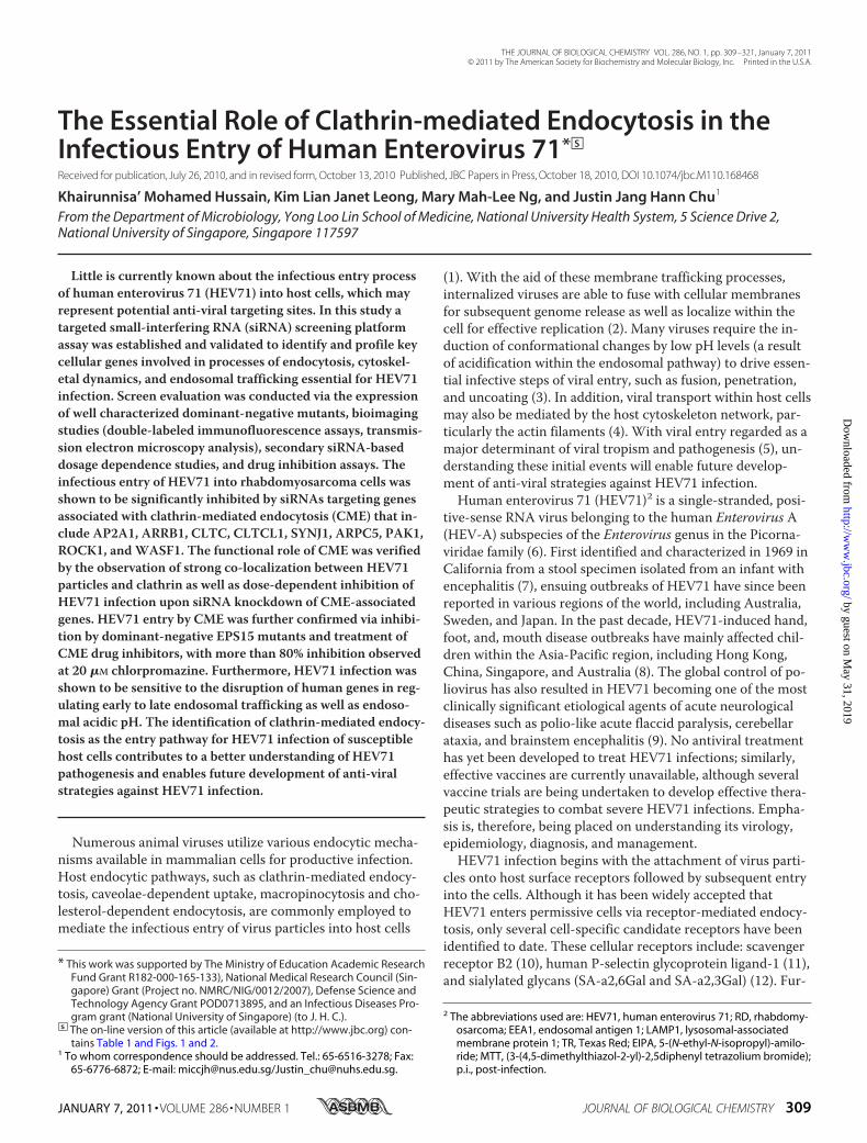
date the findings obtained from the primary screen, siRNAsmart pool-based deconvolution assays targeting several top-hit genes (Fig. 1) were performed. These genes includedAP2A1, ARPC5, CLTC, RAB3A, SYNJ1, and WASF1. siRNAsmart pool deconvolution is necessary to confirm true posi-tive phenotype observed in this screen. 30 nM concentrationsof each specific individual siRNA of the smart pool (four spe-cific siRNAs) directed against each of the respective geneswere transfected into RD cells and subsequently subjected toHEV71 infection. This experimental approach would help toensure that inhibitory effects on HEV71 infection observed inthe primary screen was specific and not due to off-target geneeffects. It can be observed that at least one of the four siRNAsdirected against each specific gene (AP2A1, ARPC5, CLTC,RAB3A, SYNJ1, and WASF1) can result in more than 50%inhibition of HEV71 infection (Fig. 2).
In addition, we have also performed siRNA dosage knock-down of the selected targeted genes (AP2A1, ARPC5, CLTC,RAB3A, SYNJ1, and WASF1) with varying concentrations ofsiRNAs (0, 5, 25, and 50 nM) on RD cells and subjected toHEV71 infection. The data showed obvious dose-dependentinhibition of HEV71 infection with increasing concentrationsof the respective siRNAs targeting AP2A1, ARPC5, CLTC,RAB3A, SYNJ1, and WASF1 upon siRNA transfection intocells (Fig. 3). Western blot detection was also carried out toensure efficient knockdown of specific protein expression(Fig. 3). Indeed, the transfection of cells with siRNAs targetingAP2A1, ARPC5, CLTC, RAB3A, SYNJ1, and WASF1 showeddose-dependent reductions in the levels of the respective pro-teins when compared with the levels in the mock-transfectedcells (Fig. 3). At the concentration of 25 nM of the transfectedsiRNA for the respective proteins, more than 60% reduction(as measured by densitometry) can be observed when com-pared with the mock-transfected samples (Fig. 3). Further-more, the different concentrations of siRNA transfected intocells have minimal cellular cytotoxicity as revealed by the cellviability assays (Fig. 3). Indeed, the data obtained from thesiRNA pool deconvolution and dose-dependent knockdownexperiments may support a functional relationship betweenthese identified genes and their inhibitory roles in the infec-tious entry of HEV71 into cells.Infectious Entry of HEV71 into RD Cells Involved Clathrin-
mediated Endocytosis—To further characterize the involve-ment of clathrin during the initial stages of HEV71 infection,a double-labeled immunofluorescence assay was performed totrack the entry process and cellular localization of HEV71within infected cells at fixed time points (0, 5, 10, 15, 20, and30 min post-infection). At time 0 min p.i., HEV71 particleswere observed predominantly attached to the plasma mem-brane of the cells with little or no co-localization of virus par-ticles with clathrin (Fig. 4a). Strong co-localization of HEV71particles and clathrin (arrows) within the cytoplasm was sub-sequently observed from 5. to 30 min p.i. (Fig. 4). These datamay suggest the involvement of clathrin in the endocytosis ofthe HEV71 particles.Transmission Electron Microscopy Analysis of HEV71 Entry
Process—To visualize synchronized entry of HEV71 at theultrastructural level, RD cells were first incubated withHEV71 (multiplicity of infection of 10) at 4 °C for 1 h. Lowtemperature treatment allows binding of HEV71 to the cellsurface receptors but prevents the internalization of virus par-ticles into the cells. Subsequently, the RD cells were warmedto 37 °C, and the virus-infected cells were processed for trans-mission electron microscopy analysis at appropriate timesafter warming. At 0 min of warming to 37 °C, attachment ofHEV71 particles (gray arrows) along the cell surface of RDcells was observed (Fig. 5a). At 5 min after warm-up, HEV71particles (gray arrows) were observed within invaginations ofthe plasma membrane (Fig. 5b). These invaginations are heav-ily decorated with clathrin molecules and resembled those ofthe clathrin-coated pits (white arrows) (35). After 10 min at37 °C, the HEV71 virus particles (gray arrows) were observedwithin structures typical of clathrin-coated vesicles (whitearrows; Fig. 5c) (35). Single virus particles were contained
FIGURE 2. Validation of siRNA knockdown of genes involved in the in-fectious entry of HEV71. The experiments shown were repeated with thedeconvoluted siRNA sequences (four individual siRNAs) from the Smart-pool. Data displayed are representative of three independent experiments.
Infectious Entry of Human Enterovirus 71
314 JOURNAL OF BIOLOGICAL CHEMISTRY VOLUME 286 • NUMBER 1 • JANUARY 7, 2011
by guest on May 31, 2019
http://ww
w.jbc.org/
Dow
nloaded from

within each of these vesicles. By 15–20 min of warm-up, vesi-cles containing numerous HEV71 virus particles (Fig. 5d, grayarrows) were observed. These virus-containing vesicles arepredominantly localized to the perinuclear region in closeassociation with the endoplasmic reticulum. Thus far, it hasbeen a challenge to visualize the uncoating process of HEV71in endocytic vesicles. It is believed that the acidification of theendocytic vesicles would probably cause the rearrangement ofthe viral capsid proteins and trigger the release of the viralRNA into the cytoplasm for the initiation of HEV71 RNAreplication.Dominant Negative EPS15 Mutants Inhibit HEV71 Entry
into Cells—Molecular inhibitors in the form of dominant-negative mutants were also used to further confirm the role ofclathrin-mediated endocytosis in the infectious entry ofHEV71. The use of dominant-negative mutants may providean alternative way to analyze the specific function of definedpathways within the cells. Eps15, a protein that binds to AP-2
(adaptor protein 2), has been shown to be necessary for inter-nalization through clathrin-coated pits (36). However, thedeletion of the EH domain of Eps15 produced a dominant-negative mutant protein that disrupts the formation of clath-rin-coated pits and inhibits the uptake of transferrin, a cellu-lar marker of clathrin-mediated endocytosis (37).In this study, RD cells were first transfected with either
pEGFP-Eps15�95/295 plasmid (dominant-negative mutant ofEps15 protein), pEGFP-Eps15DIII�2 plasmid (this constructlacks the AP2-binding sites of Eps15 and was used as a controlthat did not inhibit clathrin-mediated endocytosis), orpEGFP-C2 plasmid (coding for green fluorescent protein(GFP) as an internal control) for 48 h (37) before being in-fected with HEV71 at 4 °C for 1 h (to enable virus binding,after which the temperature was shifted rapidly to 37 °C forinternalization) and subsequently processed for immunofluo-rescence staining and microscopic imaging. As shown in Fig.6a, HEV71 particles (stained by anti-HEV71 antibodies conju-
FIGURE 3. Dose-dependent knockdown of genes involved in the infectious entry of HEV71. Gene-specific siRNA against AP2A1 (a), ARPC5 (b), CLTC (c),RAB3A (d), SYNJ1 (e), and WASF1 (f) were transfected into RD cells at different concentrations (0 –50 nM) and subjected to HEV71 infection. Dose-dependentreduction of HEV71 infection can be observed for these selected genes. Histograms represent the inhibition of virus entry determined by the percentagevirus antigen-positive cells (with S.E. bars) against siRNA concentrations. Cell viability upon siRNA transfection was unaffected as represented by the linegraphs. Western blots were also performed after treatment with siRNAs to ensure the knockdown of the specific protein expression. Dose-dependent re-ductions of protein expression were observed for the indicated genes corresponding to the concentrations of the transfected siRNAs (upper panels). At theconcentration of 25 nM transfected siRNA for the respective proteins, more than 60% reduction (as measured by densitometry) can be observed whencompared with the mock-transfected samples. The blots were also re-probed with �-actin-specific antibody. which served as a gel-loading control(lower panels).
Infectious Entry of Human Enterovirus 71
JANUARY 7, 2011 • VOLUME 286 • NUMBER 1 JOURNAL OF BIOLOGICAL CHEMISTRY 315
by guest on May 31, 2019
http://ww
w.jbc.org/
Dow
nloaded from

gated to TR (arrows)) were observed to attach onto theplasma membrane without visible signs of viral entry, withpunctuated staining of the pEGFP-Eps15�95/295 plasmidwithin the cells. In contrast, internalization of HEV71 (arrows,speckled staining) occurred within the cytoplasm of the
pEGFP-Eps15DIII�2 or GFP-expressing cells (Fig. 6, b and c,respectively). These differences were further proven uponenumerating the percentage of viral entry into cells. Althoughthe percentage of viral antigen-positive cells in the pEGFPcontrol and pEGFP-Eps15DIII�2 was similar to that of themock-transfected cells, only �40% of cells transfected withthe dominant negative mutant plasmid (pEGFP-Eps15�95/295) were viral antigen-positive (Fig. 6d). These results pro-vide further evidence that HEV71 entry into cells takes placethrough clathrin-mediated endocytosis.Drug Inhibition Analysis of Entry Pathways—The results
presented above suggested the involvement of a clathrin-me-diated endocytic pathway in HEV71 entry into RD cells. Toaffirm these results, RD cells were pretreated with drugs thatselectively inhibit clathrin-dependent endocytosis (chlor-promazine and cytochalasin B), caveolae-dependent endocy-tosis (filipin, nystatin, and methyl-�-cyclodextrin), and mac-ropinocytosis EIPA (Table 2). Possible drug-induced cytotoxiceffects were assessed by MTT cell viability assays and obser-vation of cellular morphological changes. Minimal cellularcytotoxicity was observed in drug-treated cells throughout thespectra of concentrations used in these experiments (Figs. 7and 8).Clathrin-mediated entry pathway can be inhibited by drugs
such as chlorpromazine and cytochalasin B. Chlorpromazineis a known clathrin-dependent endocytosis inhibitor (38),whereas cytochalasin B induces depolymerization of actinfilaments (39). F-actin dynamics have been shown to be nec-essary for various stages of clathrin-coated vesicle formation,including coated pit formation, constriction, and internaliza-tion (40). Both chlorpromazine and cytochalasin B are specificin inhibiting clathrin-mediated endocytosis (even at the high-est concentrations used in this study) as illustrated by the in-hibition of transferrin (a specific maker for clathrin-mediatedendocytosis) uptake (supplemental Fig. 1). Pretreatment ofRD cells with increasing concentrations of either chlorproma-zine (Fig. 7a) or cytochalasin B (Fig. 7b) revealed significantdose-dependent inhibition of HEV71 infection. More than80% inhibition at 20 �M chlorpromazine and 50% inhibitionat 4 �M cytochalasin B can be observed, further confirmingHEV71 entry via clathrin-mediated endocytosis.To eliminate the involvement of other entry pathways dur-
ing HEV71 infection, drugs known to inhibit caveolae-medi-ated endocytosis and macropinocytosis were also evaluatedon RD cells. Inhibitors of caveolae-dependent endocytosisused in this study include filipin, which disrupts caveolae-mediated endocytosis by binding specifically to cholesterolabundantly found in caveolae (41, 42), nystatin, whichbinds to sterols (42), and methyl-�-cyclodextrin, whichdepletes the cells of cholesterol, thus disrupting caveolaeformation (42). Treatment of filipin (Fig. 8a) and nystatin(Fig. 8b) did not exhibit inhibitory effects on HEV71 infec-tion at all drug concentrations used, whereas cells treatedwith methyl-�-cyclodextrin displayed slight inhibition of�30% at 7.5 mM (Fig. 8c). These results suggest minimalinvolvement of caveolae-mediated endocytosis uponHEV71 infection in RD cells. Furthermore, an inhibitor ofmacropinocytosis, EIPA (43, 44), failed to show inhibitory
FIGURE 4. Bio-imaging analysis of the interaction of clathrin moleculeswith HEV71 particles. A double-labeled immunofluorescence assay wasperformed to track the entry process and cellular localization of HEV71within infected cells at fixed time points. a, no co-localization betweenHEV71 viral particles (arrows) and clathrin molecules was observed at 0-minp.i. b, HEV71 viral particles began co-localizing with clathrin molecules (indi-cated by light gray arrows) at 5 min p.i. c, more co-localization occurred at10-min p.i. d, co-localization was observed forming around the perinuclearregion at 15 min p.i. e, enhanced co-localization was seen at 20 min p.i. f,co-localization between HEV71 viral particles and clathrin molecules wascompleted at 30 min p.i. Cell nuclei were stained blue with DAPI.
FIGURE 5. Ultrastructural analysis of HEV71 infectious entry. To visualizesynchronized HEV71 entry, RD cells were incubated with HEV71 at 4 °C for1 h, after which they were warmed to 37 °C before processing for transmis-sion electron microscopy analysis. a, at 0-min of warming to 37 °C, HEV71particles (indicated by gray arrows) were seen attached to the cell surface.b, at 5 min after warm-up, HEV71 particles were observed within invagina-tions of the plasma membrane (clathrin pits indicated by white arrows). c, at10 min of warm-up, HEV71 particles were seen enclosed within clathrin-coated vesicles. d, at 15–20 min of warm-up, vesicles containing numerousHEV71 virus particles were observed.
Infectious Entry of Human Enterovirus 71
316 JOURNAL OF BIOLOGICAL CHEMISTRY VOLUME 286 • NUMBER 1 • JANUARY 7, 2011
by guest on May 31, 2019
http://ww
w.jbc.org/
Dow
nloaded from

effects on HEV71 infection; instead, the blocking of themacropinocytotic pathway by EIPA seemed to enhanceHEV71 infection (Fig. 8d).Endocytic Trafficking of Internalized HEV71 Particles
within Cells—To further substantiate the role of clathrin-me-diated endocytosis in HEV71 infection, the importance ofendosomal trafficking of internalized HEV71 particles wastracked via microscopic analysis. Within 15 min p.i., a double-labeled immunofluorescence assay with anti-HEV71 VP1 pro-tein and anti-EEA1 antibodies showed co-localization ofHEV71 particles with early endosomes, suggesting that the
virus particles were translocated to the early endosomes afterclathrin-mediated endocytosis (Fig. 9a, upper panels). By 30min p.i., HEV71 particles were found mainly in vesicles thatwere stained with anti-LAMP1 antibodies (Fig. 9b, upper pan-els), suggesting that HEV71 were localized to the late endo-somes by this time point. The fluorescent staining was moreintense at the perinuclear region. Conversely, minimal co-localization was observed between HEV71 viral particles andlate endosomes at 15 min p.i. The majority of the HEV71 par-ticles (arrows) are localized near the plasma membrane of thecell (Fig. 9a, lower panels). Similarly, minimal co-localization
FIGURE 6. Inhibition of HEV71 entry into cells by dominant negative Eps15 mutants. RD cells were first transfected with pEGFP-Eps15�95/295 plasmid,pEGFP-Eps15DIII�2, or pEGFP-C2 plasmid for 48 h before HEV71 infection at 4 °C for 1 h to allow for virus binding. The temperature was subsequentlyshifted rapidly to 37 °C for internalization, and the cells were then processed for immunofluorescence staining and microscopic imaging. a, HEV71 viral par-ticles (arrows) were observed to attach on the surface of cell expressing Eps15 dominant negative mutant of pEGFP-Eps15�95/295. HEV71 viral particleswere observed within the cytoplasm of pEGFP-Esp15DIII�2 (b)- and pEGFP-C2 (c)-expressing cells. The expression of GFP-tagged plasmids emitted thegreen color observed (indicated here as white dots). Cell nuclei were stained blue with DAPI (indicated here in dark gray). d, the histogram represents theinhibition of virus entry determined by the percentage virus antigen-positive cells (with S.E. bars) against pEGFP-Eps15�95/295 plasmid (dominant-nega-tive mutant), pEGFP-Esp15DIII�2, pEGFP-C2 plasmid (internal control), and mock-transfection control. The plots shown are representative of three inde-pendent experiments. The asterisk indicates p values of �0.05 by Student’s t test.
TABLE 2Concentrations and functions of inhibitory drugs used in this study
Drug Concentrations used Function
Chlorpromazine 2, 10, 20, 30 �M Inhibitor of clathrin-dependent endocytosisCytochalasin B 0.2, 1, 2, 3, 4 �M Inhibitor of actin polymerizationFilipin 0.5, 1, 1.5, 2, 3 �g/ml Inhibitor of caveolin-dependent endocytosisNystatin 5, 10, 20, 40 �M Increases permeability of cell membrane of sensitive fungi by sterol bindingMethyl cyclodextrin-�-cyclodextrin 2.5, 5, 7.5, 10 mM Inhibitor of lipid raft synthesis and caveolin-dependent endocytosis5-(N-Ethyl-N-isopropyl)-amiloride 10, 25, 50, 100 �M Selective blocker of Na�/H� antiportBafilomycin A1 0.01, 0.05, 0.1, 0.5, 1 �M Inhibitor of vacuolar H�-ATPaseConcanamycin A 20, 40, 60, 80, 100 nM Inhibitor of acidification of organelles and perforin-mediated cytotoxicity
Infectious Entry of Human Enterovirus 71
JANUARY 7, 2011 • VOLUME 286 • NUMBER 1 JOURNAL OF BIOLOGICAL CHEMISTRY 317
by guest on May 31, 2019
http://ww
w.jbc.org/
Dow
nloaded from

can be observed between HEV71 viral particles and early en-dosomes at 30 min p.i. Most of the HEV71 particles (arrows)were observed as green particles in the overlay micrograph,indicating a the lack of co-localization with the early endo-somes (Fig. 9b, lower panels). Thus, these data further indi-cated that HEV71 particles do transit from early to late endo-somes during the 15–30 min infection period.Some viruses require low endosomal pH, maintained by
vacuolar proton-ATPase, to uncoat and release its viral ge-nome for replication (45). Assays to examine pH-dependententry of HEV71 in the presence of vacuolar proton-ATPaseinhibitors, bafilomycin A1 and concanamycin A, were per-formed. Bafilomycin A1 is a potent and specific inhibitor ofvacuolar proton-ATPase that inhibits endosome and lyso-some acidification (46, 47), whereas concanamycin A inhibitsacidification of organelles and perforin-mediated cytotoxicity(48, 49). Both bafilomycin A1 and concanamycin are highlyspecific in inhibiting endosomal acidification (even at thehighest concentrations used in this study) as illustrated by theabsence of acridine orange (granular orange fluorescence)staining (a specific maker for acidification of endosomes)(supplemental Fig. 2). Minimal cellular cytotoxicity was ob-served in drug-treated cells throughout the spectra of concen-trations used in these experiments (Fig. 10). Pretreatment ofcells with bafilomycin A1 (Fig. 10a) and concanamycin A (Fig.10b) showed dose-dependent inhibition of HEV71 infection,
with more than 60% inhibition at 0.5 �M and 80 nM, respec-tively, thus strongly suggesting that low endosomal pH is re-quired for the infectious entry of HEV71 into RD cells.
DISCUSSION
The application of RNA interference-based screens offersan alternative route to conventional means in identifying cel-lular proteins or components of endocytic pathways that me-diate the infectious entry of HEV71. Despite its recent discov-ery, the application of RNA interference has alreadyprofoundly enhanced the study of large scale loss-of-func-tional gene analysis in a rapid and cost-effective manner. Forthis purpose we have established and validated an RNA inter-ference screening platform assay that allows identification ofhost proteins involved in the endocytic and membrane traf-ficking process mediating the infectious entry of HEV71 intocells. A siRNA library screening using a similar subset of thehuman membrane trafficking library has previously been usedsuccessfully to screen for endocytic genes involved in respira-tory syncytial virus infection (50).In this study clathrin-mediated endocytosis has been iden-
tified as the main pathway of HEV71 internalization, withknockdown of clathrin heavy chains (CLTC and CLTCL1)and AP2A1, a subunit of the AP2 coat assembly protein com-plex linking clathrin to receptors in coated vesicles, signifi-cantly inhibiting virus uptake by up to 60% and more. In addi-tion, knockdown of several other endocytic proteins includingARRB1, ARPC5, PAK1, RAB3A, SYNJ1, ROCK1, and WASF1also significantly reduced the uptake of HEV71 into RD cells(Figs. 1–3 and Table 1).The functional role of clathrin-mediated endocytosis in
mediating HEV71 entry was independently verified by druginhibition assays, ultrastructural analysis, and microscopiccellular localization analysis as well as transfection of cellswith a well characterized dominant-negative mutant form ofEps15. Treatment with chlorpromazine, a known specific in-hibitor of clathrin-mediated endocytosis, exhibited effectiveinhibition of HEV71 infection (Fig. 7a). Co-localization ofHEV71 particles with clathrin molecules could be observedwithin the first 5 min post-infection (Fig. 6). Eps15 is an ac-cessory factor that associates with AP2 complex and is essen-tial for the formation of clathrin-coated pits at the plasmamembrane (51). The dominant-negative form of Eps15 hasbeen shown to effectively block clathrin-mediated endocytosisbut not other endocytic processes by caveolae or macropino-cytosis (37). The lack of co-localization between GFP-taggeddominant negative EPS15 and HEV71 viral particles indicatedthat clathrin-mediated endocytosis was required for HEV71viral entry. To further verify the effect of clathrin knockdownon HEV71 infection, a secondary assay was performed withincreasing siRNA concentrations to knock down CLTC, aclathrin heavy chain gene. A dose-dependent decrease inHEV71 infection as observed, thus, indicated the involvementof clathrin in HEV71 infection (Fig. 3).Furthermore, p-selectin glycoprotein ligand-1 was recently
discovered as a functional receptor for HEV71 in leukocytes(11). Setiadi et al. (52–54) found P-selectin to be rapidly inter-nalized in clathrin-coated pits, thus, enhancing the adhesive
FIGURE 7. Effects of treatment of clathrin-mediated endocytic inhibitorson HEV71-infected RD cells. RD cells were pretreated with varying con-centrations of chlorpromazine (2, 10, 20, and 30 �M) (a) and cytochalasin B(0.2, 1, 2, 3, and 4 �M) (b) for 2 h at 37 °C before HEV71 infection. At 12 h p.i.,the infected cells were processed for immunofluorescence assay. Histo-grams represent the inhibition of virus entry determined by the percentagevirus antigen-positive cells (with S.E. bars) against drug concentrations. Cellviability upon drug treatments was unaffected as represented by the linegraphs. The plots shown are representative of three independent experi-ments. The asterisk indicates p values of �0.05 by Student’s t test. UT, un-treated cells; SC, solvent control.
Infectious Entry of Human Enterovirus 71
318 JOURNAL OF BIOLOGICAL CHEMISTRY VOLUME 286 • NUMBER 1 • JANUARY 7, 2011
by guest on May 31, 2019
http://ww
w.jbc.org/
Dow
nloaded from

function of endothelial cells. Similarly, another recently dis-covered cellular receptor for HEV71, scavenger receptor B2(10), had been earlier found to undergo endocytosis by aclathrin-dependent mechanism (55). These findings furthersubstantiate our present study that HEV71 viral particles en-ter via clathrin-mediated endocytosis.The silencing of the genes PAK1, PIK3CG, PIK3C2G, and
ELKS, involved in PI3K/Akt or JNK or NK-KB signaling path-ways, also caused a reduction in viral infection. It had previ-ously been reported that HEV71-induced VCAM-1 expres-sion via PDGF receptor, PI3K/Akt, p38MAPK, JNK, andNK-KB in vascular smooth muscle cells (56). Silencing ofother genes involved in vesicle transport such as ARFIP2,RAB3A, and RAB6B also led to a decrease in viral infection,indicating the importance of vesicle transport in HEV71 in-fection. These genes can be further analyzed to evaluate theirpotential roles in HEV71 infection.Drugs used to inhibit caveolae-mediated endocytosis are
methyl-�-cyclodextrin, filipin, and nystatin, with treatment ofthe latter two failing to exhibit any inhibitory effects onHEV71 infection at all drug concentrations tested. In contrast,methyl-�-cyclodextrin-treated cells showed slight inhibitionof �30% at 7.5 mM. These results suggested that there wasminimal involvement of caveolae-mediated endocytosisHEV71 infection in RD cells. The depletion but not binding ofcholesterol inhibiting HEV71 infection could be due to theimportance of cholesterol in the formation of endocytic vesi-cles. Grimmer et al. (57) found that cholesterol depletion in-hibited the transport of endosomes to Golgi bodies, and cho-
lesterol is required for the formation of endocytic vesicles.Inhibition of HEV71 infection could, thus, result from defec-tive formation of endocytic vesicles. Furthermore, completeinhibition of infection is highly unlikely, as HEV71 may becapable of entering susceptible cells via alternative pathwaysin the event of their primary route of entry being inhibited.Many viruses are known to exhibit this feature during infec-tious entry into cells or are able to enter cells via multipleroutes, such as Simian virus 40 (caveolar and cholesterol-de-pendent pathways), influenza (both clathrin-dependent andclathrin-independent routes), echovirus 1 (caveolar and dy-namin-2 dependent pathways) (1), and adenovirus (clathrin-mediated endocytosis and macropinocytosis) (58). Neverthe-less, as shown in this study, clathrin-mediated endocytosis isfound to be the primary route of entry for HEV71 into cells.EIPA was used to target the macropinocytotic pathway.
EIPA was previously shown to inhibit propagation of humanrhinovirus 2 and Coxsackie B3 virus in HeLa cells (59). In thisstudy, EIPA failed to inhibit HEV71 infection. On the con-trary, the blocking of macropinocytosis using EIPA seemed toenhance HEV71 infection, possibly due to the activation ofreflex mechanisms in RD cells such that endocytic uptake wasincreased through other pathways. EIPA could also be suffi-ciently nonspecific to be able to trigger the activation of cellu-lar mechanisms, thus, resulting in an increase in virus uptake.In addition, the cytoskeleton, comprising of actin filaments
and microtubules, also plays a dynamic role in endocytic traf-ficking, with both up-and down-regulation of actin or micro-tubule polymerization shown to affect endocytic kinetics (60).
FIGURE 8. Effects of treatment of caveolae-mediated endocytic inhibitors (a– c) and macropinocytic inhibitor (d) on HEV71-infected RD cells.RD cells were pretreated with varying concentrations of filipin (0.5, 1, 1.5, 2, and 3 �g/ml) (a), nystatin (5, 10, 20, and 40 �M) (b), methyl-�-cyclodex-trin (2.5, 5, 7.5, and 10 mM) (c), and EIPA (10, 25, 50, and 100 �M) (d) for 2 h at 37 °C before HEV71 infection. At 12 h p.i., the infected cells were pro-cessed for immunofluorescence assay. Histograms represent the inhibition of virus entry determined by the percentage virus antigen-positive cells(with S.E. bars) against drug concentrations. Cell viability upon drug treatments was unaffected as represented by the line graphs. The plots shownare representative of three independent experiments. The asterisk indicates p values of �0.05 by Student’s t test. UT, untreated cells; SC, solventcontrol.
Infectious Entry of Human Enterovirus 71
JANUARY 7, 2011 • VOLUME 286 • NUMBER 1 JOURNAL OF BIOLOGICAL CHEMISTRY 319
by guest on May 31, 2019
http://ww
w.jbc.org/
Dow
nloaded from

Actin filaments are required for the initial uptake of ligandsvia clathrin-coated pits and subsequent degradative pathway,whereas microtubules are involved in maintaining the endo-somal traffic between peripheral early and late endosomes(60, 61). Actin cytoskeleton is shown to be closely associatedwith clathrin-coated pits, and actin polymerization may beinvolved in moving endocytic vesicles into cytosol after theyare pinched off from the plasma membrane (62). The actinbinding molecular motor, myosin VI, was also recently shownto mediate clathrin endocytosis (63). Disruption of actin fila-ments can have a dramatic effect on receptor-mediated endo-cytosis (60). In this study, the siRNA knockdown of ARPC5,ARRB1, and WASF1 genes that are important in actin poly-merization have resulted in the reduction of HEV71 infection.Arrestin B1 (ARRB1) is involved in the desensitization of re-ceptors by targeting them to clathrin-coated vesicles througha RhoA and actin-dependent mechanism (30). WASF1 is animportant downstream effector molecule involved in thetransmission of signals from tyrosine kinase receptors andsmall GTPases to the actin cytoskeleton. WASF family mem-
bers also play important roles late in clathrin-coated pit for-mation by coupling to the ARP2/3 actin-regulating complexand may act to move the coated vesicles through the cell (31).Therefore, these results suggest a potential role of these genesin regulating HEV71 endocytosis upon binding to putativecellular receptors. The involvement of actin in mediatingHEV71 entry was further confirmed by treatment with cy-tochalasin B. Cytochalasin B, an actin-disrupting drug, specif-ically affects the actin cytoskeleton by preventing its properpolymerization into microfilaments and promoting microfila-ment disassembly (64). Disruption of actin filaments wasshown to inhibit HEV71 infection in a dose-dependentmanner.Some viruses such as Semliki Forest virus, vesicular stoma-
titis virus, vaccinia virus, adenovirus, and poliovirus utilize thepH gradient to promote entry into their host cells. Polioviruspermeabilizes human cells during virus entry through the un-coating of virus particles and the functioning of the vacuolarproton-ATPase (65, 66). Inhibition of vacuolar proton-AT-Pase would disrupt endosomal pH (45). In this study, cellstreated with vacuolar proton-ATPase inhibitors bafilomycinA1 and concanamycin A1 showed dose-dependent reductionsin infection, thus understating the importance of low endoso-mal pH in HEV71 infection.
FIGURE 9. Endocytic trafficking of internalized HEV71 particles withincells. a, shown is co-localization between HEV71 viral particles and earlyendosomes (arrows) at 15 min p.i. (as observed in the Overlay, last upperpanel). The first upper panel represents the DAPI-stained cell nuclei, the sec-ond upper panel represents FITC-stained HEV71 viral particles, and the thirdupper panel represents TR-stained early endosomes. No co-localization wasobserved between HEV71 viral particles (second lower panel) and late endo-somes (third lower panel) at 15 min p.i. (as observed in the Overlay, last lowerpanel). b, co-localization between HEV71 viral particles and late endosomes(arrows) at 30 min p.i. (as observed in the Overlay, last upper panel) is shown.The first upper panel represents the DAPI-stained cell nuclei, the second up-per panel represents FITC-stained HEV71 viral particles, and the third upperpanel represents TR-stained late endosomes. No co-localization was ob-served between HEV71 viral particles (second lower panel) and early endo-somes (third lower panel) at 30 min p.i. (as observed in the Overlay, last lowerpanel).
FIGURE 10. Effects of treatment of vacuolar proton-ATPase inhibitors onHEV71-infected RD cells. RD cells were pretreated with varying concentra-tions of bafilomycin A1 (0.01, 0.05, 0.1, 0.5, and 1 �M) (a) and concanamycinA (20, 40, 60, 80, and 100 nM) (b) for 2 h at 37 °C before HEV71 infection. At12 h p.i., the infected cells were processed for immunofluorescence assay.Histograms represent the inhibition of virus entry determined by the per-centage virus antigen-positive cells (with S.E. bars) against drug concentra-tions. Cell viability upon drug treatments was unaffected as represented bythe line graphs. The plots shown are representative of three independentexperiments. The asterisk indicates p values of �0.05 by Student’s t test. UT,untreated cells; SC, solvent control.
Infectious Entry of Human Enterovirus 71
320 JOURNAL OF BIOLOGICAL CHEMISTRY VOLUME 286 • NUMBER 1 • JANUARY 7, 2011
by guest on May 31, 2019
http://ww
w.jbc.org/
Dow
nloaded from
This current work has highlighted the power of using spe-cific subset siRNA libraries to identify important cellulargenes and pathways that mediate the process of endocytosisand endocytic trafficking of HEV71 infection. The study hasprovided much in-depth analysis of human genes that is es-sential for endocytosis as well as the endocytic trafficking ofinternalized HEV71 for productive infection. Understandingthese processes may allow specific cellular pathways or mo-lecular mechanisms to be targeted pharmacologically to in-hibit the entry of HEV71 that uses the route for infection.Furthermore, the development of drugs, dominant negativemutants, and RNAi therapeutic approach targeting virus en-try can be effective in disease intervention of HEV71 or otherrelated enteroviruses. Indeed, the current screening assay canbe adapted and optimized for the screening of putative anti-viral agents and pharmacological compounds against HEV71infection.
REFERENCES1. Marsh, M., and Helenius, A. (2006) Cell 124, 729–7402. Sieczkarski, S. B., and Whittaker, G. R. (2002) J. Gen. Virol. 83,
1535–15453. Harrison, S. C. (2008) Nat. Struct. Mol. Biol. 15, 690–6984. Radtke, K., Dohner, K., and Sodeik, B. (2006) Cell. Microbiol. 8, 387–4005. Isaacson, M. K., Juckem, L. K., and Compton, T. (2008) Curr. Top. Mi-
crobiol. Immunol. 325, 85–1006. Palacios, G., and Oberste, M. S. (2005) J. Neurovirol. 11, 424–4337. Schmidt, N. J., Lennette, E. H., and Ho, H. H. (1974) J. Infect. Dis. 129,
304–3098. McMinn, P. C. (2002) FEMS Microbiol. Rev. 26, 91–1079. Nolan, M. A., Craig, M. E., Lahra, M. M., Rawlinson, W. D., Prager,
P. C., Williams, G. D., Bye, A. M., and Andrews, P. I. (2003) Neurology60, 1651–1656
10. Yamayoshi, S., Yamashita, Y., Li, J., Hanagata, N., Minowa, T., Take-mura, T., and Koike, S. (2009) Nat. Med. 15, 798–801
11. Nishimura, Y., Shimojima, M., Tano, Y., Miyamura, T., Wakita, T., andShimizu, H. (2009) Nat. Med. 15, 794–797
12. Yang, B., Chuang, H., and Yang, K. D. (2009) Virol. J. 6, 141–14613. Bergelson, J. M. (2008) Trends Microbiol. 16, 44–4714. Zeichhardt, H., Wetz, K., Willingmann, P., and Habermehl, K. O. (1985)
J. Gen. Virol. 66, 483–49215. Perez, L., and Carrasco, L. (1993) J. Virol. 67, 4543–454816. DeTulleo, L., and Kirchhausen, T. (1998) EMBO J. 17, 4585–459317. Marjomaki, V., Pietiainen, V., Matilainen, H., Upla, P., Ivaska, J., Nissi-
nen, L., Reunanen, H., Huttunen, P., Hyypia, T., and Heino, J. (2002)J. Virol. 76, 1856–1865
18. Pietiainen, V., Marjomaki, V., Upla, P., Pelkmans, L., Helenius, A., andHyypia, T. (2004)Mol. Biol. Cell 15, 4911–4925
19. Karjalainen, M., Kakkonen., E., Upla, P., Paloranta, H., Kankaanpaa, P.,Liberali, P., Renkema, G. H., Hyypia, T., Heino, J., and Marjomaki, V.(2008)Mol. Biol. Cell 19, 2857–2869
20. Simpson, K. J., Selfors, L. M., Bui, J., Reynolds, A., Leake, D., Khvorova,A., and Brugge, J. S. (2008) Nat. Cell Biol. 10, 1027–1038
21. Reynolds, A., Leake, D., Boese, Q., Scaringe, S., Marshall, W. S., and Kh-vorova, A. (2004) Nat. Biotechnol. 22, 326–330
22. Chu, J. J., and Yang, P. L. (2007) Proc. Natl. Acad. Sci. U.S.A. 104,3520–3525
23. Zhang, J. H., Chung, T. D., and Oldenburg, K. R. (1999) J. Biomol.Screen. 4, 67–73
24. Traub, L. M. (2005) Biochim. Biophys. Acta 1744, 415–43725. Young, A. (2007) Semin. Cell Dev. Biol. 18, 448–45826. Greber, U. F. (2002) Cell. Mol. Life Sci. 59, 608–62627. Geppert, M., Goda, Y., Stevens, C. F., and Sudhof, T. C. (1997) Nature
387, 810–81428. Opdam, F. J., Echard, A., Croes, H. J.., van den Hurk, J. A., van de Vor-
stenbosch, R. A., Ginsel, L. A., Goud, B., and Fransen, J. A. M. (2000)J. Cell Sci. 113, 2725–2735
29. Welch, M. D., DePace, A. H., Verma, S., Iwamatsu, A., and Mitchison,T. J. (1997) J. Cell Biol. 138, 375–384
30. Barnes, W. G., Reiter, E., Violin, J. D., Ren, X. R., Milligan, G., andLefkowitz, R. J. (2005) J. Biol. Chem. 280, 8041–8050
31. Merrifield, C. J., Qualmann, B., Kessels, M. M., and Almers, W. (2004)Eur. J. Cell Biol. 83, 13–18
32. McPherson, P. S., Garcia, E. P., Slepnev, V. I., David, C., Zhang, X.,Grabs, D., Sossin, W. S., Bauerfeind, R., Nemoto, Y., and De Camilli, P.(1996) Nature 379, 353–357
33. Cremona, O., Di Paolo, G., Wenk, M. R., Luthi, A., Kim, W. T., Takei,K., Daniell, L., Nemoto, Y., Shears, S. B., Flavell, R. A., McCormick,D. A., and De Camilli, P. (1999) Cell 99, 179–188
34. Monier, S., Jollivet, F., Janoueix-Lerosey, I., Johannes, L., and Goud, B.(2002) Traffic 3, 289–297
35. Maupin, P., and Pollard, T. D. (1983) J. Cell Biol. 96, 51–6236. Benmerah, A., Lamaze, C., Begue, B., Schmid, S. L., Dautry-Varsat, A.,
and Cerf-Bensussan, N. (1998) J. Cell Biol. 140, 1055–106237. Benmerah, A., Bayrou, M., Cerf-Bensussan, N., and Dautry-Varsat, A.
(1999) J. Cell Sci. 112, 1303–131138. Wang, L. H., Rothberg, K. G., and Anderson, R. G. (1993) J. Cell Biol.
123, 1107–111739. Sampath, P., and Pollard, T. D. (1991) Biochemistry 30, 1973–198040. Yarar, D., Waterman-Storer, C. M., and Schmid, S. L. (2005)Mol. Biol.
Cell 16, 964–97541. Rothberg, K. G., Ying, Y. S., Kamen, B. A., and Anderson, R. G. W.
(1990) J. Cell Biol. 111, 2931–293842. Rothberg, K. G., Heuser, J. E., Donzell, W. C., Ying, Y. S., Glenney, J. R.,
and Anderson, R. G. (1992) Cell 68, 673–68243. Swanson, J. A., and Watts, C. (1995) Trends Cell Biol. 5, 424–42844. Meier, O., Boucke, K., Hammer, S. V., Keller, S., Stidwill, R. P., Hemmi,
S., and Greber, U. F. (2002) J. Cell Biol. 158, 1119–113145. Carrasco, L. (1994) FEBS Lett. 350, 151–15446. Marshansky, V., and Futai, M. (2008) Curr. Opin. Cell Biol. 20, 415–42647. Gagliardi, S., Rees, M., and Farina, C. (1999) Curr. Med. Chem. 6,
1197–121248. Drose, S., Bindseil, K. U., Bowman, E. J., Siebers, A., Zeeck, A., and Alt-
endorf, K. (1993) Biochemistry 32, 3902–390649. Huss, M., Ingenhorst, G., Konig, S., Gassel, M., Drose, S., Zeeck, A., Alt-
endorf, K., and Wieczorek, H. (2002) J. Biol. Chem. 277, 40544–4054850. Kolokoltsov, A. A., Deniger, D., Fleming, E. H., Roberts, N. J., Jr.,
Karpilow, J. M., and Davey, R. A. (2007) J. Virol. 81, 7786–780051. Schmid, E. M., Ford, M. G., Burtey, A., Praefcke, G. J., Peak-Chew, S. Y.,
Mills, I. G., Benmerah, A., and McMahon, H. T. (2006) PloS Biol. 4, e26252. Setiadi, H., Disdier, M., Green, S. A., Canfield, W. M., and McEver, R. P.
(1995) J. Biol. Chem. 270, 26818–2682653. Setiadi, H., Sedgewick, G., Erlandsen, S. L., and McEver, R. P. (1998)
J. Cell Biol. 142, 859–87154. Setiadi, H., and McEver, R. P. (2003) J. Cell Biol. 163, 1385–139555. Eckhardt, E. R., Cai, L., Shetty, S., Zhao, Z., Szanto, A., Webb, N. R., and
Van der Westhuyzen, D. R. (2006) J. Biol. Chem. 281, 4348–435356. Tung, W. H., Sun, C. C., Hsieh, H. L., Wang, S. W., Horng, J. T., and
Yang, C. M. (2007) Cell. Signal. 19, 2127–213757. Grimmer, S., Spilsberg, B., Hanada, K., and Sandvig, K. (2006) Traffic 7,
1243–125358. Meier, O., and Greber, U. F. (2004) J. Gene Med. 6, S152–S16359. Harrison, D. N., Gazina, E. V., Purcell, D. F., Anderson, D. A., and Pe-
trou, S. (2008) J. Virol. 82, 1465–147360. Soldati, T., and Schliwa, M. (2006) Nat. Rev. Mol. Cell Biol. 7, 897–90861. Durrbach, A., Louvard, D., and Coudrier, E. (1996) J. Cell Sci. 109, 457–46562. Qualmann, B., and Kessels, M. M. (2002) Int. Rev. Cytol. 220, 93–14463. Buss, F., Luzio, J. P., and Kendrick-Jones, J. (2001) FEBS Lett. 508,
295–29964. Flanagan, M. D., and Lin, S. (1980) J. Biol. Chem. 255, 835–83865. Madshus, I. H., Olsnes, S., and Sandvig, K. (1984) EMBO J. 3, 1945–195066. Madshus, I. H., Olsnes, S., and Sandvig, K. (1984) J. Cell Biol. 98,
1194–1200
Infectious Entry of Human Enterovirus 71
JANUARY 7, 2011 • VOLUME 286 • NUMBER 1 JOURNAL OF BIOLOGICAL CHEMISTRY 321
by guest on May 31, 2019
http://ww
w.jbc.org/
Dow
nloaded from

Jang Hann ChuKhairunnisa' Mohamed Hussain, Kim Lian Janet Leong, Mary Mah-Lee Ng and Justin
Human Enterovirus 71The Essential Role of Clathrin-mediated Endocytosis in the Infectious Entry of
doi: 10.1074/jbc.M110.168468 originally published online October 18, 20102011, 286:309-321.J. Biol. Chem.
10.1074/jbc.M110.168468Access the most updated version of this article at doi:
Alerts:
When a correction for this article is posted•
When this article is cited•
to choose from all of JBC's e-mail alertsClick here
Supplemental material:
http://www.jbc.org/content/suppl/2010/10/18/M110.168468.DC1
http://www.jbc.org/content/286/1/309.full.html#ref-list-1
This article cites 66 references, 27 of which can be accessed free at
by guest on May 31, 2019
http://ww
w.jbc.org/
Dow
nloaded from