calcium-oxidantsignalingnetworkregulates amp ... amp-activatedproteinkinase(ampk)activationupon...
TRANSCRIPT

Calcium-Oxidant Signaling Network RegulatesAMP-activated Protein Kinase (AMPK) Activation uponMatrix Deprivation*
Received for publication, April 8, 2016, and in revised form, May 2, 2016 Published, JBC Papers in Press, May 11, 2016, DOI 10.1074/jbc.M116.731257
Ananthalakshmy Sundararaman‡1, Usha Amirtham§, and Annapoorni Rangarajan‡2
From the ‡Department of Molecular Reproduction, Development and Genetics, Indian Institute of Science, Bangalore-560012and the §Department of Pathology, Kidwai Memorial Institute of Oncology, Bangalore-560030, India
The AMP-activated protein kinase (AMPK) has recently beenimplicated in anoikis resistance. However, the molecular mech-anisms that activate AMPK upon matrix detachment remainunexplored. In this study, we show that AMPK activation is arapid and sustained phenomenon upon matrix deprivation,whereas re-attachment to the matrix leads to its dephosphory-lation and inactivation. Because matrix detachment leads to lossof integrin signaling, we investigated whether integrin signalingnegatively regulates AMPK activation. However, modulation offocal adhesion kinase or Src, the major downstream compo-nents of integrin signaling, failed to cause a correspondingchange in AMPK signaling. Further investigations revealed thatthe upstream AMPK kinases liver kinase B1 (LKB1) and Ca2�/calmodulin-dependent protein kinase kinase � (CaMKK�) con-tribute to AMPK activation upon detachment. In LKB1-defi-cient cells, we found AMPK activation to be predominantlydependent on CaMKK�. We observed no change in ATP levelsunder detached conditions at early time points suggesting thatrapid AMPK activation upon detachment was not triggered byenergy stress. We demonstrate that matrix deprivation leads to aspike in intracellular calcium as well as oxidant signaling, andboth these intracellular messengers contribute to rapid AMPKactivation upon detachment. We further show that endoplasmicreticulum calcium release-induced store-operated calciumentry contributes to intracellular calcium increase, leading toreactive oxygen species production, and AMPK activation. Weadditionally show that the LKB1/CaMKK-AMPK axis and intra-cellular calcium levels play a critical role in anchorage-indepen-dent cancer sphere formation. Thus, the Ca2�/reactive oxygenspecies-triggered LKB1/CaMKK-AMPK signaling cascade mayprovide a quick, adaptable switch to promote survival of metas-tasizing cancer cells.
Epithelial cells are known to be dependent on interactionwith specific extracellular matrix components for their survival
(1). Lack of matrix attachment triggers a form of caspase-me-diated apoptotic cell death called anoikis (2). Anoikis serves as aformidable barrier to metastasis. Cancer cells overcome anoikisthrough various genetic and epigenetic changes that sustain thesurvival pathways even under detached conditions (3). Duringmetastasis, cancer cells leaving the primary tumor site aredeprived of matrix attachment in circulation, while they may beexposed to foreign matrix components at the secondary sitethat may not be conducive for attachment (4). Besides, duringthe initial stages, glandular epithelial cancer cells that are shedinto the lumen of the duct or lobule resulting in luminal fillingare also matrix-deprived. Therefore, understanding the molec-ular mechanisms that promote cancer cell survival undermatrix-deprived conditions becomes relevant. Recent studieshave thrown light on the metabolic alterations in extracellularmatrix (ECM)3-deprived cells. Anchorage deprivation has beenlinked to reduced glucose uptake and increased ROS levels (5).Recent work from our laboratory has shown that matrix-de-prived cells activate a central metabolic regulator AMP-acti-vated protein kinase (AMPK), which contributes to anoikisresistance through phosphorylation of PEA15 (6). Moreover,AMPK-mediated suppression of mTORC1 (7) and mainte-nance of NADPH homeostasis by inhibition of ACC (8) con-tribute to anoikis resistance. However, the molecular mecha-nisms that lead to AMPK activation in the context of loss ofmatrix attachment remain unexplored.
Cell attachment to extracellular matrix is mediated by a fam-ily of adhesion proteins called integrins. Integrins are impli-cated in tumor proliferation, survival, invasion, and migration.These transmembrane proteins are heterodimers composed of� and � chains. When attached to ECM proteins like collagen,elastin, laminin, or fibronectin, integrins cluster on the mem-brane and recruit various signaling and adaptor proteins toform focal adhesions. The formation of focal adhesions lead toactivation of focal adhesion kinases (FAK), Src family kinases,
* This work was supported by grants from the Wellcome Trust-Department ofBiotechnology (DBT)-India Alliance (IA) Senior Research Fellowship500112/Z/09/Z (to A. R.) and in part from the DBT-IISc Partnership Pro-gramme. The authors declare that they have no conflicts of interest withthe contents of this article.Author’s Choice—Final version free via Creative Commons CC-BY license.
1 Recipient of CSIR-SPM Fellowship SPM-07/0079/(0094)/10 from the Councilfor Scientific and Industrial Research.
2 To whom correspondence should be addressed. Tel.: 91-80-22933263; Fax:91-80-23600999; E-mail: [email protected].
3 The abbreviations used are: ECM, extracellular matrix; AMPK, AMP-activatedprotein kinase; ROS, reactive oxygen species; CaMKK, Ca2�/calmodulin-de-pendent protein kinase kinase; LKB1, liver kinase B1; BAPTA, 1,2-bis(o-aminophenoxy)ethane-N,N,N�,N�-tetraacetic acid; MCI-186, 3-methyl-1-phenyl-2-pyrazolin-5-one; STO-609, 7-oxo-7H-benzimidazo[2,1-a]-benz[de]isoquinoline-3-carboxylic acid; NAC, N-acetyl cysteine; DCFDA,2�,7�-dichlorofluorescein diacetate; RGD, arginylglycylaspartic acid;ANOVA, analysis of variance; ER, endoplasmic reticulum; SOCE, store-oper-ated calcium entry; ITPR, inositol triphosphate receptor; 2-APB, 2-amino-ethoxydiphenyl borate; FAK, focal adhesion kinase; IHC, immunohisto-chemistry; ACC, acetyl-CoA carboxylase; pACC, phospho-ACC.
THE JOURNAL OF BIOLOGICAL CHEMISTRY VOL. 291, NO. 28, pp. 14410 –14429, July 8, 2016Author’s Choice © 2016 by The American Society for Biochemistry and Molecular Biology, Inc. Published in the U.S.A.
crossmark
14410 JOURNAL OF BIOLOGICAL CHEMISTRY VOLUME 291 • NUMBER 28 • JULY 8, 2016
by guest on June 7, 2018http://w
ww
.jbc.org/D
ownloaded from
and p130CRK associated substrate (p130CAS) (9). Integrin-me-diated signaling plays a central role in suppressing apoptosis inadherent cells; loss of integrin downstream signaling throughFAK and Src has been implicated as a major cause of anoikis(10). Although matrix deprivation leads to loss of integrin sig-naling on the one hand and AMPK activation on the otherhand, the cross-talk between these two signaling pathways hasnot been explored.
AMPK has been described as the “fuel gauge of the cell.”AMPK is regulated by phosphorylation at a key threonine res-idue at position 172 in the activation loop of the � subunit (11).Lowered cellular ATP levels and an increase in AMP are knownto activate AMPK allosterically and prevent its dephosphoryla-tion at Thr-172. Two upstream kinases, LKB1 and CaMKK�,are known to majorly phosphorylate AMPK at Thr-172 andactivate it. LKB1 is known to mediate AMPK activation underconditions of energy stress. While LKB1 is known to mediateAMPK activation under conditions of energy stress, CaMKK�activates AMPK in an AMP-independent manner, in responseto an increase in cytosolic calcium (12).
Activation of AMPK can also occur in response to oxidativestress (13). However, the mechanisms that activate AMPK inthis context remain unclear. Oxidative stress can inducechanges in the AMP/ATP ratio, leading to AMPK activation(14). Conversely, AMP-independent mechanisms like store-operated calcium entry (SOCE), in response to oxidative stress,are also known to activate AMPK (15). ROS and calcium arethus known to regulate AMPK in a context-dependent manner.
In this study, we demonstrate that AMPK is activated in arapid and sustained manner upon matrix deprivation. We findevidence to support a role for both upstream kinases LKB1 andCaMKK� in the activation of AMPK upon matrix deprivation,whereas LKB1-deficient cells depend predominantly onCaMKK�. We further show an increase in calcium and ROSlevels under detached conditions, and these signaling mole-cules contribute to rapid AMPK activation in this context. Wefurther demonstrate for the first time a calcium surge-depen-dent increase in ROS levels upon detachment. We also find thatthe AMPK signaling pathway is active in breast cancer patients,and there is a positive correlation between LKB1 and pACClevels suggesting AMPK activation through LKB1. Thus, thisstudy uncovers a novel mechanism for AMPK activation uponmatrix deprivation, which majorly depends on the intracellularcalcium and oxidant signaling.
Results
Rapid and Sustained Activation of AMPK upon De-tachment—Recent studies (7, 8, 25), including work from ourlaboratory (6), have identified AMPK activation upon matrixdeprivation and its role in anoikis. To begin to understand themolecular mechanisms that lead to AMPK activation on matrixdeprivation, we first investigated the status of AMPK signalingin MDA-MB 231 breast cancer cells upon detachment by cul-turing cells for various time periods in suspension. We per-formed immunoblotting to study the levels of phosphorylationof AMPK�, using an AMPK� Thr-172-phosphospecific anti-body, as a measure of AMPK activation (12). We found thatMDA-MB 231 cells showed elevated pAMPK� Thr-172 levels
as early as 10 min following detachment, and this was sustainedeven at 24 h of detachment (Fig. 1A). Previously we and othershave shown elevated pAMPK levels at 24 h following detach-ment as well as in cancer spheres generated in 7 days of suspen-sion culture (5, 6, 25). Together, these data revealed that matrixdetachment leads to rapid and sustained activation of AMPK.
To test whether the rapid activation of AMPK upon matrixdeprivation is cell line-specific, we took cancer cell lines fromdifferent tissues, such as breast (MCF7), cervix (HeLa S3), lung(A549), melanoma (G361) and human embryonic kidney (HEK293T), and subjected them to detachment (suspension culture)for 10 min. All of the tested cell lysates showed an increase inthe levels of pAMPK� under detached conditions (Fig. 1B).Total AMPK levels remained unchanged (Fig. 1B). To furtherconfirm whether the increase in phosphorylation of AMPK�Thr-172 also corresponds with an increase in its activity, wemeasured the phosphorylation of its downstream target,ACC (26). We observed that the levels of pACC (Ser-79)were correspondingly increased under detached conditionsin all these cell lines (Fig. 1B). This suggested that the rapidactivation of AMPK upon detachment is not a cell line-spe-cific phenomenon.
To further confirm the rapid activation of AMPK upondetachment, we undertook various approaches to gauge AMPKactivity in cells cultured under attached and detached condi-tions. For most assays on rapid activation of AMPK, we chose tosubject cells to 10 min of suspension culture followingtrypsinization prior to harvesting, unless mentioned otherwise.Immunocytochemical analysis revealed higher intensity of sig-nal for both pAMPK� and pACC under detached conditions(Fig. 1C). In addition, we performed in vitro kinase assay withAMPK immunoprecipitated from cells grown under bothattached and detached conditions using AMARA as the sub-strate peptide (27). We observed an almost 10-fold higherAMPK activity under detached conditions compared withattached culture (Fig. 1D). In yet another independent assay,cells stably expressing the AMPK activity reporter FRET con-struct AMPK-AR (17) were cultured under attached anddetached conditions, and FRET ratio was measured. Wedetected a higher FRET ratio in detached conditions indicatinghigher AMPK activity (Fig. 1E). Thus, these data confirmedrapid AMPK activation within 10 min upon cell detachment.
To address whether AMPK activation was dependent on anyspecific mode of detachment, because trypsin treatment alsoleads to the cleavage of several surface receptors besides caus-ing cell detachment, we used multiple approaches to detachcells, including cell dissociation using TrypLE (28) andmechanical detachment (19, 21, 22). pAMPK� levels wereobserved to be higher in detached cells compared with attachedconditions in all the different modes tested (Fig. 1F). Thus,AMPK activation under detached conditions is likely to be inde-pendent of the mode of detachment.
To understand whether matrix detachment-triggeredAMPK activation reverses when cells re-attach, we alloweddetached cells to re-attach on tissue culture dishes. Both immu-noblotting (Fig. 1G) and immunocytochemistry (Fig. 1H) con-firmed that indeed AMPK activation is reversed on re-attach-ment (Fig. 1, G and H). Taken together, these data revealed that
AMPK Activation upon Matrix Deprivation
JULY 8, 2016 • VOLUME 291 • NUMBER 28 JOURNAL OF BIOLOGICAL CHEMISTRY 14411
by guest on June 7, 2018http://w
ww
.jbc.org/D
ownloaded from

AMPK activation following cell detachment occurs rapidly andis independent of the mode of detachment or cell type, whereasmatrix attachment leads to its inactivation.
AMPK Phosphorylation Is Independent of FAK and SrcSignaling—Because detachment leads to a rapid loss of integrinsignaling (29) and concomitant AMPK activation, we exploreda possible negative cross-talk between integrin signaling andAMPK. We used integrin-induced autophosphorylation ofFAK at tyrosine 397 (pFAKTyr-397) or its phosphorylation by Srcat tyrosine 925 (pFAKTyr-925) as a measure of integrin signaling
(30). We found that although AMPK was active (as indicatedby pAMPK� and pACC levels) under detached conditions,pFAKTyr-397 as well as pFAKTyr-925 levels were significantlyreduced under detached conditions in HEK 293T (Fig. 2A) andMDA-MB 231 (Fig. 2B) cells. Total FAK levels remained similarunder the two conditions. These results confirmed that underattached conditions, when integrin signaling is high, AMPKactivity is low, although the reverse holds true in suspension.
To understand whether integrin signaling has a causal role toplay in the modulation of AMPK phosphorylation, we inhibited
AMPK Activation upon Matrix Deprivation
14412 JOURNAL OF BIOLOGICAL CHEMISTRY VOLUME 291 • NUMBER 28 • JULY 8, 2016
by guest on June 7, 2018http://w
ww
.jbc.org/D
ownloaded from
FAK with PZ-0117 (31). We found a corresponding reductionin autophosphorylation of FAK (pFAKTyr-397) upon PZ-0117treatment (data not shown). However, we did not find a corre-sponding increase in pAMPK� as would be expected if integrinsignaling through FAK were to contribute to a reduction inAMPK phosphorylation (Fig. 2C). To further explore the role ofintegrin signaling in regulating AMPK activity, we partiallyinhibited the pathway using soluble arginine-glycine-asparticacid (RGD) peptide (32). Addition of RGD to cells for 1 h causeda reduction in pFAKTyr-397 as well as cell rounding in MDA-MB231 cells at higher concentrations (Fig. 2, D and E). However,under these conditions, we could not detect any increase inpAMPK� levels (Fig. 2E). Thus, negative modulation of integrinfunctions under adherent conditions failed to bring aboutAMPK activation. We undertook the same experiment inanother cell type HEK 293T, where RGD (200 �M) treatmentcaused cells to round up within 10 min, and by 30 min most ofthe cells had detached. Immunoblotting for pAMPK� and itsdownstream read-out pACC revealed that cell rounding did notactivate AMPK; however, cell detachment induced by RGDcould activate AMPK (Fig. 2F). These results suggested thatAMPK activation under detached conditions could be indepen-dent of reduction in integrin signaling.
To further address the relationship between integrin signal-ing and AMPK, we induced integrin signaling in suspendedcells using a genetic approach and measured the status ofAMPK signaling in suspension. To do so, we transfected HEK293T cells with either empty vector or constitutively active Srckinase (v-Src)-expressing vector. The v-Src-expressing cellswhen cultured in detached conditions showed high pFAKTyr-925
levels over vector control suggesting that these cells have ele-vated FAK-Src signaling in detached conditions (Fig. 2G).Interestingly, when we cultured HEK 293T cells expressing vec-tor control or v-Src constructs in attached and detached con-ditions and probed for pAMPK� in these lysates, we did notobserve any change in its levels between vector control andv-Src-transfected cells, both under attached (1st and 3rd lanes)and detached conditions (2nd and 4th lanes) (Fig. 2G). Thus,the detachment-induced increase in AMPK activity was unper-turbed by constitutively active v-Src. Taken together, these
results indicated that AMPK modulation between attached anddetached conditions might be independent of integrin signalingthrough FAK and Src kinases.
Upstream Kinases LKB1 and CaMKK� Play a Role in Detach-ment-induced AMPK Activation—Upstream kinases play animportant role in activating AMPK (12). While LKB1 is impli-cated as the major kinase upstream of AMPK under severalstress conditions, CaMKK� is shown to play a major role inAMPK activation in response to elevated calcium levels. Wenext explored the roles of these two major upstream kinases inmatrix deprivation-triggered AMPK activation.
The breast cancer cell line MDA-MB 231 expresses bothLKB1 and CaMKK�. To address the roles of LKB1 andCaMKK� in AMPK activation upon detachment, we generatedcell lines stably expressing knockdown constructs targetingLKB1 and CaMKK�, respectively. We achieved around 75%knockdown of LKB1 and CaMKK� in these cells (Fig. 3A).Knockdown of both the upstream kinases led to a reduction inthe basal levels of pAMPK and pACC under adherent condi-tions (Fig. 3B) as well as under matrix deprivation (Fig. 3C),although the levels of total AMPK and ACC remainedunchanged (Fig. 3, B and C). Additional independent shRNAsequences targeting LKB1 and CaMKK� also showed a reduc-tion in AMPK activity upon detachment (data not shown). Wealso obtained similar reduction in pAMPK� levels upondetachment with the CaMKK�-specific inhibitor, STO-609(Fig. 3D) (33). Together, these data suggested that in MDA-MB231 cells, both LKB1 and CaMKK� contribute to rapid AMPKactivation following cell detachment. To investigate whetherboth of these kinases also contributed to sustained AMPK acti-vation in suspension, we subjected pGIPZ NT (non-targetingshRNA)-, shLKB1-, and shCaMKK�-expressing MDA-MB 231cells to 24 h of suspension culture. Yet again, knockdown ofeither LKB1 or CaMKK� led to a reduction in pAMPK levels(Fig. 3E). Thus, our data indicated that both upstream kinasesLKB1 and CaMKK� contribute to rapid and sustained activa-tion of AMPK signaling under detached conditions inMDA-MB 231 cells.
LKB1 phosphorylation and its cytosolic localization is knownto favor AMPK phosphorylation (34). Therefore, we next asked
FIGURE 1. Rapid and sustained activation of AMPK under detachment-induced stress. A, MDA-MB 231 cells were cultured under adherent conditions ordetached by trypsinization and subjected to suspension for the various indicated times prior to harvesting. The levels of AMPK phosphorylated at threonine172 (pAMPK�) and total AMPK were determined by Western blotting (n � 3). �-Tubulin is used as loading control in all blots. Molecular size markers aredepicted on all blots on the right margin. B, multiple cancer cell lines were cultured under attached (Att), or detached (Det 10 min) conditions. The levels ofpAMPK�, pACC, total AMPK, and ACC were determined by Western blotting. The numbers indicate relative pAMPK�/AMPK ratio and pACC/ACC ratio (n � 4).In all subsequent experiments, unless otherwise mentioned, cells were detached for 10 min. ns, non-significant. C, immunocytochemistry was performed onMDA-MB 231 cells cultured under attached and detached conditions for pAMPK� (Thr-172) and pACC (Ser-79). The representative pictures are maximumintensity projections of confocal Z stack images. Scale bar, 20 �M. Total integrated pixel intensity per cell was quantified for 30 cells in each experiment.Scatterplot depicts fold change in integrated intensity of pAMPK� with each dot constituting one biological experiment normalized to the correspondingattached value (n � 3); *, p � 0.05. Error bars represent � S.E. AU, arbitrary units. D, G361 cells were cultured under attached and detached (10 min) conditions.AMPK was immunoprecipitated from the lysates, and AMPK activity was measured by the incorporation of radioactive phosphate on AMARA peptide. Graphdepicts fold change in AMPK activity (n � 4); **, p � 0.01. Error bars represent � S.E. E, HEK 293T cells stably expressing AMPK activity reporter FRET construct(AMPK-AR) were cultured under attached and detached (10 min) conditions, and the FRET ratio (CY/CC) between acceptor emission (YFP emission) on donorexcitation (cyan excitation) abbreviated as CY to donor emission (cyan emission) on donor excitation (cyan excitation) abbreviated as CC was measured in aspectrofluorometer. Scatterplot depicts FRET ratios expressed as a percentage of attached (n � 3 biological samples each with three technical replicates); *, p �0.05. F, HEK 293T cells were detached using different modes as indicated. Cells were either scraped gently into media or detached with trypsin-EDTA (TE) orTrypLE and resuspended in media for 10 min. Cells were then harvested; pAMPK� and pACC levels were measured by Western blotting (n � 4). The numbersindicate relative pAMPK�/AMPK ratio. G, MDA-MB 231 cells were trypsinized and kept detached for 10 min or allowed to reattach to dishes immediately aftertrypsinization, for a period of 4 h, and the levels of pAMPK� and AMPK were determined by Western blotting (n � 3). Numbers indicate relative pAMPK�/AMPKratio. H, MDA-MB 231 cells cultured under detached conditions for 10 min were compared with those that were trypsinized and allowed to attach in regulartissue culture dishes for 1 and 24 h, respectively, by immunocytochemistry for pAMPK�. The representative pictures are maximum intensity projections of theconfocal Z stack images (n � 3).
AMPK Activation upon Matrix Deprivation
JULY 8, 2016 • VOLUME 291 • NUMBER 28 JOURNAL OF BIOLOGICAL CHEMISTRY 14413
by guest on June 7, 2018http://w
ww
.jbc.org/D
ownloaded from

whether LKB1 phosphorylation and/or localization is modu-lated by detachment. We probed for Ser(P)-428 on LKB1 inboth MDA-MB 231 and MCF7 cells. We observed a rapid
increase in LKB1 phosphorylation in both cell types (Fig. 3F);total LKB1 remained unchanged. LKB1 phosphorylation at Ser-428 is known to cause its nuclear to cytoplasmic translocation
FIGURE 2. AMPK phosphorylation is independent of FAK and Src signaling. HEK 293T cells (A) and MDA-MB 231 cells (B) were cultured under attached (Att)and detached (Det) (10 min) conditions, and levels of indicated total proteins and phosphoproteins were determined by Western blotting (n � 3). C, A549 andMDA-MB 231 cells were cultured under attached conditions and treated for 1 h with FAK inhibitor PZ-0117 (20 �M). pAMPK� and AMPK levels were measuredby Western blotting (n � 3). D, MDA-MB 231 cells cultured under attached conditions were treated with RGD peptide at the concentrations specified for 2 h.Phase contrast images for cell morphology changes were taken at 2 h post-treatment with RGD, and E, cells were lysed and pAMPK�, AMPK, and pFAKTyr-397
levels were measured by Western blotting (n � 5). F, HEK 293T cells were treated with RGD (200 �M) for 10 min (leading to cell rounding) or 30 min (leading tocell detachment). Cells were lysed, and pAMPK�, pACC, total AMPK, and ACC were determined by Western blotting (n � 5). The scatterplot depicts raw pAMPKdensitometric values normalized to the relative tubulin levels in each experiment .*, p � 0.05. Error bars represent � S.E. G, HEK 293T cells were transfected withvector control pcDNA3 or constitutively active v-Src construct. After 24 h, the transfected cells were then cultured in attached or detached conditions.Phosphorylation of FAK by v-Src at Tyr-925 was determined by Western blotting under detached conditions. Levels of pAMPK� and AMPK were probed byWestern blotting under attached and detached conditions (n � 3). Numbers depict relative pAMPK�/AMPK ratio. ns, non-significant; AU, arbitrary units; UT,were treated with vehicle.
AMPK Activation upon Matrix Deprivation
14414 JOURNAL OF BIOLOGICAL CHEMISTRY VOLUME 291 • NUMBER 28 • JULY 8, 2016
by guest on June 7, 2018http://w
ww
.jbc.org/D
ownloaded from

FIGURE 3. Role of upstream kinases in detachment-induced AMPK activation in LKB1 containing MDA-MB 231 cells. A, MDA-MB 231 cells stablyexpressing pGIPZ non-targeting shRNA (NT) or shRNA targeting LKB1 (shLKB1) or shRNA targeting CaMKK� (shCaMKK�) were harvested for immuno-blotting with LKB1 and CaMKK� (n � 5). Numbers indicate relative LKB1/tubulin and CaMKK�/tubulin ratio. B, MDA-MB 231 stably expressing NT, shLKB1,or shCaMKK were cultured under attached conditions. Levels of pAMPK�, pACC, AMPK, and ACC were measured by Western blotting (n � 3). Thenumbers depict relative pAMPK�/AMPK ratio. pAMPK� blot was obtained at high exposure (H.E). C, MDA-MB 231 cells expressing NT, shLKB1, orshCaMKK� were cultured under attached (Att) and detached (Det) conditions. pAMPK� and pACC were measured by Western blotting (n � 7– 8).Numbers indicate relative pAMPK�/tubulin ratio. The scatterplot depicts the fold change in pAMPK�/tubulin ratio of the knockdown cells relative tocontrol NT cells; ***, p � 0.001. Error bars represent � S.E. D, MDA-MB 231 cells were pretreated with STO-609, a specific inhibitor for CaMKK�, for 2 h.Cells were then cultured under attached and detached (10 min) conditions, and pAMPK�, pACC, total AMPK, and ACC levels were measured by Westernblotting (n � 6). The scatterplot depicts the fold change in pAMPK�/tubulin ratio; ***, p � 0.001. Error bars represent � S.E. ns, non-significant. E,MDA-MB 231 cells stably expressing NT, shLKB1, or shCaMKK� were cultured under attached and detached conditions for 24 h. pAMPK� and pACC weremeasured by Western blotting (n � 3). The numbers represent pAMPK�/tubulin ratio. F, MDA-MB 231 and MCF7 were cultured under attached anddetached conditions. pLKB1 (Ser-428) and LKB1 levels were measured by Western blotting (n � 4). Scatterplot depict relative fold change in pLKB1/LKB1ratio. Error bars represent � S.E. G, MCF7 cells were cultured under attached and detached conditions, and immunocytochemistry was performed forLKB1. Representative confocal images of equatorial sections are shown. AU, arbitrary units.
AMPK Activation upon Matrix Deprivation
JULY 8, 2016 • VOLUME 291 • NUMBER 28 JOURNAL OF BIOLOGICAL CHEMISTRY 14415
by guest on June 7, 2018http://w
ww
.jbc.org/D
ownloaded from
(34, 35). We performed immunocytochemistry with LKB1 anti-body on attached and detached cells. Interestingly, we observedthat there is a nuclear to cytoplasmic translocation of LKB1 indetached conditions (Fig. 3G). These together suggest thatmatrix deprivation might trigger AMPK activation in partthrough the phosphorylation and translocation of LKB1.
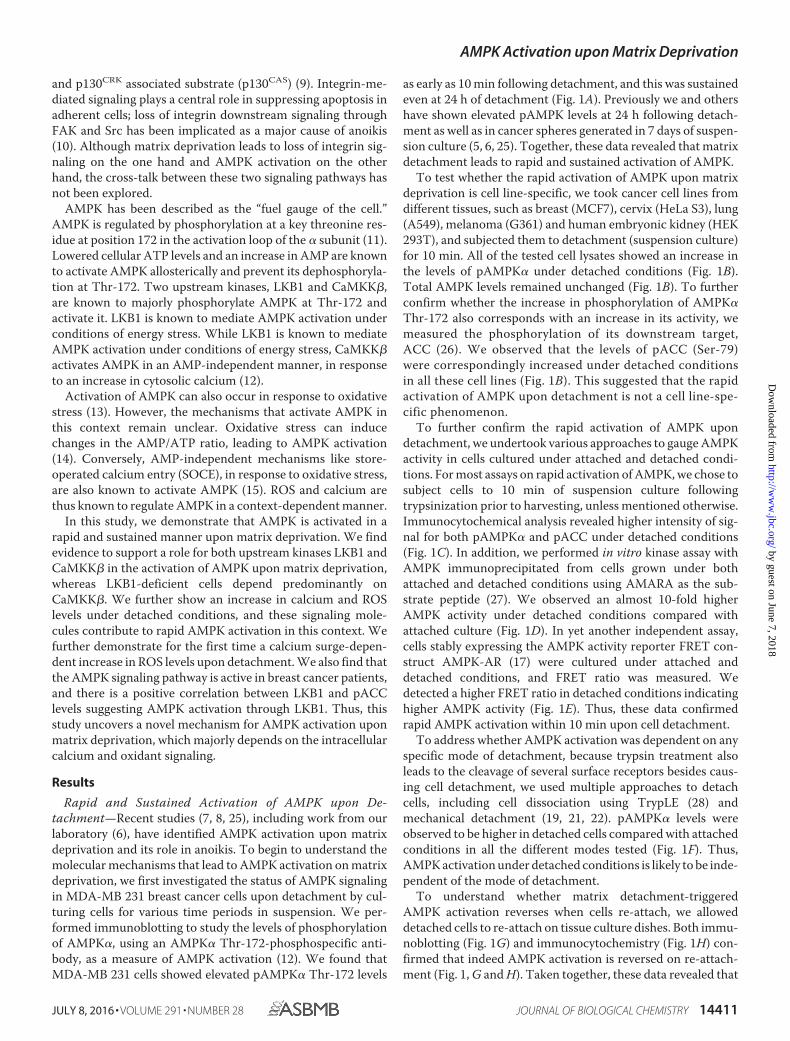
In the panel of cell lines used in this study, three cell lines,G361, A549, and HeLa S3, are known to be LKB1-deficient, andwe confirmed the same by immunoblotting (Fig. 4A). As seenpreviously, these cell lines are also capable of rapidly activatingAMPK upon detachment (Fig. 1B). Hence, we assessed the roleof CaMKK� in matrix deprivation-induced AMPK activationin the LKB1-deficient cells. Treatment of LKB1-deficient G361
cells with STO-609 led to almost complete loss of detectablepAMPK both under adherent and detached conditions (Fig.4B). Similar results were obtained for A549, another LKB1-deficient cell line, in the presence of STO-609 (data not shown).Furthermore, G361 cells stably expressing shRNA againstCaMKK� (Fig. 4C) also showed a dramatic decrease in pAMPKlevels. Additional independent shRNA sequences targetingCaMKK� also showed a reduction in AMPK activity in detach-ment (data not shown), suggesting that in LKB1-deficient cells,under detached conditions, CaMKK� is likely the majorupstream kinase activating AMPK.
Calcium Signaling Contributes to AMPK Activation uponDetachment—To understand the signals that might impinge onupstream kinases LKB1 and CaMKK� upon matrix depriva-tion, we investigated the role of known modulators of AMPKactivity. Increase in AMP levels promotes Thr-172 phosphory-lation of AMPK by upstream kinases (37). Thus, stresses thatlead to a depletion in ATP, and consequent increase in theAMP/ATP ratio, activate AMPK (37). To address whether therapid increase in pAMPK levels within 10 min of detachment isa consequence of reduced ATP levels, we measured ATP usinga bioluminescence assay kit (Sigma). Our results indicated thatATP levels remained unchanged in cells 10 min post-detach-ment (Fig. 5A). However, consistent with a previous report (5),a reduction in ATP levels was observed at 24 h (Fig. 5A). There-fore, the rapid activation of AMPK observed within 10 min maybe independent of changes in cellular ATP levels.
One of the major mechanisms implicated in the literature forAMP-independent activation of AMPK is the calcium-CaMKK� pathway (38). To investigate the possible role of cal-cium in the activation of AMPK upon detachment, we mea-sured intracellular calcium levels in detached cells using theratiometric dye Fura 2 AM (23). Because serine proteases suchas trypsin are known to increase calcium levels independent ofdetachment (39), we have used mechanical detachment by cellscraping (19, 21, 22) instead of routine trypsinization for under-standing the effects of cell detachment on the kinetics of cal-cium and its role in rapid AMPK activation. We undertookcalcium measurements in a time course format using ionomy-cin and EGTA � 1% Triton X-100 at the end of the experimentas controls (Fig. 5B).
Interestingly, in MDA-MB 231 cells loaded with Fura 2 AM,we found that the ratio of fluorescence intensities of Fura 2 AMat 340/380 nm showed a rapid spike upon detachment (Fig. 5C).We observed a similar calcium spike in LKB1-deficient G361cells subjected to detachment (Fig. 5D), together revealing thatdetachment leads to an increase in intracellular calcium levelsin these cells. We confirmed this result further by using analternative approach involving a FRET-based calcium sensor,TN-XXL (17). The FRET ratio was higher in detached cellscompared with attached cells (Fig. 5E), indicating that detachedcells have higher intracellular calcium levels. We also observedan increase in the FRET ratio of 4mtD3cpv (enhanced cyanfluorescent protein/Venus mitochondrial-targeted calciumbiosensor) (40) on detachment (Fig. 5F). The fluorescenceintensities of Rhod 2-AM (a mitochondrial calcium sensingdye) also increased in detached cells; thapsigargin was used aspositive control (Fig. 5G). These data suggested that the
FIGURE 4. Role of upstream kinases in detachment-induced AMPK activa-tion in LKB1-deficient G361 cells. A, multiple cell lines were cultured underattached conditions, and LKB1 levels were measured by Western blotting. B,G361 cells were pretreated with STO-609 and cultured under attached anddetached conditions. pAMPK� and pACC levels were measured by Westernblotting. The numbers represent relative pAMPK�/tubulin ratio. C, G361 cellswere transfected with pGIPZ non-targeting shRNA (NT) or shRNA targetingCaMKK� (shCaMKK� #3). Cells stably expressing these constructs were cul-tured under detached conditions for 10 min, and the levels of pAMPK�,CaMKK�, and AMPK were measured by Western blotting (n � 4). The numbersdepict relative pAMPK�/AMPK and CaMKK�/tubulin ratios.
AMPK Activation upon Matrix Deprivation
14416 JOURNAL OF BIOLOGICAL CHEMISTRY VOLUME 291 • NUMBER 28 • JULY 8, 2016
by guest on June 7, 2018http://w
ww
.jbc.org/D
ownloaded from

increase in cytosolic calcium also leads to an increase in mito-chondrial calcium. Importantly, in the presence of BAPTA-AM, a calcium chelator, we observed a reduction in pAMPK
levels in detached MDA-MB 231 (Fig. 5H) and LKB1-deficientA549 cells (Fig. 5I) compared with untreated cells. Takentogether, our results revealed a calcium spike upon detachment
FIGURE 5. Calcium signaling contributes to AMPK activation upon detachment. A, HEK 293T cells were cultured under attached (Att) and detached (Det)conditions and lysed at 10 min or 24 h following detachment as indicated. ATP levels were measured using ATP Bioluminescence assay kit (n � 3–7). The valueswere normalized to protein concentration in the lysate. Error bars represent � S.E.; ns � not significant; **, p � 0.01. B, cells were loaded with Fura 2 AM, aratiometric dye for calcium measurements. Graph depicts representative time course measurements of emission obtained at 510 nm by excitation of the dyeat 340 and 380 nm (340:380 ratio). The 1st black arrow represents the point of mechanical detachment in the time course. Values have been normalized to theinitial reading. The 2nd black arrow represents the point of addition of positive control ionomycin (Iono). The 3rd black arrow represents addition of EGTA alongwith 1% Triton X-100 as a negative control. MDA-MB 231 cells (C) and G361 cells (D) were loaded with Fura 2 AM, and the assay was performed as described inB (n � 4 independent experiments with three technical repeats in each experiment). Values depict mean � S.E. Arrow represents the point of mechanicaldetachment. E, HEK 293T cells were transfected with calcium-sensitive FRET construct TN-XXL and sorted to obtain a population of cells with high expressionof the FRET construct. These cells were cultured in attached conditions and detached mechanically. Intracellular calcium was measured in a plate reader format.Scatterplot depicts % change in the FRET ratio (CY/CC) normalized to attached condition (set to 1). Error bars represent � S.E.; *, p � 0.05 (n � 9). F, HEK 293Tcells were transfected with 4mtD3cpv, and FRET ratio of Venus emission (530 nm) to cyan emission (470 nm) was calculated on 430 nm excitation in attached(Att) and detached (Det) cells as a time course. The 1st arrow represents the point of mechanical detachment, and the 2nd arrow indicates the addition ofionomycin (Iono) (n � three independent experiments with three technical repeats). Solid black circle represents values from cells that remained attachedthroughout the time course, and inverted triangle represents values from cells detached at 10 min. G, MDA-MB 231 cells were loaded with Rhod 2-AM (10 �M
for 30 min), a mitochondrial calcium-sensing dye. The scatterplot depicts peak calcium under detached conditions compared with baseline attached condi-tions (n � 3). Each point represents an average of three technical repeats *, p � 0.0; ***, p � 0.001. Thapsigargin (TG) is used as positive control. H, MDA-MB 231cells were loaded with BAPTA-AM, an intracellular calcium chelator for 30 min, and then cultured under detached conditions (10 min). Cells were lysed, andpAMPK and AMPK levels were measured by Western blotting. Numbers depict relative pAMPK�/AMPK ratio (n � 7); I, A549 cells were loaded with BAPTA-AMor vehicle control DMSO and then detached mechanically. Lysates were probed by Western blotting with the antibodies indicated. Numbers depict relativepAMPK/AMPK ratio (n � 3). AU, arbitrary units.
AMPK Activation upon Matrix Deprivation
JULY 8, 2016 • VOLUME 291 • NUMBER 28 JOURNAL OF BIOLOGICAL CHEMISTRY 14417
by guest on June 7, 2018http://w
ww
.jbc.org/D
ownloaded from
and suggested a role for calcium signaling in AMPK activationupon detachment.
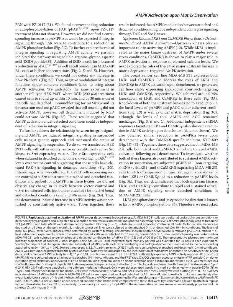
ER Release of Calcium Coupled to Store-operated CalciumEntry Contributes to AMPK Activation upon Matrix Dep-rivation—We next investigated the mechanisms that mightlead to the increase in intracellular calcium levels upon detach-ment. Within cells, ER is the major source of calcium. Calciumis released into the cytosol in response to various signalingevents that cause opening of the inositol triphosphate receptors(ITPRs) on the ER membrane (store release) (41). To under-stand whether detachment leads to calcium release from theER, we measured calcium changes using Fura 2 AM indetached cells. We observed an increase in intracellular cal-
cium, both under calcium-containing and calcium-free (toprevent entry of extracellular calcium) buffer conditions(Fig. 6A) suggesting that ER release contributes to cytosoliccalcium increase. To further gauge the involvement of ERcalcium release in AMPK activation, we studied the majorER calcium release channels, the ITPRs. An RT-PCR analysisrevealed that transcript levels of ITPR3 was severalfoldhigher than that of ITPR1 and -2 in MDA-MB 231 cells (Fig.6B). Therefore, we undertook the siRNA approach to knockdown ITPR3, and we found a significant reduction inpAMPK levels in matrix-deprived cells (Fig. 6C). This sug-gested that ER calcium release mediated by ITPR3 plays arole in AMPK activation upon matrix deprivation.
AMPK Activation upon Matrix Deprivation
14418 JOURNAL OF BIOLOGICAL CHEMISTRY VOLUME 291 • NUMBER 28 • JULY 8, 2016
by guest on June 7, 2018http://w
ww
.jbc.org/D
ownloaded from

ER calcium sensors like STIM1 (42) sense loss of calciumfrom the ER lumen beyond a threshold, and this triggers extra-cellular calcium entry through the plasma membrane calciumchannels into cells, a phenomena known as SOCE (43). As seenin Fig. 6A, the presence of extracellular calcium potentiated theincrease in intracellular calcium levels upon detachment.When ER calcium levels are depleted, STIM1 is known to trans-locate close to the plasma membrane, oligomerize, and formpuncta (43). To study the effect of matrix deprivation on STIMlocalization,wetransfectedMCF7cellswiththeSTIM-YFPcon-struct (44). Treatment of adherent cells with thapsigargin(which causes ER calcium depletion through irreversible inhi-bition of the sarco/endoplasmic reticulum Ca2�-ATPase pump(45)) led to formation of STIM1 puncta (Fig. 6D). Interestingly,we observed similar punctate appearance of STIM1 undermatrix deprivation (Fig. 6D), suggesting a possible role forSOCE in detachment-triggered intracellular calcium increase.To further confirm this, we additionally used 2-aminoethoxy-diphenyl borate (2-APB), an inhibitor of SOCE (46), to preventextracellular calcium entry upon cell detachment. Under theseconditions, we observed a significant reduction in calcium peakupon detachment (Fig. 6E), as well as a reduction in pAMPKlevels (Fig. 6F), together suggesting that the cytosolic calciumincrease upon detachment might be due to intracellular storerelease coupled with extracellular calcium entry, whichtogether lead to AMPK activation.
To further gauge whether SOCE is indeed important forAMPK activation, we used thapsigargin, which causes a deple-tion of ER calcium and subsequent induction of SOCE (47). Asexpected, thapsigargin treatment led to an increase in pAMPKlevels under attached conditions in the presence of calcium-containing buffer. However, thapsigargin-induced AMPK acti-vation was attenuated in calcium-free buffer (Fig. 6G). Similarattenuation of thapsigargin-induced AMPK activation was alsoobserved in La3� (SOCE inhibitor)-treated cells (Fig. 6H). Thissuggested that store-operated calcium entry likely plays aprominent role in AMPK activation. To further ascertainwhether SOCE is indeed involved in detachment-inducedAMPK activation, we compared pAMPK levels in cells
detached in calcium-free and calcium-containing buffers.Interestingly, we observed that AMPK activation was impairedwhen MDA-MB 231 cells were detached in calcium-free buffer(Fig. 6I). We obtained similar results in LKB1-deficient HeLa S3(Fig. 6J) and A549 cells (data not shown).
The store-operated calcium entry is facilitated by CRACchannels present on the plasma membrane (41). We investi-gated the effects of the E106D Orai1 calcium channel poremutant, which leads to enhanced pore diameter (48). Interest-ingly, we observed that MDA-MB 231 cells stably expressingOrai E106D GFP had higher basal calcium levels compared withcontrol GFP-expressing cells in both adherent and detachedconditions (Fig. 6, K and L). We compared the levels of pAMPKin vector control and Orai E106D GFP stable cells underdetached conditions. We found a significant increase in AMPKactivation in cells expressing Orai E106D GFP (Fig. 6M). Takentogether, our data indicate that detachment triggers calciumrelease from the ER and further leads to SOCE. Thus, calciumrelease from ER and extracellular calcium entry together con-tribute to rapid AMPK activation following detachment.
Oxidant Signaling Contributes to AMPK Activation—Wehave previously observed a role for upstream kinase LKB1 inrapid AMPK activation upon detachment. Calcium signaling,however, is not known to impinge on LKB1. In contrast, incertain contexts, ROS levels are known to activate AMPKthrough LKB1 in the absence of changes in the AMP/ATP ratio(49). Hence, we investigated the role of endogenous oxidantsignals as a possible regulator of AMPK activity upon matrixdeprivation. To do so, we measured the levels of ROS using2�,7�-dichlorofluorescein diacetate (DCFDA) (50) betweenattached and detached cells in a time course format using afluorescence plate reader; treatment with H2O2 at the end ofthe experiment served as the positive control (Fig. 7A). To avoidthe confounding effects of cell rounding and clumping on thefluorescence intensities, we loaded cells simultaneously withcalcein-AM and used fluorescence measured from calcein-AM-loaded cells as a normalizing control. Yet again, becauseserine proteases such as trypsin are known to increase ROSlevels independent of detachment (51), we have used mechan-
FIGURE 6. ER calcium release and store-operated calcium entry contributes to AMPK activation upon matrix deprivation. A, MDA-MB 231 cells wereloaded with Fura 2 AM and cultured in calcium-containing or calcium-free buffer. Graph depicts time course measurements of emission obtained at 510 nm byexcitation of the dye at 340 and 380 nm (340:380 ratio). The black arrow represents the point of mechanical detachment in the time course. Values have beennormalized to the initial reading. Values are expressed as mean � S.E.; ***, p � 0.001 (n � 4 with three technical repeats each). ns, non-significant. B, scatterplotdepicts 2�dct values representing the mRNA expression of ITPR receptor subtypes determined by qRT-PCR (n � 3 biological samples with three technicalrepeats each). C, MDA-MB 231 cells were transfected with either control siRNA or ITPR3 siRNA and cultured under attached (Att) and detached (Det) conditions.Cell lysates were probed by Western blotting with the antibodies indicated. D, MCF7 cells were transfected with STIM1 YFP construct and cultured underattached and detached conditions. Representative confocal equatorial sections are shown. Thapsigargin (TG) was used as positive control. E, MDA-MB 231 cellswere loaded with Fura 2 AM and pre-treated with either vehicle control (UT) or SOCE inhibitor 2-APB at 50 �M for 10 min. Graph depicts time coursemeasurements of emission obtained at 510 nm by excitation of the dye at 340 and 380 nm (340:380 ratio). The black arrow represents the point of mechanicaldetachment in the time course. Values have been normalized to the initial reading. Values are expressed as mean � S.E.; ***, p � 0.05 (n � 4 with three technicalrepeats each). F, MDA-MB 231 cells were cultured under attached conditions and pretreated with 100 �M 2-APB for 10 min prior to detachment. Cell lysateswere subjected to Western blotting with the antibodies indicated (n � 3). G, attached MDA-MB 231 cells were treated with vehicle control or thapsigargin (TG)(200 nM) for 10 min in the presence or absence of extracellular calcium as indicated. Lysates were run together in the same gel and subjected to immunoblot-ting with the antibodies indicated (n � 3). H, attached MDA-MB 231 cells were treated with vehicle control or thapsigargin (200 nM) for 10 min in the presenceor absence of La3� (10 �M). Cell lysates were subjected to Western blotting for the antibodies indicated (n � 3). I, MDA-MB 231 cells were cultured incalcium-containing or calcium-free conditions for 30 min and detached as indicated for 10 min. Lysates were subjected to immunoblotting with the antibodiesindicated (n � 3). Numbers depict relative pAMPK�/tubulin ratio. J, HeLa S3 cells were cultured in calcium-containing or calcium-free conditions for 30 min anddetached as indicated for 10 min. Lysates were subjected to immunoblotting with the antibodies indicated (n � 3). Numbers depict relative pAMPK�/tubulinratio. K and L, MDA-MB 231-GFP and MDA-MB 231 Orai E106D GFP cells were loaded with Fura 2 AM and cultured in calcium-containing KH buffer. Graph depictstime course measurements of emission obtained at 510 nm by excitation of the dye at 340 and 380 nm (340:380 ratio). The black arrow represents the point ofmechanical detachment and ionomycin addition in the time course. Values are expressed as mean � S.E.; **, p � 0.01 (n � 4 with 5 technical repeats each). M,MDA-MB 21 GFP and Orai E106D GFP cells were lysed under detached conditions, and lysates were subjected to Western blotting with the antibodies indicated.
AMPK Activation upon Matrix Deprivation
JULY 8, 2016 • VOLUME 291 • NUMBER 28 JOURNAL OF BIOLOGICAL CHEMISTRY 14419
by guest on June 7, 2018http://w
ww
.jbc.org/D
ownloaded from

ical detachment (19, 21, 22) instead of trypsin-EDTA for under-standing the effects of cell detachment on the kinetics of oxi-dant signaling and its role in the observed rapid AMPKactivation following detachment.
In MDA-MB 231 cells, measurement of ROS with DCFDArevealed an increase in oxidant signaling following detachment(Fig. 7B). A similar increase in ROS levels was also observed inLKB1-deficient cell lines like G361 following detachment (Fig.7C). To understand whether the increase in ROS levels ondetachment contributes to AMPK activation, we used the anti-oxidants 3-methyl-1-phenyl-2-pyrazolin-5-one (MCI-186) andN-acetyl cysteine (NAC) to quench cellular ROS levels by pre-
treatment of cells with these reagents before detachment. Theability of the antioxidants to quench ROS levels was confirmedby ROS measurements using DCFDA (data not shown). WhenMDA-MB 231 cells treated with MCI-186 were subjected todetachment, they showed reduced AMPK activation as gaugedby reduced pAMPK levels (Fig. 7D), as well as a reduction inpLKB1 levels (Fig. 7E). Similarly, we obtained a reduction inpAMPK levels in the presence of yet another ROS quencherNAC (Fig. 7F). Interestingly, LKB1-deficient G361 cells, thatare predominantly dependent on CaMKK� for AMPK activa-tion, also showed reduced AMPK activation upon treatmentwith antioxidants (Fig. 7G). This suggests that in LKB1-defi-
FIGURE 7. Oxidant signaling contributes to AMPK activation upon matrix deprivation. A, cells were loaded with DCFDA, a fluorescent dye for ROSmeasurements. Parallel wells were loaded with calcein AM as control. Fluorescence was measured at excitation 490/emission 520 nm in a spectrofluorometerin a plate reader format as a time course as represented in the graph. The 1st black arrow represents the point of mechanical detachment in the time course. The2nd black arrow represents the addition of positive control H2O2. Fluorescence values were normalized to initial reading. DCFDA fluorescence/calcein fluores-cence was calculated at each time point. Att, attached; Det, detached. MDA-MB 231 cells (B) G361 cells (C) were loaded with DCFDA, and ROS levels weremeasured as described above. The black arrow represents the point of mechanical detachment in the time course (n � 4 with three technical repeats each).Values are expressed as mean � S.E. D, MDA-MB 231 cells pretreated with either vehicle control or MCI-186 (200 �M), for 2 h, were cultured under attachedconditions or detached for 10 min, and Western blotting was performed to measure the levels of pAMPK� and AMPK (n � 6). The scatterplot depicts foldchange in pAMPK�/tubulin ratio; ***, p � 0.001. Error bars represent � S.E. E, MDA-MB 231 cells pretreated with either vehicle control or MCI-186 (200 �M), for2 h, were detached for 10 min, and Western blotting was performed to measure the levels of pLKB1 Ser-428 and LKB1 (n � 5). The scatterplot depicts foldchange in pLKB1/tubulin ratio; **, p � 0.01. Error bars represent � S.E. F, MDA-MB 231 cells pretreated with either vehicle control or 1 mM NAC, for 2 h, were theneither allowed to remain attached or detached for 10 min, and Western blotting was performed to measure the levels of pAMPK� and AMPK (n � 4). Thescatterplot depicts fold change in pAMPK�/tubulin ratio; **, p � 0.01. Error bars represent � S.E. G, LKB1-deficient G361 cells pretreated with either vehiclecontrol or MCI-186 (200 �M), for 2 h, were then either allowed to remain attached or detached for 10 min, and Western blotting was performed to measure thelevels of pAMPK� and AMPK (n � 4). The scatterplot depicts fold change in pAMPK�/tubulin ratio; **, p � 0.01. Error bars represent � S.E.
AMPK Activation upon Matrix Deprivation
14420 JOURNAL OF BIOLOGICAL CHEMISTRY VOLUME 291 • NUMBER 28 • JULY 8, 2016
by guest on June 7, 2018http://w
ww
.jbc.org/D
ownloaded from

cient cells ROS signaling might contribute to AMPK activation,perhaps working upstream of CaMKK�. This is consistent withreports on the role of ROS in CaMKK�-dependent AMPK acti-vation in certain contexts (15, 52). Taken together, our resultsindicate that oxidant signaling triggered upon detachment alsoplays a role in AMPK activation.
Calcium-mediated Oxidant Signaling Contributes to AMPKActivation upon Detachment—As shown above, calcium andoxidant signaling are triggered immediately following detach-ment, and both contribute to AMPK activation upon detach-ment. These molecules may function in distinct or overlappingpathways upstream of the kinases LKB1 and CaMKK�. Toinvestigate whether there is a cross-talk between oxidant andcalcium signaling under detached conditions, we first asked ifROS functions through calcium. ROS is known to cause cal-cium increase under hypoxia (15). To address this, we pre-treated MDA-MB 231 cells with the ROS quencher NAC orMCI-186 and compared the calcium surge on detachment withcontrol untreated cells. We observed no significant change inthe calcium peak on ROS inhibition (Fig. 8A), suggesting thatcalcium increase upon detachment is most likely independent
of ROS signaling. Conversely, when we inhibited calcium surgeusing 2-APB, an inhibitor of extracellular calcium entry (Fig.8B), BAPTA-AM, an intracellular calcium chelator (Fig. 8C), orby using calcium-free buffer to prevent SOCE (Fig. 8D), weobserved a significant reduction in ROS levels. These resultsindicated that intracellular calcium increase contributes toROS signaling upon detachment.
Our data for the first time revealed a calcium surge-depen-dent increase in ROS levels in detachment. To further confirmthis signaling axis under attached conditions in MDA-MB 231cells, we increased intracellular calcium levels using ionomycin,a known calcium ionophore (53). Increased cytosolic calciumlevels triggered by ionomycin can activate AMPK (54). Consis-tent with this, we observed an increase in pAMPK levels in thepresence of ionomycin (Fig. 8E). However, when we quenchedROS levels by pretreating the cells with MCI-186, ionomycin-mediated AMPK activation was partly abrogated (Fig. 8E) sug-gesting that calcium can impinge on AMPK activation, in part,through oxidant signaling.
Thus, in MDA-MB 231 cells, detachment leads to calciumincrease, which in turn increases ROS, and together these con-
FIGURE 8. Calcium mediates oxidant signaling upon detachment. A, MDA-MB 231 cells were loaded with Fura 2 AM, a ratiometric dye for calcium measure-ments. Cells were pre-treated either with vehicle control or with MCI-186 (200 �M) or NAC (1 mM) as indicated. Graph depicts peak ratio (detached/attached)obtained from time course measurements carried out as described previously (n � 6 –9). Error bars represent � S.E.; ns, non-significant. B–D, MDA-MB 231 cellswere pretreated with 2-APB (for 30 min) (B), BAPTA-AM (30 min) (C), or cultured in the presence or absence of calcium containing buffer for 30 min andsubsequently loaded with DCFDA (for 10 min). Fluorescence intensities were measured under detached (Det) and attached (Att) conditions using spectrofluo-rometer as described previously (B and C). Detached cells were analyzed by FACS (D); **, p � 0.01; *, p � 0.05. E, adherent MDA-MB 231 cells were pretreatedfor 1 h with MCI-186 (200 �M) in the indicated lanes and subsequently treated with either vehicle control (DMSO) or ionomycin (1 �M) for 5 min. pAMPK� andAMPK levels were measured by immunoblotting (n � 3). Numbers represent relative pAMPK�/AMPK ratio. The scatterplot depicts relative fold change inpAMPK/tubulin ratio; *, p � 0.05. Error bars represent � S.E. F, MDA-MB 231 cells were loaded with MitoSOX (2.5 �M) for 30 min and cultured in calcium-containing and calcium-free buffers. Cells were then detached mechanically and subjected to FACS analysis (n � 6). Rotenone, a mitochondrial electrontransport inhibitor was used as positive control; ***, p � 0.001. Error bars represent � S.E. G, MDA-MB 231 cells were treated with either vehicle control(UT) or diphenyleneiodonium chloride (DPI) and cultured under detached conditions. Cell lysates were probed by Western blotting with the antibodiesindicated (n � 3).
AMPK Activation upon Matrix Deprivation
JULY 8, 2016 • VOLUME 291 • NUMBER 28 JOURNAL OF BIOLOGICAL CHEMISTRY 14421
by guest on June 7, 2018http://w
ww
.jbc.org/D
ownloaded from

tribute to AMPK activation through CaMKK� and LKB1.These results suggest a novel mechanism involving calcium-de-pendent ROS signaling for the rapid AMPK activation underdetached conditions.
Detachment-induced Calcium Surge Triggers MitochondrialROS—To address the source of ROS that could be sensitive tocalcium changes, we measured mitochondrial ROS levels,which is known to contribute to AMPK activity in breast cancercells (55). We measured mitochondrial superoxide levels usingMitoSOX Red (56). Cells detached in calcium-containingbuffer had higher levels of mitochondrial superoxide comparedwith cells detached in calcium-free buffer (Fig. 8F). Rotenone,an electron transport chain uncoupler, served as a positive con-trol (Fig. 8F). Thus, our results indicate that detachment-in-duced increase in calcium could activate mitochondrial super-oxide production, which might further cause AMPK activation.To further understand whether yet another source of ROS, themembrane NADPH oxidases, also contributes to AMPK acti-vation, we inhibited the NADPH oxidases with diphenylenei-odonium. We observed no change in pAMPK levels (Fig. 8G)suggesting that the source of detachment-induced ROS is mito-chondrial and not membrane-bound NOXs. Taken together,our results reveal a novel calcium-mediated AMPK activationin detachment driven in part by mitochondrial ROS.
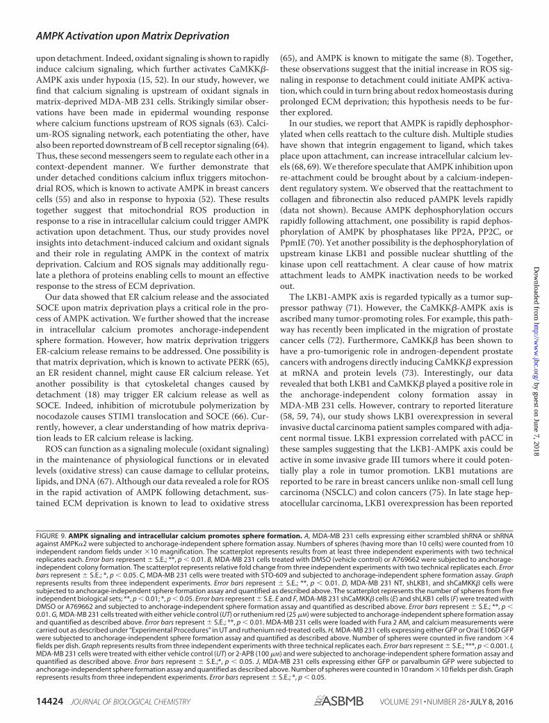
LKB1/CaMKK-AMPK Axis Promotes Anchorage-indepen-dent Colony Formation in Cancer Cells—Recent work from ourlaboratory (6) and that of others (7, 8) showed the importance ofAMPK in anchorage-independent colony formation in breastcancer cells. To understand the role of upstream kinases in thisprocess, we undertook sphere formation assays in soft agarunder conditions of inhibition or knockdown of CaMKK andLKB1. Consistent with our previous study (6), we found areduction in sphere formation in MDA-MB 231 cells withAMPK�2 knockdown compared with cells expressing scram-bled shRNA (Fig. 9A) and an increase in sphere formation incells treated with the AMPK activator A769662 (Fig. 9B). Sim-ilarly, we observed that MDA-MB 231 cells formed fewer num-bers of spheres on CaMKK� inhibition with STO-609 (Fig. 9C).We obtained similar results in LKB1-deficient A549 cellstreated with STO-609 (data not shown). Additionally,MDA-MB 231 cells expressing shCaMKK� or shLKB1 alsoformed fewer numbers of spheres as compared with MDA-MB231 NT cells (Fig. 9D). Furthermore, the reduction in colonyformation of shCaMKK� as well as shLKB1 cells was rescued bypharmacological activation of AMPK using A769662 (Fig. 9, Eand F). Yet another AMPK activator, AICAR, also rescued thereduction in colony formation observed in shCaMKK� cells(data not shown) suggesting that these upstream kinases mightfunction through AMPK to promote anchorage-independentcolony formation. These results together indicate that LKB1and CaMKK� might play a critical role in anchorage-indepen-dent colony formation through activation of AMPK.
Because our data identified the rise in intracellular calcium asa major mechanism of AMPK activation, we next sought tounderstand the role of calcium in sphere formation. To do so,we used multiple methods to modulate intracellular calciumlevels. Using ruthenium red (25 �M), we could increase thedetachment-induced calcium levels (Fig. 9G). This led to an
increase in sphere formation potential in ruthenium-treatedcells compared with untreated cells (Fig. 9G). We also used OraiE106D GFP stable cells, which showed increased basal calcium(Fig. 6K) under attached and sustained detachment. These cellsalso showed an increase in sphere formation (Fig. 9H). Treat-ment with 2-APB, which leads to a decrease in intracellularcalcium (Fig. 6E), led to a decrease in sphere formation (Fig. 9I).Alternatively, we used MDA-MB 231 cells expressing parvalbu-min nuclear exclusion signal GFP (Fig. 9J), which impairs cyto-solic calcium signals (57). In this case, we found a dramaticreduction in the sphere formation ability of MDA-MB 231 cells.Taken together, these data identify a role for calcium andupstream kinases of AMPK in promoting anchorage-indepen-dent colony formation.
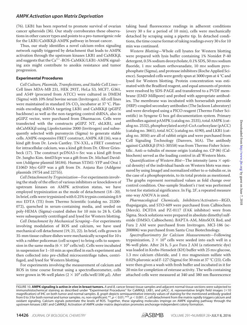
Increased LKB1 and pACC Levels in Breast Cancer PatientSpecimens—To address the relevance of the LKB1/CaMKK-AMPK axis in vivo, we undertook immunohistochemistry ontissue sections from grade III invasive carcinoma of breast com-pared with adjacent normal tissue. As shown in Fig. 10A, wedetected high levels of CaMKK� expression, which was notsignificantly altered between normal and tumor samples. Inter-estingly, and in contrast to previous reports on breast cancer(58, 59), we found an increase in LKB1 levels in breast cancersamples compared with adjacent normal tissue (Fig. 10, A andB). We used pACC as a read-out for AMPK activity, and con-sistent with a recent study (55), we found it to increase signifi-cantly in invasive breast cancers compared with adjacent nor-mal tissue (Fig. 10, A and B). We also observed a significantcorrelation between LKB1 and pACC in the cancer samples(p � 0.0104; Fisher’s exact test). Together, these results suggestthat LKB1/CaMKK�-AMPK pathway activation could poten-tially contribute to tumor progression.
Discussion
Recent work from our laboratory (6) and that of others (7, 8,25) has demonstrated AMPK activation upon matrix depriva-tion and its role in anoikis resistance. However, the molecularmechanisms that lead to AMPK activation upon matrix depri-vation are not well delineated. One of the major mechanisms ofAMPK activation is energy stress caused by reduced ATP levels(12). Although matrix deprivation causes ATP depletion by24 h (5), our data revealed that AMPK is activated very early,within minutes, following detachment. In this study, we show arapid increase in calcium and ROS levels following cell detach-ment, and we identify a novel calcium-redox signaling networkthat leads to rapid AMPK activation upon matrix detachmentthrough the upstream kinases LKB1 and CaMKK� (Fig. 10C).We speculate that this early activation of AMPK could helpprepare the cells to face the imminent energy crisis that followssustained ECM detachment.
LKB1 and CaMKK� are two well studied upstream kinasesthat are known to activate AMPK under different stress condi-tions. Although LKB1 is majorly known to activate AMPKunder energetic stress, CaMKK� is implicated under condi-tions of increased cytosolic calcium independent of the AMP/ATP ratio (60). In this study we find that upstream kinasesLKB1 as well as CaMKK� play important roles in AMPK acti-vation upon matrix detachment in MDA-MB 231 cells,
AMPK Activation upon Matrix Deprivation
14422 JOURNAL OF BIOLOGICAL CHEMISTRY VOLUME 291 • NUMBER 28 • JULY 8, 2016
by guest on June 7, 2018http://w
ww
.jbc.org/D
ownloaded from

although CaMKK� is the major player in this context in LKB1-deficient cell lines like G361 and A549. However, one cannotrule out the contributions of other recently identified AMPKkinases such as TAK1, ATM, and MLK3 (61, 62) as well asphosphatases, that may help to fine-tune AMPK activity in sus-pension; their roles in AMPK activation in matrix-deprivedcells need to be explored in the future.
Calcium and ROS are ubiquitous second messengers thatmediate various physiological functions. Although calcium isknown to activate AMPK through CaMKK� (54), oxidant sig-naling is reported to activate AMPK through LKB1 (49). Ourstudy demonstrates for the first time a rapid increase in calciumand oxidant signaling upon detachment. This increase in cal-cium and ROS contributes to the rapid activation of AMPK
AMPK Activation upon Matrix Deprivation
JULY 8, 2016 • VOLUME 291 • NUMBER 28 JOURNAL OF BIOLOGICAL CHEMISTRY 14423
by guest on June 7, 2018http://w
ww
.jbc.org/D
ownloaded from
upon detachment. Indeed, oxidant signaling is shown to rapidlyinduce calcium signaling, which further activates CaMKK�-AMPK axis under hypoxia (15, 52). In our study, however, wefind that calcium signaling is upstream of oxidant signals inmatrix-deprived MDA-MB 231 cells. Strikingly similar obser-vations have been made in epidermal wounding responsewhere calcium functions upstream of ROS signals (63). Calci-um-ROS signaling network, each potentiating the other, havealso been reported downstream of B cell receptor signaling (64).Thus, these second messengers seem to regulate each other in acontext-dependent manner. We further demonstrate thatunder detached conditions calcium influx triggers mitochon-drial ROS, which is known to activate AMPK in breast cancerscells (55) and also in response to hypoxia (52). These resultstogether suggest that mitochondrial ROS production inresponse to a rise in intracellular calcium could trigger AMPKactivation upon detachment. Thus, our study provides novelinsights into detachment-induced calcium and oxidant signalsand their role in regulating AMPK in the context of matrixdeprivation. Calcium and ROS signals may additionally regu-late a plethora of proteins enabling cells to mount an effectiveresponse to the stress of ECM deprivation.
Our data showed that ER calcium release and the associatedSOCE upon matrix deprivation plays a critical role in the pro-cess of AMPK activation. We further showed that the increasein intracellular calcium promotes anchorage-independentsphere formation. However, how matrix deprivation triggersER-calcium release remains to be addressed. One possibility isthat matrix deprivation, which is known to activate PERK (65),an ER resident channel, might cause ER calcium release. Yetanother possibility is that cytoskeletal changes caused bydetachment (18) may trigger ER calcium release as well asSOCE. Indeed, inhibition of microtubule polymerization bynocodazole causes STIM1 translocation and SOCE (66). Cur-rently, however, a clear understanding of how matrix depriva-tion leads to ER calcium release is lacking.
ROS can function as a signaling molecule (oxidant signaling)in the maintenance of physiological functions or in elevatedlevels (oxidative stress) can cause damage to cellular proteins,lipids, and DNA (67). Although our data revealed a role for ROSin the rapid activation of AMPK following detachment, sus-tained ECM deprivation is known to lead to oxidative stress
(65), and AMPK is known to mitigate the same (8). Together,these observations suggest that the initial increase in ROS sig-naling in response to detachment could initiate AMPK activa-tion, which could in turn bring about redox homeostasis duringprolonged ECM deprivation; this hypothesis needs to be fur-ther explored.
In our studies, we report that AMPK is rapidly dephosphor-ylated when cells reattach to the culture dish. Multiple studieshave shown that integrin engagement to ligand, which takesplace upon attachment, can increase intracellular calcium lev-els (68, 69). We therefore speculate that AMPK inhibition uponre-attachment could be brought about by a calcium-indepen-dent regulatory system. We observed that the reattachment tocollagen and fibronectin also reduced pAMPK levels rapidly(data not shown). Because AMPK dephosphorylation occursrapidly following attachment, one possibility is rapid dephos-phorylation of AMPK by phosphatases like PP2A, PP2C, orPpmIE (70). Yet another possibility is the dephosphorylation ofupstream kinase LKB1 and possible nuclear shuttling of thekinase upon cell reattachment. A clear cause of how matrixattachment leads to AMPK inactivation needs to be workedout.
The LKB1-AMPK axis is regarded typically as a tumor sup-pressor pathway (71). However, the CaMKK�-AMPK axis isascribed many tumor-promoting roles. For example, this path-way has recently been implicated in the migration of prostatecancer cells (72). Furthermore, CaMKK� has been shown tohave a pro-tumorigenic role in androgen-dependent prostatecancers with androgens directly inducing CaMKK� expressionat mRNA and protein levels (73). Interestingly, our datarevealed that both LKB1 and CaMKK� played a positive role inthe anchorage-independent colony formation assay inMDA-MB 231 cells. However, contrary to reported literature(58, 59, 74), our study shows LKB1 overexpression in severalinvasive ductal carcinoma patient samples compared with adja-cent normal tissue. LKB1 expression correlated with pACC inthese samples suggesting that the LKB1-AMPK axis could beactive in some invasive grade III tumors where it could poten-tially play a role in tumor promotion. LKB1 mutations arereported to be rare in breast cancers unlike non-small cell lungcarcinoma (NSCLC) and colon cancers (75). In late stage hep-atocellular carcinoma, LKB1 overexpression has been reported
FIGURE 9. AMPK signaling and intracellular calcium promotes sphere formation. A, MDA-MB 231 cells expressing either scrambled shRNA or shRNAagainst AMPK�2 were subjected to anchorage-independent sphere formation assay. Numbers of spheres (having more than 10 cells) were counted from 10independent random fields under �10 magnification. The scatterplot represents results from at least three independent experiments with two technicalreplicates each. Error bars represent � S.E.; **, p � 0.01. B, MDA-MB 231 cells treated with DMSO (vehicle control) or A769662 were subjected to anchorage-independent colony formation. The scatterplot represents relative fold change from three independent experiments with two technical replicates each. Errorbars represent � S.E.; *, p � 0.05. C, MDA-MB 231 cells were treated with STO-609 and subjected to anchorage-independent sphere formation assay. Graphrepresents results from three independent experiments. Error bars represent � S.E.; **, p � 0.01. D, MDA-MB 231 NT, shLKB1, and shCaMKK� cells weresubjected to anchorage-independent sphere formation assay and quantified as described above. The scatterplot represents the number of spheres from fiveindependent biological sets; **, p � 0.01; *, p � 0.05. Error bars represent � S.E. E and F, MDA-MB 231 shCaMKK� cells (E) and shLKB1 cells (F) were treated withDMSO or A769662 and subjected to anchorage-independent sphere formation assay and quantified as described above. Error bars represent � S.E.; **, p �0.01. G, MDA-MB 231 cells treated with either vehicle control (UT) or ruthenium red (25 �M) were subjected to anchorage-independent sphere formation assayand quantified as described above. Error bars represent � S.E.; **, p � 0.01. MDA-MB 231 cells were loaded with Fura 2 AM, and calcium measurements werecarried out as described under “Experimental Procedures” in UT and ruthenium red-treated cells. H, MDA-MB 231 cells expressing either GFP or Orai E106D GFPwere subjected to anchorage-independent sphere formation assay and quantified as described above. Number of spheres were counted in five random �4fields per dish. Graph represents results from three independent experiments with three technical replicates each. Error bars represent � S.E.; ***, p � 0.001. I,MDA-MB 231 cells were treated with either vehicle control (UT) or 2-APB (100 �M) and were subjected to anchorage-independent sphere formation assay andquantified as described above. Error bars represent � S.E.;*, p � 0.05. J, MDA-MB 231 cells expressing either GFP or parvalbumin GFP were subjected toanchorage-independent sphere formation assay and quantified as described above. Number of spheres were counted in 10 random �10 fields per dish. Graphrepresents results from three independent experiments. Error bars represent � S.E.; *, p � 0.05.
AMPK Activation upon Matrix Deprivation
14424 JOURNAL OF BIOLOGICAL CHEMISTRY VOLUME 291 • NUMBER 28 • JULY 8, 2016
by guest on June 7, 2018http://w
ww
.jbc.org/D
ownloaded from

AMPK Activation upon Matrix Deprivation
JULY 8, 2016 • VOLUME 291 • NUMBER 28 JOURNAL OF BIOLOGICAL CHEMISTRY 14425
by guest on June 7, 2018http://w
ww
.jbc.org/D
ownloaded from
(76). LKB1 has been reported to promote survival of ovariancancer spheroids (36). Our study corroborates these observa-tions in other cancer types and points to a pro-tumorigenic rolefor the LKB1/CaMKK�-AMPK pathway in breast cancer.
Thus, our study identifies a novel calcium-redox signalingnetwork rapidly triggered by detachment that leads to AMPKactivation through the upstream kinases LKB1 and CaMKK�,and suggests that the Ca2�-ROS-CaMKK/LKB1-AMPK signal-ing axis might contribute to anoikis resistance and tumorprogression.
Experimental Procedures
Cell Culture, Plasmids, Transfections, and Stable Cell Lines—Cell lines MDA-MB 231, HEK 293T, HeLa S3, MCF7, G361,and A549 (procured from ATCC) were cultured in DMEM(Sigma) with 10% fetal bovine serum (Invitrogen). All cell lineswere maintained in standard 5% CO2 incubator at 37 °C. Plas-mids encoding shRNA targeting LKB1 and CaMKK� (pGIPZbackbone) as well as the non-targeting control shRNA, also inpGIPZ vector, were purchased from Dharmacon. Cells weretransfected with the constructs pGIPZ NT, shLKB1, andshCaMKK� using Lipofectamine 2000 (Invitrogen) and subse-quently selected with puromycin (Sigma) to generate stablecells. AMPK-responsive FRET construct, AMPK AR (16), was akind gift from Dr. Lewis Cantley. TN-XXL, a FRET constructfor intracellular calcium, was a kind gift from Dr. Oliver Gries-beck (17). The construct pcDNA3-v-Src was a kind gift fromDr. Jungho Kim. 4mtD3cpv was a gift from Dr. Michael David-son (Addgene plasmid 58184). Human STIM1-YFP and Orai 1E106D Myc-GFP was a gift from Dr. Anjana Rao (Addgeneplasmids 19754 and 22755).
Cell Detachment by Trypsinization—For experiments involv-ing the study of the effect of various inhibitors or knockdown ofupstream kinases on AMPK activation status, we haveemployed trypsinization as the mode of detachment (18 –20).In brief, cells were trypsinized with 0.25% trypsin containing 0.9mM EDTA (TE) from Thermo Scientific (catalog no. 25200-072), quenched in serum-containing media, and seeded onpoly-HEMA (Sigma)-coated dishes for 10 min to 24 h. Cellswere subsequently centrifuged and lysed for Western blotting.
Cell Detachment by Mechanical Scraping—For experimentsinvolving modulation of ROS and calcium, we have usedmechanical cell detachment (19, 21, 22). In brief, cells grown in35-mm tissue culture dishes were mechanically scraped for 10 swith a rubber policeman (cell scraper) to bring cells to suspen-sion in the same media (6 � 105 cells/ml). Cells were incubatedin suspension for 1–10 min as specified in each experiment andthen collected into pre-chilled microcentrifuge tubes, centri-fuged, and lysed for Western blotting.
For experiments involving the measurement of calcium andROS in time course format using a spectrofluorometer, cellswere grown in 96-well plates (2 � 104 cells/well/100 �l). After
taking basal fluorescence readings in adherent conditions(every 30 s for a period of 10 min), cells were mechanicallydetached by scraping using a pipette tip. In detached condi-tions, the time course format of taking readings every 30 s for 10min was continued.
Western Blotting—Whole cell lysates for Western blottingwere prepared with lysis buffer containing 1% Nonidet P-40detergent, 0.5% sodium deoxycholate, 0.1% SDS, 50 mM sodiumfluoride, 1 mM sodium orthovanadate, 10 mM sodium pyro-phosphate (Sigma), and protease inhibitors (Roche Applied Sci-ence). Suspended cells were gently spun at 3000 rpm at 4 °C andlysed for Western blotting. Protein concentration was esti-mated with the Bradford reagent, and equal amounts of proteinwere resolved by SDS-PAGE and transferred to a PVDF mem-brane (EMD Millipore) and probed with appropriate antibod-ies. The membrane was incubated with horseradish peroxide(HRP)-coupled secondary antibodies (The Jackson Laboratory)and were developed using a PICO reagent (Thermo Fisher Sci-entific) in Syngene G box gel documentation system. Primaryantibodies against pAMPK (catalog no. 2535), total AMPK (cat-alog no. 2532), phosphorylated acetyl-CoA carboxylase (pACC)(catalog no. 3661), total ACC (catalog no. 4190), and LKB1 (cat-alog no. 3050) are all of rabbit origin and were purchased fromCell Signaling Technology (Danvers, MA). The antibodyagainst CaMKK� (PA5-30558) was from Thermo Fisher Scien-tific. Anti-�-tubulin of mouse origin (catalog no. CP 06) (Cal-biochem) served as the loading control in all Western blots.
Quantification of Western Blot—The intensity (area � opti-cal density) of the individual bands on Western blots was mea-sured by using ImageJ and normalized either to �-tubulin or, inthe case of a phosphoprotein, to its total protein as mentioned.The graphs represent normalized values as fold change overcontrol condition. One-sample Student’s t test was performedto test for statistical significance. In Fig. 2F, a repeated measureone-way ANOVA is used.
Pharmacological Chemicals, Inhibitors/Activators—RGD,thapsigargin, and STO-609 were purchased from Calbiochem(Merck). DCFDA and PZ-0117 (FAK inhibitor) were fromSigma. Stock solutions were prepared in absolute dimethyl sulf-oxide (DMSO, Calbiochem). BAPTA-AM, MitoSOX Red, andFura 2 AM were purchased from Invitrogen. MCI-186 (sc-200806) was purchased from Santa Cruz Biotechnology.
Spectrofluorimetry for Calcium Measurements—Followingtrypsinization, 2 � 104 cells were seeded into each well in a96-well plate. After 24 h, 5 �M Fura 2 AM (a ratiometric dye)was loaded in Krebs-Henseleit (KH) buffer with 25 mM glucose,1.3 mM calcium chloride, and 1 mM magnesium sulfate with0.02% pluronic acid F-127 (Sigma) for 30 min at 37 °C (23). Cellswere then given a wash with fresh buffer and incubated in it for20 min for completion of esterase activity. The wells containingattached cells were measured at 340 and 380 nm fluorescence
FIGURE 10. AMPK signaling is active in vivo in breast tumors. A and B, cancer breast tissue samples and adjacent normal tissue sections were subjected toimmunohistochemical staining as described under “Experimental Procedures” for CaMKK�, LKB1, and pACC. A, representative bright field images (�10magnification) of IHC on both tumor and normal samples. B, graph depicts semiquantitative intensity values of staining for the mentioned antibody gradedfrom 0 to 3 for both normal and tumor samples. ns, non-significant; **, p � 0.01; ***, p � 0.001. C, cell detachment from the matrix rapidly triggers calcium andoxidant signaling. Calcium signals potentiate the levels of ROS. Together, these signaling molecules impinge on AMPK signaling pathway through theupstream kinases LKB1 and CaMKK�. Activation of AMPK under matrix deprivation promotes anchorage-independent colony formation.
AMPK Activation upon Matrix Deprivation
14426 JOURNAL OF BIOLOGICAL CHEMISTRY VOLUME 291 • NUMBER 28 • JULY 8, 2016
by guest on June 7, 2018http://w
ww
.jbc.org/D
ownloaded from
excitation with 510 nm emission for 5–10 min with readingstaken every 30 s from at least five different regions in the well ina spectrofluorometer plate reader (Tecan Infinite M200 PRO).This served as the baseline calcium level for each well. In timecourse experiments, cells were detached gently by cell scrapingwith a pipette tip at the specified time. Following detachment,the readings were taken every 30 s for 10 min. The ratio ofemissions at 340/380 nm indicates intracellular calcium levels.At the end of each experiment, positive and negative controlswith ionomycin (1 �M) and EGTA (with 1% Triton X-100) wereperformed to determine the highest and lowest values obtain-able with Fura 2 AM for each experiment. We have used two-way repeat measures ANOVA with Bonferroni’s multiple com-parison test for statistical analysis of differences in time courseassays under different conditions.
Spectrofluorimetry and Fluorescence-activated Cell Sorting(FACS) for ROS Measurements—Following trypsinization, 2 �104 cells were seeded into each well in a 96-well plate. After24 h, DCFDA was loaded in Krebs-Henseleit (KH) buffer with25 mM glucose, 1.3 mM calcium chloride, and 1 mM magnesiumsulfate for 15 min at 37 °C. Cells were then given a wash withfresh buffer, and the plates were measured at excitation 490/emission 520 nm for DCFDA. To normalize for the fluores-cence intensity changes caused by cell rounding and clumping,we have used parallel wells loaded with calcein-AM and mea-sured the same at excitation 490/emission 520 nm. In timecourse experiments, cells were detached gently by mechanicalscraping. The ratio of fluorescence from DCFDA/calcein AMwas plotted at each time point. For measuring ROS levels byFACS, 6 � 105 cells in each 35-mm tissue culture dish werepretreated with requisite drugs as mentioned and loaded withDCFDA for cytosolic ROS and MitoSOX Red for mitochondrialsuperoxide levels. Mean fluorescence intensity was calculatedfor each condition. We have used one-way ANOVA with Bon-ferroni’s multiple comparison test to compare peak to basalratios across multiple conditions (Fig. 8A) and one sample orpaired t-tests for two data set comparisons in Fig. 8.
Immunocytochemistry and Confocal Microscopy—Cells cul-tured under attached and detached conditions were fixed with4% paraformaldehyde, and the indicated antibodies were usedfor staining. Confocal Z stack images were acquired at �60magnification in Fluoview FV10i (Olympus). Images werequantified using ImageJ by integrating pixel intensities acrossall Z sections and plotted as total intensity per cell for bothattached and detached conditions. We have used one sample ttest for statistical analysis.
Immunoprecipitation and in Vitro Kinase Assay—Cellsgrown in attached or detached conditions for the time periodsmentioned were lysed with a lysis buffer containing sodiumfluoride, sodium pyrophosphate, EDTA, EGTA, DTT, 1% Tri-ton X-100, benzamidine hydrochloride, and PMSF. Sheep anti-AMPK antibody (a kind gift from Dr. Grahame D. Hardie) wasused to immunoprecipitate AMPK from 120 �g of lysate. Sub-sequently, in vitro kinase reaction was carried out with AMPKsubstrate, AMARA, for 30 min at 37 °C. The reaction productswere spotted on P81 phosphocellulose paper and washed with1% phosphoric acid and water. Radioactive 32P incorporated
was measured in a liquid scintillation counter. We have usedone-sample Student’s t test for statistical analysis.
Immunohistochemistry (IHC)—A total of 35 breast cancerand 25–30 adjacent normal tissue samples were obtained frompreviously untreated grade III invasive ductal carcinoma cases.The paraffin blocks were collected from Kidwai MemorialInstitute of Oncology (Bangalore, KA, India). Medical EthicsCommittee (Institutional Review Board of Kidwai MemorialInstitute of Oncology) and Institutional Human Ethics Com-mittee (IISc; Bangalore, KA, India) approved the study.CaMKK� (Pierce PA530558), LKB1 (Cell Signaling Technology3570S), and pACC (Cell Signaling Technology 11818S) antibodieswere used at 1:100 dilution. IHC was performed as described pre-viously (24). Scoring was performed by an experienced histo-pathologist (U. A.). Unpaired Student’s t test was used to test forsignificant differences between groups. Fisher’s exact test was usedto test the correlation between LKB1 and pACC.
Statistical Analysis—All statistical analyses were performedusing GraphPad Prism 5.0 software. All data are presented asmeans � S.E. of the mean (S.E.). p values �0.05 were consid-ered to be statistically significant. Statistical analysis was doneusing one-sample Student’s t test for data representing fold changeover control. For other experiments, paired or unpaired, Student’st test was used based on the experimental design; *** representsp � 0.001; ** represents p � 0.01; and * represents p � 0.05. Forexperiments with three groups, one-way ANOVA with Bonferro-ni’s multiple comparison test was used. For time course experi-ments of ROS and calcium under different treatments, two-wayrepeated measures ANOVA with Bonferroni’s post hoc test wasused. For IHC experiments, unpaired t tests were used to testfor significant differences between groups, and Fisher’s exacttest was used to test for correlation.
Author Contributions—A. S. designed, performed, and analyzed theexperiments and wrote the paper. A. R. conceived and coordinatedthe study and wrote the paper. U. A. helped with acquisition, analy-sis, and interpretation of immunohistochemistry data. All authorsanalyzed the results and approved the final version of themanuscript.
Acknowledgments—We acknowledge Dr. Oliver Griesbeck and Dr.Lewis C. Cantley for TN-XXL and AMPK AR FRET constructs, respec-tively, and Dr. Graham Hardie for AMPK antibody used in kinaseassays. We also acknowledge Nehanjali Dwivedi and Rachana Sathy-endran for Western blotting; Manipa Saha and C. R. Madhwesh forhelp with in vitro kinase assay and immunocytochemistry, respec-tively; and members of the A. R. laboratory for their critical inputsduring the course of the study and for help with manuscript prepara-tion. We also thank Dr. Rekha V. Kumar and R. Anisha from KidwaiMemorial Institute of Oncology for help with immunohistochemistry.We acknowledge Drs. N. V. Joshi, Kumar Somasundaram, DeepakSaini, and Ramray Bhat for their input in the study and statisticalanalysis of the data. We acknowledge the flow cytometry facility atIISc. Support from Department of Science and Technology (DST)-Fund for Improvement of Science & Technology infrastructure in uni-versities & higher educational institutions (FIST), and UniversityGrants Commission (UGC), Govt. of India, to the Department ofMRDG are also acknowledged.
AMPK Activation upon Matrix Deprivation
JULY 8, 2016 • VOLUME 291 • NUMBER 28 JOURNAL OF BIOLOGICAL CHEMISTRY 14427
by guest on June 7, 2018http://w
ww
.jbc.org/D
ownloaded from

References1. Nelson, C. M., and Bissell, M. J. (2006) Of extracellular matrix, scaffolds,
and signaling: tissue architecture regulates development, homeostasis,and cancer. Annu. Rev. Cell Dev. Biol. 22, 287–309
2. Frisch, S. M., and Screaton, R. A. (2001) Anoikis mechanisms. Curr. Opin.Cell Biol. 13, 555–562
3. Simpson, C. D., Anyiwe, K., and Schimmer, A. D. (2008) Anoikis resis-tance and tumor metastasis. Cancer Lett. 272, 177–185
4. Mehlen, P., and Puisieux, A. (2006) Metastasis: a question of life or death.Nat. Rev. Cancer 6, 449 – 458
5. Schafer, Z. T., Grassian, A. R., Song, L., Jiang, Z., Gerhart-Hines, Z., Irie,H. Y., Gao, S., Puigserver, P., and Brugge, J. S. (2009) Antioxidant andoncogene rescue of metabolic defects caused by loss of matrix attachment.Nature 461, 109 –113
6. Hindupur, S. K., Balaji, S. A., Saxena, M., Pandey, S., Sravan, G. S., Heda,N., Kumar, M. V., Mukherjee, G., Dey, D., and Rangarajan, A. (2014)Identification of a novel AMPK-PEA15 axis in the anoikis-resistantgrowth of mammary cells. Breast Cancer Res. 16, 420
7. Ng, T. L., Leprivier, G., Robertson, M. D., Chow, C., Martin, M. J., Lader-oute, K. R., Davicioni, E., Triche, T. J., and Sorensen, P. H. (2012) TheAMPK stress response pathway mediates anoikis resistance through inhi-bition of mTOR and suppression of protein synthesis. Cell Death Differ.19, 501–510
8. Jeon, S. M., Chandel, N. S., and Hay, N. (2012) AMPK regulates NADPHhomeostasis to promote tumour cell survival during energy stress. Nature485, 661– 665
9. Desgrosellier, J. S., and Cheresh, D. A. (2010) Integrins in cancer: biolog-ical implications and therapeutic opportunities. Nat. Rev. Cancer 10, 9 –22
10. Bouchard, V., Demers, M. J., Thibodeau, S., Laquerre, V., Fujita, N., Tsu-ruo, T., Beaulieu, J. F., Gauthier, R., Vézina, A., Villeneuve, L., and Vachon,P. H. (2007) Fak/Src signaling in human intestinal epithelial cell survivaland anoikis: differentiation state-specific uncoupling with the PI3-K/Akt-1 and MEK/Erk pathways. J. Cell Physiol. 212, 717–728
11. Hardie, D. G., and Carling, D. (1997) The AMP-activated proteinkinase–fuel gauge of the mammalian cell? Eur. J. Biochem. 246, 259 –273
12. Carling, D., Sanders, M. J., and Woods, A. (2008) The regulation of AMP-activated protein kinase by upstream kinases. Int. J. Obes. 32, Suppl. 4,55–59
13. Choi, S. L., Kim, S. J., Lee, K. T., Kim, J., Mu, J., Birnbaum, M. J., Soo Kim,S., and Ha, J. (2001) The regulation of AMP-activated protein kinase byH2O2. Biochem. Biophys. Res. Commun. 287, 92–97
14. Auciello, F. R., Ross, F. A., Ikematsu, N., and Hardie, D. G. (2014) Oxida-tive stress activates AMPK in cultured cells primarily by increasing cellu-lar AMP and/or ADP. FEBS Lett. 588, 3361–3366
15. Mungai, P. T., Waypa, G. B., Jairaman, A., Prakriya, M., Dokic, D., Ball,M. K., and Schumacker, P. T. (2011) Hypoxia triggers AMPK activationthrough reactive oxygen species-mediated activation of calcium release-activated calcium channels. Mol. Cell. Biol. 31, 3531–3545
16. Tsou, P., Zheng, B., Hsu, C. H., Sasaki, A. T., and Cantley, L. C. (2011) Afluorescent reporter of AMPK activity and cellular energy stress. CellMetab. 13, 476 – 486
17. Mank, M., Santos, A. F., Direnberger, S., Mrsic-Flogel, T. D., Hofer, S. B.,Stein, V., Hendel, T., Reiff, D. F., Levelt, C., Borst, A., Bonhoeffer, T.,Hübener, M., and Griesbeck, O. (2008) A genetically encoded calciumindicator for chronic in vivo two-photon imaging. Nat. Methods 5,805– 811
18. Zhao, B., Li, L., Wang, L., Wang, C. Y., Yu, J., and Guan, K. L. (2012) Celldetachment activates the Hippo pathway via cytoskeleton reorganizationto induce anoikis. Genes Dev. 26, 54 – 68
19. Zagórska, A., Deak, M., Campbell, D. G., Banerjee, S., Hirano, M., Aizawa,S., Prescott, A. R., and Alessi, D. R. (2010) New roles for the LKB1-NUAKpathway in controlling myosin phosphatase complexes and cell adhesion.Sci. Signal. 3, ra25
20. Howe, A. K. (2001) Cell adhesion regulates the interaction between Nckand p21-activated kinase. J. Biol. Chem. 276, 14541–14544
21. Ogata, M., Takada, T., Mori, Y., Uchida, Y., Miki, T., Okuyama, A., Kosugi,A., Sawada, M., Oh-hora, M., and Hamaoka, T. (1999) Regulation of phos-
phorylation level and distribution of PTP36, a putative protein tyrosinephosphatase, by cell-substrate adhesion. J. Biol. Chem. 274, 20717–20724
22. Baron, V., and Schwartz, M. (2000) Cell adhesion regulates ubiquitin-mediated degradation of the platelet-derived growth factor receptor �.J. Biol. Chem. 275, 39318 –39323
23. Malgaroli, A., Milani, D., Meldolesi, J., and Pozzan, T. (1987) Fura-2 mea-surement of cytosolic free Ca2� in monolayers and suspensions of varioustypes of animal cells. J. Cell Biol. 105, 2145–2155
24. Mittal, S., Subramanyam, D., Dey, D., Kumar, R. V., and Rangarajan, A.(2009) Cooperation of Notch and Ras/MAPK signaling pathways in hu-man breast carcinogenesis. Molecular Cancer 8, 128
25. Fung, C., Lock, R., Gao, S., Salas, E., and Debnath, J. (2008) Induction ofautophagy during extracellular matrix detachment promotes cell survival.Mol. Biol. Cell 19, 797– 806
26. Davies, S. P., Sim, A. T., and Hardie, D. G. (1990) Location and function ofthree sites phosphorylated on rat acetyl-CoA carboxylase by the AMP-activated protein kinase. Eur. J. Biochem. 187, 183–190
27. Dale, S., Wilson, W. A., Edelman, A. M., and Hardie, D. G. (1995) Similarsubstrate recognition motifs for mammalian AMP-activated protein ki-nase, higher plant HMG-CoA reductase kinase-A, yeast SNF1, and mam-malian calmodulin-dependent protein kinase I. FEBS Lett. 361, 191–195
28. Ellerström, C., Strehl, R., Noaksson, K., Hyllner, J., and Semb, H. (2007)Facilitated expansion of human embryonic stem cells by single-cell enzy-matic dissociation. Stem Cells 25, 1690 –1696
29. Zhan, M., Zhao, H., and Han, Z. C. (2004) Signalling mechanisms of anoi-kis. Histol. Histopathol. 19, 973–983
30. Westhoff, M. A., Serrels, B., Fincham, V. J., Frame, M. C., and Carragher,N. O. (2004) SRC-mediated phosphorylation of focal adhesion kinase cou-ples actin and adhesion dynamics to survival signaling. Mol. Cell. Biol. 24,8113– 8133
31. Slack-Davis, J. K., Martin, K. H., Tilghman, R. W., Iwanicki, M., Ung, E. J.,Autry, C., Luzzio, M. J., Cooper, B., Kath, J. C., Roberts, W. G., and Par-sons, J. T. (2007) Cellular characterization of a novel focal adhesion kinaseinhibitor. J. Biol. Chem. 282, 14845–14852
32. Ruoslahti, E. (1996) RGD and other recognition sequences for integrins.Annu. Rev. Cell Dev. Biol. 12, 697–715
33. Tokumitsu, H., Inuzuka, H., Ishikawa, Y., Ikeda, M., Saji, I., and Kobayashi,R. (2002) STO-609, a specific inhibitor of the Ca2�/calmodulin-depen-dent protein kinase kinase. J. Biol. Chem. 277, 15813–15818
34. Xie, Z., Dong, Y., Scholz, R., Neumann, D., and Zou, M.-H. (2008) Phos-phorylation of LKB1 at serine 428 by protein kinase C-� is required formetformin-enhanced activation of the AMP-activated protein kinase inendothelial cells. Circulation 117, 952–962
35. Song, P., Xie, Z., Wu, Y., Xu, J., Dong, Y., and Zou, M. H. (2008) Proteinkinase Czeta-dependent LKB1 serine 428 phosphorylation increasesLKB1 nucleus export and apoptosis in endothelial cells. J. Biol. Chem. 283,12446 –12455
36. Peart, T., Ramos Valdes, Y., Correa, R. J. M., Fazio, E., Bertrand, M., Mc-Gee, J., Préfontaine, M., Sugimoto, A., DiMattia, G. E., and Shepherd, T. G.(2015) Intact LKB1 activity is required for survival of dormant ovariancancer spheroids. Oncotarget 26, 22424 –22438
37. Hardie, D. G. (2011) Sensing of energy and nutrients by AMP-activatedprotein kinase. Am. J. Clin. Nutr. 93, 891S– 896S
38. Birnbaum, M. J. (2005) Activating AMP-activated protein kinase withoutAMP. Mol. Cell 19, 289 –290
39. Davis, F. M., Azimi, I., Faville, R. A., Peters, A. A., Jalink, K., Putney, J. W.,Jr., Goodhill, G. J., Thompson, E. W., Roberts-Thomson, S. J., and Mon-teith, G. R. (2014) Induction of epithelial-mesenchymal transition (EMT)in breast cancer cells is calcium signal dependent. Oncogene 33,2307–2316
40. Palmer, A. E., Giacomello, M., Kortemme, T., Hires, S. A., Lev-Ram, V.,Baker, D., and Tsien, R. Y. (2006) Ca2� indicators based on computation-ally redesigned calmodulin-peptide pairs. Chem. Biol. 13, 521–530
41. Clapham, D. E. (2007) Calcium signaling. Cell 131, 1047–105842. Luik, R. M., Wang, B., Prakriya, M., Wu, M. M., and Lewis, R. S. (2008)
Oligomerization of STIM1 couples ER calcium depletion to CRAC chan-nel activation. Nature 454, 538 –542
43. Lewis, R. S. (2011) Store-operated calcium channels: new perspectives on
AMPK Activation upon Matrix Deprivation
14428 JOURNAL OF BIOLOGICAL CHEMISTRY VOLUME 291 • NUMBER 28 • JULY 8, 2016
by guest on June 7, 2018http://w
ww
.jbc.org/D
ownloaded from

mechanism and function. Cold Spring Harb. Perspect. Biol. 3, a00397044. Prakriya, M., Feske, S., Gwack, Y., Srikanth, S., Rao, A., and Hogan, P. G.
(2006) Orai1 is an essential pore subunit of the CRAC channel. Nature443, 230 –233
45. Lytton, J., Westlin, M., and Hanley, M. R. (1991) Thapsigargin inhibits thesarcoplasmic or endoplasmic reticulum Ca-ATPase family of calciumpumps. J. Biol. Chem. 266, 17067–17071
46. Bootman, M. D., Collins, T. J., Mackenzie, L., Roderick, H. L., Berridge,M. J., and Peppiatt, C. M. (2002) 2-Aminoethoxydiphenyl borate (2-APB)is a reliable blocker of store-operated Ca2� entry but an inconsistent in-hibitor of InsP3-induced Ca2� release. FASEB J. 16, 1145–1150
47. Treiman, M., Caspersen, C., and Christensen, S. B. (1998) A tool coming ofage: thapsigargin as an inhibitor of sarco-endoplasmic reticulum Ca2�-ATPases. Trends Pharmacol. Sci. 19, 131–135
48. Yamashita, M., Navarro-Borelly, L., McNally, B. A., and Prakriya, M.(2007) Orai1 mutations alter ion permeation and Ca2�-dependent fastinactivation of CRAC channels: evidence for coupling of permeation andgating. J. Gen. Physiol. 130, 525–540
49. Emerling, B. M., Weinberg, F., Snyder, C., Burgess, Z., Mutlu, G. M., Vi-ollet, B., Budinger, G. R., and Chandel, N. S. (2009) Hypoxic activation ofAMPK is dependent on mitochondrial ROS but independent of an in-crease in AMP/ATP ratio. Free Radic. Biol. Med. 46, 1386 –1391
50. LeBel, C. P., Ali, S. F., and Bondy, S. C. (1992) Deferoxamine inhibitsmethyl mercury-induced increases in reactive oxygen species formationin rat brain. Toxicol. Appl. Pharmacol. 112, 161–165
51. Aoshiba, K., Yasuda, K., Yasui, S., Tamaoki, J., and Nagai, A. (2001) Serineproteases increase oxidative stress in lung cells. Am. J. Physiol. Lung CellMol. Physiol. 281, L556 –564
52. Gusarova, G. A., Trejo, H. E., Dada, L. A., Briva, A., Welch, L. C., Ha-manaka, R. B., Mutlu, G. M., Chandel, N. S., Prakriya, M., and Sznajder, J. I.(2011) Hypoxia leads to Na,K-ATPase downregulation via Ca2� release-activated Ca2� channels and AMPK activation. Mol. Cell. Biol. 31,3546 –3556
53. Morgan, A. J., and Jacob, R. (1994) Ionomycin enhances Ca2� influx bystimulating store-regulated cation entry and not by a direct action at theplasma membrane. Biochem. J. 300, 665– 672
54. Woods, A., Dickerson, K., Heath, R., Hong, S.-P., Momcilovic, M., John-stone, S. R., Carlson, M., and Carling, D. (2005) Ca2�/calmodulin-depen-dent protein kinase kinase-� acts upstream of AMP-activated proteinkinase in mammalian cells. Cell Metab. 2, 21–33
55. Hart, P. C., Mao, M., de Abreu, A. L., Ansenberger-Fricano, K., Ekoue,D. N., Ganini, D., Kajdacsy-Balla, A., Diamond, A. M., Minshall, R. D.,Consolaro, M. E., Santos, J. H., and Bonini, M. G. (2015) MnSOD upregu-lation sustains the Warburg effect via mitochondrial ROS and AMPK-de-pendent signalling in cancer. Nat. Commun. 6, 6053
56. Robinson, K. M., Janes, M. S., and Beckman, J. S. (2008) The selectivedetection of mitochondrial superoxide by live cell imaging. Nat. Protoc. 3,941–947
57. Pusl, T., Wu, J. J., Zimmerman, T. L., Zhang, L., Ehrlich, B. E., Berchtold,M. W., Hoek, J. B., Karpen, S. J., Nathanson, M. H., and Bennett, A. M.(2002) Epidermal growth factor-mediated activation of the ETS domaintranscription factor Elk-1 requires nuclear calcium. J. Biol. Chem. 277,27517–27527
58. Fenton, H., Carlile, B., Montgomery, E. A., Carraway, H., Herman, J., Sa-hin, F., Su, G. H., and Argani, P. (2006) LKB1 protein expression in humanbreast cancer. Appl. Immunohistochem. Mol. Morphol. 14, 146 –153
59. Shen, Z., Wen, X. F., Lan, F., Shen, Z. Z., and Shao, Z. M. (2002) The tumorsuppressor gene LKB1 is associated with prognosis in human breast car-cinoma. Clin. Cancer Res. 8, 2085–2090
60. Alexander, A., and Walker, C. L. (2011) The role of LKB1 and AMPK incellular responses to stress and damage. FEBS Lett. 585, 952–957
61. Luo, L., Jiang, S., Huang, D., Lu, N., and Luo, Z. (2015) MLK3 phosphor-ylates AMPK independently of LKB1. PLoS ONE 10, e0123927
62. Xie, M., Zhang, D., Dyck, J. R., Li, Y., Zhang, H., Morishima, M., Mann,D. L., Taffet, G. E., Baldini, A., Khoury, D. S., and Schneider, M. D. (2006)A pivotal role for endogenous TGF-�-activated kinase-1 in the LKB1/AMP-activated protein kinase energy-sensor pathway. Proc. Natl. Acad.Sci. U.S.A. 103, 17378 –17383
63. Xu, S., and Chisholm, A. D. (2014) C. elegans epidermal wounding inducesa mitochondrial ROS burst that promotes wound repair. Dev. Cell 31,48 – 60
64. Singh, D. K., Kumar, D., Siddiqui, Z., Basu, S. K., Kumar, V., and Rao, K. V.(2005) The strength of receptor signaling is centrally controlled through acooperative loop between Ca2� and an oxidant signal. Cell 121, 281–293
65. Avivar-Valderas, A., Salas, E., Bobrovnikova-Marjon, E., Diehl, J. A., Nagi,C., Debnath, J., and Aguirre-Ghiso, J. A. (2011) PERK integrates autophagyand oxidative stress responses to promote survival during extracellularmatrix detachment. Mol. Cell. Biol. 31, 3616 –3629
66. Smyth, J. T., DeHaven, W. I., Bird, G. S., and Putney, J. W. (2007) Role ofthe microtubule cytoskeleton in the function of the store-operated Ca2�
channel activator STIM1. J. Cell Sci. 120, 3762–377167. Schieber, M., and Chandel, N. S. (2014) ROS function in redox signaling
and oxidative stress. Curr. Biol. 24, R453–R46268. Itano, N., Okamoto, S., Zhang, D., Lipton, S. A., and Ruoslahti, E. (2003)
Cell spreading controls endoplasmic and nuclear calcium: a physical generegulation pathway from the cell surface to the nucleus. Proc. Natl. Acad.Sci. U.S.A. 100, 5181–5186
69. Sjaastad, M. D., Lewis, R. S., and Nelson, W. J. (1996) Mechanisms ofintegrin-mediated calcium signaling in MDCK cells: regulation of adhe-sion by IP3- and store-independent calcium influx. Mol. Biol. Cell 7,1025–1041
70. Voss, M., Paterson, J., Kelsall, I. R., Martín-Granados, C., Hastie, C. J.,Peggie, M. W., and Cohen, P. T. (2011) Ppm1E is an in cellulo AMP-activated protein kinase phosphatase. Cell. Signal. 23, 114 –124
71. Shackelford, D. B., and Shaw, R. J. (2009) The LKB1-AMPK pathway:metabolism and growth control in tumour suppression. Nat. Rev. Cancer9, 563–575
72. Frigo, D. E., Howe, M. K., Wittmann, B. M., Brunner, A. M., Cushman, I.,Wang, Q., Brown, M., Means, A. R., and McDonnell, D. P. (2011) CaMkinase kinase �-mediated activation of the growth regulatory kinaseAMPK is required for androgen-dependent migration of prostate cancercells. Cancer Res. 71, 528 –537
73. Karacosta, L. G., Foster, B. A., Azabdaftari, G., Feliciano, D. M., and Edel-man, A. M. (2012) A regulatory feedback loop between Ca2�/calmodulin-dependent protein kinase kinase 2 (CaMKK2) and the androgen receptorin prostate cancer progression. J. Biol. Chem. 287, 24832–24843
74. Bouchekioua-Bouzaghou, K., Poulard, C., Rambaud, J., Lavergne, E., Hus-sein, N., Billaud, M., Bachelot, T., Chabaud, S., Mader, S., Dayan, G., Treil-leux, I., Corbo, L., and Le Romancer, M. (2014) LKB1 when associated withmethylated ER� is a marker of bad prognosis in breast cancer. Int. J. Can-cer 135, 1307–1318
75. Sanchez-Cespedes, M. (2007) A role for LKB1 gene in human cancerbeyond the Peutz-Jeghers syndrome. Oncogene 26, 7825–7832
76. Lee, S.-W., Li, C.-F., Jin, G., Cai, Z., Han, F., Chan, C.-H., Yang, W.-L., Li,B.-K., Rezaeian, A. H., Li, H.-Y., Huang, H.-Y., and Lin, H.-K. (2015) Skp2-dependent ubiquitination and activation of LKB1 is essential for cancercell survival under energy stress. Mol. Cell 57, 1022–1033
AMPK Activation upon Matrix Deprivation
JULY 8, 2016 • VOLUME 291 • NUMBER 28 JOURNAL OF BIOLOGICAL CHEMISTRY 14429
by guest on June 7, 2018http://w
ww
.jbc.org/D
ownloaded from

Ananthalakshmy Sundararaman, Usha Amirtham and Annapoorni Rangarajan(AMPK) Activation upon Matrix Deprivation
Calcium-Oxidant Signaling Network Regulates AMP-activated Protein Kinase
doi: 10.1074/jbc.M116.731257 originally published online May 11, 20162016, 291:14410-14429.J. Biol. Chem.
10.1074/jbc.M116.731257Access the most updated version of this article at doi:
Alerts:
When a correction for this article is posted•
When this article is cited•
to choose from all of JBC's e-mail alertsClick here
http://www.jbc.org/content/291/28/14410.full.html#ref-list-1
This article cites 76 references, 29 of which can be accessed free at
by guest on June 7, 2018http://w
ww
.jbc.org/D
ownloaded from