real-time image-guided cooperative robotic assist...
TRANSCRIPT

Real-Time Image-Guided Cooperative Robotic Assist Devicefor Deep Anterior Lamellar Keratoplasty
Mark Draelos, Brenton Keller, Gao Tang, Anthony Kuo, MD, Kris Hauser, PhD, and Joseph Izatt, PhD
Abstract— Deep anterior lamellar keratoplasty (DALK) is apromising technique for corneal transplantation that avoids thechronic immunosuppression comorbidities and graft rejectionrisk associated with penetrating keratoplasty (PKP), the stan-dard procedure. In DALK, surgeons must insert a needle 90 %through the 500 µm cornea without penetrating its underlyingmembrane. This pushes surgeons to their manipulation andvisualization limits such that 59 % of DALK attempts fail dueto corneal perforation or inadequate needle depth. We proposea robot-assisted solution to jointly solve the manipulation andvisualization challenges using a cooperatively-controlled, preciserobot arm and live optical coherence tomography (OCT) imag-ing, respectively. Our system features an interface handle, withwhich the surgeon and robot cooperatively hold the tool, and aposterior corneal boundary virtual fixture driven by real-timeOCT segmentation. A study in which three operators performedDALK needle insertions manually and cooperatively in exvivo human corneas demonstrated an 84 % improvement inperforation-free needle depth without an increased perforationrate.
Index Terms— Cooperative control, medical robotics
I. INTRODUCTION
Corneal transplants are among the most common allo-graft procedures performed worldwide. In the United States,nearly 40,000 full thickness corneal transplants, or penetrat-ing keratoplasties (PKPs), are performed annually [1]. Inthe United Kingdom, corneal transplants outnumber kidneytransplants by approximately 30 % each year [2]. Despite thesuccess and widespread use of PKP, full thickness cornealtransplants are not without drawbacks [3]. There is a substan-tial risk of immune rejection (35 % by year 10 post-surgery)which requires chronic, lifetime use of topical steroids withits side effects of glaucoma, cataracts, and infection [4]. Therisk of complete graft failure (10–22 % by year 10) leadsmany who received their grafts before middle age to requirea replacement graft later in life [4]. Furthermore, accidentaltrauma to the graft secured with fine sutures can rupture theeye and necessitate emergency repair and/or replacement.
An alternative form of corneal transplantation known asDeep Anterior Lamellar Keratoplasty (DALK) addressesmany of PKP’s drawbacks. In DALK, only the cornealepithelium and stroma are replaced, leaving the cellular
*This research is partially supported by NIH F30-EY027280, NIH T32-GM007171, and the Coulter Translational Partnership.
M. Draelos, B. Keller, and J. Izatt are with the Department of BiomedicalEngineering, Duke University, Durham, NC, USA. G. Tang is with theDepartment of Mechanical Engineering, Duke University, Durham, NC,USA. A. Kuo is with the Department of Ophthalmology, Duke UniversityMedical Center, Durham, NC, USA. K. Hauser is with the Department ofElectrical and Computer Engineering, Duke University, Durham, NC, [email protected]
and immunogenic host endothelium intact (Fig. 1). BecauseDALK preserves the endothelium, graft rejection is practi-cally eliminated, and chronic immunosuppression is thereforeunnecessary. The graft endothelium is also the predomi-nant source of time-dependent graft failure; because theendothelium is not traumatized in DALK, graft failure is notaccelerated as in PKP. Finally, as a technically extraocularprocedure, the globe remains intact and is consequently lessprone to rupture after trauma.
DALK’s major barrier to adoption, however, is its greattechnical difficulty. In DALK, the surgeon must removethe top 90 % of the cornea (epithelium and stroma) whilepreserving the 10–20 µm endothelial layer complex under-neath. This is currently accomplished via the “Big Bubble”technique wherein a needle is inserted into the cornea asclose as possible to the endothelium without penetrating it(Fig. 1) [5]. Air is then injected to pneumo-dissect alongthe stromal-endothelial boundary (Descemet’s membrane),forming a bubble that separates the stroma for excision.Inadvertently penetrating the endothelium terminates DALKand requires conversion to the usual PKP. Unfortunately, theBig Bubble technique requires extremely fine motor skillsto reach the necessary depth without perforation and con-siderable experience to judge needle depth from a standardophthalmic surgical microscope view (Fig. 2a–b). Even inexperienced hands, 10 % of DALK procedures terminatein perforation, and 54 % of the remainder fail with poor
Epithelium
Stroma
Endothelium
(a)
(b)
(c)
Needle
(d)
(e)
(f)
PKP DALK
Fig. 1. (a) Corneal microanatomical structure showing epithelium (yellow),stroma (blue), Descemet’s membrane (thick line), and endothelium (red)from top to bottom. Schematic drawing is not to scale. (a→c) PKP with fullthickness corneal excision (b) and grafting (c). (d→f) DALK with needleinsertion and bubble formation (d) followed by partial thickness cornealexcision (e) and grafting (f).

(a) (b)
(c)
Fig. 2. (a) Microscope integrated OCT for visualization during ophthalmicprocedures, adapted from [8] with permission. (b) DALK as viewed throughan ophthalmic surgical microscope with limited ability to judge needleinsertion depth, adapted from [9] under CC BY 3.0 license. (c) DALK asviewed using OCT with clearly resolved needle (white) and cornea (gray).
pneumo-dissection due to insufficient needle depth, yieldingan overall failure rate of 59 % [6]. Surgeons need advancesin both manipulation and visualization to reliably succeed atDALK.
We propose a cooperative robotic assistant with whichsurgeons can reliably complete DALK and thus avoidPKP’s morbidities. The system combines a cooperatively-controlled, precise robot arm with live optical coherencetomography (OCT) imaging and anti-perforation virtual fix-tures along the posterior corneal boundary (Fig. 2a,c). Thisassistant is most notable for equipping surgeons with sta-bilized hands and cross-sectional views needed to positiona needle to ±25 µm within the cornea. In this study ofDALK needle insertions in ex vivo human corneas, operatorsusing our system increased their corneal penetration depthby 32 % of corneal thickness, a key determinant of pneumo-dissection success [7], over manual insertions for perforation-free attempts.
II. RELATED WORK
Ophthalmic microsurgery pushes surgeons to their phys-ical limits. Surgeons not only must estimate depth using atop-down surgical microscope view but also must developthe manual dexterity to operate at microscopic scales. Evenin routine procedures, surgeons face these visualization andmanipulation hurdles. Technologies that advance ophthalmicmicrosurgery must address these challenges together.
From a visualization standpoint, optical coherence to-mography (OCT) has emerged as a leading technologyfor ophthalmic visualization [8], [10]–[13]. In particular,microscope-integrated OCT (MIOCT) provides real-timecross-sectional and volumetric imaging without surgical in-terruptions. Despite finding considerable utility in both pos-terior and anterior segment surgery, OCT offers no supportof manipulation. At best, surgeons can carefully guide theirinstruments. At worst, they can helplessly watch their tremor
disturb the tool tip.From a manipulation standpoint, several groups have ex-
plored hand stabilization and tremor reduction. The SteadyHand system uses cooperative control to attenuate tremor andimplement virtual fixtures [14]–[16]. Its micrometer stageactuators are capable of very precise motions which thesurgeon gains through the jointly held tool. The mechanicaldesign, however, features a remote center of motion, which isdisadvantageous for DALK’s peripheral and shallow needletrajectories. Furthermore, the Steady Hand has a compactworkspace that limits its flexibility for gross positioning oftools.
Similarly, the handheld Micron system uses internal minia-ture actuators to actively stabilize its needle tip and enforcevirtual fixtures over a small distance [17]–[20]. It relies onan external visual tracking system to separate handle andneedle motion. As a handheld robot, however, Micron cannothold the tool independently, and, unlike a cooperative system,the surgeon must continuously maintain gross positioning. Inaddition, several teleoperation-based ophthalmic surgery sys-tem are under development, including those by PRECEYES[21], [22] and the ARMA group [23]. These systems targetvitreoretinal surgery, and, despite their precision, are notwell-suited to the shallow needle insertions found in DALK.
There is a clear vacuum where manipulation and vi-sualization intersect in ophthalmic microsurgery. Systemsthat tightly couple fine manipulation and high-resolutionvisualization can fill that void and thereby enable surgeonswith the technologies necessary to treat their patients.
III. COOPERATIVE DALK ASSISTANT
Our cooperative DALK assistant brings together a pre-cision robot arm and high-resolution optical coherence to-mography (OCT) imaging to overcome both manipulationand visualization challenges. Section III-A introduces thesystem’s component parts while Sections III-B and III-Cdescribe the signal and imaging processing, respectively, thatdrive the control system presented in Section III-D.
A. Hardware
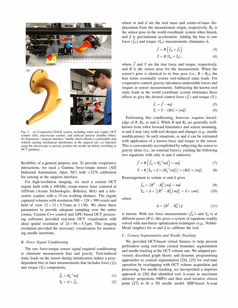
The DALK assistant has two major hardware components:a robot arm with end-effector force sensing and an OCTsystem (Fig. 3a). The robot arm and the operator jointlymanipulate the needle through an ergonomic “surgeon in-terface” handle (Fig. 3b). Both the handle-mounted needleand OCT scanner converge on the surgical site for real-time needle tracking (Fig. 3c). Together, these hardwarecomponents satisfy surgeons’ manipulation and visualizationneeds during DALK.
Due to the fine precision needed when working with the500 µm-thick cornea, we chose the IRB 120 robot arm (ABBRobotics; Zurich, Switzerland) for our system. This robotarm features 20 µm accuracy and 10 µm repeatability overa roughly 1 m3 workspace and can therefore accomplishboth gross positioning and fine tissue manipulation. With theIRB 120, we achieved sufficient precision for positioning aneedle within 50 µm as needed in DALK while retaining the
(a)
(b) (c)
Fig. 3. (a) Cooperative DALK system, including robot arm (right), OCTscanner (left), microscope (center), and artificial anterior chamber (blue).(b) Ergonomic “surgeon interface” handle which affords a comfortable gripwithout causing mechanical interference at the surgical site. (c) Operatorusing the microscope to grossly position the needle tip before switching toOCT guidance.
flexibility of a general purpose arm. To provide cooperativeinteractions, we used a Gamma force-torque sensor (ATIIndustrial Automation; Apex, NC) with ±32 N calibrationfor sensing at the surgeon interface.
For high-resolution imaging, we used a custom OCTengine built with a 100 kHz swept-source laser centered at1050 nm (Axsun Technologies; Billerica, MA) and a tele-centric scanner with a 15 cm working distance. The enginecaptured volumes with resolution 500×128×999 voxels andfield of view 12×12×5.5 mm at 1.1 Hz. We chose theseparameters to provide adequate sampling over the entirecornea. Custom C++ control and GPU-based OCT process-ing softwares provided real-time OCT visualization withideal spatial resolution of 24×94×5.5 µm. This imagingresolution provided the necessary visualization for monitor-ing needle insertions.
B. Force Signal Conditioning
The raw force-torque sensor signal required conditioningto eliminate measurement bias and gravity. Tool-inducedstatic loads on the sensor during initialization induce a pose-dependent bias in later measurements that includes force (~fb)and torque (~τb) components.
~fb = R−1b m~g (1)
~τb =~a×~fb, (2)
where m and ~a are the tool mass and center-of-mass dis-placement from the measurement origin, respectively, Rb isthe sensor pose in the world coordinate system when biased,and ~g is gravitational acceleration. Adding the bias to rawforce (~fm) and torque (~τm) measurements eliminates it.
~f = R(~fm +~fb
)(3)
~τ = R(~τm +~τb) , (4)
where ~f and ~τ are the true force and torque, respectively,and R is the sensor pose for the measurement. When thesensor’s pose is identical to its bias pose (i.e., R = Rb), thebias terms essentially restore tool-induced static loads. Forcooperative control, gravity introduces undesirable forces andtorques in sensor measurements. Subtracting the known toolstatic loads in the world coordinate system eliminates theseeffects to give the desired control force (~fc) and torque (~τc).
~fc = ~f −m~g (5)~τc =~τ− (R~a)× (m~g) (6)
Performing this conditioning, however, requires knowl-edge of R, Rb, m and ~a. While R and Rb are generally well-known from robot forward kinematics and sensor mounting,m and ~a may vary with tool designs and changes (e.g., needlemodifications). In such situations, m and ~a can be estimatedwith application of a known force and torque to the sensor.This is conveniently accomplished by subjecting the sensor togravity alone (i.e., no external forces), yielding the followingtwo equations with only m and ~a unknown.
~f = R(~fm +R−1
b m~g)= m~g (7)
~τ = R[~τm +~a×
(R−1
b m~g)]
= (R~a)× (m~g) (8)
Rearrangement to isolate m and ~a gives
~fm =(RT −RT
b)
m~g = m~u (9)~τm =~a×
[(RT −RT
b)
m~g]=~a× (m~u) (10)
where
~u =(RT −RT
b)~g (11)
is known. With raw force measurements (~fm’s and ~τm’s) atdifferent poses (R’s), this gives a system of equations readilysolved with non-linear optimization techniques (e.g., Nelder-Mead simplex) for m and ~a to calibrate the tool.
C. Cornea Segmentation and Needle Tracking
We provided OCT-based virtual fixtures to help preventperforation using real-time corneal boundary segmentationand needle tracking at the OCT volume rate. We adapted pre-viously described graph theory and dynamic programmingapproaches to corneal segmentation [24], [25] for real-timeoperation by overlapping with OCT volume acquisition andprocessing. For needle tracking, we incorporated a stepwiseapproach in [26] that identified tool A-scans in maximumintensity projections (MIPs) and then used iterative closestpoint [27] to fit a 3D needle model. MIP-based A-scan

identification exploited the needle’s hyperreflectivity in OCT.Segmentation in B-scans with needle-obscured A-scans wascorrected using image inpainting from [28].
D. Control System with Virtual Fixtures
The controller synthesized robot force sensing and OCTsegmentation and needle tracking to provide stabilized coop-erative robot-surgeon interactions and avoid corneal perfora-tion. We chose a direct Cartesian force-velocity control lawto guarantee no robot motion independent of surgeon-appliedforce.
~q = J−1o (~q)
[RH 00 RH
]G[
RH 00 RH
]T[~fc~τc
], (12)
where ~q is the target joint velocity vector, Jo(q) is the robotJacobian about the tool center of rotation, H is the rotationmatrix that defines the gain axes in the robot tool frame R,and G is the diagonal gain matrix. The surgeon thus remainsin direct control of the needle at all times (i.e., ~fc =~τc = 0implies ~q= 0). For added usability, we provided a foot switchwhich held ~q = 0 until depressed. This allowed the surgeonto release the handle while maintaining needle position.We chose G−1 = diag(40,40,40,0.9,0.75,0.4) in units ofN m−1 s and N m rad−1 s to compensate for the surgeon inter-face handle’s lever arms and discourage typically undesirableneedle rotations during DALK (e.g., rotation in horizontalplane). Our control software (Fig. 4) provided dynamicgain adjustment (0–100 %) through a joystick throttle andoperated with joint limits chosen to avoid singularities.
Using live corneal segmentation and needle tracking, thecontroller provided virtual fixtures to prevent perforation.After each OCT volume, the controller calculated the dis-tance from the needle tip to the posterior corneal boundary.If that distance reduced below 50 µm (90 % of typical corneathickness), the controller rejected control forces advancingor lowering the needle. This fixture remained active until theminimum observed distance exceeded the 50 µm threshold.For OCT volumes in which needle tracking failed, thecontroller maintained the prior fixture activation state.
We commanded the robot arm at 250 Hz through itsExternally-Guided Motion interface, which offers 20 ms re-sponse time, using a custom C++ control software. Weconfigured the robot controller for velocity-only tracking(zero position gain) and path low-pass filtering with 2 Hzbandwidth. This achieved highly responsive motion whilerejecting oscillations from noise and control lag. Low-passfiltering also attenuated the operator’s physiologic handtremor.
IV. METHODS
We designed a user study to compare DALK needleinsertions performed manually (i.e., free-hand) and coopera-tively with our system. Each operator completed training andtesting needle insertion trials in ex vivo human corneas underDuke University Medical Center IRB approval. We simulatedphysiologic corneal conditions with a pressured artificial an-terior chamber (Katena Products; Denville, NJ). For training,
Fig. 4. Software console for robot interaction, providing control force-torque readouts, sensor biasing, and force control gain adjustment.
operators performed two sets of four consecutive manual andcooperative trials in a randomized order. Operators receivedverbal and video instruction on DALK technique, coachingon cooperative system operation, and OCT visualization offinal needle depth for manual trials. For testing, operatorsperformed eight trials alternating manual and cooperativewith a randomized initial configuration. During manual trials,operators used a 27-gauge syringe-mounted needle with a45◦ bend to perform needle insertions using only light micro-scope guidance, which replicated current surgical practice.During cooperative trials, operators used a 27-gauge robot-mounted needle to perform needle insertions using lightmicroscope and real-time OCT guidance. Operators receivedno instruction or coaching once testing trials started.
For both training and testing, we asked operators to target90 % needle depth at the corneal apex as is typical insurgical practice. Operators did not complete DALK witha pneumo-dissection to allow cornea reuse for eight trials.Operators used one cornea for training and another fortesting. We rotated the cornea 45◦ after each trial to avoidreusing existing needle tracts and repressurized the artificialanterior chamber between trials at the operator’s request.For cooperative trials, we adjusted the force control gain to100 % during gross positioning, to 10 % during initial cornealpenetration, and to 2 % during advancement to the cornealapex, relative to our baseline gain settings.
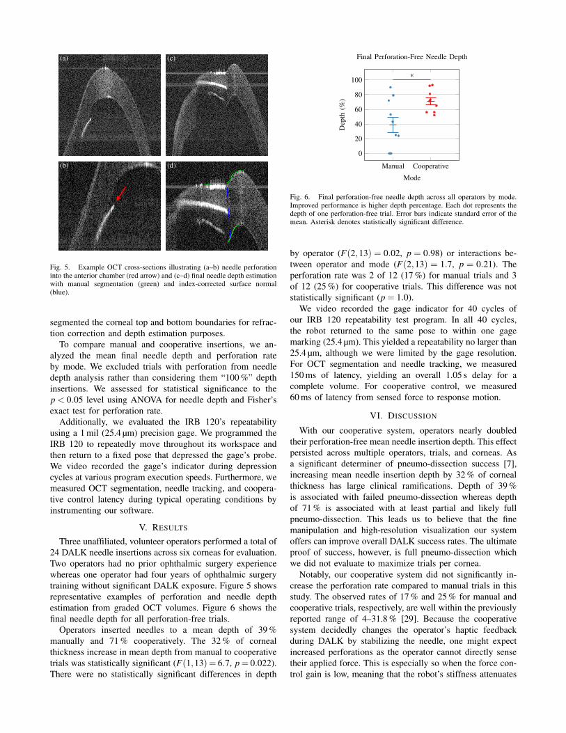
We recorded OCT volumes during the testing trials toevaluate for perforation and estimate final needle depth inlater analysis. For this study, we defined perforation asclear evidence of needle penetration through the endotheliallayer. A single grader blinded to the trial mode (manualor cooperative) reviewed each volume series for perforationand extracted the final needle depth. Using custom C++software and the approach in [7], the grader extracted finalneedle depth from refraction-corrected OCT cross-sectionsas a percentage of corneal thickness along the top surfacenormal passing through the needle tip. The grader manually
(a)
(b)
(c)
(d)
Fig. 5. Example OCT cross-sections illustrating (a–b) needle perforationinto the anterior chamber (red arrow) and (c–d) final needle depth estimationwith manual segmentation (green) and index-corrected surface normal(blue).
segmented the corneal top and bottom boundaries for refrac-tion correction and depth estimation purposes.
To compare manual and cooperative insertions, we an-alyzed the mean final needle depth and perforation rateby mode. We excluded trials with perforation from needledepth analysis rather than considering them “100 %” depthinsertions. We assessed for statistical significance to thep < 0.05 level using ANOVA for needle depth and Fisher’sexact test for perforation rate.
Additionally, we evaluated the IRB 120’s repeatabilityusing a 1 mil (25.4 µm) precision gage. We programmed theIRB 120 to repeatedly move throughout its workspace andthen return to a fixed pose that depressed the gage’s probe.We video recorded the gage’s indicator during depressioncycles at various program execution speeds. Furthermore, wemeasured OCT segmentation, needle tracking, and coopera-tive control latency during typical operating conditions byinstrumenting our software.
V. RESULTS
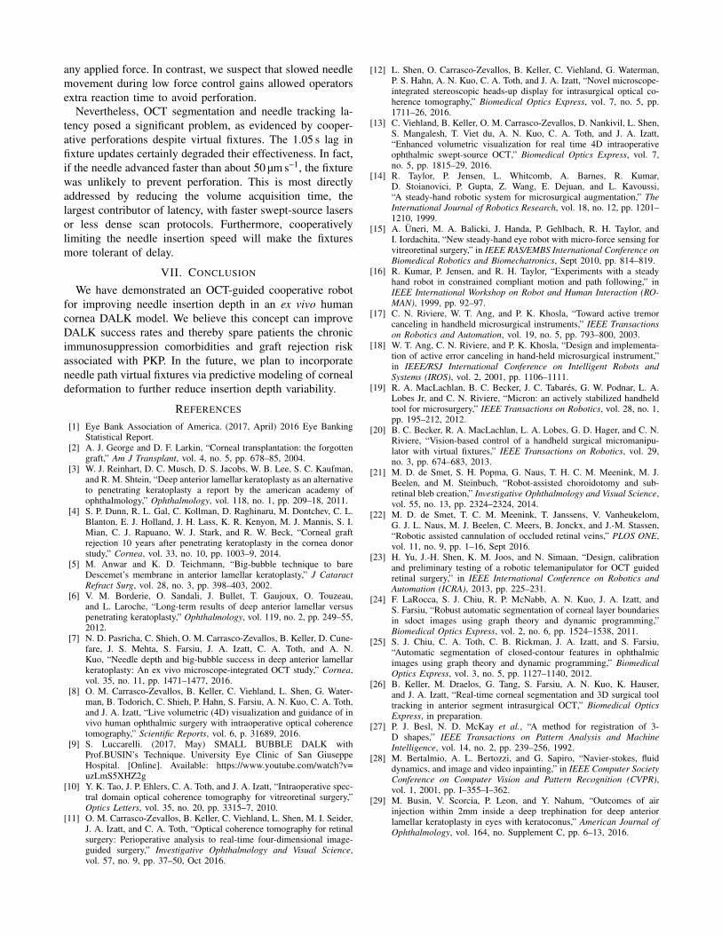
Three unaffiliated, volunteer operators performed a total of24 DALK needle insertions across six corneas for evaluation.Two operators had no prior ophthalmic surgery experiencewhereas one operator had four years of ophthalmic surgerytraining without significant DALK exposure. Figure 5 showsrepresentative examples of perforation and needle depthestimation from graded OCT volumes. Figure 6 shows thefinal needle depth for all perforation-free trials.
Operators inserted needles to a mean depth of 39 %manually and 71 % cooperatively. The 32 % of cornealthickness increase in mean depth from manual to cooperativetrials was statistically significant (F(1,13) = 6.7, p= 0.022).There were no statistically significant differences in depth
Manual Cooperative
0
20
40
60
80
100∗
Mode
Dep
th(%
)
Final Perforation-Free Needle Depth
Fig. 6. Final perforation-free needle depth across all operators by mode.Improved performance is higher depth percentage. Each dot represents thedepth of one perforation-free trial. Error bars indicate standard error of themean. Asterisk denotes statistically significant difference.
by operator (F(2,13) = 0.02, p = 0.98) or interactions be-tween operator and mode (F(2,13) = 1.7, p = 0.21). Theperforation rate was 2 of 12 (17 %) for manual trials and 3of 12 (25 %) for cooperative trials. This difference was notstatistically significant (p = 1.0).
We video recorded the gage indicator for 40 cycles ofour IRB 120 repeatability test program. In all 40 cycles,the robot returned to the same pose to within one gagemarking (25.4 µm). This yielded a repeatability no larger than25.4 µm, although we were limited by the gage resolution.For OCT segmentation and needle tracking, we measured150 ms of latency, yielding an overall 1.05 s delay for acomplete volume. For cooperative control, we measured60 ms of latency from sensed force to response motion.
VI. DISCUSSION
With our cooperative system, operators nearly doubledtheir perforation-free mean needle insertion depth. This effectpersisted across multiple operators, trials, and corneas. Asa significant determiner of pneumo-dissection success [7],increasing mean needle insertion depth by 32 % of cornealthickness has large clinical ramifications. Depth of 39 %is associated with failed pneumo-dissection whereas depthof 71 % is associated with at least partial and likely fullpneumo-dissection. This leads us to believe that the finemanipulation and high-resolution visualization our systemoffers can improve overall DALK success rates. The ultimateproof of success, however, is full pneumo-dissection whichwe did not evaluate to maximize trials per cornea.
Notably, our cooperative system did not significantly in-crease the perforation rate compared to manual trials in thisstudy. The observed rates of 17 % and 25 % for manual andcooperative trials, respectively, are well within the previouslyreported range of 4–31.8 % [29]. Because the cooperativesystem decidedly changes the operator’s haptic feedbackduring DALK by stabilizing the needle, one might expectincreased perforations as the operator cannot directly sensetheir applied force. This is especially so when the force con-trol gain is low, meaning that the robot’s stiffness attenuates
any applied force. In contrast, we suspect that slowed needlemovement during low force control gains allowed operatorsextra reaction time to avoid perforation.
Nevertheless, OCT segmentation and needle tracking la-tency posed a significant problem, as evidenced by cooper-ative perforations despite virtual fixtures. The 1.05 s lag infixture updates certainly degraded their effectiveness. In fact,if the needle advanced faster than about 50 µm s−1, the fixturewas unlikely to prevent perforation. This is most directlyaddressed by reducing the volume acquisition time, thelargest contributor of latency, with faster swept-source lasersor less dense scan protocols. Furthermore, cooperativelylimiting the needle insertion speed will make the fixturesmore tolerant of delay.
VII. CONCLUSION
We have demonstrated an OCT-guided cooperative robotfor improving needle insertion depth in an ex vivo humancornea DALK model. We believe this concept can improveDALK success rates and thereby spare patients the chronicimmunosuppression comorbidities and graft rejection riskassociated with PKP. In the future, we plan to incorporateneedle path virtual fixtures via predictive modeling of cornealdeformation to further reduce insertion depth variability.
REFERENCES
[1] Eye Bank Association of America. (2017, April) 2016 Eye BankingStatistical Report.
[2] A. J. George and D. F. Larkin, “Corneal transplantation: the forgottengraft,” Am J Transplant, vol. 4, no. 5, pp. 678–85, 2004.
[3] W. J. Reinhart, D. C. Musch, D. S. Jacobs, W. B. Lee, S. C. Kaufman,and R. M. Shtein, “Deep anterior lamellar keratoplasty as an alternativeto penetrating keratoplasty a report by the american academy ofophthalmology,” Ophthalmology, vol. 118, no. 1, pp. 209–18, 2011.
[4] S. P. Dunn, R. L. Gal, C. Kollman, D. Raghinaru, M. Dontchev, C. L.Blanton, E. J. Holland, J. H. Lass, K. R. Kenyon, M. J. Mannis, S. I.Mian, C. J. Rapuano, W. J. Stark, and R. W. Beck, “Corneal graftrejection 10 years after penetrating keratoplasty in the cornea donorstudy,” Cornea, vol. 33, no. 10, pp. 1003–9, 2014.
[5] M. Anwar and K. D. Teichmann, “Big-bubble technique to bareDescemet’s membrane in anterior lamellar keratoplasty,” J CataractRefract Surg, vol. 28, no. 3, pp. 398–403, 2002.
[6] V. M. Borderie, O. Sandali, J. Bullet, T. Gaujoux, O. Touzeau,and L. Laroche, “Long-term results of deep anterior lamellar versuspenetrating keratoplasty,” Ophthalmology, vol. 119, no. 2, pp. 249–55,2012.
[7] N. D. Pasricha, C. Shieh, O. M. Carrasco-Zevallos, B. Keller, D. Cune-fare, J. S. Mehta, S. Farsiu, J. A. Izatt, C. A. Toth, and A. N.Kuo, “Needle depth and big-bubble success in deep anterior lamellarkeratoplasty: An ex vivo microscope-integrated OCT study,” Cornea,vol. 35, no. 11, pp. 1471–1477, 2016.
[8] O. M. Carrasco-Zevallos, B. Keller, C. Viehland, L. Shen, G. Water-man, B. Todorich, C. Shieh, P. Hahn, S. Farsiu, A. N. Kuo, C. A. Toth,and J. A. Izatt, “Live volumetric (4D) visualization and guidance of invivo human ophthalmic surgery with intraoperative optical coherencetomography,” Scientific Reports, vol. 6, p. 31689, 2016.
[9] S. Luccarelli. (2017, May) SMALL BUBBLE DALK withProf.BUSIN’s Technique. University Eye Clinic of San GiuseppeHospital. [Online]. Available: https://www.youtube.com/watch?v=uzLmS5XHZ2g
[10] Y. K. Tao, J. P. Ehlers, C. A. Toth, and J. A. Izatt, “Intraoperative spec-tral domain optical coherence tomography for vitreoretinal surgery,”Optics Letters, vol. 35, no. 20, pp. 3315–7, 2010.
[11] O. M. Carrasco-Zevallos, B. Keller, C. Viehland, L. Shen, M. I. Seider,J. A. Izatt, and C. A. Toth, “Optical coherence tomography for retinalsurgery: Perioperative analysis to real-time four-dimensional image-guided surgery,” Investigative Ophthalmology and Visual Science,vol. 57, no. 9, pp. 37–50, Oct 2016.
[12] L. Shen, O. Carrasco-Zevallos, B. Keller, C. Viehland, G. Waterman,P. S. Hahn, A. N. Kuo, C. A. Toth, and J. A. Izatt, “Novel microscope-integrated stereoscopic heads-up display for intrasurgical optical co-herence tomography,” Biomedical Optics Express, vol. 7, no. 5, pp.1711–26, 2016.
[13] C. Viehland, B. Keller, O. M. Carrasco-Zevallos, D. Nankivil, L. Shen,S. Mangalesh, T. Viet du, A. N. Kuo, C. A. Toth, and J. A. Izatt,“Enhanced volumetric visualization for real time 4D intraoperativeophthalmic swept-source OCT,” Biomedical Optics Express, vol. 7,no. 5, pp. 1815–29, 2016.
[14] R. Taylor, P. Jensen, L. Whitcomb, A. Barnes, R. Kumar,D. Stoianovici, P. Gupta, Z. Wang, E. Dejuan, and L. Kavoussi,“A steady-hand robotic system for microsurgical augmentation,” TheInternational Journal of Robotics Research, vol. 18, no. 12, pp. 1201–1210, 1999.
[15] A. Uneri, M. A. Balicki, J. Handa, P. Gehlbach, R. H. Taylor, andI. Iordachita, “New steady-hand eye robot with micro-force sensing forvitreoretinal surgery,” in IEEE RAS/EMBS International Conference onBiomedical Robotics and Biomechatronics, Sept 2010, pp. 814–819.
[16] R. Kumar, P. Jensen, and R. H. Taylor, “Experiments with a steadyhand robot in constrained compliant motion and path following,” inIEEE International Workshop on Robot and Human Interaction (RO-MAN), 1999, pp. 92–97.
[17] C. N. Riviere, W. T. Ang, and P. K. Khosla, “Toward active tremorcanceling in handheld microsurgical instruments,” IEEE Transactionson Robotics and Automation, vol. 19, no. 5, pp. 793–800, 2003.
[18] W. T. Ang, C. N. Riviere, and P. K. Khosla, “Design and implementa-tion of active error canceling in hand-held microsurgical instrument,”in IEEE/RSJ International Conference on Intelligent Robots andSystems (IROS), vol. 2, 2001, pp. 1106–1111.
[19] R. A. MacLachlan, B. C. Becker, J. C. Tabares, G. W. Podnar, L. A.Lobes Jr, and C. N. Riviere, “Micron: an actively stabilized handheldtool for microsurgery,” IEEE Transactions on Robotics, vol. 28, no. 1,pp. 195–212, 2012.
[20] B. C. Becker, R. A. MacLachlan, L. A. Lobes, G. D. Hager, and C. N.Riviere, “Vision-based control of a handheld surgical micromanipu-lator with virtual fixtures,” IEEE Transactions on Robotics, vol. 29,no. 3, pp. 674–683, 2013.
[21] M. D. de Smet, S. H. Popma, G. Naus, T. H. C. M. Meenink, M. J.Beelen, and M. Steinbuch, “Robot-assisted choroidotomy and sub-retinal bleb creation,” Investigative Ophthalmology and Visual Science,vol. 55, no. 13, pp. 2324–2324, 2014.
[22] M. D. de Smet, T. C. M. Meenink, T. Janssens, V. Vanheukelom,G. J. L. Naus, M. J. Beelen, C. Meers, B. Jonckx, and J.-M. Stassen,“Robotic assisted cannulation of occluded retinal veins,” PLOS ONE,vol. 11, no. 9, pp. 1–16, Sept 2016.
[23] H. Yu, J.-H. Shen, K. M. Joos, and N. Simaan, “Design, calibrationand preliminary testing of a robotic telemanipulator for OCT guidedretinal surgery,” in IEEE International Conference on Robotics andAutomation (ICRA), 2013, pp. 225–231.
[24] F. LaRocca, S. J. Chiu, R. P. McNabb, A. N. Kuo, J. A. Izatt, andS. Farsiu, “Robust automatic segmentation of corneal layer boundariesin sdoct images using graph theory and dynamic programming,”Biomedical Optics Express, vol. 2, no. 6, pp. 1524–1538, 2011.
[25] S. J. Chiu, C. A. Toth, C. B. Rickman, J. A. Izatt, and S. Farsiu,“Automatic segmentation of closed-contour features in ophthalmicimages using graph theory and dynamic programming,” BiomedicalOptics Express, vol. 3, no. 5, pp. 1127–1140, 2012.
[26] B. Keller, M. Draelos, G. Tang, S. Farsiu, A. N. Kuo, K. Hauser,and J. A. Izatt, “Real-time corneal segmentation and 3D surgical tooltracking in anterior segment intrasurgical OCT,” Biomedical OpticsExpress, in preparation.
[27] P. J. Besl, N. D. McKay et al., “A method for registration of 3-D shapes,” IEEE Transactions on Pattern Analysis and MachineIntelligence, vol. 14, no. 2, pp. 239–256, 1992.
[28] M. Bertalmio, A. L. Bertozzi, and G. Sapiro, “Navier-stokes, fluiddynamics, and image and video inpainting,” in IEEE Computer SocietyConference on Computer Vision and Pattern Recognition (CVPR),vol. 1, 2001, pp. I–355–I–362.
[29] M. Busin, V. Scorcia, P. Leon, and Y. Nahum, “Outcomes of airinjection within 2mm inside a deep trephination for deep anteriorlamellar keratoplasty in eyes with keratoconus,” American Journal ofOphthalmology, vol. 164, no. Supplement C, pp. 6–13, 2016.