purification and properties of staphylococcal delta …iai.asm.org/content/3/3/449.full.pdfules,...
TRANSCRIPT

INFECTION AND IMMUNITY, March 1971, p. 449-465Copyright © 1971 American Society for Microbiology
Vol. 3, No. 3Printed in U.S.A.
Purification and Properties of StaphylococcalDelta Hemolysin
ARNOLD S. KREGER,' KWANG-SHIN KIM, FRANK ZABORETZKY,AND ALAN W. BERNHEIMER
Department of Microbiology, New York University School of Medicine, New York, New York 10016
Received for publication 19 November 1970
Large amounts (200 mg per liter of culture supernatant fluid) of highly purifiedstaphylococcal soluble delta hemolysin were obtained by adsorption to and selectiveelution from hydroxyapatite followed by exhaustive dialysis against water, con-
centration by polyvinylpyrrolidone or polyethylene glycol 20,000 dialysis, and a
final water dialysis. No carbohydrate, phosphorus, or inactive 280-nm absorbingmaterial was detected in the preparation; however, analysis by density gradientcentrifugation, gel filtration, analytical ultracentrifugation, carboxymethyl cellulosechromatography, polyacrylamide disc gel electrophoresis, isoelectric focusing, andelectron microscopy revealed that the lysin was molecularly heterogeneous. Thepreparation contained an acidic fibrous lysin (S20, of 11.9) and a basic lysin com-
ponent composed of a population of granular aggregates of various sizes, with a
maximum Sm,, of approximately 4.9. No other staphylococcal products were de-tected in the preparation. The lysin was active against erythrocytes from many ani-mal species and acted synergistically with staphylococcal beta hemolysin againstsheep erythrocytes. It was soluble in chloroform-methanol (2: 1), was inactivated byvarious phospholipids, normal sera, and proteolytic enzymes, but was partially re-
sistant to heat inactivation. Activity was not affected by Ca2+, Mg2+, citrate, ethyl-enediaminetetraacetic acid, or cysteine. The lysin preparation also disrupted bacterialprotoplasts and spheroplasts, erythrocyte membranes, lysosomes, and lipid spher-ules, was growth-inhibitory for certain bacteria, and clarified egg yolk-agar. Largeamounts produced dermonecrosis in rabbits and guinea pigs. The minimum lethalintravenous dose for mice and guinea pigs was approximately 110 and 30 mg/kg,respectively.
Staphylococcal delta hemolysin is differentiatedfrom the other staphylococcal hemolysins by itsability to lyse horse and human erythrocytes(51, 66) and by its ability to be inhibited byphospholipids (F. A. Kapral, Bacteriol. Proc.,p. 64, 1967) and normal sera (24, 35, 51). Prepa-rations of delta hemolysin also (i) disrupt bac-terial protoplasts and spheroplasts (7, 41),tissue culture cells (24, 29), leukocytes (23, 24,29, 34), lysosomes (9, 19), mitochondria (H. S.Kantor et al., Bacteriol. Proc., p. 78, 1970),lipid spherules (21), and erythrocyte membranes(28); (ii) are lethal for mice (42); (iii) producedermonecrosis in guinea pigs (51) and rabbits(22; F. A. Kapral, personal communication);(iv) possess limited antibacterial activity (28,32); (v) act synergistically with staphylococcalbeta hemolysin against sheep erythrocytes (51);(vi) are mitogenic for human lymphocytes (19);
1 Present address: Department of Medicine, New York Uni-versity School of Medicine, New York, N.Y. 10016.
(vii) clear egg yolk-agar (41); and (viii) possessphospholipase C activity with phosphatidyl-inositol and phosphatidylserine as substrates(67). Rigorous proof for the belief that all ofthese effects are caused by delta hemolysin islacking.
Delta hemolysin has been partially purifiedand characterized by Marks and Vaughan (51),Jackson and Little (34-36), Yoshida (68), Kayserand Raynaud (42), Kayser (38-41), Hallander(28, 29), Caird and Wiseman (12, 67), Maheswa-ran and Lindorfer (Bacteriol. Proc., p. 78, 1970),Kantor et al. (Bacteriol. Proc., p. 78, 1970),Kapral (personal communication), and Moilbyand Wadstrom (54). In this report, we (i) presenta simple and reproducible method for obtaininggram amounts of highly purified delta hemolysinand (ii) describe some of the physical, chemical,and biological properties of the purified prepa-ration.
449
on June 22, 2018 by guesthttp://iai.asm
.org/D
ownloaded from
KREGER ET AL.
MATERIALS AND METHODSOrganism. A mutant of the Staphylococcus aureus
Wood 46 strain, designated W46M and obtained byultraviolet irradiation of the Wood 46 strain, was usedfor delta hemolysin production. It is coagulase-nega-tive and has been estimated to produce only about 5%of the alpha hemolysin produced by the parent strain(7).Measurement of hemolytic activity. Alpha hemolysin
was assayed as described by Bernheimer and Schwartz(8). Beta hemolysin activity was determined as re-ported by Gow and Robinson (25). Delta hemolysinwas estimated as described for alpha hemolysin, withthe exceptions that washed horse erythrocytes wereused instead of rabbit erythrocytes and bovine serumalbumin was not added to the buffer. Unless otherwisementioned, isotonic phosphate-buffered saline (PBS),pH 7, containing 0.067M phosphate and 0.077 Msodium chloride was utilized as the solvent and diluentfor delta hemolysin. In all cases, 1 hemolytic unit(HU) was the amount of lysin required to produce50% lysis of a 0.7% (v/v) erythrocyte suspension.
Cultivation of organism. Yeast diffusate medium(6.4 liters) was prepared as previously reported (8),with the exception that the yeast extract was obtainedfrom the Fisher Scientific Co. (Fair Lawn, N.J.) ratherthan from Difco (Detroit, Mich.). The inoculum wasprepared by washing the cocci of an overnight brothculture and suspending them in 0.5 volume of sterile0.9% saline. Each 2-liter flask, containing approxi-mately 533 ml of media, was inoculated with 0.3 ml ofwashed cocci suspension and incubated for 15 to 18 hrat 37 C on a rotary shaker operating at 220 cycles/min.
Examiination for other staphylococcal products.Purified delta hemolysin (I mg) was examined for thepresence of esterase (33, 50), lipase (27, 45, 56),phospholipase C (67), protease (44), gelatinase, de-oxyribonuclease (t), ribonuclease (24), coagulase (1),staphylokinase (1, 4), hyaluronidase (1), lysozyme(31), acid phosphatase (49), and alkaline phosphatase(20) activity. The methods used are those described inthe appropriate references. Enzymes and substratesother than phospholipids were obtained from theSigma Chemical Co. (St. Louis, Mo.), WorthingtonBiochemical Corp. (Freehoid, N.J.), and MannLaboratories (New York, N.Y.). Phospholipids wereobtained from the Sylvana Co. (Millburn, N.J.) andApplied Science Laboratories (State College, Pa.).
Sucrose density gradient centrifugation. Zonalcentrifugation in a sucrose density gradient was carriedout by the method of Martin and Ames (52). A 1-mlamount of a 1% (w/v) solution of soluble delta lysinin PBS was layered on a 28-ml linear gradient of 5 to20% sucrose in PBS contained in a polyallomer tube(25 by 75 mm). After centrifugation for 24 hr at25,000 rev/min, a hole was punched in the bottom ofthe tube and eight-drop fractions were collected. Eachfraction was diluted with 1 ml of PBS, and the dilutedfractions were assayed for delta hemolysin activity andabsorbance at 280 nm.
Gel filtration chromatography. Gel ifitration wasperformed on Sephadex G-150 (Pharmacia FineChemicals, Piscataway, N.J.) and on Bio-Gel A-Sm,200 to 400 mesh (Bio-Rad Laboratories, New York,
N.Y.), packed in water-cooled (5 C) columns (typeK25/100, Pharmacia, Piscataway, N.J.). Fractionswere collected in a Buchler (Fort Lee, N.J.) refrig-erated fraction collector.
Analytical ultracentrifugation. Solutions of deltahemolysin were subjected to sedimentation velocityanalysis, by using schlieren and interference optics, ina Spinco An-D analytical rotor at 56,000 rev/min and20 C with the Spinco model E analytical ultracentri-fuge equipped with a temperature control unit. Photo-graphs of the schlieren and interference patterns weretaken at 4- or 8-min intervals after the rotor attainedtwo-thirds maximum speed. Photographic plates wereread with the aid of a Nikon profile projector (model6C), and the sedimentation coefficients were estimatedby the method described by Schachman (60). Withschlieren optics, the sedimentation coefficients weredetermined from the top (highest point) of the peak.With interference optics, the sedimentation coefficientswere determined from the position of the steepestfringe corresponding to the middle portion of theboundary. The observed sedimentation coefficientswere corrected to standard conditions by the ap-propriate viscosity and density corrections.
Carboxymethyl cellulose chromatography. What-man wet, microgranular, carboxymethyl (CM) cellu-lose (CM 52, H. Reeve Angel, Inc., Clifton, N.J.) wasequilibrated with 0.05 M acetate buffer (pH 5) andpacked (2.5 by 33.5 cm) in a water-cooled column(type K25/45, Pharmacia, Piscataway, N.J.). Afteraddition of the delta hemolysin (100 mg) dissolved inequilibrating buffer to the column, the column waswashed with approximately 250 ml of equilibratingbuffer, and the hemolytic activity was eluted from thecolumn with a gradient elution apparatus. The ap-paratus consisted of an upper reservoir containing0.05 M acetate buffer (pH 5) and 0.5 M sodium chloridewhich fed into a constant-volume mixing chambercontaining approximately 400 ml of 0.05M acetatebuffer (pH 5). The constant-volume mixing chamberdischarged into the column at a rate of approximately10 ml/hr, maintained by adjustment at the reservoir.Fractions (approximately 5 ml) were collected andassayed for hemolytic activity and absorbance at280 nm. The elution gradient was measured with aconductivity bridge (model 31, Yellow SpringsInstrument Co., Yellow Springs, Ohio).
Polyacrylamide disc gel electrophoresis. Disc gelelectrophoresis of soluble delta hemolysin was per-formed in the 7% gel alkaline system (separation atpH 9.5) described by Ornstein (57) and Davis (14) andin the 7.5% gel acidic system (separation at pH 4.3)described by Reisfeld et al. (59) with model 12 Canalcoequipment (Canal Industrial Corp., Rockville, Md.).After electrophoresis, the gels were extruded, stainedand fixed overnight in 1% aniline blue-black in 7%acetic acid, and electrolytically destained.
Chemical analyses. Protein was determined by themethod of Lowry et al. (46) with crystalline bovineserum albumin (Armour Pharmaceutical Co., Kanka-kee, Ill.) as the standard. Carbohydrate was estimatedby the orcinol, indole, and anthrone reactions (37),and hexosamine was estimated by a modified Elson-Morgan method, as described by Kabat and Mayer
450 INFEC. IMMUN.
on June 22, 2018 by guesthttp://iai.asm
.org/D
ownloaded from
STAPHYLOCOCCAL DELTA HEMOLYSIN
(37). Total phosphorus was determined as describedby De Siervo (15). Free lipid was estimated by ex-tracting soluble delta hemolysin (100 mg) with threeportions (10 ml each) of ether, evaporating the ether,and weighing the residue. Bound lipid was determinedby extracting acid-hydrolyzed delta hemolysin, pre-pared by hydrolyzing 100 mg of lysin with 10 ml of6 N hydrochloric acid for 20 hr at 110 C, with ether(10 ml), washing the ether extract four times withwater, evaporating the ether, and weighing the residue.Nitrogen (Dumas method), carbon, hydrogen, andsulfur contents were determined at the Squibb Institutefor Medical Research, New Brunswick, N.J. Nitrogenwas also determined by the micro-Kjeldahl method, asdescribed by Mayer (53).Amino acid analysis. Amino acid analyses were
performed by the method of Spackman et al. (62).Two samples (1.5 and 3 mg) of soluble delta hemolysinwere hydrolyzed for 22, 48, 72, and 97 hr at 110 Cwith 6 N HCI (1 ml) in sealed Pyrex tubes after evacua-tion and degassing. The hydrolysates were dried undervacuum, and amino acid analyses were performedwith a Beckman model 120C amino acid analyzer.Color constants for each amino acid were determinedby analyzing standard amino acid mixtures shortlybefore each determination. The amounts of threonineand serine were calculated by extrapolation to zerohydrolysis time. The amounts of valine, leucine,and isoleucine were calculated from the 97-hr hydroly-sis samples. The amounts of all other amino acids werecalculated from the averaged values of all of thesamples. The tryptophan content was estimated by thespectrophotometric method of Edelhoch (18). Half-cystine was determined as cysteic acid by the per-formic acid oxidation method described by Moore(55).
Spectral analysis. The ultraviolet absorption spec-trum was determined with a model 14 Cary recordingspectrophotometer and quartz cuvettes having a lightpath of 1 cm. The infrared absorption spectrum of thesoluble lysin (KBr pellet) was determined with aPerkin-Elmer 421 grating infrared spectrophotometer.
Electron microscopy. Lysin, dissolved in distilledwater, was negatively stained with 2% (w/v) am-monium molybdate (pH 6.8; certified reagent, FisherScientific Co., Pittsburgh, Pa.) on Formvar-supported,carbon-coated grids (11). The preparations wereexamined at an accelerating voltage of 80 kv in aSiemens Elmiskop IA equipped with a decontamina-tion device and employing a 35-,m objective aperture.
Inactivation studies. The capacity of various lipidsand normal sera to inhibit the hemolytic activity ofsoluble delta hemolysin was assayed by mixing variousamounts of test material (in 0.5 ml of PBS) with afixed amount (3 HU, in 0.5 ml PBS) of delta lysin,allowing the mixtures to stand for 10 min at roomtemperature, and adding 1 ml of a 0.7% washed horseerythrocyte suspension. After 30 min at 37 C, themixtures were centrifuged briefly, and the hemoglobinin the supernatant fluids was estimated colorimetricallyat 545 nm. Fifty per cent hemolysis was used as theend point of the titrations. Phosphatidylcholine anddiphosphatidylglycerol were obtained from SylvanaCo., Millburn, N.J.; phosphatidylserine, phospha-
tidylinositol, phosphatidylethanolamine, and sphin-gomyelin were from Applied Science Laboratories,Inc., State College, Pa.; and cholesterol was fromMatheson Coleman and Bell, East Rutherford, N.J.The capacity of various proteolytic enzymes to
destroy the hemolytic activity ofsoluble delta lysin wasdetermined by incubating 100 and 10 jg of enzymedissolved in PBS (0.1 ml) with 1 mg of lysin dissolvedin PBS (1 ml) for 30 min at 37 C and determining theresidual hemolytic activity. Trypsin (bovine pancreas,twice crystallized) was obtained from Sigma ChemicalCo., St Louis, Mo.; crystalline chymotrypsin wasfrom Armour and Co., Chicago, Ill.; Pronase (gradeB) was from Calbiochem, Los Angeles, Cal.; andcrystalline papain was from Worthington BiochemicalCorp., Freehold, N.J.The ability of soluble delta hemolysin to resist heat
inactivation was examined by heating 0.1% solutionsof lysin in PBS for 1 hr at various temperatures, fol-lowed by assaying for residual hemolytic activity.
Lysis of bacterial protoplasts and spheroplasts. Pro-toplasts of Streptococcus faecalis (ATCC 9790) wereprepared by the method of Shockman and Slade (61)by use of 1 mg of lysozyme per ml for 16 hr at 37 C.The protoplasts were sedimented by centrifugationand were suspended in 0.5 M sucrose in 0.05 M phos-phate buffer (pH 6.6) containing 0.01 M Mga+.Micrococcus lysodeikticus protoplasts were preparedwith lysozyme as reported by Bernheimer (5). Proto-plasts of Bacillus megaterium KM and Sarcina luteaand spheroplasts of Escherichia coli W [preparedwith lysozyme and ethylenediaminetetraacetic acid(EDTA)J and Vibrio metschnikovii were obtainedas previously described (10).
Test preparations (1 ml) having an optical densityat 500 nm of approximately 1.0 were examined forsusceptibility to lysis by soluble delta hemolysin byincubation for 30 min at 37 C with various amounts oflysin dissolved in the protoplast- or spheroplast-sus-pending buffer (1 ml). The amount of lysin requiredto produce half-maximal lysis (half-maximal decreasein optical density) was determined by interpolation.
Disruption of erythrocyte membranes. Rabbiterythrocyte membranes were prepared as described byDodge et al. (16). The membrane suspension (0.5 ml)having an optical density at 650 nm of approximately0.4 was examined for susceptibility to lysis by solubledelta hemolysin by incubation for 30 min at 37 C withvarious amounts of lysin dissolved in the membrane-suspending hypotonic (7.6 X 10³ M, pH 7.4) phos-phate buffer (0.5 ml). The amount of lysin required toproduce half-maximal lysis (half-maximal decrease inoptical density) was determined by interpolation.
Disruption of lysosomes. Rabbit polymorphonuclearleukocyte lysosomes were isolated as described byWeissmann et al. (65) and were suspended in 0.34 Msucrose. The lysosome suspension (0.9 ml), having anoptical density at 520 nm of approximately 0.5, wastested for susceptibility to disruption by delta lysin byincubation for 30 min at 37 C with various amounts oflysin dissolved in PBS containing 0.34 M sucrose(0.1 ml). The amount of lysin required to produce half-maximal lysis was determined by interpolation.
Lipid spherne disruption. Spherules were prepared,
VOL. 3, 1971 451
on June 22, 2018 by guesthttp://iai.asm
.org/D
ownloaded from
KREGER ET AL.
assayed for chromate leakage, and examined in theelectron microscope as previously reported (21). Thelysin was dissolved in distilled water.
Antibacterial activity. The ability of soluble deltahemolysin to inhibit the growth of various bacteriawas examined by the tube dilution method. Doublingdilutions (1 ml) of lysin in 0.9% sterile saline wereadded to double concentrated (1 ml) Trypticase SoyBroth (TSB, BBL). Todd-Hewitt Broth (BBL) wasused instead of TSB for the growth of the group AStreptococcus. Each assay mixture was inoculatedwith one loop (5 mm diameter) of a 24-hr culture ofthe test bacterium, and the mixtures were incubated at37 C for 4 days. The tubes were examined daily forgrowth.
Clearing of egg yolk-agar. Egg yolk-agar was com-posed of 1.25% (v/v) egg yolk, 1.5% (w/v) agar(Difco), and 0.01% (w/v) Merthiolate in 0.05 M tris(hydroxymethyl)aminomethane-hydrochloride buffer(pH 7.6). Ten millimeter holes were cut in the agar,and various amounts of soluble delta hemolysin, dis-solved in saline (0.2 ml), were added to the wells. Theplates were incubated for 5 days at 37 C and wereexamined daily.
Toxicity. Various amounts of soluble delta hemoly-sin, dissolved in 0.9% sterile saline, were injectedintravenously into female Swiss mice (20 to 25 g) andintracardially into female guinea pigs (330 to 350 g).Animals surviving for less than 24 hr were scored asdeaths.
Various amounts of soluble delta hemolysin and12S-free alpha hemolysin (8), dissolved in 0.1 ml of0.9% saline (containing 0.1% gelatin for the alphalysin) and sterilized by membrane (Millipore Corp.)filtration, were injected intradermally into shavedfemale New Zealand white rabbits and guinea pigs.The animals were examined daily. The ability oflecithin to inhibit the dermonecrosis produced bydelta lysin and alpha lysin was examined by allowingthe lysins to stand at room temperature for 10 minwith 0.5 mg of lecithin before injection.
RESULTSPurification procedure. All procedures were
done at approximately 5 to 10 C. The cultureswere pooled and centrifuged to remove the cocci.The supernatant fluid (approximately 6 liters,containing 300 to 400 HU/ml) was mixed for 3hr with 120 g of hydroxyapatite (Bio-Gel HTP,Bio-Rad Laboratories, New York, N.Y.), al-lowed to stand overnight, and tested for residualdelta hemolysin activity. Approximately 98%of the initial activity was adsorbed. The super-natant fluid was discarded, and the hydroxyapatitewas washed free from adhering supernatantfluid with 0.01 M potassium phosphate buffer(pH 6.8) followed by six washings (500 ml each)with 0.4 M phosphate buffer (pH 6.8) for 30 to60 min each time. These washings, which werediscarded, contained approximately 5 to 10%of the adsorbed hemolytic activity and the ma-
jority of the adsorbed pigment. The adsorbeddelta hemolysin activity was released by washingthe hydroxyapatite five to six times with 1 Mphosphate (pH 7.4) buffer (400 ml each time)for 30 to 60 min. The hemolytic supernatantfluid, containing about 50% of the adsorbedactivity, was centrifuged to remove traces ofhydroxyapatite and exhaustively dialyzed againstwater to remove the phosphate ions. The pre-cipitate appearing during dialysis was removedby centrifugation, washed three times withwater, and lyophilized. This precipitate, a buff-colored powder, was called "insoluble deltahemolysin." The fluid in the dialysis sac (ap-proximately 3.7 liters) was concentrated to ap-proximately one-fifth to one-sixth its originalvolume by dialysis against either polyvinyl-pyrrolidone (A. H. Thomas, Co., Philadelphia,Pa.) or polyethylene glycol 20,000 (Fisher Scien-tific Co., Fair Lawn, N.J.), exhaustively dialyzedagainst water, centrifuged to remove a trace ofprecipitate, and lyophilized. This final product, afluffy-white powder, was designated "solubledelta hemolysin." Each liter of culture super-natant fluid yielded approximately 50 to 70 mgof insoluble delta hemolysin having about 400HU/mg and approximately 200 to 250 mg ofsoluble delta hemolysin having about 200HU/mg. The activity of the soluble delta hemol-ysin was observed to vary by as much as 25 to50% with different lots of horse erythrocytes.The specific activity of the soluble delta hemol-ysin (HU/milligram of protein) was about 15times greater than that of the culture super-natant fluid. The total activity of both forms ofdelta lysin represents approximately 20 to 30%of the culture supernatant fluid activity.
Solubility and stability in various buffers andorganic solvents. Soluble delta hemolysin was
0.7
zA
Cic0
06
05
04
03
0.2
0
6 I0 14 18 22 26 30 34
FRACTION
42 46 50
160
40
20 E
a-
-00 u)
F-
60
40 I
20
54
FIG. 1. Sucrose density gradient centrifugation ofsoluble delta hemolysin. Fractions (eight drops) werediluted with phosphate-buffered saline (I ml) and as-sayedfor hemolytic activity (0) andfor absorbance at280nm (a).
452 INFEC. IMMUN.
-1-
on June 22, 2018 by guesthttp://iai.asm
.org/D
ownloaded from

STAPHYLOCOCCAL DELTA HEMOLYSIN
soluble and stable for at least 7 days at 5 C inwater, a variety of 0.1 M buffers ranging frompH 5 to 9, 0.1 M sodium hydroxide and aceticacid, 6 M guanidine hydrochloride, 8 M urea,and 0.1% sodium lauryl sulfate. The lysin wasinsoluble in chloroform, acetone, and ether butwas soluble in chloroform-methanol (2:1),methanol, and 75% ethanol. The insoluble deltahemolysin was poorly soluble (<0.05 mg/mi)in water and in a variety of 0.1 M buffers unlessthey contained 8 M urea. It was also soluble andstable in 0.1 M sodium hydroxide and chloro-form-methanol (2:1).
Examination for other staphylococcal products.Esterase, lipase, caseinase, gelatinase, deoxy-ribonuclease, ribonuclease, coagulase, staphylo-kinase, hyaluronidase, lysozyme, acid phospha-tase, and alkaline phosphatase activities werenot detected in the soluble delta hemolysin prepa-ration. No phospholipase C activity was de-tected when phosphatidylcholine, phosphatidyl-serine, phosphatidylinositol, phosphatidyletha-nolamine, diphosphatidylglycerol, and sphingo-myelin were used as substrates.
Sucrose density gradient centrifugation. Noinactive 280-nm absorbing peaks were observed.One broad active peak trailing toward the topof the gradient was observed (Fig. 1).
Gel filtration chromatography. No inactive280-nm absorbing peaks were observed. Activityeluted from Sephadex G-150 (Fig. 2) as a voidvolume peak trailing almost the entire length ofthe column. Activity eluted from Bio-Gel A-5m(Fig. 3) as two peaks. One peak eluted slightlyafter the void volume. Both this material andthe more slowly eluted lysin had similar activitiesper milligram (dry weight); however, the slowlyeluted lysin had approximately twice the ac-
08_' ,
0 04 _03~~~~~~~~~~~~~~~~~~~~~~~~~~~~~~~~~~~~~~~~~~~< 05_oi
026< 0 3
01100 140 180 220 260 300 340 380 420 460 5C
180
160
840 E
40
20 IiOO
mi OF EFFLUENT
FIG. 2. Sephadex G-150 gel filtration of solubledelta hemolysin. Lysin (70 mg) was applied to a
column (2.5 by 90 cm) and eluted at a flow rate of ap-proximately 15 ml/hr with 0.05 M phosphate buffer(pH 7.2) containing 0.5 M sodium chloride. Fractions(5 to 7 ml) were assayed for hemolytic activity (0)and for absorbance at 280 nm (0). Column voidvolume was approximately 166 ml.
0L7J 70 aC
< 06 - -60
0 Q5--500) D<M04 - 40
E 03--30
O -
00o 140- _180 220 260 3100 340 380 420 460 500
ml OF EFFLUENT
FIG. 3. Bio-Gel A-Sm gel filtration of soluble deltahemolysin. Lysin (50 mg) was applied to a column(2.5 by 92 cm) andeluted at aflow rate ofapproximately10 ml/hr with 0.05 M tris (hydroxymethyl) amino-methane-hydrochloride buffer (pH 7.2) containing0.5 M sodium chloride. Fractions (3 to 4 ml) were as-sayed for delta hemolysin activity (0) and for absorb-ance at 280 nm (@). Column void volume was ap-proximately 170 ml. Alpha and beta hemolysin activitywas not detected in the fractions.
tivity of the rapidly eluted lysin per 280-nmabsorbance unit; i.e., the rapidly eluted lysinhad about twice the absorbance at 280 nm perhemolytic unit as the slowly eluted lysin.
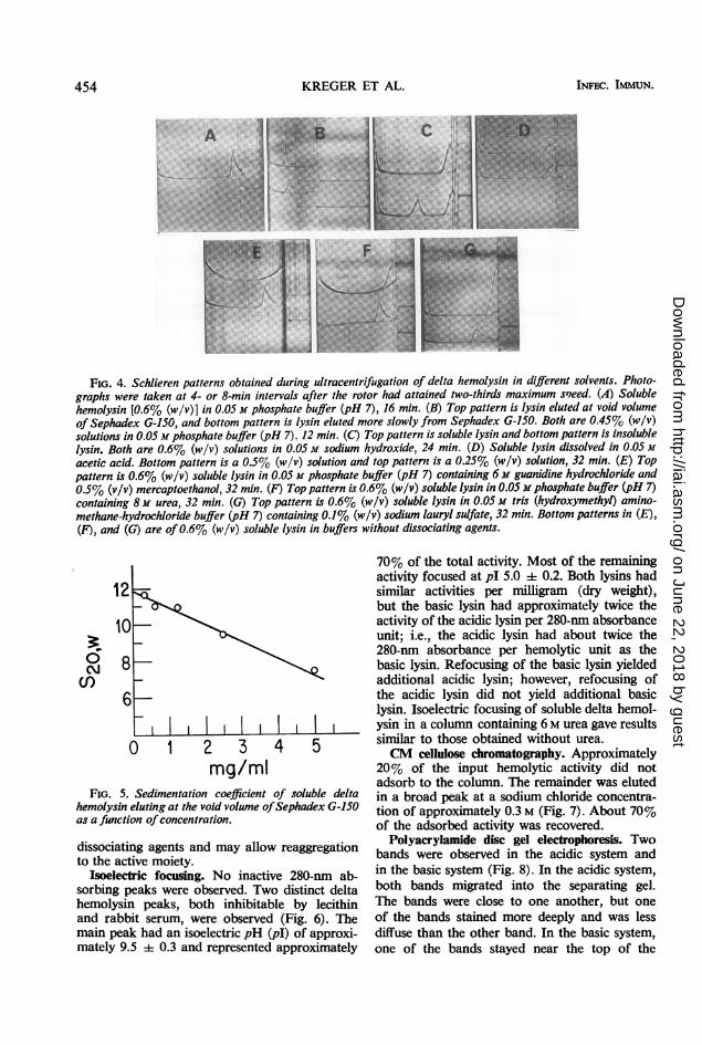
Analytical ultracentrifugation. Sedimentationof delta hemolysin in various solvents (Fig. 4)showed that the soluble lysin sedimented at pH7 as one peak having a polydisperse trailingedge and that it could be converted into moreslowly sedimenting units by high and low pH,guanidine hydrochloride, urea, and sodiumlauryl sulfate. Sedimentation analysis of thelysin fractions which were rapidly and slowlyeluted from Sephadex G-150 (Fig. 4B) showedthat the soluble delta lysin preparation wascomposed of a mixture of 11.9S lysin (Fig. 5)and a population of more slowly sedimentingmolecules, with the most rapid being approxi-mately 4.9S. The sedimentation coefficient ofthe 11.9S lysin showed a marked concentrationdependence (Fig. 5). When dissolved in 0.05 Msodium hydroxide, both the insoluble and solublelysins had molecules with an S20o of approxi-mately 1.9; however, the insoluble lysin prepa-ration also showed a peak with a sedimentationcoefficient of approximately 16S. By means of aseparation cell, a portion of the 1.9S componentwas isolated free from the 16S component. Thehemolytic activity resided in the 1.9S component.We have observed that the conversion of solubledelta hemolysin to the 1.9S form by alkali andurea is reversible upon removal of these reagents.It cannot, therefore, be conclusively stated thatthe hemolytic moiety of delta lysin has a sedi-mentation coefficient of 1.9S. The hemolyticassay involves dilutions in PBS to remove the
VOL. 3,~1971 453
t
on June 22, 2018 by guesthttp://iai.asm
.org/D
ownloaded from
KREGER ET AL.
FIG. 4. Schlieren patterns obtained during ultracentrifugation of delta hemolysin in different solvents. Photo-graphs were taken at 4- or 8-min intervals after the rotor had attained two-thirds maximum s'eed. (A) Solublehemolysin [0.6% (w/v)] in 0.05 M phosphate buffer (pH 7), 16 min. (B) Top pattern is lysin eluted at void volumeof Sephadex G-150, and bottom pattern is lysin eluted more slowly from Sephadex G-150. Both are 0.45% (w/v)solutions in 0.05 M phosphate buffer (pH 7). 12 min. (C) Top pattern is soluble lysin and bottom pattern is insolublelysin. Both are 0.6% (w/v) solutions in 0.05 M sodium hydroxide, 24 min. (D) Soluble lysin dissolved in 0.05 Macetic acid. Bottom pattern is a 0.5% (w/v) solution and top pattern is a 0.25% (w/v) solution, 32 min. (E) Toppattern is 0.6% (w/v) soluble lysin in 0.05 M phosphate buffer (pH 7) containing 6 M guanidine hydrochloride and0.5% (v/v) mercaptoethanol, 32 min. (F) Top pattern is 0.6% (w/v) soluble lysin in 0.05 m phosphate buffer (pH 7)containing 8M urea, 32 min. (G) Top pattern is 0.6% (w/v) soluble lysin in 0.05 M tris (hydroxymethyl) amino-methane-hydrochloride buffer (pH 7) containing 0.1% (w/v) sodium lauryl sulfate, 32 min. Bottom patterns in (E),(F), and (G) are of0.6% (w/v) soluble lysin in buffers without dissociating agents.
12
cn _
6--_I ] I I I I1 I IO 2 3 4 5
mg/mlFIG. 5. Sedimentation coefficient of soluble delta
hemolysin eluting at the void volume ofSephadex G-150as a function of concentration.
dissociating agents and may allow reaggregationto the active moiety.
Isoelectric focusing. No inactive 280-nm ab-sorbing peaks were observed. Two distinct deltahemolysin peaks, both inhibitable by lecithinand rabbit serum, were observed (Fig. 6). Themain peak had an isoelectric pH (pI) of approxi-mately 9.5 0.3 and represented approximately
70% of the total activity. Most of the remainingactivity focused at pI 5.0 ± 0.2. Both lysins hadsimilar activities per milligram (dry weight),but the basic lysin had approximately twice theactivity of the acidic lysin per 280-nm absorbanceunit; i.e., the acidic lysin had about twice the280-nm absorbance per hemolytic unit as thebasic lysin. Refocusing of the basic lysin yieldedadditional acidic lysin; however, refocusing ofthe acidic lysin did not yield additional basiclysin. Isoelectric focusing of soluble delta hemol-ysin in a column containing 6 M urea gave resultssimilar to those obtained without urea.CM cellulose chromatography. Approximately
20% of the input hemolytic activity did notadsorb to the column. The remainder was elutedin a broad peak at a sodium chloride concentra-tion of approximately 0.3 M (Fig. 7). About 70%of the adsorbed activity was recovered.
Polyacrylamide disc gel electrophoresis. Twobands were observed in the acidic system andin the basic system (Fig. 8). In the acidic system,both bands migrated into the separating gel.The bands were close to one another, but oneof the bands stained more deeply and was lessdiffuse than the other band. In the basic system,one of the bands stayed near the top of the
INFEC. IMMUN.454
on June 22, 2018 by guesthttp://iai.asm
.org/D
ownloaded from

STAPHYLOCOCCAL DELTA HEMOLYSIN
'
z
0
m
E0N
CLL
z
I
FRACTION
FIG. 6. Isoelectric focusing of soluble delta hemoly-sin. The gradient was prepared from a less densesolution consisting of5I ml of water, 4 ml of8% (w/v)ampholine (pH 3 to 10; LKB Instruments, Rockville,Md.), and 20 mg ofsoluble delta hemolysin and a moredense solution consisting of 32 ml of water, 8.5 ml of8% (w/v) ampholine (pH 3 to 10), and 25 g ofsucrose.Focusing was done at about 4 C for 2 days in a 110-mlelectrolysis column (LKB Instruments) with a finalpotential of 600 v. Fractions (4 ml) were examined forabsorbance at 280 nm (@), delta hemolysin activity (0),and pH at approximately 25 C (A). The 280-nmabsorbance values were corrected for backgroundabsorbance contributed by the ampholine and sucrose.About 90% of the input activity was recovered. Alphaand beta hemolysin activity was not detected in thefractions.
m
EcC>00c\
160-
120 =
80 C
0 40 I
0
0.34
0.32 co-z
_ 0.30 >-
0.28 c_ o'
_ 2
j024120 124 128 132 136 140 144 148 152 156
FRACTION NUMBER
FIG. 7. Carboxymethyl cellulose chromatographyof soluble delta hemolysin. Fractions (5 ml) wereassayed for delta hemolysin activity (0), absorbanceat 280 nm (0), and sodium chloride concentration(A). Alpha and beta hemolysin activity was not de-tected in the fractions.
separating gel but the other migrated furtherdown the gel.
Chemical analysis. The insoluble delta hemol-ysin preparation contained approximately 1to 2% phosphorus. The soluble delta hemolysinpreparation contained less than 0.025% phos-phorus, less than 1% carbohydrate (as glucose)and amino sugar (as glucosamine), and less than
.A B C .. ....D
FIG. 8. Polyacrylamide disc gel electrophoresis ofsoluble delta hemolysin. (A) A 75-pug amount of lysinin the acidic (pH 4.3) 7.5% gel system ofReisfeld et al.(59). Migration is toward the cathode. (B) Controlacidic gel (no lysin). (C) A 130-pgg amount of lysin inthe basic (pH 9.5) 7% gel system of Ornstein (57) andDavis (14). Migration is toward the anode. (D) Controlbasic gel (no lysin).
1% ash. The soluble lysin contained approxi-mately 1% bound lipid but less than 0.5% freelipid. Elemental analysis yielded 53.77% C,7.27% H, 13.38% Dumas N, 11.90% KjeldahlN, and 0.82% S.Amino acid composition. Isoleucine (18.17%),
lysine (16.66%), and aspartic acid (13.46%)were the major amino acids in the preparation(Table 1). The hydrophobic amino acids, iso-leucine, leucine, and valine, accounted for ap-proximately 31% of the amino acids in thepreparation. No histidine, arginine, proline, ortyrosine was detected even when 3.0 mg ofhydrolyzed lysin preparation was examined.Qualitative tests for these four missing aminoacids in the unhydrolyzed protein were alsonegative. The value for nitrogen content, ascalculated from the amino acid compositiondata, agrees well with that obtained by the Dumasmethod. The low value for nitrogen content, asdetermined by the Kjeldahl method, is mostlikely caused by incomplete digestion of the pro-tein with Cu2- as the catalyst. It is known that
VOL. 3, 1971 455
on June 22, 2018 by guesthttp://iai.asm
.org/D
ownloaded from

KREGER ET AL.
lysine and tryptophan are refractoryacid digestion when Cu2- is used as 1(53).
Spectral analysis. The ultraviolet
TABLE 1. Amino acid compositionidelta hemolysin
Amino acid
Lysine.Histidine........Ammonia........Arginine.........Aspartic acid....Threonine.......Serine...........Glutamic acid...Proline..........Glycine..........Alanine .........Half-cystinea....Valine..........Methionine......Isoleucine .......Leucine .........Tyrosine.........Phenylalanine.Tryptophan......
Total..........
Per cent of totalweight of amino acids
16.66Not detected
1.08Not detected
13.468.022.464.90
Not detected3.603.090.096.644.5618.176.04
Not detected7.933.30
100.00
a Determined as cysteic acid.
09-
08 /
0 64L
0 3
03 250 260 270 280 290 300 3
WAVELENGTH (nm)
FIG. 9. Ultraviolet absorption spectrundelta hemolysin. The lysin (I mg/me) wasphosphate-buffered saline (pH 7).
to sulfuric spectrum (Fig. 9) was similar to that reportedthe catalyst by Kayser and Raynaud (42). It had a maximum
at approximately 282 nm, a minimum at ap-absorption proximately 250 nm, and a distinct shoulder at
about 291 nm. The 280:260 absorbance ratio wasof soluble approximately 1.5. The infrared absorption
spectrum (Fig. 10) did not indicate the presenceof significant amounts of lipid. A distinct car-
total amino bonyl absorption peak was not observed.acid N Electron microscopy. The soluble delta hemoly-
sin preparation was composed of a mixture of3.19 fibers and granular aggregates of various sizes.
Examination of the Sephadex G-150, Bio-Gel0.95 A-5m, and isoelectric focusing fractions revealed1.42 that the fibrous material (Fig. 1lA) was the0.94 lysin which (i) eluted in the void volume of0.33 Sephadex G-150, (ii) was the main component0.47 in the first peak eluting from the Bio-Gel A-5m
column, and (iii) had a pI of approximately 5.0.0.67 The granular material (Fig. liB) was the lysin0. 49 which (i) was found in the trailing portion of0.01 the Sephadex G-150 curve, (ii) was the main0.79 component in the second peak eluting from the1.94 Bio-Gel A-5m column, and (iii) had a pI of1.94 approximately 9.5.Inactivation studies. All of the phospholipids0.67 and normal sera tested inhibited delta hemolysin0.45 activity (Table 2). Cholesterol had no effect.
All of the proteolytic enzymes tested inactivated13.39 the lysin; however, trypsin and Pronase were
most potent (Table 3). The lysin was resistantto heat inactivation. Heating for 1 hr at tempera-tures as high as 80 C had no effect. Heating for1 hr at 100 C reduced activity by approximately50%. Even after autoclaving (121 C) for 1 hr,there was still about 10% residual activity.Citrate, EDTA, Mg2+, and Ca2- at concentra-tions as high as 103 M had no effect on activity.The lysin was not cysteine-activable.
Lysis of erythrocytes from different animalspecies. Soluble delta hemolysin lysed erythro-
0 320 330 cytes from all of the animal species tested; how-l of soluble ever, horse, rabbit, and human erythrocytesdissolved in were most sensitive (Table 4).
Synergism of delta and beta hemolysins. Staph-
WAVELENGTH (MICRONS)
FREQUENCY (CM-1)
FIG. 10. Infrared absorption spectrum ofsoluble delta hemolysin. The lysin was examined as a potassium bromidepellet.
456 INFEC. IMMUN.
on June 22, 2018 by guesthttp://iai.asm
.org/D
ownloaded from
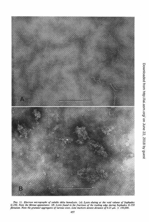
FIG. 11. Electron micrographs of soluble delta hemolysin. (A) Lysin eluting at the void volume of SephadexG-150. Note the fibrous appearance. (B) Lysin found in the fractions of the trailing edge during Sephadex G-150filtration. Note the granular aggregates of various sizes. Line markers denote distance of 0.25 jAm. X 150,000.
457
on June 22, 2018 by guesthttp://iai.asm
.org/D
ownloaded from

KREGER ET AL.
TABLE 2. Inhibition of soluble delta hemolysin byphospholipids and normal sera
Amt requiredto inhibit &i
Substance of the testamta of delta
lysin (jg)
Phosphatidylcholine (lecithin) 2Phosphatidylserine................... 2Phosphatidylinositol................. 4Phosphatidylethanolamine ........... 8Diphosphatidylglycerol (cardiolipin). 4Sphingomyelin....................... 8Cholesterol .......................... >1,000Horse serum......................... 2b
Human serum ....................... lbRabbit serum........................ 2b
a Three hemolytic units.b Measured as microliters.
TABLE 3. Inactivation of soluble delta hemolysinby proteolytic enzymes
Per cent ofcontrol
Enzyme hemolyticactivityaremaining
TrypsinlOOgg............................... 1lO,ug............................... 40
Chymotrypsin100l g............................... 10lO.g............................... 80
Pronase100 jg ............................... 2lOs,g............................... 40
Papain100,g............................... 4010,ug............................... 100
a Control hemolytic activity was 200 hemolyticunits.
TABLE 4. Lysis of erythrocytes from differentanimal species by soluble delta hemolysin
Animal HUa/mg
Horse ............................... 200
Rabbit ............................... 200
Human............................... 100
Guinea pig ............................ 50
Pig................................. 40
Calf............................... 40
Sheep............................... 40
Goat............................... 20
Cat............................... 20
Chicken............................... 20
a One hemolytic unit (HU) is the smallestamount of lysin, contained in 1 ml of phosphate-buffered saline, which will produce 50%, lysis of1 ml of a 0.7%/W0 (v/v) washed erythrocyte suspen-
sion after incubation for 30 min at 37 C.
TABLE 5. Synergistic effect of delta and betahemolysinis ont sheep erythrocytes as a function
of delta hemolysin concentration
Amt of delta lysin (ug) Per cent hemolysisa
5.00 1002.50 901.25 500.63 300.31 200.16 100 10
Controlb 0
a Determined after incubation of variousamounts of delta lysin and a constant amount(10 hemolytic units in the hot-cold assay system)of staphylococcal beta hemolysin (kindly sup-plied by W. Chesbro, Department of Microbi-ology, University of New Hampshire) in 1 ml ofbuffer used in the beta hemolysin assay with 1ml of a 0.7% washed sheep erythrocyte suspensionfor 30 min at 37 C.
b Five micrograms of delta lysin but no betalysin.
TABLE 6. Synergistic effect of delta and betahemolysins on sheep erythrocytes as a function
of beta hemolysin concentration
Amt of beta lysin (HU)' Per cent hemolysisb
10.0 1005.0 1002.5 01.3 00.6 00 0
Controlc 10
a Hemolytic units.b Determined after incubation of various
amounts of beta lysin and a constant amount (5jug, approximately 0.2 HU with sheep erythro-cytes) of delta lysin in 1 ml of buffer used in thebeta hemolysin assay with 1 ml of a 0.7% washedsheep erythrocyte suspension for 30 min at 37 C.
c Fifteen HU of beta lysin but no delta lysin.
TABLE 7. Lysis of bacterial protoplasts andspheroplasts by soluble delta hemolysin
Concn of lysinrequired to
Prepn produce half-maximal lysis
(Cgml)
Sarcina lutea protoplasts............. 12Streptococcus faecalis protoplasts 15Bacillus megaterium protoplasts .12Escherichia coli spheroplasts......... 10Micrococcus lysodeikticus protoplasts. 24Vibrio metschnikovii spheroplasts..... >1,000
458 INFEC. IMMUN.
on June 22, 2018 by guesthttp://iai.asm
.org/D
ownloaded from
'9
I
I.t.
* . 9
FIG. 12. Disruption of lipid spherules by soluble delta hemolysin. (A) Normal spherule preparation; (B) spherulepreparation incubatedfor I hr at 37 C with 200 ,ug ofsoluble delta lysin. Note the presence ofholes (h), fibrous ma-terial (f), and disorganized lipid (1). Line markers denote distance of 0.25 Am. X 150,000.
459
AZ
on June 22, 2018 by guesthttp://iai.asm
.org/D
ownloaded from

KREGER ET AL.
FIG. 13. Clearing of egg yolk-agar by soluble deltahemolysin. Egg yolk-agar was incubated for 3 days at37 C with various amounts of lysin in saline (0.2 ml).(A) A 1-mg amount, (B) 0.5 mg, (C) 0.25 mg, (D)saline control. The dark area around the wells is theopaque area ofprecipitation.
ylococcal beta and soluble delta lysins exhibiteda synergistic effect on sheep erythrocytes (Tables5 and 6). The ratio of delta to beta hemolysinhaving the optimum synergistic effect was ap-proximately 0.2 HU (with sheep erythrocytes)of delta lysin (approximately 5 4g) to 5 to 10HU of beta lysin.
Lysis of bacterial protoplasts and spheroplasts.Soluble delta hemolysin disrupted five of the sixprotoplast and spheroplast preparations (Table7). M. lysodeikticus protoplasts required abouttwice the amount of lysin to produce half-maxi-mal lysis than did the other four preparations.The lysin had no effect on V. metschnikoviispheroplasts.
TABLE 8. Lethality of soluble delta hemolysin formice and guinea pigs
No. of animals ApproxAmt of lysin injected (mg) dead/no. of survival
animals injected time (min)
Micea2.5 12/12 152.0 6/12 351.5 0/121.0 0/120 0/12
Guinea pigs'20 8/8 1010 8/8 125 0/80 0/8
a A 0.25-ml amount injected intravenously.b A 2-ml amount injected intracardially.
Disruption of rabbit erythrocyte membranesand leukocyte lysosomes. Amounts of lysin of500 and 25 ug/ml produced half-maximal lysisof the rabbit erythrocyte membrane and leuko-cyte lysosome suspensions, respectively. Theactivities were inhibited by pretreatment of thelysin with rabbit serum.
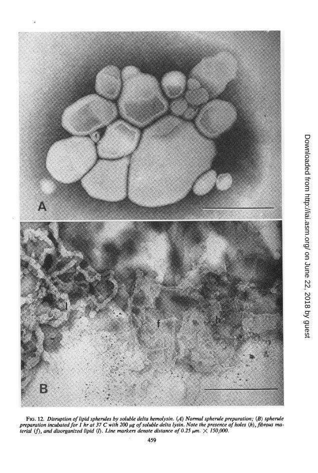
Lipid spherule disruption. Five HU of solubledelta hemolysin incubated for 1 hr at 37 C withthe spherule preparation released approximately90% of the chromate ions trapped in the spherulepreparation. Disruption of a normal spherulepreparation (Fig. 12A) by lysin could also beobserved in the electron microscope. Treatedpreparations contained holes, fibrous material,and disorganized lipid (Fig. 12B).
Antibacterial activity. Growth of B. megateriumKM was prevented for 4 days by 63 jug of solubledelta hemolysin per ml. Growth of M. lysodeikti-cus and a group A Streptococcus (strain C203s)was inhibited for 1 day by 500 Mg/ml, after whichtime growth commenced. Growth of Gaffkyatetragena (ATCC 10875), Lactobacillus casei
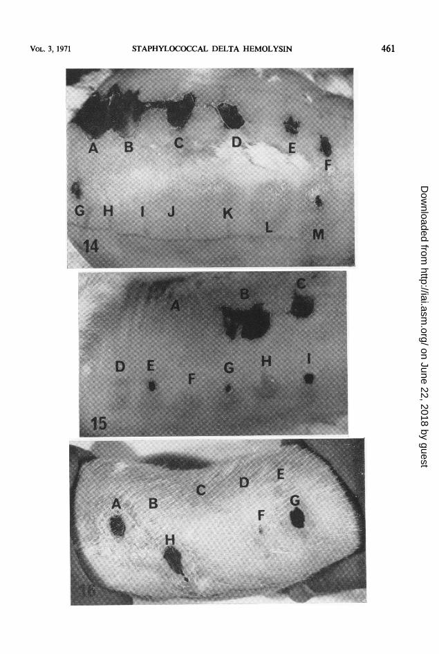
FIG. 14. Dermonecrosis produced in rabbits by staphylococcal alpha and delta hemolysins. Results shown are 7days postinjection. A through F show dermonecrosis produced by 12S-free alpha hemolysin. (A) A 10.8-,ug amount,(B) 5.4 ,ug, (C) 2.7,g, (D) 1.35,ug, (E) 0.68,ug, (F) 0.34,g. G through M show results obtained with delta hemol-ysin. (G) A 1-mg amount (lot 39, kindly supplied by F. A. Kapral, Department of Medical Microbiology, Ohio StateUniversity), (H) saline control, (1) 0.06 mg of our purified soluble delta hemolysin, (J) 0.13 mg, (K) 0.25 mg, (L)0.5 mg, and (M) 1.0 mg.
FIG. 15. Inhibition by lecithin of dermonecrosis produced in rabbits by delta hemolysin. Results shown are 6days postinjection. (A) saline-lecithin (0.5 mg) control, (B) 2.7 Mg of alpha hemolysin and 0.5 mg of lecithin, (C)2.7,ug of alpha lysin, (D) 1 mg of delta lysin and 0.5 mg of lecithin, (E) I mg of delta lysin, (F) 0.5 mg ofdeltalysin, (G) 0.5 mg ofdelta lysin (lot 17, from F. A. Kapral), (H) I mg ofdelta lysin (from F. A. Kapral) and 0.5 mgof lecithin, and (I) I mg of delta lysin (from F. A. Kapral).
FIG. 16. Dermonecrosis produced in guinea pigs by staphylococcal alpha and delta hemolysins. Results shownare 7 days postinjection. (A) A 1-mg amount of delta lysin (lot 39, from F. A. Kapral), (B) saline control, (C)0.063 mg of our purified soluble delta lysin, (D) 0.125 mg, (E) 0.25 mg, (F) 0.5 mg, (G) i mg, anid (H) 5.4 Mg ofalpha lysini.
460 INFEC. IMMUN.
on June 22, 2018 by guesthttp://iai.asm
.org/D
ownloaded from
VOL. 3, 1971 STAPHYLOCOCCAL DELTA HEMOLYSIN 461
on June 22, 2018 by guesthttp://iai.asm
.org/D
ownloaded from

KREGER ET AL.
(subspecies rhamnosus, ATCC 7469), Coryne-bacterium diphtheriae (mitis C7), S. aureus(Wood 46 and W46M strains), B. subtilis, B.cereus (ATCC 10876a), S. faecalis (ATCC 9790),E. coli K-12, Pseudomonas aeruginosa, and S.lutea was not affected by as much as 1,000 ug/ml.
Clearing of egg yolk-agar. Soluble deltahemolysin produced clearing of egg yolk-agar(Fig. 13). An opaque area of precipitation aroundthe well and a clear halo surrounding this areawere observed.
Toxicity. The minimum lethal intravenousdose (MLD) of soluble delta hemolysin for miceand guinea pigs was approximately 2.5 and 10mg, respectively (Table 8). On a weight basis,the MLD was approximately 110 mg/kg formice and 30 mg/kg for guinea pigs.Large amounts of soluble delta lysin were
required to produce dermonecrosis in rabbits(Fig. 14) and guinea pigs (Fig. 16). Lecithinprevented the dermonecrosis produced by deltalysin but not by alpha lysin (Fig. 15). A 1-mgamount of delta lysin injected intradermallyinto rabbits produced, after 1 day, a large (ap-proximately 28 mm in diameter) erythematous,hard, indurated lesion with a small (4 mm indiameter) whitish-yellow center. This smallarea became necrotic by 3 days postinjection.The other amounts of lysin injected producederythematous, indurated lesions which neverbecame necrotic. Some desquamation was ob-served by 2 days postinjection with 1 and 0.5mg. Samples of soluble delta hemolysin kindlysupplied by F. A. Kapral (Department of MedicalMicrobiology, Ohio State University) were morepotent, both in the presence and absence oflecithin, than our purified lysin. A 0.5-mg amountof his lysin also produced dermonecrosis inrabbits (Fig. 15).The lesions produced by delta lysin in guinea
pigs resembled those observed in rabbits; how-ever, the necrotic areas developed more rapidlyand were larger in the guinea pig than in therabbit. A 0.5-mg amount of delta lysin produceda small (2 mm in diameter) necrotic lesion inthe guinea pig but not in the rabbit.
DISCUSSIONPerhaps the most important aspect of the
current study is that it provides a simple andreproducible method for obtaining large amountsof highly purified delta lysin which can be usedfor further experimentation. Our success maydepend upon use of a mutant Staphylococcuswhich produces very large amounts of deltalysin but relatively small amounts of otherstaphylococcal exoproducts. Further comparison
of the culture supernatant fluids of the parentand mutant strains should show whether thisis the correct explanation.As previously observed with crude and partially
purified delta hemolysin, our highly purifiedlysin preparation (i) disrupts erythrocytes ob-tained from many animal species, bacterialprotoplasts and spheroplasts, lysosomes, lipidspherules, and erythrocyte membranes; (ii) actssynergistically with staphylococcal beta hemoly-sin; (iii) is lethal and produces dermonecrosiswhen administered in large amounts; (iv) pos-sesses limited antibacterial activity; and (v)clears egg yolk-agar. These confirmatory findingssupport the belief that all of the effects are causedby delta hemolysin and not by a contaminatingstaphylococcal product. In addition, the obser-vation that the relative sensitivities of the differentprotoplasts and spheroplasts to disruption bydelta lysin were similar to those reported (5)for alpha lysin supports the idea (7) that theprotoplast-lysing activity of alpha lysin prepa-rations is due to contamination with deltahemolysin.
Hallander (28) reported that delta lysin ob-tained from the S6 strain of S. aureus was morenegatively charged at pH 8.2 than lysin from the196E strain. In addition to the difference be-tween the two delta lysins studied, each lysin wasmolecularly heterogeneous when examined byagarose gel filtration and by Sephadex gel elec-trophoresis. Hoffmann and Streitfeld (32) sug-gested that two lysins, separable by paper chroma-tography, might be present, delta A and deltaB. Jackson and Little (36) described one heat-stable ethanol-soluble and one heat-labileethanol-insoluble lysin fraction. Guyonnet andPlommet (26) think that the heat-labile ethanol-insoluble lysin was, in reality, staphylococcalgamma hemolysin. Isoelectric focusing of apartially purified delta hemolysin preparation byMollby and Wadstrom (54) yielded only onepeak of activity; however, Maheswaran andLindorfer (Bacteriol. Proc., p. 78, 1970) isolateddelta lysins with three different isoelectric points.Our findings support the idea that staphylo-coccal delta hemolysin is molecularly heteroge-neous, i.e., it may, as staphylococcal alpha lysin(8, 64) and beta lysin (13, 30, 48), exist in morethan one active physicochemical form. Manyother bacterial toxins are known to be physico-chemically heterogeneous (2).There are at least five major unexplained
differences between our purified delta lysinpreparation and the purified preparation studiedby Caird and Wiseman (12, 67). First, theirpreparation possesses phospholipase C activitywith phosphatidylinositol and phosphatidylserine
462 INFEC. IMMUN.
on June 22, 2018 by guesthttp://iai.asm
.org/D
ownloaded from
STAPHYLOCOCCAL DELTA HEMOLYSIN
as substrates. We have not detected this activityin our preparation. Second, their preparationexhibits two peaks in the analytical ultracentri-fuge. Ours shows one peak having a polydispersetrailing edge. Third, the hemolytic activity permilligram of protein of their preparation is ap-proximately 45-fold greater than the specificactivity of our preparation. The specific activityof their starting culture supernatant is approxi-mately 20-fold greater than ours. Fourth, theyreport proline to be the N-terminal amino acidof the lysin. We have not detected proline inour preparation. Fifth, their lysin is inhibitedby cholesterol, as reported by Gladstone andvan Heyningen (23). Our lysin is not inhibitedby cholesterol, as reported by Gladstone andYoshida (24).
Hallander found (28) that partially purifieddelta lysin inhibited the growth of a strain be-longing to the genus Corynebacterium. Hoff-mann and Streitfeld (32) did not observe in-hibition of C. diphtheriae or B. megaterium bypartially purified delta lysin but reported in-hibition of G. tetragena, L. casei, and M. lyso-deikticus. We observed inhibition of B. mega-terium and M. lysodeikticus but not of G. tetra-gena, L. casei, or C. diphtheriae (mitis C7).The question of whether delta lysin is heat-
stable is also controversial. Partially purifieddelta lysin has been reported to be stable toheating at 100 C for 30 min (28) and 120 min(50) and at 115 C for 20 min (23). One of thetwo fractions described by Jackson and Little(36) was stable at 60 C for 120 min. However,the lysin obtained by Yoshida (68) lost 85%of its activity after 30 min at 100 C. Our purifiedlysin preparation was partially heat-stable. Itwas not affected by heating at 80 C for 60 minbut lost approximately 50% of its activity after60 min at 100 C and about 90% of its activityafter 60 min at 121 C.Yoshida (68) mentioned the poor solubility
of partially purified delta lysin in buffers at aneutral pH and a low ionic strength. Mollbyand Wadstrom (54) observed the formation of awhite flocculent delta lysin precipitate duringisoelectric focusing; precipitation was preventedby the addition of a nonionic detergent, TritonX-100, to the pH gradient. Our purified deltahemolysin preparation, however, is soluble atneutral pH in low ionic strength buffer and doesnot precipitate during isoelectric focusing.
Partially purified delta hemolysin has beenreported to be soluble in 75% ethanol (36) andin chloroform-methanol (2:1) (68). We haveconfirmed these observations. Our results do notsupport the idea that delta lysin is a lipid orlipoprotein; however, the high content of hy-
drophobic amino acids in the protein may ex-plain its unusual solubility in certain organicsolvents.
There are at least five important unansweredquestions concerning delta hemolysin. First,what is the interrelationship between the in-soluble delta lysin, the soluble, acidic, fibrouslysin, and the soluble, basic, granular lysin(s)?Is one an aggregate or a carrier-bound version ofanother? Kayser (38-40) and Bernheimer (6)have proposed that delta lysin may exist in acarrier-bound form. Most of the chemical andbiological property studies reported in this paperwere performed with the lysin preparation whichwas a mixture of the acidic and basic components.Additional comparative studies of the acidicand basic lysins should be done.
Second, what is the mechanism of action ofdelta lysin? Wiseman and Caird (67) have pro-posed that it is a phospholipase C; however,we have not found this activity in our preparation.In addition, Marks and Vaughan (51), Jacksonand Little (35), and Bernheimer (6) have ob-served that, when a fixed amount of delta lysinis incubated with various numbers of erythro-cytes, the absolute number or the per cent oftotal cells lysed decreases with increasing cellconcentration. This observation suggests thatdelta hemolysin is a nonenzymatic lysin, sinceif it were an enzyme it should act on many cellssequentially rather than only once, and per centhemolysis would remain constant with increasingcell concentration, as has been observed withstaphylococcal beta hemolysin (a known enzyme).A nonenzymatic mechanism of action is alsosuggested by the observation by Galston (per-sonal communication) that our delta lysin prepa-ration is surface-active.
Third, is delta lysin immunogenic? Deltahemolysin preparations show precipitin bandswhen diffused against staphylococcal antiserum;however, with the exception of Kayser andRaynaud (42) and McLeod (47), investigators(24, 28, 43, 58) have not been able to producesignificant amounts of neutralizing antibody inrabbits with delta lysin. Sera from a variety ofanimal species will inactivate the lysin; however,this activity is not primarily present in thegamma globulin fraction (24, 35). In addition,the similar amounts of inhibitor present inmany human sera (17) and its occurrence infetal calf serum (J. A. Donahue, Bacteriol.Proc., p. 93, 1969) suggest that it is not antibody.The normal serum inhibitor of delta hemolysinhas not been purified and characterized. Phos-pholipids are known inhibitors of this lysin,and it has been suggested (17) that the neutral-izing activity of serum is due to serum lipid(s)
VOL. 3, 1971 463
on June 22, 2018 by guesthttp://iai.asm
.org/D
ownloaded from

KREGER ET AL.
and that the lines of precipitation seen whendelta lysin preparations are diffused againststaphyloccal antiserum are insoluble complexesresulting from combination of delta lysin withnormal serum lipoproteins (6). Streptolysin Shas been reported (63) to be inhibited by serumlipoprotein(s).
Fourth, what is the role of delta hemolysinin staphylococcal metabolism? A very largeamount of this material is produced and ex-creted in vitro, but there does not seem to be anyclue as to its function.
Fifth, what is the role of delta lysin in staphylo-coccal infections? Partially purified lysin hasbeen reported (23, 24, 29, 34) to kill leukocytesin vitro. If the lysin is produced and can functionin vivo, it might aid in establishing or perpetuatingstaphylococcal lesions. Arbuthnott et al. (3)have suggested that delta lysin may play a partin the pathogenesis of Ritter's type of toxic
epidermal necrolysis and in extensive impetigo.
ACKNOWLEDGMENTS
We thank Charles Harman for assistance with the amino acid
analyses, Donald Green for determining the infrared absorptionspectrum, William Grosvenor for performing the Kjeldahl nitro-
gen assay, and Gerald Weissmann for supplying rabbit poly-morphonuclear leukocyte lysosomes.
This investigation was supported by a grant from the Life
Insurance Medical Research Fund and by Public Health Service
grant AI-02874 from the National Institute of Allergy and In-
fectious Diseases. A. W. Bernheimer was the recipient of Public
Health Career Program Award 5K6-AI-14-198.
LITERATURE CITEI)
1. Abramson, C., and H. Friedman. 1968. Staphylococcalhyaluronate lyase: purification and characterization studies.J. Bacteriol. 96:886-892.
2. Alouf, J. E., and M. Raynaud. 1970. Isolation and purifi-cation of bacterial toxic proteins, p. 119-182. In S. J.Ajl, S. Kadis, and T. C. Montie (ed.), Microbial toxins,vol. 1. Academic Press Inc., New York.
3. Arbuthnott, J. P., C. G. Gemmell, J. Kent, and A. Lyell.1969. Haemolysin and enzyme patterns of coagulase-positive staphylococci isolated from toxic epidermalnecrolysis, Ritter's disease and impetigo contagiosa. J.Med. Microbiol. 2:479-487.
4. Astrup, T., and S. Miillertz. 1952. The fibrin plate method forestimating fibrinolytic activity. Arch. Biochem. Biophys.40:346-351.
5. Bernheimer, A. W. 1966. Disruption of wall-less bacteria bystreptococcal and staphylococcal toxins. J. Bacteriol. 91:1677-1680.
6. Bernheimer, A. W. 1970. Cytolytic toxins of bacteria, p. 183-212. In S. J. Ajl, S. Kadis, and T. C. Montie (ed.), Microbialtoxins, vol. 1. Academic Press Inc., New York.
7. Bernheimer, A. W., L. S. Avigad, and P. Grushoff. 1968.Lytic effects of staphylococcal a-toxin and 1-hemolysin. J.Bacteriol. 96:487-491.
8. Bernheimer, A. W., and L. L. Schwartz. 1963. Isolation andcomposition of staphylococcal alpha toxin. J. Gen. Micro-biol. 30:455-468.
9. Bernheimer, A. W., and L. L. Schwartz. 1964. Lysosomaldisruption by bacterial toxins. J. Bacteriol. 87:1100-1104.
10. Bernheimer, A. W., and L. L. Schwartz. 1965. Lysis of bac-
terial protoplasts and spheroplasts by staphylococcal a-toxin and streptolysin S. J. Bacteriol. 89:1387-1392.
11. Brenner, S., and R. W. Horne. 1959. A negative stainingmethod for high resolution electron microscopy of viruses.Biochim. Biophys. Acta 34:103-110.
12. Caird, J. D., and G. M. Wiseman. 1970. Purification of thedelta toxin of Staphlylococcus aureus. Can. J. Microbiol. 16:703-708.
13. Chesbro, W. R., F. P. Heydrick, R. Martineau, and G. N.Perkins. 1965. Purification of staphylococcal,-hemolysinand its action on staphylococcal and streptococcal cell walls.J. Bacteriol. 89:378-389.
14. Davis, B. J. 1964. Disc electrophoresis. II. Clinical applica-tions. Ann. N.Y. Acad. Sci. 121:404-427.
15. De Siervo, A. J. 1969. Alterations in the phospholipid com-position of Escherichia coli B during growth at differenttemperatures. J. Bacteriol. 100:1342-1349.
16. Dodge, J. T., C. Mitchell, and D. J. Hanahan. 1963. Thepreparation and chemical characteristics of hemoglobin-free ghosts of human erythrocytes. Arch. Biochem. Biophys.100:119-130.
17. Donahue, J. A. 1969. Antistaphylococcal hemolysins anddelta hemolysin inhibitor in adult human serum. Can. J.Microbiol. 15:957-959.
18. Edelhoch, H. 1967. Spectroscopic determination of tryptophanand tyrosine in proteins. Biochemistry 6:1948-1954.
19. Evans, L., and C. H. Lack. 1969. The action of staphylococcal5-lysin on lysosomes and lymphocytes. Life Sci. 8:677-681.
20. Feingold, D. S., J. N. Goldman, and H. M. Kuritz. 1968.Locus of the lethal event in the serum bactericidal reaction.J. Bacteriol. 96:2127-2131.
21. Freer, J. H., J. P. Arbuthnott, and A. W. Bernheimner. 1968.Interaction of staphylococcal a-toxin with artificial andnatural membranes. J. Bacteriol. 95:1153-1168.
22. Gladstone, G. P. 1966. Staphylococcal haemolysins, p. 145-161. In W. T. Dobrzatiski, J. B6br, and J. Jeljaszewicz(ed.), Postepy mikrobiologii, vol. 5. Symposium on staphy-lococci and staphylococcal infections, Warsaw, 1965.
23. Gladstone, G. P., and W. E. van Heyningen. 1957. Staphy-lococcal leucocidins. Brit. J. Exp. Pathol. 38:123-137.
24. Gladstone, G. P., and A. Yoshida. 1967. The cytopathic actionof purified staphylococcal 6-haemolysin. Brit. J. Exp.Pathol. 48:11-19.
25. Gow, J. A., and J. Robinson. 1969. Properties of purifiedstaphylococcal ,-hemolysin. J. Bacteriol. 97:1026-1032.
26. Guyonnet, F., and M. Plommet. 1970. Hemolysine gamma deStaphylococcus aureus: purification et proprietes. Ann.Inst. Pasteur 118:19-33.
27. Hallander, H. 0. 1963. Fractionation of staphylococcal toxinsby gel-filtration. Acta Pathol. Microbiol. Scand. 59:543-552.
28. Hallander, H. 0. 1968. Characterization and partial purifica-tion of staphylococcal delta-lysin. Acta Pathol. Microbiol.Scand. 72:586-600.
29. Hallander, H. O., and S. Bengtsson. 1967. Studies on the celltoxicity and species specificity of purified staphylococcaltoxins. Acta Pathol. Microbiol. Scand. 70:107-119.
30. Haque, R.-U, and J. N. Baldwin. 1969. Purification andproperties of staphylococcal beta hemolysin. II. Purificationof beta hemolysin. J. Bacteriol. 100:751-759.
31. Hawiger, J. 1968. Purification and properties of lysozyme
produced by Staphylococcus aureus. J. Bacteriol. 95:376-384.
32. Hoffmann, E. M., and M. M. Streitfeld. 1965. The antibioticactivity associated with preparations of delta hemolysin ofStaphylococcus aureus. Can. J. Microbiol. 11:203-211.
33. Huggins, C., and J. Lapides. 1947. Chromogenic substrates.IV. Acetyl esters of p-nitrophenol as substrates for thecolorimetric detennination of esterase. J. Biol. Chem. 170:467-482.
34. Jackson, A. W., and R. M. Little. 1957. Leucocidal effect ofstaphylococcal 8-lysin. Can. J. Microbiol. 3:101-102.
35. Jackson, A. W., and R. M. Little. 1958. Staphylococcal toxins.
464 INFEC. IMMUN.
on June 22, 2018 by guesthttp://iai.asm
.org/D
ownloaded from
STAPHYLOCOCCAL DELTA HEMOLYSIN
II. Factors affecting hemolysis by 1-lysin. Can. J. Microbiol.4:435-444.
36. Jackson, A. W., and R. M. Little. 1958. Staphylococcal toxins.III. Partial purification and some properties of 1-lysin. Can.J. Microbiol. 4:453-461.
37. Kabat, E. A., and M. M. Mayer. 1961. Experimental im-munochemistry, 2nd ed., p. 505-507, 527-529. Charles C.Thomas, Publisher, Springfield, Ill.
38. Kayser, A. 1966. ttude d'une deuxieme hemolysine (distinctede la toxine a) presente dans les filtrats de culture de lasouche Wood 46 de Staphylococcus aureus. C. R. Acad. Sci.Paris 263:693-695.
39. Kayser, A. 1967. Fractionnement d'un complexe h6molytiquelibere par Staphylococcus aureus Wood 46, et obtentiond'une proteine G, vraisemblablement identifiable a
l'hemolysine delta. Ann. Inst. Pasteur 113:351-356.40. Kayser, A. 1968. Chromatographic d'exclusion des complexes
cuivriques des proteines. Application a la toxine staphy-lococcique. Bull Soc. Chim. Biol. 50:85-91.
41. Kayser, A. 1968. L'h6molysine staphylococcique 5G. Actionsur les erythrocytes et les stromas, sur les spheroplastesbacteriens et sur le jaune d'oeuf. Bull. Soc. Chim. Biol.50:1797-1808.
42. Kayser, A., and M. Raynaud. 1965 Etude d'une deuxiemehemolysine (distincte de lhemolysine a) presente dans lesfiltrats de culture de la souche Wood 46 de Staphylococcusaureus (h6molysine G ou 5). Ann. Inst. Pasteur 108:215-233.
43. Kleck, J. L., and J. A. Donahue. 1968. Production of thermo-stable hemolysin by cultures of Staphylococcus epidermidis.J. Infec. Dis. 118:317-323.
44. Kunitz, M. 1947. Crystalline soybean trypsin inhibitor. 2.General properties. J. Gen. Physiol. 30:291-310.
45. Kushner, D. J. 1957. An evaluation of the egg-yolk reactionas a test for lecithinase activity. J. Bacteriol. 73:297-302.
46. Lowry, 0. H., N. J. Rosebrough, A. L. Farr, and R. J.Randall. 1951. Protein measurement with the Folin phenolreagent. J. Biol. Chem. 193:265-275.
47. McLeod, J. W. 1963. Thennostable staphylococcal toxin. J.Pathol. Bacteriol. 86:35-53.
48. Maheswaran, S. K., K. L. Smith, and R. K. Lindorfer. 1967.Staphylococcal (3-hemolysin. I. Purification of 3-hemolysin.J. Bacteriol. 94:300-305.
49. Malveaux, F. J., and C. L. San Clemente. 1969. Staphylococ-cal acid phosphatase: extensive purification and characteri-zation of the loosely bound enzyme J. Bacteriol. 97:1209-1214.
50. Marks, J. 1952. Recognition of pathogenic staphylococci:with notes on non-specific staphylococcal haemolysin. J.Pathol. Bacteriol. 64:175-186.
51. Marks, J., and A. C. T. Vaughan. 1950. Staphylococcal 5-
haemolysin. J. Pathol. Bacteriol. 62:597-615.
52. Martin, R. G., and B. N. Ames. 1961. A method for deter-mining the sedimentation behavior of enzymes: applicationto protein mixtures. J. Biol. Chem. 236:1372-1379.
53. Mayer, M. M. 1961. Kjeldahl nitrogen determination, p. 476-480. In E. A. Kabat and M. M. Mayer (ed.), Experimentalimmunochemistry, 2nd ed. Charles C Thomas, Publisher,Springfield, Ill.
54. Mollby, R., and T. Wadstrom. 1970. Studies on hemolysinsfrom Staphylococcus aureus by the method of isoelectricfocusing, p. 465-469. In H. Peeters (ed.), Protides of thebiological fluids. Proceedings of the 17th colloquium,Bruges, 1969. Pergamon Press, Inc., New York.
55. Moore, S. 1963. On the determination of cystine as cysteicacid. J. Biol. Chem. 238:235-237.
56. Nachlas, M., and A. M. Seligman. 1949. Evidence for thespecificity of esterase and lipase by the use of three chroma-tographic substrates. J. Biol. Chem. 181:343-355.
57. Ornstein, L. 1964. Disc electrophoresis. I. Background andtheory. Ann. N.Y. Acad. Sci. 121:321-349.
58. Plommet, M., and C. Bouillanne. 1967. L'antigenicit6 de latoxine staphylococcique delta. Ann. Biol. Anim. Biochim.Biophys. 7:219-222.
59. Reisfeld, R. A., U. J. Lewis, and D. E. Williams. 1962. Diskelectrophoresis of basic proteins and peptides on poly-acrylamide gels. Nature (London) 195:281-283.
60. Schachman, H. K. 1959. Ultracentrifugation in biochemistry.Academic Press Inc., New York.
61. Shockman, G. D., and H. D. Slade. 1964. The cellular locationof the streptococcal group D antigen. J. Gen. Microbiol.37:297-305.
62. Spackman, D. H., W. H. Stein, and S. Moore. 1958. Auto-matic recording apparatus for use in the chromatographyof amino acids. Anal. Chem. 30:1190-1206.
63. Stollerman, G. H., A. W. Bernheimer, and C. M. MacLeod.1950. The association of lipoproteins with the inhibition ofstreptolysin S by serum. J. Clin. Invest. 29:1636-1645.
64. Wadstrom, T. 1968. Studies on extracellular proteins from
Staphylococcus aureus. IV. Separation of a-toxin by iso-electric focusing. Biochim. Biophys. Acta 168:228-242.
65. Weissmann, G., I. Spilberg, and K. Krakauer. 1969. Arthritis
induced in rabbits by lysates of granulocyte lysosomes.Arthritis Rheum. 12:103-116.
66. Williams, R. E. O., and G. J. Harper. 1947. Staphylococcalhaemolysins on sheep blood agar with evidence for a fourth
haemolysin. J. Pathol. Bacteriol. 59:69-78.67. Wiseman, G. M., and J. D. Caird. 1968. Phospholipase ac-
tivity of the delta hemolysin of Staphylococcus aureus.
Proc. Soc. Exp. Biol. Med. 128:428-430.68. Yoshida, A. 1963. Staphylococcal 5-hemolysin. I. Purification
and chemical properties. Biochim. Biophys. Acta 71:544-
553.
VOL. 3, 1971 465
on June 22, 2018 by guesthttp://iai.asm
.org/D
ownloaded from