yolk sac tumor - ucsf cme · 2 yolk sac tumor yolk sac tumor histologic patterns microcystic (aka...
TRANSCRIPT
1
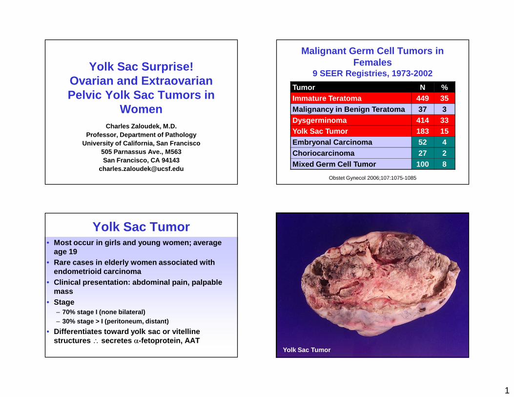
Yolk Sac Surprise!Ovarian and Extraovarian Pelvic Yolk Sac Tumors in
WomenCharles Zaloudek, M.D.
Professor, Department of PathologyUniversity of California, San Francisco
505 Parnassus Ave., M563San Francisco, CA 94143
Malignant Germ Cell Tumors in Females
9 SEER Registries, 1973-2002
Tumor N %Immature Teratoma 449 35Malignancy in Benign Teratoma 37 3Dysgerminoma 414 33Yolk Sac Tumor 183 15Embryonal Carcinoma 52 4Choriocarcinoma 27 2Mixed Germ Cell Tumor 100 8
Obstet Gynecol 2006;107:1075-1085
Yolk Sac Tumor• Most occur in girls and young women; average
age 19• Rare cases in elderly women associated with
endometrioid carcinoma• Clinical presentation: abdominal pain, palpable
mass• Stage
– 70% stage I (none bilateral)– 30% stage > I (peritoneum, distant)
• Differentiates toward yolk sac or vitelline structures ∴ secretes α-fetoprotein, AAT
Yolk Sac Tumor
2
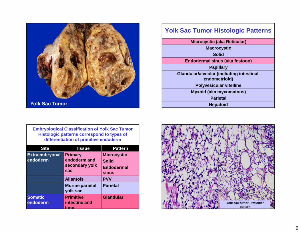
Yolk Sac Tumor
Yolk Sac Tumor Histologic Patterns
Microcystic (aka Reticular)Macrocystic
SolidEndodermal sinus (aka festoon)
PapillaryGlandular/alveolar (including intestinal,
endometrioid)Polyvesicular vitelline
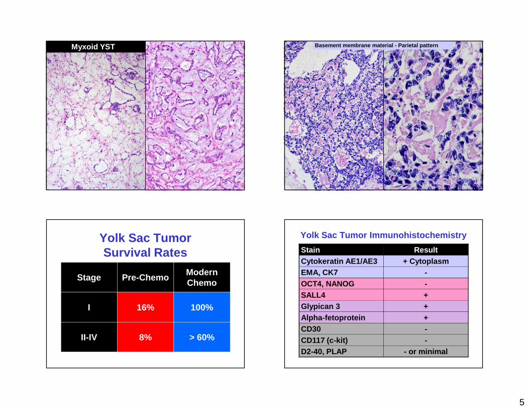
Myxoid (aka myxomatous)Parietal
Hepatoid
Embryological Classification of Yolk Sac TumorHistologic patterns correspond to types of
differentiation of primitive endoderm
Site Tissue PatternExtraembryonal endoderm
Primary endoderm and secondary yolk sac
MicrocysticSolidEndodermal sinus
Allantois PVVMurine parietal yolk sac
Parietal
Somatic endoderm
Primitive intestine and lung
GlandularYolk sac tumor - reticular
pattern
3
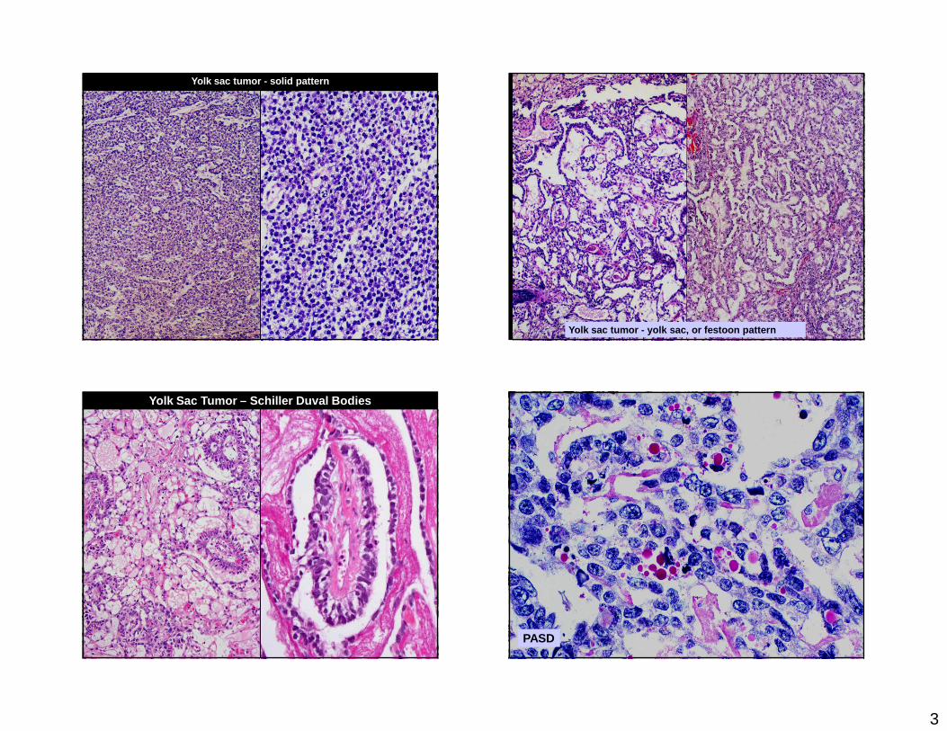
Yolk sac tumor - solid pattern
Yolk sac tumor - yolk sac, or festoon pattern
Yolk Sac Tumor – Schiller Duval Bodies
PASD
4
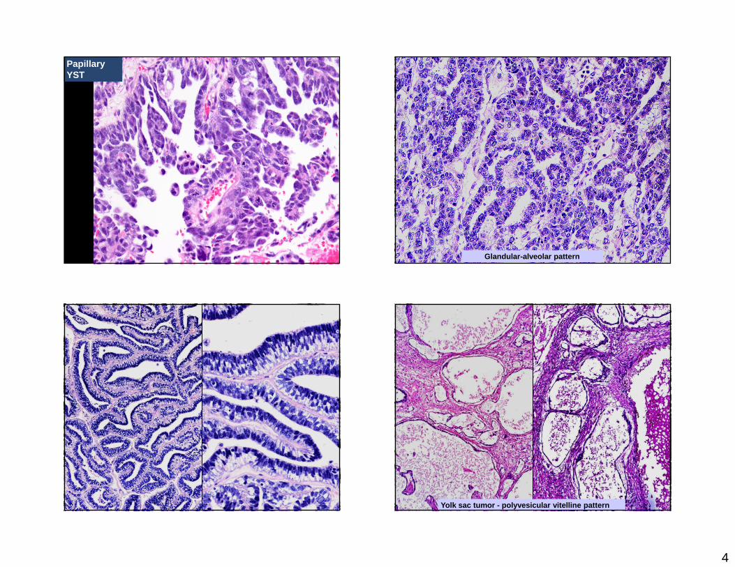
Papillary YST
Glandular-alveolar pattern
Yolk sac tumor - polyvesicular vitelline pattern
5
Myxoid YST Basement membrane material - Parietal pattern
Yolk Sac TumorSurvival Rates
Stage Pre-ChemoModernChemo
I 16% 100%
II-IV 8% > 60%
Yolk Sac Tumor Immunohistochemistry
Stain ResultCytokeratin AE1/AE3 + CytoplasmEMA, CK7 -OCT4, NANOG -SALL4 +Glypican 3 +Alpha-fetoprotein +CD30 -CD117 (c-kit) -D2-40, PLAP - or minimal
6
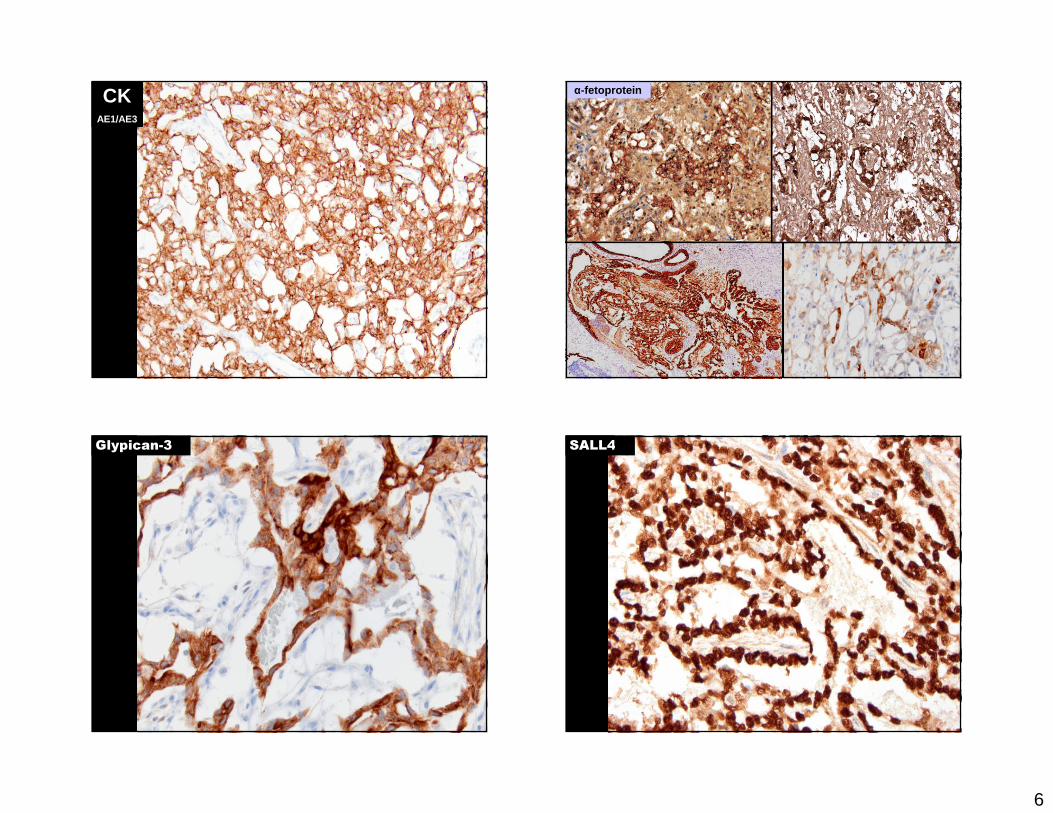
CKAE1/AE3
α-fetoprotein
Glypican-3 SALL4
7
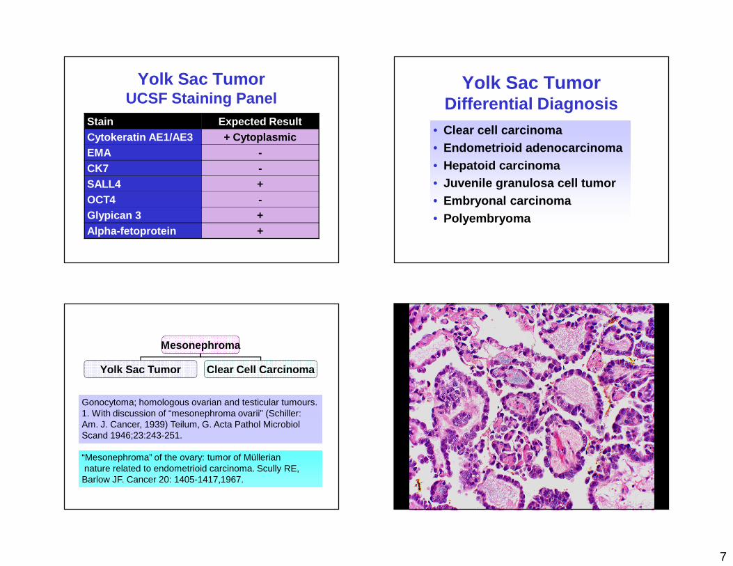
Yolk Sac TumorUCSF Staining Panel
Stain Expected ResultCytokeratin AE1/AE3 + CytoplasmicEMA -CK7 -SALL4 +OCT4 -Glypican 3 +Alpha-fetoprotein +
Yolk Sac TumorDifferential Diagnosis
• Clear cell carcinoma• Endometrioid adenocarcinoma• Hepatoid carcinoma• Juvenile granulosa cell tumor• Embryonal carcinoma• Polyembryoma
Mesonephroma
Yolk Sac Tumor Clear Cell Carcinoma
Gonocytoma; homologous ovarian and testicular tumours. 1. With discussion of “mesonephroma ovarii” (Schiller: Am. J. Cancer, 1939) Teilum, G. Acta Pathol Microbiol Scand 1946;23:243-251.
“Mesonephroma” of the ovary: tumor of Mülleriannature related to endometrioid carcinoma. Scully RE, Barlow JF. Cancer 20: 1405-1417,1967.
8
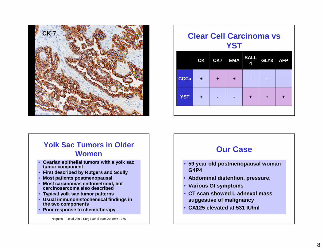
CK 7 Clear Cell Carcinoma vs YST
CK CK7 EMASALL
4GLY3 AFP
CCCa + + + - - -
YST + - - + + +
Yolk Sac Tumors in Older Women
• Ovarian epithelial tumors with a yolk sac tumor component
• First described by Rutgers and Scully• Most patients postmenopausal• Most carcinomas endometrioid, but
carcinosarcoma also described• Typical yolk sac tumor patterns• Usual immunohistochemical findings in
the two components• Poor response to chemotherapy
Nogales FF et al. Am J Surg Pathol 1996;20:1056-1066
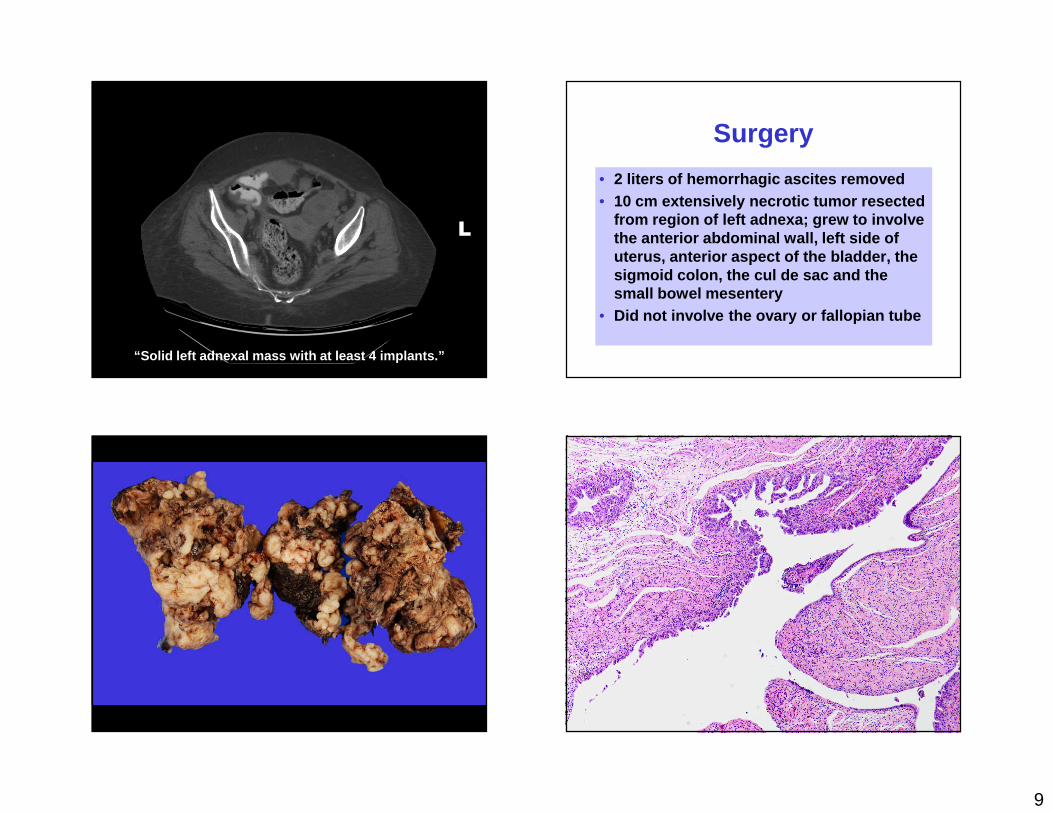
Our Case
• 59 year old postmenopausal woman G4P4
• Abdominal distention, pressure.• Various GI symptoms• CT scan showed L adnexal mass
suggestive of malignancy• CA125 elevated at 531 IU/ml
9
L
“Solid left adnexal mass with at least 4 implants.”
Surgery
• 2 liters of hemorrhagic ascites removed• 10 cm extensively necrotic tumor resected
from region of left adnexa; grew to involve the anterior abdominal wall, left side of uterus, anterior aspect of the bladder, the sigmoid colon, the cul de sac and the small bowel mesentery
• Did not involve the ovary or fallopian tube
10
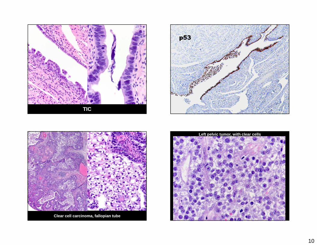
TIC
p53
Clear cell carcinoma, fallopian tube
Left pelvic tumor, with clear cells
11
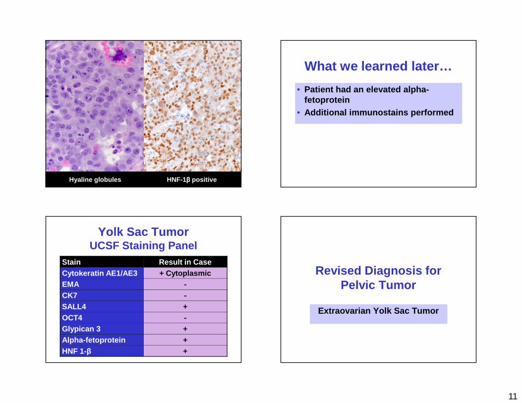
Hyaline globules HNF-1 β positive
What we learned later…
• Patient had an elevated alpha-fetoprotein
• Additional immunostains performed
Yolk Sac TumorUCSF Staining Panel
Stain Result in CaseCytokeratin AE1/AE3 + CytoplasmicEMA -CK7 -SALL4 +OCT4 -Glypican 3 +Alpha-fetoprotein +HNF 1-β +
Revised Diagnosis for Pelvic Tumor
Extraovarian Yolk Sac Tumor
12
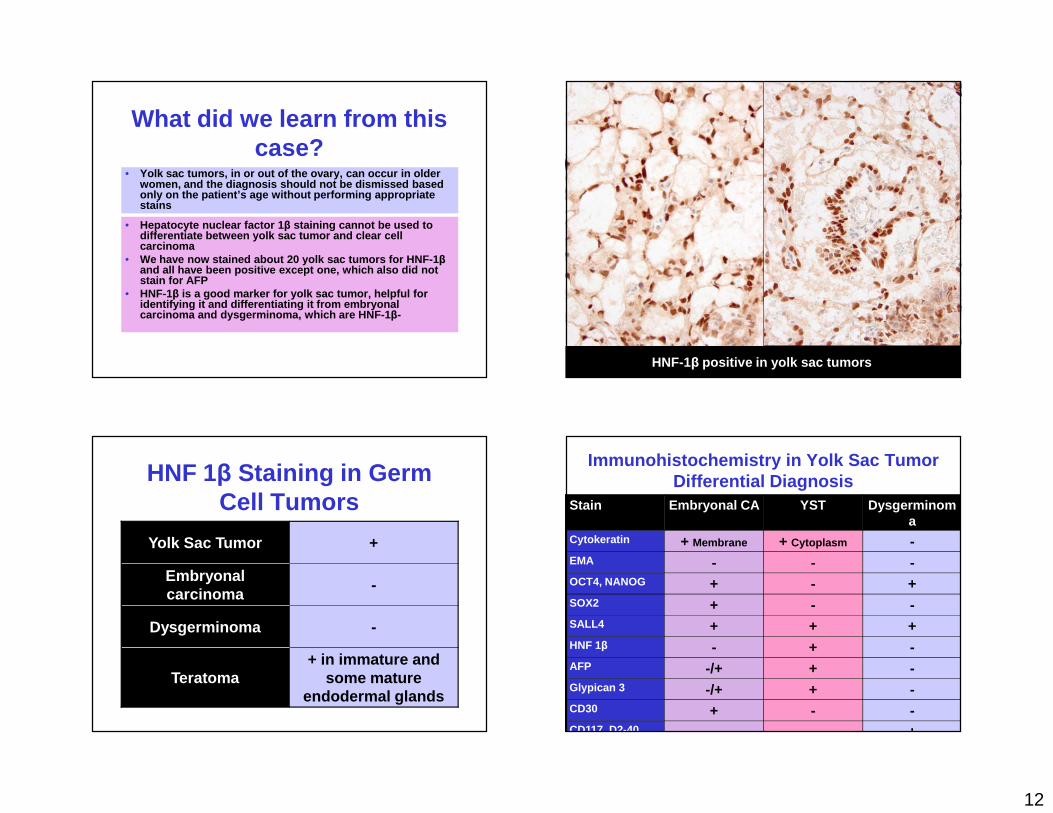
What did we learn from this case?
• Yolk sac tumors, in or out of the ovary, can occur in older women, and the diagnosis should not be dismissed ba sed only on the patient’s age without performing approp riate stains
• Hepatocyte nuclear factor 1 β staining cannot be used to differentiate between yolk sac tumor and clear cell carcinoma
• We have now stained about 20 yolk sac tumors for HN F-1βand all have been positive except one, which also d id not stain for AFP
• HNF-1β is a good marker for yolk sac tumor, helpful for identifying it and differentiating it from embryona l carcinoma and dysgerminoma, which are HNF-1 β-
HNF-1β positive in yolk sac tumors
HNF 1β Staining in Germ Cell Tumors
Yolk Sac Tumor +
Embryonal carcinoma
-
Dysgerminoma -
Teratoma+ in immature and
some mature endodermal glands
Immunohistochemistry in Yolk Sac Tumor Differential Diagnosis
Stain Embryonal CA YST Dysgerminoma
Cytokeratin + Membrane + Cytoplasm -EMA - - -OCT4, NANOG + - +SOX2 + - -SALL4 + + +HNF 1β - + -AFP -/+ + -Glypican 3 -/+ + -CD30 + - -CD117, D2-40 - - +
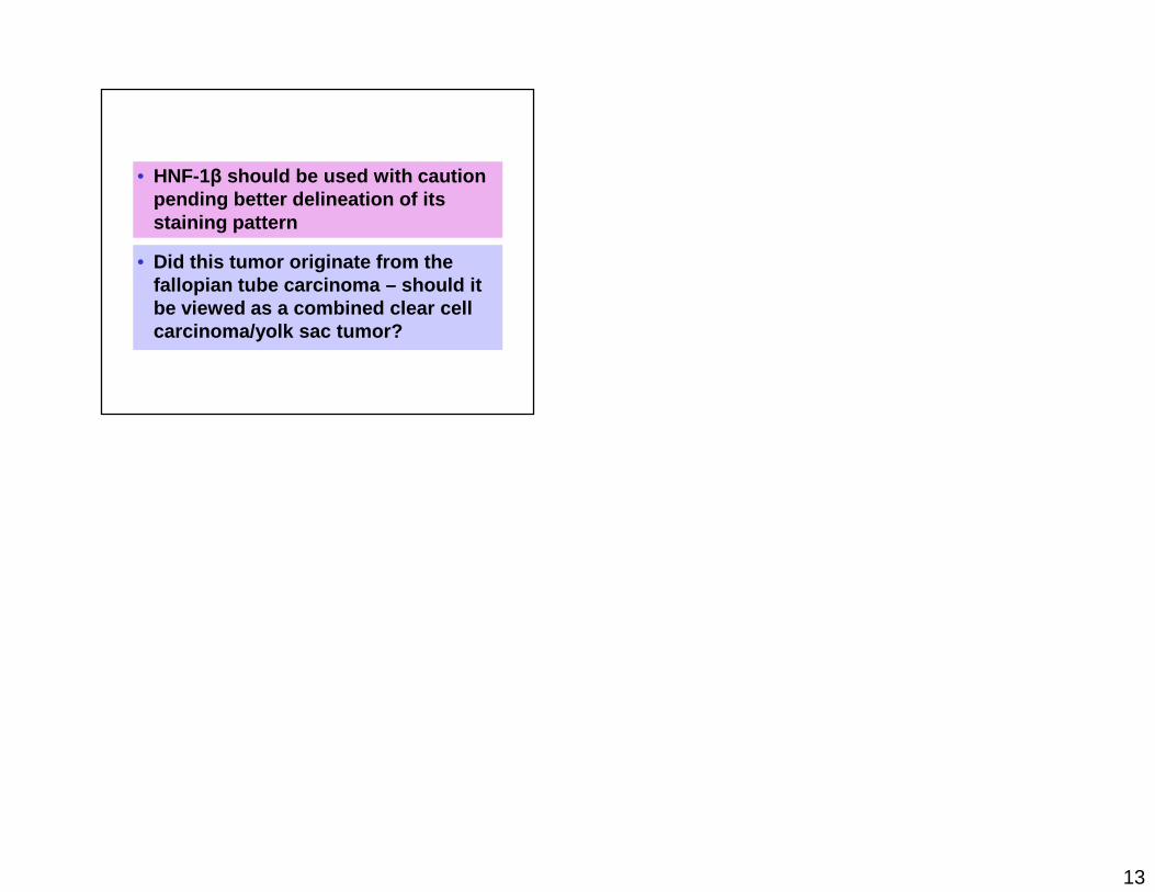
13
• HNF-1β should be used with caution pending better delineation of its staining pattern
• Did this tumor originate from the fallopian tube carcinoma – should it be viewed as a combined clear cell carcinoma/yolk sac tumor?