post-traumatic brachial plexus mri in practice · brachial plexus is made up of the anterior...
TRANSCRIPT
Diagnostic and Interventional Imaging (2013) 94, 925—943
CONTINUING EDUCATION PROGRAM: FOCUS. . .
Post-traumatic brachial plexus MRIin practice
O. Silbermann-Hoffmana,∗, F. Teboulb
a American Hospital of Paris, 63, boulevard Victor-Hugo, 92200 Neuilly-sur-Seine, Franceb Peripheral nerve and Brachial Plexus Institute of Surgery, 92, boulevard de Courcelles,75017 Paris, France
KEYWORDSMRI;Post-traumaticbrachial plexusparalysis;Avulsion;Nerve rupture;Axillary nerve
Abstract Injuries are separated into spinal nerve root avulsions (pre-ganglionic lesions) andmore distal rupture (post-ganglionic lesions). The lesions may be associated with differentnerve root levels. Spinal MRI is used to diagnose pre-ganglionic lesions, which may be presentin the absence of pseudomeningoceles. The other sequences described are used to diagnosepost-ganglionic lesions, regardless of the type of lesion. Knowledge that a graftable C5 nerveroot is present is important in the treatment strategy. Contrast enhancement in the scalenetriangle does not predict the quality of the nerve root (continuous injury with response toperoperative stimulation or division of the root needing grafting). Understanding post-traumatic
neuroma neuronal injuries to the brachial plexus. Knowing how to look for spinal MRI abnormalities andpost-ganglionic abnormalities.© 2013 Éditions françaises de radiologie. Published by Elsevier Masson SAS. All rights reserved.
Traumatic brachial plexus paralysis in adults mostly affects young people between 20 and30 years old. Ninety percent of cases are due to motorbike accidents. The mechanism ofthe injury involves stretching of the brachial plexus nerves which may lead to two typesof damage: avulsion, or tearing off of the insertion of the nerve roots from the spinalcord (pre-ganglionic lesion) and nerve rupture within the brachial plexus (post-ganglioniclesion). The neuronal injury may affect the nerve roots making up the brachial plexusin the scalene triangle, and also in the plexus’ trunks, bundles and terminal branches.Post-ganglionic injury often leads to a neuroma in continuity. Occasionally a clean rup-
ture occurs, causing nerve retraction. The investigation and treatment of these injuriesare relatively urgent and recent publications have shown the essential role of the pre-operative interval (less than 6 months) in recovery. Clinical examination combined withelectrophysiological and MRI studies enable surgeons to optimize their treatment strategy.∗ Corresponding author.E-mail address: [email protected] (O. Silbermann-Hoffman).
2211-5684/$ — see front matter © 2013 Éditions françaises de radiologie. Published by Elsevier Masson SAS. All rights reserved.http://dx.doi.org/10.1016/j.diii.2013.08.013
9
IqiriloforiEef
A
Ttiamwtvnwnoarfotf
Ffh
ttdliClraeslbdddcbsftEmnmrr
crbtmater and arachnoid stop at the foramina, when the dura
26
ndividual investigations completed in isolation are inade-uate. MRI is the first line imaging investigation: spinal CTs used if it is not possible to perform this. MRI can accu-ately diagnose pre-ganglionic and confirm post-ganglionicnjuries, although usually cannot assess whether theseesions have the potential to recover, i.e. establish whetherr not post-ganglionic injuries have the capacity to recoverunction. Functional capacity is assessed by changes in signsf renervation on repeated electromyograms. If no signs ofenervation are seen at 3 months, surgery is required, as its with lesions, which partially recover on several repeatedMGs. If a direct surgical approach is used, palpating andlectrostimulating the nerves can confirm or exclude theirunctional capacity.
natomical review
he brachial plexus is made up of the anterior branches ofhe C5 to T1 nerve roots, with a possible component com-ng from the C4 and T2 roots. The nerve roots consist ofnterior and posterior branches, which meet at the fora-en and extra-foramen junction to form a single nerve root,hich divides into an anterior branch, which forms part of
he brachial plexus, and a posterior branch, which inner-ates the paravertebral muscles. Each anterior and posteriorerve root branch is formed by the union of several rootlets,hich arise directly from the spinal cord, well before theerve leaves the foramen (Fig. 1a). These rootlets consistf three or four bands from C5 to C7 and two bands in C8nd T1 [1] with a large number of rootlets for the poste-ior part. The spinal ganglion, located in the region of theoramen (Fig. 1b), arises from the posterior part and is the
rigin of the sensory fibers, and the anterior part carrieshe motor fibers. The brachial plexus is divided into five dif-erent anatomical parts; the roots, trunks, division of theigure 1. Anatomical sections by Prof. Demondion: a: the roots emeoramen (double arrow); b: the spinal ganglion (gg) is continuous with
ead).
moi
O. Silbermann-Hoffman, F. Teboul
runks, bundles and terminal branches. The roots pass downhrough the scalene triangle between the anterior and mid-le scalene muscles. They then join and form trunks in theateral part of the scalene triangle. The superficial trunks formed from the union of the anterior roots of C5 and6 and gives rise to an important branch, the suprascapu-
ar nerve. The middle trunk is a continuation of the anterioroot of C7 and the inferior trunk arises from the union of thenterior roots of C8 and T1. The trunks arise from the lat-ral aspect of the middle scalene muscle. A landmark thathould be sought out on coronal sections is the dorsal scapu-ar artery, a branch of the subclavian artery, which passesetween the superior and middle trunks or between the mid-le and inferior trunks [2,3]. Each trunk gives rise to twoivisions, one anterior and one posterior, which lead to in sixivisions originating where these neuronal bundles cross thelavicle (costoclavicular junction), which then group intoundles. The lateral bundle arises from the anterior divi-ions of the superior and middle trunks, the medial bundlerom the anterior division of the inferior trunk and the pos-erior bundle from the posterior divisions of all three trunks.ach bundle then leads on to several terminal branches. Theain branches of the lateral bundle are the musculocuta-
eous nerve and the lateral root of the median nerve. Theedial bundle gives rise to the ulnar nerve and the medial
oot of the median nerve, and the posterior bundle to theadial nerve and the axillary nerve.
MRI myelography clearly shows the posterior nerve rootomponent, which contains more rootlets than the ante-ior component, and the smaller number of nerve rootletands in C8-T1. The rootlets leave the spine, carrying withhem the dura mater and arachnoid membrane. The dura
rge from the spine (round) proximal to their emergence from thethe posterior rootlets (arrow) behind the anterior rootlets (arrow
ater extends and merges with epineurium whereas theuter layers of the arachnoid membrane fuse with the per-neurium [4] which adheres to the anterior nerve root and
pCtagrfuarolansaa‘ttfipcwtri
M
Tisiricdtg
P
Tptcfm
tsgtrss
Post-traumatic brachial plexus MRI in practice
spinal ganglion, explaining why CSF is not seen in the fora-men unless an arachnoid diverticulum (a variant of the norm)or post-traumatic lesion is present. Pseudomeningoceles aredue to extravasation of CSF through a breach, forming anunencapsulated pouch of fluid.
Assessment of neurological functions
Clinical examination
The patient’s neurological injuries should be assessedpromptly via a full examination in order to establish whetheror not he/she is a candidate for surgery. The earlier a patientis examined, the more likely it is that the prognosis of thelesions can be established (stable damage, recovery or dete-rioration). Upper limb paralysis secondary to the stretchingof the nerves may be complete (75%) or partial (25%). Theinjuries are supraclavicular in 72% of cases and infraclavicu-lar in 28% of cases [5]. Overall, 25% of plexus injuries involvecomplete avulsions. Fifteen percent of patients with sup-raclavicular injuries have concomitant distal retro- and/orinfraclavicular damage [6]. Various clinical pictures can bedistinguished, depending on the nerve roots concerned,although, despite the fact that each nerve root correspondsto a muscle group, the distribution of nerve fibers occasion-ally varies. Muscles may be innervated by different nerveroots in different patients (for example, the radial nervemay receive most of its fibers from C7, which is usually thecase, although occasionally from C8 or even T1). Damage toC7 may therefore occur without defective elbow and wristor finger extension, and it is therefore more appropriateto describe the paralysis according to the affected function(paralysis of the shoulder, elbow flexion, wrist extensionetc.). Restoring elbow and shoulder mobility is the keyobjective.
Imaging and neurophysiological studies (nerve conduc-tion and electromyography) are essential to assess traumaticbrachial plexus paralyses in order to assess the level of thelesion, type of neuronal damage and potential for recovery.
Neurophysiological studies
A certain level of knowledge is required to understandthe results of this investigation. Seddon produced a func-tional classification of nerve injuries in 1942 [7] separatinginjuries into neuropraxia, axonotmesis and neurotmesis.Neuropraxia involves loss of nerve function, causing conduc-tion block but without anatomical injury. Recovery may takeup to 12 weeks. Axonotmesis is secondary to axonal rupturewithout damage to the Schwann linings, which results in dis-tal Wallerien regeneration with slow functional recovery asthe endoneurial tubes are preserved. Thirdly, neurotmesisinvolves division of all of the parts of the nerve (dam-age to the axons and linings), in which case no recoveryoccurs.
Neurophysiological study should not be performed lessthan 4 weeks after the injury (the Wallerien degen-
eration time) with repeat studies at 6-week intervals.Sensory potentials can differentiate between pre- frompost-ganglionic injury. The sensory neuron cell body islocated in the posterior ganglion at the foramen, and in aad[E
927
re-ganglionic injury (avulsion) the ganglion remains intact.omplete clinical lack of sensation is found in the terri-ory investigated, with preserved sensory nerve potentialmplitudes. On the other hand, a mixed pre- and post-anglionic injury cannot be excluded if these potentials areeduced. The extent of muscle denervation and number ofunctional motor neurons is established by ELG. A motornit is made up of the motor neuron cell body, its axonnd all of the muscle fibers, which it innervates, and isesponsible for bi- or tri-phasic waves. The resting tracef a normally innervated muscle is flat. Muscle contractioneads to the appearance of spikes, the number and appear-nce of which depend on the contraction force — i.e. theumber of motor units used. A denervated muscle exhibitshort, low potentials on the resting trace, which developpproximately 3 weeks after the accident, i.e. once thexons have completely degenerated (these are known as‘fibrillation potentials’’). In addition, intentional attemptso contract the muscle, or stimulation of the nerve, do notrigger spikes. Neuronal regeneration results in a fall in thebrillation potentials and the emergence of regenerationotentials after intentional attempts to contract the mus-le. The presence of polyphasic resting fibrillation waves,hich indicate denervation, and poor voluntary contrac-
ion traces indicate a lack of functional units. Signs ofe-innervation develop between 2 and 4 months after thenjury.
RI
he modified Nagano classification of brachial plexusnjuries describes four zones. Zone I involves the avul-ion of rootlets from the spine; zone IIA contains ganglionnjuries within the foramen; zone IIB comprises the nerveoots up to the division of the trunks; zone III when thenjured area involves the trunks; and zone IV when theentre of the injury is on the bundles [8]. Zone IIA isefined as a post-ganglionic injury, although it is, in fact,he equivalent of avulsion as it is too proximal to berafted.
re-ganglionic injuries
here are both direct and indirect signs suggestingre-ganglionic damage. These injuries are studied by inves-igating the rootlets, analyzing meningeal linings, anyontrast enhancement on the path of a nerve or on the sur-ace of the spine [9] and by examining the paravertebraluscles [10].Spinal CT has long been the reference investigation for
hese injuries. Published results have shown equivalentensitivity and specificity results when they are investi-ated by MRI if high quality myelography sequences areaken, providing excellent contrast between CSF and theootlets, combined with excellent spatial resolution. Sen-itivity ranges between 89% and 96.1% against the ‘‘goldtandard’’ of spinal CT with a specificity of between 95
nd 96.6% [11—13]. Myelography MRI is named differentlyepending on the manufacturers (Gradient 3D Echo: fiestaGE] CISS [Siemens] True-Fisp [Philips]; Ultra Fast Spincho T2: SSFSE [GE] SSTSE [Haste Siemens] SSIT-TSE [UFSE
9
Prsla
vamcvatsnsiaaaii(F5tgivt(cosats
EBtcarabaiOrrfira
EPadrrpdet
IRlc
Fv
28
hilips]). These enable volume acquisition with secondaryeconstructions in the different spatial planes. This type ofequence is essential for detailed analysis of pre-ganglionicesions and should be repeated if artefacts are produced as
result of movements.We use the 3D FIESTA sequence (T2/T1 ratio), which pro-
ides high signal to noise images with few flow artefacts,lthough this sequence is sensitive to swallowing and move-ent artefacts. In addition, the bone marrow and spinal
ord appear very black restricting the examination of theertebrae and for spinal edema [14] and only offering goodnalysis of the rootlets in 60% of patients because of mul-iple large pseudomeningoceles. The same occurred withpinal CT. These large pseudomeningoceles are rarely seenow, probably because accidents are at lower speeds (radarpeed traps, license penalty points). We record our imagesn the sagittal plane, although other groups use frontal orxial planes [15] with the patient lying on his/her back, armslong the length of the body, and with their head and necks straight as possible. A 21-phase array head and neck coils used. The volume is placed on the axial ‘‘scout view’’,ncluding the spinal cord and a proximal part of the foraminaFig. 2a) using the following settings: TR = 5.5, TE = min full.ield 22 cm, matrix 288 × 320 antero-posterior frequency,2 loc, 3 excitations, flip angle 45◦, band width 62.5 sectionhickness 0.8 mm or 1 mm. The patient is asked to breatheently, using their stomach, and to try not to swallow dur-ng the acquisition. Axial reconstructions are made from theolume recorded in the middle plane of the discs from C4o T1-T2 in 1 mm thicknesses with an interval of 0.5 mmFig. 2b). This results in approximately 200 sections, whichan be seen in cine mode on the console. The direct signsbserved are whether or not the rootlets arising from the
pinal cord, which leave through the foramen are visible,nd spinal signal abnormalities at the point of insertion ofhe rootlets into the spinal cord or into the rootlets them-elves.iccs
igure 2. a: the 3D myelography image is positioned on the scout axisibility; b: axial reconstructions from the sagittal volume are made in
O. Silbermann-Hoffman, F. Teboul
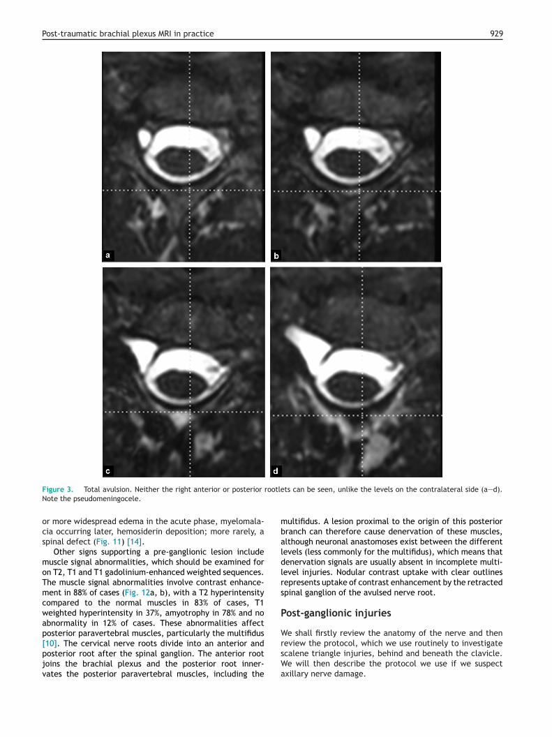
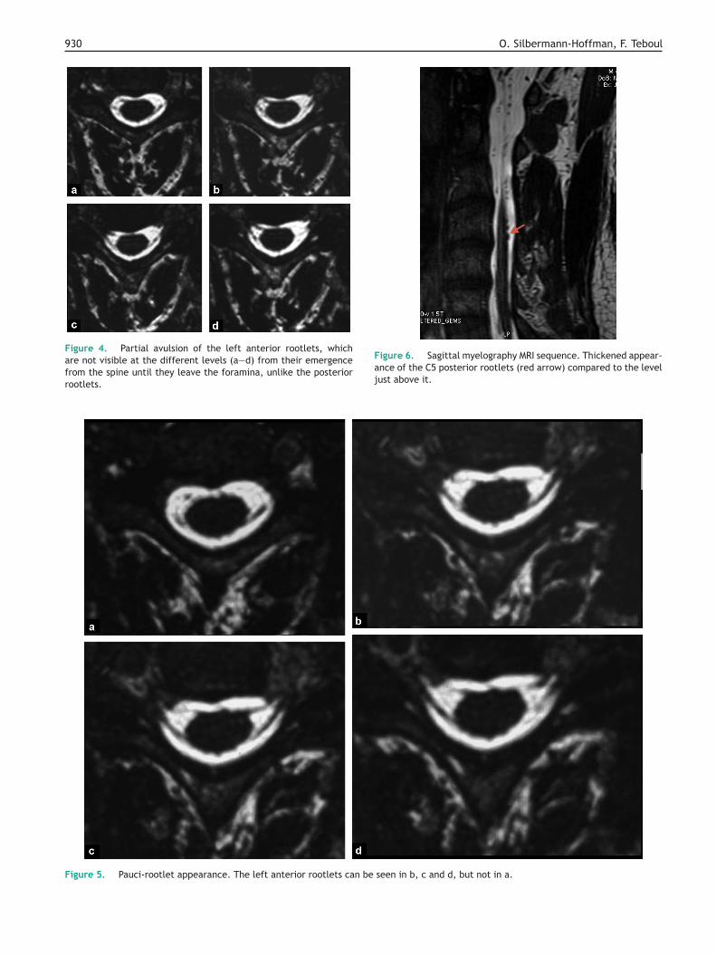
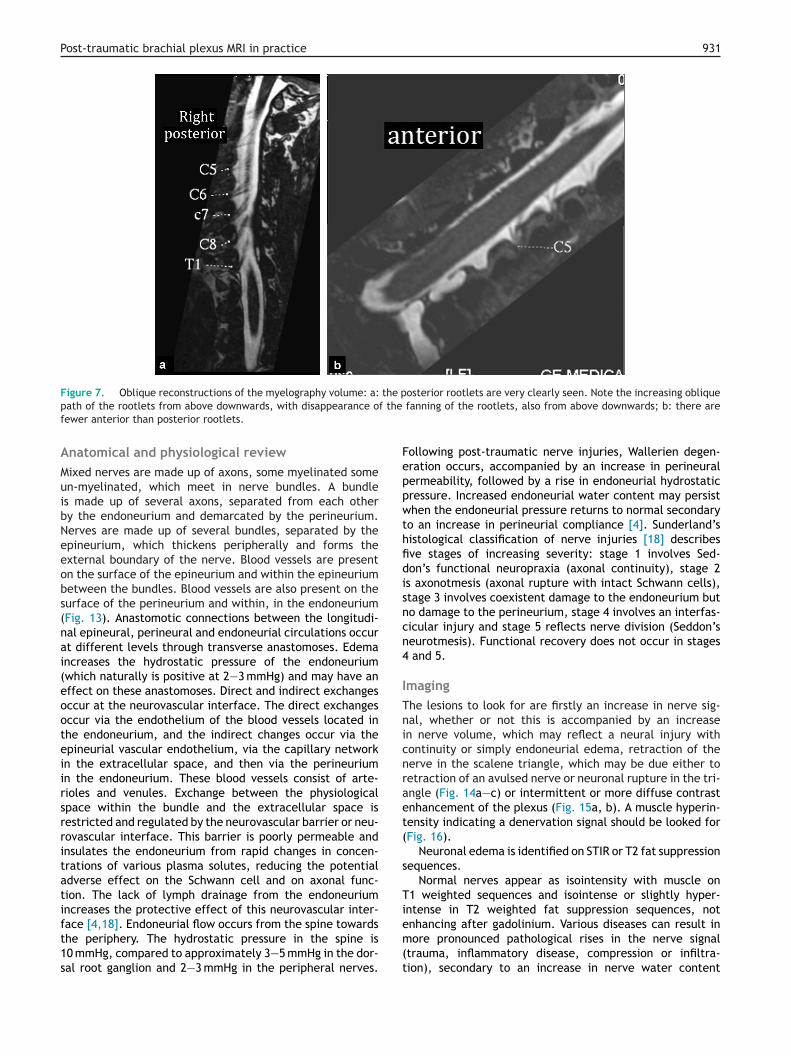
xamination of the rootletsoth the anterior and posterior rootlets can be torn off fromhe spinal cord at all levels (total avulsion) (Fig. 3a—d). Lessommonly, all of the individual anterior or posterior rootletsre torn off (partial avulsion) (Fig. 4a—d). Some of the ante-ior and/or anterior and posterior rootlets (pauci-rootletppearance) may occur (Fig. 5a—d) wherein the rootlets maye present but thickened (Fig. 6). Partial avulsion injuriesre rare and were found in 4.6% (5 out of 105 rootlets)n Tsai’s series [16] and in 5.6% in Gasparotti’s series [17].blique reconstructions are performed to identify the poste-
ior rootlets (Fig. 7a), together with frontal and/or obliqueeconstructions for the anterior rootlets (Fig. 7b). Summarylms containing approximately six to eight images per nerveoot level are given to the clinician with the nerve root levelsnnotated on them (Fig. 8a—h).
xamination of the meningesseudomeningoceles are present in 80% of avulsions, i.e.pproximately 20% of avulsions occur without forming pseu-omeningoceles. In their series, Gasparotti et al. [11]eported five cases of pseudomeningoceles without nerveoot abnormalities, something we have never seen. Theseseudomeningoceles may be large or small (saccular oriverticular), immediately against the foramen, they mayxtend over several levels, or they may lie anterior or pos-erior peri-spinally (Fig. 9a—c).
ndirect signsootlet contrast enhancement (12/250) (Fig. 10) or nodu-ar contrast enhancement on the surface of the spinalord (42/250) is rare and very suggestive of pre-ganglionic
njuries (7/12 and 38/42) [9]. Spinal abnormalities mayoexist with pre-ganglionic injuries in approximately 20% ofases, such as displacement of the spinal cord on the healthyide, spinal edema around the emergence of the rootletsial, represented here by an axial myelography section for betterthe middle plane of the discs of C4-C5 to C7-T1.
Post-traumatic brachial plexus MRI in practice 929
ootle
mbaldlrs
P
W
Figure 3. Total avulsion. Neither the right anterior or posterior rNote the pseudomeningocele.
or more widespread edema in the acute phase, myelomala-cia occurring later, hemosiderin deposition; more rarely, aspinal defect (Fig. 11) [14].
Other signs supporting a pre-ganglionic lesion includemuscle signal abnormalities, which should be examined foron T2, T1 and T1 gadolinium-enhanced weighted sequences.The muscle signal abnormalities involve contrast enhance-ment in 88% of cases (Fig. 12a, b), with a T2 hyperintensitycompared to the normal muscles in 83% of cases, T1weighted hyperintensity in 37%, amyotrophy in 78% and noabnormality in 12% of cases. These abnormalities affectposterior paravertebral muscles, particularly the multifidus
[10]. The cervical nerve roots divide into an anterior andposterior root after the spinal ganglion. The anterior rootjoins the brachial plexus and the posterior root inner-vates the posterior paravertebral muscles, including thersWa
ts can be seen, unlike the levels on the contralateral side (a—d).
ultifidus. A lesion proximal to the origin of this posteriorranch can therefore cause denervation of these muscles,lthough neuronal anastomoses exist between the differentevels (less commonly for the multifidus), which means thatenervation signals are usually absent in incomplete multi-evel injuries. Nodular contrast uptake with clear outlinesepresents uptake of contrast enhancement by the retractedpinal ganglion of the avulsed nerve root.
ost-ganglionic injuries
e shall firstly review the anatomy of the nerve and then
eview the protocol, which we use routinely to investigatecalene triangle injuries, behind and beneath the clavicle.e will then describe the protocol we use if we suspectxillary nerve damage.
930 O. Silbermann-Hoffman, F. Teboul
Figure 4. Partial avulsion of the left anterior rootlets, whichare not visible at the different levels (a—d) from their emergencefrom the spine until they leave the foramina, unlike the posteriorrootlets.
Figure 6. Sagittal myelography MRI sequence. Thickened appear-ance of the C5 posterior rootlets (red arrow) compared to the leveljust above it.
Figure 5. Pauci-rootlet appearance. The left anterior rootlets can be seen in b, c and d, but not in a.
Post-traumatic brachial plexus MRI in practice 931
Figure 7. Oblique reconstructions of the myelography volume: a: the posterior rootlets are very clearly seen. Note the increasing obliquef the
Feppwthfidisncn4
ITnicnraet(
s
Ti
path of the rootlets from above downwards, with disappearance ofewer anterior than posterior rootlets.
Anatomical and physiological reviewMixed nerves are made up of axons, some myelinated someun-myelinated, which meet in nerve bundles. A bundleis made up of several axons, separated from each otherby the endoneurium and demarcated by the perineurium.Nerves are made up of several bundles, separated by theepineurium, which thickens peripherally and forms theexternal boundary of the nerve. Blood vessels are presenton the surface of the epineurium and within the epineuriumbetween the bundles. Blood vessels are also present on thesurface of the perineurium and within, in the endoneurium(Fig. 13). Anastomotic connections between the longitudi-nal epineural, perineural and endoneurial circulations occurat different levels through transverse anastomoses. Edemaincreases the hydrostatic pressure of the endoneurium(which naturally is positive at 2—3 mmHg) and may have aneffect on these anastomoses. Direct and indirect exchangesoccur at the neurovascular interface. The direct exchangesoccur via the endothelium of the blood vessels located inthe endoneurium, and the indirect changes occur via theepineurial vascular endothelium, via the capillary networkin the extracellular space, and then via the perineuriumin the endoneurium. These blood vessels consist of arte-rioles and venules. Exchange between the physiologicalspace within the bundle and the extracellular space isrestricted and regulated by the neurovascular barrier or neu-rovascular interface. This barrier is poorly permeable andinsulates the endoneurium from rapid changes in concen-trations of various plasma solutes, reducing the potentialadverse effect on the Schwann cell and on axonal func-tion. The lack of lymph drainage from the endoneuriumincreases the protective effect of this neurovascular inter-
face [4,18]. Endoneurial flow occurs from the spine towardsthe periphery. The hydrostatic pressure in the spine is10 mmHg, compared to approximately 3—5 mmHg in the dor-sal root ganglion and 2—3 mmHg in the peripheral nerves.em(t
fanning of the rootlets, also from above downwards; b: there are
ollowing post-traumatic nerve injuries, Wallerien degen-ration occurs, accompanied by an increase in perineuralermeability, followed by a rise in endoneurial hydrostaticressure. Increased endoneurial water content may persisthen the endoneurial pressure returns to normal secondary
o an increase in perineurial compliance [4]. Sunderland’sistological classification of nerve injuries [18] describesve stages of increasing severity: stage 1 involves Sed-on’s functional neuropraxia (axonal continuity), stage 2s axonotmesis (axonal rupture with intact Schwann cells),tage 3 involves coexistent damage to the endoneurium buto damage to the perineurium, stage 4 involves an interfas-icular injury and stage 5 reflects nerve division (Seddon’seurotmesis). Functional recovery does not occur in stages
and 5.
maginghe lesions to look for are firstly an increase in nerve sig-al, whether or not this is accompanied by an increasen nerve volume, which may reflect a neural injury withontinuity or simply endoneurial edema, retraction of theerve in the scalene triangle, which may be due either toetraction of an avulsed nerve or neuronal rupture in the tri-ngle (Fig. 14a—c) or intermittent or more diffuse contrastnhancement of the plexus (Fig. 15a, b). A muscle hyperin-ensity indicating a denervation signal should be looked forFig. 16).
Neuronal edema is identified on STIR or T2 fat suppressionequences.
Normal nerves appear as isointensity with muscle on1 weighted sequences and isointense or slightly hyper-
ntense in T2 weighted fat suppression sequences, not
nhancing after gadolinium. Various diseases can result inore pronounced pathological rises in the nerve signaltrauma, inflammatory disease, compression or infiltra-ion), secondary to an increase in nerve water content
932 O. Silbermann-Hoffman, F. Teboul
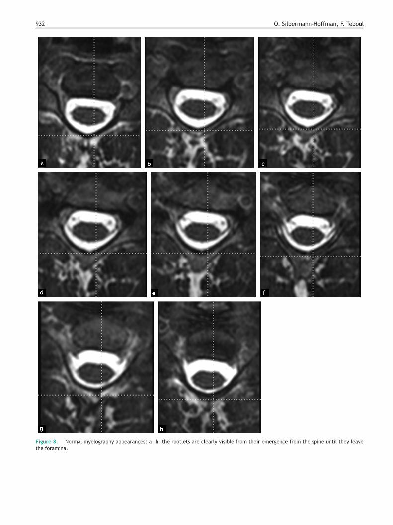
Figure 8. Normal myelography appearances: a—h: the rootlets are clearly visible from their emergence from the spine until they leavethe foramina.
Post-traumatic brachial plexus MRI in practice 933
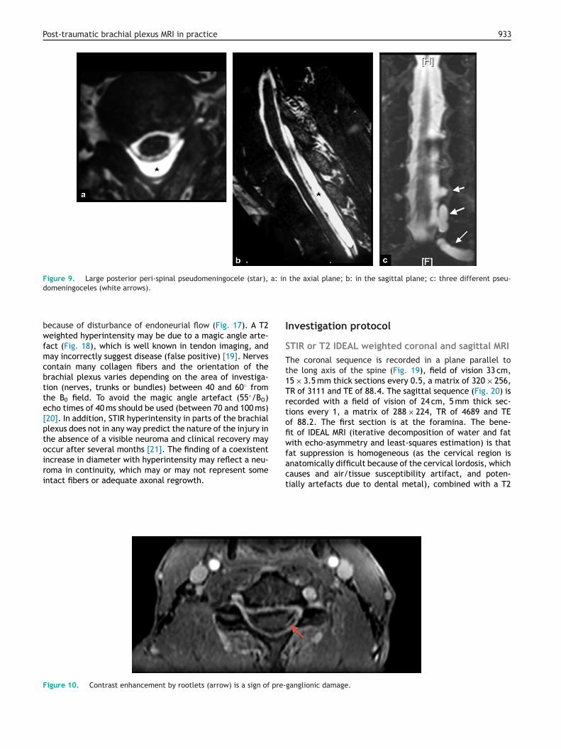
Figure 9. Large posterior peri-spinal pseudomeningocele (star), a: in the axial plane; b: in the sagittal plane; c: three different pseu-
I
STt1Trtofiwfat suppression is homogeneous (as the cervical region isanatomically difficult because of the cervical lordosis, which
domeningoceles (white arrows).
because of disturbance of endoneurial flow (Fig. 17). A T2weighted hyperintensity may be due to a magic angle arte-fact (Fig. 18), which is well known in tendon imaging, andmay incorrectly suggest disease (false positive) [19]. Nervescontain many collagen fibers and the orientation of thebrachial plexus varies depending on the area of investiga-tion (nerves, trunks or bundles) between 40 and 60◦ fromthe B0 field. To avoid the magic angle artefact (55◦/BO)echo times of 40 ms should be used (between 70 and 100 ms)[20]. In addition, STIR hyperintensity in parts of the brachialplexus does not in any way predict the nature of the injury inthe absence of a visible neuroma and clinical recovery mayoccur after several months [21]. The finding of a coexistentincrease in diameter with hyperintensity may reflect a neu-
roma in continuity, which may or may not represent someintact fibers or adequate axonal regrowth.ct
Figure 10. Contrast enhancement by rootlets (arrow) is a sign of pre-
nvestigation protocol
TIR or T2 IDEAL weighted coronal and sagittal MRIhe coronal sequence is recorded in a plane parallel tohe long axis of the spine (Fig. 19), field of vision 33 cm,5 × 3.5 mm thick sections every 0.5, a matrix of 320 × 256,R of 3111 and TE of 88.4. The sagittal sequence (Fig. 20) isecorded with a field of vision of 24 cm, 5 mm thick sec-ions every 1, a matrix of 288 × 224, TR of 4689 and TEf 88.2. The first section is at the foramina. The bene-t of IDEAL MRI (iterative decomposition of water and fatith echo-asymmetry and least-squares estimation) is that
auses and air/tissue susceptibility artifact, and poten-ially artefacts due to dental metal), combined with a T2
ganglionic damage.
934 O. Silbermann-Hoffman, F. Teboul
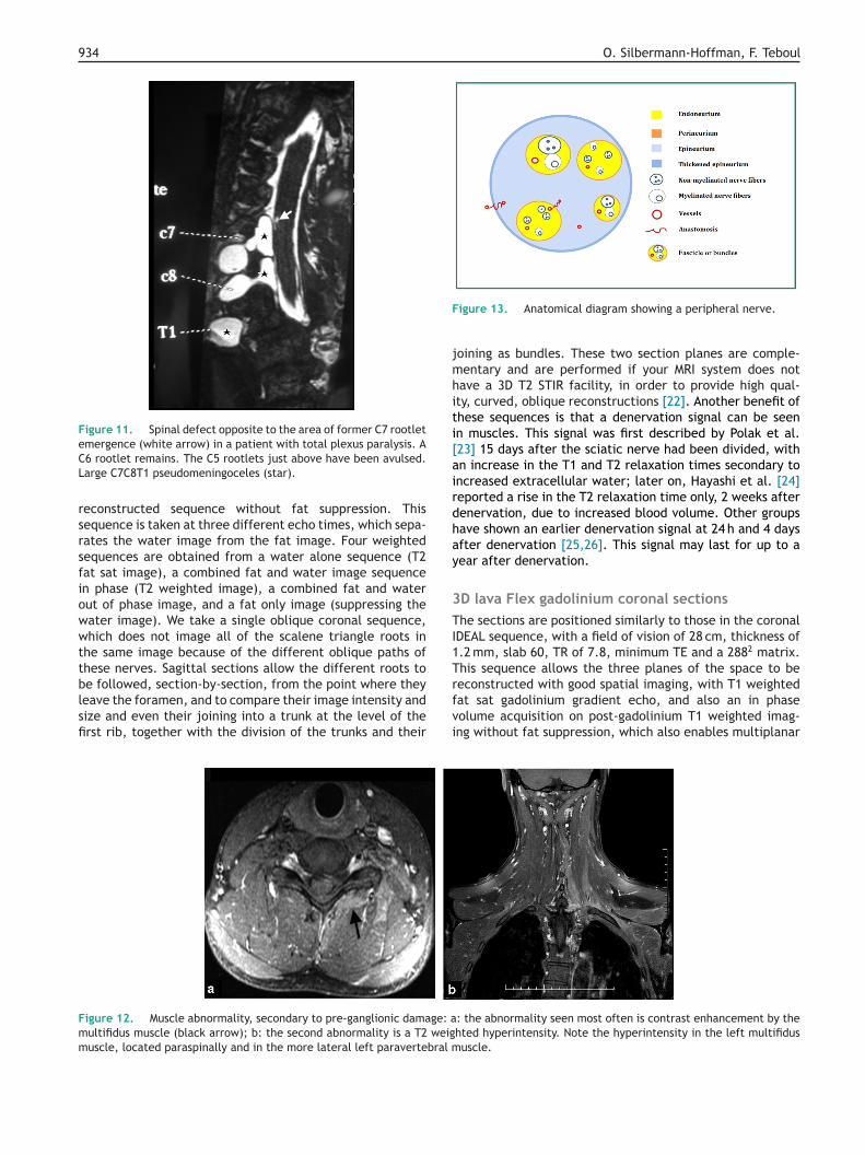
Figure 11. Spinal defect opposite to the area of former C7 rootletemergence (white arrow) in a patient with total plexus paralysis. ACL
rsrsfiowwttblsfi
F
jmhiti[airdhay
3TI1T
Fmm
6 rootlet remains. The C5 rootlets just above have been avulsed.arge C7C8T1 pseudomeningoceles (star).
econstructed sequence without fat suppression. Thisequence is taken at three different echo times, which sepa-ates the water image from the fat image. Four weightedequences are obtained from a water alone sequence (T2at sat image), a combined fat and water image sequencen phase (T2 weighted image), a combined fat and waterut of phase image, and a fat only image (suppressing theater image). We take a single oblique coronal sequence,hich does not image all of the scalene triangle roots in
he same image because of the different oblique paths ofhese nerves. Sagittal sections allow the different roots to
e followed, section-by-section, from the point where theyeave the foramen, and to compare their image intensity andize and even their joining into a trunk at the level of therst rib, together with the division of the trunks and theirrfvi
igure 12. Muscle abnormality, secondary to pre-ganglionic damage: aultifidus muscle (black arrow); b: the second abnormality is a T2 weiguscle, located paraspinally and in the more lateral left paravertebral m
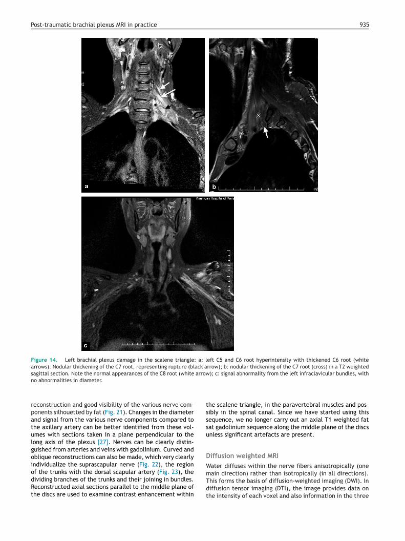
igure 13. Anatomical diagram showing a peripheral nerve.
oining as bundles. These two section planes are comple-entary and are performed if your MRI system does not
ave a 3D T2 STIR facility, in order to provide high qual-ty, curved, oblique reconstructions [22]. Another benefit ofhese sequences is that a denervation signal can be seenn muscles. This signal was first described by Polak et al.23] 15 days after the sciatic nerve had been divided, withn increase in the T1 and T2 relaxation times secondary toncreased extracellular water; later on, Hayashi et al. [24]eported a rise in the T2 relaxation time only, 2 weeks afterenervation, due to increased blood volume. Other groupsave shown an earlier denervation signal at 24 h and 4 daysfter denervation [25,26]. This signal may last for up to aear after denervation.
D lava Flex gadolinium coronal sectionshe sections are positioned similarly to those in the coronal
DEAL sequence, with a field of vision of 28 cm, thickness of.2 mm, slab 60, TR of 7.8, minimum TE and a 2882 matrix.his sequence allows the three planes of the space to beeconstructed with good spatial imaging, with T1 weighted
at sat gadolinium gradient echo, and also an in phaseolume acquisition on post-gadolinium T1 weighted imag-ng without fat suppression, which also enables multiplanar: the abnormality seen most often is contrast enhancement by thehted hyperintensity. Note the hyperintensity in the left multifidususcle.
Post-traumatic brachial plexus MRI in practice 935
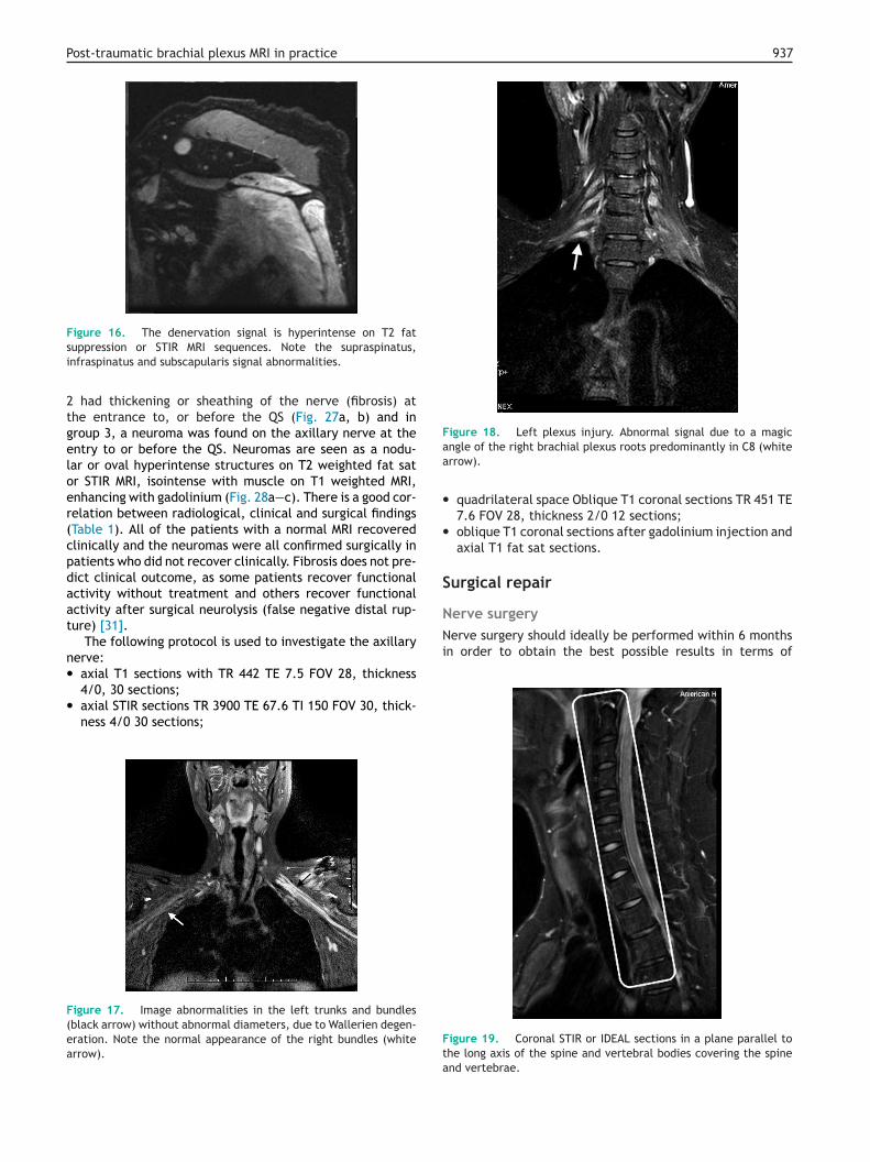
Figure 14. Left brachial plexus damage in the scalene triangle: a: left C5 and C6 root hyperintensity with thickened C6 root (whitearrows). Nodular thickening of the C7 root, representing rupture (black arrow); b: nodular thickening of the C7 root (cross) in a T2 weightedsagittal section. Note the normal appearances of the C8 root (white arrow); c: signal abnormality from the left infraclavicular bundles, with
tsssu
DW
no abnormalities in diameter.
reconstruction and good visibility of the various nerve com-ponents silhouetted by fat (Fig. 21). Changes in the diameterand signal from the various nerve components compared tothe axillary artery can be better identified from these vol-umes with sections taken in a plane perpendicular to thelong axis of the plexus [27]. Nerves can be clearly distin-guished from arteries and veins with gadolinium. Curved andoblique reconstructions can also be made, which very clearlyindividualize the suprascapular nerve (Fig. 22), the region
of the trunks with the dorsal scapular artery (Fig. 23), thedividing branches of the trunks and their joining in bundles.Reconstructed axial sections parallel to the middle plane ofthe discs are used to examine contrast enhancement withinmTdt
he scalene triangle, in the paravertebral muscles and pos-ibly in the spinal canal. Since we have started using thisequence, we no longer carry out an axial T1 weighted fatat gadolinium sequence along the middle plane of the discsnless significant artefacts are present.
iffusion weighted MRIater diffuses within the nerve fibers anisotropically (one
ain direction) rather than isotropically (in all directions).his forms the basis of diffusion-weighted imaging (DWI). Iniffusion tensor imaging (DTI), the image provides data onhe intensity of each voxel and also information in the three
936 O. Silbermann-Hoffman, F. Teboul
Figure 15. a: contrast enhancement by a retracted spinal ganglion in the scalene triangle (arrow); b: contrast enhancement either sideof an intra-scalene C7 rupture. Note the hemosiderin deposits in this gradient echo sequence.
sbataldi[ggrplsitoparoid4dtitutaltr
A
RTbhsrolthtpttnaouwauapsids
patial planes of each voxel. Each voxel can be representedy a vector along the main direction of the nerve: the sum ofll of these vectors in the volume produces a linear image ofhe nerve path. Water molecule diffusion is restricted to thexons because of the myelin sheath. Intra-axonal metabo-ites such as NE (N-Acetyl-Aspartate) and intra-axonal wateriffusion is also restricted. Use of a low b factor (< 1000)s more sensitive for the inter-axonal extracellular space28]. Diffusion weighted imaging visualizes the sensory nerveanglia, post-ganglionic nerve roots in the scalene trian-le, the trunks, and also the lymph nodes. This sequenceequires processing with MIP reconstruction in the frontallane to visualize these neuronal structures and suppress theymph nodes, which may be superimposed on the neuronaltructures as well as possible. This technique does not visual-ze the nerve roots above C5 or the pre-ganglionic portion ofhe nerve [15]. The infraclavicular region can be seen in 60%f normal subjects. Reduced signal intensity in traumaticost-ganglionic lesions has been reported [29] and a recentrticle [17] using diffusion tensor imaging with tractography,eported that intact nerve roots from C5 to T1 were visiblen the healthy side in 100% of the patients suffering a plexusnjury, with a sensitivity of 88.1% and specificity of 98.8% toiagnose avulsion. The procedure, however, lasts 11 min and0 s, and cannot therefore be used in everyday practice! Aiffusion tensor sequence takes time, both for the acquisi-ion and particularly for processing, thus we do not performt routinely. The parameters used are a b of 900 and 9 direc-ion gradient or a b of 1000 with 12 direction gradients (somese 30 direction gradients [15]). In practice, we do not findhis sequence to be a discriminating one in terms of analysisnd we found no signal defect on a post-ganglionic continuity
esion, which produced no response to electrical stimula-ion preoperatively and therefore reflected intra-fascicularupture with superficial epineural continuity.UAwM
xillary nerve damage
eview of anatomy and pathologyhe sensorimotor axillary nerve is one of the terminalranches of the posterior bundle and runs behind theumeral artery in contact with the anterior region of theubscapularis muscle, before passing from anterior to poste-ior along an oblique path into the quadrilateral space givingut collaterals to the teres minor, occasionally the subscapu-aris, the gleno-humeral joint and the sensory branch tohe external aspect of the shoulder. It passes around theumerus and then runs along the deep surface of the del-oid muscle, giving out several intramuscular branches, fromosterior to anterior. The deltoid is involved in abduction ofhe arm, antepulsion through its anterior fibers and con-ributes to extension of the arm via its posterior fibers. Theerve can be damaged in arm traction movements followingntero-internal dislocation of the humeral head. One studyn 63 patients showed that the damage was due to rupture,sually at the entrance to the quadrilateral space (41/63),here the nerve is fixed inside the space itself (21/63), and
third lesion was identified as a nerve which was contin-ous but sheathed in the fibrous tissue (14/63) [30]. Thexillary nerve is clearly seen on T1 weighted coronal MRI,arallel to the quadrilateral space (QS) (Fig. 24). It appearsurrounded by fat, proximal to the space, and in the spacetself (Fig. 25). On axial sections it can occasionally beifficult to distinguish it from its accompanying axillary ves-els (Fig. 26).
se of MRI prospective study has been carried out on 15 patientsith post-traumatic axillary nerve paralysis and found threeRI groups of patients: group 1 had a normal MRI, group
Post-traumatic brachial plexus MRI in practice 937
Figure 16. The denervation signal is hyperintense on T2 fat
Figure 18. Left plexus injury. Abnormal signal due to a magicaa
•
•
S
Nerve surgeryNerve surgery should ideally be performed within 6 monthsin order to obtain the best possible results in terms of
suppression or STIR MRI sequences. Note the supraspinatus,infraspinatus and subscapularis signal abnormalities.
2 had thickening or sheathing of the nerve (fibrosis) atthe entrance to, or before the QS (Fig. 27a, b) and ingroup 3, a neuroma was found on the axillary nerve at theentry to or before the QS. Neuromas are seen as a nodu-lar or oval hyperintense structures on T2 weighted fat sator STIR MRI, isointense with muscle on T1 weighted MRI,enhancing with gadolinium (Fig. 28a—c). There is a good cor-relation between radiological, clinical and surgical findings(Table 1). All of the patients with a normal MRI recoveredclinically and the neuromas were all confirmed surgically inpatients who did not recover clinically. Fibrosis does not pre-dict clinical outcome, as some patients recover functionalactivity without treatment and others recover functionalactivity after surgical neurolysis (false negative distal rup-ture) [31].
The following protocol is used to investigate the axillarynerve:• axial T1 sections with TR 442 TE 7.5 FOV 28, thickness
4/0, 30 sections;• axial STIR sections TR 3900 TE 67.6 TI 150 FOV 30, thick-
ness 4/0 30 sections;
Figure 17. Image abnormalities in the left trunks and bundles(black arrow) without abnormal diameters, due to Wallerien degen-eration. Note the normal appearance of the right bundles (whitearrow).
Fta
ngle of the right brachial plexus roots predominantly in C8 (whiterrow).
quadrilateral space Oblique T1 coronal sections TR 451 TE7.6 FOV 28, thickness 2/0 12 sections;oblique T1 coronal sections after gadolinium injection andaxial T1 fat sat sections.
urgical repair
igure 19. Coronal STIR or IDEAL sections in a plane parallel tohe long axis of the spine and vertebral bodies covering the spinend vertebrae.
938 O. Silbermann-Hoffman, F. Teboul
FI
rmdea•
Fphc
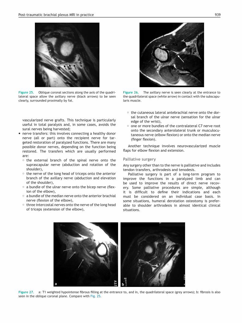
Figure 22. Oblique reconstruction from a 3D lava sequence withfat suppression: the superior trunk is very clearly seen (white arrow)and the suprascapular nerve is seen as far as the supra-coracoidnotch (arrow head).
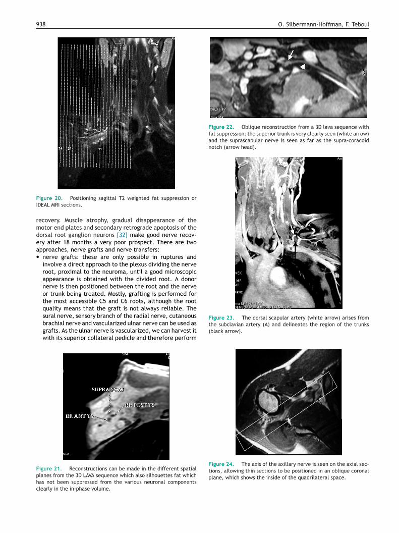
igure 20. Positioning sagittal T2 weighted fat suppression orDEAL MRI sections.
ecovery. Muscle atrophy, gradual disappearance of theotor end plates and secondary retrograde apoptosis of theorsal root ganglion neurons [32] make good nerve recov-ry after 18 months a very poor prospect. There are twopproaches, nerve grafts and nerve transfers:nerve grafts: these are only possible in ruptures andinvolve a direct approach to the plexus dividing the nerveroot, proximal to the neuroma, until a good microscopicappearance is obtained with the divided root. A donornerve is then positioned between the root and the nerveor trunk being treated. Mostly, grafting is performed forthe most accessible C5 and C6 roots, although the rootquality means that the graft is not always reliable. Thesural nerve, sensory branch of the radial nerve, cutaneous
brachial nerve and vascularized ulnar nerve can be used asgrafts. As the ulnar nerve is vascularized, we can harvest itwith its superior collateral pedicle and therefore performigure 21. Reconstructions can be made in the different spatiallanes from the 3D LAVA sequence which also silhouettes fat whichas not been suppressed from the various neuronal componentslearly in the in-phase volume.
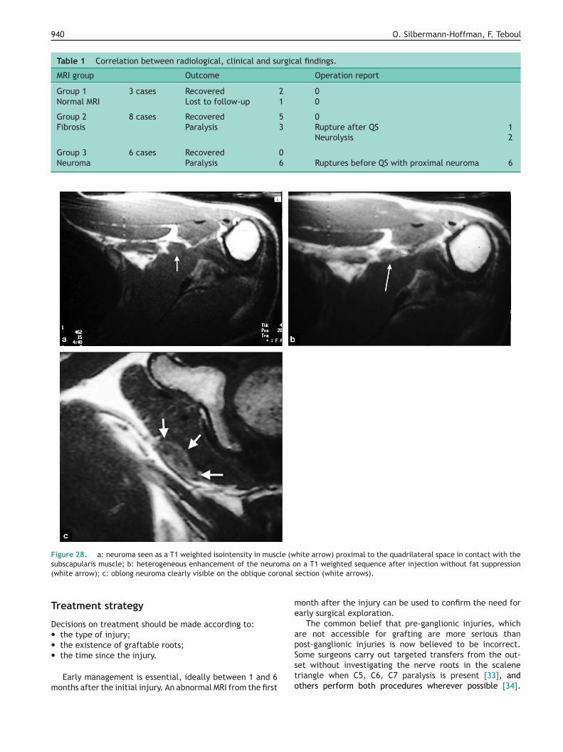
Figure 23. The dorsal scapular artery (white arrow) arises fromthe subclavian artery (A) and delineates the region of the trunks(black arrow).
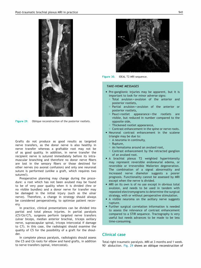
Figure 24. The axis of the axillary nerve is seen on the axial sec-tions, allowing thin sections to be positioned in an oblique coronalplane, which shows the inside of the quadrilateral space.
Post-traumatic brachial plexus MRI in practice 939
Figure 25. Oblique coronal sections along the axis of the quadri-lateral space allow the axillary nerve (black arrows) to be seenclearly, surrounded proximally by fat.
Figure 26. The axillary nerve is seen clearly at the entrance totl
fl
PAt
ibeimust be considered on an individual case basis. Insome situations, humeral derotation osteotomy is prefer-
vascularized nerve grafts. This technique is particularlyuseful in total paralysis and, in some cases, avoids thesural nerves being harvested;
• nerve transfers: this involves connecting a healthy donornerve (all or part) onto the recipient nerve for tar-geted restoration of paralyzed functions. There are manypossible donor nerves, depending on the function beingrestored. The transfers which are usually performedare:◦ the external branch of the spinal nerve onto the
suprascapular nerve (abduction and rotation of theshoulder),
◦ the nerve of the long head of triceps onto the anteriorbranch of the axillary nerve (abduction and elevationof the shoulder),
◦ a bundle of the ulnar nerve onto the bicep nerve (flex-ion of the elbow),
◦ a bundle of the median nerve onto the anterior brachialnerve (flexion of the elbow),
◦ three intercostal nerves onto the nerve of the long headof triceps (extension of the elbow),
Figure 27. a: T1 weighted hypointense fibrous filling at the entrance
seen in the oblique coronal plane. Compare with Fig. 25.
as
he quadrilateral space (white arrow) in contact with the subscapu-aris muscle.
◦ the cutaneous lateral antebrachial nerve onto the dor-sal branch of the ulnar nerve (sensation for the ulnaredge of the wrist),
◦ one or more bundles of the contralateral C7 nerve rootonto the secondary anterolateral trunk or musculocu-taneous nerve (elbow flexion) or onto the median nerve(finger flexion).
Another technique involves neurovascularized muscleaps for elbow flexion and extension.
alliative surgeryny surgery other than to the nerve is palliative and includesendon transfers, arthrodesis and tenodesis.
Palliative surgery is part of a long-term program tomprove the functions in a paralyzed limb and cane used to improve the results of direct nerve recov-ry. Some palliative procedures are simple, althought is difficult to define their indications and each
to, and in, the quadrilateral space (grey arrows); b: fibrosis is also
ble to shoulder arthrodesis in almost identical clinicalituations.
940 O. Silbermann-Hoffman, F. Teboul
Table 1 Correlation between radiological, clinical and surgical findings.
MRI group Outcome Operation report
Group 1 3 cases Recovered 2 0Normal MRI Lost to follow-up 1 0
Group 2 8 cases Recovered 5 0Fibrosis Paralysis 3 Rupture after QS 1
Neurolysis 2
Group 3 6 cases Recovered 0Neuroma Paralysis 6 Ruptures before QS with proximal neuroma 6
Figure 28. a: neuroma seen as a T1 weighted isointensity in muscle (white arrow) proximal to the quadrilateral space in contact with thes ma o( onal
T
D•••
m
me
apSome surgeons carry out targeted transfers from the out-set without investigating the nerve roots in the scalene
ubscapularis muscle; b: heterogeneous enhancement of the neurowhite arrow); c: oblong neuroma clearly visible on the oblique cor
reatment strategy
ecisions on treatment should be made according to:the type of injury;the existence of graftable roots;the time since the injury.
Early management is essential, ideally between 1 and 6onths after the initial injury. An abnormal MRI from the first
to
n a T1 weighted sequence after injection without fat suppressionsection (white arrows).
onth after the injury can be used to confirm the need forarly surgical exploration.
The common belief that pre-ganglionic injuries, whichre not accessible for grafting are more serious thanost-ganglionic injuries is now believed to be incorrect.
riangle when C5, C6, C7 paralysis is present [33], andthers perform both procedures wherever possible [34].
Post-traumatic brachial plexus MRI in practice 941
Figure 29. Oblique reconstruction of the posterior rootlets.
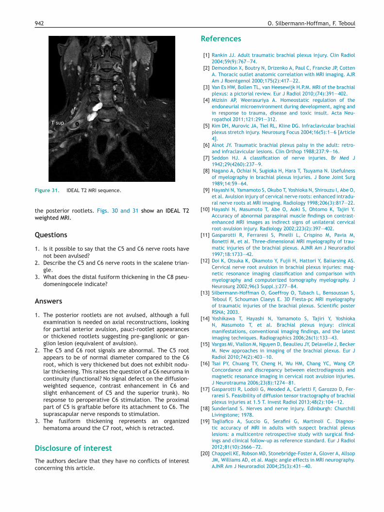
Figure 30. IDEAL T2 MRI sequence.
TAKE-HOME MESSAGES
• Pre-ganglionic injuries may be apparent, but it isimportant to look for minor adverse signs:◦ Total avulsion = avulsion of the anterior and
posterior rootlets,◦ Partial avulsion = avulsion of the anterior or
posterior rootlets,◦ Pauci-rootlet appearance = the rootlets are
visible, but reduced in number compared to theopposite side,
◦ Thickened rootlet appearance,◦ Contrast enhancement in the spine or nerve roots.
• Neuronal contrast enhancement in the scalenetriangle may be due to:◦ A neuroma in continuity,◦ Rupture,◦ An hematoma around an avulsed root,◦ Contrast enhancement by the retracted ganglion
of an avulsed root.• A brachial plexus T2 weighted hyperintensity
may represent reversible endoneurial edema, orreversible or irreversible Wallerien degeneration.The combination of a signal abnormality andincreased nerve diameter suggests a poorerprognosis. Functionality cannot be assessed by MRIexcept when the nerve is divided.
• MRI on its own is of no use except in obvious totalavulsion, and needs to be used in tandem withrepeated electromyograms to determine the surgicalstrategy, with or without peroperative stimulation.
• A visible neuroma on the axillary nerve suggestsrupture.
• Further surgical correlation information is neededto assess the relevance of contrast enhancementcompared to a STIR sequence. Tractography is veryuseful but needs advances to be made to be less
C
Total right traumatic paralysis. MRI at 3 months and 1 week.90◦ abduction. Fig. 29 shows an oblique reconstruction of
Grafts do not produce as good results as targetednerve transfers, as the donor nerve is also healthy innerve transfer whereas a graftable root may not beof as good quality. In addition, in nerve transfer therecipient nerve is sutured immediately before its intra-muscular branching and therefore no donor nerve fibersare lost in the sensory fibers or those destined forother nerves (no axonal confusion) and only one neuronalsuture is performed (unlike a graft, which requires twosutures!).
Preoperative planning may change during the proce-dure: a root which has not been avulsed may be foundto be of very poor quality when it is divided (few orno visible bundles) and a donor nerve for transfer maybe damaged in the initial injury (such as the ulnarnerve). Therefore, a change in strategy should alwaysbe considered peroperatively, to optimize patient recov-ery.
In practice, clinical presentations can be divided intopartial and total plexus injuries. In partial paralysis(C5/C6/C7), surgeons perform targeted nerve transfers(ulnar biceps, median anterior brachial, triceps axillarynerve, suprascapular spinal, triceps intercostal if damageto C7). In this case, the radiologist should examine thequality of C5 for the possibility of a graft for the shoul-der.
In complete plexus paralysis, radiologists should assessthe C5 and C6 roots for elbow and hand grafts, in additionto nerve transfers (spinal, intercostal).
time-consuming.
linical case
942
F
tw
Q
1
2
3
A
1
2
3
D
Tc
R
[
[
[
[
[
[
[
[
[
[
2012;81(10):2666—72.
igure 31. IDEAL T2 MRI sequence.
he posterior rootlets. Figs. 30 and 31 show an IDEAL T2eighted MRI.
uestions
. Is it possible to say that the C5 and C6 nerve roots havenot been avulsed?
. Describe the C5 and C6 nerve roots in the scalene trian-gle.
. What does the distal fusiform thickening in the C8 pseu-domeningocele indicate?
nswers
. The posterior rootlets are not avulsed, although a fullexamination is needed on axial reconstructions, lookingfor partial anterior avulsion, pauci-rootlet appearancesor thickened rootlets suggesting pre-ganglionic or gan-glion lesion (equivalent of avulsion).
. The C5 and C6 root signals are abnormal. The C5 rootappears to be of normal diameter compared to the C6root, which is very thickened but does not exhibit nodu-lar thickening. This raises the question of a C6 neuroma incontinuity (functional? No signal defect on the diffusion-weighted sequence, contrast enhancement in C6 andslight enhancement of C5 and the superior trunk). Noresponse to peroperative C6 stimulation. The proximalpart of C5 is graftable before its attachment to C6. Thesuprascapular nerve responds to stimulation.
. The fusiform thickening represents an organizedhematoma around the C7 root, which is retracted.
isclosure of interest
he authors declare that they have no conflicts of interestoncerning this article.
[
O. Silbermann-Hoffman, F. Teboul
eferences
[1] Rankin JJ. Adult traumatic brachial plexus injury. Clin Radiol2004;59(9):767—74.
[2] Demondion X, Boutry N, Drizenko A, Paul C, Francke JP, CottenA. Thoracic outlet anatomic correlation with MRI imaging. AJRAm J Roentgenol 2000;175(2):417—22.
[3] Van Es HW, Bollen TL, van Heesewijk H.P.M. MRI of the brachialplexus: a pictorial review. Eur J Radiol 2010;(74):391—402.
[4] Mizisin AP, Weerasuriya A. Homeostatic regulation of theendoneurial microenvironment during development, aging andin response to trauma, disease and toxic insult. Acta Neu-ropathol 2011;121:291—312.
[5] Kim DH, Murovic JA, Tiel RL, Kline DG. Infraclavicular brachialplexus stretch injury. Neurosurg Focus 2004;16(5):1—6 [Article4].
[6] Alnot JY. Traumatic brachial plexus palsy in the adult: retro-and infraclavicular lesions. Clin Orthop 1988;237:9—16.
[7] Seddon HJ. A classification of nerve injuries. Br Med J1942;29(4260):237—9.
[8] Nagano A, Ochiai N, Sugioka H, Hara T, Tsuyama N. Usefulnessof myelography in brachial plexus injuries. J Bone Joint Surg1989;14:59—64.
[9] Hayashi N, Yamamoto S, Okubo T, Yoshioka N, Shirouzu I, Abe O,et al. Avulsion injury of cervical nerve roots: enhanced intradu-ral nerve roots at MRI imaging. Radiology 1998;206(3):817—22.
10] Hayashi N, Masumoto T, Abe O, Aoki S, Ohtomo K, Tajiri Y.Accuracy of abnormal paraspinal muscle findings on contrast-enhanced MRI images as indirect signs of unilateral cervicalroot-avulsion injury. Radiology 2002;223(2):397—402.
11] Gasparotti R, Ferraresi S, Pinelli L, Crispino M, Pavia M,Bonetti M, et al. Three-dimensional MRI myelography of trau-matic injuries of the brachial plexus. AJNR Am J Neuroradiol1997;18:1733—42.
12] Doi K, Otsuka K, Okamoto Y, Fujii H, Hattori Y, Baliarsing AS.Cervical nerve root avulsion in brachial plexus injuries: mag-netic resonance imaging classification and comparison withmyelography and computerized tomography myelography. JNeurosurg 2002;96(3 Suppl.):277—84.
13] Silbermann-Hoffman O, Goeffroy O, Tubach L, Bensoussan S,Teboul F, Schouman Claeys E. 3D Fiesta-pc MRI myelographyof traumatic injuries of the brachial plexus. Scientific posterRSNA; 2003.
14] Yoshikawa T, Hayashi N, Yamamoto S, Tajiri Y, YoshiokaN, Masumoto T, et al. Brachial plexus injury: clinicalmanifestations, conventional imaging findings, and the latestimaging techniques. Radiographics 2006;26(1):133—43.
15] Vargas MI, Viallon M, Nguyen D, Beaulieu JY, Delavelle J, BeckerM. New approaches in imaging of the brachial plexus. Eur JRadiol 2010;74(2):403—10.
16] Tsai PY, Chuang TY, Cheng H, Wu HM, Chang YC, Wang CP.Concordance and discrepancy between electrodiagnosis andmagnetic resonance imaging in cervical root avulsion injuries.J Neurotrauma 2006;23(8):1274—81.
17] Gasparotti R, Lodoli G, Meoded A, Carletti F, Garozzo D, Fer-raresi S. Feasibility of diffusion tensor tractography of brachialplexus injuries at 1.5 T. Invest Radiol 2013;48(2):104—12.
18] Sunderland S. Nerves and nerve injury. Edinburgh: ChurchillLivingstone; 1978.
19] Tagliafico A, Succio G, Serafini G, Martinoli C. Diagnos-tic accuracy of MRI in adults with suspect brachial plexuslesions: a multicentre retrospective study with surgical find-ings and clinical follow-up as reference standard. Eur J Radiol
20] Chappell KE, Robson MD, Stonebridge-Foster A, Glover A, AllsopJM, Williams AD, et al. Magic angle effects in MRI neurography.AJNR Am J Neuroradiol 2004;25(3):431—40.
[
[
[
[
[
[
Post-traumatic brachial plexus MRI in practice
[21] Chhabra A, Thawait GK, Soldatos T, Thakkar RS, Grande FD,Chalian M, et al. High-resolution 3T MRI neurography of thebrachial plexus and its branches, with emphasis on 3D imaging.AJNR Am J Neuroradiol 2013;34(3):486—97.
[22] Viallon M, Vargas MI, Jlassi H, Lövblad KO, Delavelle J. High-resolution and functional magnetic resonance imaging of thebrachial plexus using an isotropic 3D T2 STIR (Short Term Inver-sion Recovery) SPACE sequence and diffusion tensor imaging.Eur Radiol 2008;18:1018—23.
[23] Polak JF, Jolesz FA, Adams DF. MRI imaging of skeletal muscle:prolongation of T1 and T2 subsequent to denervation. InvestRadiol 1988;23:365—9.
[24] Hayashi Y, Ikata T, Takai H, Takata S, Ishikawa M, Sogabe T, et al.Effect of peripheral nerve injury on nuclear MRI relaxationtimes of rat skeletal muscle. Invest Radiol 1997;32:135—9.
[25] West GA, Haynor DR, Goodkin, et al. MRI imaging signal inten-sity changes in denervated muscles after peripheral nerveinjury. Neurosurgery 1994;35:1077—86.
[26] Bendszus M, Koltzenburg M, Wessig C, Solymosi L. Sequential
MRI imaging of denervated muscle: experimental study. Am JNeuroradiol 2002;23:1427—31.[27] Sureka J, Cherian RA, Alexander M, Thomas BP. MRI of brachialplexopathies. Clin Radiol 2009;64:208—18.
[
943
28] Le Bihan D, Mangin JF, Poupon C, Clark CA, Pappata S, MolkoN, et al. Diffusion tensor imaging: concepts and applications.J Magn Reson Imaging 2001;13(4):534—46.
29] Takahara T, Hendrikse J, Yamashita T, Mali WP, Kwee TC,Imai Y, et al. Diffusion-weighted MRI neurography of thebrachial plexus: feasibility study. Radiology 2008;249(2):653—60.
30] Lésions du nerf axillaire. Thesis written by Coenes LN 1985University of Leiden panel chairman Pr Narakas.
31] Miroux F [Thèse d’exercice de médecine] IRM et pathologietraumatique du nerf axillaire: à propos de 18 observations.Paris: Université Paris-12 Créteil; 1996.
32] Burnett MG, Zager EL. Pathophysiology of peripheral nerveinjury: a brief review. Neurosurg Focus 2004;16(5):1—7[article 1].
33] Garg R, Merrell GA, Hillstrom HJ, Wolfe SW. Comparison ofnerve transfers and nerve grafting for traumatic upper plexuspalsy: a systematic review and analysis. J Bone Joint Surg Am2011;93(9):819—29.
34] Sulaiman OA, Kim DD, Burkett C, Kline DG. Nerve transfersurgery for adult brachial plexus injury: a 10-year experi-ence at Louisiana State University. Neurosurgery 2009;65(4Suppl.):A55—62.