pathology of digestive system-1 - bihar animal sciences
TRANSCRIPT

Pathology of digestive system-1
Dr. Sanjiv Kumar, Assistant Professor,
Department of Pathology Bihar Veterinary College, Patna

Introduction
The digestive tract includes:
• the oral cavity and associated organs (lips, teeth, tongue, and salivary glands),
• the esophagus,
• the forestomachs (reticulum, rumen, omasum) of ruminants and the true stomach in all species,
• the small intestine,
• the liver,
• the exocrine pancreas,
• the large intestine, and
• the rectum and anus.
Gut-associated lymphoid tissue (tonsils, Peyer’s patches, diffuse lymphoid tissue) is distributed along the GI tract.


Histology of mouth

Histology of Alimentary canal
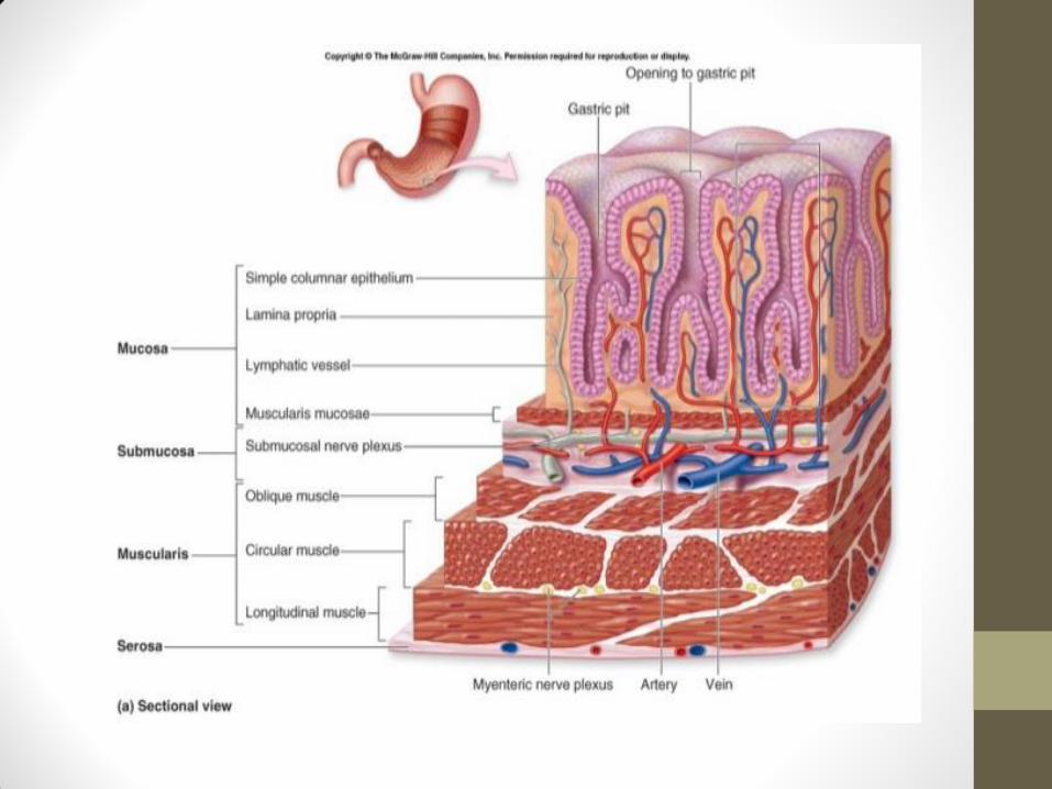
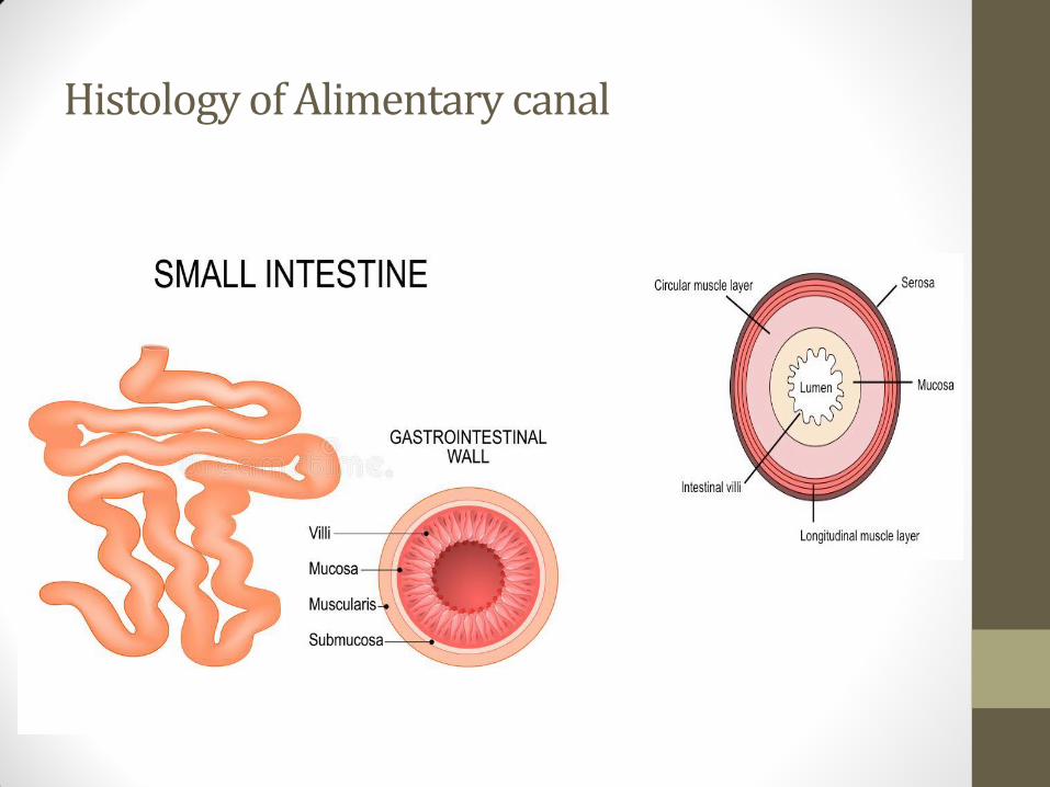
Histology of Alimentary canal


PATHOLOGY OF ORAL CAVITY STOMATITIS: Stomatitis in the inflammation
of mucosa of oral cavity
• Gingivitis : Inflammation of gums
• Glossitis : Inflammation of tongue
• Cheilitis : Inflammation of lips
• Tonsilitis : Inflammation of tonsil
• Palatitis / Lampas: Inflammation of palates.
Malformations: Cleft lip (Chelioschisis) and cleft
palate (Palatoschisis)

Aetiology • It may be a primary affection or may occur as secondary to other diseases
viz. gastritis or infectious diseases.
• The causes are
• Physical
• Trauma by awns, thorns, burrs, wood pieces, glass pieces, sharp bits, irregular sharp teeth, sharp edged feeding utensils.
• Thermal injuries: Hot drenches and eating frozen foods.
• Chemical : caustic alkalies, corrosive acids, fertilizers.
• Deficiency of vitamins
• Hypovitaminosis A especially in fowl
• Niacin deficiency : Black tongue in dogs
• Microorganisms
• Bacteria: Actinomyces bovis; Actinobacillus lignieresi; Fusobacterium necrophorum; Pseudomonas aeruginosa; Streptococci andStaphylococci.
• Fungi: Candida albicans and Oidium pullorum in poultry.
• Viruses: Foot and Mouth disease; Rinderpest: Virus diarrhoea -mucosal disease; Infectious canine hepatitis.

Macroscopic features
• Catarrhal : Mucous exudation in oral cavity
• Fibrinous : False membrane in oral mucosa
• Vesicular : Vesicles in oral mucosal e.g. FMD
• Erosive : Erosions in oral mucosa e.g. Rinderpest
• Ulcerative : Presence of ulcers in oral mucosa e.g. mucosal disease
Microscopic features
• Congestion of oral mucosa
• Presence of erosions, vesicles or ulcers
• Infiltration of neutrophils, lymphocytes and macrophages.
• Presence of fibrinous exudates in the form of diphtheritic membrane
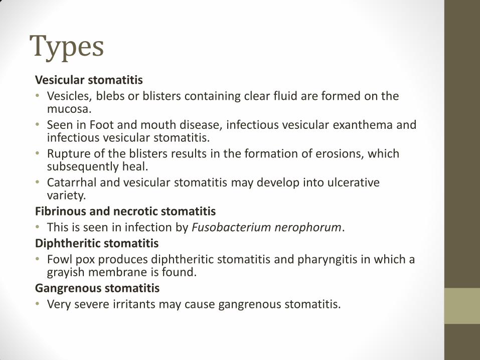
Types Vesicular stomatitis • Vesicles, blebs or blisters containing clear fluid are formed on the
mucosa. • Seen in Foot and mouth disease, infectious vesicular exanthema and
infectious vesicular stomatitis. • Rupture of the blisters results in the formation of erosions, which
subsequently heal. • Catarrhal and vesicular stomatitis may develop into ulcerative
variety. Fibrinous and necrotic stomatitis • This is seen in infection by Fusobacterium nerophorum. Diphtheritic stomatitis • Fowl pox produces diphtheritic stomatitis and pharyngitis in which a
grayish membrane is found. Gangrenous stomatitis • Very severe irritants may cause gangrenous stomatitis.

PATHOLOGY OF ESOPHAGUS
Choke is esophageal obstruction when the esophagus is obstructed by food or foreign objects.
Occurrence
• It occurs in horses and cattle, but more common in the former.
• Aetiology
• Old age
• In cattle, large objects of food- beet root, carrot, apples, potatoes, fetal membranes, sticks and wire. In dogs, large bones.
• Impacted masses of feed due to improper chewing, bad teeth and rapid gulping of dry feed.
• Lesions of esophagus – stenosis or diverticulum cause repeated choking.
• Enlarged lymph nodes- mediastinal and cervical.
• Enlarged thyroids.
• Neoplasms of adjacent tissue especially thymus - thymoma in new-born animals.

Choke may be complete or incomplete.
• Complete choke
• In complete choke, feed will be returned and water will flow through the nostrils when animal is watered.
• Aspiration of the feed will cause secondary foreign-body pneumonia.
• In cattle, complete obstruction will cause dangerous tympany.
• Because of pressure, ischemia and resultant necrosis and gangrene may develop.
• Infection may spread to the surrounding tissues- cellulitis or to the lungs- gangrenous pneumonia.
• Resultant sapremia or toxaemia is the cause of death in fatal case.
• Partial choke
• Partial obstruction will give rise to dilatation of esophagus above the obstruction - the esophageal diverticulum .

Macroscopic features
• Tympany
• Gangrene, sapremia and toxaemia
• Sac like dialatation “Esophageal diverticulum”
• Perforation due to sharp bone ends.
Microscopic features
• Necrosis gangrene at a point of obstruction
• Congestion hemorrhage in perforated cases

ESOPHAGITIS: This is inflammation of the oesophageal
mucosa.
Occurrence
• It is rare in animals because of the thick and resistant condition
of the mucosa.
• Aetiology
• Trauma by probing, stomach tube or foreign bodies.
• Chemicals – corrosives.
• In the fowl, thallium sulphate poisoning.
• Avitaminosis A in Fowls.
• Viral enteritis and mucosal disease in cattle.
• Parasites – bot fly larvae in horses and hypoderma larvae in
cattle.
• Persistent vomiting in dogs and pigs.

Macroscopic features
• Congestion
• Ulcer formation
• Red streaks of catarrhal inflammation
• Stenosis due to fibrous nodules or inflammatory exudates
• Enlargement of glands
Microscopic features
• Congestion, haemorrhage
• Ulceration
• Infiltration of neutrophils, lymphocytes
• Sub-epithelial fibrosis/nodules by Spirocerea lupi

PATHOLOGY OF STOMACH
GASTRITIS : Inflammation of the stomach is called gastritis.
Occurrence: Gastritis is a fairly common condition in animals.
Predisposing causes: Close confinement and unsanitary conditions where bacteria thrive contaminating feeds and feeding utensils
Aetiology
•Physical
• Faulty dentition prevents mastication
• Foreign bodies may traumatize the gastric mucosa.
• Feeding very coarse material
• Feeding with frozen foods
• Spoiled, mouldy and fermented hay and silage or feeding easily fermentable foods.
• When heavily fatigued animals are fed, the feed is not easily digested, stagnates, ferments and so produces irritation.
• Too sudden changes of feed

Chemicals • Uremia; (Excretion gastritis is caused by the excretion of the toxic substances
through gastric glands) • Caustic and corrosive chemicals like mercury, lead, copper, arsenic and
phosphorus Bacterial • In calves - enterotoxemia and colibacillosis; in pigs - erysipelas, vibrionic
dysentery, salmonellosis and colibacillosis. Viruses • Pig- hog cholera; transmissible gastro-enteritis in baby pigs • Cattle - rinderpest, mucosal disease. • In chicken - Ranikhet disease causes haemorrhagic proventriculitis.
Fungi • Mucormycosis, moniliasis and aspergillosis cause gastritis in many animals.
Parasites • Stomach worms - Trichostrongylus sp., Hemonchus sp., Ostertagia sp., larval
paramphistomes in ruminants. • Larvae of Habronema sp. and Gastrophilus equi in horses. • In pigs Hyostrongylus rubidus, Ascarops strongylina and Physocephalus sexalatus.
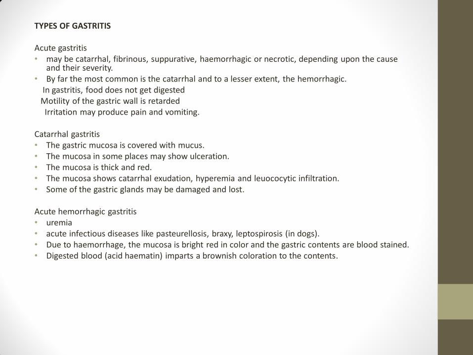
TYPES OF GASTRITIS Acute gastritis • may be catarrhal, fibrinous, suppurative, haemorrhagic or necrotic, depending upon the cause
and their severity. • By far the most common is the catarrhal and to a lesser extent, the hemorrhagic. In gastritis, food does not get digested Motility of the gastric wall is retarded Irritation may produce pain and vomiting.
Catarrhal gastritis • The gastric mucosa is covered with mucus. • The mucosa in some places may show ulceration. • The mucosa is thick and red. • The mucosa shows catarrhal exudation, hyperemia and leuococytic infiltration. • Some of the gastric glands may be damaged and lost.
Acute hemorrhagic gastritis • uremia • acute infectious diseases like pasteurellosis, braxy, leptospirosis (in dogs). • Due to haemorrhage, the mucosa is bright red in color and the gastric contents are blood stained. • Digested blood (acid haematin) imparts a brownish coloration to the contents.

Parasitic Gastritis • Cattle and sheep: Hemonchus contortus, Ostertagia ostertagi, Trichostrongylus axei • Horses: Habronema larvae, Trichostrongylus axei and Gastrophilus equi larvae. • Pig: Hyostrongylus rubidus, Physocephalus sexalatus. Simondsia paradoxa. • Cats: Gnathostoma spinigerum • The strongyles are blood suckers and they produce minute injuries on the mucosa. • The larvae may burrow into the mucosa for completion of their life cycle and thereby cause
damage to the epithelium and glands. • Heavy infestation besides causing anemia will produce catarrhal gastritis. • Gastrophilus sp. in the stomach may produce ulcers • Habronema larvae live in granulomatous nodules which may be infected by secondary
bacteria and form abscesses.
Chronic gastritis • The mucous membrane is thickened and covered with tenacious, viscid glassy mucus. • This condition is usually of a hypertrophic type with thickening of the gastric wall. • There is exfoliation of the epithelium • The mucosa may be thrown into polypoid folds (polypoid gastritis). • The interstitial connective tissue hyperplasia exaggerates the mucosal foldings. • Occlusion of glands results into development of retention cysts.

Tympany/ Bloat TYMPANY/ Bloat: Excessive accumulation of gas. Can occur
• When the gas is produced at too rapid a rate than can be eructated.
• When the eructation mechanism is faulty.
Types
• Based on course, bloat may be acute or chronic.
• Based on nature of gas, bloat may be dry or frothy.
Acute bloat
• This may be due to complete choke in esophagus.
• It may also be due to sudden changes of feed.
• Excessive feeding on legumes that are wet with dew or rain.
Chronic bloat
• The chronic variety occurs whenever there is any hindrance to eructation in the esophagus either within or without pressure by tumors, foreign bodies, enlarged lymph nodes, abscesses, constrictions or diverticula.
• It may occur in lesions of the rumen causing decreased contractions of the ruminal wall as in atony, serosal adhesions, paresis, diffuse lymphomatosis.
Dry bloat
• The dry bloat is less harmful, since in this condition, the gases can be more easily got rid of by eructation.
Frothy bloat
• In the frothy bloat, the gas is trapped as small bubbles in the fluid forming a foamy mass which is not easily eructated.

Aetiology
• Saponin and Water-soluble proteins of the legumes are capable of forming froth.
• Normally, in rumen due to bacterial activity, fatty acids are produced which increase the surface tension. If the production of these fatty acids is decreased, will favour froth production.
Pathogenesis
• Some legumes contain HCN causing paralysis of the ruminal or reticular musculature. Phosphatase can accelerates fermentation producing a large quantity of CO2.
• H2S, CO2 and CO produced in large quantities causes paralysis of ruminal muscles.
• Ruminal mucinolytic bacteria may destroy salivary mucin thereby producing frothy bloat. Polysaccharides produced by capsulated ruminal bacteria may be another etiological factor in bloat.
• Interference with the nerve pathways that are responsible for the eructation reflex may also lead to tympany.
• Distended rumen compresses other abdominal organs and forward thrust on the diaphragm.
• The result of this is hypoxia and ultimate asphyxia and death.

Macroscopic features
• Rumen is distended due to excessive accumulation of gases (CO2, H2S, CO)
• Distended rumen compresses diaphragm to hinder respiration
• Tarry color blood, pale liver and rupture of diaphragm
• On rupture of rumen gas comes out (dry tympany)
Microscopic features
• Hemorrhage in lungs, pericardium, trachea and lymph nodes
• Atelectasis

RUMENITIS
Macroscopic features
• Ulcers
• Spherical white nodules of 1-2 cm diameter size
• Sloughing of mucosa
Microscopic features
• Seropurrulent exudates
• Ulcers
• Infiltration of lymphocytes and neutrophils
• Fibrous nodules due to hyperplasia of fibroblasts
• Parakeratosis

RETICULITIS
Macroscopic features
• Perforation of reticulum by foreign body.
• Abscessation/suppuration
• Peritonitis, adhesions of reticulum with diaphragm
• Pericarditis due to foreign body (traumatic reticulo pericarditis)
Microscopic features
• Infiltration of neutrophils, macrophages, lymphocytes
• Proliferation of fibroblasts producing adhesions
• Liquifactive necrosis

ABOMASITIS
Macroscopic features
• Presence of ulcer (button ulcers in Hog cholera)
• Congestion, oedema of abomasal folds, haemorrhage in braxy
Microscopic features
• Catarrhal, hemorrhagic abomasits
• Presence of gram positive rods in case of braxy
• Neutrophilic and lymphocytic infilteration
• Congestion and haemorrhages
• Ulceration with lymphocytic infiltration

IMPACTION OF RUMEN AND RETICULUM
Macroscopic features
• Atony of rumen due to lactic acid production
• Rumen is filled with hard, caked undigested food with foul odour
• Hemoconcentration, anuria, blood becomes dark in colour
Microscopic features
• Hemorrhage in lungs
• Desquamation of ruminal epithelium
• Lesions of acidosis/toxicosis