pictures in digestive pathology - scielo españascielo.isciii.es/pdf/diges/v106n3/imagen1.pdf ·...
TRANSCRIPT

PICTURES IN DIGESTIVE PATHOLOGY
Deep infiltrating rectosigmoid endometriosis. Diagnostic keys
M.ª Alcázar Iribarren-Marín1, Herminia Pérez-Vega1, Manuel Martínez-Moya1, Lourdes Gómez-Izquierdo2 and Cristina Martínez-Polanco1
1Diagnostic Imaging Unit. Radiology Department. 2Pathological Anatomy Service. Hospital Universitario Virgen del Rocío. Seville, Spain
1130-0108/2014/106/3/212-213Revista española de enfeRmedades digestivasCopyRight © 2014 aRán ediCiones, s. l.
Rev esp enfeRm dig (MadridVol. 106, N.º 3, pp. 212-213, 2014
CASE REPORT
A 36 year-old woman was admitted with dysmenorrhea associated with rectal bleeding. The colonoscopy showed a stenosis at the rectosigmoid (RS) junction which did not exceed the endoscope. A barium enema (Figs. 1 A and B) showed traction of the sigmoid mucosa at the lower margin of the RS junction. CT examination (Figs. 1 C and D) showed an en-dometrial implant in the Douglas pouch, the pedicle connected to the colon wall and the “mushroom cap” configuration of the endometrial implant in the RS wall, all with a density similar to that of muscle tissue. A sigmoidectomy was performed (Fig. 2) with histological (Fig. 3) and immunohistochemical analysis, which demonstrated deep infiltrating rectosigmoid endometriosis (DIRSE) as the resulting diagnosis.
DISCUSSION
Endometriosis is defined as the presence of functioning ectopic endometrial tissue (endometrial glands and stroma) outside the uterus and at a distance from and unconnected with uterine endometrial tissue. The ectopic endometrium is sensitive to hormonal variation during the menstrual cycle. These results in chronic inflammation that is the cause of cyclic pelvic pain found in these patients. In the DIRSE, the ectopic tissue would respond to the ovarian hormonal cycles causing inflammation, bleeding, fibrosis and metaplasia or hyperplasia of the smooth intestinal muscle possibly affecting the sero-sa, submucosa and rarely the mucosa, which gives rise to a thickening of the intestinal wall and in some cases may evolve
Fig. 1. A and B. Barium enema. Filling compression defect which causes extrinsic traction of the sigmoid colon mucosa (arrows) produced by deep infiltration of endometrial wall. C and D. Abdominal CT with iodinated contrast. A. Coronal reconstruction. Right ovarian endometrioma (*). B. Sagittal reconstruction. Endometrial implant in the Douglas pouch (black arrow). Pedicle connecting the implant to the colon wall (hole arrow). Endometrial infiltration adopting the mushroom configuration (white arrows).
A B C D
Vol. 106, N.º 3, 2014 DEEP INFILTRATING RECToSIGMoID ENDoMETRIoSIS. DIAGNoSTIC kEyS 213
Rev esp enfeRm Dig 2014; 106 (3): 212-213
into an obstruction (1-3). The “mushroom cap” sign has been described in the presumed diagnosis because the tumor grows towards the RS colonic lumen (4). The differential diagnosis is reached from the neoplasic processes seen in this area.
REFERENCES
1. Chamiè LP, Blasbalg R, Pereira RM, Warmbrand G, Serafini, PC. Findings of pelvic endometriosis at transvaginal US, MR imaging and laparoscopy. RadioGraphics 2011;31:E77-100.
2. Nasim H, Sikafi D, Nasr A. Sigmoid endometriosis and a diagnostic dilema-A case report and literature review. Int J Surg Case Rep 2011;2:181-4.3. Fernández-Rey CL, Álvarez-González SA, Díaz-Solís P, Blanco-González A, Costilla-García S. Endometriosis ileal como causa de obstrucción de intestino
delgado: diagnóstico por tomografía computarizada multicorte. Rev Esp Enferm Dig 2009;101:872-4. 4. yoon JH, Choi D, Jang kT, kim Ck, kim H, Lee SJ, et al. Depp rectosigmoid endometriosis: “mushroom cup” sing on T2-weighted MR imaging. Abdom
Imaging 2010;35:726-31.
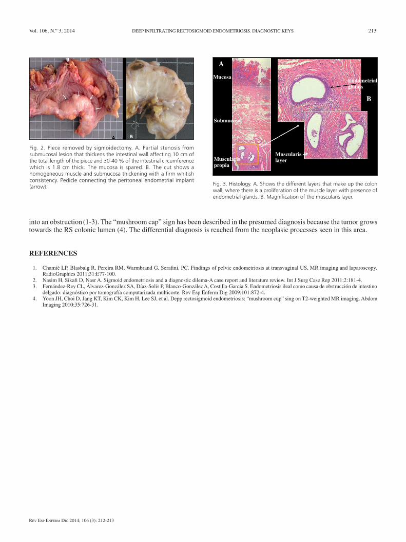
Fig. 2. Piece removed by sigmoidectomy. A. Partial stenosis from submucosal lesion that thickens the intestinal wall affecting 10 cm of the total length of the piece and 30-40 % of the intestinal circumference which is 1.8 cm thick. The mucosa is spared. B. The cut shows a homogeneous muscle and submucosa thickening with a firm whitish consistency. Pedicle connecting the peritoneal endometrial implant (arrow). Fig. 3. Histology. A. Shows the different layers that make up the colon
wall, where there is a proliferation of the muscle layer with presence of endometrial glands. B. Magnification of the muscularis layer.
Mucosa
Submucosa
Muscularis layerMuscularis
propia
Endometrial glands
A
B