ns20: scalp, face, parotid
TRANSCRIPT

20/02/2013
1
Cranial Fossae, Meninges,
Sinuses, Intracranial Bleeds
Mohammed A Al-Muharraqi MBChB, BDS, MSc,
MRCS Glas, FFD RCS Irel, MFDS RCS Eng
e-mail: [email protected]
Learning Outcomes
• The Cranial Fossae
– Anterior
– Middle
– Posterior
• The Meninges
– Pia Mater
– Arachnoid Mater
– Dura Mater
• The Dural Folds
• The Venous Sinuses
• Intracranial Bleeds
– Spontaneous
• SAH
– Traumatic • Extradural Haematoma
• Subdural Haematoma
• Intracerebral Haematoma
Parietal Bone
Occipital Bone (Posterior Cranial Fossa)
Foramen Magnum (Posterior Cranial Fossa)
Sphenoid Bone (Middle/Anterior Cranial
Fossa)
Temporal Bone (Middle Cranial Fossa)
Frontal Bone (Anterior Cranial Fossa) Ethmoid Bone
(Anterior Cranial Fossa)
Cranial Fossae
Three Cranial Fossae
1. ANTERIOR – Orbital Roof
(Ethmoid, Frontal, Sphenoid)
2. MIDDLE – Middle Ear (Sphenoid,
Petrosal/Temporal)
3. POSTERIOR – Foramen Magnum
(Occipital)
Maxilla
Nasal Bone
Zygomatic Bone
Meninges
Cranial Dura Mater: Meningeal Layer The cranial dura mater is the tough outermost layer of meninges that surround the brain and spinal cord. It is split into two layers: the outer layer adheres to the inner surface of the skull and is known as the periosteal
layer and the inner layer is reflected as sheet-like protrusions into the cranial cavity and is known as the meningeal layer, which are continuous with the spinal dura mater. The meningeal layer adheres to the arachnoid mater, but can be easily separated from it.
Cranial Dura Mater: Periosteal Layer The cranial dura mater forms four dural reflections/folds : • Falx cerebri • Tentorium cerebelli • Falx cerebelli . • Diaphragma sellae.
Dural venous sinuses form at the dural reflections and drain blood and cerebrospinal fluid from the brain into the internal jugular vein.
Cranial Arachnoid Mater The cranial arachnoid mater is the middle layer of meninges that surround the brain. It is a thin transparent membrane, which is continuous inferiorly with the spinal arachnoid mater. It is closely applied to the deep
aspect of the dura mater, being separated by the subdural space.
Cranial Pia Mater The cranial pia mater is the innermost layer of meninges that surround the brain. It is a thin transparent membrane, 1-2 c ells thick, that closely adheres to the contours of the brain and is continuous inferiorly with the
spinal pia mater.
Cerebrum (Telencephalon) The c erebrum accounts for 85% of the brain and occupies the anterior and middle cerebral fossae. It is directly related to the cranial vault and is divided into two
cerebral hemispheres that are joined at the bottom by the corpus callosum. The surface of the cerebrum is covered in a layer of grey matter; this cortex covers a core of white matter. Thick folds called gyri that form shallow grooves
called sulci increase the surface area of the c erebrum. Several prominent sulci divide the cerebrum into 5 lobes: Frontal, Parietal, Temporal, Occipital, Insula. The c erebrum controls and
integrates motor , sensory, and higher mental functions, such as thought, reason, emotion and memory.
Meninges Meninges
Cranial Dura Mater: Falx Cerebri
Cranial Dura Mater: Meningeal
Layer
Sigmoid Sinus
Superior Bulb of the Internal Jugular Vein
Intercavernous Sinus
Basilar Venous Plexus
Cavernous Sinus
Sphenoparietal Sinus Superior Sagittal
Sinus
Superior Petrosal
Sinus
Cerebellum
Cerebrum (Telencephalon)

20/02/2013
2
Meninges Cranial Dura Mater: Falx Cerebri
Cranial Dura Mater: Tentorium Cerebelli
Cranial Dura Mater: Meningeal
Layer
Sigmoid Sinus Occipital Sinus
Superior Bulb of the Internal Jugular Vein
Intercavernous Sinus
Basilar Venous Plexus
Cavernous Sinus
Sphenoparietal Sinus
Inferior Sagittal Sinus
Superior Sagittal Sinus
Straight Sinus
Superior Petrosal
Sinus
Dural Folds The falx cerebri is a sickle-shaped fold of dura that lies along the median sagittal plane in the longitudinal cerebral fissure between the two cerebral hemispheres. Anteriorly, it is attached to the crista galli. It arches backwards; it’s attachment to the skull continuing supero-posteriorly along the margins of the superior sagittal venous sinus to the internal occipital protuberance. It is continuous with the tentorium cerebelli.
The tentorium cerebelli lies between the cerebellum and the occipital lobes. Anteriorly its free edge forms a hiatus/notch around the brainstem and attaches to the dorsum sellae of the sphenoid bone. This notch is called the ‘tentorial incisure’. The margin is named the ‘free border of the tentorium’. The lateral edges attach to the occipital bone and enclose the transverse sinuses. The anterior lateral edges are attached to the upper borders of the petrous parts of the temporal bones and contain the superior petrosal sinuses.
Cranial Dura Mater: Falx Cerebri
Cranial Dura Mater: Tentorium Cerebelli
Sinuses The venous sinuses are encased
between the two layers of the dura
mater. Sinuses differ from ordinary
veins in having no smooth muscle in
their walls and maintain their patency,
because their walls are held by
attachments to surrounding connective
tissue. Important sinuses located
opposite the base of the brain are the
cavernous sinus, the superior and
inferior petrosal sinuses, and the
occipital sinus. Important sinuses
embedded in the dura lining the
remainder of the cranial vault are the
superior sagittal sinus, inferior sagittal
sinus, straight sinus, confluence of
sinuses, transverse sinus, and sigmoid
sinus. Drainage converges on the
jugular bulb. The jugular bulb is located
in the jugular foramen. Venous blood,
especially that from the anterior base
of the brain, drains through the
cavernous sinus anteriorly to the veins
of the face.
Cranial Dura Mater: Falx Cerebri
Cranial Dura Mater: Tentorium Cerebelli
Cavernous Sinus
Intercavernous Sinus
Sphenoparietal Sinus
Inferior Sagittal Sinus
Superior Sagittal Sinus
Superior Petrosal
Sinus
Sigmoid Sinus Occipital Sinus
Basilar Venous Plexus
Superior Bulb of the Internal Jugular Vein
Straight Sinus
Transverse Sinus
Sinuses Blood from the deep central portions
of the brain, collects into the internal
cerebral vein (great vein of Galen) and
meets with the inferior sagittal sinus
posteriorly to form the straight sinus.
The inferior sagittal sinus is positioned
in the lower free margin of the falx
cerebri, and the straight sinus runs
from anterior to posterior between the
dural layers in the midline of the
tentorium cerebelli. Blood draining
from the superolateral surfaces of the
brain collects into cortical veins, which
extend across the subarachnoid space
to drain into the superior sagittal sinus,
which conveys blood posteriorly to an
anastomosis with the posterior end of
the straight sinus, formed when the
inferior sagittal sinus unites with the
internal cerebral vein. This region,
where the superior sagittal sinus and
the straight sinus meet, is known as the
confluence of sinuses.
Cranial Dura Mater: Falx Cerebri
Inferior Sagittal Sinus
Superior Sagittal Sinus
Transverse Sinus
Superior Petrosal
Sinus
Sigmoid Sinus Intercavernous Sinus
Cavernous Sinus
Straight Sinus Superior Bulb of
the Internal Jugular Vein
Sphenoparietal Sinus
Cerebellum
Sinuses From the confluence of sinuses,
there originates a pair of
transverse sinuses, extending
laterally in grooves on the inner
surface of the skull. The transverse
sinuses are located in the posterior
margins of the tentorium cerebelli.
Each transverse sinus then turns
sharply toward the anterior to
form a sigmoid sinus, which travels
toward the jugular foramen to
drain into the jugular bulb. Where
the transverse sinus becomes the
sigmoid sinus, it receives drainage
from the superior petrosal sinus, a
diagonal venous channel travelling
along the petrosal ridge of the
temporal bone, connecting
anteriorly with the cavernous
sinus.
Cranial Dura Mater: Falx Cerebri
Cranial Dura Mater: Tentorium Cerebelli
Confluence Of Sinuses
Superior Sagittal Sinus
Inferior Sagittal Sinus
Sigmoid Sinus
Transverse Sinus
Intercavernous Sinus
Cerebellum
Occipital Sinus
Superior Bulb of the Internal Jugular Vein
Sphenoparietal Sinus
Sinuses The venous sinuses in the superolateral parts of
the dura mater, especially the superior sagittal
sinus, are invaginated by tufts of the nearby
layers of arachnoid mater. These tufts of
arachnoid membrane, called arachnoid
granulations, actually protrude into the centre
of the lumen of the sagittal sinus, and it is at
these sites that cerebrospinal fluid from the
subarachnoid space diffuses back into the
venous circulation. Sinuses in the superolateral
part of the cranial cavity have connections
through small emissary veins with the vascular
spaces between the two layers of the skull, and
thence to the scalp. The importance of these
veins is that infections in the scalp can
potentially spread to the venous sinuses inside
the cranial cavity and lead to meningitis.
Infections of the scalp should always be treated
aggressively to avoid this potential
complication.

20/02/2013
3
Cavernous Sinus The cavernous sinuses are blood-filled spaces
between the two layers of dura mater located
on each side of the central portion of the
sphenoid bone, surrounding the sphenoid sinus
and the sella turcica. The interior is criss-
crossed by lacy connective tissue filaments, and
blood flow through the cavernous sinuses is
slow and prone to thrombus formation. In the
centre of the cavernous sinus are found the
internal carotid artery and the abducent nerve
(VI). Lateral to the cavernous sinus are found,
from superior to inferior, the oculomotor (III),
trochlear (IV), ophthalmic (V1) and maxillary
(V2) nerves. All of these structures appear to be
embedded in the cavernous sinus but are
outside the vascular endothelium and not truly
within the cavernous sinus.
Structures Piercing the Dura in the Base of the Skull
Structures Piercing the Dura in the Base of the Skull
Middle Meningeal Artery The middle meningeal artery is a branch of the
maxillary and is the largest of the meningeal
arteries. It travels through the foramen spinosum.
Once inside the skull, it follows a groove on the
temporal bone and divides into frontal (anterior)
and parietal (posterior) branches. The frontal
branch crosses the greater wing of the sphenoid
and divides into branches. The branches travel in-
between the dura mater and the cranium, where
they travel to the vertex and the occipital region.
The parietal branch bends backwards where it
divides into five named branches to supply the
posterior portions of the cranium and cranial dura
mater. The branches anastomose with each other
as well as the anterior and posterior meningeal
arteries. It supplies the facial nerve and ganglion
in the tympanic cavity. Additional branches are
the superior tympanic artery, which supplies
tensor tympani via its canal and an anastomotic
branch enters the superior orbital fissure to
anastomose with the recurrent branch of the
lacrimal artery. There are the ganglionic branches,
which supply the trigeminal ganglion.
Spontaneous Intracranial Haemorrhage
1. Subarachnoid Haemorrhage (SAH)
2. Intracerebral Haemorrhage (ICH)
3. Intraventricular Haemorrhage
SAH
• Bleeding into the Subarachnoid Space
• Incidence of spontaneous SAH 6/100,000/yr
• Potentially fatal if diagnosis missed
• Even with treatment 46% 30 day mortality – 10% die at scene
– 20% die in first week
– 50% die in first month
– 50% survivors have major disability
– 66% of ‘successful’ patients never return to their previous occupation
• Usually underlying berry aneurysm
• Sometimes AVM or no underlying cause found

20/02/2013
4
SAH 1. Sudden Onset Severe
Headache ‘Hit by Bat’
2. Collapse
3. Vomiting
4. Neck Pain/Stiffness
5. Photophobia
6. ↓ Conscious Level
7. Focal Neurological
Deficit (Dysphasia,
Hemiparesis, CNIII
Palsy)
8. Fundoscopy – Retinal or
Vitreous Haemorrhage
Differential Diagnosis: • Subarachnoid Haemorrhage
• Thunderclap Headache
• Benign Coital Cephalgia
Investigating SAH • CT Scan
– May be negative if >3days post ictus
– Negative in 15% of patients who have bled
• Lumbar Puncture
– Safe in alert patient with no focal neurological deficit and no papilloedema, or after normal CT scan
– Bloodstained or xanthochromic CSF (6-48hr)
– Differentiate from ‘traumatic tap’
• Cerebral Angiography
– Seldinger technique via femoral artery
– 4 vessel angiography with multiple views
– Gold standard but may miss an aneurysm due to vasospasm
– MRI techniques developing
Complications of SAH • Re-bleeding
– Often fatal
– 20% risk in first 14 days, 50% risk in first 6 months
– Aneurysm clipping
– Endovascular techniques
• Delayed Ischaemic Neurological Deficit (DIND)
– Days 3-12
– altered conscious level or focal deficit
– Vasospasm
– Nimodipine
– High fluid intake ‘Triple H therapy’
• Hydrocephalus
– Increased intracranial CSF pressure
– 6% symptomatic
– Increasing headache or altered conscious level
– Treatment - CSF drainage - LP,
EVD, Shunt
• Seizures
• Hyponatraemia – SIADH or ‘cerebral salt wasting’
– Do not fluid restrict
– Supplement Sodium Intake
– Fludrocortisone
Traumatic Intracranial Haemorrhage
Head Injury – UK 1983
• 1,000,000 patients / year seen in A&E
• 100,000 admitted to hospital for observation
• 10,000 admitted to neurosurgical unit
Trauma is the leading cause of death under 45
years – About half of these deaths are from a
head injury
Head Injury
It is really “Brain Injury”
• Primary from time of injury – At the site of trauma
• Direct disruption of brain
• Shearing of axons
• Intracerebral haemorrhage
– Opposite the site of trauma
• Contre-coup
• Secondary due to
consequences of injury
partly preventable – Brain oedema → ↑ICP, ↓BP
→ Cerebral Ischemia
Types of Head Injury
• Open = Missile =
Penetrating
or
• Closed = Non-Missile

20/02/2013
5
Physical Findings in Head Injury
• Scalp swelling / laceration
• Skull Vault Fracture
– Linear (Simple)
– Depressed
– Compound
• Basal Skull Fracture
– CSF Rhinorrhoea, Blood behind eardrum
– Anterior Cranial Fossa “Raccoon” or “Panda” Eyes
– Middle Cranial Fossa “Battle Sign”
Head Injury Assessment of Consciousness
Glasgow Coma Scale (GCS)
• Eye opening (4) o Spontaneously
o To Command
o To Pain
o None
• Best Motor Response (6) o Obeys Commands
o Localises Pain
o Flexes to Pain
o Abnormal Flexion
o Extension
o None
• Verbal Response (5) o Orientated
o Confused
o Inappropriate Words
o Incomprehensible Sounds
o None
GCS of 3 Means the Patient is
Dead (or close to it)!
Types of Traumatic Intracranial Haematoma
1. Extradural Haematoma Middle Meningeal Artery bleeds into Extradural Space
2. Subdural Haematoma Cortical Veins ‘ooze’ into Subdural Space
3. Traumatic Subarachnoid Haemorrhage
4. Intracerebral Haematoma Most common
5. Intracerebral Contusion
6. Intraventricular Haemorrhage

20/02/2013
6
Extradural Haematoma
Subdural Haematoma
Intracerebral Haematoma
Frontal Contusions
Intracranial Pressure (ICP)
Monro-Kellie Doctrine • Skull is an inelastic closed box
• Volume inside skull is constant and has 3 components
– Brain
– Blood (mainly in venous phase)
– CSF
• An increase in volume of one component must produce a decrease in another component otherwise ICP will rise
Brain Blood CSF
Adult ICP is normally 12 -15 cm H2O
Cerebral Perfusion Pressure (CPP)
• CPP = MAP - ICP Cerebral Perfusion Pressure = Mean Arterial Pressure – Intracranial Pressure
• Hypotension has a major influence on CPP
• Blood pressure is much easier to measure and control
• Aim for CPP of > 70mmHg after head injury, i.e., MAP > 90mmHg
What Kills Patients With Head Injury?
• Hypoxia → ventilatory support/mechanical ventilation
• Hypotension → ionotropic support, pushing fluids
• Raised ICP → evacuation of haematoma or shunting hydrocephalus, forced ventilation
Why are ABC’s important in Head Injury?
• Brain is only 2% of body weight but uses;
– 15% of cardiac output
– 20% of carried oxygen
– 12% of carried glucose
Irreversible Neuronal Damage Within 5 Minutes
of Circulatory Arrest
Cerebral Protection Following Injury
• Mannitol – Improves micro-perfusion
• Steroids – trial 2005, no evidence
• Hypothermia – weak evidence
• Barbiturates – weak evidence
• Hyperbaric therapy – weak evidence
20/02/2013
7
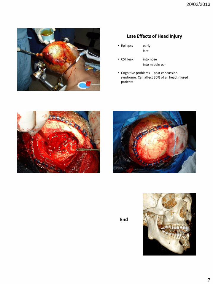
Late Effects of Head Injury
• Epilepsy early
late
• CSF leak into nose
into middle ear
• Cognitive problems – post concussion syndrome. Can affect 30% of all head injured patients
End