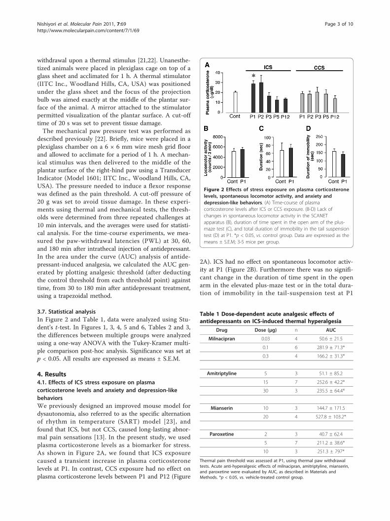
nishiyori molecular pain ......figure 2 effects of stress exposure on plasma corticosterone levels,...
TRANSCRIPT

RESEARCH Open Access
Permanent relief from intermittent cold stress-induced fibromyalgia-like abnormal pain byrepeated intrathecal administration ofantidepressantsMichiko Nishiyori1, Hitoshi Uchida1, Jun Nagai1, Kohei Araki1, Takehiro Mukae1, Shiroh Kishioka2 and Hiroshi Ueda1*
Abstract
Background: Fibromyalgia (FM) is characterized by chronic widespread pain, which is often refractory toconventional painkillers. Numerous clinical studies have demonstrated that antidepressants are effective in treatingFM pain. We previously established a mouse model of FM-like pain, induced by intermittent cold stress (ICS).
Results: In this study, we find that ICS exposure causes a transient increase in plasma corticosterone concentration,but not in anxiety or depression-like behaviors. A single intrathecal injection of an antidepressant, such asmilnacipran, amitriptyline, mianserin or paroxetine, had an acute analgesic effect on ICS-induced thermalhyperalgesia at post-stress day 1 in a dose-dependent manner. In addition, repeated daily antidepressanttreatments during post-stress days 1-5 gradually reversed the reduction in thermal pain threshold, and thisrecovery was maintained for at least 7 days after the final treatment. In addition, relief from mechanical allodynia,induced by ICS exposure, was also observed at day 9 after the cessation of antidepressant treatment. In contrast,the intravenous administration of these antidepressants at conventional doses failed to provide relief.
Conclusions: These results suggest that the repetitive intrathecal administration of antidepressants permanentlycures ICS-induced FM pain in mice.
Keywords: fibromyalgia, cold stress, vicious circle, antidepressant, allodynia, hyperalgesia
2. BackgroundFibromyalgia (FM) is characterized by generalized ten-derness and chronic widespread pain that affects 2-4% ofthe population in industrialized nations and primarilyaffects females [1]. Although its etiology and pathogen-esis are largely unknown, emerging evidence indicatesthat pain amplification within the central nervous system(CNS) plays a critical role in the pathology of FM pain[2]. Recent studies, including functional imaging, haverevealed that this central amplification process depends,in part, on deficits in endogenous descending pain inhibi-tory pathways [3,4] and abnormal pain processing [5]. Inaddition, FM pain is often refractory to treatment using
conventional painkillers, such as non-steroidal anti-inflammatory drugs and opioids [6]. However, numerousstudies have demonstrated the effectiveness of antide-pressants and antiepileptics, such as gabapentin andpregabalin, in the treatment of FM pain [7,8].There are several animal models of FM pain, induced
by either intramuscular injection of acidic saline [9],vagotomy [10], sound stress [11] or depletion of bio-genic amines [12]. However, in order to better under-stand the molecular basis of the underlying painmechanisms, it is necessary to establish an animalmodel which accurately reflects the pathological andpharmacotherapeutic features of the disease.Recently, we established a mouse model of FM using
intermittent cold stress (ICS), which produces long-last-ing thermal hyperalgesia and mechanical allodynia, pre-dominantly in females [13]. We found that gabapentin,
* Correspondence: [email protected] of Molecular Pharmacology and Neuroscience, Nagasaki UniversityGraduate School of Biomedical Sciences, 1-14 Bunkyo-machi, Nagasaki 852-8521, JapanFull list of author information is available at the end of the article
Nishiyori et al. Molecular Pain 2011, 7:69http://www.molecularpain.com/content/7/1/69 MOLECULAR PAIN
© 2011 Nishiyori et al; licensee BioMed Central Ltd. This is an Open Access article distributed under the terms of the CreativeCommons Attribution License (http://creativecommons.org/licenses/by/2.0), which permits unrestricted use, distribution, andreproduction in any medium, provided the original work is properly cited.

particularly when injected intracerebroventricularly, hadpotent anti-hyperalgesic and anti-allodynic effects in thismodel [13]. In addition, systemically and intracerebro-ventricularly-administered morphine was found to haveno analgesic effect in ICS-exposed mice, due to a failureto activate descending pain inhibitory pathways [14].These findings indicate that our ICS model might accu-rately reflect the pathological and pharmacotherapeuticfeatures of FM pain. In this study, we examine whethervarious antidepressants can ameliorate the abnormalpain sensations in this model.
3. Materials and methods3.1. AnimalsMale C57BL/6J mice weighing 18-22 g were used. Theywere kept in a room with an ambient temperature of 21± 2°C, with free access to a standard laboratory diet andtap water. All procedures were approved by the Naga-saki University Animal Care Committee and compliedwith the recommendations of the International Associa-tion for the Study of Pain [15].
3.2. Drug treatmentsAntidepressants were obtained from Sigma (St. Louis,MO, USA). Milnacipran, paroxetine, and amitriptylinewere dissolved in artificial cerebrospinal fluid (aCSF; 125mM NaCl, 3.8 mM KCl, 2.0 mM CaCl2, 1.0 mM MgCl2,1.2 mM KH2PO4, 26 mM NaHCO3, 10 mM glucose, pH7.4). Mianserin was dissolved in physiological saline. Forvehicle treatments, aCSF or saline was injected. Intrathecal(i.t.) injections were administered according to Hylden andWilcox [16] using a 30-gauge needle. The site of injectionwas chosen to be between spinal L5 and L6–near wherethe spinal cord ends and the cauda equina begins. Thisallowed us to maximize inter-vertebral accessibility and tominimize the possibility of spinal damage. After sufficienttraining, the experimenters were able to perform the tech-nique without causing injury to the animals.
3.3. Experimental model of fibromyalgiaICS exposure and constant cold stress (CCS) were per-formed as previously reported [13]. Briefly, for the ICSmodel, mice were placed on stainless mesh plate in acold room at 4°C overnight (from 4:30 pm to 10:00 am),followed by ICS with environmental temperatures alter-nating between 24 and 4°C every 30 min, from 10:00 amto 4:30 pm. These procedures were repeated twice. Onday 3, the mice were adapted to 24°C for 1 h before beha-vior testing. We designated day 3 following the onset ofstress exposure as day 1 post-stress exposure (P1). Forthe CCS model, mice were placed in the cold room from4:30 pm on day 1 to 10:00 am on day 3, followed byadaptation at 24°C for 1 h. Mice in the control groupwere kept at 24°C for all 3 days (from 4:30 pm on day 1
to 10:00 am on day 3). During the stress period, two micewere kept in each cage (12 × 15 × 10.5 cm), with freeaccess to food and agar as alternate drink water in placeof fluid. Although the body weight of mice was decreasedduring and after the ICS stress, it attained to the controlmice level as early as 4 day after the stress (Figure 1).
3.4. Measurement of plasma corticosteronePlasma corticosterone levels were measured as describedpreviously [17]. Briefly, plasma was separated by centri-fugation at 3 000 g for 15 min at 4°C and collected intoice-chilled tubes containing 0.1% EDTA and stored at-80°C until use. Blood samples were collected at 9:00pm in order to exclude the effect of circadian rhythmson circulating plasma corticosterone. The plasma corti-costerone level was estimated fluorometrically, accordingto the method of Zenker and Bernstein [18].
3.5. Assessment of stress-related behaviorsSpontaneous locomotor activity was measured in theopen filed (22 × 33 cm) for 3 min, using SCANET appa-ratus (Melquest, Japan). In the elevated plus-maze testused to estimate anxiety, the time spent in the openarm was recorded during a 6-min period. To assessdepression-like behaviors, the tail-suspension test wasperformed [19,20]. Mice were suspended 30 cm abovethe floor using adhesive tape, and the total duration ofimmobility during a 6-min period was measured.
3.6. Nociception testsIn the thermal paw withdrawal test, the nociceptionthreshold was assessed using the latency of paw
Figure 1 Changes in body weight in ICS treated mice. Resultsrepresent the percentage of body weight of mice, compared to thevalue at 1 day before ICS stress (Pre). *p < 0.05, vs. control group.Data are the means ± S.E.M; 6 mice per group.
Nishiyori et al. Molecular Pain 2011, 7:69http://www.molecularpain.com/content/7/1/69
Page 2 of 10
withdrawal upon a thermal stimulus [21,22]. Unanesthe-tized animals were placed in plexiglass cage on top of aglass sheet and acclimated for 1 h. A thermal stimulator(IITC Inc., Woodland Hills, CA, USA) was positionedunder the glass sheet and the focus of the projectionbulb was aimed exactly at the middle of the plantar sur-face of the animal. A mirror attached to the stimulatorpermitted visualization of the plantar surface. A cut-offtime of 20 s was set to prevent tissue damage.The mechanical paw pressure test was performed as
described previously [22]. Briefly, mice were placed in aplexiglass chamber on a 6 × 6 mm wire mesh grid floorand allowed to acclimate for a period of 1 h. A mechan-ical stimulus was then delivered to the middle of theplantar surface of the right-hind paw using a TransducerIndicator (Model 1601; IITC Inc., Woodland Hills, CA,USA). The pressure needed to induce a flexor responsewas defined as the pain threshold. A cut-off pressure of20 g was set to avoid tissue damage. In these experi-ments using thermal and mechanical tests, the thresh-olds were determined from three repeated challenges at10 min intervals, and the averages were used for statisti-cal analysis. For the time-course experiments, we mea-sured the paw-withdrawal latencies (PWL) at 30, 60,and 180 min after intrathecal injection of antidepressant.In the area under the curve (AUC) analysis of antide-pressant-induced analgesia, we calculated the AUC gen-erated by plotting analgesic threshold (after deductingthe control threshold from each threshold point) againsttime, from 30 to 180 min after antidepressant treatment,using a trapezoidal method.
3.7. Statistical analysisIn Figure 2 and Table 1, data were analyzed using Stu-dent’s t-test. In Figures 1, 3, 4, 5 and 6, Tables 2 and 3,the differences between multiple groups were analyzedusing a one-way ANOVA with the Tukey-Kramer multi-ple comparison post-hoc analysis. Significance was set atp < 0.05. All results are expressed as means ± S.E.M.
4. Results4.1. Effects of ICS stress exposure on plasmacorticosterone levels and anxiety and depression-likebehaviorsWe previously designed an improved mouse model fordysautonomia, also referred to as the specific alternationof rhythm in temperature (SART) model [23], andfound that ICS, but not CCS, caused long-lasting abnor-mal pain sensations [13]. In the present study, we usedplasma corticosterone levels as a biomarker for stress.As shown in Figure 2A, we found that ICS exposurecaused a transient increase in plasma corticosteronelevels at P1. In contrast, CCS exposure had no effect onplasma corticosterone levels between P1 and P12 (Figure
2A). ICS had no effect on spontaneous locomotor activ-ity at P1 (Figure 2B). Furthermore there was no signifi-cant change in the duration of time spent in the openarm in the elevated plus-maze test or in the total dura-tion of immobility in the tail-suspension test at P1
Figure 2 Effects of stress exposure on plasma corticosteronelevels, spontaneous locomotor activity, and anxiety anddepression-like behaviors. (A) Time-course of plasmacorticosterone levels after ICS or CCS exposure. (B-D) Lack ofchanges in spontaneous locomotor activity in the SCANETapparatus (B), duration of time spent in the open arm of the plus-maze test (C), and total duration of immobility in the tail suspensiontest (D) at P1. *p < 0.05, vs. control group. Data are expressed as themeans ± S.E.M; 3-5 mice per group.
Table 1 Dose-dependent acute analgesic effects ofantidepressants on ICS-induced thermal hyperalgesia
Drug Dose (μg) n AUC
Milnacipran 0.03 4 50.6 ± 21.5
0.1 6 281.9 ± 71.3*
0.3 4 166.2 ± 31.3*
Amitriptyline 5 3 51.1 ± 85.2
15 7 252.6 ± 42.2*
30 3 235.5 ± 64.4*
Mianserin 10 3 144.7 ± 171.5
20 4 527.8 ± 103.2*
Paroxetine 2 3 40.7 ± 62.4
5 7 211.2 ± 38.6*
10 3 251.3 ± 797*
Thermal pain threshold was assessed at P1, using thermal paw withdrawaltests. Acute anti-hyperalgesic effects of milnacipran, amitriptyline, mianserin,and paroxetine were evaluated by AUC, as described in Materials andMethods. *p < 0.05, vs. vehicle-treated control group.
Nishiyori et al. Molecular Pain 2011, 7:69http://www.molecularpain.com/content/7/1/69
Page 3 of 10

Figure 3 Antidepressant-induced acute analgesic effects in ICS treated mice. Thermal pain threshold was assessed at P1 after control or ICStreatment, using the thermal paw withdrawal test. Results represent the time course of thermal paw withdrawal latencies (PWL, in seconds) aftera single intrathecal injection of antidepressants. (A-D) Each data point in [control + vehicle] and [ICS + vehicle] groups is common. *p < 0.05, vs.vehicle-treated control group; #p < 0.05, vs. vehicle-treated and ICS-exposed groups. Data are expressed as the means ± S.E.M.; 4-8 mice pergroup.
Figure 4 Permanent relief from ICS-induced thermal hyperalgesia by repeated intrathecal administration of milnacipran. Intrathecalinjections of milnacipran (0.1 μg) were given once daily at 11:30 a.m. from P1-P5 after assessment of nociceptive thresholds at 11:00 a.m. Resultsrepresent the basal threshold as the latency to paw-withdrawal from thermal stimuli (PWL, in seconds), just before the daily injection of vehicleor milnacipran. *p < 0.05, vs. vehicle-treated control group; #p < 0.05, vs. vehicle-treated and ICS-exposed groups. Data are expressed as themeans ± S.E.M.;4-8 mice per group.
Nishiyori et al. Molecular Pain 2011, 7:69http://www.molecularpain.com/content/7/1/69
Page 4 of 10

(Figures 2C, D). In addition, there were no gross beha-vioral changes in mice as early as 1 h after the transferfrom 4°C to 24°C room.
4.2. Antidepressant-induced acute analgesic effects onthermal hyperalgesia in ICS-exposed micePrevious reports demonstrated that thermal hyperalgesiais elicited at P1 after ICS exposure and lasts for at least12 days [13,14]. As shown in Figure 3, the nociceptivethermal threshold was significantly reduced and stablethroughout experiments for 180 min. A single intrathe-cal injection of milnacipran (0.1 μg) had no effect onthe nociceptive threshold in control mice (Figure 3A),but produced significant anti-hyperalgesic effects thatpersisted for at least 180 min post-injection at P1 (Fig-ure 3A). This effect of milnacipran was dose-dependentin the range of 0.03-0.1 μg, but declined at 0.3 μg(Table 1). Statistical significance was observed at 0.1 and0.3 μg. Similar results were observed with other antide-pressants, such as amitriptyline (5-30 μg), mianserin (10and 20 μg), and paroxetine (2-10 μg), as shown in Fig-ures 3B-D and Table 1. However, with 20 μg of mian-serin, a significant analgesic effect was observed at 60min in the control mice, and anti-hyperalgesic effectswere observed until 180 min (Figure 3C). Both amitrip-tyline and paroxetine showed significant anti-hyperalge-sic effects, but no significance was observed at 180 min(Figures 3B, D).
4.3. Permanent relief of abnormal pain by repeatedcentral administrationAs the anti-hyperalgesic effect of milnacipran remained180 min after intrathecal administration at day P1 afterICS stress (threshold: ~ 7.46 ± 0.2 s), we measured the
nociceptive threshold at 11:00 a.m. on day P2. As seenin Figure 4, a significant anti-hyperalgesic effect stillremained (threshold: ~ 7.67 ± 0.6 s). The second admin-istration of milnacipran was performed at 11:30 a.m.The basal nociceptive threshold at 11:00 a.m. on day P3further increased to 8.56 ± 0.8 s. The increase in basalthreshold was maintained by daily administration of mil-nacipran. Complete recovery to the normal pain thresh-old was observed on P6, the day following the lastadministration, and lasted until P12. Similar completereversals of hyperalgesia on P5 and P12 were observedafter 5-day administrations of amitriptyline (15 μg),mianserin (20 μg), and paroxetine (5 μg), as seen inTable 2. Complete recovery was also observed with ICS-induced mechanical allodynia, even on P14, following a5-day administration of the antidepressants (Figure 5).
4.4. Lack of beneficial effects by repeated systemicadministrationWhen milnacipran was given by intravenous (i.v.) injec-tion (10 mg/kg), there was a significant analgesic effectin the thermal nociception test at 30 min in controlmice. However, there was no significant suppression inthe ICS mouse model using this dose of antidepressantup to 180 min on P1 (Figure 6A). The absence of anameliorative effect on ICS-induced hyperalgesia was alsoobserved with amitriptyline (3 mg/kg, i.v.), mianserin(10 mg/kg, i.v.), and paroxetine (1 mg/kg, i.v.), despiteproducing significant acute analgesia at 30 min in con-trol mice (Figures 6B-D). In addition, the repeated sys-temic administration of milnacipran for 5 days did notaffect the basal threshold throughout the experiment(Figure 6E). Repeated administrations of amitriptyline,mianserin or paroxetine also did not provide relief fromICS-induced hyperalgesia (Table 3).
5. DiscussionPatients with FM exhibit widespread pain, with diversesymptoms, such as fatigue, depression, and sleep distur-bance. Although the pathogenesis of FM is not clearlyunderstood, certain biological stressors, such as auto-nomic nervous system disorder and psychological dis-tress seem to be closely related to the development ofFM [24]. An important role for such stressors is sup-ported by studies using animal models in which rats ormice are subjected to stressors, such as chemical, sound,or surgery stress, which induce long-lasting abnormalpain [9-11,25]. Recently, we reported that ICS produceslong-lasting thermal hyperalgesia and mechanical allody-nia in mice [13,14]. The ICS-induced pain is bilateraland female-predominant (after gonadectomy) [13],which are also features found in FM patients [26].In this study, mice subjected to ICS exhibited a transi-
ent increase in plasma corticosterone levels on P1. In
Figure 5 Complete relief from ICS-induced mechanicalallodynia. Basal mechanical paw-withdrawal threshold (PWT, ingrams) was assessed at P14, using paw pressure tests. Intrathecalinjection of milnacipran (0.1 μg), amitriptyline (15 μg), mianserin (20μg), or paroxetine (5 μg), was given once daily from P1-P5, asdescribed in Figure 4. *p < 0.05, vs. vehicle-treated control group; #p< 0.05, vs. vehicle-treated and ICS-exposed groups. Data areexpressed as the means ± S.E.M.; 3-6 mice per group.
Nishiyori et al. Molecular Pain 2011, 7:69http://www.molecularpain.com/content/7/1/69
Page 5 of 10

Figure 6 Lack of anti-hyperalgesic effects by systemic administration of antidepressants. Thermal pain threshold was assessed at P1 aftercontrol or ICS-treatment, using thermal paw withdrawal tests. (A-D) Results represent the time-course of thermal paw-withdrawal latencies (PWL,in seconds) after a single i.v. injection of antidepressants. (A-D) Each data point in [control + vehicle] and [ICS + vehicle] groups is common. (E)Milnacipran was given i.v. once daily for 5 days, as described in Figure 4. Results represent the basal threshold as the latency to paw-withdrawalfrom thermal stimuli (PWL, in seconds), just before the daily injection of vehicle or milnacipran. *p < 0.05, vs. vehicle-treated control group; #p <0.05, vs. vehicle-treated and ICS-exposed groups. Data are expressed as the means ± S.E.M.; 3-6 mice per group.
Nishiyori et al. Molecular Pain 2011, 7:69http://www.molecularpain.com/content/7/1/69
Page 6 of 10

contrast, there was no significant change in corticoster-one levels in mice subjected to CCS. Considering thatthe abnormal pain in CCS mice was only transient, andnot long-lasting [13,14], the rise in corticosterone levelsin ICS mice likely played a role in the appearance ofabnormal pain. A recent report suggests that the stress-induced increase in corticosterone concentration may berelated to abnormal pain behavior in an FM-like animalmodel, possibly through a mechanism involving epi-nephrine release [27].In our ICS model, the mice did not show significant
changes in the tail-suspension test, a behavioral testdesigned to assess depression-like behavior [28]. This isin contrast to a study using less frequent temperaturealternation (the SART model), in which mice exhibitedhyperalgesia for only a week [29], and there was a tran-sient reduction of immobility duration in forced swim-ming test, followed by gradual recovery in 5-6 days [30].As the forced swimming causes a facilitation of immobi-lity in an antidepressant-reversible manner [31], it is notclear whether the transient reduction of immobility
duration reflects depression. From this point of view,the tail suspension test seems to be a better method forevaluation of depression-related despair behavior. Gaba-pentin and pregabalin are widely used to treat FMpatients in the clinic [32,33]. These medicines alleviateabnormal pain and the accompanying fatigue andinsomnia, without affecting depressive symptoms[33,34]. Therefore, the presence of depression-like beha-vior is unlikely to be necessary in animal models of FM.Consequently, the ICS model may be more clinicallyrelevant than the SART model for evaluating long-termpain.Various antidepressants have been used for FM in the
clinic [35,36]. Recently milnacipran and duloxetine, ser-otonin/norepinephrine reuptake inhibitors, and seroto-nin-specific reuptake inhibitors have been approved fortreating FM pain by the United States Food and DrugAdministration. As the antinociceptive activities of thesecompounds are largely independent of their effects onmood, making them potentially efficacious for patientswith or without depressive [37], it appears to reflect the
Table 2 Permanent relief from ICS-induced thermal hyperalgesia by repeated intrathecal (i.t.) administration ofamitriptyline, mianserin or paroxetine
n Pre(sec)
P1(sec)
P5(sec)
P12(sec)
Control-vehicle(i.t.)
8 9.84 ± 0.33 9.81 ± 0.45 10.24 ± 0.23 9.99 ± 0.22
ICS-vehicle(i.t.)
8 10.22 ± 0.28 5.76 ± 0.15* 6.04 ± 0.16* 6.28 ± 0.21*
ICS- amitriptyline(15 μg, i.t.)
4 10.48 ± 0.23 5.35 ± 0.36 8.44 ± 0.16# 9.35 ± 0.66#
ICS-mianserin(20 μg, i.t.)
4 9.68 ± 0.30 5.35 ± 0.36 8.13 ± 0.25# 9.95 ± 0.66#
ICS- paroxetine(5 μg, i.t.)
4 9.82 ± 0.44 5.47 ± 0.13 9.08 ± 0.46# 9.52 ± 0.21#
Antidepressants were administered between P1-P5, as described in Figure 3. Thermal pain threshold was assessed using the thermal paw withdrawal test. *p <0.05, vs. vehicle-treated control group; #p < 0.05, vs. vehicle-treated and ICS-exposed groups.
Table 3 Lack of anti-hyperalgesic effects by repeated systemic administration of antidepressants
n Pre(sec)
P1(sec)
P5(sec)
P12(sec)
Control-vehicle(i.v.)
8 9.51 ± 0.22 9.80 ± 0.18 10.00 ± 0.29 9.57 ± 0.30
ICS-vehicle(i.v.)
8 9.67 ± 0.23 5.78 ± 0.36 6.02 ± 0.12 6.36 ± 0.21
ICS- amitriptyline(3 mg/kg, i.v.)
4 9.55 ± 0.42 5.85 ± 0.37 5.79 ± 0.36 6.72 ± 0.35
ICS-mianserin(10 mg/kg, i.v.)
4 9.77 ± 0.34 5.14 ± 0.56 5.98 ± 0.39 6.66 ± 0.41
ICS- paroxetine(1 mg/kg, i.v.)
4 9.34 ± 0.36 5.29 ± 0.34 6.89 ± 0.30 6.96 ± 0.65
ICS- milnacipran(10 mg/kg, i.v.)
4 9.75 ± 0.47 6.25 ± 0.25 6.71 ± 0.21 7.54 ± 0.53
Amitriptyline, mianserin, paroxetine or milnacipran were given intravenously (i.v.) and assessment of basal nociceptive thresholds was performed as described inFigure 5. Thermal pain threshold was assessed using the thermal paw withdrawal test. *p < 0.05, vs. vehicle-treated control group.
Nishiyori et al. Molecular Pain 2011, 7:69http://www.molecularpain.com/content/7/1/69
Page 7 of 10
importance of central descending monoaminergic path-ways in pain regulation [38,39]. Recent studies revealedthat polymorphisms in the 5-HT receptor, transporter,and metabolic enzyme can contribute to the etiology ofFM [40-42]. The fMRI study also demonstrates thatbrain regions involved in descending pain inhibitorypathways appear to have decreased activity in FMpatients [43]. Although serotonergic and/or noradrener-gic pathways are well documented as descending paininhibitory pathways [39], there is no report that theabnormality of such descending monoaminergic systemsis observed in FM patients. However, it would be chal-lenging to examine the effects of representative antide-pressants on ICS-induced abnormal pain by introducingthe drugs into the intrathecal space, very close to targetregions.Our study shows that the repeated intrathecal admin-
istration of different antidepressants gradually sup-pressed ICS-induced pain. The gradual reversal ofabnormal pain may be related to the down-regulation ofb-adrenoceptors or abnormal monoaminergic metabo-lism [44-46]. Alternative mechanisms may include thealtered expression of multiple receptors and ion chan-nels, such as the NMDA receptor, opioid receptors, andsodium channels [47-49]. It should be noted that thereversal of abnormal pain continued after the cessationof drug treatment, for each of the antidepressants tested.Although further investigation is required to clarify themolecular mechanisms of antidepressant action and toprovide a permanent cure for ICS-induced abnormalpain, it is interesting to speculate that the chronic painmay be due to a vicious cycle of pain elicited by reducedinhibitory input from monoaminergic pathways. Thus,the rescue of pain-inhibitory mechanisms by repeatedantidepressant treatment should halt chronic pain. Simi-lar observations were made in our previous study usingcentral administration of gabapentin [13,14]. In thatstudy, using the ICS model, a single intracerebroventri-cular administration of gabapentin produced a 4-dayperiod of anti-hyperalgesia. As the injection had noeffect on peripheral nerve injury-induced neuropathicpain [13,14], and the gabapentin was unlikely to haveremained in the brain for 4 days, it is interesting tospeculate that the observed effect is due to the inhibi-tion of the pain cycle, possibly through enhancement ofinhibitory transmission. However, the present studydemonstrates that systemic administration of variousantidepressants had no significant beneficial effect onICS-induced hyperalgesia, though they had a significantacute analgesic effect in control mice. As the clinicallybeneficial effects of oral antidepressants to FM patientswere evident when they are treated for more than sev-eral weeks [50], the lack of effects of intravenous antide-pressants in the present study may be attributed to the
shortage of treatments (5 days). In this meaning it issurprising that only 5 days repetitive intrathecal treat-ments abolishes abnormal pain even after the cession oftreatments. Furthermore, although the mechanismsunderlying the lack of antihyperalgesic effect remain elu-sive, it may be worthwhile to investigate possible invol-vements of interference of spinal effects by peripheralpain facilitating serotonergic actions or by descendingpain facilitating monoaminergic systems [39]. Thus, weexpect that repetitive intrathecal administration of anti-depressants are likely to be more effective at treatingFM-like pain in mouse models.Finally, this study demonstrates that the ICS model has
similarities to clinical features of FM in terms of the sen-sitivity to analgesics or adjuvant analgesics. In our pre-vious findings, we observed that the effective dose ofgabapentin was 3 mg/kg for ICS-induced pain, but wasover 30 mg/kg for nerve injury-induced neuropathic painin mice [13,14], consistent with the fact that the clini-cally-effective dose of gabapentin for FM patients islower than that for neuropathic pain [51]. In addition, weobserved that ICS-induced thermal hyperalgesia wasresistant to morphine treatment [13,14], consistent withthe clinical evidence [52]. Considering that other experi-mental animal models of FM-like pain exhibit morphineanalgesia (albeit with low potency) [53-56], the ICSmodel may be pharmacologically distinct from the others.
6. ConclusionThis study demonstrates that repeated intrathecal anti-depressant treatment provides a complete cure of ICS-induced FM-like abnormal pain. Based on the pharma-cological similarity of ICS-induced pain to clinical FM,the ICS model appears to be suitable for investigatingthe pathogenesis of FM and for evaluating therapeuticstrategies for this debilitating illness.
List of abbreviations usedaCSF: artificial cerebrospinal fluid; AUC: area under the curve: CCS: constantcold stress; FM: fibromyalgia; ICS: intermittent cold stress; PWL: pawwithdrawal latency; PWT: paw withdrawal threshold.
AcknowledgementsThe authors thank Drs. L Ma and W Xie for technical assistance. This study wassupported in part by MEXT KAKENHI (17109015 to Hiroshi Ueda) and HealthLabor Sciences Research Grants from the Ministry of Health, Labor and Welfareof Japan (to Hiroshi Ueda): Research on Allergic disease and Immunology andThird Term Comprehensive Control Research for Cancer (398-49).
Author details1Division of Molecular Pharmacology and Neuroscience, Nagasaki UniversityGraduate School of Biomedical Sciences, 1-14 Bunkyo-machi, Nagasaki 852-8521, Japan. 2Department of Pharmacology, Wakayama Medical University,811-1 Kimiidera, Wakayama 641-0012, Japan.
Authors’ contributionsMN participated in the experimental designing, collection and analyses ofdata, and drafted the manuscript in equal contribution. HU and JN
Nishiyori et al. Molecular Pain 2011, 7:69http://www.molecularpain.com/content/7/1/69
Page 8 of 10

performed the statistical analyses and carried out surgical manipulation, datacollection, and drafted the manuscript. KA and TM performed stressexposing and participated nociceptive behavior assay. SK measured plasmacorticosterone levels. HU conceived of the study, participated in its designand coordination. All authors read and approved the final manuscript.
Competing interestsThe authors declare that they have no competing interests.
Received: 11 August 2011 Accepted: 21 September 2011Published: 21 September 2011
References1. Wolfe F, Ross K, Anderson J, Russell IJ, Hebert L: The prevalence and
characteristics of fibromyalgia in the general population. Arthritis Rheum1995, 38(1):19-28.
2. Schmidt-Wilcke T, Clauw DJ: Pharmacotherapy in fibromyalgia (FM)–implications for the underlying pathophysiology. Pharmacol Ther127(3):283-294.
3. Jensen KB, Kosek E, Petzke F, Carville S, Fransson P, Marcus H, Williams SC,Choy E, Giesecke T, Mainguy Y, et al: Evidence of dysfunctional paininhibition in Fibromyalgia reflected in rACC during provoked pain. Pain2009, 144(1-2):95-100.
4. Julien N, Goffaux P, Arsenault P, Marchand S: Widespread pain infibromyalgia is related to a deficit of endogenous pain inhibition. Pain2005, 114(1-2):295-302.
5. Clauw DJ: Fibromyalgia: an overview. Am J Med 2009, 122(12 Suppl):S3-S13.
6. Arnold LM, Bradley LA, Clauw DJ, Glass JM, Goldenberg DL:Multidisciplinary care and stepwise treatment for fibromyalgia. J ClinPsychiatry 2008, 69(12):e35.
7. Arnold LM, Goldenberg DL, Stanford SB, Lalonde JK, Sandhu HS, Keck PE Jr,Welge JA, Bishop F, Stanford KE, Hess EV, et al: Gabapentin in thetreatment of fibromyalgia: a randomized, double-blind, placebo-controlled, multicenter trial. Arthritis Rheum 2007, 56(4):1336-1344.
8. Arnold LM, Russell IJ, Diri EW, Duan WR, Young JP Jr, Sharma U, Martin SA,Barrett JA, Haig G: A 14-week, randomized, double-blinded, placebo-controlled monotherapy trial of pregabalin in patients with fibromyalgia.J Pain 2008, 9(9):792-805.
9. Sluka KA, Kalra A, Moore SA: Unilateral intramuscular injections of acidicsaline produce a bilateral, long-lasting hyperalgesia. Muscle Nerve 2001,24(1):37-46.
10. Khasar SG, Miao JP, Janig W, Levine JD: Modulation of bradykinin-inducedmechanical hyperalgesia in the rat by activity in abdominal vagalafferents. Eur J Neurosci 1998, 10(2):435-444.
11. Khasar SG, Dina OA, Green PG, Levine JD: Sound Stress-Induced Long-Term Enhancement of Mechanical Hyperalgesia in Rats Is Maintained bySympathoadrenal Catecholamines. J Pain 2009, 10(10):1073-1077.
12. Nagakura Y, Oe T, Aoki T, Matsuoka N: Biogenic amine depletion causeschronic muscular pain and tactile allodynia accompanied by depression:A putative animal model of fibromyalgia. Pain 2009, 146(1-2):26-33.
13. Nishiyori M, Ueda H: Prolonged gabapentin analgesia in an experimentalmouse model of fibromyalgia. Mol Pain 2008, 4:52.
14. Nishiyori M, Nagai J, Nakazawa T, Ueda H: Absence of morphine analgesiaand its underlying descending serotonergic activation in anexperimental mouse model of fibromyalgia. Neurosci Lett 2010,472(3):184-187.
15. Zimmermann M: Ethical guidelines for investigations of experimentalpain in conscious animals. Pain 1983, 16(2):109-110.
16. Hylden JL, Wilcox GL: Intrathecal morphine in mice: a new technique. EurJ Pharmacol 1980, 67(2-3):313-316.
17. Inoue M, Mishina M, Ueda H: Locus-specific rescue of GluRepsilon1NMDA receptors in mutant mice identifies the brain regions importantfor morphine tolerance and dependence. J Neurosci 2003,23(16):6529-6536.
18. Zenker N, Bernstein DE: The estimation of small amounts ofcorticosterone in rat plasma. J Biol Chem 1958, 231(2):695-701.
19. Pellow S: Anxiolytic and anxiogenic drug effects in a novel test ofanxiety: are exploratory models of anxiety in rodents valid? Methods FindExp Clin Pharmacol 1986, 8(9):557-565.
20. Steru L, Chermat R, Thierry B, Simon P: The tail suspension test: a newmethod for screening antidepressants in mice. Psychopharmacology (Berl)1985, 85(3):367-370.
21. Hargreaves K, Dubner R, Brown F, Flores C, Joris J: A new and sensitivemethod for measuring thermal nociception in cutaneous hyperalgesia.Pain 1988, 32(1):77-88.
22. Inoue M, Rashid MH, Fujita R, Contos JJ, Chun J, Ueda H: Initiation ofneuropathic pain requires lysophosphatidic acid receptor signaling. NatMed 2004, 10(7):712-718.
23. Kita T, Hata T, Iida J, Yoneda R, Isida S: Decrease in pain threshold in SARTstressed mice. Jpn J Pharmacol 1979, 29(3):479-482.
24. Smith HS, Harris R, Clauw D: Fibromyalgia: an afferent processing disorderleading to a complex pain generalized syndrome. Pain Physician 14(2):E217-245.
25. Khasar SG, Green PG, Levine JD: Repeated sound stress enhancesinflammatory pain in the rat. Pain 2005, 116(1-2):79-86.
26. Yunus MB: The role of gender in fibromyalgia syndrome. Curr RheumatolRep 2001, 3(2):128-134.
27. Khasar SG, Burkham J, Dina OA, Brown AS, Bogen O, Alessandri-Haber N,Green PG, Reichling DB, Levine JD: Stress induces a switch of intracellularsignaling in sensory neurons in a model of generalized pain. J Neurosci2008, 28(22):5721-5730.
28. Imbe H, Iwai-Liao Y, Senba E: Stress-induced hyperalgesia: animal modelsand putative mechanisms. Front Biosci 2006, 11:2179-2192.
29. Ohara H, Kawamura M, Namimatsu A, Miura T, Yoneda R, Hata T:Mechanism of hyperalgesia in SART stressed (repeated cold stress) mice:antinociceptive effect of neurotropin. Jpn J Pharmacol 1991,57(2):243-250.
30. Porsolt RD, Bertin A, Jalfre M: Behavioral despair in mice: a primaryscreening test for antidepressants. Arch Int Pharmacodyn Ther 1977,229(2):327-336.
31. Hata T, Nishikawa H, Itoh E, Watanabe A: Depressive state with anxiety inrepeated cold-stressed mice in forced swimming tests. Jpn J Pharmacol1999, 79(2):243-249.
32. Tzellos TG, Toulis KA, Goulis DG, Papazisis G, Zampeli VA, Vakfari A,Kouvelas D: Gabapentin and pregabalin in the treatment of fibromyalgia:a systematic review and a meta-analysis. J Clin Pharm Ther 35(6):639-656.
33. Hauser W, Bernardy K, Uceyler N, Sommer C: Treatment of fibromyalgiasyndrome with gabapentin and pregabalin - A meta-analysis ofrandomized controlled trials. Pain 2009, 145(1-2):69-81.
34. Crofford LJ, Rowbotham MC, Mease PJ, Russell IJ, Dworkin RH, Corbin AE,Young JP Jr, LaMoreaux LK, Martin SA, Sharma U: Pregabalin for thetreatment of fibromyalgia syndrome: results of a randomized, double-blind, placebo-controlled trial. Arthritis Rheum 2005, 52(4):1264-1273.
35. Rao SG, Bennett RM: Pharmacological therapies in fibromyalgia. Best PractRes Clin Rheumatol 2003, 17(4):611-627.
36. Gendreau RM, Thorn MD, Gendreau JF, Kranzler JD, Ribeiro S, Gracely RH,Williams DA, Mease PJ, McLean SA, Clauw DJ: Efficacy of milnacipran inpatients with fibromyalgia. J Rheumatol 2005, 32(10):1975-1985.
37. Arnold LM, Hudson JI, Wang F, Wohlreich MM, Prakash A, Kajdasz DK,Chappell AS: Comparisons of the efficacy and safety of duloxetine forthe treatment of fibromyalgia in patients with versus without majordepressive disorder. Clin J Pain 2009, 25(6):461-468.
38. Fishbain DA, Cutler R, Rosomoff HL, Rosomoff RS: Evidence-based datafrom animal and human experimental studies on pain relief withantidepressants: a structured review. Pain Med 2000, 1(4):310-316.
39. Millan MJ: Descending control of pain. Prog Neurobiol 2002, 66(6):355-474.40. Offenbaecher M, Bondy B, de Jonge S, Glatzeder K, Kruger M, Schoeps P,
Ackenheil M: Possible association of fibromyalgia with a polymorphismin the serotonin transporter gene regulatory region. Arthritis Rheum 1999,42(11):2482-2488.
41. Gursoy S: Absence of association of the serotonin transporter genepolymorphism with the mentally healthy subset of fibromyalgiapatients. Clin Rheumatol 2002, 21(3):194-197.
42. Tander B, Gunes S, Boke O, Alayli G, Kara N, Bagci H, Canturk F:Polymorphisms of the serotonin-2A receptor and catechol-O-methyltransferase genes: a study on fibromyalgia susceptibility.Rheumatol Int 2008, 28(7):685-691.
43. Mainguy Y: Functional magnetic resonance imagery (fMRI) infibromyalgia and the response to milnacipran. Hum Psychopharmacol2009, 24(Suppl 1):S19-23.
Nishiyori et al. Molecular Pain 2011, 7:69http://www.molecularpain.com/content/7/1/69
Page 9 of 10

44. Banerjee SP, Kung LS, Riggi SJ, Chanda SK: Development of beta-adrenergic receptor subsensitivity by antidepressants. Nature 1977,268(5619):455-456.
45. Wolfe BB, Harden TK, Sporn JR, Molinoff PB: Presynaptic modulation ofbeta adrenergic receptors in rat cerebral cortex after treatment withantidepressants. J Pharmacol Exp Ther 1978, 207(2):446-457.
46. Antkiewicz-Michaluk L, Romanska I, Michaluk J, Vetulani J: Role of calciumchannels in effects of antidepressant drugs on responsiveness to pain.Psychopharmacology (Berl) 1991, 105(2):269-274.
47. Yaron I, Shirazi I, Judovich R, Levartovsky D, Caspi D, Yaron M: Fluoxetineand amitriptyline inhibit nitric oxide, prostaglandin E2, and hyaluronicacid production in human synovial cells and synovial tissue cultures.Arthritis Rheum 1999, 42(12):2561-2568.
48. Petrie RX, Reid IC, Stewart CA: The N-methyl-D-aspartate receptor,synaptic plasticity, and depressive disorder. A critical review. PharmacolTher 2000, 87(1):11-25.
49. Wattiez AS, Libert F, Privat AM, Loiodice S, Fialip J, Eschalier A, Courteix C:Evidence for a differential opioidergic involvement in the analgesiceffect of antidepressants: prediction for efficacy in animal models ofneuropathic pain? Br J Pharmacol 163(4):792-803.
50. Arnold LM: Biology and therapy of fibromyalgia. New therapies infibromyalgia. Arthritis Res Ther 2006, 8(4):212.
51. Tzellos TG, Papazisis G, Toulis KA, Sardeli C, Kouvelas D: A2delta ligandsgabapentin and pregabalin: future implications in daily clinical practice.Hippokratia 14(2):71-75.
52. Sorensen J, Bengtsson A, Backman E, Henriksson KG, Bengtsson M: Painanalysis in patients with fibromyalgia. Effects of intravenous morphine,lidocaine, and ketamine. Scand J Rheumatol 1995, 24(6):360-365.
53. Harris RE, Clauw DJ, Scott DJ, McLean SA, Gracely RH, Zubieta JK:Decreased central mu-opioid receptor availability in fibromyalgia. JNeurosci 2007, 27(37):10000-10006.
54. Nielsen AN, Mathiesen C, Blackburn-Munro G: Pharmacologicalcharacterisation of acid-induced muscle allodynia in rats. Eur J Pharmacol2004, 487(1-3):93-103.
55. Sluka KA, Rohlwing JJ, Bussey RA, Eikenberry SA, Wilken JM: Chronic musclepain induced by repeated acid Injection is reversed by spinallyadministered mu- and delta-, but not kappa-, opioid receptor agonists. JPharmacol Exp Ther 2002, 302(3):1146-1150.
56. Furuta S, Shimizu T, Narita M, Matsumoto K, Kuzumaki N, Horie S, Suzuki T:Subdiaphragmatic vagotomy promotes nociceptive sensitivity of deeptissue in rats. Neuroscience 2009, 164(3):1252-1262.
doi:10.1186/1744-8069-7-69Cite this article as: Nishiyori et al.: Permanent relief from intermittentcold stress-induced fibromyalgia-like abnormal pain by repeatedintrathecal administration of antidepressants. Molecular Pain 2011 7:69.
Submit your next manuscript to BioMed Centraland take full advantage of:
• Convenient online submission
• Thorough peer review
• No space constraints or color figure charges
• Immediate publication on acceptance
• Inclusion in PubMed, CAS, Scopus and Google Scholar
• Research which is freely available for redistribution
Submit your manuscript at www.biomedcentral.com/submit
Nishiyori et al. Molecular Pain 2011, 7:69http://www.molecularpain.com/content/7/1/69
Page 10 of 10