neuronal microrna deregulation in response to alzheimer… · neuronal microrna deregulation in...
TRANSCRIPT

Neuronal MicroRNA Deregulation in Response toAlzheimer’s Disease Amyloid-bNicole Schonrock1*, Yazi D. Ke1, David Humphreys2, Matthias Staufenbiel3, Lars M. Ittner1, Thomas
Preiss2,4, Jurgen Gotz1*
1 Alzheimer’s and Parkinson’s Disease Laboratory, Brain and Mind Research Institute, University of Sydney, Sydney, New South Wales, Australia, 2 Molecular Genetics
Division, Victor Chang Cardiac Research Institute (VCCRI), Darlinghurst, Sydney, New South Wales, Australia, 3 Novartis Institute for BioMedical Research, Basel,
Switzerland, 4 School of Biotechnology and Biomolecular Sciences and St. Vincent’s Clinical School, University of New South Wales, Sydney, New South Wales, Australia
Abstract
Normal brain development and function depends on microRNA (miRNA) networks to fine tune the balance between thetranscriptome and proteome of the cell. These small non-coding RNA regulators are highly enriched in brain where theyplay key roles in neuronal development, plasticity and disease. In neurodegenerative disorders such as Alzheimer’s disease(AD), brain miRNA profiles are altered; thus miRNA dysfunction could be both a cause and a consequence of disease. Ourstudy dissects the complexity of human AD pathology, and addresses the hypothesis that amyloid-b (Ab) itself, a knowncausative factor of AD, causes neuronal miRNA deregulation, which could contribute to the pathomechanisms of AD. Weused sensitive TaqMan low density miRNA arrays (TLDA) on murine primary hippocampal cultures to show that about half ofall miRNAs tested were down-regulated in response to Ab peptides. Time-course assays of neuronal Ab treatments showthat Ab is in fact a powerful regulator of miRNA levels as the response of certain mature miRNAs is extremely rapid.Bioinformatic analysis predicts that the deregulated miRNAs are likely to affect target genes present in prominent neuronalpathways known to be disrupted in AD. Remarkably, we also found that the miRNA deregulation in hippocampal cultureswas paralleled in vivo by a deregulation in the hippocampus of Ab42-depositing APP23 mice, at the onset of Ab plaqueformation. In addition, the miRNA deregulation in hippocampal cultures and APP23 hippocampus overlaps with thoseobtained in human AD studies. Taken together, our findings suggest that neuronal miRNA deregulation in response to aninsult by Ab may be an important factor contributing to the cascade of events leading to AD.
Citation: Schonrock N, Ke YD, Humphreys D, Staufenbiel M, Ittner LM, et al. (2010) Neuronal MicroRNA Deregulation in Response to Alzheimer’s Disease Amyloid-b.PLoS ONE 5(6): e11070. doi:10.1371/journal.pone.0011070
Editor: Mel B. Feany, Brigham and Women’s Hospital, Harvard Medical School, United States of America
Received February 23, 2010; Accepted May 21, 2010; Published June 11, 2010
Copyright: � 2010 Schonrock et al. This is an open-access article distributed under the terms of the Creative Commons Attribution License, which permitsunrestricted use, distribution, and reproduction in any medium, provided the original author and source are credited.
Funding: N.S. is supported by the Human Frontier Science Program. L.I. is supported by the National Health and Medical Research Council (NHMRC) and theAustralian Research Council (ARC), and J.G. is supported by grants from the University of Sydney, the National Health and Medical Research Council (NHMRC), theAustralian Research Council (ARC), and the J.O. & J.R. Wicking Trust. Postgraduate scholarship support has been provided by the Wenkart Foundation,GlaxoSmithKline and Alzheimer’s Australia. Novartis (via Matthias Staufenbiel) provided the APP23 mouse strain. All experiments were performed in Sydney, andNovartis did not fund any aspect of this work. The funders had no role in study design, data collection and analysis, decision to publish, or preparation of themanuscript.
Competing Interests: The authors have a purely academic collaboration with Matthias Staufenbiel of Novartis. There is no commercial interest attached to thiswork with him. All PLoS ONE policies regarding the sharing of data and materials will be adhered to.
* E-mail: [email protected] (NS); [email protected] (JG)
Introduction
Alzheimer’s disease (AD) is a prominent neurodegenerative
disorder characterized by progressive loss of memory and other
cognitive functions. Histopathologically, AD is characterized by
neurofibrillary tangles (NFTs) consisting of the microtubule-
associated protein tau and neuritic plaques composed of
amyloid-b (Ab). Ab is a naturally occurring, predominantly 40
amino acid long polypeptide (Ab40) derived from the larger
amyloid precursor protein (APP) [1]. Increases in the proportion of
the longer, more neurotoxic form, Ab42, result in the formation of
higher order aggregates and subsequently, plaque deposition. In
familial AD (FAD), the increases in Ab42 are caused by aberrant
processing of APP due to mutations in either the APP gene itself or
in genes that encode subunits of the APP processing machinery. In
addition, APP promoter polymorphisms [2], gene duplications [3]
or trisomy 21 [4] can cause increased APP expression levels,
resulting in elevated Ab42. While increased Ab levels characterize
AD pathology, the precise mechanism(s) and signaling cascades it
uses to cause cellular toxicity and cell death are not fully
understood [5,6].
To better understand disease initiation and progression,
transgenic animal models have been developed that model aspects
of AD [7]. APP23 mice over-express the FAD mutant human APP
in brain, and develop amyloid plaques similar to the human
pathology [8]. These mice mimic several of the histopathological,
biochemical, cognitive and behavioral alterations characteristic for
AD. More recently, the research focus has shifted away from
plaque formation to earlier events in disease progression such as
the deregulation of genes whose impact on disease is still largely
unknown [9]. A substantial portion of post-transcriptional gene
regulation is controlled by microRNA (miRNA) networks, hence
an alteration in the expression of miRNAs is emerging as a
significant contributing factor to human neurodegenerative disease
[10,11]. miRNAs are evolutionarily conserved non-coding RNAs
of ,22 nucleotides that negatively regulate gene expression in a
PLoS ONE | www.plosone.org 1 June 2010 | Volume 5 | Issue 6 | e11070
sequence-specific manner. Indeed, profiling of postmortem human
AD brain has verified that significant changes in miRNA
expression occur in several brain regions [10]. This includes
miRNAs that regulate genes such as APP itself, and BACE1, that
encodes an enzyme involved in APP processing [12,13,14].
However, whether the deregulated miRNAs are a cause or a
consequence of disease, and what triggers miRNA dysfunction in
AD is unknown. We therefore explored the hypothesis that Abitself causes neuronal miRNA deregulation which could contribute
to the pathology associated with AD. To remove the complexity
inherently associated with human studies, we used mature murine
primary hippocampal cultures to determine the effects of Abspecifically on neuronal miRNAs.
Sensitive TaqMan low density miRNA arrays (TLDA) revealed
that 47% of all miRNAs tested were down-regulated in response to
Ab42. This response may be extremely rapid and bioinformatic
analysis predicts that the deregulated miRNAs are likely to affect
target genes present in prominent neuronal pathways disrupted in
AD. Remarkably, when we analyzed hippocampi of APP23 mice
at the onset of Ab plaque formation, we found a similar miRNA
deregulation as in our in vitro model. These findings support the
notion that an insult by Ab peptides causes a considerable
neuronal miRNA deregulation that may be an important factor in
the pathocascade of events leading to AD.
Materials and Methods
Ethics StatementAll animal experiments were approved by the Animal Ethics
Committee (AEC) of the University of Sydney under AEC
approval numbers K00/1-2009/3/4914 and K00/1-2009/3/
4915.
Cell culture and Ab treatmentsPrimary hippocampal neurons were prepared from 16.5-day-
old embryonic C57BL/6 mice (E16.5) as described [15]. 600,000
cells were plated per dish and cultivated in Neurobasal medium
supplemented with 1% (v/v) B27 supplements (Gibco) and 0.25%
(v/v) 200 mM L-glutamine (Gibco) to minimize growth of
astrocytes and microglia. Synthetic Ab42 peptides (Bachem,
Germany) dissolved in PBS were aged by incubation at 37uC for
24 h with shaking at 1000 rpm to allow fibril formation [16]. We
applied a protocol as described which used a range of biophysical
methods to determine the fibrillar nature of our preparation
[17,18]. At 23 days in vitro (DIV) cells were treated for 0, 1, 6, 15
or 24 hours with either 5 mM aged Ab42 or a mock treatment
containing PBS. Following treatments, cells were washed once in
PBS and lysed by resuspension in QIAzol reagent (Qiagen). Cell
lysates were stored in QIAzol at 280uC until further use.
Experiments were performed in triplicate.
Transgenic mouse strainAPP23 mice expressing human APP751 cDNA containing the
Swedish double mutation (K651M and N652L) were used for this
study [8]. Brains from mice at different ages (ranging from two to
thirteen months) were harvested following cervical dislocation and
the hippocampus was isolated, snap frozen and stored at 280uCuntil use. Hippocampi were not pooled and analysis was
performed on individual animals. Non-transgenic littermates were
used as controls.
RNA extraction and microarray analysisRNA was extracted from mouse primary hippocampal neurons
and dissected hippocampi using the miRNeasy Kit (Qiagen)
according to the manufacturer’s instructions. RNA quantity was
routinely assessed on a NanoDrop 1000 spectrophotometer
(Thermo Scientific). For microarray analysis, RNA quality was
determined on a Bioanalyser 2100 (Agilent) and only RNA
samples with an RNA integrity number (RIN) between 8 and 10
were used. Megaplex profiling using rodent TaqMan Low Density
miRNA Arrays (TLDA) (Applied Biosystems) was used to assay the
expression of 380 miRNAs as described by the manufacturer.
Briefly, 100 ng of total RNA obtained from Ab42- or control-
treated primary hippocampal cells was used in the megaplex
reverse transcription (RT) reaction containing about 450 miRNA-
specific RT primers provided by the manufacturer. No prior
miRNA preamplification step was needed. The RT product was
mixed with 2X TaqMan Universal PCR Master Mix, No
AmpErase UNG (Applied Biosystems) and loaded onto the TLDA
containing the 48-plex PCR reaction mix. TLDAs were run on a
7900HT Thermocycler (Applied Biosystems) using Sequence
Detection Systems (SDS) software version 2.3. A single TLDA
was used per Ab- or control-treated sample. Manual inspection of
all amplification plots was performed and miRNAs were excluded
from the analysis if: Ct values were too high (above 35, indicating a
miRNA expression too low for accurate detection), if amplification
was not achieved in all six samples, or if very high variation was
found. Data analysis was performed using SDS RQ manager v1.2
(Applied Biosystems) which utilizes the delta-delta CT method
[19]. The endogenous small nucleolar control RNA, snoRNA234,
was used for normalization. Significance was calculated using the
student’s T-test.
Quantitative real-time PCRIndividual TaqMan assays (Applied Biosystems) were used to
analyse the expression of the following mature mouse miRNAs:
miR-181c, miR-9, miR-20b, miR-21, miR-30c, miR-148b, miR-
361, miR-409-3p and Let-7i. 10 ng of total RNA was used in the
RT reaction and the transcribed cDNA was then used for
subsequent PCR amplification using TaqMan 2X Universal PCR
Master Mix, No AmpErase UNG (Applied Biosystems) as
described by the manufacturer. Assays were run on an
Mx3000P thermocycler (Stratagene) as follows: 95uC for 10 min,
and 40 cycles at 95uC for 151s followed by 60uC for 1 min. To
avoid any miRNA degradation, RNA extractions, reverse
transcription reactions and real-time runs were all performed on
the same day. Mouse snoRNA135 expression was assayed for
normalization. All reactions were performed in triplicate, and
relative miRNA expression was normalized against endogenous
controls using the comparative delta-delta CT method calculated
using MxPRO Software V4.0 (Stratagene).
In-situ hybridisation and immunohistochemistryKetamine/xylazine (Troy Laboratories, Australia) -anaesthe-
tized mice were perfused with 20 ml PBS. Brain tissue was
dissected and postfixed over night at 4uC in 4% paraformaldehyde
(Sigma, Australia). Tissue embedding in paraffin was done in a
Shandon Excelsior tissue processor (Thermo, USA). In-situ
hybridization was performed as described [20]. Briefly, 15 mm
paraffin-embedded sections of three month old mice were
rehydrated, permeabilized with proteinase K (10 mg/ml for
5 min), and then refixed for 15 min in 4% PFA before hybridizing
at 65uC overnight to a digoxigenin-labeled probe in a humidifying
chamber. The 760 bp APP probe used for hybridization
corresponds to the 39 end of human APP751 following a BamHI
digest. Slides were subsequently washed, prepared for immuno-
histochemistry with an alkaline phosphatase-conjugated antidigox-
Ab-Mediated miRNA Deregulation
PLoS ONE | www.plosone.org 2 June 2010 | Volume 5 | Issue 6 | e11070
igenin antibody (Sigma), and developed in NBT/BCIP solution
(Sigma).
For immunohistochemistry, antigen retrieval of 5 mm sections of
APP23 and WT brain was performed in 10 mM citrate buffer,
pH 5.8 in a RHS-1 microwave vacuum histoprocessor (Milestone,
USA) at 120uC. For standardization, all stainings were carried out
in Sequenza racks (Thermo, USA). Sections were blocked with
PBS containing 3% heat inactivated goat serum and 5% BSA for
1 hour at room temperature followed by incubation at 4uC over
night with the primary antibody 6E10 (1:1000, Signet, USA),
which is reactive against amino acid residues 1-16 of Ab. Antibody
staining was visualized using the AP-ABC Elite Kit (Vector, USA).
ImmunocytochemistryCoverslips containing mouse primary hippocampal neurons
grown for 24 DIV were fixed with 4% paraformaldehyde in
80 mM PIPES, 1 mM MgCl2, and 1 mM EGTA, pH 6.8. Cells
were permeabilized with 0.1% Triton in phosphate-buffered saline
and stained with primary antibodies to rabbit b3 tubulin (1:400,
Covance, USA). Antibody staining was visualized using Alexa
labeled secondary antibodies (Molecular Probes, USA). Pictures
were taken with a BX51 fluorescence microscope equipped with a
DP70 CCD color camera (Olympus, USA).
Pathway enrichment analysis of deregulated miRNAsTargetScanMouse v5.1 [21] was used to generate a list of potential
target genes for each of our significantly deregulated miRNAs found
from the microarray analysis. Due to miRNA sequence similarities
between family members, TargetScan mostly predicts target genes
for miRNA families rather than for individual family members. We
performed an enrichment analysis of target gene lists predicted for all
significantly deregulated miRNAs using the DAVID (Database for
Annotation, Visualization and Interrogated Discovery) bioinfor-
matics database [22,23]. Gene lists were uploaded into DAVID and
enrichment analysis was performed by comparing each set of genes
to all available biological pathways provided by the Kyoto
Encyclopedia of Genes and Genomes (KEGG) [24]. A cut-off P-
value of 0.01 was used to show KEGG Pathways likely to be affected
by predicted targets of deregulated miRNAs.
Results
Neuronal miRNA expression changes upon exposure toamyloid-b
To determine whether neuronal miRNAs are deregulated in
response to Ab we used a cell culture model of murine primary
hippocampal neurons [15]. Neurons were matured in vitro and
neuronal b3 tubulin staining showed that they had developed dense
axonal networks indicative of fully differentiated and healthy
neurons (Fig 1A). At 23 DIV, triplicate cultures were exposed for
24 hours to either 5 mM of aged Ab42 preparations or a PBS
control. Under these conditions, no evidence of Ab toxicity or cell
death was observed. Propidium iodide (PI) uptake was negligible in
Ab-treated neurons indicative of viable cultures and b3 tubulin
staining of Ab-treated cells showed no neuronal fragmentation, an
early sign of degeneration (data not shown). Total RNA was then
isolated and expression of 381 miRNAs analyzed by qRT-PCR
using rodent TaqMan Low Density miRNA Arrays (TLDA)
(Applied Biosystems). Careful manual inspection of all amplification
plots excluded miRNAs which did not amplify in all six samples,
had very high variation, or had Ct values above 35 indicating that
their expression was too low for accurate analysis. Relative miRNA
expression was normalized against the endogenous control
snoRNA234 using the comparative delta-delta CT method as
calculated using SDS RQ manager v1.2 software. For comparison,
normalization was also performed using two other endogenous
controls, snoRNA135 and 18S, included in each reverse transcrip-
tion (RT) primer pool and the same end result was achieved (data
not shown). 230 miRNAs (60%) were reliably detected on the array
(Supplementary Table S1) of which 35% were considered
unchanged as their expression levels varied only up to 15% from
untreated controls. Interestingly, Ab42 induced a considerable
down-regulation of miRNAs with 47% showing decreased levels
compared to untreated controls (Fig 1B). A much smaller fraction
(18%) of miRNAs was up-regulated in response to Ab42. When
applying a cut-off P-value of ,0.05, twenty miRNAs showed a
significant down-regulation, of up to four fold, with the exception of
miR-376b whose expression seemed to be strongly induced (2.6
fold) by Ab treatment (Fig 1C). Amongst the strongest significantly
down-regulated miRNAs were 409-3p, 361, 20b, 21, 181c and
148b. miRNAs 700, 146a, 365, 30c and 301 showed a moderate
decrease, while miRNAs 9, 664, 187, 125b, 433, 137, 30b, Let-7i
and Let-7g had a mild but significant down-regulation in response
to Ab. One advantage of using TaqMan assays is that their design,
with three levels of specificity per miRNA, allows differentiation
between mature miRNAs differing in only a few nucleotides.
Despite the fact that the Let-7 family consists of eight members
varying only by one to two nucleotides, only Let-7i, Let-7g and
miR-98 were down-regulated by Ab, whilst the others remained
mostly unchanged. In addition, specific members of the miR-30
family (30c and 30b) were also significantly down-regulated in
response to Ab. Thus, treatment of mouse primary neurons with
Ab42 does indeed evoke a strong change in miRNA profiles with a
substantial portion of miRNAs being down-regulated.
Neuronal miRNA response to amyloid-b occurs rapidlyDue to the low amounts of total RNA obtained from primary
neuronal cells we chose to validate our microarray results by
quantitative PCR using individual TaqMan assays on independent
preparations of Ab42-treated primary cultures. Eight significantly
down-regulated miRNAs from the array data were selected for
further validation and analysis. This selection was based on the
fact that these miRNAs produced the most significant fold-changes
in our study. In addition, an interesting overlap between human
studies and ours was observed (miR-9, 181c, 30c, 148b, 20b and
Let-7i) (Table 1) and therefore it was of great interest to validate
and analyze these miRNAs in particular [13,25]. miRNA
expression was thus assayed in independent 24 DIV cultures
treated with 5 mM aged Ab42. As Ab is known to cause cellular
toxicity and cell death [26], a time course of 1, 6 and 15 hour
treatments was used to exclude any effects due to apoptosis and to
gauge the rapidity of the miRNA response evoked by Ab. Similar
to our TLDA microarray results, all of the eight tested miRNAs
showed a significant down-regulation upon exposure to Ab at
some stage during the Ab time-course treatment compared to
untreated controls (Fig 2). Interestingly, Ab caused an extremely
rapid neuronal response of distinct mature miRNA sequences with
miR-9, 181c, 409-3p and 361 responding even after a one hour
Ab treatment. Expression of miRNAs 148b, 21, 20b and Let-7i
was more variable and therefore no conclusions can be made
concerning the rapidity of their response to Ab. Thus our analysis
not only validates the microarray data but further shows that Ab42
is a powerful regulator of certain mature miRNA sequences.
Deregulated miRNAs may affect targets in key pathwaysaltered in AD
A single miRNA is predicted to regulate several target genes,
whilst a single gene can also be regulated by several miRNAs;
Ab-Mediated miRNA Deregulation
PLoS ONE | www.plosone.org 3 June 2010 | Volume 5 | Issue 6 | e11070

Figure 1. Deregulated miRNAs in mouse primary hippocampal cells treated with Ab42. A. Mouse primary hippocampal neurons grown for 24DIV (days in vitro) stained with neuronal b3 tubulin showing dense axonal networks indicative of healthy mature neurons. Scale bar = 25 um. B.Neuronal miRNA response to Ab treatment. Overview of directional miRNA changes after Ab treatment. miRNAs altered by 15% compared to untreatedcontrols were considered unchanged. C. Summary of significantly deregulated microRNAs in primary hippocampal cells with or without Ab42 treatment(n = 3) analyzed by rodent TLDA. miRNA expression levels can be gauged using average (Ave) Ct values. T-test P-value significance: **P,0.01, *P,0.05.doi:10.1371/journal.pone.0011070.g001
Table 1. Overlap in miRNA changes between Ab42-treated murine hippocampal neurons and APP23 hippocampus found in thisstudy and sporadic human AD brain.
Mouse Neurons APP23 Hippocampus Human AD Brain
miRNA FC P-value FC P-value FC P-value Brain Region Reference
miR-9 0.76 0.01146 0.54 0.00000002 0.71 0.0053 Anterior temporal cortex [14]
0.38 (–1.39*) 0.0231 Hippocampus, Braak 5,6 [26]
miR-181c 0.35 0.01771 0.66 0.00006444 0.71 0.0018 Anterior temporal cortex [14]
0.1 (–3.30*) 0.0015 Parietal lobe cortex [51]
let-7i 0.75 0.02112 0.74 0.00000481 0.88 0.0283 Anterior temporal cortex [14]
miR-30c 0.74 0.04302 0.72 0.00008285 0.3 (–1.76*) 0.0194 Hippocampus, Braak 3,4 [26]
miR-148b 0.38 0.03563 0.76 0.00139502 0.21 (–2.24*) 0.0062 Parietal lobe cortex [51]
miR-20b 0.28 0.0188 0.80 0.25607460 0.28 (–1.84*) 0.0085 Parietal lobe cortex [51]
Deregulated miRNA fold changes (FC) and significance (P-value) found in our study on Ab-treated mouse primary neurons and APP23 hippocampus compared to thosefound in human AD profiling experiments on various brain regions (using the Braak staging). * FC values represented in the original reference from some human studieswere in log form.doi:10.1371/journal.pone.0011070.t001
Ab-Mediated miRNA Deregulation
PLoS ONE | www.plosone.org 4 June 2010 | Volume 5 | Issue 6 | e11070

hence changes in miRNA expression can have profound effects on
biological systems [21]. To increase the likelihood of identifying
biological processes most relevant to the miRNAs deregulated by
Ab we performed a gene ontology (GO) enrichment analysis on
predicted target genes. Of the available prediction algorithms such
as miRBase, PicTar, miRanda, PITA and TargetScan, all of
which use site conservation as a prediction criterion, the latter was
shown to result in the most accurate predictions upon target
validation [27,28]. Therefore, we used TargetScanMouse v5.1 to
generate lists of predicted target genes regulated by our miRNAs
of interest. Lists of target genes can be found at http://www.
targetscan.org/mmu_50/. To extract biological meaning associ-
ated with these large gene lists we used the bioinformatics database
DAVID. Pathway enrichment analysis was performed by
comparing each list of target genes to all available biological
pathways provided by the Kyoto Encyclopedia of Genes and
Genomes (KEGG) [24]. Encouragingly, many pathways associat-
ed with brain function were enriched in the pathway prediction
analysis (Table 2). Axon guidance was among the most significant
pathways to be affected by the predicted target genes and was the
top prediction for miR-9, miR-30 and miR-20. These three
miRNAs that are down-regulated by Ab potentially target a total
Figure 2. miRNA expression pattern in response to Ab time course in murine primary hippocampal neurons assessed by real-timePCR using TaqMan assays. Independent primary neuronal preparations treated with Ab42 for 1, 6 or 15 hours were assayed for miRNA expressionrelative to untreated controls. T-test P-value significance: ***P,0.001, **P,0.01, *P,0.05. Expression was normalized to snoRNA135.doi:10.1371/journal.pone.0011070.g002
Ab-Mediated miRNA Deregulation
PLoS ONE | www.plosone.org 5 June 2010 | Volume 5 | Issue 6 | e11070

Table 2. Pathway enrichment analysis for deregulated miRNAs in mouse primary hippocampal neurons after Ab treatment.
miRNATotal Targetsin mouse*
Targets assignedto KEGG pathways** KEGG Pathway***
# Targetsin Pathway**** P-value
miR-9 742 87 Axon guidance 16 (80) 0.000150
Focal adhesion 17 (133) 0.004500
Renal cell carcinoma 9 (48) 0.005800
MAPK signaling pathway 20 (179) 0.006100
Glutamate metabolism 6 (47) 0.006600
ErbB signaling pathway 10 (60) 0.007700
Regulation of actin cytoskeleton 17 (139) 0.009300
miR-30 871 69 Axon guidance 16 (80) 0.000097
Ubiquitin mediated proteolysis 13 (118) 0.004800
miR-20b 741 56 Axon guidance 14 (80) 0.000006
MAPK signaling pathway 14 (179) 0.006100
miR-181 639 83 MAPK signaling pathway 23 (179) 0.000020
Long-term potentiation 11 (42) 0.000032
Dorso-ventral axis formation 7 (24) 0.000160
T cell receptor signaling pathway 11 (80) 0.000950
Renal cell carcinoma 9 (48) 0.001600
TGF-beta signaling pathway 10 (46) 0.002900
Chronic myeloid leukemia 9 (51) 0.003300
Colorectal cancer 9 (64) 0.006100
mTOR signaling pathway 7 (30) 0.006800
ErbB signaling pathway 9 (60) 0.007100
Prostate cancer 9 (68) 0.007600
Focal adhesion 14 (133) 0.010000
miR-21 143 23 MAPK signaling pathway 10 (179) 0.000840
Colorectal cancer 5 (64) 0.010000
Let-7 683 72 MAPK signaling pathway 26 (179) 0.000016
Pancreatic cancer 11 (51) 0.000550
Bladder cancer 7 (33) 0.005200
Glycan structures - biosynthesis 12 (115) 0.005700
Melanoma 9 (38) 0.006300
Chronic myeloid leukemia 9 (51) 0.010000
Axon guidance 12 (80) 0.011000
miR-148b 368 53 Focal adhesion 13 (133) 0.001200
Regulation of actin cytoskeleton 13 (139) 0.002300
TGF-beta signaling pathway 8 (46) 0.004300
Pancreatic cancer 7 (51) 0.006000
Chronic myeloid leukemia 7 (51) 0.007200
miR-361 91 8 Huntington’s disease 3 (143) 0.009000
miR-137 678 18 Glycosphingolipid biosynthesis 3 (25) 0.009400
miR-365 134 15 Small cell lung cancer 6 (65) 0.001000
Prostate cancer 6 (68) 0.001000
mTOR signaling pathway 5 (30) 0.001100
Glioma 5 (39) 0.002200
Melanoma 5 (38) 0.003400
Focal adhesion 7 (133) 0.006900
miR-409-3p 101 1 P-value cutoff not met , ,
miR-433 146 10 P-value cutoff not met , ,
miR-376b 94 8 P-value cutoff not met , ,
miR-146 85 0 No Pathways predicted , ,
Ab-Mediated miRNA Deregulation
PLoS ONE | www.plosone.org 6 June 2010 | Volume 5 | Issue 6 | e11070
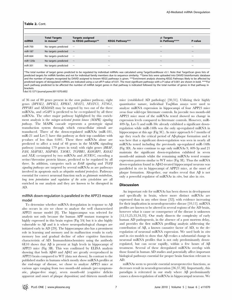
of 36 out of 80 genes present in the axon guidance pathway, eight
genes (DPYSL2, DPYSL5, EPHA7, NFAT5, NFATC3, NTNG1,
PPP3R1 and SEMA6D) may be targeted by two out of the three
miRNAs, and srGAP3 is predicted to be co-regulated by all three
miRNAs. The other major pathway highlighted by this enrich-
ment analysis is the mitogen-activated protein kinase (MAPK) signaling
pathway. The MAPK cascade represents a prototypic signal
transduction system through which extracellular stimuli are
transduced. Three of the down-regulated miRNAs (miR-181,
miR-21 and Let-7) have this pathway as their top candidate with
p-values of less than 0.001. These three miRNAs alone are
predicted to affect a total of 48 genes in the MAPK signaling
pathway (containing 179 genes in total) with eight genes (BRAF,
FASl, MAP3K1, MAP4K4, TAOK1, TGFBR1, RASGRP1, RASA2
and NLK) co-targeted by two miRNAs and ACVR1C, encoding a
serine/threonine protein kinase, predicted to be regulated by all
three. In addition, categories such as ErbB signaling and TGFbsignaling pathways are targeted by several miRNAs as are pathways
involved in apoptosis such as ubiquitin mediated proteolysis. Pathways
essential for correct neuronal function such as glutamate metabolism,
long term potentiation and regulation of the actin cytoskeleton are all
enriched in our analysis and they are known to be disrupted in
AD.
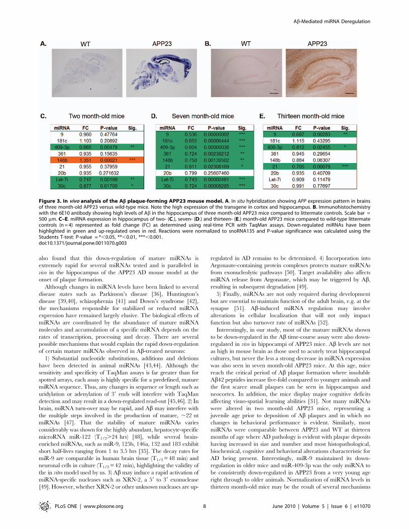
miRNA down-regulation is paralleled in the APP23 mousemodel
To determine whether miRNA deregulation in response to Abalso occurs in vivo we chose to analyze the well characterized
APP23 mouse model [8]. The hippocampus was selected for
analysis not only because the human APP mutant transgene is
highly expressed in this brain region (Fig 3A) but it is also highly
vulnerable to Ab and it is where neuropathological changes are
initiated early in AD [29]. The hippocampus also has a prominent
role in learning and memory and its malfunction results in early
memory loss and gradual decline of other cognitive functions
characteristic of AD. Immunohistochemistry using the antibody
6E10 shows that Ab is present at high levels in hippocampi of
APP23 mice (Fig 3B). This was confirmed by ELISA analysis
showing that both Ab40 and Ab42 are greatly overproduced in
APP23 brain compared to WT (data not shown). In contrast to the
published studies in humans which mostly show miRNA profiles at
the end-stage of disease, we chose to analyze APP23 mice at
various ages ranging from two month-old animals (pre-symptom-
atic, plaque-free stage), seven month-old (cognitive deficits
apparent and onset of plaque deposition) and thirteen month-old
mice (established AD pathology) [30,31]. Utilizing their highly
quantitative nature, individual TaqMan assays were used to
analyze miRNA expression in hippocampi of four APP23 mice
versus four wild-type littermate controls. In juvenile two month-old
APP23 mice most of the miRNAs tested showed no change in
expression levels compared to littermate controls. However, miR-
409-3p, Let-7i and miR-30c already exhibited a significant down-
regulation while miR-148b was the only up-regulated miRNA in
hippocampus at this age (Fig 3C). As mice approach 6-7 months of
age they reach the critical period of Ab-plaque formation and it
was here that a significant down-regulation was seen in nearly all
miRNAs tested including the previously up-regulated miR-148b
(Fig 3D). As mice continue to age only miRNAs 9, 409-3p and 21
maintain the significant down-regulation exhibited in seven
month-old animals whilst the remaining miRNAs tested resume
expression patterns similar to WT mice (Fig 3E). Thus the miRNA
down-regulation found in Ab42-treated hippocampal neurons was
paralleled in vivo in hippocampi of APP23 mice, at the onset of
plaque formation. Altogether, our studies reveal that Ab is not
only a powerful regulator of miRNAs in vitro, but also in vivo.
Discussion
An important role for miRNAs has been shown in development
and specifically in brain, where more distinct miRNAs are
expressed than in any other tissue [32]; with evidence increasing
for their implication in neurodegenerative disease [10,11]. miRNA
profiles are known to be altered in several regions of the AD brain,
however what is cause or consequence of the disease is unknown
[12,13,25,33,34,35]. Our study dissects the complexity of early
human AD pathogenesis, in the absence of a post mortem delay,
and provides the first miRNA profiling analysis addressing the
contribution of Ab, a known causative factor of AD, to the de-
regulation of neuronal miRNA expression. We used both in vitro
and in vivo models to show that Ab evokes a substantial change in
neuronal miRNA profiles that is not only predominantly down-
regulated, but can occur rapidly, within a few hours of Abtreatment. Several of these deregulated miRNAs overlap with
those found in human AD studies and potentially affect important
biological pathways essential for proper brain function relevant to
AD.
miRNAs seem to provide essential neuroprotective functions, as
decreases result in neurodegeneration [36,37,38]. Importantly, this
paradigm is reiterated in our study where Ab predominantly
causes a down-regulation of miRNAs in hippocampal neurons. We
miRNATotal Targetsin mouse*
Targets assignedto KEGG pathways** KEGG Pathway***
# Targetsin Pathway**** P-value
miR-700 No targets predicted , , , ,
miR-187 No targets predicted , , , ,
miR-664 No targets predicted , , , ,
miR-125b No targets predicted , , , ,
miR-301 No targets predicted , , , ,
*The total number of target genes predicted to be regulated by individual miRNAs was calculated using TargetScanMouse v5.1. Note that TargetScan gives lists ofpredicted targets for miRNA families and not for individual family members due to sequence similarity. **Gene lists were uploaded into DAVID bioinformatic databaseand the number of targets recognized by DAVID assigned to known KEGG pathways is given. ***Enrichment analysis showing KEGG Pathways likely to be affected bypredicted targets of deregulated miRNAs are indicated using a cut-off P-value of 0.01. The most significant pathways with a P-value of 0.001 are shown in bold. ****Foreach pathway predicted to be affected the number of miRNA target genes in that pathway is indicated followed by the total number of genes in that pathway inbrackets.doi:10.1371/journal.pone.0011070.t002
Table 2. Cont.
Ab-Mediated miRNA Deregulation
PLoS ONE | www.plosone.org 7 June 2010 | Volume 5 | Issue 6 | e11070
also found that this down-regulation of mature miRNAs is
extremely rapid for several miRNAs tested and is paralleled in
vivo in the hippocampus of the APP23 AD mouse model at the
onset of plaque formation.
Although changes in miRNA levels have been linked to several
disease states such as Parkinson’s disease [36], Huntington’s
disease [39,40], schizophrenia [41] and Down’s syndrome [42],
the mechanisms responsible for stabilized or reduced miRNA
expression have remained largely elusive. The biological effects of
miRNAs are coordinated by the abundance of mature miRNA
molecules and accumulation of a specific miRNA depends on the
rates of transcription, processing and decay. There are several
possible mechanisms that would explain the rapid down-regulation
of certain mature miRNAs observed in Ab-treated neurons:
1) Substantial nucleotide substitutions, additions and deletions
have been detected in animal miRNAs [43,44]. Although the
sensitivity and specificity of TaqMan assays is far greater than for
spotted arrays, each assay is highly specific for a predefined, mature
miRNA sequence. Thus, any changes in sequence or length such as
uridylation or adenylation of 39 ends will interfere with TaqMan
detection and may result in a down-regulated read-out [45,46]. 2) In
brain, miRNA turn-over may be rapid, and Ab may interfere with
the multiple steps involved in the production of mature, ,22 nt
miRNAs [47]. That the stability of mature miRNAs varies
considerably was shown for the highly abundant, hepatocyte-specific
microRNA miR-122 (T1/2.24 hrs) [48], while several brain-
enriched miRNAs, such as miR-9, 125b, 146a, 132 and 183 exhibit
short half-lives ranging from 1 to 3.5 hrs [35]. The decay rates for
miR-9 are comparable in human brain tissue (T1/2 = 48 min) and
neuronal cells in culture (T1/2 = 42 min), highlighting the validity of
the in vitro model used by us. 3) Ab may induce a rapid activation of
miRNA-specific nucleases such as XRN-2, a 59 to 39 exonuclease
[49]. However, whether XRN-2 or other unknown nucleases are up-
regulated in AD remains to be determined. 4) Incorporation into
Argonaute-containing protein complexes protects mature miRNAs
from exonucleolytic pathways [50]. Target availability also affects
miRNA release from Argonaute, which may be triggered by Ab,
resulting in subsequent degradation [49].
5) Finally, miRNAs are not only required during development
but are essential to maintain function of the adult brain, e.g. at the
synapse [51]. Ab-induced miRNA regulation may involve
alterations in cellular localization that will not only impact
function but also turnover rate of miRNAs [52].
Interestingly, in our study, most of the mature miRNAs shown
to be down-regulated in the Ab time-course assay were also down-
regulated in vivo in hippocampi of APP23 mice. Ab levels are not
as high in mouse brain as those used to acutely treat hippocampal
cultures, but never the less a strong decrease in miRNA expression
was also seen in seven month-old APP23 mice. At this age, mice
reach the critical period of Ab plaque formation where insoluble
Ab42 peptides increase five-fold compared to younger animals and
the first scarce small plaques can be seen in hippocampus and
neocortex. In addition, the mice display major cognitive deficits
affecting visuo-spatial learning abilities [31]. Not many miRNAs
were altered in two month-old APP23 mice, representing a
juvenile age prior to deposition of Ab plaques and in which no
changes in behavioral performance is evident. Similarly, most
miRNAs were comparable between APP23 and WT at thirteen
months of age where AD pathology is evident with plaque deposits
having increased in size and number and most histopathological,
biochemical, cognitive and behavioral alterations characteristic for
AD being present. Interestingly, miR-9 maintained its down-
regulation in older mice and miR-409-3p was the only miRNA to
be consistently down-regulated in APP23 from a very young age
right through to older animals. Normalization of miRNA levels in
thirteen month-old mice may be the result of several mechanisms
Figure 3. In vivo analysis of the Ab plaque-forming APP23 mouse model. A. In situ hybridization showing APP expression pattern in brainsof three month-old APP23 versus wild-type mice. Note the high expression of the transgene in cortex and hippocampus. B. Immunohistochemistrywith the 6E10 antibody showing high levels of Ab in the hippocampus of three month-old APP23 mice compared to littermate controls. Scale bar =500 mm. C–E. miRNA expression in hippocampus of two- (C.), seven- (D.) and thirteen- (E.) month-old APP23 mice compared to wild-type littermatecontrols (n = 4) represented as fold change (FC) as determined using real-time PCR with TaqMan assays. Down-regulated miRNAs have beenhighlighted in green and up-regulated ones in red. Reactions were normalized to snoRNA135 and P-value significance was calculated using theStudents T-test: P-value = *,0.05, **,0.01, ***,0.001.doi:10.1371/journal.pone.0011070.g003
Ab-Mediated miRNA Deregulation
PLoS ONE | www.plosone.org 8 June 2010 | Volume 5 | Issue 6 | e11070
including the induction of compensatory physiological responses
induced by prolonged acute over-expression of proteins in
transgenic mice or qualitative changes in Ab such as aggregation
of Ab into oligomeric or fibrillar species. However, the exact
mechanisms responsible for these patterns of miRNA deregulation
occurring in vivo are likely complex and remain to be determined.
Of great interest is the possible overlap in miRNA deregulation
between the models used in our study with the existing profiling
studies performed on human AD brain (Table 1). In general,
miRNA expression studies on AD patients revealed either no or
only very little overlap in miRNA changes (reviewed in [10]).
However, similar to our finding, Hebert et al showed that in AD
temporal cortex the deregulated miRNAs were also mostly down-
regulated compared to controls [13]. Importantly, this human study
showed that miR-9, 181c and Let-7i were down-regulated in AD
brain. miR-9 has also been reported to be down-regulated in an
independent human profiling study of various brain regions
including hippocampus [25]. In addition, this study showed that
miR-30c was down-regulated in hippocampus at an early stage of
disease (Braak stages 3 and 4). A recent study by Nunez-Iglesias et al
found forty-eight significantly deregulated miRNAs in human AD
parietal lobe cortex, of which miR-148b, 20b and 181c were down-
regulated [33]. Our in vivo analysis of APP23 hippocampus showed
down-regulation of miR-9, 181c, 30c, 20b, 148b and Let-7i, all of
which were altered in human AD brain. The overlap between
human AD and our in vitro and in vivo AD models indicates that
amongst the complex pathology in human AD brain, down-
regulation of miR-9, miR-181c, miR-30c, miR-20b, miR-148b and
Let-7i could be attributed at least in part to the presence of Ab.
miR-9, the most abundant human brain miRNA [53], is a
recurring candidate from several AD profiling studies. In contrast to
the above studies including ours, miR-9 was found to be up-
regulated in human AD CA1 [34] and temporal cortex [35]. Studies
performed in zebrafish and mice revealed that miR-9 is essential in
patterning, neurogenesis and differentiation and thus ideally placed
to impact various aspects of brain function. Over-expression of
miR-9 accelerates neuronal differentiation, while its inhibition in
the medial pallium of E11.5 mouse embryos results in defective
differentiation of Cajal-Retzius cells, the first neurons to populate
the embryonic cortex. Similarly, loss of miR-9 in zebrafish embryos
decreases the relative numbers of differentiated neurons in the
anterior hindbrain [54,55,56]. Neurogenesis is not only important
in the developing brain but is a process which continues in the adult
hippocampus, a region heavily affected by Ab pathology in AD
[57]. Interestingly, AD patients exhibit altered expression of early
neuronal markers in the hippocampus which has been attributed to
increased neurogenesis [58]. Decreased expression of miR-9 may
therefore impact adult brain function.
It is encouraging to see that most of the pathways predicted to
be affected by miR-9 target genes are related to brain function. In
comparison, miR-21, miR-181 and Let-7 have well characterized
roles in cancer and it is not surprising therefore that their target
genes result in enrichment for cancer-related pathways as well.
The MAPK pathway was one of the top candidate pathways to be
affected by Ab-mediated down-regulation of miRNAs. This
signaling cascade is involved in various cellular functions including
hippocampal synaptic plasticity and learning. Indeed, even very
low concentrations of oligomeric Ab42 activate MAPK in human
neuroblastoma cells [59]. In addition, MAPK activation was
observed in hippocampal slice cultures of Ab-forming Tg2576
mice [60]. In rodent hippocampus, MAPK is essential for LTP
formation, and several APP mutant mouse strains exhibit deficits
in hippocampal LTP and hippocampus-dependent associative
learning paradigms, including contextual fear conditioning and
escape training in the Morris water maze [30,61,62]. Thus, Ab42-
induced miRNA deregulation of the MAPK cascade may in part
underlie the learning and memory deficits attributed to hippo-
campal dysfunction in AD.
Axon guidance was the other major pathway over-represented in
our enrichment analysis. It represents a key stage in the formation of
neuronal networks known to be disrupted in AD. The down-
regulated miRNAs miR-9, miR-30 and miR-20 were all strongly
predicted to affect target genes involved in axonal guidance.
Interestingly, dihydropyrimidinase-related protein 2, DPYSL2, a
highly abundant protein in brain, is targeted by miR-30, 20 and 181
and has been shown to be up-regulated in proteomic studies on
APP23 mice already at a very early age [63]. Also called collapsin
response mediator protein 2 (CRMP2), DPYSL2 is a signal
mediator of Semaphorin 3A in the guidance of axonal growth.
Dysregulation of DPYSL2 has also been reported in other AD
proteomic studies [64,65,66,67], along with its aberrant phosphor-
ylation [68] and association with NFTs [69]. However, whether or
not miRNAs play a role in its regulation remains to be determined.
Together, our work provides insight into previously unknown
effects of Ab on neuronal miRNA networks. We show that Ab is a
powerful regulator of miRNA expression, causing a rapid decrease
of certain mature miRNA populations, which could have
profound impacts on biological processes affecting the pathogen-
esis of AD. Ab is well positioned to target several mechanisms that
affect the stability of mature miRNAs, including alterations in cis-
acting modifications, protein complex formation or the exposure
to nucleases, to name a few [70]. However, the exact mechanism
of the rapid Ab-mediated down-regulation of mature brain
miRNAs remains to be determined. The close overlap of our
miRNA profiles in the cell culture model, APP23 hippocampus
and human AD suggests the established APP23 mouse model is an
ideal system to investigate further the role of Ab-induced miRNA
deregulation in AD. Our study uncovers an unexplored mecha-
nism of how Ab may impact the pathology of AD and the
identification of key miRNAs affected by Ab will allow further
analysis of target genes and biological pathways contributing to
pathomechanisms in AD.
Supporting Information
Table S1 miRNA changes in mouse primary hippocampal cells
evoked by Ab42 treatment. Expression profiling of microRNAs in
mouse primary hippocampal cells with and without Ab42
treatment using Rodent TaqMan Low Denisty miRNA Arrays.
Shown are the results for 230 miRNAs in ascending order out of
the 381 miRNAs present on the TLDA whose amplification plots
where above the cutoff threshold in the triplicate analysis. miRNA
expression levels can be gauged using Average (Ave) Ct values.
miRNAs highlighted in bold are those significantly deregulated. T-
test P-value significance: **P,0.01, *P,0.05.
Found at: doi:10.1371/journal.pone.0011070.s001 (0.47 MB
DOC)
Acknowledgments
We thank Denise Nergenau for technical assistance, Dr Christine Biben for
help with in-situ hybridization and Dr Romaric Bouveret for fruitful
discussions.
Author Contributions
Conceived and designed the experiments: NS TP JG. Performed the
experiments: NS YDK DTH LMI. Analyzed the data: NS. Contributed
reagents/materials/analysis tools: MS TP JG. Wrote the paper: NS MS TP
JG.
Ab-Mediated miRNA Deregulation
PLoS ONE | www.plosone.org 9 June 2010 | Volume 5 | Issue 6 | e11070
References
1. Haass C (2004) Take five–BACE and the gamma-secretase quartet conduct
Alzheimer’s amyloid beta-peptide generation. EMBO J 23: 483–488.
2. Theuns J, Brouwers N, Engelborghs S, Sleegers K, Bogaerts V, et al. (2006)Promoter mutations that increase amyloid precursor-protein expression are
associated with Alzheimer disease. Am J Hum Genet 78: 936–946.
3. Rovelet-Lecrux A, Hannequin D, Raux G, Le Meur N, Laquerriere A, et al.(2006) APP locus duplication causes autosomal dominant early-onset Alzheimer
disease with cerebral amyloid angiopathy. Nat Genet 38: 24–26.
4. Podlisny MB, Lee G, Selkoe DJ (1987) Gene dosage of the amyloid beta
precursor protein in Alzheimer’s disease. Science 238: 669–671.
5. Gotz J, Schild A, Hoerndli F, Pennanen L (2004) Amyloid-inducedneurofibrillary tangle formation in Alzheimer’s disease: insight from transgenic
mouse and tissue-culture models. Int J Dev Neurosci 22: 453–465.
6. Gotz J, Ittner LM, Schonrock N, Cappai R (2008) An update on the toxicity ofAbeta in Alzheimer’s disease. Neuropsychiatr Dis Treat 4: 1033–1042.
7. Gotz J, Deters N, Doldissen A, Bokhari L, Ke Y, et al. (2007) A decade of tau
transgenic animal models and beyond. Brain Pathol 17: 91–103.
8. Sturchler-Pierrat C, Abramowski D, Duke M, Wiederhold KH, Mistl C, et al.(1997) Two amyloid precursor protein transgenic mouse models with Alzheimer
disease-like pathology. Proc Natl Acad Sci U S A 94: 13287–13292.
9. Walsh DM, Selkoe DJ (2007) A beta oligomers - a decade of discovery.J Neurochem 101: 1172–1184.
10. Hebert SS, De Strooper B (2009) Alterations of the microRNA network cause
neurodegenerative disease. Trends Neurosci 32: 199–206.
11. Lau P, de Strooper B (2010) Dysregulated microRNAs in neurodegenerativedisorders. Semin Cell Dev Biol.
12. Wang WX, Rajeev BW, Stromberg AJ, Ren N, Tang G, et al. (2008) The
expression of microRNA miR-107 decreases early in Alzheimer’s disease and
may accelerate disease progression through regulation of beta-site amyloidprecursor protein-cleaving enzyme 1. J Neurosci 28: 1213–1223.
13. Hebert SS, Horre K, Nicolai L, Papadopoulou AS, Mandemakers W, et al.
(2008) Loss of microRNA cluster miR-29a/b-1 in sporadic Alzheimer’s diseasecorrelates with increased BACE1/beta-secretase expression. Proc Natl Acad
Sci U S A 105: 6415–6420.
14. Hebert SS, Horre K, Nicolai L, Bergmans B, Papadopoulou AS, et al. (2009)MicroRNA regulation of Alzheimer’s Amyloid precursor protein expression.
Neurobiol Dis 33: 422–428.
15. Fath T, Ke YD, Gunning P, Gotz J, Ittner LM (2009) Primary support cultures
of hippocampal and substantia nigra neurons. Nat Protoc 4: 78–85.
16. Ferrari A, Hoerndli F, Baechi T, Nitsch RM, Gotz J (2003) beta-Amyloidinduces paired helical filament-like tau filaments in tissue culture. J Biol Chem
278: 40162–40168.
17. Habicht G, Haupt C, Friedrich RP, Hortschansky P, Sachse C, et al. (2007)Directed selection of a conformational antibody domain that prevents mature
amyloid fibril formation by stabilizing Abeta protofibrils. Proc Natl AcadSci U S A 104: 19232–19237.
18. Eckert A, Hauptmann S, Scherping I, Meinhardt J, Rhein V, et al. (2008)
Oligomeric and fibrillar species of beta-amyloid (A beta 42) both impair
mitochondrial function in P301L tau transgenic mice. J Mol Med 86:1255–1267.
19. Schmittgen TD, Livak KJ (2008) Analyzing real-time PCR data by the
comparative C(T) method. Nat Protoc 3: 1101–1108.
20. Biben C, Harvey RP (1997) Homeodomain factor Nkx2-5 controls left/rightasymmetric expression of bHLH gene eHand during murine heart development.
Genes Dev 11: 1357–1369.
21. Lewis BP, Burge CB, Bartel DP (2005) Conserved seed pairing, often flanked byadenosines, indicates that thousands of human genes are microRNA targets. Cell
120: 15–20.
22. Dennis G, Jr., Sherman BT, Hosack DA, Yang J, Gao W, et al. (2003) DAVID:Database for Annotation, Visualization, and Integrated Discovery. Genome Biol
4: P3.
23. Huang da W, Sherman BT, Lempicki RA (2009) Systematic and integrative
analysis of large gene lists using DAVID bioinformatics resources. Nat Protoc 4:44–57.
24. Kanehisa M, Goto S (2000) KEGG: kyoto encyclopedia of genes and genomes.
Nucleic Acids Res 28: 27–30.
25. Cogswell JP, Ward J, Taylor IA, Waters M, Shi Y, et al. (2008) Identification ofmiRNA changes in Alzheimer’s disease brain and CSF yields putative
biomarkers and insights into disease pathways. J Alzheimers Dis 14: 27–41.
26. Gotz J, Ittner LM, Fandrich M, Schonrock N (2008) Is tau aggregation toxic orprotective: a sensible question in the absence of sensitive methods? J Alzheimers
Dis 14: 423–429.
27. Selbach M, Schwanhausser B, Thierfelder N, Fang Z, Khanin R, et al. (2008)Widespread changes in protein synthesis induced by microRNAs. Nature 455:
58–63.
28. Baek D, Villen J, Shin C, Camargo FD, Gygi SP, et al. (2008) The impact of
microRNAs on protein output. Nature 455: 64–71.
29. Gotz J, Schonrock N, Vissel B, Ittner LM (2009) Alzheimer’s disease selectivevulnerability and modeling in transgenic mice. J Alzheimers Dis 18: 243–251.
30. Van Dam D, Vloeberghs E, Abramowski D, Staufenbiel M, De Deyn PP (2005)
APP23 mice as a model of Alzheimer’s disease: an example of a transgenicapproach to modeling a CNS disorder. CNS Spectr 10: 207–222.
31. Van Dam D, D’Hooge R, Staufenbiel M, Van Ginneken C, Van Meir F, et al.(2003) Age-dependent cognitive decline in the APP23 model precedes amyloid
deposition. Eur J Neurosci 17: 388–396.
32. Fineberg SK, Kosik KS, Davidson BL (2009) MicroRNAs potentiate neural
development. Neuron 64: 303–309.
33. Nunez-Iglesias J, Liu CC, Morgan TE, Finch CE, Zhou XJ (2010) Joint genome-wide profiling of miRNA and mRNA expression in Alzheimer’s disease cortex
reveals altered miRNA regulation. PLoS One 5: e8898.
34. Lukiw WJ (2007) Micro-RNA speciation in fetal, adult and Alzheimer’s diseasehippocampus. Neuroreport 18: 297–300.
35. Sethi P, Lukiw WJ (2009) Micro-RNA abundance and stability in human brain:
specific alterations in Alzheimer’s disease temporal lobe neocortex. Neurosci Lett459: 100–104.
36. Kim J, Inoue K, Ishii J, Vanti WB, Voronov SV, et al. (2007) A MicroRNAfeedback circuit in midbrain dopamine neurons. Science 317: 1220–1224.
37. Schaefer A, O’Carroll D, Tan CL, Hillman D, Sugimori M, et al. (2007)
Cerebellar neurodegeneration in the absence of microRNAs. J Exp Med 204:1553–1558.
38. Bilen J, Liu N, Burnett BG, Pittman RN, Bonini NM (2006) MicroRNA
pathways modulate polyglutamine-induced neurodegeneration. Mol Cell 24:157–163.
39. Packer AN, Xing Y, Harper SQ, Jones L, Davidson BL (2008) The bifunctional
microRNA miR-9/miR-9* regulates REST and CoREST and is downregulatedin Huntington’s disease. J Neurosci 28: 14341–14346.
40. Johnson R, Zuccato C, Belyaev ND, Guest DJ, Cattaneo E, et al. (2008) AmicroRNA-based gene dysregulation pathway in Huntington’s disease. Neuro-
biol Dis 29: 438–445.
41. Perkins DO, Jeffries CD, Jarskog LF, Thomson JM, Woods K, et al. (2007)microRNA expression in the prefrontal cortex of individuals with schizophrenia
and schizoaffective disorder. Genome Biol 8: R27.
42. Kuhn DE, Nuovo GJ, Martin MM, Malana GE, Pleister AP, et al. (2008)Human chromosome 21-derived miRNAs are overexpressed in down syndrome
brains and hearts. Biochem Biophys Res Commun 370: 473–477.
43. Thomson JM, Newman M, Parker JS, Morin-Kensicki EM, Wright T, et al.(2006) Extensive post-transcriptional regulation of microRNAs and its
implications for cancer. Genes Dev 20: 2202–2207.
44. Ebhardt HA, Tsang HH, Dai DC, Liu Y, Bostan B, et al. (2009) Meta-analysis of
small RNA-sequencing errors reveals ubiquitous post-transcriptional RNA
modifications. Nucleic Acids Res 37: 2461–2470.
45. Jones MR, Quinton LJ, Blahna MT, Neilson JR, Fu S, et al. (2009) Zcchc11-
dependent uridylation of microRNA directs cytokine expression. Nat Cell Biol
11: 1157–1163.
46. Katoh T, Sakaguchi Y, Miyauchi K, Suzuki T, Kashiwabara S, et al. (2009)
Selective stabilization of mammalian microRNAs by 39 adenylation mediated by
the cytoplasmic poly(A) polymerase GLD-2. Genes Dev 23: 433–438.
47. Winter J, Jung S, Keller S, Gregory RI, Diederichs S (2009) Many roads to
maturity: microRNA biogenesis pathways and their regulation. Nat Cell Biol 11:228–234.
48. Gatfield D, Le Martelot G, Vejnar CE, Gerlach D, Schaad O, et al. (2009)
Integration of microRNA miR-122 in hepatic circadian gene expression. GenesDev 23: 1313–1326.
49. Chatterjee S, Grosshans H (2009) Active turnover modulates mature microRNA
activity in Caenorhabditis elegans. Nature 461: 546–549.
50. Diederichs S, Haber DA (2007) Dual role for argonautes in microRNA
processing and posttranscriptional regulation of microRNA expression. Cell 131:
1097–1108.
51. Schratt G (2009) microRNAs at the synapse. Nat Rev Neurosci 10: 842–849.
52. Nelson PT, Wilfred BR (2009) In situ hybridization is a necessary experimentalcomplement to microRNA (miRNA) expression profiling in the human brain.
Neurosci Lett 466: 69–72.
53. Mattick JS, Makunin IV (2005) Small regulatory RNAs in mammals. Hum MolGenet 14 Spec No 1: R121–132.
54. Shibata M, Kurokawa D, Nakao H, Ohmura T, Aizawa S (2008) MicroRNA-9
modulates Cajal-Retzius cell differentiation by suppressing Foxg1 expression inmouse medial pallium. J Neurosci 28: 10415–10421.
55. Zhao C, Sun G, Li S, Shi Y (2009) A feedback regulatory loop involving
microRNA-9 and nuclear receptor TLX in neural stem cell fate determination.Nat Struct Mol Biol 16: 365–371.
56. Leucht C, Stigloher C, Wizenmann A, Klafke R, Folchert A, et al. (2008)MicroRNA-9 directs late organizer activity of the midbrain-hindbrain boundary.
Nat Neurosci 11: 641–648.
57. Hallbergson AF, Gnatenco C, Peterson DA (2003) Neurogenesis and braininjury: managing a renewable resource for repair. J Clin Invest 112: 1128–1133.
58. Jin K, Peel AL, Mao XO, Xie L, Cottrell BA, et al. (2004) Increased
hippocampal neurogenesis in Alzheimer’s disease. Proc Natl Acad Sci U S A101: 343–347.
59. Young KF, Pasternak SH, Rylett RJ (2009) Oligomeric aggregates of amyloid
beta peptide 1-42 activate ERK/MAPK in SH-SY5Y cells via the alpha7nicotinic receptor. Neurochem Int 55: 796–801.
60. Dineley KT, Westerman M, Bui D, Bell K, Ashe KH, et al. (2001) Beta-amyloidactivates the mitogen-activated protein kinase cascade via hippocampal alpha7
Ab-Mediated miRNA Deregulation
PLoS ONE | www.plosone.org 10 June 2010 | Volume 5 | Issue 6 | e11070
nicotinic acetylcholine receptors: In vitro and in vivo mechanisms related to
Alzheimer’s disease. J Neurosci 21: 4125–4133.61. Hsiao K, Chapman P, Nilsen S, Eckman C, Harigaya Y, et al. (1996) Correlative
memory deficits, Abeta elevation, and amyloid plaques in transgenic mice.
Science 274: 99–102.62. Chapman PF, White GL, Jones MW, Cooper-Blacketer D, Marshall VJ, et al.
(1999) Impaired synaptic plasticity and learning in aged amyloid precursorprotein transgenic mice. Nat Neurosci 2: 271–276.
63. Guerreiro N, Staufenbiel M, Gomez-Mancilla B (2008) Proteomic 2-D DIGE
profiling of APP23 transgenic mice brain from pre-plaque and plaquephenotypes. J Alzheimers Dis 13: 17–30.
64. David DC, Ittner LM, Gehrig P, Nergenau D, Shepherd C, et al. (2006) Beta-amyloid treatment of two complementary P301L tau-expressing Alzheimer’s
disease models reveals similar deregulated cellular processes. Proteomics 6:6566–6577.
65. Lubec G, Nonaka M, Krapfenbauer K, Gratzer M, Cairns N, et al. (1999)
Expression of the dihydropyrimidinase related protein 2 (DRP-2) in Down
syndrome and Alzheimer’s disease brain is downregulated at the mRNA and
dysregulated at the protein level. J Neural Transm Suppl 57: 161–177.66. Sizova D, Charbaut E, Delalande F, Poirier F, High AA, et al. (2007) Proteomic
analysis of brain tissue from an Alzheimer’s disease mouse model by two-
dimensional difference gel electrophoresis. Neurobiol Aging 28: 357–370.67. Vercauteren FG, Clerens S, Roy L, Hamel N, Arckens L, et al. (2004) Early
dysregulation of hippocampal proteins in transgenic rats with Alzheimer’sdisease-linked mutations in amyloid precursor protein and presenilin 1. Brain
Res Mol Brain Res 132: 241–259.
68. Gu Y, Hamajima N, Ihara Y (2000) Neurofibrillary tangle-associated collapsinresponse mediator protein-2 (CRMP-2) is highly phosphorylated on Thr-509,
Ser-518, and Ser-522. Biochemistry 39: 4267–4275.69. Yoshida H, Watanabe A, Ihara Y (1998) Collapsin response mediator protein-2
is associated with neurofibrillary tangles in Alzheimer’s disease. J Biol Chem 273:9761–9768.
70. Kai ZS, Pasquinelli AE (2010) MicroRNA assassins: factors that regulate the
disappearance of miRNAs. Nat Struct Mol Biol 17: 5–10.
Ab-Mediated miRNA Deregulation
PLoS ONE | www.plosone.org 11 June 2010 | Volume 5 | Issue 6 | e11070