neurological effects of non-ionizing electromagnetic fields...radiations. lustenberger et al. (2013)...
TRANSCRIPT

SECTION 9
Neurological Effects of Non-Ionizing Electromagnetic Fields
2014 Supplement
Prof. Henry Lai, PhD (Ret.)
Department of Bioengineering University of Washington
Seattle, WA USA
Prepared for the BioInitiative Working Group March 2014

2
I. INTRODUCTION Neurological effects are caused by changes in the nervous system. Factors that act
directly or indirectly on the nervous system causing morphological, chemical, or electrical
changes in the nervous system can lead to neurological effects. The final manifestation of these
effects can be seen in psychological changes, e.g., memory, learning and perception. The
nervous system is an electrical organ. Thus, it should not be surprising that exposure to
electromagnetic fields could lead to neurological changes. Morphological, chemical, electrical,
and behavioral changes have been reported in animals and cells after exposure to nonionizing
electromagnetic fields (EMF) across a range of frequencies. The consequences of physiological
changes in the nervous system are very difficult to assess. We don’t quite understand how the
nervous system functions and reacts to external perturbations. The highly flexible nervous
system could easily compensate for external disturbances. On the other hand, the consequence
of neural perturbation is also situation-dependent. An EMF-induced change in brain electrical
activity, for instance, could lead to different consequences depending on whether a person is
watching TV or driving a car.
The following is a summary of the research literature on the neurological effects of EMF
exposure published between 2007-2014. The literature on radiofrequency and extremely-low
frequency EMFs are placed in two separate sections. Each section has a discussion and a list of
publications with abstracts. Summary sentences in the abstracts are underlined for reader
convenience. Where additional information is relevant, some earlier papers, or papers not
specifically related to neurological effects, are also included with citations contained within the
discussion.
In this paper, as in the update paper on genetic effects, analyses show that there are more
publications showing effects than no effects with the recent neurological literature. With E
representing a biological effect, and NE representing no biological effects, the recent literature
finds in 211 studies, RFR-neurological effects at: E=144 publications (68%); NE=67
publications (32%); and 105 ELF-neurological effects studies: E=95 (90%); NE=10 (10%).
Appendix A has references and abstracts for the RFR literature. Appendix B has references and abstracts for the ELF-EMF literature.

3
II. NEUROLOGICAL EFFECTS OF RADIOFREQUENCY RADIATION (RFR) - (2007-2014)
Discussion
(1) There are many new studies on human subjects. Many of them are on changes in brain electrical activities after acute exposure to cell phone radiation. Bak et al (2010) reported effects on event-related potentials. Maganioti et al. (2010) further reported that RFR affected the gender-specific components of event-related potentials (see also Hountala et al., 2008). Croft et al (2008) reported changes of the alpha-wave power of EEG. The same authors (Croft et al., 2010) further reported that effects differed between various new cell phone transmission systems, which have different signaling characteristics. They observed effects after exposure to second generation (2G), but not third generation (3G) radiation, whereas Leung et al. (2011) found similar EEG effects with both 2G and 3G radiations. Lustenberger et al. (2013) found increased slow-wave activity in humans during exposure to pulse-modulated RF EMF toward the end of the sleep period. Vecchio and associates reported that cell phone RFR affected EEG and the spread of neural synchronization conveyed by interhemispherical functional coupling of EEG rhythms (Vecchio et al., 2007) and enhanced human cortical neural efficiency (Vecchio et al., 2012a). An interesting finding is that RFR could interact with the activity of brain epileptic foci in epileptic patients (Tombini et al., 2012; Vecchio et al., 2012b). However, no significant effect on EEG was reported by Parentos et al. (2007) or Trunk et al. (2012), and Kleinlogel et al. (2008 a, b) also reported no significant effects on resting EEG and event-related potentials in humans after exposure to cell phone RFR. Furthermore, Krause et al. (2007) reported no significant effect of cell phone radiation on brain oscillatory activity, and Inomata-Terada et al. (2007) concluded that cell phone radiation does not affect the electrical activity of the motor cortex.
(2) There are studies on the interaction of cell phone radiation on EEG during sleep. Changes
in sleep EEG have been reported by Hung et al. (2007), Regel et al. (2007), Lowden et al (2011), Schmid et al. (2012), and Loughran et al. (2012), whereas, no significant effect was reported by Fritzer et al (2007), Mohler et al. (2010, 2012) and Nakatani-Enomoto et al. (2013). Loughran et al. (2012) provided an interesting conclusion in their paper: “These results confirm previous findings of mobile phone-like emissions affecting the EEG during non-REM sleep. Importantly, this low-level effect was also shown to be sensitive to individual variability. Furthermore, this indicates that “previous negative results are not strong evidence for a lack of an effect…” Increase in REM sleep was reported by Pelletier et al. (2012) in developing rats after chronic exposure. Mohammed et al. (2013) reported a disturbance in REM sleep EEG in the rat after long term exposure (1 hr/day for 1 month) to a 900-MHz modulated RFR.
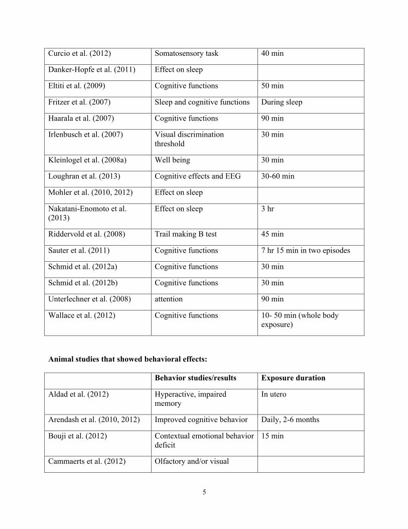
(3) With these electrophysiological changes in the brain, what behavioral effects have been
reported? The outcomes are summarized in the tables below. The animal studies are mostly studies on rodents (i.e., rat and mouse).

4
Human studies that showed behavioral effects:
Behavior studies/results Exposure duration
de Tommaso et al. (2009) Reduction in behavioral arousal
10 min
Hung et al. (2007) Sleep latency 30 min
Leung et al. (2011) Cognitive functions 10 min
Luria et al. (2009) Spatial working memory (In a subsequent study (Hareuveny et al., 2011), the authors indicated that some of the effects observed may not be related to RFR exposure.)
60 min
Lustenberger et al. (2013) Sleep-dependent motor-task performance improvement
All-night
Redmayne et al. (2013) Well-being Use of cellphone and cordless phone
Regel et al. (2007) Cognitive functions 30 min
Thomas et al. (2010b) Overall behavioral problems in adolescents
Vecchio et al. (2012b) Enhanced cognitive-motor processes
45 min
Vecsei et al. (2013) Thermal pain threshold 30 min
Wiholm et al. (2009) ‘Virtual’ spatial navigation task
150 min
Human studies that did not show behavioral effects:
Behavior studies/results Exposure duration
Cinel et al. (2007) Order threshold task 40 min
Cinel et al. (2008) Subjective symptoms 40 min
Curcio et al. (2008) Reaction time task, sequential figure tapping task
3 x 15 min
5
Curcio et al. (2012) Somatosensory task 40 min
Danker-Hopfe et al. (2011) Effect on sleep
Eltiti et al. (2009) Cognitive functions 50 min
Fritzer et al. (2007) Sleep and cognitive functions During sleep
Haarala et al. (2007) Cognitive functions 90 min
Irlenbusch et al. (2007) Visual discrimination threshold
30 min
Kleinlogel et al. (2008a) Well being 30 min
Loughran et al. (2013) Cognitive effects and EEG 30-60 min
Mohler et al. (2010, 2012) Effect on sleep
Nakatani-Enomoto et al. (2013)
Effect on sleep 3 hr
Riddervold et al. (2008) Trail making B test 45 min
Sauter et al. (2011) Cognitive functions 7 hr 15 min in two episodes
Schmid et al. (2012a) Cognitive functions 30 min
Schmid et al. (2012b) Cognitive functions 30 min
Unterlechner et al. (2008) attention 90 min
Wallace et al. (2012) Cognitive functions 10- 50 min (whole body exposure)
Animal studies that showed behavioral effects:
Behavior studies/results Exposure duration
Aldad et al. (2012) Hyperactive, impaired memory
In utero
Arendash et al. (2010, 2012) Improved cognitive behavior Daily, 2-6 months
Bouji et al. (2012) Contextual emotional behavior deficit
15 min
Cammaerts et al. (2012) Olfactory and/or visual

6
memory deficit in ants
Cammaerts et al. (2013) Food collection behavior of ants
180 hr
Daniels et al. (2009) Decreased motor activity
Deshmukh et al. (2013) Cognitive functions 2 hr/day, 30 days
Fragopoulou et al. (2010) Spatial memory deficit 2 hr/day, 4 days
Hao et al. (2012) Learning and memory deficit 6 hr/day, 5 days/wk, 10 wk
İkinci et al. (2013) Learning behavior deficit Prenatal exposure
Júnior et al. (2014) Stress behavioral patterns 25 sec every 2 min for 3 days
Kumar et al. (2009) hypoactivity 50 missed call/day, 4 wk
Kumlin et al. (2007) Improved learning and memory
2 hr/day. 5 days/wk, 5 wk
Lu et al. (2012) Spatial memory deficit 3 hr/day, 30 days
Maaroufi et al. (2013) Spatial learning and memory deficit
1 hr/day, 21 days
Mathur (2008) Analgesic effect 2 hr/day, 45 days
Megha et al. (2012) Cognitive functions 2 hr/day, 30 days
Narayanan et al. (2009) Learning deficit 50 missed call/day, 4 wk
Narayanan et al. (2010) Passive avoidance deficit 50 missed call/day, 4 wk
Narayanan et al. (2012) Elevated plus maze- emotionality test
28 days
Nittby et al. (2008) Reduced memory functions 2 hr/wk, 55 wk
Ntzouni et al. (2011) Non-spatial memory deficit 90 min/day, 17 days
Ntzouni et al. (2013) Spatial and non-spatial memory deficit
90 min/day, 66-148 days
Odacı et al. (2013) Motor function Prenatal exposure
Pelletier et al. (2012) Food intake increase 5 weeks
Qin et al. (2014) Learning and memory deficits 2 hr/day, 30 days

7
Razavinasab et al. (2014) Learning and memory deficits In utero
Sarapultseva et al. (2013) Motor activity in protozoa 0.05-10 hr
Sharma et al. (2013) Spatial memory deficit 2 hr/day, 30 days
Sokolovic et al. (2012) Anxiety-related behavior 4 hr/day for 20, 40, 60 days
Vácha et al. (2009) Magnetoreception in cockroach
Wang et al. (2013) Spatial memory deficit 6 min
Animal studies that did not show behavioral effects:
Behavior studies/results Exposure duration
Ammari et al. (2008c) spatial memory 15 min/day, 8 or 24 wk
Haghani et al. (2013) Motor function 6 hr/day during gestation period
Almost all the animal studies reported effects, whereas more human studies
reported no effects than effects. This may be caused by several possible factors: (a) Humans are less susceptible to the effects of RFR than are rodents. (b) It may be more difficult to do human than animal experiments, since it is, in general, easier to control the variables and confounding factors in an animal experiment. (c) In the animal studies, the cumulative exposure duration was generally longer and studies were carried out after exposure, whereas in the human studies, the exposure was generally one time and testing was done during exposure. This raises the question of whether the effects of RFR are cumulative. This consideration could have very important implication on real life human exposure to EMF. However, it must be pointed out that neurophysiological and behavioral changes have been reported in both animals and humans after acute (one time) exposure to RFR, and most of the EEG studies mentioned above are acute exposure experiments. (In the 2007-2013 papers listed below, see those marked ‘(E)’ and not classified as ‘CE’). (d) In the animal studies, the effects studies were mostly learning and memory functions. The hippocampus in the brain, particularly the cholinergic system, plays a major role in learning and memory functions. Various studies (2007-2013) indicated that RFR affected the activities/morphology/chemistry of the hippocampus in animals (Aboul Ezz et al., 2013; Ammari et al., 2010; Barcal et al., 2007; Baş et al., 2009, 2013; Carballo-Quintas et al., 2011; Fragopoulous et al., 2012; Hao et al., 2012; İkinci et al., 2013; Kesari et al., 2011; Lopez-Martin et al., 2009; Lu et al., 2012; Maskey et al., 2010 a,b, 2012; Narayanan et al., 2010; Ning et al., 2007; Nittby et al., 2008; Odaci et al., 2008; Razavinasab et al., 2014; Tong et al., 2013; Wang et al., 2013; Yang et al., 2012). (Reports on effects of the hippocampus can also be found in the ELF section below). As early as 1987, we have reported that RFR affected cholinergic system in the hippocampus
8
of the rat (Lai H, Horita A, Chou CK, Guy AW. Low-level microwave irradiation affects central cholinergic activity in the rat. J Neurochem. 48:40-45, 1987). Thus, it is not surprising that ‘learning and memory’ functions are affected in the rodents by RFR. In the human studies listed above, the most common effect studied was cognitive function. Since the exposure in most of these human studies was localized in the brain, particularly in the temporal cortical area, it is questionable whether the psychological tests used were appropriate.
(4) There are studies on the effects of cell phone radiation and the auditory system. Most research (Kwon 2009, 2010a, b; Parazzini et al., 2009; Stefanics et al., 2007, 2008) reported no effects, which seems to agree with the pre-2007 studies in this area. However, there are two reports by Kaprana et al. (2011) and Khullar et al (2013) showing effects on auditory brainstem response, two papers by Panda et al (2010, 2011) that concluded: “Long-term and intensive GSM and CDMA mobile phone use may cause damage to cochlea as well as the auditory cortex.”, and a paper (Mandala et al., 2013) reporting effect on auditory-evoked cohlear nerve response. Maskey et al. (2013) reported chemical changes in the superiou olivary complex, a neural component of the auditory system, in mice after chronic exposure to RFR. Velayutham et al. (2014) reported hearing loss in cell phone users and Sudan et al. (2013) observed weak associations between cell phone use and hearing loss in children at age 7. These effects may not be caused by the radiation.
(5) There are several studies that showed neurological changes in humans after use of
wireless devices, but those changes apparently were not caused by exposure to the radiation. Abramson et al. (2009) reported changes in cognitive functions in young adolescents. (“The accuracy of working memory was poorer, reaction time for a simple learning task shorter, associative learning response time shorter and accuracy poorer in children reporting more mobile phone voice calls”). Arns et al. (2007) observed more focused attention in frequent cell phone users, which was probably a “cognitive training effect”. Yuan et al. (2011) reported morphological changes in the brain of adolescents with “internet addiction disorder”.
(6) There are several studies showing differential effects of different waveforms. This is an important consideration in understanding how EMF interacts with living organisms and nonthermal effects. Croft et al. (2010) reported that 2G, but not 3G, cell phone radiation affected resting EEG. Hung et al. (2007) showed that 2, 8, 217 Hz-modulated RFR differentially affected sleep. Lopez-Martin et al. (2009) reported that modulated and non-modulated RFR had different effects on gene expression in the brain. Nylund et al. (2010) found that different carrier-frequencies (900 MHz verses 1800 MHz) had different effects on protein expression. Schmid et al. (2012) concluded that “modulation frequency components (of a RFR) within a physiological range may be sufficient to induce changes in sleep EEG”. Zhang et al. (2008) reported that an intermittent exposure to RFR had a more potent effect on gene expression in the brain than a continuous exposure. Apparently, ELF-modulation plays a role on determining the biological effects of RFR. Indeed, in the following section on the neurological effects of ELF EMF, one can find many studies showing EEG and behavioral effects in animals after exposure to ELF

9
fields (Capone et al., 2009; Carrubba et al., 2007, 2010; Cook et al., 2009; Corbacio et al., 2011; Cvetkovic and Cosic, 2009; Legros et al., 2012; Perentos et al., 2008; Ross et al., 2008; Shafiei et al., 2012; Shin et al., 2007, 2011; Stevens, 2007). This is of considerable importance, since all cell phone signals are modulated by low frequency components.
(7) In the 2007-2014 literaure below on the neurological effects of RFR, there are several papers indicating that oxidative stress played a role in the effects observed: Cetin et al., 2014; Dasdag et al., 2009, 2012; Del Vecchio et al., 2009; Deshmukh et al., 2013a; Dragicevic et al., 2011; Eser et al., 2013; Gao et al., 2013; Imge et al., 2010; Jing et al., 2012; Kesari et al., 2011; Liu et al., 2011; Maaroufi et al., 2013; Megha et al., 2012; Meral et al., 2007; Nazıroğlu et al., 2012; Qin et al., 2014; Sokolovic et al., 2009; Xu et al., 2010. (Dragicevic et al. (2011) reported a decrease in mitochondrial free radical production in the hippocampus and cerebral cortex of the mouse after RFR exposure.) There was one study (Poulletier de Gannes et al, 2011) that found no significant oxidative stress in brain cells after exposure to Enhanced Data rate for GSM Evolution (EDGE) signal. Kang et al (2013) reported that “neither combined RF radiation alone nor combined RF radiation with menadione or H2O2 influences the intracellular ROS level in neuronal cells.” The mediating roles of cellular free radicals and oxidative status on the biological effects of EMF are worth looking into.
(8) An important issue that has been extensively debated in the media is whether children are
more vulnerable to the effect of cell phone radiation than adults? The claim that children have thinner skulls and thus absorb more energy is not valid. And the claim that a child’s head absorbs more energy from a cell phone is also debatable. It is quite possible that the pattern of energy distribution of cell phone energy absorption in the head is significantly different between a child and an adult (cf. Christ A, Kuster N. Differences in RF energy absorption in the heads of adults and children. Bioelectromagnetics. Suppl 7:S31-44. 2005; Christ A, Gosselin MC, Christopoulou M, Kühn S, Kuster N. Age-dependent tissue-specific exposure of cell phone users. Phys. Med. Biol. 55(7):1767-1783, 2010; Gandhi OP, Morgan LL, de Salles AA, Han YY, Herberman RB, Davis DL. Exposure limits: the underestimation of absorbed cell phone radiation, especially in children. Electromagn. Biol. Med. 31(1):34-51, 2012. ). Scientific data on whether a child is biologically more vulnerable to cell phone radiation is sparse. In the 2007-2014 literature that I surveyed, there are several studies that indicate that animals (including humans) of different ages respond differently to cell phone radiation. Bouji et al. (2012) reported differences in neuro-immunity, stress, and behavioral responses to GSM signals between ‘young adult’ (6 weeks-old) and ‘middle age’ (12 month-old) rats. Croft et al. (2010) showed that GSM signals affected certain electrical activities of the brain in young human adults (19-40 years old) but not in adolescents (13-15 years old) or elderly (55-70 years old) subjects. Leung et al. (2011) reported that performance in a cognitive test was affected by GSM signal in adolescents but not in young or old human subjects. Noor et al. (2011) reported differences in neurochemical responses to 900-MHz RFR between adult and young rats. And, Vecchio et al. (2010) found differences in brain electric activities between young and elderly human subjects responding to GSM signals. It must be pointed out that although these studies reported an age-dependent effect of cell
10
phone radiation, they do not necessarily imply that children are more vulnerable to cell phone radiation than adults. (See also: Sekeroğlu V, Akar A, Sekeroğlu ZA. Cytotoxic and genotoxic effects of high-frequency electromagnetic fields (GSM 1800 MHz) on immature and mature rats. Ecotoxicol Environ Saf. 80:140-144, 2012.) There are several papers showing effects of exposure to RFR during perinatal periods on the development and functions of the nervous system (Aldad et al., 2012; Bas et al., 2013; Cetin et al., 2014; Divan et al., 2008, 2011, 2012; Gao et al., 2013; Haghani et al., 2013; İkinci et al., 2013; Jing et al., 2012; Kokturk et al., 2013; Odaci et al., 2008, 2013; Ragbetli e al., 2010; Razavinasab et al., 2014; Zareen et a., 2009). The cerebellum seems to be a structure especially vulnerable to the exposure (Eser et al. 2013; Haghani et al., 2013; Kokturk et al., 2013; Ragbetli e al., 2010).
(9) In many of these studies, a cell phone was used in the exposure of animals and humans. But information on how the cell phone was activated, in many instances, was not provided. Thus, the amount of energy deposited in the body was not known. Some studies used the phone in ‘stand-by’ mode. Kjell Mild and his associates reported that when a stationary cell phone is on ‘stand-by’ mode, it actually infrequently emits a very small amount of energy (Mild KH, Andersen JB, Pedersen GF. Is there any exposure from a mobile phone in stand-by mode? Electromagn Biol Med. 31(1):52-56, 2012).
(10) I think that a few words should be said about ‘thermal’ and ‘nonthermal’ effects. It is not
easy to conclude that an RFR effect is ‘nonthermal’, because of the uneven distribution of the energy in the body. On the other hand, it is also not easy to prove that an effect is ‘thermal’. There is an important criterion for the proof of ‘nonthermal’ effect. It is ‘modulation effect’. If you expose an animal or cells at the same frequency and SAR (thus, the same distribution and amount of energy) but at different modulations (i.e., energy is delivered with different time sequences) and produce different effects, then it is good proof of a nonthermal effect. Most studies do not include different modulations. Thus, the effects reported by these studies cannot be concluded as ‘nonthermal’. There are some studies, however, that reported different biological effects with RFRs of the same frequency and intensity but different modulations (see point #6 above and the section on ‘genetic effects’, and some of my earlier papers). From these; I would conclude that nonthermal effects probably exist. Another important argument for EMF nonthermal effects is that low-level ELF-EMF can produce biological effects. The energy carried by ELF-EMF is very small and thermal effect is unlikely. (High intensity ELF-EMF can produce electric currents in the body and possibly heating.) The ‘thermal/nonthermal’ distinction is purely a scientific question. In public exposure policy, we only need to know at what level of exposure an effect occurs. Exposure guideline should be set based on it, and it doesn’t matter whether the effect is thermal or nonthermal.

11
III. NEUROLOGICAL EFFECTS OF EXTREMELY-LOW FREQUENCY ELECTROMAGNETIC FIELDS (ELF-EMF) (2007-2014)
Discussion
The following is a summary of the research literature on the neurological effects of ELF EMF published in 2007-2014. (In most studies, even only magnetic field was mentioned; there was no explicit statement that electric fields had been eliminated. In most ELF EMF exposure systems used in laboratory system, electric fields were also generated unless grounding was done. Thus, cells or animals were actually exposed to both magnetic and electric fields.)
1. Neurotransmitters are chemicals that carry (transmit) signals from one nerve cell to another. Neurotransmitters are released from one nerve cell and react with molecules called receptors on another nerve cell. The reaction alters the activity of the second nerve cell. Activities in nerve cell could also change the properties of these receptors (mainly by changing the concentration or the affinity of the receptors to neurotransmitters). In the updated EMF literature, all the studies are on the effects of ELF EMF exposure on neurotransmitter receptors. Manikonda et al. (2007) reported effects of chronic ELF EMF exposure on NMDA receptors in the hippocampus of the rat. Salunke et al. (2013) reported that ELF EMF-induced anxiety in the rat involved NMDA receptors in the brain. There is a report on effects of magnetic field serotonin and dopamine receptors in the brain of the rat (Janac et al., 2009). Changes in a subtypes of serotonin receptors 5HT(2A) in the prefrontal cortex was reported. However, Masuda et al. (2011) reported that another types of serotonin receptor 5HT (1B) was not significantly affected after magnetic field exposure in an in vitro experiment. The research were trying to replicate two experiments carried out previously showing magnetic field exposure affected 5HT(1B) receptor. Some of the co-authors of the Musuda study were actually co-authors of one of these earlier studies. However, the 5HT(2A) receptors , particularly in the frontal cortex, are believed to be related to the psychiatric syndromes of depression in humans. Kitaoka et al. (2013) and Szemerszky et al. (2010) did report depression-like behavior in mice and rats, respectively, after chronic exposure to magnetic fields. There are two reports on dopamine receptors. Shin et al. (2007, 2011) reported an increase in D-1 dopamine receptors and activity in the striatum of the rat after magnetic field exposure. Dopamine in the striatum is involved in Parkinson’s disease. Wang et al. (2008) reported that ELF magnetic fields potentiated morphine-induced decrease in D-2 dopamine receptors. The implication of these data is not readily clear. Both D-1 and D-2 dopamine receptors in the brain are involved in depression and drug addiction. There is one study on the cholinergic system. Ravera et al. (2010) reported changes in the enzyme acetylcholinesterase in cell membrane isolated from the cerebellum after magnetic field exposure. Interesting, these researchers also reported ‘frequency window’ effects in their experiment. Window effects, i.e., effects are observed at a certain range(s) of EMF frequency or intensity, were first reported by Ross Adey and Susan Bawin and Carl Blackman in the 1980s. A recently study by Fournier et al. (2012) reported an ‘intensity window’ effect of ELF magnetic field on neurodevelopment in the rat. The cholinergic systems in the brain play a major role in learning and memory functions. There were a

12
series of studies carried out more than a decade ago showing effects of ELF magnetic field on the cholinergic systems, e.g., Lai and Carino (1999) (60-Hz magnetic field and central cholinergic activity: effects of exposure intensity and duration. Bioelectromagnetics 20:284-289, 1999). Not many studies have been carried out in recent years to further investigate the effects of EMF on this important neurological function.
2. Behavioral effects of ELF EMF have been further substantiated in recent research. These included: changes in locomotor activity (Balassa et al., 2009; Dimitrijevic et al., 2014; Janac et al., 2012; Legros et al., 2012; Raus et al., 2012b; Shin et al., 2007, 2011; Todorovic et al., 2012), learning and memory functions (Che et al., 2007; Corbacio et al., 2011; Cui et al., 2012; Duan et al., 2013; Fournier et al., 2012; Fu et al., 2008; Harakawa et al., 2008; He et al., 2011; Liu et al., 2008b; Sun et al., 2010), anxiety (Balassa et al., 2009; He et al., 2011; Korpinar et al., 2012; Liu et al., 2008a; Salunke et al., 2013); depression-like behavior (Kitaoka et al., 2013; Szemerszky et al., 2011), perception (Ross et al., 2008), cognitive dysfunction (Davanipour et al., 2014), emotional state (Stevens, 2007), sleep onset (Hung et al., 2007), and comb building in hornets (Ishay et al., 2007). Since different behavioral effects have been observed in different exposure conditions, species of animals, and testing paradigms, they provide the strongest evidence that exposure to ELF EMF can affect the nervous system.
3. In some of these observed neurological effects, oxidative changes (free radicals) again seemed to play a role (Akdag et al., 2010, 2013; Akpinar et al., 2013; Cho et al., 2012; Chu et al., 2011; Ciejka et al., 2011; Deng et al., 2013; Coskun et al., 2009; Cui et al., 2012; Cui et al., 2012; Di Loreto et al., 2009; Duan et al., 2013; Falone et al., 2008; Manikonda et al., 2013; Martinez-Samano et al., 2012; Rauš Balind et al., 2014; Selaković et al., 2013; Tassel et al., 2012a, Turkozer et al., 2008). Increase in free radicals causes cellular damages. Most of these effects are changes in enzymes involved in maintenance of oxidative balance in cells. A paper by Falone et al. (2008) reported an interesting finding. The researchers observed that, after magnetic field exposure, the brain of young rats showed an increase in anti-oxidative enzymes and defense against oxidative damage, whereas that of old rat showed a decrease. Thus, aging may make an individual more susceptible to the detrimental effects of ELF EMF. There are other factors that could affect an animal’s response to ELF EMF. Janac et al. (2012) reported age-dependent effects of ELF EMF on locomotor activity in the Gerbils. Reyes-Guerrero et al. (2010) found that the fields affected olfactory bulb estrogen receptors in female but not in male rats. Sun et al. (2010) reported that, after in ovo (in the egg) exposure to ELF EMF, chicks showed memory deficit only when they were under stress. Indeed, Lahijani et al. (2011) reported histological changes in the brain of chicks exposed to ELF EMF in ovo.
4. The possible medical applications of ELF EMF should be given more attention. Several studies indicate that ELF EMF could enhance recovery of functions after nervous system damage and have protective effects against development of neurodegenerative diseases. Cuccurazzu et al. (2010) reported an ELF EMF-induced neurogenesis and repair of the nervous system after damage. Kumar et al. (2010) and Das et al. (2012) showed an enhanced restoration of functions after spinal injury in the rat. Kumar et al. (2013) further showed that ELF EMF exposure restored spinal cord injury-induced tonic pain and

13
changes in neurotransmitter concentrations in the brain of the rat. Maestú et al. (2013) reported improvement in pain sensation in fibromyalgia patients after magnetic field stimulation. A possible beneficial effect on cerebral ischemia has been reported by Rauš Balind et al. (2014). Piacentini et al. (2008) reported a promotion of neural differentiation by ELF EMF. Kim et al. (2013) and Bai et al. (2013) reported stimulation by ELF EMF on neural differentiation of stem cells. Effects on stem cells and hippocampal neurogenesis also have been reported by Podda et al. (2013) and Leone et al. (2014). Protective effects of ELF EMF have been reported by Raus et al (2012a, b) after cerebral ischemia, Tassel et al. (2012a, b) on the development of Huntington’s Disease, and Manjhi et al. (2013) on spinal cord injury induced osteoporosis. Furthermore, Cvetkovic et al. (2009) reported alteration of EEG by application of certain frequencies of magnetic fields. This may be useful in the treatment of certain neurological disorders such as sleep and psychiatric disorders. Static magnetic field has been shown by Wang et al. (2010) to act like an anti-Parkinson drug. Static magnetic field also has been shown to have antiangiogenesis property (Wang Z, Yang P, Xu H, Qian A, Hu L, Shang P. Inhibitory effects of a gradient static magnetic field on normal angiogenesis. Bioelectromagnetics. 30(6):446-453, 2009), which can be translated into an anticancer activity. Use of ELF EMF for cancer treatment has been extensively investigated. There is a study showed that pulsed electromagnetic fields turned on adenosine receptors in brain cancer cells that inhibit cancer growth (Vincenzi F, Targa M, Corciulo C, Gessi S, Merighi S, Setti S, Cadossi R, Borea PA, Varani K. The anti-tumor effect of A3 adenosine receptors is potentiated by pulsed electromagnetic fields in cultured neural cancer cells. PLoS One 7(6):e39317, 2012). Interesting, this effect was not observed when normal brain cells were exposed to magnetic field. The waveform of the fields may play an important role in the effect produced. There are several studies on pulsed (instead of sinusoidal) magnetic fields (Aldinucci et al., 2009; Capone et al., 2009; Cook et al. 2009; Glover et al., 2009) and complex fields (Ross et al., 2008). It has been speculated that intermittent EMF or fields that have a transient nature could be more biologically potent than constant fields. The conditions and parameters of the fields that could produce either detrimental or beneficial effects need further investigation. Furthermore, it is still not clear whether acute (one time) exposure would elicit effects different from chronic/repeated exposure. In the 2007-2012 literature, there are many studies investigated the effects of chronic/repeated exposure. The study by Liu et al. (2008a) indicates that duration of exposure could be an important factor.
5. The majority of the studies used magnetic fields above 0.1 mT (1 gauss; the highest was 8 mT). The intensities are much higher than those in the public environment. Thus, caution should be taken in extrapolating the high-intensity cell and animal studies to environmental human exposure situation. Exposure to magnetic fields of 0.4 µT (0.0004 mT) has been implication in an increased risk of childhood leukemia. And, the recent report by Li et al. (Li DK, Ferber JR, Odouli R, Quesenberry CP Jr. A Prospective Study of In-utero Exposure to Magnetic Fields and the Risk of Childhood Obesity. Sci Rep. 2:540, 2012) on an increased risk of obesity of humans exposed prenatally to magnetic field at 0.25 µT (0.00025 mT). There is also a report of a blood pressure lowering effect in humans with mild-to-moderate hypertension after exposure to magnetic fields at 1 µT (0.001mT) (Nishimura T, Tada H, Guo X, Murayama T, Teramukai S, Okano H, Yamada J, Mohri K, Fukushima M. A 1-µT extremely low-frequency electromagnetic field vs.

14
sham control for mild-to-moderate hypertension: a double-blind, randomized study. Hypertens Res. 34(3):372-377, 2011.) Apparently, humans are sensitive to magnetic field at level less than 1 µT. There are a study by Ross et al (2008) showing ‘perception’ alternation in human subjects exposed to magnetic field at 10 nT (0.00001 mT), a study by Fournier et al (2012) on effect of brain development in the rat at 30 nT (0.00003 mT), and a study by Stevens (2007) indicating changes in emotional states in humans exposed to 8-12 Hz magnetic field at 5 µT (0.005 mT). These data do suggest magnetic fields at very low intensities could cause neurological effects in humans. In the 1990s, there was a series of more than 20 studies published by Reuven Sandyk showing that pulsed magnetic fields at pT (1 pT = 0.000000001 mT) levels could have therapeutic effects on Parkinson’s disease and multiple sclerosis (see e.g., Sandyk R. Reversal of cognitive impairment in an elderly Parkinsonian patient by transcranial application of picotesla electromagnetic fields. Int J Neurosci. 91(1-2):57-68, 1997, or, search for ‘Sandyk R’ in the PubMed.) However, Sandyk’s findings have never been independently confirmed.
6. In summary, both RF and ELF EMF affect neurological functions and behavior in animals and humans. There is no definite data showing that these effects are detrimental to human health. However, since effects have been observed, it is advisable that one should limit one’s exposure to EMF.

15
III. NEUROLOGICAL EFFECTS OF EXTREMELY-LOW FREQUENCY ELECTROMAGNETIC FIELDS (ELF-EMF) (2007-2013)
Discussion
The following is a summary of the research literature on the neurological effects of ELF EMF published in 2007-2013. (In most studies, even only magnetic field was mentioned; there was no explicit statement that electric fields had been eliminated. In most ELF EMF exposure systems used in laboratory system, electric fields were also generated unless grounding was done. Thus, cells or animals were actually exposed to both magnetic and electric fields.)
7. Neurotransmitters are chemicals that carry (transmit) signals from one nerve cell to another. Neurotransmitters are released from one nerve cell and react with molecules called receptors on another nerve cell. The reaction alters the activity of the second nerve cell. Activities in nerve cell could also change the properties of these receptors (mainly by changing the concentration or the affinity of the receptors to neurotransmitters). In the updated EMF literature, all the studies are on the effects of ELF EMF exposure on neurotransmitter receptors. Manikonda et al. (2007) reported effects of chronic ELF EMF exposure on NMDA receptors in the hippocampus of the rat. There is a report on effects of magnetic field serotonin and dopamine receptors in the brain of the rat (Janac et al., 2009). Changes in a subtypes of serotonin receptors 5HT(2A) in the prefrontal cortex was reported. However, Masuda et al. (2011) reported that another types of serotonin receptor 5HT (1B) was not significantly affected after magnetic field exposure in an in vitro experiment. The research were trying to replicate two experiments carried out previously showing magnetic field exposure affected 5HT(1B) receptor. Some of the co-authors of the Musuda study were actually co-authors of one of these earlier studies. However, the 5HT(2A) receptors , particularly in the frontal cortex, are believed to be related to the psychiatric syndromes of depression in humans. Kitaoka et al. (2013) and Szemerszky et al. (2010) did report depression-like behavior in mice and rats, respectively, after chronic exposure to magnetic fields. There are two reports on dopamine receptors. Shin et al. (2007, 2011) reported an increase in D-1 dopamine receptors and activity in the striatum of the rat after magnetic field exposure. Dopamine in the striatum is involved in Parkinson’s disease. Wang et al. (2008) reported that ELF magnetic fields potentiated morphine-induced decrease in D-2 dopamine receptors. The implication of these data is not readily clear. Both D-1 and D-2 dopamine receptors in the brain are involved in depression and drug addiction. There is one study on the cholinergic system. Ravera et al. (2010) reported changes in the enzyme acetylcholinesterase in cell membrane isolated from the cerebellum after magnetic field exposure. Interesting, these researchers also reported ‘frequency window’ effects in their experiment. Window effects, i.e., effects are observed at a certain range(s) of EMF frequency or intensity, were first reported by Ross Adey and Susan Bawin and Carl Blackman in the 1980s. A recently study by Fournier et al. (2012) reported an ‘intensity window’ effect of ELF magnetic field on neurodevelopment in the rat. The cholinergic systems in the brain play a major role in learning and memory functions. There were a series of studies carried out more than a decade ago showing effects of ELF magnetic field on the cholinergic systems, e.g., Lai

16
and Carino (1999) (60-Hz magnetic field and central cholinergic activity: effects of exposure intensity and duration. Bioelectromagnetics 20:284-289, 1999). Not many studies have been carried out in recent years to further investigate the effects of EMF on this important neurological function.
8. Behavioral effects of ELF EMF have been further substantiated in recent research. These included: changes in locomotor activity (Balassa et al., 2009; Dimitrijevic et al., 2014; Janac et al., 2012; Legros et al., 2012; Raus et al., 2012b; Shin et al., 2007, 2011; Todorovic et al., 2012), learning and memory functions (Che et al., 2007; Corbacio et al., 2011; Cui et al., 2012; Duan et al., 2013; Fournier et al., 2012; Fu et al., 2008; Harakawa et al., 2008; He et al., 2011; Liu et al., 2008b; Sun et al., 2010), anxiety (Balassa et al., 2009; He et al., 2011; Korpinar et al., 2012; Liu et al., 2008a); depression-like behavior (Kitaoka et al., 2013; Szemerszky et al., 2011), perception (Ross et al., 2008), emotional state (Stevens, 2007), sleep onset (Hung et al., 2007), and comb building in hornets (Ishay et al., 2007). Since different behavioral effects have been observed in different exposure conditions, species of animals, and testing paradigms, they provide the strongest evidence that exposure to ELF EMF can affect the nervous system.
9. In some of these observed neurological effects, oxidative changes (free radicals) again seemed to play a role (Akdag et al., 2010, 2013; Akpinar et al., 2013; Cho et al., 2012; Chu et al., 2011; Ciejka et al., 2011; Deng et al., 2013; Coskun et al., 2009; Cui et al., 2012; Cui et al., 2012; Di Loreto et al., 2009;Duan et al., 2013; Falone et al., 2008; Manikonda et al., 2013; Martinez-Samano et al., 2012; Selaković et al., 2013; Tassel et al., 2012a, Turkozer et al., 2008). Increase in free radicals causes cellular damages. Most of these effects are changes in enzymes involved in maintenance of oxidative balance in cells. A paper by Falone et al. (2008) reported an interesting finding. The researchers observed that, after magnetic field exposure, the brain of young rats showed an increase in anti-oxidative enzymes and defense against oxidative damage, whereas that of old rat showed a decrease. Thus, aging may make an individual more susceptible to the detrimental effects of ELF EMF. There are other factors that could affect an animal’s response to ELF EMF. Janac et al. (2012) reported age-dependent effects of ELF EMF on locomotor activity in the Gerbils. Reyes-Guerrero et al. (2010) found that the fields affected olfactory bulb estrogen receptors in female but not in male rats. Sun et al. (2010) reported that, after in ovo (in the egg) exposure to ELF EMF, chicks showed memory deficit only when they were under stress. Indeed, Lahijani et al. (2011) reported histological changes in the brain of chicks exposed to ELF EMF in ovo.
10. The possible medical applications of ELF EMF should be given more attention. Several studies indicate that ELF EMF could enhance recovery of functions after nervous system damage and have protective effects against development of neurodegenerative diseases. Cuccurazzu et al. (2010) reported an ELF EMF-induced neurogenesis and repair of the nervous system after damage. Kumar et al. (2010) and Das et al. (2012) showed an enhanced restoration of functions after spinal injury in the rat. Piacentini et al. (2008) reported a promotion of neural differentiation by ELF EMF. Kim et al. (2013) reported stimulation by ELF EMF on nural differentiation of stem cells. Protective effects of ELF EMF have been reported by Raus et al (2012a, b) after cerebral ischemia, Tassel et al. (2012a, b) on the development of Huntington’s Disease, and Manjhi et al. (2013) on

17
spinal cord injury induced osteoporosis. Furthermore, Cvetkovic et al. (2009) reported alteration of EEG by application of certain frequencies of magnetic fields. This may be useful in the treatment of certain neurological disorders such as sleep and psychiatric disorders. Static magnetic field has been shown by Wang et al. (2010) to act like an anti-Parkinson drug. Static magnetic field also has been shown to have antiangiogenesis property (Wang Z, Yang P, Xu H, Qian A, Hu L, Shang P. Inhibitory effects of a gradient static magnetic field on normal angiogenesis. Bioelectromagnetics. 30(6):446-453, 2009), which can be translated into an anticancer activity. Use of ELF EMF for cancer treatment has been extensively investigated. There is a study showed that pulsed electromagnetic fields turned on adenosine receptors in brain cancer cells that inhibit cancer growth (Vincenzi F, Targa M, Corciulo C, Gessi S, Merighi S, Setti S, Cadossi R, Borea PA, Varani K. The anti-tumor effect of A3 adenosine receptors is potentiated by pulsed electromagnetic fields in cultured neural cancer cells. PLoS One 7(6):e39317, 2012). Interesting, this effect was not observed when normal brain cells were exposed to magnetic field. The waveform of the fields may play an important role in the effect produced. There are several studies on pulsed (instead of sinusoidal) magnetic fields (Aldinucci et al., 2009; Capone et al., 2009; Cook et al. 2009; Glover et al., 2009) and complex fields (Ross et al., 2008). It has been speculated that intermittent EMF or fields that have a transient nature could be more biologically potent than constant fields. The conditions and parameters of the fields that could produce either detrimental or beneficial effects need further investigation. Furthermore, it is still not clear whether acute (one time) exposure would elicit effects different from chronic/repeated exposure. In the 2007-2012 literature, there are many studies investigated the effects of chronic/repeated exposure. The study by Liu et al. (2008a) indicates that duration of exposure could be an important factor.
11. The majority of the studies used magnetic fields above 0.1 mT (1 gauss; the highest was 8 mT). The intensities are much higher than those in the public environment. Thus, caution should be taken in extrapolating the high-intensity cell and animal studies to environmental human exposure situation. Exposure to magnetic fields of 0.4 µT (0.0004 mT) has been implication in an increased risk of childhood leukemia. And, the recent report by Li et al. (Li DK, Ferber JR, Odouli R, Quesenberry CP Jr. A Prospective Study of In-utero Exposure to Magnetic Fields and the Risk of Childhood Obesity. Sci Rep. 2:540, 2012) on an increased risk of obesity of humans exposed prenatally to magnetic field at 0.25 µT (0.00025 mT). There is also a report of a blood pressure lowering effect in humans with mild-to-moderate hypertension after exposure to magnetic fields at 1 µT (0.001mT) (Nishimura T, Tada H, Guo X, Murayama T, Teramukai S, Okano H, Yamada J, Mohri K, Fukushima M. A 1-µT extremely low-frequency electromagnetic field vs. sham control for mild-to-moderate hypertension: a double-blind, randomized study. Hypertens Res. 34(3):372-377, 2011.) Apparently, humans are sensitive to magnetic field at level less than 1 µT. There are a study by Ross et al (2008) showing ‘perception’ alternation in human subjects exposed to magnetic field at 10 nT (0.00001 mT), a study by Fournier et al (2012) on effect of brain development in the rat at 30 nT (0.00003 mT), and a study by Stevens (2007) indicating changes in emotional states in humans exposed to 8-12 Hz magnetic field at 5 mT (0.005 mT). These data do suggest magnetic fields at very low intensities could cause neurological effects in humans. In the 1990s, there was a series of more than 20 studies published by Reuven Sandyk showing that pulsed

18
magnetic fields at pT (1 pT = 0.000000001 mT) levels could have therapeutic effects on Parkinson’s disease and multiple sclerosis (see e.g., Sandyk R. Reversal of cognitive impairment in an elderly Parkinsonian patient by transcranial application of picotesla electromagnetic fields. Int J Neurosci. 91(1-2):57-68, 1997, or, search for ‘Sandyk R’ in the PubMed.) However, Sandyk’s findings have never been independently confirmed.
12. In summary, both RF and ELF EMF affect neurological functions and behavior in animals and humans. There is no definite data showing that these effects are detrimental to human health. However, since effects have been observed, it is advisable that one should limit one’s exposure to EMF.

19
APPENDIX A: ABSTRACTS OF STUDIES ON NEUROLOGICAL EFFECTS OF RADIOFREQUENCY RADIATION (RFR) - (2007-2014)
Below is a key to abbreviations used throughout the following list of abstracts for recent papers published since 2007 and serve as my comments to help the reader identify the significance of each paper. (E)-effect observed; (NE)- no significant observed; HU- human study; AS- animal study; CS-cell study; LI- low intensity/cell tower; CE- chronic/repeated exposure; BE- behavioral effect; DE- developmental effect; CC- cellular effects; CH-chemical changes; ME- morphological effect; PE-physiological effect; EE- electrophysiological effect; OX- oxidative changes; AD- age-dependent effect; SL- effect on sleep; MA- possible medical application; WS- waveform specific effect; IA- interaction with other factors. (E) Abdel-Rassoul G, El-Fateh OA, Salem MA, Michael A, Farahat F, El-Batanouny M, Salem E. Neurobehavioral effects among inhabitants around mobile phone base stations. Neurotoxicology. 28(2):434-440, 2007. (HU, CE, BE, LI, SL)
S
(E) Aboul Ezz HS, Khadrawy YA, Ahmed NA, Radwan NM, El Bakry MM. The effect of pulsed electromagnetic radiation from mobile phone on the levels of monoamine neurotransmitters in four different areas of rat brain. Eur Rev Med Pharmacol Sci. 17(13):1782-1788, 2013. (AS, CE, CH) BACKGROUND: The use of mobile phones is rapidly increasing all over the world. Few studies deal with the effect of electromagnetic radiation (EMR) on monoamine neurotransmitters in the different brain areas of adult rat. AIM: The aim of the present study was to investigate the effect of EMR on the concentrations of dopamine (DA), norepinephrine (NE) and serotonin (5-HT) in the hippocampus, hypothalamus, midbrain and medulla oblongata of adult rats. MATERIALS AND METHODS: Adult rats were exposed daily to EMR (frequency 1800 MHz, specific absorption rate 0.843 W/kg, power density 0.02 mW/cm2, modulated at 217 Hz) and sacrificed after 1, 2 and 4 months of daily EMR exposure as well as after stopping EMR for 1 month (after 4 months of daily EMR exposure). Monoamines were determined by high performance liquid chromatography coupled with fluorescence detection (HPLC-FD) using their native properties. RESULTS: The exposure to EMR resulted in significant changes in DA, NE and 5-HT in the four selected areas of adult rat brain. CONCLUSIONS: The exposure of adult rats to EMR may cause disturbances in monoamine neurotransmitters and this may underlie many of the adverse effects reported after EMR including memory, learning, and stress. *(E) Abramson MJ, Benke GP, Dimitriadis C, Inyang IO, Sim MR, Wolfe RS, Croft RJ. Mobile telephone use is associated with changes in cognitive function in young adolescents. Bioelectromagnetics. 30(8):678-686, 2009. (HU, BE) (*Effects observed probably not caused by exposure to RFR.)
As part of the Mobile Radiofrequency Phone Exposed Users' Study (MoRPhEUS), a cross-sectional epidemiological study examined cognitive function in secondary school students. We recruited 317, 7th grade students (144 boys, 173 girls, median age 13 years) from 20 schools

20
around Melbourne, Australia. Participants completed an exposure questionnaire based on the Interphone study, a computerised cognitive test battery, and the Stroop colour-word test. The principal exposure metric was the total number of reported mobile phone voice calls per week. Linear regression models were fitted to cognitive test response times and accuracies. Age, gender, ethnicity, socio-economic status and handedness were fitted as covariates and standard errors were adjusted for clustering by school. The accuracy of working memory was poorer, reaction time for a simple learning task shorter, associative learning response time shorter and accuracy poorer in children reporting more mobile phone voice calls. There were no significant relationships between exposure and signal detection, movement monitoring or estimation. The completion time for Stroop word naming tasks was longer for those reporting more mobile phone voice calls. The findings were similar for total short message service (SMS, also known as text) messages per week, suggesting these cognitive changes were unlikely due to radiofrequency (RF) exposure. Overall, mobile phone use was associated with faster and less accurate responding to higher level cognitive tasks. These behaviours may have been learned through frequent use of a mobile phone.
(NE) Ahlers MT, Ammermüller J. No influence of acute RF exposure (GSM-900, GSM-1800, and UMTS) on mouse retinal ganglion cell responses under constant temperature conditions. Bioelectromagnetics. 2013 Sep 21. doi: 10.1002/bem.21811. [Epub ahead of print] (CS, CC) Possible non-thermal effects of radio frequency electromagnetic fields (RF-EMF) on retinal ganglion cells were studied in vitro under conditions of constant temperature. Isolated mouse retinae were exposed to GSM-900, GSM-1800, and universal mobile telecommunication system (UMTS) RF-EMF applying specific absorption rates (SAR) of 0 (sham), 0.02, 0.2, 2, and 20 W/kg. Temperature was kept constant within ±0.5 to 1 °C for GSM-900 and ±0.5 °C for GSM-1800 and UMTS. Responses of retinal ganglion cells to light stimuli of three intensities (0.5, 16, and 445 lx) were recorded before, during, and up to 35 min after exposure. Experiments were performed under double-blind conditions. Changes in light responses during and after exposure were determined for each condition (RF-EMF; SAR value; light intensity) with respect to the responses before exposure, respectively. Changes were calculated using the Euclidian distance of the n-dimensional response vectors, respectively. Some changes already occurred during sham (0 W/kg) exposure, reflecting the intrinsic variability in retinal ganglion cell responses. Comparison of the distance values from sham exposure with those from actual exposure yielded no significant differences. In addition, linear regression analysis of the distance values versus SAR values yielded no consistent dependence of light response changes. From these results we conclude that RF-EMF exposure at three mobile phone frequencies (GSM-900, GSM-1800, UMTS) and SARs up to 20 W/kg has no acute effects on retinal ganglion cell responses under constant temperature conditions. (NE) Aït-Aïssa S, de Gannes FP, Taxile M, Billaudel B, Hurtier A, Haro E, Ruffié G, Athané A, Veyret B, Lagroye I. In Situ Expression of Heat-Shock Proteins and 3-Nitrotyrosine in Brains of Young Rats Exposed to a WiFi Signal In Utero and In Early Life. Radiat Res. 2013 May 10. [Epub ahead of print] (AS, CE, CH, DE, OX) The bioeffects of exposure to Wireless High-Fidelity (WiFi) signals on the developing nervous systems of young rodents was investigated by assessing the in vivo and in situ expression levels

21
of three stress markers: 3-Nitrotyrosine (3-NT), an oxidative stress marker and two heat-shock proteins (Hsp25 and Hsp70). These biomarkers were measured in the brains of young rats exposed to a 2450 MHz WiFi signal by immunohistochemistry. Pregnant rats were first exposed or sham exposed to WiFi from day 6 to day 21 of gestation. In addition three newborns per litter were further exposed up to 5 weeks old. Daily 2-h exposures were performed blind in a reverberation chamber and whole-body specific absorption rate levels were 0, 0.08, 0.4 and 4 W/kg. 3-NT and stress protein expression was assayed in different areas of the hippocampus and cortex. No significant difference was observed among exposed and sham-exposed groups. These results suggest that repeated exposure to WiFi during gestation and early life has no deleterious effects on the brains of young rats. (E) Aldad TS, Gan G, Gao XB, Taylor HS. Fetal radiofrequency radiation exposure from 800-1900 MHz-rated cellular telephones affects neurodevelopment and behavior in mice. Sci Rep. 2:312, 2012. (AS, CS, DE, BE, CE, CC)
Neurobehavioral disorders are increasingly prevalent in children, however their etiology is not well understood. An association between prenatal cellular telephone use and hyperactivity in children has been postulated, yet the direct effects of radiofrequency radiation exposure on neurodevelopment remain unknown. Here we used a mouse model to demonstrate that in-utero radiofrequency exposure from cellular telephones does affect adult behavior. Mice exposed in-utero were hyperactive and had impaired memory as determined using the object recognition, light/dark box and step-down assays. Whole cell patch clamp recordings of miniature excitatory postsynaptic currents (mEPSCs) revealed that these behavioral changes were due to altered neuronal developmental programming. Exposed mice had dose-responsive impaired glutamatergic synaptic transmission onto layer V pyramidal neurons of the prefrontal cortex. We present the first experimental evidence of neuropathology due to in-utero cellular telephone radiation. Further experiments are needed in humans or non-human primates to determine the risk of exposure during pregnancy.
(E) Ammari M, Brillaud E, Gamez C, Lecomte A, Sakly M, Abdelmelek H, de Seze R. Effect of a chronic GSM 900 MHz exposure on glia in the rat brain. Biomed Pharmacother. 62(4):273-281, 2008a. (AS, CE, CC)
Extension of the mobile phone technology raises concern about the health effects of 900 MHz microwaves on the central nervous system (CNS). In this study we measured GFAP expression using immunocytochemistry method, to evaluate glial evolution 10 days after a chronic exposure (5 days a week for 24 weeks) to GSM signal for 45 min/day at a brain-averaged specific absorption rate (SAR)=1.5 W/kg and for 15 min/day at a SAR=6 W/kg in the following rat brain areas: prefrontal cortex (PfCx), caudate putamen (Cpu), lateral globus pallidus of striatum (LGP), dentate gyrus of hippocampus (DG) and cerebellum cortex (CCx). In comparison to sham or cage control animals, rats exposed to chronic GSM signal at 6 W/kg have increased GFAP stained surface areas in the brain (p<0.05). But the chronic exposure to GSM at 1.5 W/kg did not increase GFAP expression. Our results indicated that chronic exposure to GSM 900 MHz microwaves (SAR=6 W/kg) may induce persistent astroglia activation in the rat brain (sign of a potential gliosis).

22
(E) Ammari M, Lecomte A, Sakly M, Abdelmelek H, de-Seze R. Exposure to GSM 900 MHz electromagnetic fields affects cerebral cytochrome c oxidase activity. Toxicology. 250(1):70-74, 2008b. (AS, CE, CH)
The world-wide and rapidly growing use of mobile phones has raised serious concerns about the biological and health-related effects of radio frequency (RF) radiation, particularly concerns about the effects of RFs upon the nervous system. The goal of this study was conducted to measure cytochrome oxidase (CO) levels using histochemical methods in order to evaluate regional brain metabolic activity in rat brain after exposure to a GSM 900 MHz signal for 45 min/day at a brain-averaged specific absorption rate (SAR) of 1.5 W/Kg or for 15 min/day at a SAR of 6 W/Kg over seven days. Compared to the sham and control cage groups, rats exposed to a GSM signal at 6 W/Kg showed decreased CO activity in some areas of the prefrontal and frontal cortex (infralimbic cortex, prelimbic cortex, primary motor cortex, secondary motor cortex, anterior cingulate cortex areas 1 and 2 (Cg1 and Cg2)), the septum (dorsal and ventral parts of the lateral septal nucleus), the hippocampus (dorsal field CA1, CA2 and CA3 of the hippocampus and dental gyrus) and the posterior cortex (retrosplenial agranular cortex, primary and secondary visual cortex, perirhinal cortex and lateral entorhinal cortex). However, the exposure to GSM at 1.5 W/Kg did not affect brain activity. Our results indicate that 6 W/Kg GSM 900 MHz microwaves may affect brain metabolism and neuronal activity in rats.
(NE) Ammari M, Jacquet A, Lecomte A, Sakly M, Abdelmelek H, de Seze R. Effect of head-only sub-chronic and chronic exposure to 900-MHz GSM electromagnetic fields on spatial memory in rats. Brain Inj. 22(13-14):1021-1029, 2008c. (AS, CE, BE)
PRIMARY OBJECTIVE: This study was carried out to investigate the behavioural effects of sub-chronic and chronic head-only exposure to 900 MHz GSM (Global System for Mobile communications) in male rats. METHODS: Rats were exposed for 45 minutes per day, at a brain-averaged specific absorption rate (SAR) = 1.5 W Kg(-1) or 15 minutes per day at a SAR = 6 W Kg(-1), during 8 or 24 weeks. Then, their spatial memory was tested using the radial-arm maze. In the first phase (10 days), rats were trained to visit the eight arms of the maze without returning to an arm already visited. In the second phase (8 days), a 45-minute intra-trial delay was introduced after four visited arms. RESULTS: Performance of exposed rats (1.5 or 6 W Kg(-1)) was compared with that of sham, negative control and positive control rats. Scopolamine treatment in the positive control rats induced deficit in spatial memory task in the second phase of the test. However, spatial memory task was unaffected in exposed rats. CONCLUSION: Sub-chronic and chronic head-only exposure of rats to GSM 900 MHz signal (45-minutes, SAR = 1.5 or 15-minutes, SAR = 6 W Kg(-1)) did not induce spatial memory deficit in the radial-arm maze.
(E) Ammari M, Gamez C, Lecomte A, Sakly M, Abdelmelek H, De Seze R. GFAP expression in the rat brain following sub-chronic exposure to a 900 MHz electromagnetic field signal. Int J Radiat Biol. 86(5):367-375, 2010. (AS, CE, CC)
PURPOSE: The rapid development and expansion of mobile communications contributes to the general debate on the effects of electromagnetic fields emitted by mobile phones on the nervous system. This study aims at measuring the glial fibrillary acidic protein (GFAP) expression in 48

23
rat brains to evaluate reactive astrocytosis, three and 10 days after long-term head-only sub-chronic exposure to a 900 MHz electromagnetic field (EMF) signal, in male rats. METHODS: Sprague-Dawley rats were exposed for 45 min/day at a brain-averaged specific absorption rate (SAR) = 1.5 W/kg or 15 min/day at a SAR = 6 W/kg for five days per week during an eight-week period. GFAP expression was measured by the immunocytochemistry method in the following rat brain areas: Prefrontal cortex, cerebellar cortex, dentate gyrus of the hippocampus, lateral globus pallidus of the striatum, and the caudate putamen. RESULTS: Compared to the sham-treated rats, those exposed to the sub-chronic GSM (Global System for mobile communications) signal at 1.5 or 6 W/kg showed an increase in GFAP levels in the different brain areas, three and ten days after treatment. CONCLUSION: Our results show that sub-chronic exposures to a 900 MHz EMF signal for two months could adversely affect rat brain (sign of a potential gliosis).
(E) Arendash GW, Sanchez-Ramos J, Mori T, Mamcarz M, Lin X, Runfeldt M, Wang L, Zhang G, Sava V, Tan J, Cao C. Electromagnetic field treatment protects against and reverses cognitive impairment in Alzheimer's disease mice. J Alzheimers Dis. 19(1):191-210, 2010. (AS, CE, CH, BE, MA)
Despite numerous studies, there is no definitive evidence that high-frequency electromagnetic field (EMF) exposure is a risk to human health. To the contrary, this report presents the first evidence that long-term EMF exposure directly associated with cell phone use (918 MHz; 0.25 w/kg) provides cognitive benefits. Both cognitive-protective and cognitive-enhancing effects of EMF exposure were discovered for both normal mice and transgenic mice destined to develop Alzheimer's-like cognitive impairment. The cognitive interference task utilized in this study was designed from, and measure-for-measure analogous to, a human cognitive interference task. In Alzheimer's disease mice, long-term EMF exposure reduced brain amyloid-beta (Abeta) deposition through Abeta anti-aggregation actions and increased brain temperature during exposure periods. Several inter-related mechanisms of EMF action are proposed, including increased Abeta clearance from the brains of Alzheimer's disease mice, increased neuronal activity, and increased cerebral blood flow. Although caution should be taken in extrapolating these mouse studies to humans, we conclude that EMF exposure may represent a non-invasive, non-pharmacologic therapeutic against Alzheimer's disease and an effective memory-enhancing approach in general.
(E) Arendash GW, Mori T, Dorsey M, Gonzalez R, Tajiri N, Borlongan C. Electromagnetic treatment to old Alzheimer's mice reverses β-amyloid deposition, modifies cerebral blood flow, and provides selected cognitive benefit. PLoS One. 7(4):e35751, 2012. (AS, CE, CH, BE, MA) Few studies have investigated physiologic and cognitive effects of "long-term" electromagnetic field (EMF) exposure in humans or animals. Our recent studies have provided initial insight into the long-term impact of adulthood EMF exposure (GSM, pulsed/modulated, 918 MHz, 0.25-1.05 W/kg) by showing 6+ months of daily EMF treatment protects against or reverses cognitive impairment in Alzheimer's transgenic (Tg) mice, while even having cognitive benefit to normal mice. Mechanistically, EMF-induced cognitive benefits involve suppression of brain β-amyloid (Aβ) aggregation/deposition in Tg mice and brain mitochondrial enhancement in both Tg and normal mice. The present study extends this work by showing that daily EMF treatment given to

24
very old (21-27 month) Tg mice over a 2-month period reverses their very advanced brain Aβ aggregation/deposition. These very old Tg mice and their normal littermates together showed an increase in general memory function in the Y-maze task, although not in more complex tasks. Measurement of both body and brain temperature at intervals during the 2-month EMF treatment, as well as in a separate group of Tg mice during a 12-day treatment period, revealed no appreciable increases in brain temperature (and no/slight increases in body temperature) during EMF "ON" periods. Thus, the neuropathologic/cognitive benefits of EMF treatment occur without brain hyperthermia. Finally, regional cerebral blood flow in cerebral cortex was determined to be reduced in both Tg and normal mice after 2 months of EMF treatment, most probably through cerebrovascular constriction induced by freed/disaggregated Aβ (Tg mice) and slight body hyperthermia during "ON" periods. These results demonstrate that long-term EMF treatment can provide general cognitive benefit to very old Alzheimer's Tg mice and normal mice, as well as reversal of advanced Aβ neuropathology in Tg mice without brain heating. Results further underscore the potential for EMF treatment against AD.
*(E) Arns M, Van Luijtelaar G, Sumich A, Hamilton R, Gordon E. Electroencephalographic, personality, and executive function measures associated with frequent mobile phone use. Int J Neurosci. 117(9):1341-1360, 2007. (HU, BE) (*Effects observed probably not caused by exposure to RFR.)
The present study employs standardized data acquired from the Brain Resource International Database to study the relationship between mobile phone usage, personality, and brain function (n = 300). Based on the frequency and duration of mobile phone usage, three groups were formed. The findings suggest a subtle slowing of brain activity related to mobile phone use that is not explained by differences in personality. These changes are still within normal physiological ranges. Better executive function in mobile phone users may reflect more focused attention, possibly associated with a cognitive training effect (i.e., frequently making phone calls in distracting places), rather than a direct effect of mobile phone use on cognition.
(E) Bak M, Dudarewicz A, Zmyślony M, Sliwinska-Kowalska M. Effects of GSM signals during exposure to event related potentials (ERPs). Int J Occup Med Environ Health. 23(2):191-199, 2010. (HU, EE)
OBJECTIVES: The primary aim of this work was to assess the effect of electromagnetic field (EMF) from the GSM mobile phone system on human brain function. The assessment was based on the assay of event related potentials (ERPs). MATERIAL AND METHODS: The study group consisted of 15 volunteers, including 7 men and 8 women. The test protocol comprised determination of P300 wave in each volunteer during exposure to the EMF. To eliminate possible effects of the applied test procedure on the final result, the test was repeated without EMF exposure. P300 latency, amplitude, and latency of the N1, N2, P2 waves were analysed. RESULTS: The statistical analysis revealed an effect of EMF on P300 amplitude. In the experiment with EMF exposure, lower P300 amplitudes were observed only at the time in which the volunteers were exposed to EMF; when the exposure was discontinued, the values of the amplitude were the same as those observed before EMF application. No such change was observed when the experiment was repeated with sham exposure, which may be considered as an indirect proof that lower P300 amplitude values were due to EMF exposure. No statistically significant changes were noted in the latencies of the N1, N2, P2 waves that precede the P300

25
wave, nor in the latency of the P300 itself. CONCLUSIONS: The results suggest that exposure to GSM EMF exerts some effects on CNS, including effects on long latency ERPs.
(E) Barcal J, Vozeh F. Effect of whole-body exposure to high-frequency electromagnetic field on the brain cortical and hippocampal activity in mouse experimental model. NeuroQuantology 5:292-302, 2007. (AS, EE) Evaluation of the direct registration of brain cortical and hippocampal activity during a high-frequency electromagnetic field (HF-EMF) exposure was performed. Experimental procedures were done under general anesthesia (urethane, 20%, 2g/kg i.p.) in Lurcher mutant mice, wild type (healthy littermates) were used as controls. Animals were exposed to the HF-EMF with frequency corresponding to cellular phones (900 MHz). We used of gel electrodes (silicon tubes or glass microcapillary filled with agar) where the connection with classical electrodes was located out of HF-EMF space. ECoG evaluation showed a distinct shift to lower frequency components but clear effect has been observed only in wild type (healthy) mice whereas in Lurcher mutant mice only gentle differences between frequency spectra were found. Measurement of hippocampal rhythmicity showed gentle changes with increase of higher frequencies (i.e. opposite effect than in cortex) and changes in theta oscillations registered from a dentate gyrus and CA1 area in both types of animals (healthy and mutant). These findings support an idea about possible influencing the central nervous system by HF-EMF exposure and support also some recent results about possible health risks resulting from cellular phones use.
(E) Bas O, Odaci E, Kaplan S, Acer N, Ucok K, Colakoglu S. 900 MHz electromagnetic field exposure affects qualitative and quantitative features of hippocampal pyramidal cells in the adult female rat. Brain Res. 1265:178-185, 2009. (AS, CE, ME)
The effects of electromagnetic fields (EMFs) emitted by mobile phones on humans hold special interest due to their use in close proximity to the brain. The current study investigated the number of pyramidal cells in the cornu ammonis (CA) of the 16-week-old female rat hippocampus following postnatal exposure to a 900 megahertz (MHz) EMF. In this study were three groups of 6 rats: control (Cont), sham exposed (Sham), and EMF exposed (EMF). EMF group rats were exposed to 900 MHz EMF (1 h/day for 28 days) in an exposure tube. Sham group was placed in the exposure tube but not exposed to EMF (1 h/day for 28 days). Cont group was not placed into the exposure tube nor were they exposed to EMF during the study period. In EMF group rats, the specific energy absorption rate (SAR) varied between 0.016 (whole body) and 2 W/kg (locally in the head). All of the rats were sacrificed at the end of the experiment and the number of pyramidal cells in the CA was estimated using the optical fractionator technique. Histopathological evaluations were made on sections of the CA region of the hippocampus. Results showed that postnatal EMF exposure caused a significant decrease of the pyramidal cell number in the CA of the EMF group (P<0.05). Additionally, cell loss can be seen in the CA region of EMF group even at qualitative observation. These results may encourage researchers to evaluate the chronic effects of 900 MHz EMF on teenagers' brains.

26
(E) Baş O, Sönmez OF, Aslan A, İkinci A, Hancı H, Yıldırım M, Kaya H, Akça M, Odacı E. Pyramidal Cell Loss in the Cornu Ammonis of 32-day-old Female Rats Following Exposure to a 900 Megahertz Electromagnetic Field During Prenatal Days 13–21. NeuroQuantology 11:591-599, 2013. (AS, CE, ME, DE) The number of studies reporting that the electromagnetic field (EMF) emitted by mobile phones affects human health is increasing by the day. In previous studies we reported that a 900 megahertz (MHz) EMF applied throughout the prenatal period reduced the number of pyramidal cells in the cornu ammonis of rat pups in the postnatal period. In this study we investigated the effect of a 900 MHz EMF applied on days 13-21 of the prenatal period on the number of pyramidal cells in the cornu ammonis of rat pups in the postnatal period. For that purpose, pregnant rats were divided into experimental and control groups. Experimental group pregnant rats were exposed to the effect of a 900 MHz EMF on days 13-21 of pregnancy. No procedure was applied to the control group. Newborn female rat pups were added to the study, and no procedure was performed on these after birth. Five newborn female rats were obtained from the experimental group and six from the control group. All female rat pups were decapitated on the postnatal 32nd day, and histological procedures were performed on the brain tissues. Sections were stained with Cresyl fast violet. The optical dissector technique was used to estimate the total number of pyramidal cells in the cornu ammonis. Sections of cornu ammonis were subjected to histopathological evaluations. Our results showed that exposure to 900 MHz EMF during prenatal days 13-21 led to a significant decrease in the number of pyramidal cells in the cornu ammonis of the experimental group female rat pups (P<0.05). Histopathological examination revealed picnotic cells in the cornu ammonis in experimental female rat pups. The pyramidal cell loss in the cornu ammonis may therefore be attributed to exposure to 900 MHz EMF in days 13-21 of the prenatal period. (E) Bodera P, Stankiewicz W, Antkowiak B, Paluch M, Kieliszek J, Sobiech J, Zdanowski R, Wojdas A, Siwicki AK, Skopińska-Rózewska E. Suppressive effect of electromagnetic field on analgesic activity of tramadol in rats. Pol J Vet Sci. 15(1):95-100, 2012. (AS, PE, IA)
The electromagnetic fields (EMFs) have been shown to alter animal and human behavior, such as directional orientation, learning, pain perception (nociception or analgesia) and anxiety-related behaviors. The aim of this study was to evaluate the influence of electromagnetic fields of high-frequency microwaves on pain perception and anti-nociceptive activity of tramadol (TRAM) - analgetic effective in the treatment of moderate to severe acute and chronic pain states. Electromagnetic fields exposures of a)1500 MHz frequency and b) modulated, 1800 MHz (which is identical to that generated by mobile phones) were applied. Paw withdrawal latency (PWL) to thermal stimulus was measured in vehicle or tramadol (TRAM) treated animals before and after 30, 60 and 90 minutes from injections. The differences in the level of pain (PWL) between control group and rats exposed to EMF alone in three measurements, were not observed. Tramadol alone significantly increased PWLs to thermal stimulus in comparison to vehicle results at 30 (p < 0.001) and 60 minutes (p < 0.05) after drug injection. EMF exposure of both frequencies transiently suppressed analgesic effect of tramadol, significantly reducing paw withdrawal latency in animals treated with this drug at 30 minutes from the drug injection.

27
(E) Bouji M, Lecomte A, Hode Y, de Seze R, Villégier AS. Effects of 900 MHz radiofrequency on corticosterone, emotional memory and neuroinflammation in middle-aged rats. Exp Gerontol. 47(6):444-451, 2012. (AS, CC, BE, AD)
The widespread use of mobile phones raises the question of the effects of electromagnetic fields (EMF, 900 MHz) on the brain. Previous studies reported increased levels of the glial fibrillary acidic protein (GFAP) in the rat's brain after a single exposure to 900 MHz global system for mobile (GSM) signal, suggesting a potential inflammatory process. While this result was obtained in adult rats, no data is currently available in older animals. Since the transition from middle-age to senescence is highly dependent on environment and lifestyle, we studied the reactivity of middle-aged brains to EMF exposure. We assessed the effects of a single 15 min GSM exposure (900 MHz; specific absorption rate (SAR)=6 W/kg) on GFAP expression in young adults (6 week-old) and middle-aged rats (12 month-old). Brain interleukin (IL)-1β and IL-6, plasmatic levels of corticosterone (CORT), and emotional memory were also assessed. Our data indicated that, in contrast to previously published work, acute GSM exposure did not induce astrocyte activation. Our results showed an IL-1β increase in the olfactory bulb and enhanced contextual emotional memory in GSM-exposed middle-aged rats, and increased plasmatic levels of CORT in GSM-exposed young adults. Altogether, our data showed an age dependency of reactivity to GSM exposure in neuro-immunity, stress and behavioral parameters. Reproducing these effects and studying their mechanisms may allow a better understanding of mobile phone EMF effects on neurobiological parameters.
(E) Brillaud E, Piotrowski A, de Seze R. Effect of an acute 900 MHz GSM exposure on glia in the rat brain: a time-dependent study. Toxicology. 238(1):23-33, 2007. (AS, CC)
Because of the increasing use of mobile phones, the possible risks of radio frequency electromagnetic fields adverse effects on the human brain has to be evaluated. In this work we measured GFAP expression, to evaluate glial evolution 2, 3, 6 and 10 days after a single GSM exposure (15min, brain averaged SAR=6W/kg, 900 MHz signal) in the rat brain. A statistically significant increase of GFAP stained surface area was observed 2 days after exposure in the frontal cortex and the caudate putamen. A smaller statistically significant increase was noted 3 days after exposure in the same areas and in the cerebellum cortex. Our results confirm the Mausset-Bonnefont et al. study [Mausset-Bonnefont, A.L., Hirbec, H., Bonnefont, X., Privat, A., Vignon, J., de Seze, R., 2004. Acute exposure to GSM 900MHz electromagnetic fields induces glial reactivity and biochemical modifications in the rat brain. Neurobiol. Dis. 17, 445-454], showing the existence of glial reactivity after a 15min GSM acute exposure at a brain averaged SAR of 6W/kg. We conclude to a temporary effect, probably due to a hypertrophy of glial cells, with a temporal and a spatial modulation of the effect. Whether this effect could be harmful remains to be studied.
(E) Calabrò E, Condello S, Currò M, Ferlazzo N, Caccamo D, Magazù S, Ientile R. Modulation of heat shock protein response in SH-SY5Y by mobile phone microwaves. World J Biol Chem. 3(2):34-40, 2012. (CS, CH)
AIM: To investigate putative biological damage caused by GSM mobile phone frequencies by assessing electromagnetic fields during mobile phone working. METHODS: Neuron-like cells, obtained by retinoic-acid-induced differentiation of human neuroblastoma SH-SY5Y cells, were

28
exposed for 2 h and 4 h to microwaves at 1800 MHz frequency bands. RESULTS: Cell stress response was evaluated by MTT assay as well as changes in the heat shock protein expression (Hsp20, Hsp27 and Hsp70) and caspase-3 activity levels, as biomarkers of apoptotic pathway. Under our experimental conditions, neither cell viability nor Hsp27 expression nor caspase-3 activity was significantly changed. Interestingly, a significant decrease in Hsp20 expression was observed at both times of exposure, whereas Hsp70 levels were significantly increased only after 4 h exposure. CONCLUSION: The modulation of the expression of Hsps in neuronal cells can be an early response to radiofrequency microwaves.
(E) Cammaerts MC, De Doncker P, Patris X, Bellens F, Rachidi Z, Cammaerts D. GSM 900 MHz radiation inhibits ants' association between food sites and encountered cues. Electromagn Biol Med. 31(2):151-165, 2012. (AS, BE)
The kinetics of the acquisition and loss of the use of olfactory and visual cues were previously obtained in six experimental colonies of the ant Myrmica sabuleti meinert 1861, under normal conditions. In the present work, the same experiments were conducted on six other naive identical colonies of M. sabuleti, under electromagnetic radiation similar to those surrounding GSM and communication masts. In this situation, no association between food and either olfactory or visual cues occurred. After a recovery period, the ants were able to make such an association but never reached the expected score. Such ants having acquired a weaker olfactory or visual score and still undergoing olfactory or visual training were again submitted to electromagnetic waves. Not only did they lose all that they had memorized, but also they lost it in a few hours instead of in a few days (as under normal conditions when no longer trained). They kept no visual memory at all (instead of keeping 10% of it as they normally do). The impact of GSM 900 MHz radiation was greater on the visual memory than on the olfactory one. These communication waves may have such a disastrous impact on a wide range of insects using olfactory and/or visual memory, i.e., on bees.
(E) Cammaerts MC, Rachidi Z, Bellens F, De Doncker P. Food collection and response to pheromones in an ant species exposed to electromagnetic radiation. Electromagn Biol Med. 2013 Jan 15. [Epub ahead of print] (AS, BE)
We used the ant species Myrmica sabuleti as a model to study the impact of electromagnetic waves on social insects' response to their pheromones and their food collection. We quantified M. sabuleti workers' response to their trail, area marking and alarm pheromone under normal conditions. Then, we quantified the same responses while under the influence of electromagnetic waves. Under such an influence, ants followed trails for only short distances, no longer arrived at marked areas and no longer orientated themselves to a source of alarm pheromone. Also when exposed to electromagnetic waves, ants became unable to return to their nest and recruit congeners; therefore, the number of ants collecting food increases only slightly and slowly. After 180 h of exposure, their colonies deteriorated. Electromagnetic radiation obviously affects social insects' behavior and physiology.
(E) Carballo-Quintás M, Martínez-Silva I, Cadarso-Suárez C, Alvarez-Figueiras M, Ares-Pena FJ, López-Martín E. A study of neurotoxic biomarkers, c-fos and GFAP after acute exposure to GSM radiation at 900 MHz in the picrotoxin model of rat brains. Neurotoxicology. 32(4):478-494, 2011. (AS, CH)

29
The acute effects of microwave exposure from the Global System for Mobile Communication (GSM) were studied in rats, using 900 MHz radiation at an intensity similar to mobile phone emissions. Acute subconvulsive doses of picrotoxin were then administered to the rats and an experimental model of seizure-proneness was created from the data. Seventy-two adult male Sprague-Dawley rats underwent immunochemical testing of relevant anatomical areas to measure induction of the c-fos neuronal marker after 90min and 24h, and of the glial fibrillary acidic protein (GFAP) 72h after acute exposure to a 900MHz electromagnetic field (EMF). The experimental set-up facilitated measurement of absorbed power, from which the average specific absorption rate was calculated using the finite-difference time-domain (FDTD) 2h after exposure to EMF radiation at 1.45W/kg in picrotoxin-treated rats and 1.38W/kg in untreated rats. Ninety minutes after radiation high levels of c-fos expression were recorded in the neocortex and paleocortex along with low hippocampus activation in picrotoxin treated animals. Most brain areas, except the limbic cortical region, showed important increases in neuronal activation 24h after picrotoxin and radiation. Three days after picrotoxin treatment, radiation effects were still apparent in the neocortex, dentate gyrus and CA3, but a significant decrease in activity was noted in the piriform and entorhinal cortex. During this time, glial reactivity increased with every seizure in irradiated, picrotoxin-treated brain regions. Our results reveal that c-fos and glial markers were triggered by the combined stress of non-thermal irradiation and the toxic effect of picrotoxin on cerebral tissues.
(E) Cetin H, Nazıroğlu M, Celik O, Yüksel M, Pastacı N, Ozkaya MO. Liver antioxidant stores protect the brain from electromagnetic radiation (900 and 1800 MHz)-induced oxidative stress in rats during pregnancy and the development of offspring. J Matern Fetal Neonatal Med. 2014 Mar 3. [Epub ahead of print] (AS, CE, CH, OX, DE)
Objectives: The present study determined the effects of mobile phone (900 and 1800 MHz)-induced electromagnetic radiation (EMR) exposure on oxidative stress in the brain and liver as well as the element levels in growing rats from pregnancy to 6 weeks of age. Methods: Thirty-two rats and their offspring were equally divided into 3 different groups: the control, 900 MHz, and 1800 MHz groups. The 900 MHz and 1800 MHz groups were exposed to EMR for 60 min/day during pregnancy and neonatal development. At the 4th, 5th, and 6th weeks of the experiment, brain samples were obtained. Results: Brain and liver glutathione peroxidase (GSH-Px) activities, as well as liver vitamin A and β-carotene concentrations decreased in the EMR groups, although brain iron, vitamin A, and β-carotene concentrations increased in the EMR groups. In the 6th week, selenium concentrations in the brain decreased in the EMR groups. There were no statistically significant differences in glutathione, vitamin E, chromium, copper, magnesium, manganese, and zinc concentrations between the 3 groups. Conclusion: EMR-induced oxidative stress in the brain and liver was reduced during the development of offspring. Mobile phone-induced EMR could be considered as a cause of oxidative brain and liver injury in growing rats.
(NE) Cinel C, Boldini A, Russo R, Fox E. Effects of mobile phone electromagnetic fields on an auditory order threshold task. Bioelectromagnetics. 28(6):493-496, 2007. (HU, BE)

30
The effect of acute exposure to radio frequency electromagnetic fields (RF EMF) generated by mobile phones on an auditory threshold task was investigated. 168 participants performed the task while exposed to RF EMF in one testing session (either global system for mobile communication (GSM) or unmodulated signals) while in a separate session participants were exposed to sham signals. Lateralization effects were tested by exposing participants either on the left side or on the right side of the head. No significant effect of exposure to RF EMF was detected, suggesting that acute exposure to RF EMFs does not affect performance in the order threshold task.
(NE) Cinel C, Russo R, Boldini A, Fox E. Exposure to mobile phone electromagnetic fields and subjective symptoms: a double-blind study. Psychosom Med. 70(3):345-348, 2008. (HU, BE)
OBJECTIVES: The objective of this study was to examine whether acute exposure to radio frequency electromagnetic fields (REFs) emitted by mobile phone may affect subjective symptoms. METHODS: Three large groups of volunteers (total 496) were exposed to REFs emitted by mobile phones in one session and sham signals in a different session. REF and sham exposure sessions were counterbalanced and double blinded. Participants were exposed to either Global System for Mobile Communication (GSM) or unmodulated signals, and the mobile phone was positioned either on the left or on the right side of the head. Before and after REF and sham exposure participants completed a questionnaire to rate five symptoms. Any changes in the severity of the symptoms after REF exposure were compared with changes after sham exposure. RESULTS: For one group of participants (N = 160), it was found that dizziness was affected by GSM exposure, but this was not consistently found with the other two groups of participants. No other significant effects were found. CONCLUSIONS: We did not find consistent evidence suggesting that exposure to mobile phone REFs affect subjective symptoms. Even though we acknowledge that more research is needed, we believe that our results give an important contribution to the research on mobile phone use and subjective symptoms.
(E) Croft RJ, Hamblin DL, Spong J, Wood AW, McKenzie RJ, Stough C. The effect of mobile phone electromagnetic fields on the alpha rhythm of human electroencephalogram. Bioelectromagnetics. 29(1):1-10, 2008. (HU, EE)
Mobile phones (MP) emit low-level electromagnetic fields that have been reported to affect neural function in humans; however, demonstrations of such effects have not been conclusive. The purpose of the present study was to test one of the strongest findings in the literature; that of increased "alpha" power in response to MP-type radiation. Healthy participants (N = 120) were tested using a double-blind counterbalanced crossover design, with each receiving a 30-min Active and a 30-min Sham Exposure 1 week apart, while electroencephalogram (EEG) data were recorded. Resting alpha power (8-12 Hz) was then derived as a function of time, for periods both during and following exposure. Non-parametric analyses were employed as data could not be normalized. Previous reports of an overall alpha power enhancement during the MP exposure were confirmed (relative to Sham), with this effect larger at ipsilateral than contralateral sites over posterior regions. No overall change to alpha power was observed following exposure cessation; however, there was less alpha power contralateral to the exposure source during this period (relative to ipsilateral). Employing a strong methodology, the current findings support previous research that has reported an effect of MP exposure on EEG alpha power.

31
(E) Croft RJ, Leung S, McKenzie RJ, Loughran SP, Iskra S, Hamblin DL, Cooper NR. Effects of 2G and 3G mobile phones on human alpha rhythms: Resting EEG in adolescents, young adults, and the elderly. Bioelectromagnetics. 31(6):434-444, 2010. (HU, EE, AD, WS)
The present study was conducted to determine whether adolescents and/or the elderly are more sensitive to mobile phone (MP)-related bioeffects than young adults, and to determine this for both 2nd generation (2G) GSM, and 3rd generation (3G) W-CDMA exposures. To test this, resting alpha activity (8-12 Hz band of the electroencephalogram) was assessed because numerous studies have now reported it to be enhanced by MP exposure. Forty-one 13-15 year olds, forty-two 19-40 year olds, and twenty 55-70 year olds were tested using a double-blind crossover design, where each participant received Sham, 2G and 3G exposures, separated by at least 4 days. Alpha activity, during exposure relative to baseline, was recorded and compared between conditions. Consistent with previous research, the young adults' alpha was greater in the 2G compared to Sham condition, however, no effect was seen in the adolescent or the elderly groups, and no effect of 3G exposures was found in any group. The results provide further support for an effect of 2G exposures on resting alpha activity in young adults, but fail to support a similar enhancement in adolescents or the elderly, or in any age group as a function of 3G exposure.
(NE) Curcio G, Valentini E, Moroni F, Ferrara M, De Gennaro L, Bertini M. Psychomotor performance is not influenced by brief repeated exposures to mobile phones. Bioelectromagnetics. 29(3):237-241, 2008. (HU, BE)
The present study investigated the presence of a cumulative effect of brief and repeated exposures to a GSM mobile phone (902.40 MHz, 217 Hz modulated; peak power of 2 W; average power of 0.25 W; SAR = 0.5 W/kg) on psychomotor functions. To this end, after each of 3 15-min exposures, both an acoustic simple reaction time task (SRTT) and a sequential finger tapping task (SFTT) were administered to 24 subjects. The present study was unable to detect the cumulative effects of brief and repeated EMF exposure on human psychomotor performance, although there was a non-statistical trend to shorter reaction times. In summary, these data show an absence of effects with these particular exposure conditions; however, possible cognitive effects induced by different signal characteristics cannot be excluded.
(E) Curcio G, Ferrara M, Limongi T, Tempesta D, Di Sante G, De Gennaro L, Quaresima V, Ferrari M. Acute mobile phones exposure affects frontal cortex hemodynamics as evidenced by functional near-infrared spectroscopy. J Cereb Blood Flow Metab. 29(5):903-910, 2009. (HU, PE)
This study aimed to evaluate by functional near-infrared spectroscopy (fNIRS), the effects induced by an acute exposure (40 mins) to a GSM (Global System for Mobile Communications) signal emitted by a mobile phone (MP) on the oxygenation of the frontal cortex. Eleven healthy volunteers underwent two sessions (Real and Sham exposure) after a crossover, randomized, double-blind paradigm. The whole procedure lasted 60 mins: 10-mins baseline (Bsl), 40-mins (Exposure), and 10-mins recovery (Post-Exp). Together with frontal hemodynamics, heart rate, objective and subjective vigilance, and self-evaluation of subjective symptoms were also assessed. The fNIRS results showed a slight influence of the GSM signal on frontal cortex, with

32
a linear increase in [HHb] as a function of time in the Real exposure condition (F(4,40)=2.67; P=0.04). No other measure showed any GSM exposure-dependent changes. These results suggest that fNIRS is a convenient tool for safely and noninvasively investigating the cortical activation in MP exposure experimental settings. Given the short-term effects observed in this study, the results should be confirmed on a larger sample size and using a multichannel instrument that allows the investigation of a wider portion of the frontal cortex.
(NE) Curcio G, Nardo D, Perrucci MG, Pasqualetti P, Chen TL, Del Gratta C, Romani GL, Rossini PM. Effects of mobile phone signals over BOLD response while performing a cognitive task. Clin Neurophysiol. 123(1):129-136, 2012. (HU, BE, PE)
OBJECTIVE: The aim of this study was to investigate the effects induced by an exposure to a GSM signal (Global System for Mobile Communication) on brain BOLD (blood-oxygen-level dependent) response, as well as its time course while performing a Go-NoGo task. METHODS: Participants were tested twice, once in presence of a "real" exposure to GSM radiofrequency signal and once under a "sham" exposure (placebo condition). BOLD response of active brain areas and reaction times (RTs) while performing the task were measured both before and after the exposure. RESULTS: RTs to the somatosensory task did not change as a function of exposure (real vs sham) to GSM signal. BOLD results revealed significant activations in inferior parietal lobule, insula, precentral and postcentral gyri associated with Go responses after both ''real'' and ''sham'' exposure, whereas no significant effects were observed in the ROI analysis. CONCLUSIONS: The present fMRI study did not detect any brain activity changes by mobile phones. Also RTs in a somatosensory task resulted unaffected. SIGNIFICANCE: No changes in BOLD response have been observed as a consequence of RF-EMFs exposure.
(E) Daniels WM, Pitout IL, Afullo TJ, Mabandla MV. The effect of electromagnetic radiation in the mobile phone range on the behaviour of the rat. Metab Brain Dis. 24(4):629-641, 2009. (AS, ME, BE)
Electromagnetic radiation (EMR) is emitted from electromagnetic fields that surround power lines, household appliances and mobile phones. Research has shown that there are connections between EMR exposure and cancer and also that exposure to EMR may result in structural damage to neurons. In a study by Salford et al. (Environ Health Perspect 111:881-883, 2003) the authors demonstrated the presence of strongly stained areas in the brains of rats that were exposed to mobile phone EMR. These darker neurons were particularly prevalent in the hippocampal area of the brain. The aim of our study was to further investigate the effects of EMR. Since the hippocampus is involved in learning and memory and emotional states, we hypothesised that EMR will have a negative impact on the subject's mood and ability to learn. We subsequently performed behavioural, histological and biochemical tests on exposed and unexposed male and female rats to determine the effects of EMR on learning and memory, emotional states and corticosterone levels. We found no significant differences in the spatial memory test, and morphological assessment of the brain also yielded non-significant differences between the groups. However, in some exposed animals there were decreased locomotor activity, increased grooming and a tendency of increased basal corticosterone levels. These findings suggested that EMR exposure may lead to abnormal brain functioning.

33
*(NE) Danker-Hopfe H, Dorn H, Bornkessel C, Sauter C. Do mobile phone base stations affect sleep of residents? Results from an experimental double-blind sham-controlled field study. Am J Hum Biol. 22(5):613-618, 2010. (HU, BE, LI, SL) (*Effects observed probably not caused by exposure to RFR.)
OBJECTIVES: The aim of the present double-blind, sham-controlled, balanced randomized cross-over study was to disentangle effects of electromagnetic fields (EMF) and non-EMF effects of mobile phone base stations on objective and subjective sleep quality. METHODS: In total 397 residents aged 18-81 years (50.9% female) from 10 German sites, where no mobile phone service was available, were exposed to sham and GSM (Global System for Mobile Communications, 900 MHz and 1,800 MHz) base station signals by an experimental base station while their sleep was monitored at their homes during 12 nights. Participants were randomly exposed to real (GSM) or sham exposure for five nights each. Individual measurement of EMF exposure, questionnaires on sleep disorders, overall sleep quality, attitude towards mobile communication, and on subjective sleep quality (morning and evening protocols) as well as objective sleep data (frontal EEG and EOG recordings) were gathered. RESULTS: Analysis of the subjective and objective sleep data did not reveal any significant differences between the real and sham condition. During sham exposure nights, objective and subjective sleep efficiency, wake after sleep onset, and subjective sleep latency were significantly worse in participants with concerns about possible health risks resulting from base stations than in participants who were not concerned. CONCLUSIONS: The study did not provide any evidence for short-term physiological effects of EMF emitted by mobile phone base stations on objective and subjective sleep quality. However, the results indicate that mobile phone base stations as such (not the electromagnetic fields) may have a significant negative impact on sleep quality.
(NE) Danker-Hopfe H, Dorn H, Bahr A, Anderer P, Sauter C. Effects of electromagnetic fields emitted by mobile phones (GSM 900 and WCDMA/UMTS) on the macrostructure of sleep. J Sleep Res. 20(1 Pt 1):73-81, 2011. (HU, BE, SL)
In the present double-blind, randomized, sham-controlled cross-over study, possible effects of electromagnetic fields emitted by Global System for Mobile Communications (GSM) 900 and Wideband Code-Division Multiple Access (WCDMA)/Universal Mobile Telecommunications System (UMTS) cell-phones on the macrostructure of sleep were investigated in a laboratory environment. An adaptation night, which served as screening night for sleep disorders and as an adjustment night to the laboratory environment, was followed by 9 study nights (separated by a 2-week interval) in which subjects were exposed to three exposure conditions (sham, GSM 900 and WCDMA/UMTS). The sample comprised 30 healthy male subjects within the age range 18-30 years (mean ± standard deviation: 25.3 ± 2.6 years). A cell-phone usage at maximum radio frequency (RF) output power was simulated and the transmitted power was adjusted in order to approach, but not to exceed, the specific absorption rate (SAR) limits of the International Commission on Non-Ionizing Radiation Protection (ICNIRP) guidelines for general public exposure (SAR(10g) = 2.0 W kg(-1)). In this study, possible effects of long-term (8 h) continuous RF exposure on the central nervous system were analysed during sleep, because sleep is a state in which many confounding intrinsic and extrinsic factors (e.g. motivation, personality, attitude) are eliminated or controlled. Thirteen of 177 variables characterizing the initiation and maintenance of sleep in the GSM 900 and three in the WCDMA exposure condition differed from the sham condition. The few significant results are not indicative of a negative impact on

34
sleep architecture. From the present results there is no evidence for a sleep-disturbing effect of GSM 900 and WCDMA exposure.
(E) Dasdag S, Akdag MZ, Ulukaya E, Uzunlar AK, Ocak AR. Effect of mobile phone exposure on apoptotic glial cells and status of oxidative stress in rat brain. Electromagn Biol Med. 28(4):342-354, 2009. (AS, CE, CC, OX)
The aim of this study was to investigate the effects of mobile phone exposure on glial cells in brain. The study carried out on 31 Wistar Albino adult male rats. The rat heads in a carousel exposed to 900 MHz microwave. For the study group (n:14), rats exposed to the radiation 2 h per day (7 days in a week) for 10 months. For the sham group (n:7), rats were placed into the carousel and the same procedure was applied except that the generator was turned off. For the cage control (n:10), nothing applied to rats in this group. In this study, rats were euthanized after 10 months of exposure periods and brains were removed. Brain tissues were immunohistochemically stained for the active (cleaved) caspase-3, which is a well-known apoptosis marker, and p53. The expression of the proteins was evaluated by a semi-quantitative scoring system. However, total antioxidative capacity (TAC), catalase, total oxidant status (TOS), and oxidative stress index were measured in rat brain. Final score for apoptosis in the exposed group was significantly lower than the sham (p < 0.001) and the cage control groups (p < 0.01). p53 was not significantly changed by the exposure (p > 0.05). The total antioxidant capacity and catalase in the experimental group was found higher than that in the sham group (p < 0.001, p < 0.05). In terms of the TOS and oxidative stress index, there was no statistically significant difference between exposure and sham groups (p > 0.05). In conclusion, the final score for apoptosis, total antioxidant capacity and catalase in rat brain might be altered by 900 MHz radiation produced by a generator to represent exposure of global systems for mobile communication (GSM) cellular phones.
(E) Dasdag S, Akdag MZ, Kizil G, Kizil M, Cakir DU, Yokus B. Effect of 900 MHz radio frequency radiation on beta amyloid protein, protein carbonyl, and malondialdehyde in the brain. Electromagn Biol Med. 31(1):67-74, 2012. (AS, CE, CH, OX)
Recently, many studies have been carried out in relation to 900 MHz radiofrequency radiation (RF) emitted from a mobile phone on the brain. However, there is little data concerning possible mechanisms between long-term exposure of RF radiation and biomolecules in brain. Therefore, we aimed to investigate long-term effects of 900 MHz radiofrequency radiation on beta amyloid protein, protein carbonyl, and malondialdehyde in the rat brain. The study was carried out on 17 Wistar Albino adult male rats. The rat heads in a carousel were exposed to 900 MHz radiofrequency radiation emitted from a generator, simulating mobile phones. For the study group (n: 10), rats were exposed to the radiation 2 h per day (7 days a week) for 10 months. For the sham group (n: 7), rats were placed into the carousel and the same procedure was applied except that the generator was turned off. In this study, rats were euthanized after 10 months of exposure and their brains were removed. Beta amyloid protein, protein carbonyl, and malondialdehyde levels were found to be higher in the brain of rats exposed to 900 MHz radiofrequency radiation. However, only the increase of protein carbonyl in the brain of rats exposed to 900 MHz radiofrequency radiation was found to be statistically significant (p<0.001). In conclusion, 900 MHz radiation emitted from mobile/cellular phones can be an agent to alter some biomolecules such as protein. However, further studies are necessary.
35
(NE) de Gannes FP, Billaudel B, Taxile M, Haro E, Ruffié G, Lévêque P, Veyret B, Lagroye I. Effects of head-only exposure of rats to GSM-900 on blood-brain barrier permeability and neuronal degeneration. Radiat Res. 172(3):359-367, 2009. (AS, CE, ME, CC)
Salford et al. reported in 2003 that a single 2-h exposure to GSM-900 mobile telephony signals induced brain damage (increased permeability of the blood-brain barrier and presence of dark neurons) 50 days after exposure. In our study, 16 Fischer 344 rats (14 weeks old) were exposed head-only to the GSM-900 signal for 2 h at various brain-averaged SARs (0, 0.14 and 2.0 W/kg) or were used as cage or positive controls. Albumin leakage and neuron degeneration were evaluated 14 and 50 days after exposure. No apoptotic neurons were found 14 days after the last exposure using the TUNEL method. No statistically significant albumin leakage was observed. Neuronal degeneration, assessed using cresyl violet or the more specific marker Fluoro-Jade B, was not significantly different among the tested groups. No apoptotic neurons were detected. The findings of our study did not confirm the previous results of Salford et al.
(E) de Tommaso M, Rossi P, Falsaperla R, Francesco Vde V, Santoro R, Federici A. Mobile phones exposure induces changes of contingent negative variation in humans. Neurosci Lett. 464(2):79-83, 2009. (HU, EE)
Event-related potentials have been largely employed to test effects of GSM emissions on human brain. The aim of the present study was the evaluation of initial contingent negative variation (iCNV) changes, induced by 900 MHz GSM exposure, in a double blind design in healthy volunteers, subjected to a threefold experimental condition, EXPOSED (A), a real GSM phone emitting electromagnetic power, SHAM (B), a real phone where the electromagnetic power was dissipated on an internal load and OFF (C), a phone completely switched-off. Ten healthy right-handed volunteers were evaluated. The CNV was recorded during a 10 min time interval in each of the three experimental conditions A, B, and C, in order to assess the iCNV amplitude and habituation. The iCNV amplitude decreased and habituation increased during both A and B conditions, compared with condition C. This effect was diffuse over the scalp, and there was no significant prevalence of iCNV amplitude reduction on the left side, were the phones were located. Mobile Phones exposures A and B seemed to act on brain electrical activity, reducing the arousal and expectation of warning stimulus. This evidence, limited by the low number of subjects investigated, could be explained in terms of an effect induced by both the GSM signal and the extremely low frequency magnetic field produced by battery and internal circuits.
(E) Del Vecchio G, Giuliani A, Fernandez M, Mesirca P, Bersani F, Pinto R, Ardoino L, Lovisolo GA, Giardino L, Calzà L. Effect of radiofrequency electromagnetic field exposure on in vitro models of neurodegenerative disease. Bioelectromagnetics. 30(7):564-572, 2009. (CS, CE, IA, OX)
In this work we tested viability, proliferation, and vulnerability of neural cells, after continuous radiofrequency (RF) electromagnetic fields exposure (global system for mobile telecommunications (GSM) modulated 900 MHz signal at a specific absorption rate (SAR) of 1 W/kg and maximum duration 144 h) generated by transverse electromagnetic cells. We used two cellular systems, SN56 cholinergic for example, SN56 cholinergic cell line and rat primary cortical neurons, and well-known neurotoxic challenges, such as glutamate, 25-35AA

36
beta-amyloid, and hydrogen peroxide. Exposure to RF did not change viability/proliferation rate of the SN56 cholinergic cells or viability of cortical neurons. Co-exposure to RF exacerbated neurotoxic effect of hydrogen peroxide in SN56, but not in primary cortical neurons, whereas no cooperative effects of RF with glutamate and 25-35AA beta-amyloid were found. These data suggest that only under particular circumstances exposure to GSM modulated, 900 MHz signal act as a co-stressor for oxidative damage of neural cells.
(E) Del Vecchio G, Giuliani A, Fernandez M, Mesirca P, Bersani F, Pinto R, Ardoino L, Lovisolo GA, Giardino L, Calzà L. Continuous exposure to 900MHz GSM-modulated EMF alters morphological maturation of neural cells. Neurosci Lett. 455(3):173-177, 2009. (CS, ME, DE)
The effects of radiofrequency electromagnetic field (RF-EMF) exposure on neuronal phenotype maturation have been studied in two different in vitro models: murine SN56 cholinergic cell line and rat primary cortical neurons. The samples were exposed at a dose of 1W/kg at 900 MHz GSM modulated. The phenotype analysis was carried out at 48 and 72 h (24 and 48 h of SN56 cell line differentiation) or at 24, 72, 120 h (2, 4 and 6 days in vitro for cortical neurons) of exposure, on live and immunolabeled neurons, and included the morphological study of neurite emission, outgrowth and branching. Moreover, cortical neurons were studied to detect alterations in the expression pattern of cytoskeleton regulating factors, e.g. beta-thymosin, and of early genes, e.g. c-Fos and c-Jun through real-time PCR on mRNA extracted after 24h exposure to EMF. We found that RF-EMF exposure reduced the number of neurites generated by both cell systems, and this alteration correlates to increased expression of beta-thymosin mRNA.
(E) Deshmukh PS, Banerjee BD, Abegaonkar MP, Megha K, Ahmed RS, Tripathi AK, Mediratta PK. Effect of low level microwave radiation exposure on cognitive function and oxidative stress in rats. Indian J Biochem Biophys. 50(2):114-119, 2013a. (AS, LI, CE, BE, OX) Use of wireless communicating devices is increasing at an exponential rate in present time and is raising serious concerns about possible adverse effects of microwave (MW) radiation emitted from these devices on human health. The present study aimed to evaluate the effects of 900 MHz MW radiation exposure on cognitive function and oxidative stress in blood of Fischer rats. Animals were divided into two groups (6 animals/group): Group I (MW-exposed) and Group II (Sham-exposed). Animals were subjected to MW exposure (Frequency 900 MHz; specific absorption rate 8.4738 x 10(-5) W/kg) in Gigahertz transverse electromagnetic cell (GTEM) for 30 days (2 h/day, 5 days/week). Subsequently, cognitive function and oxidative stress parameters were examined for each group. Results showed significant impairment in cognitive function and increase in oxidative stress, as evidenced by the increase in levels of MDA (a marker of lipid peroxidation) and protein carbonyl (a marker of protein oxidation) and unaltered GSH content in blood. Thus, the study demonstrated that low level MW radiation had significant effect on cognitive function and was also capable of leading to oxidative stress. (E) Deshmukh PS, Megha K, Banerjee BD, Ahmed RS, Chandna S, Abegaonkar MP, Tripathi AK. Detection of Low Level Microwave Radiation Induced Deoxyribonucleic Acid Damage Vis-à-vis Genotoxicity in Brain of Fischer Rats. Toxicol Int. 20(1):19-24, 2013b. (AS, LI, CE, CH)

37
BACKGROUND: Non-ionizing radiofrequency radiation has been increasingly used in industry, commerce, medicine and especially in mobile phone technology and has become a matter of serious concern in present time. OBJECTIVE: The present study was designed to investigate the possible deoxyribonucleic acid (DNA) damaging effects of low-level microwave radiation in brain of Fischer rats. MATERIALS AND METHODS: Experiments were performed on male Fischer rats exposed to microwave radiation for 30 days at three different frequencies: 900, 1800 and 2450 MHz. Animals were divided into 4 groups: Group I (Sham exposed): Animals not exposed to microwave radiation but kept under same conditions as that of other groups, Group II: Animals exposed to microwave radiation at frequency 900 MHz at specific absorption rate (SAR) 5.953 × 10(-4) W/kg, Group III: Animals exposed to 1800 MHz at SAR 5.835 × 10(-4) W/kg and Group IV: Animals exposed to 2450 MHz at SAR 6.672 × 10(-4) W/kg. At the end of the exposure period animals were sacrificed immediately and DNA damage in brain tissue was assessed using alkaline comet assay. RESULTS: In the present study, we demonstrated DNA damaging effects of low level microwave radiation in brain. CONCLUSION: We concluded that low SAR microwave radiation exposure at these frequencies may induce DNA strand breaks in brain tissue. (E) Divan HA, Kheifets L, Obel C, Olsen J. Prenatal and postnatal exposure to cell phone use and behavioral problems in children. Epidemiology. 19(4):523-529, 2008. (HU, DE, BE)
BACKGROUND: The World Health Organization has emphasized the need for research into the possible effects of radiofrequency fields in children. We examined the association between prenatal and postnatal exposure to cell phones and behavioral problems in young children. METHODS: Mothers were recruited to the Danish National Birth Cohort early in pregnancy. When the children of those pregnancies reached 7 years of age in 2005 and 2006, mothers were asked to complete a questionnaire regarding the current health and behavioral status of children, as well as past exposure to cell phone use. Mothers evaluated the child's behavior problems using the Strength and Difficulties Questionnaire. RESULTS: Mothers of 13,159 children completed the follow-up questionnaire reporting their use of cell phones during pregnancy as well as current cell phone use by the child. Greater odds ratios for behavioral problems were observed for children who had possible prenatal or postnatal exposure to cell phone use. After adjustment for potential confounders, the odds ratio for a higher overall behavioral problems score was 1.80 (95% confidence interval = 1.45-2.23) in children with both prenatal and postnatal exposure to cell phones. CONCLUSIONS: Exposure to cell phones prenatally-and, to a lesser degree, postnatally-was associated with behavioral difficulties such as emotional and hyperactivity problems around the age of school entry. These associations may be noncausal and may be due to unmeasured confounding. If real, they would be of public health concern given the widespread use of this technology.
(NE) Divan HA, Kheifets L, Olsen J. Prenatal cell phone use and developmental milestone delays among infants. Scand J Work Environ Health. 37(4):341-348, 2011. (HU, DE, BE)
OBJECTIVE: The aim of this study was to examine if prenatal use of cell phones by pregnant mothers is associated with developmental milestones delays among offspring up to 18 months of age. METHODS: Our work is based upon the Danish National Birth Cohort (DNBC), which recruited pregnant mothers from 1996-2002, and was initiated to collect a variety of detailed information regarding in utero exposures and various health outcomes. At the end of 2008, over

38
41,000 singleton, live births had been followed with the Age-7 questionnaire, which collected cell phone use exposure for mothers during pregnancy. Outcomes for developmental milestones were obtained from telephone interviews completed by mothers at age 6 and 18 months postpartum. RESULTS: A logistic regression model estimated the odds ratios (OR) for developmental milestone delays, adjusted for potential confounders. Less than 5% of children at age 6 and 18 months had cognitive/language or motor developmental delays. At 6 months, the adjusted OR was 0.8 [95% confidence interval (95% CI) 0.7-1.0] for cognitive/language delay and 0.9 (95% CI 0.8-1.1) for motor development delay. At 18 months, the adjusted OR were 1.1 (95% CI 0.9-1.3) and 0.9 (95% CI 0.8-1.0) for cognitive/language and motor development delay, respectively. CONCLUSIONS: No evidence of an association between prenatal cell phone use and motor or cognitive/language developmental delays among infants at 6 and 18 months of age was observed. Even when considering dose-response associations for cell phone, associations were null.
(E) Divan HA, Kheifets L, Obel C, Olsen J. Cell phone use and behavioural problems in young children. J Epidemiol Community Health. 66(6):524-529, 2012. (HU, DE, BE)
BACKGROUND: Potential health effects of cell phone use in children have not been adequately examined. As children are using cell phones at earlier ages, research among this group has been identified as the highest priority by both national and international organisations. The authors previously reported results from the Danish National Birth Cohort (DNBC), which looked at prenatal and postnatal exposure to cell phone use and behavioural problems at age 7 years. Exposure to cell phones prenatally, and to a lesser degree postnatally, was associated with more behavioural difficulties. The original analysis included nearly 13 000 children who reached age 7 years by November 2006. METHODS: To see if a larger, separate group of DNBC children would produce similar results after considering additional confounders, children of mothers who might better represent current users of cell phones were analysed. This 'new' dataset consisted of 28 745 children with completed Age-7 Questionnaires to December 2008. RESULTS: The highest OR for behavioural problems were for children who had both prenatal and postnatal exposure to cell phones compared with children not exposed during either time period. The adjusted effect estimate was 1.5 (95% CI 1.4 to 1.7). CONCLUSIONS: The findings of the previous publication were replicated in this separate group of participants demonstrating that cell phone use was associated with behavioural problems at age 7 years in children, and this association was not limited to early users of the technology. Although weaker in the new dataset, even with further control for an extended set of potential confounders, the associations remained.
(NE) Dogan M, Turtay MG, Oguzturk H, Samdanci E, Turkoz Y, Tasdemir S, Alkan A, Bakir S. Effects of electromagnetic radiation produced by 3G mobile phones on rat brains: magnetic resonance spectroscopy, biochemical, and histopathological evaluation. Hum Exp Toxicol. 31(6):557-564, 2012. (AS, CE, OX, CC, CH)
Objective: The effects of electromagnetic radiation (EMR) produced by a third-generation (3G) mobile phone (MP) on rat brain tissues were investigated in terms of magnetic resonance spectroscopy (MRS), biochemistry, and histopathological evaluations. Methods: The rats were randomly assigned to two groups: Group 1 is composed of 3G-EMR-exposed rats (n = 9) and Group 2 is the control group (n = 9). The first group was subjected to EMR for 20 days. The

39
control group was not exposed to EMR. Choline (Cho), creatinin (Cr), and N-acetylaspartate (NAA) levels were evaluated by MRS. Catalase (CAT) and glutathione peroxidase (GSH-Px) enzyme activities were measured by spectrophotometric method. Histopathological analyses were carried out to evaluate apoptosis in the brain tissues of both groups. Results: In MRS, NAA/Cr, Cho/Cr, and NAA/Cho ratios were not significantly different between Groups 1 and 2. Neither the oxidative stress parameters, CAT and GSH-Px, nor the number of apoptotic cells were significantly different between Groups 1 and 2. Conclusions: Usage of short-term 3G MP does not seem to have a harmful effect on rat brain tissue.
(E) Dragicevic N, Bradshaw PC, Mamcarz M, Lin X, Wang L, Cao C, Arendash GW. Long-term electromagnetic field treatment enhances brain mitochondrial function of both Alzheimer's transgenic mice and normal mice: a mechanism for electromagnetic field-induced cognitive benefit? Neuroscience 185:135-149, 2011. (AS, CE, CC, OX, MA)
We have recently reported that long-term exposure to high frequency electromagnetic field (EMF) treatment not only prevents or reverses cognitive impairment in Alzheimer's transgenic (Tg) mice, but also improves memory in normal mice. To elucidate the possible mechanism(s) for these EMF-induced cognitive benefits, brain mitochondrial function was evaluated in aged Tg mice and non-transgenic (NT) littermates following 1 month of daily EMF exposure. In Tg mice, EMF treatment enhanced brain mitochondrial function by 50-150% across six established measures, being greatest in cognitively-important brain areas (e.g. cerebral cortex and hippocampus). EMF treatment also increased brain mitochondrial function in normal aged mice, although the enhancement was not as robust and less widespread compared to that of Tg mice. The EMF-induced enhancement of brain mitochondrial function in Tg mice was accompanied by 5-10 fold increases in soluble Aβ1-40 within the same mitochondrial preparations. These increases in mitochondrial soluble amyloid-β peptide (Aβ) were apparently due to the ability of EMF treatment to disaggregate Aβ oligomers, which are believed to be the form of Aβ causative to mitochondrial dysfunction in Alzheimer's disease (AD). Finally, the EMF-induced mitochondrial enhancement in both Tg and normal mice occurred through non-thermal effects because brain temperatures were either stable or decreased during/after EMF treatment. These results collectively suggest that brain mitochondrial enhancement may be a primary mechanism through which EMF treatment provides cognitive benefit to both Tg and NT mice. Especially in the context that mitochondrial dysfunction is an early and prominent characteristic of Alzheimer's pathogenesis, EMF treatment could have profound value in the disease's prevention and treatment through intervention at the mitochondrial level.
(E) Eberhardt JL, Persson BR, Brun AE, Salford LG, Malmgren LO. Blood-brain barrier permeability and nerve cell damage in rat brain 14 and 28 days after exposure to microwaves from GSM mobile phones. Electromagn Biol Med. 27(3):215-229, 2008. (AS, ME, CC, LI)
We investigated the effects of global system for mobile communication (GSM) microwave exposure on the permeability of the blood-brain barrier and signs of neuronal damage in rats using a real GSM programmable mobile phone in the 900 MHz band. Ninety-six non-anaesthetized rats were either exposed to microwaves or sham exposed in TEM-cells for 2 h at specific absorption rates of average whole-body Specific Absorption Rates (SAR) of 0.12, 1.2, 12, or 120 mW/kg. The rats were sacrificed after a recovery time of either 14 or 28 d, following

40
exposure and the extravazation of albumin, its uptake into neurons, and occurrence of damaged neurons was assessed. Albumin extravazation and also its uptake into neurons was seen to be enhanced after 14 d (Kruskal Wallis test: p = 0.02 and 0.002, respectively), but not after a 28 d recovery period. The occurrence of dark neurons in the rat brains, on the other hand, was enhanced later, after 28 d (p = 0.02). Furthermore, in the 28-d brain samples, neuronal albumin uptake was significantly correlated to occurrence of damaged neurons (Spearman r = 0.41; p < 0.01).
(NE) Eltiti S, Wallace D, Ridgewell A, Zougkou K, Russo R, Sepulveda F, Fox E. Short-term exposure to mobile phone base station signals does not affect cognitive functioning or physiological measures in individuals who report sensitivity to electromagnetic fields and controls. Bioelectromagnetics. 30(7):556-563, 2009. (HU, BE, LI)
Individuals who report sensitivity to electromagnetic fields often report cognitive impairments that they believe are due to exposure to mobile phone technology. Previous research in this area has revealed mixed results, however, with the majority of research only testing control individuals. Two studies using control and self-reported sensitive participants found inconsistent effects of mobile phone base stations on cognitive functioning. The aim of the present study was to clarify whether short-term (50 min) exposure at 10 mW/m(2) to typical Global System for Mobile Communication (GSM) and Universal Mobile Telecommunications System (UMTS) base station signals affects attention, memory, and physiological endpoints in sensitive and control participants. Data from 44 sensitive and 44 matched-control participants who performed the digit symbol substitution task (DSST), digit span task (DS), and a mental arithmetic task (MA), while being exposed to GSM, UMTS, and sham signals under double-blind conditions were analyzed. Overall, cognitive functioning was not affected by short-term exposure to either GSM or UMTS signals in the current study. Nor did exposure affect the physiological measurements of blood volume pulse (BVP), heart rate (HR), and skin conductance (SC) that were taken while participants performed the cognitive tasks.
(E) Eser O, Songur A, Aktas C, Karavelioglu E, Caglar V, Aylak F, Ozguner F, Kanter M. The effect of electromagnetic radiation on the rat brain: an experimental study. Turk Neurosurg. 23(6):707-715, 2013. (AS, CE, OX, ME) AIM: The aim of this study is to determine the structural changes of electromagnetic waves in the frontal cortex, brain stem and cerebellum. MATERIAL and METHODS: 24 Wistar Albino adult male rats were randomly divided into four groups: group I consisted of control rats, and groups II-IV comprised electromagnetically irradiated (EMR) with 900, 1800 and 2450 MHz. The heads of the rats were exposed to 900, 1800 and 2450 MHz microwaves irradiation for 1h per day for 2 months. RESULTS: While the histopathological changes in the frontal cortex and brain stem were normal in the control group, there were severe degenerative changes, shrunken cytoplasm and extensively dark pyknotic nuclei in the EMR groups. Biochemical analysis demonstrated that the Total Antioxidative Capacity level was significantly decreased in the EMR groups and also Total Oxidative Capacity and Oxidative Stress Index levels were significantly increased in the frontal cortex, brain stem and cerebellum. IL-1β level was significantly increased in the EMR groups in the brain stem. CONCLUSION: EMR causes to structural changes in the frontal cortex, brain stem and cerebellum and impair the oxidative stress and

41
inflammatory cytokine system. This deterioration can cause to disease including loss of these areas function and cancer development. (E) Favre D. Mobile phone-induced honeybee worker piping Apidologie 42:270–279, 2011. (AS, BE)
The worldwide maintenance of the honeybee has major ecological, economic, and political implications. In the present study, electromagnetic waves originating from mobile phones were tested for potential effects on honeybee behavior. Mobile phone handsets were placed in the close vicinity of honeybees. The sound made by the bees was recorded and analyzed. The audiograms and spectrograms revealed that active mobile phone handsets have a dramatic impact on the behavior of the bees, namely by inducing the worker piping signal. In natural conditions, worker piping either announces the swarming process of the bee colony or is a signal of a disturbed bee colony.
(NE) Finnie JW, Blumbergs PC, Cai Z, Manavis J. Expression of the water channel protein, aquaporin-4, in mouse brains exposed to mobile telephone radiofrequency fields. Pathology. 41(5):473-475, 2009. (AS, CE, CC)
AIM: To determine whether exposure to mobile telephone radiofrequency (RF) fields, either acutely or long-term, produces up-regulation of the water channel protein, aquaporin-4 (AQP-4). METHODS: Using a purpose-designed exposure system at 900 MHz, mice were given a single, far-field whole body exposure at a specific absorption rate of 4 W/kg for 60 minutes or a similar exposure on 5 successive days/week for 104 weeks. Control mice were sham-exposed or freely mobile in a cage to control for any stress caused by restraint in the exposure module. A positive control group was given a clostridial toxin known to cause microvascular endothelial injury, severe vasogenic oedema and upregulation of AQP-4. Brains were perfusion fixed with 4% paraformaldehyde, coronal sections cut from six levels, and immunostained for the principal water channel protein in brain, AQP-4. RESULTS: There was no increase in AQP-4 expression in brains exposed to mobile phone microwaves compared to control (sham exposed and freely moving caged mice) brains after short or protracted exposure, while AQP-4 was substantially upregulated in the brains of mice given the clostridial toxin. CONCLUSION: Brains exposed to mobile telephone RF fields for a short (60 minutes) or long (2 years) duration did not show any immunohistochemically detectable up-regulation of the water channel protein, AQP-4, suggesting that there was no significant increase in blood-brain barrier permeability.
(NE) Finnie JW, Chidlow G, Blumbergs PC, Manavis J, Cai Z. Heat shock protein induction in fetal mouse brain as a measure of stress after whole of gestation exposure to mobile telephony radiofrequency fields. Pathology. 41(3):276-279, 2009. (AS, LE, CC, DE)
AIM: To determine whether whole of gestation exposure of fetal mouse brain to mobile telephone radiofrequency fields produces a stress response detectable by induction of heat shock proteins (HSPs). METHODS: Using a purpose-designed exposure system at 900 MHz, pregnant mice were given a single, far-field, whole body exposure at a specific absorption rate of 4 W/kg for 60 min/day from day 1 to day 19 of gestation. Control mice were sham-exposed or freely mobile in a cage to control for any stress caused by restraint in the exposure module. Immediately prior to parturition on day 19, fetal brains were collected, fixed in 4%

42
paraformaldehyde and paraffin-embedded. Three coronal sections encompassing a wide range of anatomical regions were cut from each brain and any stress response detected by immunostaining for HSP25, 32 and 70. RESULTS: There was no induction of HSP32 or 70 in any brains, while HSP25 expression was limited to two brainstem nuclei and occurred consistently in exposed and non-exposed brains. CONCLUSION: Whole of gestation exposure of fetal mouse brains to mobile phone radiofrequency fields did not produce any stress response using HSPs as an immunohistochemical marker.
(NE) Finnie JW, Cai Z, Manavis J, Helps S, Blumbergs PC. Microglial activation as a measure of stress in mouse brains exposed acutely (60 minutes) and long-term (2 years) to mobile telephone radiofrequency fields. Pathology. 42(2):151-154, 2010. (AS, CE, CC)
AIM: To determine whether acute or long-term exposure of the brain to mobile telephone radiofrequency (RF) fields produces activation of microglia, which normally respond rapidly to any change in their microenvironment. METHODS: Using a purpose designed exposure system at 900 MHz, mice were given a single, far-field whole body exposure at a specific absorption rate (SAR) of 4 W/kg for 60 min (acute) or on five successive days per week for 104 weeks (long-term). Control mice were sham-exposed or freely mobile in a cage to control for any stress caused by immobilisation in the exposure module. Positive control brains subjected to a stab wound were also included to confirm the ability of microglia to react to any neural stress. Brains were perfusion-fixed with 4% paraformaldehyde and representative regions of the cerebral cortex and hippocampus immunostained for ionised calcium binding adaptor molecule (Iba1), a specific microglial marker. RESULTS: There was no increase in microglial Iba1 expression in brains short or long-term exposed to mobile telephony microwaves compared to control (sham-exposed or freely moving caged mice) brains, while substantial microglial activation occurred in damaged positive control neural tissue. CONCLUSION: Acute (60 minutes) or longer duration (2 years) exposure of murine brains to mobile telephone RF fields did not produce any microglial activation detectable by Iba1 immunostaining.
(E) Fragopoulou AF, Miltiadous P, Stamatakis A, Stylianopoulou F, Koussoulakos SL, Margaritis LH. Whole body exposure with GSM 900MHz affects spatial memory in mice. Pathophysiology. 17(3):179-187, 2010. (AS, BE)
Extended work has been performed worldwide on the effects of mobile phone radiation upon rats' cognitive functions, however there is great controversy to the existence or not of deficits. The present work has been designed in order to test the effects of mobile phone radiation on spatial learning and memory in mice Mus musculus Balb/c using the Morris water maze (a hippocampal-dependent spatial memory task), since there is just one other study on mice with very low SAR level (0.05W/kg) showing no effects. We have applied a 2h daily dose of pulsed GSM 900MHz radiation from commercially available mobile phone for 4 days at SAR values ranging from 0.41 to 0.98W/kg. Statistical analysis revealed that during learning, exposed animals showed a deficit in transferring the acquired spatial information across training days (increased escape latency and distance swam, compared to the sham-exposed animals, on the first trial of training days 2-4). Moreover, during the memory probe-trial sham-exposed animals showed the expected preference for the target quadrant, while the exposed animals showed no preference, indicating that the exposed mice had deficits in consolidation and/or retrieval of the

43
learned spatial information. Our results provide a basis for more thorough investigations considering reports on non-thermal effects of electromagnetic fields (EMFs).
(E) Fragopoulou AF, Samara A, Antonelou MH, Xanthopoulou A, Papadopoulou A, Vougas K, Koutsogiannopoulou E, Anastasiadou E, Stravopodis DJ, Tsangaris GT, Margaritis LH. Brain proteome response following whole body exposure of mice to mobile phone or wireless DECT base radiation. Electromagn Biol Med. 31(4):250-274, 2012. (AS, CE, CH, LI)
The objective of this study was to investigate the effects of two sources of electromagnetic fields (EMFs) on the proteome of cerebellum, hippocampus, and frontal lobe in Balb/c mice following long-term whole body irradiation. Three equally divided groups of animals (6 animals/group) were used; the first group was exposed to a typical mobile phone, at a SAR level range of 0.17-0.37 W/kg for 3 h daily for 8 months, the second group was exposed to a wireless DECT base (Digital Enhanced Cordless Telecommunications/Telephone) at a SAR level range of 0.012-0.028 W/kg for 8 h/day also for 8 months and the third group comprised the sham-exposed animals. Comparative proteomics analysis revealed that long-term irradiation from both EMF sources altered significantly (p < 0.05) the expression of 143 proteins in total (as low as 0.003 fold downregulation up to 114 fold overexpression). Several neural function related proteins (i.e., Glial Fibrillary Acidic Protein (GFAP), Alpha-synuclein, Glia Maturation Factor beta (GMF), and apolipoprotein E (apoE)), heat shock proteins, and cytoskeletal proteins (i.e., Neurofilaments and tropomodulin) are included in this list as well as proteins of the brain metabolism (i.e., Aspartate aminotransferase, Glutamate dehydrogenase) to nearly all brain regions studied. Western blot analysis on selected proteins confirmed the proteomics data. The observed protein expression changes may be related to brain plasticity alterations, indicative of oxidative stress in the nervous system or involved in apoptosis and might potentially explain human health hazards reported so far, such as headaches, sleep disturbance, fatigue, memory deficits, and brain tumor long-term induction under similar exposure conditions.
(NE) Fritzer G, Göder R, Friege L, Wachter J, Hansen V, Hinze-Selch D, Aldenhoff JB. Effects of short- and long-term pulsed radiofrequency electromagnetic fields on night sleep and cognitive functions in healthy subjects. Bioelectromagnetics. 28(4):316-325, 2007. (HU, BE, EE, SL)
There has been wide public discussion on whether the electromagnetic fields of mobile telephones and their base stations affect human sleep or cognitive functioning. As there is evidence for learning and memory-consolidating effects of sleep and particularly of REM sleep, disturbance of sleep by radiofrequency electromagnetic fields might also impair cognitive functions. Previously realized sleep studies yielded inconsistent results regarding short-term exposure. Moreover, data are lacking on the effect that short- and long-term exposure might have on sleep as well as on cognitive functions. Therefore, 10 healthy young male subjects were included and nocturnal sleep was recorded during eight consecutive nights. In the second, third, and last night, we investigated polysomnographic night sleep and cognitive functions. After the adaptation and baseline nights, the participants were exposed to a defined radiofrequency electromagnetic field during the following six nights. We analyzed polysomnographic night sleep according to Rechtschaffen and Kales [1968, Manual of Standardized Terminology, Techniques and Scoring System for Sleep of Human Subjects] as well as by power spectra and
44
correlation dimension. Cognitive functions were investigated by an array of neuropsychological tests. Data analysis was done by comparing the baseline night with the first and last exposure night and the first two sleep cycles of the respective nights. We did not find significant effects, either on conventional sleep parameters or on power spectra and correlation dimension, nor were there any significant effects on cognitive functions. With our results, we are unable to reveal either short-term or cumulative long-term effects of radiofrequency electromagnetic fields on night sleep and cognitive functions in healthy young male subjects.
(E) Gao X, Luo R, Ma B, Wang H, Liu T, Zhang J, Lian Z, Cui X. [Interference of vitamin E on the brain tissue damage by electromagnetic radiation of cell phone in pregnant and fetal rats]. Wei Sheng Yan Jiu. 42(4):642-646, 2013.[Article in Chinese] (AS, CE, ME, OX, DE) OBJECTIVE: To investigate the interference ot vitamin E on brain tissue damage by electromagnetic radiation of cell phone in pregnant and fetal rats. METHODS: 40 pregnant rats were randomly divided into five groups (positive control, negative control, low, middle and high dosage of vitamin E groups). The low, middle and high dosage of vitamin E groups were supplemented with 5, 15 and 30 mg/ml vitamin E respectively since the first day of pregnancy. And the negative control group and the positive control group were given peanut oil without vitamin E. All groups except for the negative control group were exposed to 900MHz intensity of cell phone radiation for one hour each time, three times per day for 21 days. After accouchement, the right hippocampus tissue of fetal rats in each group was taken and observed under electron microscope. The vitality of superoxide dismutase (SOD) and glutathione peroxidase (GSH-Px), and the content of malondialdehyde (MDA) in pregnant and fetal rats' brain tissue were tested. RESULTS: Compared with the negative control group, the chondriosomes in neuron and neuroglia of brain tissues was swelling, mild edema was found around the capillary, chromatin was concentrated and collected, and bubbles were formed in vascular endothelial cells (VEC) in the positive fetal rat control group, whereas the above phenomenon was un-conspicuous in the middle and high dosage of vitamin E groups. We can see uniform chromatin, abundant mitochondrion, rough endoplasmic reticulum and free ribosomes in the high dosage group. The apoptosis has not fond in all groups'sections. In the antioxidase activity analysis, compared with the negative control group, the vitality of SOD and GSH-Px significantly decreased and the content of MDA significantly increased both in the pregnant and fetal rats positive control group (P < 0.05). In fetal rats, the vitality of SOD and GSH-Px significantly increased in the brain tissues of all three different vitamin E dosages groups when compared with the positive control group, and the content of MDA was found significantly decreased in both middle and high dosage of vitamin E groups(P < 0.05). The same results have also been found in high dosage pregnant rat group, but in middle dosage group only SOD activity was found increased with significance (P < 0.05). With the dosage increase of vitamin E, the vitality of SOD and GSH-Px was increasing and the content of MDA was decreasing. CONCLUSION: Under the experimental dosage, vitamin E has certain interference on damage of antioxidant capacity and energy metabolization induced by electromagnetic radiation of cell phone in pregnant rats and fetal rats. (NE) Grafström G, Nittby H, Brun A, Malmgren L, Persson BR, Salford LG, Eberhardt J. Histopathological examinations of rat brains after long-term exposure to GSM-900 mobile phone radiation. Brain Res Bull. 77(5):257-263, 2008. (AS, CE, ME, CH, LI)

45
In order to mimic the real life situation, with often life-long exposure to the electromagnetic fields emitted by mobile phones, we have investigated in a rat model the effects of repeated exposures under a long period to Global System for Mobile Communication-900 MHz (GSM-900) radiation. Out of a total of 56 rats, 32 were exposed once weekly in a 2-h period, for totally 55 weeks, at different average whole-body specific absorption rates (SAR) (of in average 0.6 and 60 mW/kg at the initiation of the experimental period). The animals were exposed in a transverse electromagnetic transmission line chamber (TEM-cell) to radiation emitted by a GSM-900 test phone. Sixteen animals were sham exposed and eight animals were cage controls, which never left the animal house. After behavioural tests, 5-7 weeks after the last exposure, the brains were evaluated for histopathological alterations such as albumin extravasation, dark neurons, lipofuscin aggregation and signs of cytoskeletal and neuritic neuronal changes of the type seen in human ageing. In this study, no significant alteration of any these histopathological parameters was found, when comparing the GSM exposed animals to the sham exposed controls.
(NE) Guxens M, van Eijsden M, Vermeulen R, Loomans E, Vrijkotte TG, Komhout H, van Strien RT, Huss A. Maternal cell phone and cordless phone use during pregnancy and behaviour problems in 5-year-old children. J Epidemiol Community Health. 2013 Feb 5. [Epub ahead of print] (HU, DE, BE)
BACKGROUND: A previous study found an association between maternal cell phone use during pregnancy and maternal-reported child behaviour problems at age 7. Together with cell phones, cordless phones represent the main exposure source of radiofrequency-electromagnetic fields to the head. Therefore, we assessed the association between maternal cell phone and cordless phone use during pregnancy and teacher-reported and maternal-reported child behaviour problems at age 5. METHODS: The study was embedded in the Amsterdam Born Children and their Development study, a population-based birth cohort study in Amsterdam, the Netherlands (2003-2004). Teachers and mothers reported child behaviour problems using the Strength and Difficulties Questionnaire at age 5. Maternal cell phone and cordless phone use during pregnancy was asked when children were 7 years old. RESULTS: A total of 2618 children were included. As compared to non-users, those exposed to prenatal cell phone use showed an increased but non-significant association of having teacher-reported overall behaviour problems, although without dose-response relationship with the number of calls (OR=2.12 (95% CI 0.95 to 4.74) for <1 call/day, OR=1.58 (95% CI 0.69 to 3.60) for 1-4 calls/day and OR=2.04 (95% CI 0.86 to 4.80) for ≥5 calls/day). ORs for having teacher-reported overall behaviour problems across categories of cordless phone use were below 1 or close to unity. Associations of maternal cell phone and cordless phone use with maternal-reported overall behaviour problems remained non-significant. Non-significant associations were found for the specific behaviour problem subscales. CONCLUSION: Our results do not suggest that maternal cell phone or cordless phone use during pregnancy increases the odds of behaviour problems in their children.
(NE) Haarala C, Takio F, Rintee T, Laine M, Koivisto M, Revonsuo A, Hämäläinen H. Pulsed and continuous wave mobile phone exposure over left versus right hemisphere: effects on human cognitive function. Bioelectromagnetics. 28(4):289-295, 2007. (HU, BE)
The possible effects of continuous wave (CW) and pulse modulated (PM) electromagnetic field (EMF) on human cognition was studied in 36 healthy male subjects. They performed cognitive tasks while exposed to CW, PM, and sham EMF. The subjects performed the same tasks twice

46
during each session; once with left-sided and once with right-sided exposure. The EMF conditions were spread across three testing sessions, each session separated by 1 week. The exposed hemisphere, EMF condition, and test order were counterbalanced over all subjects. We employed a double-blind design: both the subject and the experimenter were unaware of the EMF condition. The EMF was created with a signal generator connected via amplifier to a dummy phone antenna, creating a power output distribution similar to the original commercial mobile phone. The EMF had either a continuous power output of 0.25 W (CW) or pulsed power output with a mean of 0.25 W. An additional control group of 16 healthy male volunteers performed the same tasks without any exposure equipment to see if mere presence of the equipment could have affected the subjects' performance. No effects were found between the different EMF conditions, separate hemisphere exposures, or between the control and experimental group. In conclusion, the current results indicate that normal mobile phones have no discernible effect on human cognitive function as measured by behavioral tests.
(E) Haghani M, Shabani M, Moazzami K. Maternal mobile phone exposure adversely affects the electrophysiological properties of Purkinje neurons in rat offspring. Neuroscience. 2013 Jul 29. pii: S0306-4522(13)00643-X. doi: 10.1016/j.neuroscience.2013.07.049. [Epub ahead of print] (AS, CE, EE, CC, DE) no behavioral effect. Electromagnetic field (EMF) radiations emitted from mobile phones may cause structural damage to neurons. With the increased usage of mobile phones worldwide, concerns about their possible effects on the nervous system are rising. In the present study, we aimed to elucidate the possible effects of prenatal EMF exposure on the cerebellum of offspring Wistar rats. Rats in EMF group were exposed to 900 MHz Pulse-EMF irradiation for six hours per day during all gestation period. Ten offspring's per each group were evaluated for behavioral and electrophysiological evaluations. Cerebellum - related behavioral dysfunctions were analyzed using motor learning and cerebellum-dependent functional tasks (Accelerated Rotarod, Hanging and Open field tests). Whole cell- patch clamp recordings were used for electrophysiological evaluations. The results of the present study failed to show any behavioral abnormalities in rats exposed to chronic EMF radiation. However, whole cell patch clamp recordings revealed decreased neuronal excitability of Purkinje cells in rats exposed to EMF. The most prominent changes included afterhyperpolarization amplitude, spike frequency, half width and first spike latency. In conclusion, the results of the present study show that prenatal EMF exposure results in altered electrophysiological properties of Purkinje neurons. However, these changes may not be severe enough to alter the cerebellum-dependent functional tasks. (E) Hao D, Yang L, Chen S, Tong J, Tian Y, Su B, Wu S, Zeng Y. Effects of long-term electromagnetic field exposure on spatial learning and memory in rats. Neurol Sci. 2012 Feb 24. [Epub ahead of print] (AS, CE, BE, CC, EE)
With the development of communications industry, mobile phone plays an important role in daily life. Whether or not the electromagnetic radiation emitted by mobile phone causes any adverse effects on brain function has become of a great concern. This paper investigated the effect of electromagnetic field on spatial learning and memory in rats. 32 trained Wistar rats were divided into two groups: exposure group and control group. The exposure group was

47
exposed to 916 MHz, 10w/m2 mobile phone electromagnetic field (EMF) 6 h a day, 5 days a week, 10 weeks. The completion time, number of total errors and the neuron discharge signals were recorded while the rats were searching for food in an eight-arm radial maze at every weekend. The neuron signals of one exposed rat and one control rat in the maze were obtained by the implanted microelectrode arrays in their hippocampal regions. It can be seen that during the weeks 4-5 of the experiment, the average completion time and error rate of the exposure group were longer and larger than that of control group (p < 0.05). During the weeks 1-3 and 6-9, they were close to each other. The hippocampal neurons showed irregular firing patterns and more spikes with shorter interspike interval during the whole experiment period. It indicates that the 916 MHz EMF influence learning and memory in rats to some extent in a period during exposure, and the rats can adapt to long-term EMF exposure.
(E) Hardell L, Söderqvist F, Carlberg M, Zetterberg H, Mild KH. Exposure to wireless phone emissions and serum beta-trace protein. Int J Mol Med. 26(2):301-306, 2010. (HU, CH, SL)
The lipocalin type of prostaglandin D synthase or beta-trace protein is synthesized in the choroid plexus, lepto-meninges and oligodendrocytes of the central nervous system and is secreted into the cerebrospinal fluid. beta-trace protein is the key enzyme in the synthesis of prostaglandin D2, an endogenous sleep-promoting neurohormone in the brain. Electromagnetic fields (EMF) in the radio frequency (RF) range have in some studies been associated with disturbed sleep. We studied the concentration of beta-trace protein in blood in relation to emissions from wireless phones. This study included 62 persons aged 18-30 years. The concentration of beta-trace protein decreased with increasing number of years of use of a wireless phone yielding a negative beta coefficient = -0.32, 95% confidence interval -0.60 to -0.04. Also cumulative use in hours gave a negative beta coefficient, although not statistically significant. Of the 62 persons, 40 participated in an experimental study with 30 min exposure to an 890-MHz GSM signal. No statistically significant change of beta-trace protein was found. In a similar study of the remaining 22 participitants with no exposure, beta-trace protein increased significantly over time, probably due to a relaxed situation. EMF emissions may down-regulate the synthesis of beta-trace protein. This mechanism might be involved in sleep disturbances reported in persons exposed to RF fields. The results must be interpreted with caution since use of mobile and cordless phones were self-reported. Awareness of exposure condition in the experimental study may have influenced beta-trace protein concentrations.
(NE) Hareuveny R, Eliyahu I, Luria R, Meiran N, Margaliot M. Cognitive effects of cellular phones: a possible role of non-radiofrequency radiation factors. Bioelectromagnetics. 32(7):585-588, 2011. (See also: Luria et al., 2009) (HU, BE)
Some studies found that cognitive functions of human beings may be altered while exposed to radiofrequency radiation (RFR) emitted by cellular phones. In two recent studies, we have found that experiment duration and exposure side (i.e., phone's location--right or left) may have a major influence on the detection of such effects. In this brief follow-up experiment, 29 right-handed male subjects were divided into two groups. Each subject had two standard cellular phones attached to both sides of his head. The subjects performed a spatial working memory task that required either a left-hand or a right-hand response under one of the two exposure conditions: left side of the head or right side. Contrary to our previous studies, in this work external antennas

48
located far away from the subjects were connected to the cellular phones. This setup prevents any emission of RFR from the internal antenna, thus drastically reducing RFR exposure. Despite that, the results remain similar to those obtained in our previous work. These results indicate that some of the effects previously attributed to RFR can be the result of some confounders.
(NE) Heinrich S, Thomas S, Heumann C, von Kries R, Radon K. Association between exposure to radiofrequency electromagnetic fields assessed by dosimetry and acute symptoms in children and adolescents: a population based cross-sectional study. Environ Health. 9:75, 2010. (HU, BE)
BACKGROUND: The increase in numbers of mobile phone users was accompanied by some concern that exposure to radiofrequency electromagnetic fields (RF EMF) might adversely affect acute health especially in children and adolescents. The authors investigated this potential association using personal dosimeters. METHODS: A 24-hour exposure profile of 1484 children and 1508 adolescents was generated in a population-based cross-sectional study in Germany between 2006 and 2008 (participation 52%). Personal interview data on socio-demographic characteristics, self-reported exposure and potential confounders were collected. Acute symptoms were assessed twice during the study day using a symptom diary. RESULTS: Only few of the large number of investigated associations were found to be statistically significant. At noon, adolescents with a measured exposure in the highest quartile during morning hours reported a statistically significant higher intensity of headache (Odd Ratio: 1.50; 95% confidence interval: 1.03, 2.19). At bedtime, adolescents with a measured exposure in the highest quartile during afternoon hours reported a statistically significant higher intensity of irritation in the evening (4th quartile 1.79; 1.23, 2.61), while children reported a statistically significant higher intensity of concentration problems (4th quartile 1.55; 1.02, 2.33). CONCLUSIONS: We observed few statistically significant results which are not consistent over the two time points. Furthermore, when the 10% of the participants with the highest exposure are taken into consideration the significant results of the main analysis could not be confirmed. Based on the pattern of these results, we assume that the few observed significant associations are not causal but rather occurred by chance.
(NE) Hirose H, Sakuma N, Kaji N, Nakayama K, Inoue K, Sekijima M, Nojima T, Miyakoshi J. Mobile phone base station-emitted radiation does not induce phosphorylation of Hsp27. Bioelectromagnetics. 28(2):99-108, 2007. (CS, CH, LI)
An in vitro study focusing on the effects of low-level radiofrequency (RF) fields from mobile radio base stations employing the International Mobile Telecommunication 2000 (IMT-2000) cellular system was conducted to test the hypothesis that modulated RF fields act to induce phosphorylation and overexpression of heat shock protein hsp27. First, we evaluated the responses of human cells to microwave exposure at a specific absorption rate (SAR) of 80 mW/kg, which corresponds to the limit of the average whole-body SAR for general public exposure defined as a basic restriction in the International Commission on Non-Ionizing Radiation Protection (ICNIRP) guidelines. Second, we investigated whether continuous wave (CW) and Wideband Code Division Multiple Access (W-CDMA) modulated signal RF fields at 2.1425 GHz induced activation or gene expression of hsp27 and other heat shock proteins (hsps). Human glioblastoma A172 cells were exposed to W-CDMA radiation at SARs of 80 and 800 mW/kg for 2-48 h, and CW radiation at 80 mW/kg for 24 h. Human IMR-90 fibroblasts from

49
fetal lungs were exposed to W-CDMA at 80 and 800 mW/kg for 2 or 28 h, and CW at 80 mW/kg for 28 h. Under the RF field exposure conditions described above, no significant differences in the expression levels of phosphorylated hsp27 at serine 82 (hsp27[pS82]) were observed between the test groups exposed to W-CDMA or CW signal and the sham-exposed negative controls, as evaluated immediately after the exposure periods by bead-based multiplex assays. Moreover, no noticeable differences in the gene expression of hsps were observed between the test groups and the negative controls by DNA Chip analysis. Our results confirm that exposure to low-level RF field up to 800 mW/kg does not induce phosphorylation of hsp27 or expression of hsp gene family.
(NE) Hirose H, Sasaki A, Ishii N, Sekijima M, Iyama T, Nojima T, Ugawa Y. 1950 MHz IMT-2000 field does not activate microglial cells in vitro. Bioelectromagnetics. 31(2):104-112, 2010. (CS, CC)
Given the widespread use of the cellular phone today, investigation of potential biological effects of radiofrequency (RF) fields has become increasingly important. In particular, much research has been conducted on RF effects on brain function. To examine any biological effects on the central nervous system (CNS) induced by 1950 MHz modulation signals, which are controlled by the International Mobile Telecommunication-2000 (IMT-2000) cellular system, we investigated the effect of RF fields on microglial cells in the brain. We assessed functional changes in microglial cells by examining changes in immune reaction-related molecule expression and cytokine production after exposure to a 1950 MHz Wideband Code Division Multiple Access (W-CDMA) RF field, at specific absorption rates (SARs) of 0.2, 0.8, and 2.0 W/kg. Primary microglial cell cultures prepared from neonatal rats were subjected to an RF or sham field for 2 h. Assay samples obtained 24 and 72 h after exposure were processed in a blind manner. Results showed that the percentage of cells positive for major histocompatibility complex (MHC) class II, which is the most common marker for activated microglial cells, was similar between cells exposed to W-CDMA radiation and sham-exposed controls. No statistically significant differences were observed between any of the RF field exposure groups and the sham-exposed controls in percentage of MHC class II positive cells. Further, no remarkable differences in the production of tumor necrosis factor-alpha (TNF-alpha), interleukin-1beta (IL-1beta), and interleukin-6 (IL-6) were observed between the test groups exposed to W-CDMA signal and the sham-exposed negative controls. These findings suggest that exposure to RF fields up to 2 W/kg does not activate microglial cells in vitro.
(E) Hountala CD, Maganioti AE, Papageorgiou CC, Nanou ED, Kyprianou MA, Tsiafakis VG, Rabavilas AD, Capsalis CN. The spectral power coherence of the EEG under different EMF conditions. Neurosci Lett. 441(2):188-192, 2008. (HU, EE)
The present study introduces the concept of spectral power coherence (SPC), which reflects the pattern of coordination of the four basic EEG bands (delta, theta, alpha, and beta) at a specific location of the brain. The SPC was calculated for the pre-stimulus EEG signal during an auditory memory task under different electromagnetic field (EMF) conditions (900 MHz and 1800 MHz). The results showed that delta rhythm is less consequential in the overall cooperation between the bands than the higher frequency theta, alpha and beta rhythms. Additionally, it has been shown that the radiation effect on SPC is different for the two genders. In the absence of radiation males

50
exhibit higher overall SPC than females. These differences disappear in the presence of 900 MHz and are reversed in the presence of 1800 MHz.
(E) Hung CS, Anderson C, Horne JA, McEvoy P. Mobile phone 'talk-mode' signal delays EEG-determined sleep onset. Neurosci Lett. 421(1):82-86, 2007. (HU, EE, BE, WS, SL)
Mobile phones signals are pulse-modulated microwaves, and EEG studies suggest that the extremely low-frequency (ELF) pulse modulation has sleep effects. However, 'talk', 'listen' and 'standby' modes differ in the ELF (2, 8, and 217Hz) spectral components and specific absorption rates, but no sleep study has differentiated these modes. We used a GSM900 mobile phone controlled by a base-station simulator and a test SIM card to simulate these three specific modes, transmitted at 12.5% (23dBm) of maximum power. At weekly intervals, 10 healthy young adults, sleep restricted to 6h, were randomly and single-blind exposed to one of: talk, listen, standby and sham (nil signal) modes, for 30 min, at 13:30 h, whilst lying in a sound-proof, lit bedroom, with a thermally insulated silent phone beside the right ear. Bipolar EEGs were recorded continuously, and subjective ratings of sleepiness obtained every 3 min (before, during and after exposure). After exposure the phone and base-station were switched off, the bedroom darkened, and a 90 min sleep opportunity followed. We report on sleep onset using: (i) visually scored latency to onset of stage 2 sleep, (ii) EEG power spectral analysis. There was no condition effect for subjective sleepiness. Post-exposure, sleep latency after talk mode was markedly and significantly delayed beyond listen and sham modes. This condition effect over time was also quite evident in 1-4Hz EEG frontal power, which is a frequency range particularly sensitive to sleep onset. It is possible that 2, 8, 217Hz modulation may differentially affect sleep onset.
(E) İkinci A, Odacı E, Yıldırım M, Kaya H, Akça M, Hancı H, Aslan A, Sönmez OF, Baş O. The Effects of Prenatal Exposure to a 900 Megahertz Electromagnetic Field on Hippocampus Morphology and Learning Behavior in Rat Pups. NeuroQuantology. 11(4):582-590, 2013. (AS, BE, ME, CE, DE) The purpose of this study was to examine the effect on hippocampus morphology and learning behavior in rat pups following prenatal exposure to a 900 megahertz (MHz) electromagnetic field (EMF). Female Sprague Dawley rats weighing 180-250 g were left to mate with males. The following day, pregnant rats identified as such by the vaginal smear test were divided into two groups, control (n=3) and EMF (n=3). No procedures were performed on the control group. The rats in the EMF group were exposed to 900 MHz EMF on days 13 to 21 of pregnancy, for 1 h a day. Female rat pups were removed from their mothers at 22 days old. We then established two newborn rat groups, a 13 member control group and a 10 member EMF group. Radial arm maze and passive avoidance tests were used to measure rat pups’ learning and memory performance. All rats were decapitated on the postnatal 32nd day. Routine histological procedures were performed on the brain tissues, and sections were stained with Cresyl fast violet. The radial arm maze (p=0.007) and passive avoidance (p=0.032) tests were administered to both groups under identical conditions, and compromised learning behavior was determined in the EMF group rats. Morphological compromise was also determined in the EMF group sections. Our results show that the application of a 900 MHz EMF in the prenatal period adversely affected female pups’ learning behavior and also resulted in histopathological changes appearing in the hippocampus.

51
(E) Imge EB, Kiliçoğlu B, Devrim E, Cetin R, Durak I. Effects of mobile phone use on brain tissue from the rat and a possible protective role of vitamin C - a preliminary study. Int J Radiat Biol. 86(12):1044-1049, 2010. (AS, CE, CH, OX)
PURPOSE: To evaluate effects of mobile phone use on brain tissue and a possible protective role of vitamin C. MATERIALS AND METHODS: Forty female rats were divided into four groups randomly (Control, mobile phone, mobile phone plus vitamin C and, vitamin C alone). The mobile phone group was exposed to a mobile phone signal (900 MHz), the mobile phone plus vitamin C group was exposed to a mobile phone signal (900 MHz) and treated with vitamin C administered orally (per os). The vitamin C group was also treated with vitamin C per os for four weeks. Then, the animals were sacrificed and brain tissues were dissected to be used in the analyses of malondialdehyde (MDA), antioxidant potential (AOP), superoxide dismutase, catalase (CAT), glutathione peroxidase (GSH-Px), xanthine oxidase, adenosine deaminase (ADA) and 5'nucleotidase (5'-NT). RESULTS: Mobile phone use caused an inhibition in 5'-NT and CAT activities as compared to the control group. GSH-Px activity and the MDA level were also found to be reduced in the mobile phone group but not significantly. Vitamin C caused a significant increase in the activity of GSH-Px and non-significant increase in the activities of 5'-NT, ADA and CAT enzymes. CONCLUSION: Our results suggest that vitamin C may play a protective role against detrimental effects of mobile phone radiation in brain tissue.
(NE) Inomata-Terada S, Okabe S, Arai N, Hanajima R, Terao Y, Frubayashi T, Ugawa Y. Effects of high frequency electromagnetic field (EMF) emitted by mobile phones on the human motor cortex. Bioelectromagnetics. 28(7):553-561, 2007. (HU, EE)
We investigated whether the pulsed high frequency electromagnetic field (EMF) emitted by a mobile phone has short term effects on the human motor cortex. We measured motor evoked potentials (MEPs) elicited by single pulse transcranial magnetic stimulation (TMS), before and after mobile phone exposure (active and sham) in 10 normal volunteers. Three sites were stimulated (motor cortex (CTX), brainstem (BST) and spinal nerve (Sp)). The short interval intracortical inhibition (SICI) of the motor cortex reflecting GABAergic interneuronal function was also studied by paired pulse TMS method. MEPs to single pulse TMS were also recorded in two patients with multiple sclerosis showing temperature dependent neurological symptoms (hot bath effect). Neither MEPs to single pulse TMS nor the SICI was affected by 30 min of EMF exposure from mobile phones or sham exposure. In two MS patients, mobile phone exposure had no effect on any parameters of MEPs even though conduction block occurred at the corticospinal tracts after taking a bath. As far as available methods are concerned, we did not detect any short-term effects of 30 min mobile phone exposure on the human motor cortical output neurons or interneurons even though we can not exclude the possibility that we failed to detect some mild effects due to a small sample size in the present study. This is the first study of MEPs after electromagnetic exposure from a mobile phone in neurological patients.
(NE) Irlenbusch L, Bartsch B, Cooper J, Herget I, Marx B, Raczek J, Thoss F. Influence of a 902.4 MHz GSM signal on the human visual system: investigation of the discrimination threshold. Bioelectromagnetics. 28(8):648-654, 2007. (HU, EE, LI)
The proximity of a mobile phone to the human eye raises the question as to whether radiofrequency (RF) electromagnetic fields (EMF) affect the visual system. A basic

52
characteristic of the human eye is its light sensitivity, making the visual discrimination threshold (VDThr) a suitable parameter for the investigation of potential effects of RF exposure on the eye. The VDThr was measured for 33 subjects under standardized conditions. Each subject took part in two experiments (RF-exposure and sham-exposure experiment) on different days. In each experiment, the VDThr was measured continuously in time intervals of about 10 s for two periods of 30 min, having a break of 5 min in between. The sequence of the two experiments was randomized, and the study was single blinded. During the RF exposure, a GSM signal of 902.4 MHz (pulsed with 217 Hz) was applied to the subjects. The power flux density of the electromagnetic field at the subject location (in the absence of the subject) was 1 W/m(2), and numerical dosimetry calculations determined corresponding maximum local averaged specific absorption rate (SAR) values in the retina of SAR(1 g) = 0.007 W/kg and SAR(10 g) = 0.003 W/kg. No statistically significant differences in the VDThr were found in comparing the data obtained for RF exposure with those for sham exposure.
(E) Jing J, Yuhua Z, Xiao-qian Y, Rongping J, Dong-mei G, Xi C. The influence of microwave radiation from cellular phone on fetal rat brain. Electromagn Biol Med. 31(1):57-66, 2012. (AS, CE, CH, OX, DE)
The increasing use of cellular phones in our society has brought focus on the potential detrimental effects to human health by microwave radiation. The aim of our study was to evaluate the intensity of oxidative stress and the level of neurotransmitters in the brains of fetal rats chronically exposed to cellular phones. The experiment was performed on pregnant rats exposed to different intensities of microwave radiation from cellular phones. Thirty-two pregnant rats were randomly divided into four groups: CG, GL, GM, and GH. CG accepted no microwave radiation, GL group radiated 10 min each time, GM group radiated 30 min, and GH group radiated 60 min. The 3 experimental groups were radiated 3 times a day from the first pregnant day for consecutively 20 days, and on the 21st day, the fetal rats were taken and then the contents of superoxide dismutase (SOD), glutathione peroxidase (GSH-Px), malondialdehyde (MDA), noradrenaline (NE), dopamine (DA), and 5-hydroxyindole acetic acid (5-HT) in the brain were assayed. Compared with CG, there were significant differences (P<0.05) found in the contents of SOD, GSH-Px, and MDA in GM and GH; the contents of SOD and GSH-Px decreased and the content of MDA increased. The significant content differences of NE and DA were found in fetal rat brains in GL and GH groups, with the GL group increased and the GH group decreased. Through this study, we concluded that receiving a certain period of microwave radiation from cellular phones during pregnancy has certain harm on fetal rat brains.
(E) Jorge-Mora T, Folgueiras MA, Leiro-Vidal JM, Jorge-Barreiro FJ, Ares-Pena FJ, López-Martin E. Exposure to 2.45 GHz Microwave Radiation Provokes Cerebral Changes in Induction of Hsp-90 α/β Heat Shock Protein in Rat. Prog Electromagn Res, 100:351-379, 2010. (AS, CC, CH) Physical agents such as non-ionizing continuous-wave 2.45 GHz radiation may cause damage that alters cellular homeostasis and may trigger activation of the genes that encode heat shock proteins (HSP). We used Enzyme-Linked ImmunoSorbent Assay (ELISA) and immunohistochemistry to analyze the changes in levels of HSP-90 and its distribution in the brain of Sprague-Dawley rats, ninety minutes and twenty-four hours after acute (30min) continuous exposure to 2.45 GHz radiation in a the Gigahertz Transverse Electromagnetic

53
(GTEM cell). In addition, we studied further indicators of neuronal insult: dark neurons, chromatin condensation and nucleus fragmentation, which were observed under optical conventional or fluorescence microscopy after DAPI staining. The cellular distribution of protein HSP-90 in the brain increased with each corresponding SAR (0.034 + 3.10-3, 0.069 + 5.10-3, 0.27 + 21.10-3 W/kg), in hypothalamic nuclei, limbic cortex and somatosensorial cortex after exposure to the radiation. At twenty-four hours post-irradiation, levels of HSP-90 protein remained high in all hypothalamic nuclei for all SARs, and in the parietal cortex, except the limbic system, HSP-90 levels were lower than in non-irradiated rats, almost half the levels in rats exposed to the highest power radiation. Non-apoptotic cellular nuclei and some dark neurons were found ninety minutes and twenty-four hours after maximal SAR exposure. The results suggest that acute exposure to electromagnetic fields triggered an imbalance in anatomical HSP- 90 levels but the anti-apoptotic mechanism is probably sufficient to compensate the non-ionizing stimulus. Further studies are required to determine the regional effects of chronic electromagnetic pollution on heat shock proteins and their involvement in neurological processes and neuronal damage. (NE) Joubert V, Leveque P, Cueille M, Bourthoumieu S, Yardin C. No apoptosis is induced in rat cortical neurons exposed to GSM phone fields. Bioelectromagnetics. 28(2):115-121, 2007. (CS, CC)
The aim of this study was to investigate the radiofrequency (RF) electromagnetic fields (EMF) effects on neuronal apoptosis in vitro. Primary cultured neurons from cortices of embryonic Wistar rats were exposed to a 900-MHz global system for mobile communication (GSM) RF field for 24 h in a wire-patch cell. The average-specific absorption rate (SAR) used was 0.25 W/kg. Apoptosis rate was assessed immediately or 24 h after exposure using three methods: (i) DAPI staining; (ii) flow cytometry using double staining with TdT-mediated dUTP nick-end labeling (TUNEL) and propidium iodide (PI); and (iii) measurement of caspase-3 activity by fluorimetry. No statistically significant difference in the apoptosis rate was observed between controls and 24 h GSM-exposed neurons, either 0 h or 24 h post-exposure. All three methods used to assess apoptosis were concordant. These results showed that, under the conditions of experiment used, GSM-exposure does not significantly increase the apoptosis rate in rat primary neuronal cultures. This work is in accordance with other studies performed on cell lines and, to our knowledge, is the first one performed on cultured cortical neurons.
**(E) Joubert, V., Bourthoumieu, S., Leveque, P. and Yardin, C. Apoptosis is Induced by Radiofrequency Fields through the Caspase-Independent Mitochondrial Pathway in Cortical Neurons. Radiat. Res. 169, 38-45, 2008. (CS, CC)
In the present study, we investigated whether continuous-wave (CW) radiofrequency (RF) fields induce neuron apoptosis in vitro. Rat primary neuronal cultures were exposed to a CW 900 MHz RF field with a specific absorption rate (SAR) of 2 W/kg for 24 h. During exposure, an increase of 2 degrees C was measured in the medium; control experiments with neurons exposed to 39 degrees C were then performed. Apoptosis was assessed by condensation of nuclei with 4',6-diamino-2-phenylindole (DAPI) staining observed with an epifluorescence microscope and fragmentation of DNA with TdT-mediated dUTP nick-end labeling (TUNEL) analyzed by flow cytometry. A statistically significant difference in the rate of apoptosis was found in the RF-field-exposed neurons compared to the sham-, 37 degrees C- and 39 degrees C-exposed

54
neurons either 0 or 24 h after exposure using both methods. To assess whether the observed apoptosis was caspase-dependent or -independent, assays measuring caspase 3 activity and apoptosis-inducing factor (AIF) labeling were performed. No increase in the caspase 3 activity was found, whereas the percentage of AIF-positive nuclei in RF-field-exposed neurons was increased by three- to sevenfold compared to other conditions. Our results show that, under the experimental conditions used, exposure of primary rat neurons to CW RF fields may induce a caspase-independent pathway to apoptosis that involves AIF.
(E) Júnior LC, Guimarães ED, Musso CM, Stabler CT, Garcia RM, Mourão-Júnior CA, Andreazzi AE. Behavior and memory evaluation of Wistar rats exposed to 1·8 GHz radiofrequency electromagnetic radiation. Neurol Res. 2014 Jan 27:1743132813Y0000000276. [Epub ahead of print] (AS, CE, BE)
Background: The development of communication systems has brought great social and economic benefits to society. As mobile phone use has become widespread, concerns have emerged regarding the potential adverse effects of radiofrequency electromagnetic radiation (RF-EMR) used by these devices. Objective: To verify potential effects of mobile phone radiation on the central nervous system (CNS) in an animal model. Methods: Male Wistar rats (60 days old) were exposed to RF-EMR from a Global System for Mobile (GSM) cell phone (1·8 GHz) for 3 days. At the end of the exposure, the following behavioral tests were performed: open field and object recognition. Results: Our results showed that exposed animals did not present anxiety patterns or working memory impairment, but stress behavior actions were observe. Conclusion: Given the results of the present study, we speculate that RF-EMR does not promote CNS impairment, but suggest that it may lead to stressful behavioral patterns.
(NE) Kang KA, Lee HC, Lee JJ, Hong MN, Park MJ, Lee YS, Choi HD, Kim N, Ko YK, Lee JS. Effects of combined radiofrequency radiation exposure on levels of reactive oxygen species in neuronal cells. J Radiat Res. 2013 Oct 8. [Epub ahead of print] (CS, OX, IA) The objective of this study was to investigate the effects of the combined RF radiation (837 MHz CDMA plus 1950 MHz WCDMA) signal on levels of intracellular reactive oxygen species (ROS) in neuronal cells. Exposure of the combined RF signal was conducted at specific absorption rate values of 2 W/kg of CDMA plus 2 W/kg of WCDMA for 2 h. Co-exposure to combined RF radiation with either H2O2 or menadione was also performed. The experimental exposure groups were incubator control, sham-exposed, combined RF radiation-exposed with or without either H2O2 or menadione groups. The intracellular ROS level was measured by flow cytometry using the fluorescent probe dichlorofluorescein diacetate. Intracellular ROS levels were not consistently affected by combined RF radiation exposure alone in a time-dependent manner in U87, PC12 or SH-SY5Y cells. In neuronal cells exposed to combined RF radiation with either H2O2 or menadione, intracellular ROS levels showed no statically significant alteration compared with exposure to menadione or H2O2 alone. These findings indicate that neither combined RF radiation alone nor combined RF radiation with menadione or H2O2 influences the intracellular ROS level in neuronal cells such as U87, PC12 or SH-SY5Y.
(E) Kaprana AE, Chimona TS, Papadakis CE, Velegrakis SG, Vardiambasis IO, Adamidis G, Velegrakis GA. Auditory brainstem response changes during exposure to GSM-900 radiation: an experimental study. Audiol Neurootol. 16(4):270-276, 2011. (HU, EE)

55
The objective of the present study was to investigate the possible electrophysiological time-related changes in auditory pathway during mobile phone electromagnetic field exposure. Thirty healthy rabbits were enrolled in an experimental study of exposure to GSM-900 radiation for 60 min and auditory brainstem responses (ABRs) were recorded at regular time-intervals during exposure. The study subjects were radiated via an adjustable power and frequency radio transmitter for GSM-900 mobile phone emission simulation, designed and manufactured according to the needs of the experiment. The mean absolute latency of waves III-V showed a statistically significant delay (p < 0.05) after 60, 45 and 15 min of exposure to electromagnetic radiation of 900 MHz, respectively. Interwave latency I-III was found to be prolonged after 60 min of radiation exposure in correspondence to wave III absolute latency delay. Interwave latencies I-V and III-V were found with a statistically significant delay (p < 0.05) after 30 min of radiation. No statistically significant delay was found for the same ABR parameters in recordings from the ear contralateral to the radiation source at 60 min radiation exposure compared with baseline ABR. The ABR measurements returned to baseline recordings 24 h after the exposure to electromagnetic radiation of 900 MHz. The prolongation of interval latencies I-V and III-V indicates that exposure to electromagnetic fields emitted by mobile phone can affect the normal electrophysiological activity of the auditory system, and these findings fit the pattern of general responses to a stressor.
(E) Karaca E, Durmaz B, Aktug H, Yildiz T, Guducu C, Irgi M, Koksal MG, Ozkinay F, Gunduz C, Cogulu O. The genotoxic effect of radiofrequency waves on mouse brain. J Neurooncol. 106(1):53-58, 2012. (CS, CH)
Concerns about the health effects of radiofrequency (RF) waves have been raised because of the gradual increase in usage of cell phones, and there are scientific questions and debates about the safety of those instruments in daily life. The aim of this study is to evaluate the genotoxic effects of RF waves in an experimental brain cell culture model. Brain cell cultures of the mice were exposed to 10.715 GHz with specific absorbtion rate (SAR) 0.725 W/kG signals for 6 h in 3 days at 25°C to check for the changes in the micronucleus (MNi) assay and in the expression of 11 proapoptotic and antiapoptotic genes. It was found that MNi rate increased 11-fold and STAT3 expression decreased 7-fold in the cell cultures which were exposed to RF. Cell phones which spread RF may damage DNA and change gene expression in brain cells.
(E) Kesari KK, Kumar S, Behari J. 900-MHz microwave radiation promotes oxidation in rat brain. Electromagn Biol Med. 30(4):219-234, 2011. (AS, CE, CH, OX)
Recently, there have been several reports referring to detrimental effects due to radio frequency electromagnetic fields (RF-EMF) exposure. Special attention was given to investigate the effect of mobile phone exposure on the rat brain. Since the integrative mechanism of the entire body lies in the brain, it is suggestive to analyze its biochemical aspects. For this, 35-day old Wistar rats were exposed to a mobile phone for 2 h per day for a duration of 45 days where specific absorption rate (SAR) was 0.9 W/Kg. Animals were divided in two groups: sham exposed (n = 6) and exposed group (n = 6). Our observations indicate a significant decrease (P < 0.05) in the level of glutathione peroxidase, superoxide dismutase, and an increase in catalase activity. Moreover, protein kinase shows a significant decrease in exposed group (P < 0.05) of hippocampus and whole brain. Also, a significant decrease (P < 0.05) in the level of pineal

56
melatonin and a significant increase (P < 0.05) in creatine kinase and caspase 3 was observed in exposed group of whole brain as compared with sham exposed. Finally, a significant increase in the level of ROS (reactive oxygen species) (P < 0.05) was also recorded. The study concludes that a reduction or an increase in antioxidative enzyme activities, protein kinase C, melatonin, caspase 3, and creatine kinase are related to overproduction of reactive oxygen species (ROS) in animals under mobile phone radiation exposure. Our findings on these biomarkers are clear indications of possible health implications.
(E) Khullar S1, Sood A2, Sood S3. Auditory Brainstem Responses and EMFs Generated by Mobile Phones. Indian J Otolaryngol Head Neck Surg. 65(Suppl 3):645-649, 2013. (HU, EE) There has been a manifold increase in the number of mobile phone users throughout the world with the current number of users exceeding 2 billion. However this advancement in technology like many others is accompanied by a progressive increase in the frequency and intensity of electromagnetic waves without consideration of the health consequences. The aim of our study was to advance our understanding of the potential adverse effects of GSM mobile phones on auditory brainstem responses (ABRs). 60 subjects were selected for the study and divided into three groups of 20 each based on their usage of mobile phones. Their ABRs were recorded and analysed for latency of waves I-V as well as interpeak latencies I-III, I-V and III-V (in ms). Results revealed no significant difference in the ABR parameters between group A (control group) and group B (subjects using mobile phones for maximum 30 min/day for 5 years). However the latency of waves was significantly prolonged in group C (subjects using mobile phones for 10 years for a maximum of 30 min/day) as compared to the control group. Based on our findings we concluded that long term exposure to mobile phones may affect conduction in the peripheral portion of the auditory pathway. However more research needs to be done to study the long term effects of mobile phones particularly of newer technologies like smart phones and 3G. (NE) Kim HS, An YS, Paik MJ, Lee YS, Choi HD, Kim BC, Pack JK, Kim N, Ahn YH. The effects of exposure to 915 MHz radiofrequency identification on cerebral glucose metabolism in rat: A [F-18] FDG micro-PET study. Int J Radiat Biol. 2013 May 7. [Epub ahead of print] (AS, CE, CC, CH) Purpose: We investigated the effect of whole-body exposure to 915-MHz radiofrequency identification (RFID) on rat cortical glucose metabolism by using 18F-deoxyglucose positron emission tomography (FDG-PET). Materials and methods: Male Sprague-Dawley rats were divided into three groups: Cage-control, sham-exposed and RFID-exposed groups. Rats were exposed to the 915-MHz RFID for 8 h daily, 5 days per week, for 2 or 16 weeks. The whole-body average specific absorption rate (SAR) was 4 W/kg for the field of the 915 MHz RFID signal. FDG-PET images were obtained the day after RFID exposure, using micro-PET with a FDG tracer. With a Xeleris functional imaging workstation, absolute values in regions of interest (ROI) in the frontal, temporal and parietal cortexes and cerebellum were measured. Cortical ROI values were normalized to the cerebellar value and compared. Results: The data showed that the relative cerebral glucose metabolic rate was unchanged in the frontal, temporal and parietal cortexes of the 915 MHz RFID-exposed rats, compared with rats in cage-control and

57
sham-exposed groups. Conclusion: Our results suggest that 915 MHz RFID radiation exposure did not cause a significant long lasting effect on glucose metabolism in the rat brain. (NE) Kim TH, Huang TQ, Jang JJ, Kim MH, Kim HJ, Lee JS, Pack JK, Seo JS, Park WY. Local exposure of 849 MHz and 1763 MHz radiofrequency radiation to mouse heads does not induce cell death or cell proliferation in brain. Exp Mol Med. 40(3):294-303, 2008. (AS, CE, CC) Erratum in: Exp Mol Med. 2008 Aug 31;40(4):477. Kim, Tae-Hyoung [corrected to Kim, Tae-Hyung]. Even though there is no direct evidence to prove the cellular and molecular changes induced by radiofrequency (RF) radiation itself, we cannot completely exclude the possibility of any biological effect of mobile phone frequency radiation. We established a carousel-type exposure chamber for 849 MHz or 1763 MHz of mobile phone RF radiation to expose RF to the heads of C57BL mice. In this chamber, animals were irradiated intermittently at 7.8 W/kg for a maximum of 12 months. During this period, the body weights of 3 groups-sham, 849 MHz RF, and 1763 MHz RF-did not show any differences between groups. The brain tissues were obtained from 3 groups at 6 months and 12 months to examine the differences in histology and cell proliferation between control and RF exposure groups, but we could not find any change upon RF radiation. Likewise, we could not find changes in the expression and distribution of NeuN and GFAP in hippocampus and cerebellum, or in cell death by TUNEL assay in RF exposure groups. From these data, we conclude that the chronic exposure to 849 MHz and 1763 MHz RF radiation at a 7.8 W/kg specific absorption rate (SAR) could not induce cellular alterations such as proliferation, death, and reactive gliosis. (NE) Kleinlogel H, Dierks T, Koenig T, Lehmann H, Minder A, Berz R. Effects of weak mobile phone - electromagnetic fields (GSM, UMTS) on well-being and resting EEG. Bioelectromagnetics. 29(6):479-487, 2008a. (HU, BE, EE)
Modern mobile phones emit electromagnetic fields (EMFs) ranging from 900 to 2000 MHz which are suggested to have an influence on well-being, attention and neurological parameters in mobile phone users. To date most studies have investigated Global System for Mobile Communications (GSM)-EMF and only very few studies were concerned with Universal Mobile Telecommunications System (UMTS)-EMF. Consequently, we tested the effects of both types of EMF, 1950 MHz UMTS (SAR 0.1 and 1 W/kg) and pulsed 900 MHz GSM (1 W/kg), on well-being and vigilance-controlled resting electroencephalogram (eyes closed) in 15 healthy, right-handed subjects. A double-blind, randomised, crossover application of the test procedure was used. Neither the UMTS- nor the GSM-EMF produced any significant changes in the measured parameters compared to sham exposure. The results do not give any evidence for a deleterious effect of the EMF on normal healthy mobile phone users.
(NE) Kleinlogel H, Dierks T, Koenig T, Lehmann H, Minder A, Berz R. Effects of weak mobile phone - electromagnetic fields (GSM, UMTS) on event related potentials and cognitive functions. Bioelectromagnetics. 29(6):488-497, 2008b. (HU, EE, BE)
Modern mobile phones emit electromagnetic fields (EMF) ranging from 900 to 2000 MHz which are suggested to have an influence on well-being, attention and neurological parameters in mobile phone users. Until now most studies have investigated Global System for Mobile

58
Communications (GSM)-EMF and only very few studies have focused on Universal Mobile Telecommunications System (UMTS)-EMF. Therefore, we tested the effects of both types of unilaterally presented EMF, 1950 UMTS (0.1 and 1 W/kg) and pulsed 900 MHz GSM (1 W/kg), on visually evoked occipital P100, the P300 of a continuous performance test, auditory evoked central N100 and the P300 during an oddball task as well as on the respective behavioral parameters, reaction time and false reactions, in 15 healthy, right handed subjects. A double-blind, randomized, crossover application of the test procedure was used. Neither the UMTS- nor the GSM-EMF produced any significant changes in the measured parameters compared to sham exposure. The results do not give any evidence for a deleterious effect of the EMF on normal healthy mobile phone users.
(E) Köktürk S, Yardimoglu M, Celikozlu SD, Dolanbay EG, Cimbiz A. Effect of Lycopersicon esculentum extract on apoptosis in the rat cerebellum, following prenatal and postnatal exposure to an electromagnetic field. Exp Ther Med. 6(1):52-56, 2013. (AS, CE, DE, CC) The expansion of mobile phone technology has raised concerns regarding the effect of 900-MHz electromagnetic field (EMF) exposure on the central nervous system. At present, the developing human brain is regularly exposed to mobile telephones, pre- and postnatally. Several studies have demonstrated the acute effects of EMF exposure during pre- or postnatal periods; however, the chronic effects of EMF exposure are less understood. Thus, the aim of the present study was to determine the chronic effects of EMF on the pre- and postnatal rat cerebellum. The control group was maintained in the same conditions as the experimental groups, without the exposure to EMF. In the EMF1 group, the rats were exposed to EMF during pre- and postnatal periods (until postnatal day 80). In the EMF2 group, the rats were also exposed to EMF pre- and postnatally; in addition, however, they were provided with a daily oral supplementation of Lycopersicon esculentum extract (∼2 g/kg). The number of caspase-3-labeled Purkinje neurons and granule cells present in the rats in the control and experimental groups were then counted. The neurodegenerative changes were studied using cresyl violet staining, and these changes were evaluated. In comparison with the control animals, the EMF1 group demonstrated a significant increase in the number of caspase-3-labeled Purkinje neurons and granule cells present in the cerebellum (P<0.001). However, in comparison with the EMF1 group, the EMF2 group exhibited significantly fewer caspase-3-labeled Purkinje neurons and granule cells in the cerebellum. In the EMF1 group, the Purkinje neurons were revealed to have undergone dark neuron degenerative changes. However, the presence of dark Purkinje neurons was reduced in the EMF2 group, compared with the EMF1 group. The results indicated that apoptosis and neurodegeneration in rats exposed to EMF during pre- and postnatal periods may be reduced with Lycopersicon esculentum extract therapy. (NE) Krause CM, Pesonen M, Haarala Björnberg C, Hämäläinen H. Effects of pulsed and continuous wave 902 MHz mobile phone exposure on brain oscillatory activity during cognitive processing. Bioelectromagnetics. 28(4):296-308, 2007. (HU, EE)
The aim of the current double-blind studies was to partially replicate the studies by Krause et al. [2000ab, 2004] and to further investigate the possible effects of electromagnetic fields (EMF) emitted by mobile phones (MP) on the event-related desynchronisation/synchronisation (ERD/ERS) EEG (electroencephalogram) responses during cognitive processing. Two groups,

59
both consisting of 36 male participants, were recruited. One group performed an auditory memory task and the other performed a visual working memory task in six exposure conditions: SHAM (no EMF), CW (continuous wave EMF) and PM (pulse modulated EMF) during both left- and right-side exposure, while the EEG was recorded. In line with our previous studies, we observed that the exposure to EMF had modest effects on brain oscillatory responses in the alpha frequency range ( approximately 8-12 Hz) and had no effects on the behavioural measures. The effects on the EEG were, however, varying, unsystematic and inconsistent with previous reports. We conclude that the effects of EMF on brain oscillatory responses may be subtle, variable and difficult to replicate for unknown reasons.
(E) Kumar RS, Sareesh NN, Nayak S, Mailankot M. Hypoactivity of Wistar rats exposed to mobile phone on elevated plus maze. Indian J Physiol Pharmacol. 53(3):283-286, 2009. (AS, BE)
No abstract available. From discussion section: “In conclusion, our preliminary results indicate mobile phone exposure induced behavioral changes in rats, expressed as deficit in open arm exploration on elevated plus-maze.”
(E) Kumlin T, Iivonen H, Miettinen P, Juvonen A, van Groen T, Puranen L, Pitkäaho R, Juutilainen J, Tanila H. Mobile phone radiation and the developing brain: behavioral and morphological effects in juvenile rats. Radiat Res. 168(4):471-479, 2007. (AS, CE, ME, BE)
The increasing use of mobile phones by children and teenagers has raised concerns about their safety. Addressing such concerns is difficult, because no data are available on possible effects from long-term exposure to radiofrequency (RF) fields during the development of the nervous system. Possible morphological and functional changes were evaluated in the central nervous system of young male Wistar rats exposed to 900 MHz mobile phone signal for 2 h/day on 5 days/week. After 5 weeks of exposure at whole-body average specific energy absorption rates of 0.3 or 3.0 W/kg or sham exposure, six rats per group were examined histologically, and the remaining 18 rats per group were subjected to behavioral tests. No degenerative changes, dying neurons, or effects on the leakage of the blood-brain barrier were detected. No group differences were observed in the open-field test, plus maze test or acoustic startle response tests. In the water maze test, however, significantly improved learning (P = 0.012) and memory (P = 0.01) were detected in rats exposed to RF fields. The results do not indicate a serious threat to the developing brain from mobile phone radiation at intensities relevant to human exposure. However, the interesting finding of improved learning and memory warrants further studies.
(NE) Kwon MS, Jääskeläinen SK, Toivo T, Hämäläinen H. No effects of mobile phone electromagnetic field on auditory brainstem response. Bioelectromagnetics. 31(1):48-55, 2010a. (HU, EE)
The present study investigated the possible effects of the electromagnetic field (EMF) emitted by an ordinary GSM mobile phone (902.4 MHz pulsed at 217 Hz) on brainstem auditory processing. Auditory brainstem responses (ABR) were recorded in 17 healthy young adults, without a mobile phone at baseline, and then with a mobile phone on the ear under EMF-off and EMF-on conditions. The amplitudes, latencies, and interwave intervals of the main ABR components (waves I, III, V) were compared among the three conditions. ABR waveforms showed no

60
significant differences due to exposure, suggesting that short-term exposure to mobile phone EMF did not affect the transmission of sensory stimuli from the cochlea up to the midbrain along the auditory nerve and brainstem auditory pathways.
(NE) Kwon MS, Huotilainen M, Shestakova A, Kujala T, Näätänen R, Hämäläinen H. No effects of mobile phone use on cortical auditory change-detection in children: an ERP study. Bioelectromagnetics. 31(3):191-199, 2010b. (HU, EE)
We investigated the effect of mobile phone use on the auditory sensory memory in children. Auditory event-related potentials (ERPs), P1, N2, mismatch negativity (MMN), and P3a, were recorded from 17 children, aged 11-12 years, in the recently developed multi-feature paradigm. This paradigm allows one to determine the neural change-detection profile consisting of several different types of acoustic changes. During the recording, an ordinary GSM (Global System for Mobile Communications) mobile phone emitting 902 MHz (pulsed at 217 Hz) electromagnetic field (EMF) was placed on the ear, over the left or right temporal area (SAR(1g) = 1.14 W/kg, SAR(10g) = 0.82 W/kg, peak value = 1.21 W/kg). The EMF was either on or off in a single-blind manner. We found that a short exposure (two 6 min blocks for each side) to mobile phone EMF has no statistically significant effects on the neural change-detection profile measured with the MMN. Furthermore, the multi-feature paradigm was shown to be well suited for studies of perception accuracy and sensory memory in children. However, it should be noted that the present study only had sufficient statistical power to detect a large effect size.
(NE) Kwon MS, Vorobyev V, Kännälä S, Laine M, Rinne JO, Toivonen T, Johansson J, Teräs M, Joutsa J, Tuominen L, Lindholm H, Alanko T, Hämäläinen H. No effects of short-term GSM mobile phone radiation on cerebral blood flow measured using positron emission tomography. Bioelectromagnetics. 33(3):247-256, 2012. (HU, PE)
The present study investigated the effects of 902.4 MHz global system for mobile communications (GSM) mobile phone radiation on cerebral blood flow using positron emission tomography (PET) with the (15) O-water tracer. Fifteen young, healthy, right-handed male subjects were exposed to phone radiation from three different locations (left ear, right ear, forehead) and to sham exposure to test for possible exposure effects on brain regions close to the exposure source. Whole-brain [¹⁵O]H₂O-PET images were acquired 12 times, 3 for each condition, in a counterbalanced order. Subjects were exposed for 5 min in each scan while performing a simple visual vigilance task. Temperature was also measured in the head region (forehead, eyes, cheeks, ear canals) during exposure. The exposure induced a slight temperature rise in the ear canals but did not affect brain hemodynamics and task performance. The results provided no evidence for acute effects of short-term mobile phone radiation on cerebral blood flow.
(E) Kwon MS, Vorobyev V, Kännälä S, Laine M, Rinne JO, Toivonen T, Johansson J, Teräs M, Lindholm H, Alanko T, Hämäläinen H. GSM mobile phone radiation suppresses brain glucose metabolism. J Cereb Blood Flow Metab. 31(12):2293-2301, 2011. (HU, PE)
We investigated the effects of mobile phone radiation on cerebral glucose metabolism using high-resolution positron emission tomography (PET) with the (18)F-deoxyglucose (FDG) tracer.

61
A long half-life (109 minutes) of the (18)F isotope allowed a long, natural exposure condition outside the PET scanner. Thirteen young right-handed male subjects were exposed to a pulse-modulated 902.4 MHz Global System for Mobile Communications signal for 33 minutes, while performing a simple visual vigilance task. Temperature was also measured in the head region (forehead, eyes, cheeks, ear canals) during exposure. (18)F-deoxyglucose PET images acquired after the exposure showed that relative cerebral metabolic rate of glucose was significantly reduced in the temporoparietal junction and anterior temporal lobe of the right hemisphere ipsilateral to the exposure. Temperature rise was also observed on the exposed side of the head, but the magnitude was very small. The exposure did not affect task performance (reaction time, error rate). Our results show that short-term mobile phone exposure can locally suppress brain energy metabolism in humans.
(NE) Kwon MS, Kujala T, Huotilainen M, Shestakova A, Näätänen R, Hämäläinen H. Preattentive auditory information processing under exposure to the 902 MHz GSM mobile phone electromagnetic field: a mismatch negativity (MMN) study. Bioelectromagnetics. 30(3):241-248, 2009. (HU, EE)
Previous studies on the effects of the mobile phone electromagnetic field (EMF) on various event-related potential (ERP) components have yielded inconsistent and even contradictory results, and often failed in replication. The mismatch negativity (MMN) is an auditory ERP component elicited by infrequent (deviant) stimuli differing in some physical features from the repetitive frequent (standard) stimuli in a sound sequence. The MMN provides a sensitive measure for cortical auditory stimulus feature discrimination, regardless of attention and other contaminating factors. In this study, MMN responses to duration, intensity, frequency, and gap changes were recorded in healthy young adults (n = 17), using a multifeature paradigm including several types of auditory change in the same stimulus sequence, while a GSM mobile phone was placed on either ear with the EMF (902 MHz pulsed at 217 Hz; SAR(1g) = 1.14 W/kg, SAR(10g) = 0.82 W/kg, peak value = 1.21 W/kg, measured with an SAM phantom) on or off. An MMN was elicited by all deviant types, while its amplitude and latency showed no significant differences due to EMF exposure for any deviant types. In the present study, we found no conclusive evidence that acute exposure to GSM mobile phone EMF affects cortical auditory change detection processing reflected by the MMN.
(E) Lee KS, Choi JS, Hong SY, Son TH, Yu K. Mobile phone electromagnetic radiation activates MAPK signaling and regulates viability in Drosophila. Bioelectromagnetics. 29(5):371-379, 2008. (AS, CC)
Mobile phones are widely used in the modern world. However, biological effects of electromagnetic radiation produced by mobile phones are largely unknown. In this report, we show biological effects of the mobile phone 835 MHz electromagnetic field (EMF) in the Drosophila model system. When flies were exposed to the specific absorption rate (SAR) 1.6 W/kg, which is the proposed exposure limit by the American National Standards Institute (ANSI), more than 90% of the flies were viable even after the 30 h exposure. However, in the SAR 4.0 W/kg strong EMF exposure, viability dropped from the 12 h exposure. These EMF exposures triggered stress response and increased the production of reactive oxygen species. The EMF exposures also activated extracellular signal regulated kinase (ERK) and c-Jun N-terminal kinase (JNK) signaling, but not p38 kinase signaling. Interestingly, SAR 1.6 W/kg activated
62
mainly ERK signaling and expression of an anti-apoptotic gene, whereas SAR 4.0 W/kg strongly activated JNK signaling and expression of apoptotic genes. In addition, SAR 4.0 W/kg amplified the number of apoptotic cells in the fly brain. These findings demonstrate that the exposure limit on electromagnetic radiation proposed by ANSI triggered ERK-survival signaling but the strong electromagnetic radiation activated JNK-apoptotic signaling in Drosophila.
(E) Leung S, Croft RJ, McKenzie RJ, Iskra S, Silber B, Cooper NR, O'Neill B, Cropley V, Diaz-Trujillo A, Hamblin D, Simpson D. Effects of 2G and 3G mobile phones on performance and electrophysiology in adolescents, young adults and older adults. Clin Neurophysiol. 122(11):2203-2216, 2011. (HU, AD, BE, EE)
OBJECTIVE: This study examined sensory and cognitive processing in adolescents, young adults and older adults, when exposed to 2nd (2G) and 3rd (3G) generation mobile phone signals. METHODS: Tests employed were the auditory 3-stimulus oddball and the N-back. Forty-one 13-15 year olds, forty-two 19-40 year olds and twenty 55-70 year olds were tested using a double-blind cross-over design, where each participant received Sham, 2G and 3G exposures, separated by at least 4 days. RESULTS: 3-Stimulus oddball task: Behavioural: accuracy and reaction time of responses to targets were not affected by exposure. Electrophysiological: augmented N1 was found in the 2G condition (independent of age group). N-back task: Behavioural: the combined groups performed less accurately during the 3G exposure (compared to Sham), with post hoc tests finding this effect separately in the adolescents only. Electrophysiological: delayed ERD/ERS responses of the alpha power were found in both 3G and 2G conditions (compared to Sham; independent of age group). CONCLUSION: Employing tasks tailored to each individual's ability level, this study provides support for an effect of acute 2G and 3G exposure on human cognitive function. SIGNIFICANCE: The subtlety of mobile phone effect on cognition in our study suggests that it is important to account for individual differences in future mobile phone research.
(NE) Lipping T, Rorarius M, Jäntti V, Annala K, Mennander A, Ferenets R, Toivonen T, Toivo T, Värri A, Korpinen L. Using the nonlinear control of anaesthesia-induced hypersensitivity of EEG at burst suppression level to test the effects of radiofrequency radiation on brain function. Nonlinear Biomed Phys. 3(1):5, 2009. (AS, IA, EE)
BACKGROUND: In this study, investigating the effects of mobile phone radiation on test animals, eleven pigs were anaesthetised to the level where burst-suppression pattern appears in the electroencephalogram (EEG). At this level of anaesthesia both human subjects and animals show high sensitivity to external stimuli which produce EEG bursts during suppression. The burst-suppression phenomenon represents a nonlinear control system, where low-amplitude EEG abruptly switches to very high amplitude bursts. This switching can be triggered by very minor stimuli and the phenomenon has been described as hypersensitivity. To test if also radio frequency (RF) stimulation can trigger this nonlinear control, the animals were exposed to pulse modulated signal of a GSM mobile phone at 890 MHz. In the first phase of the experiment electromagnetic field (EMF) stimulation was randomly switched on and off and the relation between EEG bursts and EMF stimulation onsets and endpoints were studied. In the second phase a continuous RF stimulation at 31 W/kg was applied for 10 minutes. The ECG, the EEG, and the subcutaneous temperature were recorded. RESULTS: No correlation between the

63
exposure and the EEG burst occurrences was observed in phase I measurements. No significant changes were observed in the EEG activity of the pigs during phase II measurements although several EEG signal analysis methods were applied. The temperature measured subcutaneously from the pigs' head increased by 1.6 degrees C and the heart rate by 14.2 bpm on the average during the 10 min exposure periods. CONCLUSION: The hypothesis that RF radiation would produce sensory stimulation of somatosensory, auditory or visual system or directly affect the brain so as to produce EEG bursts during suppression was not confirmed.
(E) Liu ML, Wen JQ, Fan YB. Potential protection of green tea polyphenols against 1800 MHz electromagnetic radiation-induced injury on rat cortical neurons. Neurotox Res. 20(3):270-276, 2011. (CS, IA, CC, OX)
Radiofrequency electromagnetic fields (EMF) are harmful to public health, but the certain anti-irradiation mechanism is not clear yet. The present study was performed to investigate the possible protective effects of green tea polyphenols against electromagnetic radiation-induced injury in the cultured rat cortical neurons. In this study, green tea polyphenols were used in the cultured cortical neurons exposed to 1800 MHz EMFs by the mobile phone. We found that the mobile phone irradiation for 24 h induced marked neuronal cell death in the MTT (3-(4,5-dimethylthiazole-2-yl)-2,5-diphenyl-tetrazolium bromide) and TUNEL (TdT mediated biotin-dUTP nicked-end labeling) assay, and protective effects of green tea polyphenols on the injured cortical neurons were demonstrated by testing the content of Bcl-2 Assaciated X protein (Bax) in the immunoprecipitation assay and Western blot assay. In our study results, the mobile phone irradiation-induced increases in the content of active Bax were inhibited significantly by green tea polyphenols, while the contents of total Bax had no marked changes after the treatment of green tea polyphenols. Our results suggested a neuroprotective effect of green tea polyphenols against the mobile phone irradiation-induced injury on the cultured rat cortical neurons.
(E) Liu YX, Tai JL, Li GQ, Zhang ZW, Xue JH, Liu HS, Zhu H, Cheng JD, Liu YL, Li AM, Zhang Y. Exposure to 1950-MHz TD-SCDMA Electromagnetic Fields Affects the Apoptosis of Astrocytes via Caspase-3-Dependent Pathway. PLoS One. 7(8):e42332, 2012. (CS, CC)
The usage of mobile phone increases globally. However, there is still a paucity of data about the impact of electromagnetic fields (EMF) on human health. This study investigated whether EMF radiation would alter the biology of glial cells and act as a tumor-promoting agent. We exposed rat astrocytes and C6 glioma cells to 1950-MHz TD-SCDMA for 12, 24 and 48 h respectively, and found that EMF exposure had differential effects on rat astroctyes and C6 glioma cells. A 48 h of exposure damaged the mitochondria and induced significant apoptosis of astrocytes. Moreover, caspase-3, a hallmark of apoptosis, was highlighted in astrocytes after 48 h of EMF exposure, accompanied by a significantly increased expression of bax and reduced level of bcl-2. The tumorigenicity assays demonstrated that astrocytes did not form tumors in both control and exposure groups. In contrast, the unexposed and exposed C6 glioma cells show no significant differences in both biological feature and tumor formation ability. Therefore, our results implied that exposure to the EMF of 1950-MHz TD-SCDMA may not promote the tumor formation, but continuous exposure damaged the mitochondria of astrocytes and induce apoptosis through a caspase-3-dependent pathway with the involvement of bax and bcl-2.

64
(E) López-Martín E, Bregains J, Relova-Quinteiro JL, Cadarso-Suárez C, Jorge-Barreiro FJ, Ares-Pena FJ. The action of pulse-modulated GSM radiation increases regional changes in brain activity and c-Fos expression in cortical and subcortical areas in a rat model of picrotoxin-induced seizure proneness. J Neurosci Res. 87(6):1484-1499, 2009. (AS, CC, WS)
The action of the pulse-modulated GSM radiofrequency of mobile phones has been suggested as a physical phenomenon that might have biological effects on the mammalian central nervous system. In the present study, GSM-exposed picrotoxin-pretreated rats showed differences in clinical and EEG signs, and in c-Fos expression in the brain, with respect to picrotoxin-treated rats exposed to an equivalent dose of unmodulated radiation. Neither radiation treatment caused tissue heating, so thermal effects can be ruled out. The most marked effects of GSM radiation on c-Fos expression in picrotoxin-treated rats were observed in limbic structures, olfactory cortex areas and subcortical areas, the dentate gyrus, and the central lateral nucleus of the thalamic intralaminar nucleus group. Nonpicrotoxin-treated animals exposed to unmodulated radiation showed the highest levels of neuronal c-Fos expression in cortical areas. These results suggest a specific effect of the pulse modulation of GSM radiation on brain activity of a picrotoxin-induced seizure-proneness rat model and indicate that this mobile-phone-type radiation might induce regional changes in previous preexcitability conditions of neuronal activation.
(E) Loughran SP, McKenzie RJ, Jackson ML, Howard ME, Croft RJ. Individual differences in the effects of mobile phone exposure on human sleep: rethinking the problem. Bioelectromagnetics. 33(1):86-93, 2012. (HU, EE, SL)
Mobile phone exposure-related effects on the human electroencephalogram (EEG) have been shown during both waking and sleep states, albeit with slight differences in the frequency affected. This discrepancy, combined with studies that failed to find effects, has led many to conclude that no consistent effects exist. We hypothesised that these differences might partly be due to individual variability in response, and that mobile phone emissions may in fact have large but differential effects on human brain activity. Twenty volunteers from our previous study underwent an adaptation night followed by two experimental nights in which they were randomly exposed to two conditions (Active and Sham), followed by a full-night sleep episode. The EEG spectral power was increased in the sleep spindle frequency range in the first 30 min of non-rapid eye movement (non-REM) sleep following Active exposure. This increase was more prominent in the participants that showed an increase in the original study. These results confirm previous findings of mobile phone-like emissions affecting the EEG during non-REM sleep. Importantly, this low-level effect was also shown to be sensitive to individual variability. Furthermore, this indicates that previous negative results are not strong evidence for a lack of an effect and, given the far-reaching implications of mobile phone research, we may need to rethink the interpretation of results and the manner in which research is conducted in this field.
(NE) Loughran SP, Benz DC, Schmid MR, Murbach M, Kuster N, Achermann P. No increased sensitivity in brain activity of adolescents exposed to mobile phone-like emissions. Clin Neurophysiol. 124(7):1303-1308, 2013. (HU, BE, EE, AD)

65
OBJECTIVE: To examine the potential sensitivity of adolescents to radiofrequency electromagnetic field (RF EMF) exposures, such as those emitted by mobile phones. METHODS: In a double-blind, randomized, crossover design, 22 adolescents aged 11-13years (12 males) underwent three experimental sessions in which they were exposed to mobile phone-like RF EMF signals at two different intensities, and a sham session. During exposure cognitive tasks were performed and waking EEG was recorded at three time-points subsequent to exposure (0, 30 and 60min). RESULTS: No clear significant effects of RF EMF exposure were found on the waking EEG or cognitive performance. CONCLUSIONS: Overall, the current study was unable to demonstrate exposure-related effects previously observed on the waking EEG in adults, and also provides further support for a lack of an influence of mobile phone-like exposure on cognitive performance. SIGNIFICANCE: Adolescents do not appear to be more sensitive than adults to mobile phone RF EMF emissions.
(E) Lowden A, Akerstedt T, Ingre M, Wiholm C, Hillert L, Kuster N, Nilsson JP, Arnetz B. Sleep after mobile phone exposure in subjects with mobile phone-related symptoms. Bioelectromagnetics. 32(1):4-14, 2011. (HU, EE, SL)
Several studies show increases in activity for certain frequency bands (10-14 Hz) and visually scored parameters during sleep after exposure to radiofrequency electromagnetic fields. A shortened REM latency has also been reported. We investigated the effects of a double-blind radiofrequency exposure (884 MHz, GSM signaling standard including non-DTX and DTX mode, time-averaged 10 g psSAR of 1.4 W/kg) on self-evaluated sleepiness and objective EEG measures during sleep. Forty-eight subjects (mean age 28 years) underwent 3 h of controlled exposure (7:30-10:30 PM; active or sham) prior to sleep, followed by a full-night polysomnographic recording in a sleep laboratory. The results demonstrated that following exposure, time in Stages 3 and 4 sleep (SWS, slow-wave sleep) decreased by 9.5 min (12%) out of a total of 78.6 min, and time in Stage 2 sleep increased by 8.3 min (4%) out of a total of 196.3 min compared to sham. The latency to Stage 3 sleep was also prolonged by 4.8 min after exposure. Power density analysis indicated an enhanced activation in the frequency ranges 0.5-1.5 and 5.75-10.5 Hz during the first 30 min of Stage 2 sleep, with 7.5-11.75 Hz being elevated within the first hour of Stage 2 sleep, and bands 4.75-8.25 Hz elevated during the second hour of Stage 2 sleep. No pronounced power changes were observed in SWS or for the third hour of scored Stage 2 sleep. No differences were found between controls and subjects with prior complaints of mobile phone-related symptoms. The results confirm previous findings that RF exposure increased the EEG alpha range in the sleep EEG, and indicated moderate impairment of SWS. Furthermore, reported differences in sensitivity to mobile phone use were not reflected in sleep parameters.
(E) Lu Y, Xu S, He M, Chen C, Zhang L, Liu C, Chu F, Yu Z, Zhou Z, Zhong M. Glucose administration attenuates spatial memory deficits induced by chronic low-power-density microwave exposure. Physiol Behav. 106(5):631-637, 2012. (AS, CE, BE)
Extensive evidence indicates that glucose administration attenuates memory deficits in rodents and humans, and cognitive impairment has been associated with reduced glucose metabolism and uptake in certain brain regions including the hippocampus. In the present study, we investigated whether glucose treatment attenuated memory deficits caused by chronic low-power-density microwave (MW) exposure, and the effect of MW exposure on hippocampal glucose uptake. We exposed Wistar rats to 2.45 GHz pulsed MW irradiation at a power density

66
of 1 mW/cm(2) for 3 h/day, for up to 30 days. MW exposure induced spatial learning and memory impairments in rats. Hippocampal glucose uptake was also reduced by MW exposure in the absence or presence of insulin, but the levels of blood glucose and insulin were not affected. However, these spatial memory deficits were reversed by systemic glucose treatment. Our results indicate that glucose administration attenuates the spatial memory deficits induced by chronic low-power-density MW exposure, and reduced hippocampal glucose uptake may be associated with cognitive impairment caused by MW exposure.
(E) Luria R, Eliyahu I, Hareuveny R, Margaliot M, Meiran N. Cognitive effects of radiation emitted by cellular phones: the influence of exposure side and time. Bioelectromagnetics. 30(3):198-204, 2009. (See also Hareuveny et al., 2011) (HU, BE)
This study examined the time dependence effects of exposure to radiofrequency radiation (RFR) emitted by standard GSM cellular phones on the cognitive functions of humans. A total of 48 healthy right-handed male subjects performed a spatial working memory task (that required either a left-hand or a right-hand response) while being exposed to one of two GSM phones placed at both sides of the head. The subjects were randomly divided into three groups. Each group was exposed to one of three exposure conditions: left-side of the head, right-side, or sham-exposure. The experiment consisted of 12 blocks of trials. Response times (RTs) and accuracy of the responses were recorded. It was found that the average RT of the right-hand responses under left-side exposure condition was significantly longer than those of the right-side and sham-exposure groups averaged together during the first two time blocks. These results confirmed the existence of an effect of exposure on RT, as well as the fact that exposure duration (together with the responding hand and the side of exposure) may play an important role in producing detectable RFR effects on performance. Differences in these parameters might be the reason for the failure of certain studies to detect or replicate RFR effects.
(E) Lustenberger C, Murbach M, Durr R, Schmid MR, Kuster N, Achermann P, Huber R. Stimulation of the brain with radiofrequency electromagnetic field pulses affects sleep-dependent performance improvement. Brain Stimul 6(5):805-811, 2013. (HU, BE, EE, SL)
Background: Sleep-dependent performance improvements seem to be closely related to sleep spindles (12–15 Hz) and sleep slow-wave activity (SWA, 0.75–4.5 Hz). Pulse-modulated radiofrequency electromagnetic fields (RF EMF, carrier frequency 900 MHz) are capable to modulate these electroencephalographic (EEG) characteristics of sleep. Objective: The aim of our study was to explore possible mechanisms how RF EMF affects cortical activity during sleep and to test whether such effects on cortical activity during sleep interact with sleep-dependent performance changes. Methods: Sixteen male subjects underwent 2 experimental nights, one of them with all-night 0.25–0.8 Hz pulsed RF EMF exposure. All-night EEG was recorded. To investigate RF EMF induced changes in overnight performance improvement, subjects were trained for both nights on a motor task in the evening and the morning. Results: We obtained good sleep quality in all subjects under both conditions (mean sleep efficiency > 90%). After pulsed RF EMF we found increased SWA during exposure to pulse-modulated RF EMF compared to sham exposure (P < 0.05) toward the end of the sleep period. Spindle activity was not affected. Moreover, subjects showed an increased RF EMF burst-related response in the SWA range, indicated by an increase in event-related EEG spectral power and phase changes in the SWA range. Notably, during exposure, sleep-dependent performance improvement in the

67
motor sequence task was reduced compared to the sham condition (−20.1%, P = 0.03). Conclusion: The changes in the time course of SWA during the exposure night may reflect an interaction of RF EMF with the renormalization of cortical excitability during sleep, with a negative impact on sleep-dependent performance improvement.
(E) Lv B, Chen Z, Wu T, Shao Q, Yan D, Ma L, Lu K, Xie Y. The alteration of spontaneous low frequency oscillations caused by acute electromagnetic fields exposure. Clin Neurophysiol. 2013 Sep 4. pii: S1388-2457(13)00976-0. doi: 10.1016/j.clinph.2013.07.018. [Epub ahead of print] (HU, EE, PE) OBJECTIVE: The motivation of this study is to evaluate the possible alteration of regional resting state brain activity induced by the acute radiofrequency electromagnetic field (RF-EMF) exposure (30min) of Long Term Evolution (LTE) signal. METHODS: We designed a controllable near-field LTE RF-EMF exposure environment. Eighteen subjects participated in a double-blind, crossover, randomized and counterbalanced experiment including two sessions (real and sham exposure). The radiation source was close to the right ear. Then the resting state fMRI signals of human brain were collected before and after the exposure in both sessions. We measured the amplitude of low frequency fluctuation (ALFF) and fractional ALFF (fALFF) to characterize the spontaneous brain activity. RESULTS: We found the decreased ALFF value around in left superior temporal gyrus, left middle temporal gyrus, right superior temporal gyrus, right medial frontal gyrus and right paracentral lobule after the real exposure. And the decreased fALFF value was also detected in right medial frontal gyrus and right paracentral lobule. CONCLUSIONS: The study provided the evidences that 30min LTE RF-EMF exposure modulated the spontaneous low frequency fluctuations in some brain regions. SIGNIFICANCE: With resting state fMRI, we found the alteration of spontaneous low frequency fluctuations induced by the acute LTE RF-EMF exposure. (E) Maaroufi K, Had-Aissouni L, Melon C, Sakly M, Abdelmelek H, Poucet B, Save E. Spatial learning, monoamines and oxidative stress in rats exposed to 900MHz electromagnetic field in combination with iron overload. Behav Brain Res. 2013 Oct 18. pii: S0166-4328(13)00624-4. doi: 10.1016/j.bbr.2013.10.016. [Epub ahead of print] (AS, CE, BE, CH) The increasing use of mobile phone technology over the last decade raises concerns about the impact of high frequency electromagnetic fields (EMF) on health. More recently, a link between EMF, iron overload in the brain and neurodegenerative disorders including Parkinson's and Alzheimer's diseases has been suggested. Co-exposure to EMF and brain iron overload may have a greater impact on brain tissues and cognitive processes than each treatment by itself. To examine this hypothesis, Long-Evans rats submitted to 900MHz exposure or combined 900MHz EMF and iron overload treatments were tested in various spatial learning tasks (navigation task in the Morris water maze, working memory task in the radial-arm maze, and object exploration task involving spatial and non spatial processing). Biogenic monoamines and metabolites (dopamine, serotonin) and oxidative stress were measured. Rats exposed to EMF were impaired in the object exploration task but not in the navigation and working memory tasks. They also showed alterations of monoamine content in several brain areas but mainly in the hippocampus. Rats that received combined treatment did not show greater behavioral and neurochemical
68
deficits than EMF-exposed rats. None of the two treatments produced global oxidative stress. These results show that there is an impact of EMF on the brain and cognitive processes but this impact is revealed only in a task exploiting spontaneous exploratory activity. In contrast, there are no synergistic effects between EMF and a high content of iron in the brain. (E) Maganioti AE, Hountala CD, Papageorgiou CC, Kyprianou MA, Rabavilas AD, Capsalis CN. Principal component analysis of the P600 waveform: RF and gender effects. Neurosci Lett. 478(1):19-23, 2010. (HU, EE)
The aim of the present study was to examine the patterns of activation of the P600 waveform of the event-related potentials (ERP), applying principal component analysis (PCA) and repeated measures ANOVA, and whether these patterns are RF and gender dependent. The ERPs of thirty-nine healthy subjects (20 male and 19 female) were recorded during an auditory memory task in the presence and absence of RF, similar to that emitted by mobile phones. Both PCA and ANOVA produced congruent results, showing that activation of the P600 component occurs early and more intensely in the region of the posterior electrodes and in a less intense manner in the central electrodes. Conversely, the activation at the anterior electrodes arises later with a considerably reduced intensity. In the absence of RF female subjects exhibited significantly lower amplitudes at anterior electrodes and earlier latencies at central electrodes than male subjects. These differences disappear in the presence of RF. Consequently, the P600 component follows distinct patterns of activation in the anterior, central and posterior brain areas and gender differences are observed simultaneously at several electrodes within these areas. Finally, the gender-related functional architecture with regard the P600 component appears to be RF sensitive. In conclusion, the application of the PCA procedure provides an adequate model of the spatially distributed event-related dynamics that correspond to the P600 waveform.
(E) Mandalà M, Colletti V, Sacchetto L, Manganotti P, Ramat S, Marcocci A, Colletti L. Effect of Bluetooth headset and mobile phone electromagnetic fields on the human auditory nerve. Laryngoscope. 2013 Apr 25. doi: 10.1002/lary.24103. [Epub ahead of print] (HU, EE) OBJECTIVES/HYPOTHESIS: The possibility that long-term mobile phone use increases the incidence of astrocytoma, glioma and acoustic neuroma has been investigated in several studies. Recently, our group showed that direct exposure (in a surgical setting) to cell phone electromagnetic fields (EMFs) induces deterioration of auditory evoked cochlear nerve compound action potential (CNAP) in humans. To verify whether the use of Bluetooth devices reduces these effects, we conducted the present study with the same experimental protocol. STUDY DESIGN: Randomized trial. METHODS: Twelve patients underwent retrosigmoid vestibular neurectomy to treat definite unilateral Ménière's disease while being monitored with acoustically evoked CNAPs to assess direct mobile phone exposure or alternatively the EMF effects of Bluetooth headsets. RESULTS: We found no short-term effects of Bluetooth EMFs on the auditory nervous structures, whereas direct mobile phone EMF exposure confirmed a significant decrease in CNAPs amplitude and an increase in latency in all subjects. CONCLUSIONS: The outcomes of the present study show that, contrary to the finding that the latency and amplitude of CNAPs are very sensitive to EMFs produced by the tested mobile phone, the EMFs produced by a common Bluetooth device do not induce any significant change in cochlear nerve activity. The conditions of exposure, therefore, differ from those of everyday

69
life, in which various biological tissues may reduce the EMF affecting the cochlear nerve. Nevertheless, these novel findings may have important safety implications. (E) Masuda H, Hirata A, Kawai H, Wake K, Watanabe S, Arima T, Poulletier de Gannes F, Lagroye I, Veyret B. Local exposure of the rat cortex to radiofrequency electromagnetic fields increases local cerebral blood flow along with temperature. J Appl Physiol. 110(1):142-148, 2011. (AS, PE)
Few studies have shown that local exposure to radiofrequency electromagnetic fields (RF) induces intensity-dependent physiological changes, especially in the brain. The aim of the present study was to detect reproducible responses to local RF exposure in the parietal cortex of anesthetized rats and to determine their dependence on RF intensity. The target cortex tissue was locally exposed to 2-GHz RF using a figure-eight loop antenna within a range of averaged specific absorption rates (10.5, 40.3, 130, and 263 W/kg averaged over 4.04 mg) in the target area. Local cerebral blood flow (CBF) and temperatures in three regions (target area, rectum, and calf hypodermis) were measured using optical fiber blood flow meters and thermometers during RF exposure. All parameters except for the calf hypodermis temperature increased significantly in exposed animals compared with sham-exposed ones during 18-min exposures. Dependence of parameter values on exposure intensity was analyzed using linear regression models. The elevation of local CBF was correlated with temperature rise in both target and rectum at the end of RF exposure. However, the local CBF elevation seemed to be elevated by the rise in target temperature, but not by that of the rectal temperature, in the early part of RF exposure or at low-intensity RF exposure. These findings suggest that local RF exposure of the rat cortex drives a regulation of CBF accompanied by a local temperature rise, and our findings may be helpful for discussing physiological changes in the local cortex region, which is locally exposed to RF.
(E) Maskey D, Kim M, Aryal B, Pradhan J, Choi IY, Park KS, Son T, Hong SY, Kim SB, Kim HG, Kim MJ. Effect of 835 MHz radiofrequency radiation exposure on calcium binding proteins in the hippocampus of the mouse brain. Brain Res. 1313:232-241, 2010a. (AS, CE, ME, CH)
Worldwide expansion of mobile phones and electromagnetic field (EMF) exposure has raised question of their possible biological effects on the brain and nervous system. Radiofrequency (RF) radiation might alter intracellular signaling pathways through changes in calcium (Ca(2+)) permeability across cell membranes. Changes in the expression of calcium binding proteins (CaBP) like calbindin D28-k (CB) and calretinin (CR) could indicate impaired Ca(2+)homeostasis due to EMF exposure. CB and CR expression were measured with immunohistochemistry in the hippocampus of mice after EMF exposure at 835 MHz for different exposure times and absorption rates, 1 h/day for 5 days at a specific absorption rate (SAR)=1.6 W/kg, 1 h/day for 5 days at SAR=4.0 W/kg, 5 h/day for 1 day at SAR=1.6 W/kg, 5 h/day for 1 day at SAR=4.0 W/kg, daily exposure for 1 month at SAR=1.6 W/kg. Body weights did not change significantly. CB immunoreactivity (IR) displayed moderate staining of cells in the cornu ammonis (CA) areas and prominently stained granule cells. CR IR revealed prominently stained pyramidal cells with dendrites running perpendicularly in the CA area. Exposure for 1 month produced almost complete loss of pyramidal cells in the CA1 area. CaBP differences could cause changes in cellular Ca(2+)levels, which could have deleterious effect on normal hippocampal functions concerned with neuronal connectivity and integration.

70
(E) Maskey D, Pradhan J, Aryal B, Lee CM, Choi IY, Park KS, Kim SB, Kim HG, Kim MJ. Chronic 835-MHz radiofrequency exposure to mice hippocampus alters the distribution of calbindin and GFAP immunoreactivity. Brain Res. 1346:237-246, 2010b. (AS, CE, ME, CH)
Exponential interindividual handling in wireless communication system has raised possible doubts in the biological aspects of radiofrequency (RF) exposure on human brain owing to its close proximity to the mobile phone. In the nervous system, calcium (Ca(2+)) plays a critical role in releasing neurotransmitters, generating action potential and membrane integrity. Alterations in intracellular Ca(2+) concentration trigger aberrant synaptic action or cause neuronal apoptosis, which may exert an influence on the cellular pathology for learning and memory in the hippocampus. Calcium binding proteins like calbindin D28-K (CB) is responsible for the maintaining and controlling Ca(2+) homeostasis. Therefore, in the present study, we investigated the effect of RF exposure on rat hippocampus at 835 MHz with low energy (specific absorption rate: SAR=1.6 W/kg) for 3 months by using both CB and glial fibrillary acidic protein (GFAP) specific antibodies by immunohistochemical method. Decrease in CB immunoreactivity (IR) was noted in exposed (E1.6) group with loss of interneurons and pyramidal cells in CA1 area and loss of granule cells. Also, an overall increase in GFAP IR was observed in the hippocampus of E1.6. By TUNEL assay, apoptotic cells were detected in the CA1, CA3 areas and dentate gyrus of hippocampus, which reflects that chronic RF exposure may affect the cell viability. In addition, the increase of GFAP IR due to RF exposure could be well suited with the feature of reactive astrocytosis, which is an abnormal increase in the number of astrocytes due to the loss of nearby neurons. Chronic RF exposure to the rat brain suggested that the decrease of CB IR accompanying apoptosis and increase of GFAP IR might be morphological parameters in the hippocampus damages.
(E) Maskey D, Kim HJ, Kim HG, Kim MJ. Calcium-binding proteins and GFAP immunoreactivity alterations in murine hippocampus after 1 month of exposure to 835 MHz radiofrequency at SAR values of 1.6 and 4.0 W/kg. Neurosci Lett. 506(2):292-296, 2012. (AS, CE, ME, CH)
Widespread use of wireless mobile communication has raised concerns of adverse effect to the brain owing to the proximity during use due to the electromagnetic field emitted by mobile phones. Changes in calcium ion concentrations via binding proteins can disturb calcium homeostasis; however, the correlation between calcium-binding protein (CaBP) immunoreactivity (IR) and glial cells has not been determined with different SAR values. Different SAR values [1.6 (E1.6 group) and 4.0 (E4 group) W/kg] were applied to determine the distribution of calbindin D28-k (CB), calretinin (CR), and glial fibrillary acidic protein (GFAP) IR in murine hippocampus. Compared with sham control group, decreased CB and CR IRs, loss of CB and CR immunoreactive cells and increased GFAP IR exhibiting hypertrophic cytoplasmic processes were noted in both experimental groups. E4 group showed a prominent decrement in CB and CR IR than the E1.6 group due to down-regulation of CaBP proteins and neuronal loss. GFAP IR was more prominent in the E4 group than the E1.6 group. Decrement in the CaBPs can affect the calcium-buffering capacity leading to cell death, while increased GFAP IR and changes in astrocyte morphology, may mediate brain injury due to radiofrequency exposure.
71
(E) Maskey D, Kim MJ. Immunohistochemical Localization of Brain-derived Neurotrophic Factor and Glial Cell Line-derived Neurotrophic Factor in the Superior Olivary Complex of Mice after Radiofrequency Exposure. Neuroscience Letters. Available online February 16, 2014. (AS, CE, CH) Raising health concerns about the biological effects from radiofrequency exposure, even with conflicting results, has prompted calls for formulation of a guideline of the biological safety level. Given the close proximity between a mobile phone and the ear, it has been suggested that the central auditory system may be detrimentally influenced by radiofrequency exposure. In the auditory system, neurotrophins are important in the regulation of neuron survival, especially mammalian cochlear neurons. Neurotrophic factors like brain-derived neurotrophic factor (BDNF) and glial-derived neurotrophic factor (GDNF) present in the auditory system are responsible for the maintenance of auditory neurons. BDNF and GDNF may protect against acoustic trauma and prevent from hearing defect. The present study applied radiofrequency at a specific absorption rate (SAR) of 1.6 W/kg (E1.6) or 0 W/kg group to determine the distribution of BDNF and GDNF in the nuclei of superior olivary complex (SOC). In the E1.6 group, significant decrements of BDNF immunoreactivity (IR) were noted in the lateral superior olive, medial superior olive, superior paraolivary nucleus and medial nucleus of the trapezoid body. GDNF IR was also significantly decreased (p < 0.001) in all SOC nuclei of the E1.6 group. The decrease in the IR of these neurotrophic factors in the SOC of the E1.6 group suggests a detrimental effect of RF exposure in the auditory nuclei.
(E) Mathur R. Effect of chronic intermittent exposure to AM radiofrequency field on responses to various types of noxious stimuli in growing rats. Electromagn Biol Med. 27(3):266-276, 2008. (AS, CE, BE)
There are several reports of altered pain sensation after exposure (from a few minutes to hours in single or repeated doses for 2-3 weeks) to electromagnetic fields (EMF) in adults. The commonly utilized noxious stimulus is radiant heat. The nociceptive responses are known to be influenced by characteristics of stimulus, organism, and environment. We studied the pattern of nociceptive responses to various noxious stimuli in growing rats exposed to radiofrequency field (73.5 MHz amplitude modulated, 16 Hz power density 1.33 mw/cm(2), SAR = 0.4 w/kg) for 45 d (2 h/d). Threshold current for stimulation of nociceptive afferents to mediate motor response of tail (TF), vocalization during stimulus (VD), and vocalization after discharge (VA); the withdrawal latency of tail (TFL) and hind paw (HPL) to thermal noxious stimulus and tonic pain responses were recorded in every rat. The TFL was not affected, HPL was decreased (p < 0.01), and the thresholds of TF and VD were not affected, while, that of VA was significantly decreased. The tonic pain rating was decreased (p < 0.01). A decrease in the threshold of VA (p < 0.01) is indicative of an increase in the emotional component of the response to the phasic pain, whereas a decrease in the pain rating indicates analgesia in response to the tonic pain. The results of our study suggest that chronic (45 d), intermittent (2 h/d) amplitude modulated RF field exposure to the peripubertal rat increases the emotional component of phasic pain over a basal eaualgesic state, while late response to tonic pain is decreased. The data suggest that amplitude modulated RF field differentially affects the mechanisms involved in the processing of various noxious stimuli.

72
(E) Megha K, Deshmukh PS, Banerjee BD, Tripathi AK, Abegaonkar MP. Microwave radiation induced oxidative stress, cognitive impairment and inflammation in brain of Fischer rats. Indian J Exp Biol. 50(12):889-896, 2012. (AS, LI, CE, BE, OX, CH) Public concerns over possible adverse effects of microwave radiation emitted by mobile phones on health are increasing. To evaluate the intensity of oxidative stress, cognitive impairment and inflammation in brain of Fischer rats exposed to microwave radiation, male Fischer-344 rats were exposed to 900 MHz microwave radiation (SAR = 5.953 x 10(-4) W/kg) and 1800 MHz microwave radiation (SAR = 5.835 x 10(-4) W/kg) for 30 days (2 h/day). Significant impairment in cognitive function and induction of oxidative stress in brain tissues of microwave exposed rats were observed in comparison with sham exposed groups. Further, significant increase in level of cytokines (IL-6 and TNF-alpha) was also observed following microwave exposure. Results of the present study indicated that increased oxidative stress due to microwave exposure may contribute to cognitive impairment and inflammation in brain.
(E) Meral I, Mert H, Mert N, Deger Y, Yoruk I, Yetkin A, Keskin S. Effects of 900-MHz electromagnetic field emitted from cellular phone on brain oxidative stress and some vitamin levels of guinea pigs. Brain Res. 1169:120-124, 2007. (AS, CE, OX)
This study was designed to demonstrate the effects of 900-MHz electromagnetic field (EMF) emitted from cellular phone on brain tissue and also blood malondialdehyde (MDA), glutathione (GSH), retinol (vitamin A), vitamin D(3) and tocopherol (vitamin E) levels, and catalase (CAT) enzyme activity of guinea pigs. Fourteen male guinea pigs, weighing 500-800 g were randomly divided into one of two experimental groups: control and treatment (EMF-exposed), each containing seven animals. Animals in treatment group were exposed to 890- to 915-MHz EMF (217-Hz pulse rate, 2-W maximum peak power, SAR 0.95 w/kg) of a cellular phone for 12 h/day (11-h 45-min stand-by and 15-min spiking mode) for 30 days. Control guinea pigs were housed in a separate room without exposing EMF of a cellular phone. Blood samples were collected through a cardiac puncture and brains were removed after decapitation for the biochemical analysis at the end of the 30 days of experimental period. It was found that the MDA level increased (P<0.05), GSH level and CAT enzyme activity decreased (P<0.05), and vitamins A, E and D(3) levels did not change (P>0.05) in the brain tissues of EMF-exposed guinea pigs. In addition, MDA, vitamins A, D(3) and E levels, and CAT enzyme activity increased (P<0.05), and GSH level decreased (P<0.05) in the blood of EMF-exposed guinea pigs. It was concluded that electromagnetic field emitted from cellular phone might produce oxidative stress in brain tissue of guinea pigs. However, more studies are needed to demonstrate whether these effects are harmful or/and affect the neural functions.
(NE) Mohler E, Frei P, Braun-Fahrländer C, Fröhlich J, Neubauer G, Röösli M; Qualifex Team. Effects of everyday radiofrequency electromagnetic-field exposure on sleep quality: a cross-sectional study. Radiat Res. 174(3):347-356, 2010. (HU, SL)
The aim of this cross-sectional study was to investigate the association between exposure to various sources of radiofrequency electromagnetic fields (RF EMFs) in the everyday environment and sleep quality, which is a common public health concern. We assessed self-reported sleep disturbances and daytime sleepiness in a random population sample of 1,375 inhabitants from the area of Basel, Switzerland. Exposure to environmental far-field RF EMFs

73
was predicted for each individual using a prediction model that had been developed and validated previously. Self-reported cordless and mobile phone use as well as objective mobile phone operator data for the previous 6 months were also considered in the analyses. In multivariable regression models, adjusted for relevant confounders, no associations between environmental far-field RF EMF exposure and sleep disturbances or excessive daytime sleepiness were observed. The 10% most exposed participants had an estimated risk for sleep disturbances of 1.11 (95% CI: 0.50 to 2.44) and for excessive daytime sleepiness of 0.58 (95% CI: 0.31 to 1.05). Neither mobile phone use nor cordless phone use was associated with decreased sleep quality. The results of this large cross-sectional study did not indicate an impairment of subjective sleep quality due to exposure from various sources of RF EMFs in everyday life.
(NE) Mohler E, Frei P, Fröhlich J, Braun-Fahrländer C, Röösli M; QUALIFEX-team. Exposure to radiofrequency electromagnetic fields and sleep quality: a prospective cohort study. PLoS One. 7(5):e37455, 2012. (HU, SL)
BACKGROUND: There is persistent public concern about sleep disturbances due to radiofrequency electromagnetic field (RF-EMF) exposure. The aim of this prospective cohort study was to investigate whether sleep quality is affected by mobile phone use or by other RF-EMF sources in the everyday environment. METHODS: We conducted a prospective cohort study with 955 study participants aged between 30 and 60 years. Sleep quality and daytime sleepiness was assessed by means of standardized questionnaires in May 2008 (baseline) and May 2009 (follow-up). We also asked about mobile and cordless phone use and asked study participants for consent to obtain their mobile phone connection data from the mobile phone operators. Exposure to environmental RF-EMF was computed for each study participant using a previously developed and validated prediction model. In a nested sample of 119 study participants, RF-EMF exposure was measured in the bedroom and data on sleep behavior was collected by means of actigraphy during two weeks. Data were analyzed using multivariable regression models adjusted for relevant confounders. RESULTS: In the longitudinal analyses neither operator-recorded nor self-reported mobile phone use was associated with sleep disturbances or daytime sleepiness. Also, exposure to environmental RF-EMF did not affect self-reported sleep quality. The results from the longitudinal analyses were confirmed in the nested sleep study with objectively recorded exposure and measured sleep behavior data. CONCLUSIONS: We did not find evidence for adverse effects on sleep quality from RF-EMF exposure in our everyday environment.
(E) Mohammed HS, Fahmy HM, Radwah NM, Elsayed AA. Non-thermal continuous and modulated electromagnetic radiation fields effects on sleep EEG of rats. J Adv Res 4(2) 181-187, 2013. (AS, EE, SL, WS) In the present study, the alteration in the sleep EEG in rats due to chronic exposure to low-level non-thermal electromagnetic radiation was investigated. Two types of radiation fields were used; 900 MHz unmodulated wave and 900 MHz modulated at 8 and 16 Hz waves. Animals has exposed to radiation fields for 1 month (1 h/day). EEG power spectral analyses of exposed and control animals during slow wave sleep (SWS) and rapid eye movement sleep (REM sleep) revealed that the REM sleep is more susceptible to modulated radiofrequency radiation fields (RFR) than the SWS. The latency of REM sleep increased due to radiation exposure indicating a

74
change in the ultradian rhythm of normal sleep cycles. The cumulative and irreversible effect of radiation exposure was proposed and the interaction of the extremely low frequency radiation with the similar EEG frequencies was suggested. (E) Moretti D, Garenne A, Haro E, Poulletier de Gannes F, Lagroye I, Lévêque P, Veyret B, Lewis N. In-vitro exposure of neuronal networks to the GSM-1800 signal. Bioelectromagnetics. 2013 Aug 1. doi: 10.1002/bem.21805. [Epub ahead of print] (CS, EE) The central nervous system is the most likely target of mobile telephony radiofrequency (RF) field exposure in terms of biological effects. Several electroencephalography (EEG) studies have reported variations in the alpha-band power spectrum during and/or after RF exposure, in resting EEG and during sleep. In this context, the observation of the spontaneous electrical activity of neuronal networks under RF exposure can be an efficient tool to detect the occurrence of low-level RF effects on the nervous system. Our research group has developed a dedicated experimental setup in the GHz range for the simultaneous exposure of neuronal networks and monitoring of electrical activity. A transverse electromagnetic (TEM) cell was used to expose the neuronal networks to GSM-1800 signals at a SAR level of 3.2 W/kg. Recording of the neuronal electrical activity and detection of the extracellular spikes and bursts under exposure were performed using microelectrode arrays (MEAs). This work provides the proof of feasibility and preliminary results of the integrated investigation regarding exposure setup, culture of the neuronal network, recording of the electrical activity, and analysis of the signals obtained under RF exposure. In this pilot study on 16 cultures, there was a 30% reversible decrease in firing rate (FR) and bursting rate (BR) during a 3 min exposure to RF. Additional experiments are needed to further characterize this effect. (NE) Nakatani-Enomoto S, Furubayashi T, Ushiyama A, Groiss SJ, Ueshima K, Sokejima S, Simba AY, Wake K, Watanabe SI, Nishikawa M, Miyawaki K, Taki M, Ugawa Y. Effects of electromagnetic fields emitted from W-CDMA-like mobile phones on sleep in humans. Bioelectromagnetics. 2013 Aug 22. doi: 10.1002/bem.21809. [Epub ahead of print] (HU, EE, SL) In this study, we investigated subjective and objective effects of mobile phones using a Wideband Code Division Multiple Access (W-CDMA)-like system on human sleep. Subjects were 19 volunteers. Real or sham electromagnetic field (EMF) exposures for 3 h were performed before their usual sleep time on 3 consecutive days. They were exposed to real EMF on the second or third experimental day in a double-blind design. Sleepiness and sleep insufficiency were evaluated the next morning. Polysomnograms were recorded for analyses of the sleep variables and power spectra of electroencephalograms (EEG). No significant differences were observed between the two conditions in subjective feelings. Sleep parameters including sleep stage percentages and EEG power spectra did not differ significantly between real and sham exposures. We conclude that continuous wave EMF exposure for 3 h from a W-CDMA-like system has no detectable effects on human sleep. (E) Narayanan SN, Kumar RS, Potu BK, Nayak S, Mailankot M. Spatial memory performance of Wistar rats exposed to mobile phone. Clinics (Sao Paulo). 64(3):231-234, 2009. (AS, CE, BE)
75
INTRODUCTION: With the tremendous increase in number of mobile phone users world wide, the possible risks of this technology have become a serious concern. OBJECTIVE: We tested the effects of mobile phone exposure on spatial memory performance. MATERIALS AND METHODS: Male Wistar rats (10-12 weeks old) were exposed to 50 missed calls/day for 4 weeks from a GSM (900/1800 MHz) mobile phone in vibratory mode (no ring tone). After the experimental period, the animals were tested for spatial memory performance using the Morris water maze test. RESULTS: Both phone exposed and control animals showed a significant decrease in escape time with training. Phone exposed animals had significantly (approximately 3 times) higher mean latency to reach the target quadrant and spent significantly (approximately 2 times) less time in the target quadrant than age- and sex-matched controls. CONCLUSION: Mobile phone exposure affected the acquisition of learned responses in Wistar rats. This in turn points to the poor spatial navigation and the object place configurations of the phone-exposed animals.
(E) Narayanan SN, Kumar RS, Potu BK, Nayak S, Bhat PG, Mailankot M. Effect of radio-frequency electromagnetic radiations (RF-EMR) on passive avoidance behaviour and hippocampal morphology in Wistar rats. Ups J Med Sci. 115(2):91-96, 2010. (AS, CE, ME, BE)
INTRODUCTION: The interaction of mobile phone radio-frequency electromagnetic radiation (RF-EMR) with the brain is a serious concern of our society. OBJECTIVE: We evaluated the effect of RF-EMR from mobile phones on passive avoidance behaviour and hippocampal morphology in rats. MATERIALS AND METHODS: Healthy male albino Wistar rats were exposed to RF-EMR by giving 50 missed calls (within 1 hour) per day for 4 weeks, keeping a GSM (0.9 GHz/1.8 GHz) mobile phone in vibratory mode (no ring tone) in the cage. After the experimental period, passive avoidance behaviour and hippocampal morphology were studied. RESULTS: Passive avoidance behaviour was significantly affected in mobile phone RF-EMR-exposed rats demonstrated as shorter entrance latency to the dark compartment when compared to the control rats. Marked morphological changes were also observed in the CA(3) region of the hippocampus of the mobile phone-exposed rats in comparison to the control rats. CONCLUSION: Mobile phone RF-EMR exposure significantly altered the passive avoidance behaviour and hippocampal morphology in rats.
(E) Narayanan SN, Kumar RS, Paval J, Kedage V, Bhat MS, Nayak S, Bhat PG. Analysis of emotionality and locomotion in radio-frequency electromagnetic radiation exposed rats. Neurol Sci. 34(7):1117-1124, 2013. (AS, CE, BE)
In the current study the modulatory role of mobile phone radio-frequency electromagnetic radiation (RF-EMR) on emotionality and locomotion was evaluated in adolescent rats. Male albino Wistar rats (6-8 weeks old) were randomly assigned into the following groups having 12 animals in each group. Group I (Control): they remained in the home cage throughout the experimental period. Group II (Sham exposed): they were exposed to mobile phone in switch-off mode for 28 days, and Group III (RF-EMR exposed): they were exposed to RF-EMR (900 MHz) from an active GSM (Global system for mobile communications) mobile phone with a peak power density of 146.60 µW/cm(2) for 28 days. On 29th day, the animals were tested for emotionality and locomotion. Elevated plus maze (EPM) test revealed that, percentage of entries into the open arm, percentage of time spent on the open arm and distance travelled on the open
76
arm were significantly reduced in the RF-EMR exposed rats. Rearing frequency and grooming frequency were also decreased in the RF-EMR exposed rats. Defecation boli count during the EPM test was more with the RF-EMR group. No statistically significant difference was found in total distance travelled, total arm entries, percentage of closed arm entries and parallelism index in the RF-EMR exposed rats compared to controls. Results indicate that mobile phone radiation could affect the emotionality of rats without affecting the general locomotion.
(E) Nazıroğlu M, Çelik Ö, Özgül C, Çiğ B, Doğan S, Bal R, Gümral N, Rodríguez AB, Pariente JA. Melatonin modulates wireless (2.45 GHz)-induced oxidative injury through TRPM2 and voltage gated Ca(2+) channels in brain and dorsal root ganglion in rat. Physiol Behav. 105(3):683-692, 2012. (AS, CE, CH, EE, OX)
We aimed to investigate the protective effects of melatonin and 2.45 GHz electromagnetic radiation (EMR) on brain and dorsal root ganglion (DRG) neuron antioxidant redox system, Ca(2+) influx, cell viability and electroencephalography (EEG) records in the rat. Thirty two rats were equally divided into four different groups namely group A1: Cage control, group A2: Sham control, group B: 2.45 GHz EMR, group C: 2.45 GHz EMR+melatonin. Groups B and C were exposed to 2.45 GHz EMR during 60 min/day for 30 days. End of the experiments, EEG records and the brain cortex and DRG samples were taken. Lipid peroxidation (LP), cell viability and cytosolic Ca(2+) values in DRG neurons were higher in group B than in groups A1 and A2 although their concentrations were increased by melatonin, 2-aminoethyldiphenyl borinate (2-APB), diltiazem and verapamil supplementation. Spike numbers of EEG records in group C were lower than in group B. Brain cortex vitamin E concentration was higher in group C than in group B. In conclusion, Melatonin supplementation in DRG neurons and brain seems to have protective effects on the 2.45 GHz-induced increase Ca(2+) influx, EEG records and cell viability of the hormone through TRPM2 and voltage gated Ca(2+) channels.
(E) Ning W, Xu SJ, Chiang H, Xu ZP, Zhou SY, Yang W, Luo JH. Effects of GSM 1800 MHz on dendritic development of cultured hippocampal neurons. Acta Pharmacol Sin. 28(12):1873-1880, 2007. (CS, CE, DE, ME)
AIM: To evaluate the effects of global system for mobile communications (GSM) 1800 MHz microwaves on dendritic filopodia, dendritic arborization, and spine maturation during development in cultured hippocampal neurons in rats. METHODS: The cultured hippocampal neurons were exposed to GSM 1800 MHz microwaves with 2.4 and 0.8 W/kg, respectively, for 15 min each day from 6 days in vitro (DIV6) to DIV14. The subtle structures of dendrites were displayed by transfection with farnesylated enhanced green fluorescent protein (F-GFP) and GFP-actin on DIV5 into the hippocampal neurons. RESULTS: There was a significant decrease in the density and mobility of dendritic filopodia at DIV8 and in the density of mature spines at DIV14 in the neurons exposed to GSM 1800 MHz microwaves with 2.4 W/kg. In addition, the average length of dendrites per neuron at DIV10 and DIV14 was decreased, while the dendritic arborization was unaltered in these neurons. However, there were no significant changes found in the neurons exposed to the GSM 1800 MHz microwaves with 0.8 W/kg. CONCLUSION: These data indicate that the chronic exposure to 2.4 W/kg GSM 1800 MHz microwaves during the early developmental stage may affect dendritic development and the formation of excitatory synapses of hippocampal neurons in culture.

77
(E) Nittby H, Widegren B, Krogh M, Grafström G, Berlin H, Rehn G, Eberhardt JL, Malmgren L, Persson BRR, Salford L. Exposure to radiation from global system for mobile communications at 1,800 MHz significantly changes gene expression in rat hippocampus and cortex. Environmentalist 28(4), 458-465, 2008. (AS, CH, LI)
We have earlier shown that radio frequency electromagnetic fields can cause significant leakage of albumin through the blood–brain barrier of exposed rats as compared to non-exposed rats, and also significant neuronal damage in rat brains several weeks after a 2 h exposure to a mobile phone, at 915 MHz with a global system for mobile communications (GSM) frequency modulation, at whole-body specific absorption rate values (SAR) of 200, 20, 2, and 0.2 mW/kg. We have now studied whether 6 h of exposure to the radiation from a GSM mobile test phone at 1,800 MHz (at a whole-body SAR-value of 13 mW/kg, corresponding to a brain SAR-value of 30 mW/kg) has an effect upon the gene expression pattern in rat brain cortex and hippocampus—areas where we have observed albumin leakage from capillaries into neurons and neuronal damage. Microarray analysis of 31,099 rat genes, including splicing variants, was performed in cortex and hippocampus of 8 Fischer 344 rats, 4 animals exposed to global system for mobile communications electromagnetic fields for 6 h in an anechoic chamber, one rat at a time, and 4 controls kept as long in the same anechoic chamber without exposure, also in this case one rat at a time. Gene ontology analysis (using the gene ontology categories biological processes, molecular functions, and cell components) of the differentially expressed genes of the exposed animals versus the control group revealed the following highly significant altered gene categories in both cortex and hippocampus: extracellular region, signal transducer activity, intrinsic to membrane, and integral to membrane. The fact that most of these categories are connected with membrane functions may have a relation to our earlier observation of albumin transport through brain capillaries.
(E) Nittby H, Grafström G, Tian DP, Malmgren L, Brun A, Persson BR, Salford LG, Eberhardt J. Cognitive impairment in rats after long-term exposure to GSM-900 mobile phone radiation. Bioelectromagnetics. 29(3):219-232, 2008. (AS, CE, BE, LI)
Considering the frequent use of mobile phones, we have directed attention to possible implications on cognitive functions. In this study we investigated in a rat model the long-term effects of protracted exposure to Global System for Mobile Communication-900 MHz (GSM-900) radiation. Out of a total of 56 rats, 32 were exposed for 2 h each week for 55 weeks to radio-frequency electromagnetic radiation at different SAR levels (0.6 and 60 mW/kg at the initiation of the experimental period) emitted by a (GSM-900) test phone. Sixteen animals were sham exposed and eight animals were cage controls, which never left the animal house. After this protracted exposure, GSM-900 exposed rats were compared to sham exposed controls. Effects on exploratory behaviour were evaluated in the open-field test, in which no difference was seen. Effects on cognitive functions were evaluated in the episodic-like memory test. In our study, GSM exposed rats had impaired memory for objects and their temporal order of presentation, compared to sham exposed controls (P = 0.02). Detecting the place in which an object was presented was not affected by GSM exposure. Our results suggest significantly reduced memory functions in rats after GSM microwave exposure (P = 0.02).
(E) Nittby H, Brun A, Eberhardt J, Malmgren L, Persson BR, Salford LG. Increased blood-brain barrier permeability in mammalian brain 7 days after exposure to the

78
radiation from a GSM-900 mobile phone. Pathophysiology. 16(2-3):103-112, 2009. (AS, ME, LI)
Microwaves were for the first time produced by humans in 1886 when radio waves were broadcasted and received. Until then microwaves had only existed as a part of the cosmic background radiation since the birth of universe. By the following utilization of microwaves in telegraph communication, radars, television and above all, in the modern mobile phone technology, mankind is today exposed to microwaves at a level up to 10(20) times the original background radiation since the birth of universe. Our group has earlier shown that the electromagnetic radiation emitted by mobile phones alters the permeability of the blood-brain barrier (BBB), resulting in albumin extravasation immediately and 14 days after 2h of exposure. In the background section of this report, we present a thorough review of the literature on the demonstrated effects (or lack of effects) of microwave exposure upon the BBB. Furthermore, we have continued our own studies by investigating the effects of GSM mobile phone radiation upon the blood-brain barrier permeability of rats 7 days after one occasion of 2h of exposure. Forty-eight rats were exposed in TEM-cells for 2h at non-thermal specific absorption rates (SARs) of 0mW/kg, 0.12mW/kg, 1.2mW/kg, 12mW/kg and 120mW/kg. Albumin extravasation over the BBB, neuronal albumin uptake and neuronal damage were assessed. Albumin extravasation was enhanced in the mobile phone exposed rats as compared to sham controls after this 7-day recovery period (Fisher's exact probability test, p=0.04 and Kruskal-Wallis, p=0.012), at the SAR-value of 12mW/kg (Mann-Whitney, p=0.007) and with a trend of increased albumin extravasation also at the SAR-values of 0.12mW/kg and 120mW/kg. There was a low, but significant correlation between the exposure level (SAR-value) and occurrence of focal albumin extravasation (r(s)=0.33; p=0.04). The present findings are in agreement with our earlier studies where we have seen increased BBB permeability immediately and 14 days after exposure. We here discuss the present findings as well as the previous results of altered BBB permeability from our and other laboratories.
(E) Nittby H, Moghadam MK, Sun W, Malmgren L, Eberhardt J, Persson BR, Salford LG. Analgetic effects of non-thermal GSM-1900 radiofrequency electromagnetic fields in the land snail Helix pomatia. Int J Radiat Biol. 88(3):245-252, 2012. (AS, BE, MA, LI)
PURPOSE: To investigate whether mobile phone radiation might affect snail nociception, employing radiofrequency (RF) electromagnetic fields (EMF) which, to our knowledge, have hitherto not been studied in a snail model. Exposure to extremely low frequency (ELF) magnetic fields has however been shown to significantly affect nociceptive responses. MATERIALS AND METHODS: In the present study, we exposed 29 land snails of the strain Helix pomatia to global system for mobile communications (GSM) EMF at 1900 MHz at the non-thermal level 48 mW/kg for 1 hour each and 29 snails were sham controls. The experiments took place during the onset of summer, with all snails being well out of hibernation. Before and after GSM or sham exposure, the snails were subjected to thermal pain by being placed on a hot plate. The reaction time for retraction from the hot plate was measured by two blinded observers. RESULTS: Comparing the reaction pattern of each snail before and after exposure, the GSM-exposed snails were less sensitive to thermal pain as compared to the sham controls, indicating that RF exposure induces a significant analgesia (Mann-Whitney p < 0.001). CONCLUSION: This study might support earlier findings, describing beneficial effects of EMF exposure upon nociception.
79
(E) Noor NA, Mohammed HS, Ahmed NA, Radwan NM. Variations in amino acid neurotransmitters in some brain areas of adult and young male albino rats due to exposure to mobile phone radiation. Eur Rev Med Pharmacol Sci. 15(7):729-742, 2011. (AS, CE, CH, AD)
BACKGROUND AND OBJECTIVES: Mobile phone radiation and health concerns have been raised, especially following the enormous increase in the use of wireless mobile telephony throughout the world. The present study aims to investigate the effect of one hour daily exposure to electromagnetic radiation (EMR) with frequency of 900 Mz (SAR 1.165 w/kg, power density 0.02 mW/cm2) on the levels of amino acid neurotransmitters in the midbrain, cerebellum and medulla of adult and young male albino rats. MATERIALS AND METHODS: Adult and young rats were divided into two main groups (treated and control). The treated group of both adult and young rats was exposed to EMR for 1 hour daily. The other group of both adult and young animals was served as control. The determination of amino acid levels was carried out after 1 hour, 1 month, 2 months and 4 months of EMR exposure as well as after stopping radiation. RESULTS: Data of the present study showed a significant increase in both excitatory and inhibitory amino acids in the cerebellum of adult and young rats and midbrain of adult animals after 1 hour of EMR exposure. In the midbrain of adult animals, there was a significant increase in glycine level after 1 month followed by significant increase in GABA after 4 months. Young rats showed significant decreases in the midbrain excitatory amino acids. In the medulla, the equilibrium ratio percent (ER%) calculations showed a state of neurochemical inhibition after 4 months in case of adult animals, whereas in young animals, the neurochemical inhibitory state was observed after 1 month of exposure due to significant decrease in glutamate and aspartate levels. This state was converted to excitation after 4 months due to the increase in glutamate level. CONCLUSION: The present changes in amino acid concentrations may underlie the reported adverse effects of using mobile phones.
(E) Ntzouni MP, Stamatakis A, Stylianopoulou F, Margaritis LH. Short-term memory in mice is affected by mobile phone radiation. Pathophysiology. 18(3):193-199, 2011. (AS, CE, BE)
The effects of mobile phone electromagnetic fields (EMFs) were studied on a non-spatial memory task (Object Recognition Task - ORT) that requires entorhinal cortex function. The task was applied to three groups of mice Mus musculus C57BL/6 (exposed, sham-exposed and control) combined with 3 different radiation exposure protocols. In the first protocol designated "acute exposure", mice 45 days old (PND45 - postnatal day 45) were exposed to mobile phone (MP) radiation (SAR value 0.22W/kg) during the habituation, the training and the test sessions of the ORT, but not during the 10min inter-trial interval (ITI) where consolidation of stored object information takes place. On the second protocol designated "chronic exposure-I", the same mice were exposed for 17 days for 90min/per day starting at PND55 to the same MP radiation. ORT recognition memory was performed at PND72 with radiation present only during the ITI phase. In the third protocol designated "chronic exposure-II", mice continued to be exposed daily under the same conditions up to PND86 having received radiation for 31 days. One day later the ORT test was performed without irradiation present in any of the sessions. The ORT-derived discrimination indices in all three exposure protocols revealed a major effect on the "chronic exposure-I" suggesting a possible severe interaction of EMF with the consolidation phase of recognition memory processes. This may imply that the primary EMF target may be the

80
information transfer pathway connecting the entorhinal-parahippocampal regions which participate in the ORT memory task.
(E) Ntzouni MP, Skouroliakou A, Kostomitsopoulos N, Margaritis LH. Transient and cumulative memory impairments induced by GSM 1.8 GHz cell phone signal in a mouse model. Electromagn Biol Med. 2013 Jan 15. [Epub ahead of print] (AS, CE, BE)
This study was designed to investigate the transient and cumulative impairments in spatial and non-spatial memory of C57Bl/6J mice exposed to GSM 1.8 GHz signal for 90 min daily by a typical cellular (mobile) phone at a specific absorption rate value of 0.11 W/kg. Free-moving male mice 2 months old were irradiated in two experimental protocols, lasting for 66 and for 148 days respectively. Each protocol used three groups of animals (n = 8 each for exposed, sham exposed and controls) in combination with two behavioural paradigms, the object recognition task and the object location task sequentially applied at different time points. One-way analysis of variance revealed statistically significant impairments of both types of memory gradually accumulating, with more pronounced effects on the spatial memory. The impairments persisted even 2 weeks after interruption of the 8 weeks daily exposure, whereas the memory of mice as detected by both tasks showed a full recovery approximately 1 month later. Intermittent every other day exposure for 1 month had no effect on both types of memory. The data suggest that visual information processing mechanisms in hippocampus, perirhinal and entorhinal cortex are gradually malfunctioning upon long-term daily exposure, a phenotype that persists for at least 2 weeks after interruption of radiation, returning to normal memory performance levels 4 weeks later. It is postulated that cellular repair mechanisms are operating to eliminate the memory affecting molecules. The overall contribution of several possible mechanisms to the observed cumulative and transient impairments in spatial and non-spatial memory is discussed.
(NE) Nylund R, Kuster N, Leszczynski D. Analysis of proteome response to the mobile phone radiation in two types of human primary endothelial cells. Proteome Sci. 8:52, 2010. (CS, CH, WS)
BACKGROUND: Use of mobile phones has widely increased over the past decade. However, in spite of the extensive research, the question of potential health effects of the mobile phone radiation remains unanswered. We have earlier proposed, and applied, proteomics as a tool to study biological effects of the mobile phone radiation, using as a model human endothelial cell line EA.hy926. Exposure of EA.hy926 cells to 900 MHz GSM radiation has caused statistically significant changes in expression of numerous proteins. However, exposure of EA.hy926 cells to 1800 MHz GSM signal had only very small effect on cell proteome, as compared with 900 MHz GSM exposure. In the present study, using as model human primary endothelial cells, we have examined whether exposure to 1800 MHz GSM mobile phone radiation can affect cell proteome. RESULTS: Primary human umbilical vein endothelial cells and primary human brain microvascular endothelial cells were exposed for 1 hour to 1800 MHz GSM mobile phone radiation at an average specific absorption rate of 2.0 W/kg. The cells were harvested immediately after the exposure and the protein expression patterns of the sham-exposed and radiation-exposed cells were examined using two dimensional difference gel electrophoresis-based proteomics (2DE-DIGE). There were observed numerous differences between the proteomes of human umbilical vein endothelial cells and human brain microvascular endothelial cells (both sham-exposed). These differences are most likely representing

81
physiological differences between endothelia in different vascular beds. However, the exposure of both types of primary endothelial cells to mobile phone radiation did not cause any statistically significant changes in protein expression. CONCLUSIONS: Exposure of primary human endothelial cells to the mobile phone radiation, 1800 MHz GSM signal for 1 hour at an average specific absorption rate of 2.0 W/kg, does not affect protein expression, when the proteomes were examined immediately after the end of the exposure and when the false discovery rate correction was applied to analysis. This observation agrees with our earlier study showing that the 1800 MHz GSM radiation exposure had only very limited effect on the proteome of human endothelial cell line EA.hy926, as compared with the effect of 900 MHz GSM radiation.
(NE) O'Connor RP, Madison SD, Leveque P, Roderick HL, Bootman MD. Exposure to GSM RF fields does not affect calcium homeostasis in human endothelial cells, rat pheocromocytoma cells or rat hippocampal neurons. PLoS One. 5(7):e11828, 2010. (CS, CC, CH)
In the course of modern daily life, individuals are exposed to numerous sources of electromagnetic radiation that are not present in the natural environment. The strength of the electromagnetic fields from sources such as hairdryers, computer display units and other electrical devices is modest. However, in many home and office environments, individuals can experience perpetual exposure to an "electromagnetic smog", with occasional peaks of relatively high electromagnetic field intensity. This has led to concerns that such radiation can affect health. In particular, emissions from mobile phones or mobile phone masts have been invoked as a potential source of pathological electromagnetic radiation. Previous reports have suggested that cellular calcium (Ca2+) homeostasis is affected by the types of radiofrequency fields emitted by mobile phones. In the present study, we used a high-throughput imaging platform to monitor putative changes in cellular Ca2+ during exposure of cells to 900 MHz GSM fields of differing power (specific absorption rate 0.012-2 W/Kg), thus mimicking the type of radiation emitted by current mobile phone handsets. Data from cells experiencing the 900 Mhz GSM fields were compared with data obtained from paired experiments using continuous wave fields or no field. We employed three cell types (human endothelial cells, PC-12 neuroblastoma and primary hippocampal neurons) that have previously been suggested to be sensitive to radiofrequency fields. Experiments were designed to examine putative effects of radiofrequency fields on resting Ca2+, in addition to Ca2+ signals evoked by an InsP(3)-generating agonist. Furthermore, we examined putative effects of radiofrequency field exposure on Ca2+ store emptying and store-operated Ca2+ entry following application of the Ca2+ATPase inhibitor thapsigargin. Multiple parameters (e.g., peak amplitude, integrated Ca2+ signal, recovery rates) were analysed to explore potential impact of radiofrequency field exposure on Ca2+ signals. Our data indicate that 900 MHz GSM fields do not affect either basal Ca2+ homeostasis or provoked Ca2+ signals. Even at the highest field strengths applied, which exceed typical phone exposure levels, we did not observe any changes in cellular Ca2+ signals. We conclude that under the conditions employed in our experiments, and using a highly-sensitive assay, we could not detect any consequence of RF exposure.

82
(E) Odaci E, Bas O, Kaplan S. Effects of prenatal exposure to a 900 MHz electromagnetic field on the dentate gyrus of rats: a stereological and histopathological study. Brain Res. 1238:224-229, 2008. (AS, CE, DE, ME)
Electromagnetic fields (EMFs) inhibit the formation and differentiation of neural stem cells during embryonic development. In this study, the effects of prenatal exposure to EMF on the number of granule cells in the dentate gyrus of 4-week-old rats were investigated. This experiment used a control (Cont) group and an EMF exposed (EMF) group (three pregnant rats each group). The EMF group consisted of six offspring (n=6) of pregnant rats that were exposed to an EMF of up to 900 megahertz (MHz) for 60 min/day between the first and last days of gestation. The control group consisted of five offspring (n=5) of pregnant rats that were not treated at all. The offspring were sacrificed when they were 4 weeks old. The numbers of granule cells in the dentate gyrus were analyzed using the optical fractionator technique. The results showed that prenatal EMF exposure caused a decrease in the number of granule cells in the dentate gyrus of the rats (P<0.01). This suggests that prenatal exposure to a 900 MHz EMF affects the development of the dentate gyrus granule cells in the rat hippocampus. Cell loss might be caused by an inhibition of granule cell neurogenesis in the dentate gyrus.
(E) Odacı E, İkinci A, Yıldırım M, Kaya H, Akça M, Hancı H, Sönmez OF, Aslan A, Okuyan M, Baş O. The Effects of 900 Megahertz Electromagnetic Field Applied in the Prenatal Period on Spinal Cord Morphology and Motor Behavior in Female Rat Pups. NeuroQuantology 11:573-581, 2013. (AS, CE, DE, BE, ME) This study investigated the effect of a 900 megahertz (MHz) electromagnetic field (EMF) applied in the prenatal period on the spinal cord and motor behavior of female rat pups. Beginning of the study, female Sprague Dawley rats (180–250 g) were left to mate with male rats. Rats identified as pregnant were then divided into control (n=3) and EMF groups (n=3). The EMF group was exposed to 1-h 900 MHz EMF daily between days 13 and 21 of pregnancy. At 21 days old, rat pups were removed from their mothers and divided into two newborn rat groups, control (n=13) and EMF (n=10). The rotarod test was applied to the rat pups to assess motor functions and the open field test to evaluate locomotor activity. On day 32 of the study, the rat pups were decapitated, and the spinal cord in the upper thoracic region was removed. Following routine histological tests, they were stained with Cresyl fast violet. Rotarod test results revealed a significant increase in EMF group rat pups’ motor functions (p=0.037). However, no difference was observed in the open field test results (p>0.05). In the EMF group’ rat pups, we observed pathological changes in the spinal cord. On the basis of our results, 900 MHz EMF applied in the prenatal period affected spinal cord development. This effect was observed in the form of pathological changes in the spinal cord of rat pups, and it may be that these pathological changes led to an increase in rat pups’ motor activities.
(NE) Ogawa K, Nabae K, Wang J, Wake K, Watanabe S, Kawabe M, Fujiwara O, Takahashi S, Ichihara T, Tamano S, Shirai T. Effects of gestational exposure to 1.95-GHz W-CDMA signals for IMT-2000 cellular phones: Lack of embryotoxicity and teratogenicity in rats. Bioelectromagnetics. 30(3):205-212, 2009. (AS, CE, DE)

83
The present study was designed to evaluate whether gestational exposure to an EMF targeting the head region, similar to that from cellular phones, might affect embryogenesis in rats. A 1.95-GHz wide-band code division multiple access (W-CDMA) signal, which is one applied for the International Mobile Telecommunication 2000 (IMT-2000) system and used for the freedom of mobile multimedia access (FOMA), was employed for exposure to the heads of four groups of pregnant CD(SD) IGS rats (20 per group) for gestational days 7-17. The exposure was performed for 90 min/day in the morning. The spatial average specific absorption rate (SAR) for individual brains was designed to be 0.67 and 2.0 W/kg with peak brain SARs of 3.1 and 7.0 W/kg for low (group 3) and high (group 4) exposures, respectively, and a whole-body average SAR less than 0.4 W/kg so as not to cause thermal effects due to temperature elevation. Control and sham exposure groups were also included. At gestational day 20, all dams were killed and fetuses were taken out by cesarean section. There were no differences in maternal body weight gain. No adverse effects of EMF exposure were observed on any reproductive and embryotoxic parameters such as number of live (243-271 fetuses), dead or resorbed embryos, placental weights, sex ratios, weights or external, visceral or skeletal abnormalities of live fetuses.
(NE) Okano T, Terao Y, Furubayashi T, Yugeta A, Hanajima R, Ugawa Y. The effect of electromagnetic field emitted by a mobile phone on the inhibitory control of saccades. Clin Neurophysiol. 121(4):603-611, 2010. (HU, PE)
OBJECTIVE: To investigate whether exposure to a pulsed high-frequency electromagnetic field (pulsed EMF) emitted by a mobile phone has short-term effects on the inhibitory control of saccades. METHODS: A double-blind, counterbalanced crossover study design was employed. We assessed the performance of 10 normal subjects on antisaccade (AS) and cued saccade (CUED) tasks as well as two types of overlap saccade (OL1, OL2) task before and after 30 min of exposure to EMF emitted by a mobile phone or sham exposure. RESULTS: After EMF or sham exposure, we observed a slight but significant shortening of latency in the CUED and OL2 tasks. AS amplitude decreased as well as the saccade velocities in the AS, CUED, and OL1 tasks after exposure. These changes occurred regardless of whether exposure was real or sham. The frequencies of pro-saccades in the AS task, saccades to cue in the CUED task, and prematurely initiated saccades in the overlap (OL2) task did not change significantly after real or sham EMF exposure. CONCLUSIONS: Thirty minutes of mobile phone exposure has no significant short-term effect on the inhibitory control of saccades. SIGNIFICANCE: The cortical processing responsible for saccade inhibition is not affected by exposure to EMF emitted by a mobile phone. (E) Panda NK, Jain R, Bakshi J, Munjal S. Audiologic disturbances in long-term mobile phone users. J Otolaryngol Head Neck Surg. 39(1):5-11, 2010. (HU, CE, PE)
INTRODUCTION: There is general concern regarding the possible hazardous health effects of exposure to radiofrequency electromagnetic radiation emitted from mobile phones. This study aimed to assess the effects of chronic exposure to electromagnetic waves emitted from Global System for Mobile Communication (GSM) mobile phones on auditory functions. MATERIAL AND METHODS: A retrospective, cross-sectional, randomized, case control study was carried out in a tertiary care hospital. One hundred twelve subjects who were long-term mobile phone users (more than 1 year) and 50 controls who had never used a mobile phone underwent a battery of audiologic investigations including pure-tone audiometry (both speech and high frequency), tympanometry, distortion product otoacoustic emissions, auditory brain responses, and middle latency responses. Changes in the various parameters were studied in the mobile phone- and non-mobile phone-using ears of subjects and corresponding ears of the controls to ascertain the effects of electromagnetic exposure. RESULTS: There was no significant difference between

84
users and controls for any of the audiologic parameters. However, trends for audiologic abnormalities were seen within the users. High-frequency loss and absent distortion product otoacoustic emissions were observed with an increase in the duration of mobile phone use, excessive use of mobile phones, and age more than 30 years. Additionally, users with some complaints during mobile phone use demonstrated absent distortion product otoacoustic emissions and abnormalities in auditory brainstem response. CONCLUSION: Long-term and intensive mobile phone use may cause inner ear damage. A large sample size would be required to reach definitive conclusions.
(E) Panda NK, Modi R, Munjal S, Virk RS. Auditory changes in mobile users: is evidence forthcoming? Otolaryngol Head Neck Surg. 144(4):581-585, 2011. (HU, CE, PE)
OBJECTIVE: Genuine concerns are being raised as to the potential health risks posed by electromagnetic frequency exposure secondary to mobile phone usage. This study was undertaken to assess and compare potential changes in hearing function at the level of the inner ear and central auditory pathway due to chronic exposure to electromagnetic waves from both global system for mobile communications (GSM) and code division multiple access (CDMA) mobile phone usage. DESIGN: Cohort study. SETTING: Tertiary referral center. SUBJECTS AND METHODS: One hundred twenty-five subjects who were long-term mobile phone users (more than 1 year; 63 GSM and 62 CDMA) and 58 controls who had never used mobile phones underwent audiological investigations including pure tone audiometry (250-12 kHz), tympanometry, distortion product otoacoustic emissions (DPOAE), auditory brain responses (ABR), and middle latency responses (MLRs). The changes in various parameters were studied in mobile-using and non-mobile-using ears of both GSM and CDMA subjects and corresponding ears of the controls to ascertain the effects of electromagnetic exposure. RESULTS: GSM and CDMA users were found to be at a significantly higher risk of having DPOAE absent as compared with controls (P < .05). They were found to have higher speech frequency thresholds and lower MLR wave and Na and Pa amplitudes. More than 3 years of mobile phone usage emerged as a risk factor (P < .05). The damage done was bilateral, with the quantum of damage being the same for both GSM and CDMA. CONCLUSION: Long-term and intensive GSM and CDMA mobile phone use may cause damage to cochlea as well as the auditory cortex.
(E) Papageorgiou CC, Hountala CD, Maganioti AE, Kyprianou MA, Rabavilas AD, Papadimitriou GN, Capsalis CN. Effects of wi-fi signals on the p300 component of event-related potentials during an auditory hayling task. J Integr Neurosci. 10(2):189-202, 2011 (HU, EE)
The P300 component of event-related potentials (ERPs) is believed to index attention and working memory (WM) operation of the brain. The present study focused on the possible gender-related effects of Wi-Fi (Wireless Fidelity) electromagnetic fields (EMF) on these processes. Fifteen male and fifteen female subjects, matched for age and education level, were investigated while performing a modified version of the Hayling Sentence Completion test adjusted to induce WM. ERPs were recorded at 30 scalp electrodes, both without and with the exposure to a Wi-Fi signal. P300 amplitude values at 18 electrodes were found to be significantly lower in the response inhibition condition than in the response initiation and baseline conditions. Independent of the above effect, within the response inhibition condition there was also a significant gender X radiation interaction effect manifested at 15 leads by decreased P300

85
amplitudes of males in comparison to female subjects only at the presence of EMF. In conclusion, the present findings suggest that Wi-Fi exposure may exert gender-related alterations on neural activity associated with the amount of attentional resources engaged during a linguistic test adjusted to induce WM.
(NE) Paparini A, Rossi P, Gianfranceschi G, Brugaletta V, Falsaperla R, De Luca P, Romano Spica V. No evidence of major transcriptional changes in the brain of mice exposed to 1800 MHz GSM signal. Bioelectromagnetics. 29(4):312-323, 2008. (AS, CH)
To analyze possible effects of microwaves on gene expression, mice were exposed to global system for mobile communication (GSM) 1800 MHz signal for 1 h at a whole body SAR of 1.1 W/kg. Gene expression was studied in the whole brain, where the average SAR was 0.2 W/kg, by expression microarrays containing over 22,600 probe sets. Comparison of data from sham and exposed animals showed no significant difference in gene expression modulation. However, when less stringent constraints were adopted to analyze microarray results, 75 genes were found to be modulated following exposure. Forty-two probes showed fold changes ranging from 1.5 to 2.8, whereas 33 were down-regulated from 0.67- to 0.29-fold changes, but these differences in gene expression were not confirmed by real-time PCR. Under these specific limited conditions, no consistent indication of gene expression modulation in whole mouse brain was found associated to GSM 1800 MHz exposure.
(E) Parazzini M, Ravazzani P, Tognola G, Thuróczy G, Molnar FB, Sacchettini A, Ardesi G, Mainardi LT. Electromagnetic fields produced by GSM cellular phones and heart rate variability. Bioelectromagnetics. 28(2):122-129, 2007. (HU, PE)
In this study, 26 healthy young volunteers were submitted to 900 MHz (2 W) GSM cellular phone exposure and to sham exposure in separate sessions. The study was designed to assess cardiac regulatory mechanism in different autonomic nervous system (ANS) states during exposure to low-intensity EMF. Rest-to-stand protocol was applied to evaluate ANS in quiet condition (rest, vagal prevalence) and after a sympathetic activation (stand). The procedure is conducted twice in a double-blind design: once with a genuine EMF exposure and once with a sham exposure (at least 24 h apart). During each session three-leads electrocardiograms were recorded and RR series extracted off-line. Time domain and frequency domain HRV parameters were calculated in every phase of the protocol and during different exposures. The analysis of the data show there was no statistically significant effect due to EMF exposure both on main (i.e., RR mean) and most of the other HRV parameters. A weak interaction between some HRV parameters (i.e., SDNN, TINN, and triangular index in time domain and LF power in frequency domain analysis) and RF exposure was observed and this effect seems to be gathered around the sympathetic response to stand.
(NE) Parazzini M, Sibella F, Lutman ME, Mishra S, Moulin A, Sliwinska-Kowalska M, Woznicka E, Politanski P, Zmyslony M, Thuroczy G, Molnár F, Kubinyi G, Tavartkiladze G, Bronyakin S, Uloziene I, Uloza V, Gradauskiene E, Ravazzani P. Effects of UMTS cellular phones on human hearing: results of the European project EMFnEAR. Radiat Res. 172(2):244-251, 2009. (HU, PE)

86
The European project EMFnEAR was undertaken to assess potential changes in human auditory function after a short-term exposure to radiofrequency (RF) radiation produced by UMTS (Universal Mobile Telecommunication System) mobile phones. Participants were healthy young adults with no hearing or ear disorders. Auditory function was assessed immediately before and after exposure to radiofrequency radiation, and only the exposed ear was tested. Tests for the assessment of auditory function were hearing threshold level (HTL), distortion product otoacoustic emissions (DPOAE), contralateral suppression of transiently evoked otoacoustic emission (CAS effect on TEOAE), and auditory evoked potentials (AEP). The exposure consisted of speech at a typical conversational level delivered via an earphone to one ear, plus genuine or sham RF-radiation exposure produced by a commercial phone controlled by a personal computer. Results from 134 participants did not show any consistent pattern of effects on the auditory system after a 20-min UMTS exposure at the maximum output of the phone with 69 mW/kg SAR in the cochlea region in a double blind comparison of genuine and sham exposure. An isolated effect on the hearing threshold at high frequencies was identified, but this was statistically nonsignificant after correction for multiple comparisons. It is concluded that UMTS short-term exposure at the maximum output of consumer mobile phones does not cause measurable immediate effects on the human auditory system.
(E) Partsvania B, Sulaberidze T, Shoshiashvili L, Modebadze Z. Acute effect of exposure of mollusk single neuron to 900-MHz mobile phone radiation. Electromagn Biol Med. 30(3):170-179, 2011. (CS, EE)
The goal of the present work was to explore the influence of commercially available cell phone irradiation on the single neuron excitability and memory processes. A Transverse Electromagnetic Cell (TEM Cell) was used to expose single neurons of mollusk to the electromagnetic field. Finite-Difference Time-Domain (FDTD) method was used for modeling the TEM Cell and the electromagnetic field interactions with living nerve ganglion and neurons. Neuron electrophysiology was investigated using standard microelectrode technique. The specific absorption rate (SAR) deposited into the single neuron was calculated to be 0.63 W/kg with a temperature increment of 0.1°C. After acute exposure, average firing threshold of the action potentials was not changed. However, the average latent period was significantly decreased. This indicates that together with latent period the threshold and the time of habituation might be altered during exposure. However, these alterations are transient and only latent period remains on the changed level.
(E) Pelletier A, Delanaud S, Décima P, Thuroczy G, de Seze R, Cerri M, Bach V, Libert JP, Loos N. Effects of chronic exposure to radiofrequency electromagnetic fields on energy balance in developing rats. Environ Sci Pollut Res Int. 2012 Nov 10. [Epub ahead of print] (AS, LI, CE, BE, PE, SL)
The effects of radiofrequency electromagnetic fields (RF-EMF) on the control of body energy balance in developing organisms have not been studied, despite the involvement of energy status in vital physiological functions. We examined the effects of chronic RF-EMF exposure (900 MHz, 1 V m(-1)) on the main functions involved in body energy homeostasis (feeding behaviour, sleep and thermoregulatory processes). Thirteen juvenile male Wistar rats were exposed to continuous RF-EMF for 5 weeks at 24 °C of air temperature (T (a)) and compared with 11 non-exposed animals. Hence, at the beginning of the 6th week of exposure, the functions were

87
recorded at T (a) of 24 °C and then at 31 °C. We showed that the frequency of rapid eye movement sleep episodes was greater in the RF-EMF-exposed group, independently of T (a) (+42.1 % at 24 °C and +31.6 % at 31 °C). The other effects of RF-EMF exposure on several sleep parameters were dependent on T (a). At 31 °C, RF-EMF-exposed animals had a significantly lower subcutaneous tail temperature (-1.21 °C) than controls at all sleep stages; this suggested peripheral vasoconstriction, which was confirmed in an experiment with the vasodilatator prazosin. Exposure to RF-EMF also increased daytime food intake (+0.22 g h(-1)). Most of the observed effects of RF-EMF exposure were dependent on T (a). Exposure to RF-EMF appears to modify the functioning of vasomotor tone by acting peripherally through α-adrenoceptors. The elicited vasoconstriction may restrict body cooling, whereas energy intake increases. Our results show that RF-EMF exposure can induce energy-saving processes without strongly disturbing the overall sleep pattern.
(NE) Perentos N, Croft RJ, McKenzie RJ, Cvetkovic D, Cosic I. Comparison of the effects of continuous and pulsed mobile phone like RF exposure on the human EEG. Australas Phys Eng Sci Med. 30(4):274-280, 2007. (HU, EE)
It is not clear yet whether Global System for Mobiles (GSM) mobile phone radiation has the ability to interfere with normal resting brain function. There have been reports that GSM exposure increases alpha band power, and does so only when the signal is modulated at low frequencies (Huber, R., Treyer, V., Borbely, A. A., Schuderer, J., Gottselig, J. M., Landolt, H.P., Werth, E., Berthold,T., Kuster, N., Buck, A and Achermann, P. Electromagnetic fields, such as those from mobile phones, alter regional cerebral blood flow and sleep and waking EEG. J Sleep Res 11, 289-295, 2002.) However, as that research employed exposure distributions that are not typical of normal GSM handset usage (deep brain areas were overexposed), it remains to be determined whether a similar result patterning would arise from a more representative exposure. In this fully counterbalanced cross-over design, we recruited 12 participants and tried to replicate the modulation linked post exposure alpha band power increase described above, but with an exposure source (dipole antenna) more closely resembling that of a real GSM handset. Exposures lasted for 15 minutes. No changes to alpha power were found for either modulated or unmodulated radiofrequency fields, and thus we failed to replicate the above results. Possible reasons for this failure to replicate are discussed, with the main reason argued to be the lower and more representative exposure distribution employed in the present study. In addition we investigated the possible GSM exposure related effects on the non-linear features of the resting electroencephalogram using the Approximate Entropy (ApEn) method of analysis. Again, no effect was demonstrated for either modulated or unmodulated radiofrequency exposures.
(NE) Platano D, Mesirca P, Paffi A, Pellegrino M, Liberti M, Apollonio F, Bersani F, Aicardi G. Acute exposure to low-level CW and GSM-modulated 900 MHz radiofrequency does not affect Ba 2+ currents through voltage-gated calcium channels in rat cortical neurons. Bioelectromagnetics. 28(8):599-607, 2007. (CS, EE)
We have studied the non-thermal effects of radiofrequency (RF) electromagnetic fields (EMFs) on Ba(2+) currents (I Ba 2+) through voltage-gated calcium channels (VGCC), recorded in primary cultures of rat cortical neurons using the patch-clamp technique. To assess whether low-level acute RF field exposure could modify the amplitude and/or the voltage-dependence of

88
I Ba 2+, Petri dishes containing cultured neurons were exposed for 1-3 periods of 90 s to 900 MHz RF-EMF continuous wave (CW) or amplitude-modulated according to global system mobile communication standard (GSM) during whole-cell recording. The specific absorption rates (SARs) were 2 W/kg for CW and 2 W/kg (time average value) for GSM-modulated signals, respectively. The results obtained indicate that single or multiple acute exposures to either CW or GSM-modulated 900 MHz RF-EMFs do not significantly alter the current amplitude or the current-voltage relationship of I Ba 2+, through VGCC.
(NE) Poulletier de Gannes F, Haro E, Hurtier A, Taxile M, Ruffié G, Billaudel B, Veyret B, Lagroye I. Effect of exposure to the edge signal on oxidative stress in brain cell models. Radiat Res. 175(2):225-230, 2011. (CS, OX)
In this study we investigated the effect of the Enhanced Data rate for GSM Evolution (EDGE) signal on cells of three human brain cell lines, SH-SY5Y, U87 and CHME5, used as models of neurons, astrocytes and microglia, respectively, as well as on primary cortical neuron cultures. SXC-1800 waveguides (IT'IS-Foundation, Zürich, Switzerland) were modified for in vitro exposure to the EDGE signal radiofrequency (RF) radiation at 1800 MHz. Four exposure conditions were tested: 2 and 10 W/kg for 1 and 24 h. The production of reactive oxygen species (ROS) was measured by flow cytometry using the dichlorofluorescein diacetate (DCFH-DA) probe at the end of the 24-h exposure or 24 h after the 1-h exposure. Rotenone treatment was used as a positive control. All cells tested responded to rotenone treatment by increasing ROS production. These findings indicate that exposure to the EDGE signal does not induce oxidative stress under these test conditions, including 10 W/kg. Our results are in agreement with earlier findings that RF radiation alone does not increase ROS production.
(NE) Prochnow N, Gebing T, Ladage K, Krause-Finkeldey D, El Ouardi A, Bitz A, Streckert J, Hansen V, Dermietzel R. Electromagnetic field effect or simply stress? Effects of UMTS exposure on hippocampal longterm plasticity in the context of procedure related hormone release. PLoS One. 6(5):e19437, 2011. (AS, EE)
Harmful effects of electromagnetic fields (EMF) on cognitive and behavioural features of humans and rodents have been controversially discussed and raised persistent concern about adverse effects of EMF on general brain functions. In the present study we applied radio-frequency (RF) signals of the Universal Mobile Telecommunications System (UMTS) to full brain exposed male Wistar rats in order to elaborate putative influences on stress hormone release (corticosteron; CORT and adrenocorticotropic hormone; ACTH) and on hippocampal derived synaptic long-term plasticity (LTP) and depression (LTD) as electrophysiological hallmarks for memory storage and memory consolidation. Exposure was computer controlled providing blind conditions. Nominal brain-averaged specific absorption rates (SAR) as a measure of applied mass-related dissipated RF power were 0, 2, and 10 W/kg over a period of 120 min. Comparison of cage exposed animals revealed, regardless of EMF exposure, significantly increased CORT and ACTH levels which corresponded with generally decreased field potential slopes and amplitudes in hippocampal LTP and LTD. Animals following SAR exposure of 2 W/kg (averaged over the whole brain of 2.3 g tissue mass) did not differ from the sham-exposed group in LTP and LTD experiments. In contrast, a significant reduction in LTP and LTD was observed at the high power rate of SAR (10 W/kg). The results demonstrate that a rate of 2 W/kg displays no adverse impact on LTP and LTD, while 10 W/kg leads to significant

89
effects on the electrophysiological parameters, which can be clearly distinguished from the stress derived background. Our findings suggest that UMTS exposure with SAR in the range of 2 W/kg is not harmful to critical markers for memory storage and memory consolidation, however, an influence of UMTS at high energy absorption rates (10 W/kg) cannot be excluded.
(E) Qin F, Yuan H, Nie J, Cao Y, Tong J. [Effects of nano-selenium on cognition performance of mice exposed in 1800 MHz radiofrequency fields]. Wei Sheng Yan Jiu. 43(1):16-21, 2014. [Article in Chinese] (AS, CE, BE, CH, OX)
OBJECTIVE: To study the effects of nano-selenium (NSe) on cognition performance of mice exposed to 1800 MHz radiofrequency fields (RF).METHODS: Male mice were randomly divided into four groups, control and nano-Se low, middle and high dose groups (L, M, H). Each group was sub-divided into three groups, RF 0 min, RF 30 min and RF 120 min. Nano-se solution (2, 4 and 8 microg/ml) were administered to mice of L, M, H groups by intra-gastric injection respectively, 0.5 ml/d for 50 days, the control group was administered with distilled water. At the 21st day, the mice in RF subgroup were exposed to 208 microW/cm2 1800 MHz radiofrequency fields (0, 30 and 120 min/d respectively) for 30 days. The cognitive ability of the mice were tested with Y-maze. Further, the levels of MDA, GABA, Glu, Ach and the activities of CAT and GSH-Px in cerebra were measured. RESULTS: Significant impairments in learning and memory (P < 0.05) were observed in the RF 120 min group, and with reduction of the Ach level and the activities of CAT and GSH-Px and increase of the content of GABA, Glu and MDA in cerebrum. NSe enhanced cognitive performance of RF mice, decreased GABA, Glu and MDA levels, increased Ach levels, GSH-Px and CAT activities. CONCLUSION: NSe could improve cognitive impairments of mice exposed to RF, the mechanism of which might involve the increasing antioxidation, decreasing free radical content and the changes of cerebra neurotransmitters.
(NE) Rağbetli MC, Aydinlioğlu A, Koyun N, Rağbetli C, Karayel M. Effect of prenatal exposure to mobile phone on pyramidal cell numbers in the mouse hippocampus: a stereological study. Int J Neurosci. 119(7):1031-1041, 2009. (AS, ME, DE)
Because of the possible risk factor for the health, World Health Organization (WHO) recommended the study with animals on the developing nervous system concerning the exposure to radiofrequency (RF) field. A few studies related to hippocampal exposure are available, which indicate the impact of RF field in some parameters. The present study investigated the effect of exposure to mobile phone on developing hippocampus. Male and female Swiss albino mice were housed as control and mobile phone exposed groups. The pregnant animals in tested group were exposed to the effects of mobile phone in a room possessing the exposure system. The left hemispheres of the brains were processed by frozen microtome. The sections obtained were stained with Hematoxylin & Eosin. For cell counting by the optical fractionator method, a pilot study was first performed. Hippocampal areas were analyzed using Axiovision software running on a personal computer. The optical dissector, systematically and randomly spaced, was focused to the widest profile of the pyramidal cell nucleus. No significant difference in pyramidal cell number of total Cornu Ammonis (CA) sectors of hippocampus was found between the control and the mobile phone exposed groups (p > .05). It was concluded that further study is needed in this field due to popular use of mobile telephones and relatively high exposure to the developing brain.

90
(E) Rağbetli MC, Aydinlioğlu A, Koyun N, Rağbetli C, Bektas S, Ozdemir S. The effect of mobile phone on the number of Purkinje cells: a stereological study. Int J Radiat Biol. 86(7):548-554, 2010. (AS, ME, DE)
PURPOSE: The World Health Organisation proposed an investigation concerning the exposure of animals to radiofrequency fields because of the possible risk factor for health. At power frequencies there is evidence to associate both childhood leukaemia and brain tumours with magnetic field exposures. There is also evidence of the effect of mobile phone exposure on both cognitive functions and the cerebellum. Purkinje cells of the cerebellum are also sensitive to high dose microwave exposure in rats. The present study investigated the effect of exposure to mobile phone on the number of Purkinje and granule neurons in the developing cerebellum. MATERIAL AND METHODS: Male and female Swiss albino mice were housed as control and mobile phone-exposed groups. Pregnant animals in the experimental group were exposed to Global System for Mobile Communication (GSM) mobile phone radiation at 890-915 MHz at 0.95 W/Kg specific absorption rate (SAR). The cerebella were processed by frozen microtome. The sections obtained were stained with Haematoxylin-eosin and cresyl violet. For cell counting by the optical fractionator method, a pilot study was firstly performed. Cerebellar areas were analysed by using Axiovision software running on a personal computer. The optical dissectors were systematically spaced at random, and focused to the widest profile of the neuron cell nucleus. RESULTS: A significant decrease in the number of Purkinje cells and a tendency for granule cells to increase in cerebellum was found. CONCLUSION: Further studies in this area are needed due to the popular use of mobile telephones and relatively high exposure on developing brain.
(E) Razavinasab M, Moazzami K, Shabani M. Maternal mobile phone exposure alters intrinsic electrophysiological properties of CA1 pyramidal neurons in rat offspring. Toxicol Ind Health. 2014 Mar 6. [Epub ahead of print] (AS, CE, BE, DE, EE)
Some studies have shown that exposure to electromagnetic field (EMF) may result in structural damage to neurons. In this study, we have elucidated the alteration in the hippocampal function of offspring Wistar rats (n = 8 rats in each group) that were chronically exposed to mobile phones during their gestational period by applying behavioral, histological, and electrophysiological tests. Rats in the EMF group were exposed to 900 MHz pulsed-EMF irradiation for 6 h/day. Whole cell recordings in hippocampal pyramidal cells in the mobile phone groups did show a decrease in neuronal excitability. Mobile phone exposure was mostly associated with a decrease in the number of action potentials fired in spontaneous activity and in response to current injection in both male and female groups. There was an increase in the amplitude of the afterhyperpolarization (AHP) in mobile phone rats compared with the control. The results of the passive avoidance and Morris water maze assessment of learning and memory performance showed that phone exposure significantly altered learning acquisition and memory retention in male and female rats compared with the control rats. Light microscopy study of brain sections of the control and mobile phone-exposed rats showed normal morphology. Our results suggest that exposure to mobile phones adversely affects the cognitive performance of both female and male offspring rats using behavioral and electrophysiological techniques.

91
(E) Redmayne M, Smith E, and Abramson MJ. The relationship between adolescents' well-being and their wireless phone use: a cross-sectional study. Environmental Health 12(1):90, 2013. (HU, BE) Background. The exposure of young people to radiofrequency electromagnetic fields (RF-EMFs) has increased rapidly in recent years with their increased use of cellphones and use of cordless phones and WiFi. We sought to ascertain associations between New Zealand early-adolescents' subjective well-being and self-reported use of, or exposure to, wireless telephone and internet technology. Methods. In this cross-sectional survey, participants completed questionnaires in class about their cellphone and cordless phone use, their self-reported well-being, and possible confounding information such as whether they had had influenza recently or had a television in the bedroom. Parental questionnaires provided data on whether they had WiFi at home and cordless phone ownership and model. Data were analysed with Ordinal Logistic Regression adjusting for common confounders. Odds ratios (OR) and 95% confidence intervals were calculated. Results. The number and duration of cellphone and cordless phone calls were associated with increased risk of headaches (>6 cellphone calls over 10 minutes weekly, adjusted OR 2.4, CI 1.2-4.8; >15 minutes cordless use daily adjusted OR 1.74, CI 1.1-2.9)). Texting and extended use of wireless phones was related to having a painful 'texting' thumb). Using a wired cellphone headset was associated with tinnitus (adjusted OR 1.8, CI 1.0-3.3), while wireless headsets were associated with headache (adjusted OR 2.2, CI 1.1-4.5), feeling down/depressed (adjusted OR 2.0, CI 1.1-3.8), and waking in the night (adjusted OR 2.4, CI 1.2-4.8). Several cordless phone frequencies bands were related to tinnitus, feeling down/depressed and sleepiness at school, while the last of these was also related to modulation. Waking nightly was less likely for those with WiFi at home (adjusted OR 0.7, CI 0.4-0.99). Being woken at night by a cellphone was strongly related to tiredness at school (OR 4.1, CI 2.2-7.7). Conclusions . There were more statistically significant associations (36%) than could be expected by chance (5%). Several were dose-dependent relationships. To safeguard young people's well-being, we suggest limiting their use of cellphones and cordless phones to less than 15 minutes daily, and employing a speaker-phone device for longer daily use. We recommend parental measures are taken to prevent young people being woken by their cellphones.
(E) Regel SJ, Tinguely G, Schuderer J, Adam M, Kuster N, Landolt HP, Achermann P. Pulsed radio-frequency electromagnetic fields: dose-dependent effects on sleep, the sleep EEG and cognitive performance. J Sleep Res. 16(3):253-258, 2007. (HU, EE, BE, SL) To establish a dose-response relationship between the strength of electromagnetic fields (EMF) and previously reported effects on the brain, we investigated the influence of EMF exposure by varying the signal intensity in three experimental sessions. The head of 15 healthy male subjects was unilaterally exposed for 30 min prior to sleep to a pulse-modulated EMF (GSM handset like signal) with a 10 g-averaged peak spatial specific absorption rate of (1) 0.2 W kg(-1), (2) 5 W kg(-1), or (3) sham exposed in a double-blind, crossover design. During exposure, subjects performed two series of three computerized cognitive tasks, each presented in a fixed order [simple reaction time task, two-choice reaction time task (CRT), 1-, 2-, 3-back task]. Immediately after exposure, night-time sleep was polysomnographically recorded for 8 h. Sleep architecture was not affected by EMF exposure. Analysis of the sleep electroencephalogram (EEG) revealed a dose-dependent increase of power in the spindle frequency range in non-REM

92
sleep. Reaction speed decelerated with increasing field intensity in the 1-back task, while accuracy in the CRT and N-back task were not affected in a dose-dependent manner. In summary, this study reveals first indications of a dose-response relationship between EMF field intensity and its effects on brain physiology as demonstrated by changes in the sleep EEG and in cognitive performance. (NE) Riddervold IS, Pedersen GF, Andersen NT, Pedersen AD, Andersen JB, Zachariae R, Mølhave L, Sigsgaard T, Kjaergaard SK. Cognitive function and symptoms in adults and adolescents in relation to rf radiation from UMTS base stations. Bioelectromagnetics. 29(4):257-267, 2008. (HU, BE)
There is widespread public concern about the potential adverse health effects of mobile phones in general and their associated base stations in particular. This study was designed to investigate the acute effects of radio frequency (RF) electromagnetic fields (EMF) emitted by the Universal Mobile Telecommunication System (UMTS) mobile phone base stations on human cognitive function and symptoms. Forty adolescents (15-16 years) and 40 adults (25-40 years) were exposed to four conditions: (1) sham, (2) a Continuous Wave (CW) at 2140 MHz, (3) a signal at 2140 MHz modulated as UMTS and (4) UMTS at 2140 MHz including all control features in a randomized, double blinded cross-over design. Each exposure lasted 45 min. During exposure the participants performed different cognitive tasks with the Trail Making B (TMB) test as the main outcome and completed a questionnaire measuring self reported subjective symptoms. No statistically significant differences between the UMTS and sham conditions were found for performance on TMB. For the adults, the estimated difference between UMTS and sham was -3.2% (-9.2%; 2.9%) and for the adolescents 5.5% (-1.1%; 12.2%). No significant changes were found in any of the cognitive tasks. An increase in 'headache rating' was observed when data from the adolescents and adults were combined (P = 0.027), an effect that may be due to differences at baseline. In conclusion, the primary hypothesis that UMTS radiation reduces general performance in the TMB test was not confirmed. However, we suggest that the hypothesis of subjective symptoms and EMF exposure needs further research.
(NE) Sakurai T, Kiyokawa T, Narita E, Suzuki Y, Taki M, Miyakoshi J. Analysis of gene expression in a human-derived glial cell line exposed to 2.45 GHz continuous radiofrequency electromagnetic fields. J Radiat Res. 52(2):185-192, 2011. (CS, CH)
The increasing use of mobile phones has aroused public concern regarding the potential health risks of radiofrequency (RF) fields. We investigated the effects of exposure to RF fields (2.45 GHz, continuous wave) at specific absorption rate (SAR) of 1, 5, and 10 W/kg for 1, 4, and 24 h on gene expression in a normal human glial cell line, SVGp12, using DNA microarray. Microarray analysis revealed 23 assigned gene spots and 5 non-assigned gene spots as prospective altered gene spots. Twenty-two genes out of the 23 assigned gene spots were further analyzed by reverse transcription-polymerase chain reaction to validate the results of microarray, and no significant alterations in gene expression were observed. Under the experimental conditions used in this study, we found no evidence that exposure to RF fields affected gene expression in SVGp12 cells.

93
(E) Sarapultseva EI, Igolkina JV, Tikhonov VN, Dubrova YE.THE IN VIVO EFFECTS OF LOW-INTENSITY RADIOFREQUENCY FIELDS ON THE MOTOR ACTIVITY OF PROTOZOA. Int J Radiat Biol. 2013 Nov 25. [Epub ahead of print] (AS, BE, LI) Purpose: To analyze the direct and transgenerational effects of exposure to low-dose 1 GHz (mobile phone/wireless telecommunication range) and 10 GHz (radar/satellite communication range) radiofrequency electromagnetic fields (RF-EMF) on the motility of ciliates Spirostomum ambiguum. Materials and Methods: S. ambiguum were exposed to 1 GHz and 10 GHz RF-EMF with power flux densities (PD) ranging from 0.05 to 0.5 W/m2 over a period of time from 0.05 to 10 h. The motility of directly exposed ciliates and their non-exposed progeny across 10-15 generations was measured. Results: Exposure to 0.1 W/m2 of either 1 or 10 GHz RF-EMF resulted in a significant decrease in the motility. The dose of exposure capable of altering the mobility of ciliates was inversely correlated with the flux density of RF-EMF. The motility of the non-exposed progeny of ciliates irradiated with 0.1 W/m2 of 10 GHz RF-EMF remained significantly compromised, at least, across 10-15 generations, thus indicating the presence of transgenerational effects. Conclusions: The results of our study show that low-dose exposure to RF-EMF can significantly affect the motility of irradiated ciliates and their non-exposed offspring, thus providing further insights into the unknown mechanisms underlying the in vivo effects of RF-EMF. (NE) Sauter C, Dorn H, Bahr A, Hansen ML, Peter A, Bajbouj M, Danker-Hopfe H. Effects of exposure to electromagnetic fields emitted by GSM 900 and WCDMA mobile phones on cognitive function in young male subjects. Bioelectromagnetics. 32(3):179-190, 2011. (HU, BE)
Results of studies on the possible effects of electromagnetic fields emitted by mobile phones on cognitive functions are contradictory, therefore, possible effects of long-term (7 h 15 min) electromagnetic field (EMF) exposure to handset-like signals of Global System for Mobile Communications (GSM) 900 and Wideband Code-Division Multiple Access (WCDMA) on attention and working memory were studied. The sample comprised 30 healthy male subjects (mean ± SD: 25.3 ± 2.6 years), who were tested on nine study days in which they were exposed to three exposure conditions (sham, GSM 900 and WCDMA) in a randomly assigned and balanced order. All tests were presented twice (morning and afternoon) on each study day within a fixed timeframe. Univariate comparisons revealed significant changes when subjects were exposed to GSM 900 compared to sham, only in the vigilance test. In the WCDMA exposure condition, one parameter in the vigilance and one in the test on divided attention were altered compared to sham. Performance in the selective attention test and the n-back task was not affected by GSM 900 or WCDMA exposure. Time-of-day effects were evident for the tests on divided and selective attention, as well as for working memory. After correction for multiple testing, only time-of-day effects remained significant in two tests, resulting in faster reactions in the afternoon trials. The results of the present study do not provide any evidence of an EMF effect on human cognition, but they underline the necessity to control for time of day.
(E) Schmid MR, Loughran SP, Regel SJ, Murbach M, Bratic Grunauer A, Rusterholz T, Bersagliere A, Kuster N, Achermann P. Sleep EEG alterations: effects of different pulse-modulated radio frequency electromagnetic fields. J Sleep Res. 21(1):50-58, 2012a. (HU, EE, BE, SL, WS)

94
Previous studies have observed increases in electroencephalographic power during sleep in the spindle frequency range (approximately 11-15 Hz) after exposure to mobile phone-like radio frequency electromagnetic fields (RF EMF). Results also suggest that pulse modulation of the signal is crucial to induce these effects. Nevertheless, it remains unclear which specific elements of the field are responsible for the observed changes. We investigated whether pulse-modulation frequency components in the range of sleep spindles may be involved in mediating these effects. Thirty young healthy men were exposed, at weekly intervals, to three different conditions for 30 min directly prior to an 8-h sleep period. Exposure consisted of a 900-MHz RF EMF, pulse modulated at 14 Hz or 217 Hz, and a sham control condition. Both active conditions had a peak spatial specific absorption rate of 2 W kg(-1) . During exposure subjects performed three different cognitive tasks (measuring attention, reaction speed and working memory), which were presented in a fixed order. Electroencephalographic power in the spindle frequency range was increased during non-rapid eye movement sleep (2nd episode) following the 14-Hz pulse-modulated condition. A similar but non-significant increase was also observed following the 217-Hz pulse-modulated condition. Importantly, this exposure-induced effect showed considerable individual variability. Regarding cognitive performance, no clear exposure-related effects were seen. Consistent with previous findings, our results provide further evidence that pulse-modulated RF EMF alter brain physiology, although the time-course of the effect remains variable across studies. Additionally, we demonstrated that modulation frequency components within a physiological range may be sufficient to induce these effects.
(E) Schmid MR, Murbach M, Lustenberger C, Maire M, Kuster N, Achermann P, Loughran SP. Sleep EEG alterations: effects of pulsed magnetic fields versus pulse-modulated radio frequency electromagnetic fields. J Sleep Res. 21(6):620-629, 2012b. (HU, EE, SL)
Studies have repeatedly shown that electroencephalographic power during sleep is enhanced in the spindle frequency range following radio frequency electromagnetic field exposures pulse-modulated with fundamental frequency components of 2, 8, 14 or 217 Hz and combinations of these. However, signals used in previous studies also had significant harmonic components above 20 Hz. The current study aimed: (i) to determine if modulation components above 20 Hz, in combination with radio frequency, are necessary to alter the electroencephalogram; and (ii) to test the demodulation hypothesis, if the same effects occur after magnetic field exposure with the same pulse sequence used in the pulse-modulated radio frequency exposure. In a randomized double-blind crossover design, 25 young healthy men were exposed at weekly intervals to three different conditions for 30 min before sleep. Cognitive tasks were also performed during exposure. The conditions were a 2-Hz pulse-modulated radio frequency field, a 2-Hz pulsed magnetic field, and sham. Radio frequency exposure increased electroencephalogram power in the spindle frequency range. Furthermore, delta and theta activity (non-rapid eye movement sleep), and alpha and delta activity (rapid eye movement sleep) were affected following both exposure conditions. No effect on sleep architecture and no clear impact of exposure on cognition was observed. These results demonstrate that both pulse-modulated radio frequency and pulsed magnetic fields affect brain physiology, and the presence of significant frequency components above 20 Hz are not fundamental for these effects to occur. Because responses were not identical for all exposures, the study does not support the hypothesis that effects of radio frequency exposure are based on demodulation of the signal only.

95
(E) Sharma A, Sisodia R, Bhatnagar D, Saxena VK. Spatial memory and learning performance and its relationship to protein synthesis of Swiss albino mice exposed to 10 GHz microwaves. Int J Radiat Biol. 2013 Aug 19. [Epub ahead of print] (AS, CE, BE, CH) Purpose: To study the possible role of microwave (MW) exposure on spatial memory of Swiss albino mice and its relationship to protein concentration in whole brain. Materials and methods: Mice were exposed to 10 GHz (Giga Hertz) microwaves with the power density of 0.25 mW/cm2 (milliwatt per centimeter square) with average whole body specific absorption rate (SAR) 0.1790 W/kg daily for 2 hours per day (h/day) for 30 days. After exposure mice were tested for spatial memory performance using Morris water maze test (MWT). For this purpose mice (6-8 weeks old) were divided into two groups (i) sham exposed and, (ii) microwaves exposed. After initial training for two days, MWT was performed for another 6 days. Protein was estimated 48 hours after exposure and immediately after completion of MWT. Results: Both sham exposed and microwave exposed animals showed a significant decrease in escape time with training. Microwave exposed animals had statistically significant higher mean latency to reach the target quadrant compared to sham exposed. A concurrent decrease in protein levels was estimated in whole brain of the exposed mice compared to sham exposed mice. Conclusions: It can be concluded from the current study that exposure to microwave radiation caused decrements in the ability of mice to learn the special memory task, this may be due to simultaneous decrease in protein levels in the brain of mice. (E) Sirav B, Seyhan N. Effects of radiofrequency radiation exposure on blood-brain barrier permeability in male and female rats. Electromagn Biol Med. 30(4):253-260, 2011. (AS, ME)
During the last several decades, numerous studies have been performed aiming at the question of whether or not exposure to radiofrequency radiation (RFR) influences the permeability of the blood-brain barrier (BBB). The objective of this study was to investigate the effect of RFR on the permeability of BBB in male and female Wistar albino rats. Right brain, left brain, cerebellum, and total brain were analyzed separately in the study. Rats were exposed to 0.9 and 1.8 GHz continuous-wave (CW) RFR for 20 min (at SARs of 4.26 mW/kg and 1.46 mW/kg, respectively) while under anesthesia. Control rats were sham-exposed. Disruption of BBB integrity was detected spectrophotometrically using the Evans-blue dye, which has been used as a BBB tracer and is known to be bound to serum albumin. Right brain, left brain, cerebellum, and total brain were evaluated for BBB permeability. In female rats, no albumin extravasation was found in in the brain after RFR exposure. A significant increase in albumin was found in the brains of the RF-exposed male rats when compared to sham-exposed male brains. These results suggest that exposure to 0.9 and 1.8 GHz CW RFR at levels below the international limits can affect the vascular permeability in the brain of male rats. The possible risk of RFR exposure in humans is a major concern for the society. Thus, this topic should be investigated more thoroughly in the future.
(E) Söderqvist F, Carlberg M, Hardell L. Mobile and cordless telephones, serum transthyretin and the blood-cerebrospinal fluid barrier: a cross-sectional study. Environ Health. 21; 8:19, 2009. (HU, PE)
96
BACKGROUND: Whether low-intensity radiofrequency radiation damages the blood-brain barrier has long been debated, but little or no consideration has been given to the blood-cerebrospinal fluid barrier. In this cross-sectional study we tested whether long-term and/or short-term use of wireless telephones was associated with changes in the serum transthyretin level, indicating altered transthyretin concentration in the cerebrospinal fluid, possibly reflecting an effect of radiation. METHODS: One thousand subjects, 500 of each sex aged 18-65 years, were randomly recruited using the population registry. Data on wireless telephone use were assessed by a postal questionnaire and blood samples were analyzed for serum transthyretin concentrations determined by standard immunonephelometric techniques on a BN Prospec instrument. RESULTS: The response rate was 31.4%. Logistic regression of dichotomized TTR serum levels with a cut-point of 0.31 g/l on wireless telephone use yielded increased odds ratios that were statistically not significant. Linear regression of time since first use overall and on the day that blood was withdrawn gave different results for males and females: for men significantly higher serum concentrations of TTR were seen the longer an analogue telephone or a mobile and cordless desktop telephone combined had been used, and in contrast, significantly lower serum levels were seen the longer an UMTS telephone had been used. Adjustment for fractions of use of the different telephone types did not modify the effect for cumulative use or years since first use for mobile telephone and DECT, combined. For women, linear regression gave a significant association for short-term use of mobile and cordless telephones combined, indicating that the sooner blood was withdrawn after the most recent telephone call, the higher the expected transthyretin concentration. CONCLUSION: In this hypothesis-generating descriptive study time since first use of mobile telephones and DECT combined was significantly associated with higher TTR levels regardless of how much each telephone type had been used. Regarding short-term use, significantly higher TTR concentrations were seen in women the sooner blood was withdrawn after the most recent telephone call on that day.
(E) Söderqvist F, Carlberg M, Hansson Mild K, Hardell L. Exposure to an 890-MHz mobile phone-like signal and serum levels of S100B and transthyretin in volunteers. Toxicol Lett. 189(1):63-66, 2009. (HU, PE)
Whether low-intensity non-thermal microwave radiation alters the integrity of the blood-brain barrier has been debated since the late 1970s, yet no experimental study has been carried out on humans. The aim of this study was to test, using peripheral markers, whether exposure to a mobile phone-like signal alters the integrity of the human blood-brain and blood-cerebrospinal fluid barriers. A provocation study was carried out that exposed 41 volunteers to a 30 min GSM 890 MHz signal with an average specific energy absorption rate distribution of 1.0 W/kg in the temporal area of the head as measured over any 1g of contiguous tissue. The outcome was assessed by changes in serum concentrations of two putative markers of brain barrier integrity, S100B and transthyretin. Repeated blood sampling before and after the provocation showed no statistically significant increase in the serum levels of S100B, while for transthyretin a statistically significant increase was seen in the final blood sample 60 min after the end of the provocation as compared to the prior sample taken immediately after provocation (p=0.02). The clinical significance of this finding, if any, is unknown. Further randomized studies with use of additional more brain specific markers are needed.

97
(NE) Söderqvist F, Carlberg M, Hardell L. Use of wireless telephones and serum S100B levels: a descriptive cross-sectional study among healthy Swedish adults aged 18-65 years. Sci Total Environ. 407(2):798-805, 2009. (HU, PE)
BACKGROUND: Since the late 1970s, experimental animal studies have been carried out on the possible effects of low-intensive radiofrequency fields on the blood-brain barrier (BBB), but no epidemiological study has been published to date. OBJECTIVE: Using serum S100B as a putative marker of BBB dysfunction we performed a descriptive cross-sectional study to investigate whether protein levels were higher among frequent than non-frequent users of mobile and cordless desktop phones. METHOD: One thousand subjects, 500 of each sex aged 18-65 years, were randomly recruited using the population registry. Data on wireless phone use were assessed by a postal questionnaire and blood samples were analyzed for S100B. RESULTS: The response rate was 31.4%. The results from logistic and linear regression analyses were statistically insignificant, with one exception: the linear regression analysis of latency for UMTS use, which after stratifying on gender remained significant only for men (p = 0.01; n = 31). A low p-value (0.052) was obtained for use of cordless phone (n = 98) prior to giving the blood samples indicating a weak negative association. Total use of mobile and cordless phones over time yielded odds ratio (OR) 0.8 and 95% confidence interval (CI) 0.3-2.0 and use on the same day as giving blood yielded OR=1.1, CI=0.4-2.8. CONCLUSIONS: This study failed to show that long- or short-term use of wireless telephones was associated with elevated levels of serum S100B as a marker of BBB integrity. The finding regarding latency of UMTS use may be interesting but it is based on small numbers. Generally, S100B levels were low and to determine whether this association - if causal - is clinically relevant, larger studies with sufficient follow-up are needed.
(E) Söderqvist F, Hardell L, Carlberg M, Mild KH. Radiofrequency fields, transthyretin, and Alzheimer's disease. J Alzheimers Dis. 20(2):599-606, 2010. (HU, PE, MA)
Radiofrequency field (RF) exposure provided cognitive benefits in an animal study. In Alzheimer's disease (AD) mice, exposure reduced brain amyloid-beta (Abeta) deposition through decreased aggregation of Abeta and increase in soluble Abeta levels. Based on our studies on humans on RF from wireless phones, we propose that transthyretin (TTR) might explain the findings. In a cross-sectional study on 313 subjects, we used serum TTR as a marker of cerebrospinal fluid TTR. We found a statistically significantly positive beta coefficient for TTR for time since first use of mobile phones and desktop cordless phones combined (P=0.03). The electromagnetic field parameters were similar for the phone types. In a provocation study on 41 persons exposed for 30 min to an 890-MHz GSM signal with specific absorption rate of 1.0 Watt/kg to the temporal area of the brain, we found statistically significantly increased serum TTR 60 min after exposure. In our cross-sectional study, use of oral snuff also yielded statistically significantly increased serum TTR concentrations and nicotine has been associated with decreased risk for AD and to upregulate the TTR gene in choroid plexus but not in the liver, another source of serum TTR. TTR sequesters Abeta, thereby preventing the formation of Abeta plaques in the brain. Studies have shown that patients with AD have lowered TTR concentrations in the cerebrospinal fluid and have attributed the onset of AD to insufficient sequestering of Abeta by TTR. We propose that TTR might be involved in the findings of RF exposure benefit in AD mice.

98
(E) Sokolovic D, Djindjic B, Nikolic J, Bjelakovic G, Pavlovic D, Kocic G, Krstic D, Cvetkovic T, Pavlovic V. Melatonin reduces oxidative stress induced by chronic exposure of microwave radiation from mobile phones in rat brain. J Radiat Res. 49(6):579-586, 2008. (AS, CE, CH, OX)
PURPOSE: The aim of the study was to evaluate the intensity of oxidative stress in the brain of animals chronically exposed to mobile phones and potential protective effects of melatonin in reducing oxidative stress and brain injury. MATERIALS AND METHODS: Experiments were performed on Wistar rats exposed to microwave radiation during 20, 40 and 60 days. Four groups were formed: I group (control)- animals treated by saline, intraperitoneally (i.p.) applied daily during follow up, II group (Mel)- rats treated daily with melatonin (2 mg kg(-1) body weight i.p.), III group (MWs)- microwave exposed rats, IV group (MWs + Mel)- MWs exposed rats treated with melatonin (2 mg kg(-1) body weight i.p.). The microwave radiation was produced by a mobile test phone (SAR = 0.043-0.135 W/kg). RESULTS: A significant increase in the brain tissue malondialdehyde (MDA) and carbonyl group concentration was registered during exposure. Decreased activity of catalase (CAT) and increased activity of xanthine oxidase (XO) remained after 40 and 60 days of exposure to mobile phones. Melatonin treatment significantly prevented the increase in the MDA content and XO activity in the brain tissue after 40 days of exposure while it was unable to prevent the decrease of CAT activity and increase of carbonyl group contents. CONCLUSION: We demonstrated two important findings; that mobile phones caused oxidative damage biochemically by increasing the levels of MDA, carbonyl groups, XO activity and decreasing CAT activity; and that treatment with the melatonin significantly prevented oxidative damage in the brain.
(E) Sokolovic D, Djordjevic B, Kocic G, Babovic P, Ristic G, Stanojkovic Z, Sokolovic DM, Veljkovic A, Jankovic A, Radovanovic Z. The effect of melatonin on body mass and behaviour of rats during an exposure to microwave radiation from mobile phone. Bratisl Lek Listy. 113(5):265-269, 2012. (AS, CE, PE, BE)
BACKGROUND: Microwave radiation (MW) produced by wireless telecommunications and a number of electrical devices used in household or in healthcare institutions may cause various disorders in human organism. On the other hand, melatonin is a potent antioxidant, immunostimulator and neuromodulator. The aim of this research was to determine body mass and behaviour changes in rats after a chronic microwave exposure, as well as to determine the effects of melatonin on body mass and behaviour in irradiated rats. METHODS: Wistar rats were divided into the four experimental groups: I group (control) - rats treated with 0,9 % saline, II group (Mel) - rats treated with melatonin (2 mg/kg), III group (MW) - rats exposed to MW radiation (4 h/day), IV group (MW+Mel) - rats, which were both exposed to MW radiation and received melatonin premedication (2 mg/kg). RESULTS: A significant body mass reduction was noted in animals exposed to MW radiation when compared to controls after 20, 40 and 60 days (p<0.001). Furthermore, body weight was significantly increased (p<0.05) in irradiated rats, which received melatonin pretreatment (MW+Mel) in comparison to irradiated group (MW) after 20 days. Microwave radiation exposed animals showed an anxiety related behaviour (agitation, irritability) after 10 days of exposure. After the radiation source removal, changes in behaviour were less noticeable. Melatonin administration to irradiated rats caused a decrease in the stress induced behaviour. CONCLUSION: Microwave radiation causes body mass decrease
99
and anxiety related behaviour in rats, however melatonin causes a reverse of those effects on both body weight and behaviour of irradiated animals (Fig. 2, Ref. 32).
(E) Sonmez OF, Odaci E, Bas O, Kaplan S. Purkinje cell number decreases in the adult female rat cerebellum following exposure to 900 MHz electromagnetic field. Brain Res. 1356:95-101, 2010. (AS, CE, ME)
The biological effects of electromagnetic field (EMF) exposure from mobile phones have growing concern among scientists since there are some reports showing increased risk for human health, especially in the use of mobile phones for a long duration. In the presented study, the effects on the number of Purkinje cells in the cerebellum of 16-week (16 weeks) old female rats were investigated following exposure to 900 MHz EMF. Three groups of rats, a control group (CG), sham exposed group (SG) and an electromagnetic field exposed group (EMFG) were used in this study. While EMFG group rats were exposed to 900 MHz EMF (1h/day for 28 days) in an exposure tube, SG was placed in the exposure tube but not exposed to EMF (1h/day for 28 days). The specific energy absorption rate (SAR) varied between 0.016 (whole body) and 2 W/kg (locally in the head). The CG was not placed into the exposure tube nor was it exposed to EMF during the study period. At the end of the experiment, all of the female rats were sacrificed and the number of Purkinje cells was estimated using a stereological counting technique. Histopathological evaluations were also done on sections of the cerebellum. Results showed that the total number of Purkinje cells in the cerebellum of the EMFG was significantly lower than those of CG (p<0.004) and SG (p<0.002). In addition, there was no significant difference at the 0.05 level between the rats' body and brain weights in the EMFG and CG or SG. Therefore, it is suggested that long duration exposure to 900 MHz EMF leads to decreases of Purkinje cell numbers in the female rat cerebellum.
(E) Spichtig S, Scholkmann F, Chin L, Lehmann H, Wolf M. Assessment of intermittent UMTS electromagnetic field effects on blood circulation in the human auditory region using a near-infrared system. Bioelectromagnetics. 33(1):40-54, 2012. (HU, PE)
The aim of the present study was to assess the potential effects of intermittent Universal Mobile Telecommunications System electromagnetic fields (UMTS-EMF) on blood circulation in the human head (auditory region) using near-infrared spectroscopy (NIRS) on two different timescales: short-term (effects occurring within 80 s) and medium-term (effects occurring within 80 s to 30 min). For the first time, we measured potential immediate effects of UMTS-EMF in real-time without any interference during exposure. Three different exposures (sham, 0.18 W/kg, and 1.8 W/kg) were applied in a controlled, randomized, crossover, and double-blind paradigm on 16 healthy volunteers. In addition to oxy-, deoxy-, and total haemoglobin concentrations ([O(2) Hb], [HHb], and [tHb], respectively), the heart rate (HR), subjective well-being, tiredness, and counting speed were recorded. During exposure to 0.18 W/kg, we found a significant short-term increase in Δ[O(2) Hb] and Δ[tHb], which is small (≈17%) compared to a functional brain activation. A significant decrease in the medium-term response of Δ[HHb] at 0.18 and 1.8 W/kg exposures was detected, which is in the range of physiological fluctuations. The medium-term ΔHR was significantly higher (+1.84 bpm) at 1.8 W/kg than for sham exposure. The other parameters showed no significant effects. Our results suggest that intermittent exposure to UMTS-EMF has small short- and medium-term effects on cerebral blood circulation and HR.

100
(NE) Stefanics G, Kellényi L, Molnár F, Kubinyi G, Thuróczy G, Hernádi I. Short GSM mobile phone exposure does not alter human auditory brainstem response. BMC Public Health. 7:325, 2007. (HU, EE)
BACKGROUND: There are about 1.6 billion GSM cellular phones in use throughout the world today. Numerous papers have reported various biological effects in humans exposed to electromagnetic fields emitted by mobile phones. The aim of the present study was to advance our understanding of potential adverse effects of the GSM mobile phones on the human hearing system. METHODS: Auditory Brainstem Response (ABR) was recorded with three non-polarizing Ag-AgCl scalp electrodes in thirty young and healthy volunteers (age 18-26 years) with normal hearing. ABR data were collected before, and immediately after a 10 minute exposure to 900 MHz pulsed electromagnetic field (EMF) emitted by a commercial Nokia 6310 mobile phone. Fifteen subjects were exposed to genuine EMF and fifteen to sham EMF in a double blind and counterbalanced order. Possible effects of irradiation was analyzed by comparing the latency of ABR waves I, III and V before and after genuine/sham EMF exposure. RESULTS: Paired sample t-test was conducted for statistical analysis. Results revealed no significant differences in the latency of ABR waves I, III and V before and after 10 minutes of genuine/sham EMF exposure. CONCLUSION: The present results suggest that, in our experimental conditions, a single 10 minute exposure of 900 MHz EMF emitted by a commercial mobile phone does not produce measurable immediate effects in the latency of auditory brainstem waves I, III and V.
(NE) Stefanics G, Thuróczy G, Kellényi L, Hernádi I. Effects of twenty-minute 3G mobile phone irradiation on event related potential components and early gamma synchronization in auditory oddball paradigm. Neuroscience. 157(2):453-462, 2008. (HU, EE)
We investigated the potential effects of 20 min irradiation from a new generation Universal Mobile Telecommunication System (UMTS) 3G mobile phone on human event related potentials (ERPs) in an auditory oddball paradigm. In a double-blind task design, subjects were exposed to either genuine or sham irradiation in two separate sessions. Before and after irradiation subjects were presented with a random series of 50 ms tone burst (frequent standards: 1 kHz, P=0.8, rare deviants: 1.5 kHz, P=0.2) at a mean repetition rate of 1500 ms while electroencephalogram (EEG) was recorded. The subjects' task was to silently count the appearance of targets. The amplitude and latency of the N100, N200, P200 and P300 components for targets and standards were analyzed in 29 subjects. We found no significant effects of electromagnetic field (EMF) irradiation on the amplitude and latency of the above ERP components. In order to study possible effects of EMF on attentional processes, we applied a wavelet-based time-frequency method to analyze the early gamma component of brain responses to auditory stimuli. We found that the early evoked gamma activity was insensitive to UMTS RF exposition. Our results support the notion, that a single 20 min irradiation from new generation 3G mobile phones does not induce measurable changes in latency or amplitude of ERP components or in oscillatory gamma-band activity in an auditory oddball paradigm.
(NE) Stovner LJ, Oftedal G, Straume A, Johnsson A. Nocebo as headache trigger: evidence from a sham-controlled provocation study with RF fields. Acta Neurol Scand Suppl. 188:67-71, 2008. (HU, PE)

101
BACKGROUND: A large proportion of the population in Norway has experienced headache in connection with mobile phone use, but several double-blind provocation studies with radiofrequency (RF) and sham exposures have shown no relation between headache and mobile phone RF fields. AIMS: To investigate the type and location of headache experienced by participants in one provocation study in order to gain insight into possible causes and mechanisms of the headaches. METHOD: Questionnaire about headache, indication on figure of location of headache after exposure, interview with neurologist about headache features to make headache diagnoses. RESULTS: The 17 participants went through 130 trials (sham or RF exposure). No significant difference existed in headache type, laterality or location between the headaches experienced with the two exposures types. In most participants, the headache was compatible with tension-type headache. DISCUSSION: As participants experienced their typical 'mobile phone headache' both with and without RF exposure, and since the experiment did not involve the stress or the arm/head position of mobile phone use, the most likely explanation is that the headache in this situation is caused by negative expectations (nocebo). CONCLUSION: This and other similar studies indicate that headache occurring in connection with mobile phone use is not related to RF fields, and that a nocebo effect is important for this and possibly other headache triggers.
(E) Sudan M, Kheifets L, Arah OA, Olsen J. Cell phone exposures and hearing loss in children in the Danish National Birth Cohort. Paediatr Perinat Epidemiol. 27(3):247-257, 2013. (HU, BE)
BACKGROUND: Children today are exposed to cell phones early in life, and may be the most vulnerable if exposure is harmful to health. We investigated the association between cell phone use and hearing loss in children. METHODS: The Danish National Birth Cohort (DNBC) enrolled pregnant women between 1996 and 2002. Detailed interviews were conducted during gestation, and when the children were 6 months, 18 months and 7 years of age. We used multivariable-adjusted logistic regression, marginal structural models (MSM) with inverse-probability weighting, and doubly robust estimation (DRE) to relate hearing loss at age 18 months to cell phone use at age 7 years, and to investigate cell phone use reported at age 7 in relation to hearing loss at age 7. RESULTS: Our analyses included data from 52 680 children. We observed weak associations between cell phone use and hearing loss at age 7, with odds ratios and 95% confidence intervals from the traditional logistic regression, MSM and DRE models being 1.21 [95% confidence interval [CI] 0.99, 1.46], 1.23 [95% CI 1.01, 1.49] and 1.22 [95% CI 1.00, 1.49], respectively. CONCLUSIONS: Our findings could have been affected by various biases and are not sufficient to conclude that cell phone exposures have an effect on hearing. This is the first large-scale epidemiologic study to investigate this potentially important association among children, and replication of these findings is needed.
(NE) Terao Y, Okano T, Furubayashi T, Yugeta A, Inomata-Terada S, Ugawa Y. Effects of thirty-minute mobile phone exposure on saccades. Clin Neurophysiol. 118(7):1545-1556, 2007. (HU PE)
OBJECTIVE: To investigate whether exposure to pulsed high-frequency electromagnetic field (pulsed EMF) emitted by a mobile phone has short-term effects on saccade performances. METHODS: A double blind, counterbalanced crossover design was employed. In 10 normal subjects, we studied the performance of visually guided saccade (VGS), gap saccade (GAP), and memory guided saccade (MGS) tasks before and after exposure to EMF emitted by a mobile

102
phone for thirty minutes or sham exposure. We also implemented a hand reaction time (RT) task in response to a visual signal. RESULTS: With the exception of VGS and MGS latencies, the parameters of VGS, GAP and MGS tasks were unchanged before and after real or sham EMF exposure. In addition, the latencies of VGS and MGS did not change differently after real and sham exposure. The hand RT shortened with the repetition of trials, but again this trend was of similar magnitude for real and sham exposures. CONCLUSIONS: Thirty minutes of mobile phone exposure has no significant short-term effect on saccade performances. SIGNIFICANCE: This is the first study to investigate saccade performance in relation to mobile phone exposure. No significant effect of mobile phone use was demonstrated on the performance of various saccade tasks, suggesting that the cortical processing for saccades and attention is not affected by exposure to EMF emitted by a mobile phone.
(NE) Thomas S, Benke G, Dimitriadis C, Inyang I, Sim MR, Wolfe R, Croft RJ, Abramson MJ. Use of mobile phones and changes in cognitive function in adolescents. Occup Environ Med. 67(12):861-866, 2010a. (HU, BE)
BACKGROUND: Several studies have investigated the impact of mobile phone exposure on cognitive function in adults. However, children and adolescents are of special interest due to their developing nervous systems. METHODS: Data were derived from the Australian Mobile Radiofrequency Phone Exposed Users' Study (MoRPhEUS) which comprised a baseline examination of year 7 students during 2005/2006 and a 1-year follow-up. Sociodemographic and exposure data were collected with a questionnaire. Cognitive functions were assessed with a computerised test battery and the Stroop Color-Word test. RESULTS: 236 students participated in both examinations. The proportion of mobile phone owners and the number of voice calls and short message services (SMS) per week increased from baseline to follow-up. Participants with more voice calls and SMS at baseline showed less reductions in response times over the 1-year period in various computerised tasks. Furthermore, those with increased voice calls and SMS exposure over the 1-year period showed changes in response time in a simple reaction and a working memory task. No associations were seen between mobile phone exposure and the Stroop test. CONCLUSIONS: We have observed that some changes in cognitive function, particularly in response time rather than accuracy, occurred with a latency period of 1 year and that some changes were associated with increased exposure. However, the increased exposure was mainly applied to those who had fewer voice calls and SMS at baseline, suggesting that these changes over time may relate to statistical regression to the mean, and not be the effect of mobile phone exposure.
(E) Thomas S, Heinrich S, von Kries R, Radon K. Exposure to radio-frequency electromagnetic fields and behavioural problems in Bavarian children and adolescents. Eur J Epidemiol. 25(2):135-141, 2010b. (HU, BE)
Only few studies have so far investigated possible health effects of radio-frequency electromagnetic fields (RF EMF) in children and adolescents, although experts discuss a potential higher vulnerability to such fields. We aimed to investigate a possible association between measured exposure to RF EMF fields and behavioural problems in children and adolescents. 1,498 children and 1,524 adolescents were randomly selected from the population registries of four Bavarian (South of Germany) cities. During an Interview data on participants' mental health, socio-demographic characteristics and potential confounders were collected. Mental health behaviour was assessed using the German version of the Strengths and Difficulties
103
Questionnaire (SDQ). Using a personal dosimeter, we obtained radio-frequency EMF exposure profiles over 24 h. Exposure levels over waking hours were expressed as mean percentage of the reference level. Overall, exposure to radiofrequency electromagnetic fields was far below the reference level. Seven percent of the children and 5% of the adolescents showed an abnormal mental behaviour. In the multiple logistic regression analyses measured exposure to RF fields in the highest quartile was associated to overall behavioural problems for adolescents (OR 2.2; 95% CI 1.1-4.5) but not for children (1.3; 0.7-2.6). These results are mainly driven by one subscale, as the results showed an association between exposure and conduct problems for adolescents (3.7; 1.6-8.4) and children (2.9; 1.4-5.9). As this is one of the first studies that investigated an association between exposure to mobile telecommunication networks and mental health behaviour more studies using personal dosimetry are warranted to confirm these findings.
(E) Thomée S, Härenstam A, Hagberg M. Mobile phone use and stress, sleep disturbances, and symptoms of depression among young adults--a prospective cohort study. BMC Public Health. 11:66, 2011. (HU, BE) (Effects may not be caused by RFR exposure.)
BACKGROUND: Because of the quick development and widespread use of mobile phones, and their vast effect on communication and interactions, it is important to study possible negative health effects of mobile phone exposure. The overall aim of this study was to investigate whether there are associations between psychosocial aspects of mobile phone use and mental health symptoms in a prospective cohort of young adults. METHODS: The study group consisted of young adults 20-24 years old (n = 4156), who responded to a questionnaire at baseline and 1-year follow-up. Mobile phone exposure variables included frequency of use, but also more qualitative variables: demands on availability, perceived stressfulness of accessibility, being awakened at night by the mobile phone, and personal overuse of the mobile phone. Mental health outcomes included current stress, sleep disorders, and symptoms of depression. Prevalence ratios (PRs) were calculated for cross-sectional and prospective associations between exposure variables and mental health outcomes for men and women separately. RESULTS: There were cross-sectional associations between high compared to low mobile phone use and stress, sleep disturbances, and symptoms of depression for the men and women. When excluding respondents reporting mental health symptoms at baseline, high mobile phone use was associated with sleep disturbances and symptoms of depression for the men and symptoms of depression for the women at 1-year follow-up. All qualitative variables had cross-sectional associations with mental health outcomes. In prospective analysis, overuse was associated with stress and sleep disturbances for women, and high accessibility stress was associated with stress, sleep disturbances, and symptoms of depression for both men and women. CONCLUSIONS: High frequency of mobile phone use at baseline was a risk factor for mental health outcomes at 1-year follow-up among the young adults. The risk for reporting mental health symptoms at follow-up was greatest among those who had perceived accessibility via mobile phones to be stressful. Public health prevention strategies focusing on attitudes could include information and advice, helping young adults to set limits for their own and others' accessibility.
(E) Tombini M, Pellegrino G, Pasqualetti P, Assenza G, Benvenga A, Fabrizio E, Rossini PM Mobile phone emissions modulate brain excitability in patients with focal epilepsy. Brain Stimul. 2012 Aug 9. [Epub ahead of print] (HU, EE, MA)

104
BACKGROUND: Electromagnetic fields (EMFs) emitted by mobile phones had been shown to increase cortical excitability in healthy subjects following 45 min of continuous exposure on the ipsilateral hemisphere. OBJECTIVE: Using Transcranial Magnetic Stimulation (TMS), the current study assessed the effects of acute exposure to mobile phone EMFs on the cortical excitability in patients with focal epilepsy. METHODS: Ten patients with cryptogenic focal epilepsy originating outside the primary motor area (M1) were studied. Paired-pulse TMS were applied to the M1 of both the hemisphere ipsilateral (IH) and contralateral (CH) to the epileptic focus before and immediately after real/sham exposure to the GSM-EMFs (45 min). The TMS study was carried out in all subjects in three different experimental sessions (IH and CH exposure, sham), 1 week apart, according to a crossover, double-blind and counter-balanced paradigm. RESULTS: The present study clearly demonstrated that an acute and relatively prolonged exposure to GSM-EMFs modulates cortical excitability in patients affected by focal epilepsy; however, in contrast to healthy subjects, these effects were evident only after EMFs exposure over the hemisphere contralateral to the epileptic focus (CH). They were characterized by a significant cortical excitability increase in the exposed hemisphere paired with slight excitability decrease in the other one (IH). Both sham and real EMFs exposure of the IH did not affect brain excitability. CONCLUSION: Present results suggest a significant interaction between the brain excitability changes induced by EMFs and the epileptic focus, which eliminated the excitability enhancing effects of EMFs evident only in the CH.
(E) Tong J, Chen S, Liu XM, Hao DM. [Effect of electromagnetic radiation on discharge activity of neurons in the hippocampus CA1 in rats]. Zhongguo Ying Yong Sheng Li Xue Za Zhi. 29(5):423-427, 2013. [Article in Chinese] (AS, CE, EE)
OBJECTIVE: In order to explore effect of electromagnetic radiation on learning and memory ability of hippocampus neuron in rats, the changes in discharge patterns and overall electrical activity of hippocampus neuron after electromagnetic radiation were observed. METHODS: Rat neurons discharge was recorded with glass electrode extracellular recording technology and a polygraph respectively. Radiation frequency of electromagnetic wave was 900 MHZ and the power was 10 W/m2. In glass electrode extracellular recording, the rats were separately irradiated for 10, 20, 30, 40, 50 and 60 min, every points repeated 10 times and updated interval of 1h, observing the changes in neuron discharge and spontaneous discharge patterns after electromagnetic radiation. In polygraph recording experiments, irradiation group rats for five days a week, 6 hours per day, repeatedly for 10 weeks, memory electrical changes in control group and irradiation group rats when they were feeding were repeatedly monitored by the implanted electrodes, observing the changes in peak electric digits and the largest amplitude in hippocampal CA1 area, and taking some electromagnetic radiation sampling sequence for correlation analysis. RESULTS: (1) Electromagnetic radiation had an inhibitory role on discharge frequency of the hippocampus CA1 region neurons. After electromagnetic radiation, discharge frequency of the hippocampus CA1 region neurons was reduced, but the changes in scale was not obvious. (2) Electromagnetic radiation might change the spontaneous discharge patterns of hippocampus CA1 region neurons, which made the explosive discharge pattern increased obviously. (3) Peak potential total number within 5 min in irradiation group was significantly reduced, the largest amplitude was less than that of control group. (4) Using mathematical method to make the correlation analysis of the electromagnetic radiation sampling sequence, that of irradiation group was less than that of control group, indicating that there was a tending to be inhibitory connection between neurons in irradiation group after electromagnetic

105
radiation. CONCLUSION: Electromagnetic radiation may cause structure and function changes of transfer synaptic in global, make hippocampal CA1 area neurons change in the overall discharge characteristic and discharge patterns, thus lead to decrease in the ability of learning and memory.
(E) Trosić I, Pavicić I, Milković-Kraus S, Mladinić M, Zeljezić D. Effect of electromagnetic radiofrequency radiation on the rats' brain, liver and kidney cells measured by comet assay. Coll Antropol. 35(4):1259-1264, 2011. (AS, CE, CH)
The goal of study was to evaluate DNA damage in rat's renal, liver and brain cells after in vivo exposure to radiofrequency/microwave (Rf/Mw) radiation of cellular phone frequencies range. To determine DNA damage, a single cell gel electrophoresis/comet assay was used. Wistar rats (male, 12 week old, approximate body weight 350 g) (N = 9) were exposed to the carrier frequency of 915 MHz with Global System Mobile signal modulation (GSM), power density of 2.4 W/m2, whole body average specific absorption rate SAR of 0.6 W/kg. The animals were irradiated for one hour/day, seven days/week during two weeks period. The exposure set-up was Gigahertz Transversal Electromagnetic Mode Cell (GTEM--cell). Sham irradiated controls (N = 9) were apart of the study. The body temperature was measured before and after exposure. There were no differences in temperature in between control and treated animals. Comet assay parameters such as the tail length and tail intensity were evaluated. In comparison with tail length in controls (13.5 +/- 0.7 microm), the tail was slightly elongated in brain cells of irradiated animals (14.0 +/- 0.3 microm). The tail length obtained for liver (14.5 +/- 0.3 microm) and kidney (13.9 +/- 0.5 microm) homogenates notably differs in comparison with matched sham controls (13.6 +/- 0.3 microm) and (12.9 +/- 0.9 microm). Differences in tail intensity between control and exposed animals were not significant. The results of this study suggest that, under the experimental conditions applied, repeated 915 MHz irradiation could be a cause of DNA breaks in renal and liver cells, but not affect the cell genome at the higher extent compared to the basal damage.
(NE) Trunk A, Stefanics G, Zentai N, Kovács-Bálint Z, Thuróczy G, Hernádi I. No effects of a single 3G UMTS mobile phone exposure on spontaneous EEG activity, ERP correlates, and automatic deviance detection. Bioelectromagnetics. 2012 Jun 4. doi: 10.1002/bem.21740. [Epub ahead of print] (HU, EE)
Potential effects of a 30 min exposure to third generation (3G) Universal Mobile Telecommunications System (UMTS) mobile phone-like electromagnetic fields (EMFs) were investigated on human brain electrical activity in two experiments. In the first experiment, spontaneous electroencephalography (sEEG) was analyzed (n = 17); in the second experiment, auditory event-related potentials (ERPs) and automatic deviance detection processes reflected by mismatch negativity (MMN) were investigated in a passive oddball paradigm (n = 26). Both sEEG and ERP experiments followed a double-blind protocol where subjects were exposed to either genuine or sham irradiation in two separate sessions. In both experiments, electroencephalograms (EEG) were recorded at midline electrode sites before and after exposure while subjects were watching a silent documentary. Spectral power of sEEG data was analyzed in the delta, theta, alpha, and beta frequency bands. In the ERP experiment, subjects were presented with a random series of standard (90%) and frequency-deviant (10%) tones in a passive binaural oddball paradigm. The amplitude and latency of the P50, N100, P200, MMN,

106
and P3a components were analyzed. We found no measurable effects of a 30 min 3G mobile phone irradiation on the EEG spectral power in any frequency band studied. Also, we found no significant effects of EMF irradiation on the amplitude and latency of any of the ERP components. In summary, the present results do not support the notion that a 30 min unilateral 3G EMF exposure interferes with human sEEG activity, auditory evoked potentials or automatic deviance detection indexed by MMN.
(NE) Unterlechner M, Sauter C, Schmid G, Zeitlhofer J. No effect of an UMTS mobile phone-like electromagnetic field of 1.97 GHz on human attention and reaction time. Bioelectromagnetics. 29(2):145-153, 2008. (HU, BE)
Several studies in the past reported influences of electromagnetic emissions of GSM phones on reaction time in humans. However, there are currently only a few studies available dealing with possible effects of the electromagnetic fields emitted by UMTS mobile phones. In our study, 40 healthy volunteers (20 female, 20 male), aged 26.0 years (range 21-30 years) underwent four different computer tests measuring reaction time and attention under three different UMTS mobile phone-like exposure conditions (two exposure levels plus sham exposure). Exposure of the subjects was accomplished by small helical antennas operated close to the head and fed by a generic signal representing the emissions of a UMTS mobile phone under constant receiving conditions as well as under a condition of strongly varying transmit power. In the high exposure condition the resulting peak spatial average exposure of the test subjects in the cortex of the left temporal lobe of the brain was 0.63 W/kg (min. 0.25 W/kg, max. 1.49 W/kg) in terms of 1 g averaged SAR and 0.37 W/kg (min. 0.16 W/kg, max. 0.84 W/kg) in terms of 10 g averaged SAR, respectively. Low exposure condition was one-tenth of high exposure and sham was at least 50 dB below low exposure. Statistical analysis of the obtained test parameters showed that exposure to the generic UMTS signal had no statistically significant immediate effect on attention or reaction. Therefore, this study does not provide any evidence that exposure of UMTS mobiles interferes with attention under short-term exposure conditions.
(E) Vácha M, Puzová T, Kvícalová M. Radio frequency magnetic fields disrupt magnetoreception in American cockroach. J Exp Biol. 212(Pt 21):3473-3477, 2009. (AS, LI, BE) The sense that allows birds to orient themselves by the Earth's magnetic field can be disabled by an oscillating magnetic field whose intensity is just a fraction of the geomagnetic field intensity and whose oscillations fall into the medium or high frequency radio wave bands. This remarkable phenomenon points very clearly at one of two existing alternative magnetoreception mechanisms in terrestrial animals, i.e. the mechanism based on the radical pair reactions of specific photosensitive molecules. As the first such study in invertebrates, our work offers evidence that geomagnetic field reception in American cockroach is sensitive to a weak radio frequency field. Furthermore, we show that the 'deafening' effect at Larmor frequency 1.2 MHz is stronger than at different frequencies. The parameter studied was the rise in locomotor activity of cockroaches induced by periodic changes in the geomagnetic North positions by 60 deg. The onset of the disruptive effect of a 1.2 MHz field was found between 12 nT and 18 nT whereas the threshold of a doubled frequency field 2.4 MHz fell between 18 nT and 44 nT. A 7 MHz field showed no impact even in maximal 44 nT magnetic flux density. The results indicate resonance

107
effects rather than non-specific bias of procedure itself and suggest that insects may be equipped with the same magnetoreception system as the birds. (E) Vecchio F, Babiloni C, Ferreri F, Curcio G, Fini R, Del Percio C, Rossini PM. Mobile phone emission modulates interhemispheric functional coupling of EEG alpha rhythms. Eur J Neurosci. 25(6):1908-1913, 2007. (HU, EE) We tested the working hypothesis that electromagnetic fields from mobile phones (EMFs) affect interhemispheric synchronization of cerebral rhythms, an important physiological feature of information transfer into the brain. Ten subjects underwent two electroencephalographic (EEG) recordings, separated by 1 week, following a crossover double-blind paradigm in which they were exposed to a mobile phone signal (global system for mobile communications; GSM). The mobile phone was held on the left side of the subject head by a modified helmet, and orientated in the normal position for use over the ear. The microphone was orientated towards the corner of the mouth, and the antenna was near the head in the parietotemporal area. In addition, we positioned another similar phone (but without battery) on the right side of the helmet, to balance the weight and to prevent the subject localizing the side of GSM stimulation (and consequently lateralizing attention). In one session the exposure was real (GSM) while in the other it was Sham; both sessions lasted 45 min. Functional interhemispheric connectivity was modelled using the analysis of EEG spectral coherence between frontal, central and parietal electrode pairs. Individual EEG rhythms of interest were delta (about 2-4 Hz), theta (about 4-6 Hz), alpha 1 (about 6-8 Hz), alpha 2 (about 8-10 Hz) and alpha 3 (about 10-12 Hz). Results showed that, compared to Sham stimulation, GSM stimulation modulated the interhemispheric frontal and temporal coherence at alpha 2 and alpha 3 bands. The present results suggest that prolonged mobile phone emission affects not only the cortical activity but also the spread of neural synchronization conveyed by interhemispherical functional coupling of EEG rhythms.
(E) Vecchio F, Buffo P, Sergio S, Iacoviello D, Rossini PM, Babiloni C. Mobile phone emission modulates event-related desynchronization of α rhythms and cognitive-motor performance in healthy humans. Clin Neurophysiol. 123(1):121-128, 2012a. (HU, EE, BE)
OBJECTIVES: It has been shown that electromagnetic fields of Global System for Mobile Communications phone (GSM-EMFs) affect human brain rhythms (Vecchio et al., 2007, 2010), but it is not yet clear whether these effects are related to alterations of cognitive functions. METHODS: Eleven healthy adults underwent two electroencephalographic (EEG) sessions separated by 1 week, following a cross-over, placebo-controlled, double-blind paradigm. In both sessions, they performed a visual go/no-go task before real exposure to GSM-EMFs or after a sham condition with no EMF exposure. In the GSM real session, temporal cortex was continuously exposed to GSM-EMFs for 45 min. In the sham session, the subjects were not aware that the EMFs had been switched off for the duration of the experiment. In the go/no-go task, a central fixation stimulus was followed by a green (50% of probability) or red visual stimulus. Subjects had to press the mouse button after the green stimuli (go trials). With reference to a baseline period, power decrease of low- (about 8-10 Hz) and high-frequency (about 10-12 Hz) alpha rhythms indexed the cortical activity. RESULTS: It was found less power decrease of widely distributed high-frequency alpha rhythms and faster reaction time to go stimuli in the post- than pre-exposure period of the GSM session. No effect was found in the

108
sham session. CONCLUSIONS: These results suggest that the peak amplitude of alpha ERD and the reaction time to the go stimuli are modulated by the effect of the GSM-EMFs on the cortical activity. SIGNIFICANCE: Exposure to GSM-EMFs for 45 min may enhance human cortical neural efficiency and simple cognitive-motor processes in healthy adults.
(E) Vecchio F, Tombini M, Buffo P, Assenza G, Pellegrino G, Benvenga A, Babiloni C, Rossini PM. Mobile phone emission increases inter-hemispheric functional coupling of electroencephalographic alpha rhythms in epileptic patients. Int J Psychophysiol. 84(2):164-171, 2012b. (HU, EE, MA)
It has been reported that GSM electromagnetic fields (GSM-EMFs) of mobile phones modulate - after a prolonged exposure - inter-hemispheric synchronization of temporal and frontal resting electroencephalographic (EEG) rhythms in normal young and elderly subjects (Vecchio et al., 2007, 2010). Here we tested the hypothesis that this can be even more evident in epileptic patients, who typically suffer from abnormal mechanisms governing synchronization of rhythmic firing of cortical neurons. Eyes-closed resting EEG data were recorded in ten patients affected by focal epilepsy in real and sham exposure conditions. These data were compared with those obtained from 15 age-matched normal subjects of the previous reference studies. The GSM device was turned on (45 min) in the "GSM" condition and was turned off (45 min) in the other condition ("sham"). The mobile phone was always positioned on the left side in both patients and control subjects. Spectral coherence evaluated the inter-hemispheric synchronization of EEG rhythms at the following frequency bands: delta (about 2-4 Hz), theta (about 4-6 Hz), alpha1 (about 6-8 Hz), alpha2 (about 8-10 Hz), and alpha3 (about 10-12 Hz). The effects on the patients were investigated comparing the inter-hemispheric EEG coherence in the epileptic patients with the control group of subjects evaluated in the previous reference studies. Compared with the control subjects, epileptic patients showed a statistically significant higher inter-hemispheric coherence of temporal and frontal alpha rhythms (about 8-12 Hz) in the GSM than "Sham" condition. These results suggest that GSM-EMFs of mobile phone may affect inter-hemispheric synchronization of the dominant (alpha) EEG rhythms in epileptic patients. If confirmed by future studies on a larger group of epilepsy patients, the modulation of the inter-hemispheric alpha coherence due to the GSM-EMFs could have clinical implications and be related to changes in cognitive-motor function.
(E) Vecchio F, Babiloni C, Ferreri F, Buffo P, Cibelli G, Curcio G, van Dijkman S, Melgari JM, Giambattistelli F, Rossini PM. Mobile phone emission modulates inter-hemispheric functional coupling of EEG alpha rhythms in elderly compared to young subjects. Clin Neurophysiol. 121(2):163-171, 2010. (HU, EE, AD)
OBJECTIVE: It has been reported that GSM electromagnetic fields (GSM-EMFs) of mobile phones modulate--after a prolonged exposure--inter-hemispheric synchronization of temporal and frontal resting electroencephalographic (EEG) rhythms in normal young subjects [Vecchio et al., 2007]. Here we tested the hypothesis that this effect can vary on physiological aging as a sign of changes in the functional organization of cortical neural synchronization. METHODS: Eyes-closed resting EEG data were recorded in 16 healthy elderly subjects and 5 young subjects in the two conditions of the previous reference study. The GSM device was turned on (45 min) in one condition and was turned off (45 min) in the other condition. Spectral coherence evaluated the inter-hemispheric synchronization of EEG rhythms at the following bands: delta (about 2-4

109
Hz), theta (about 4-6 Hz), alpha 1 (about 6-8 Hz), alpha 2 (about 8-10 Hz), and alpha 3 (about 10-12 Hz). The aging effects were investigated comparing the inter-hemispheric EEG coherence in the elderly subjects vs. a young group formed by 15 young subjects (10 young subjects of the reference study; Vecchio et al., 2007). RESULTS: Compared with the young subjects, the elderly subjects showed a statistically significant (p<0.001) increment of the inter-hemispheric coherence of frontal and temporal alpha rhythms (about 8-12 Hz) during the GSM condition. CONCLUSIONS: These results suggest that GSM-EMFs of a mobile phone affect inter-hemispheric synchronization of the dominant (alpha) EEG rhythms as a function of the physiological aging. SIGNIFICANCE: This study provides further evidence that physiological aging is related to changes in the functional organization of cortical neural synchronization.
(E) Vecsei Z, Csathó A, Thuróczy G, Hernádi I. Effect of a single 30 min UMTS mobile phone-like exposure on the thermal pain threshold of young healthy volunteers. Bioelectromagnetics. 2013 Jun 20. doi: 10.1002/bem.21801. [Epub ahead of print] (HU, BE) One of the most frequently investigated effects of radiofrequency electromagnetic fields (RF EMFs) on the behavior of complex biological systems is pain sensitivity. Despite the growing body of evidence of EMF-induced changes in pain sensation, there is no currently accepted experimental protocol for such provocation studies for the healthy human population. In the present study, therefore, we tested the effects of third generation Universal Mobile Telecommunications System (UMTS) RF EMF exposure on the thermal pain threshold (TPT) measured on the surface of the fingers of 20 young adult volunteers. The protocol was initially validated with a topical capsaicin treatment. The exposure time was 30 min and the genuine (or sham) signal was applied to the head through a patch antenna, where RF EMF specific absorption rate (SAR) values were controlled and kept constant at a level of 1.75 W/kg. Data were obtained using randomized, placebo-controlled trials in a double-blind manner. Subjective pain ratings were tested blockwise on a visual analogue rating scale (VAS). Compared to the control and sham conditions, the results provide evidence for intact TPT but a reduced desensitization effect between repeated stimulations within the individual blocks of trials, observable only on the contralateral side for the genuine UMTS exposure. Subjective pain perception (VAS) data indicated marginally decreased overall pain ratings in the genuine exposure condition only. The present results provide pioneering information about human pain sensation in relation to RF EMF exposure and thus may contribute to cover the existing gap between safety research and applied biomedical science targeting the potential biological effects of environmental RF EMFs. (E) Velayutham P, Govindasamy GK, Raman R, Prepageran N, Ng KH. High-frequency hearing loss among mobile phone users. Indian J Otolaryngol Head Neck Surg. 2014 Jan;66(Suppl 1):169-72. doi: 10.1007/s12070-011-0406-4. Epub 2011 Dec 15. (HU, BE) The objective of this study is to assess high frequency hearing (above 8 kHz) loss among prolonged mobile phone users is a tertiary Referral Center. Prospective single blinded study. This is the first study that used high-frequency audiometry. The wide usage of mobile phone is so profound that we were unable to find enough non-users as a control group. Therefore we compared the non-dominant ear to the dominant ear using audiometric measurements. The study was a blinded study wherein the audiologist did not know which was the dominant ear. A total of 100 subjects were studied. Of the subjects studied 53% were males and 47% females. Mean age

110
was 27. The left ear was dominant in 63%, 22% were dominant in the right ear and 15% did not have a preference. This study showed that there is significant loss in the dominant ear compared to the non-dominant ear (P < 0.05). Chronic usage mobile phone revealed high frequency hearing loss in the dominant ear (mobile phone used) compared to the non dominant ear.
(E) Volkow ND, Tomasi D, Wang GJ, Vaska P, Fowler JS, Telang F, Alexoff D, Logan J, Wong C. Effects of cell phone radiofrequency signal exposure on brain glucose metabolism. JAMA. 305(8):808-813, 2011. (HU, PE) CONTEXT: The dramatic increase in use of cellular telephones has generated concern about possible negative effects of radiofrequency signals delivered to the brain. However, whether acute cell phone exposure affects the human brain is unclear. OBJECTIVE: To evaluate if acute cell phone exposure affects brain glucose metabolism, a marker of brain activity. DESIGN, SETTING, AND PARTICIPANTS: Randomized crossover study conducted between January 1 and December 31, 2009, at a single US laboratory among 47 healthy participants recruited from the community. Cell phones were placed on the left and right ears and positron emission tomography with ((18)F)fluorodeoxyglucose injection was used to measure brain glucose metabolism twice, once with the right cell phone activated (sound muted) for 50 minutes ("on" condition) and once with both cell phones deactivated ("off" condition). Statistical parametric mapping was used to compare metabolism between on and off conditions using paired t tests, and Pearson linear correlations were used to verify the association of metabolism and estimated amplitude of radiofrequency-modulated electromagnetic waves emitted by the cell phone. Clusters with at least 1000 voxels (volume >8 cm(3)) and P < .05 (corrected for multiple comparisons) were considered significant. MAIN OUTCOME MEASURE: Brain glucose metabolism computed as absolute metabolism (µmol/100 g per minute) and as normalized metabolism (region/whole brain). RESULTS: Whole-brain metabolism did not differ between on and off conditions. In contrast, metabolism in the region closest to the antenna (orbitofrontal cortex and temporal pole) was significantly higher for on than off conditions (35.7 vs 33.3 µmol/100 g per minute; mean difference, 2.4 [95% confidence interval, 0.67-4.2]; P = .004). The increases were significantly correlated with the estimated electromagnetic field amplitudes both for absolute metabolism (R = 0.95, P < .001) and normalized metabolism (R = 0.89; P < .001). CONCLUSIONS: In healthy participants and compared with no exposure, 50-minute cell phone exposure was associated with increased brain glucose metabolism in the region closest to the antenna. This finding is of unknown clinical significance. (NE) Wallace D, Eltiti S, Ridgewell A, Garner K, Russo R, Sepulveda F, Walker S, Quinlan T, Dudley S, Maung S, Deeble R, Fox E. Cognitive and physiological responses in humans exposed to a TETRA base station signal in relation to perceived electromagnetic hypersensitivity. Bioelectromagnetics. 33(1):23-39, 2012. (HU, BE)
Terrestrial Trunked Radio (TETRA) technology ("Airwave") has led to public concern because of its potential interference with electrical activity in the brain. The present study is the first to examine whether acute exposure to a TETRA base station signal has an impact on cognitive functioning and physiological responses. Participants were exposed to a 420 MHz TETRA signal at a power flux density of 10 mW/m(2) as well as sham (no signal) under double-blind conditions. Fifty-one people who reported a perceived sensitivity to electromagnetic fields as well as 132 controls participated in a double-blind provocation study. Forty-eight sensitive and

111
132 control participants completed all three sessions. Measures of short-term memory, working memory, and attention were administered while physiological responses (blood volume pulse, heart rate, skin conductance) were monitored. After applying exclusion criteria based on task performance for each aforementioned cognitive measure, data were analyzed for 36, 43, and 48 sensitive participants for these respective tasks and, likewise, 107,125, and 129 controls. We observed no differences in cognitive performance between sham and TETRA exposure in either group; physiological response also did not differ between the exposure conditions. These findings are similar to previous double-blind studies with other mobile phone signals (900-2100 MHz), which could not establish any clear evidence that mobile phone signals affect health or cognitive function.
(E) Wang H, Peng R, Zhou H, Wang S, Gao Y, Wang L, Yong Z, Zuo H, Zhao L, Dong J, Xu X, Su Z. Impairment of long-term potentiation induction is essential for the disruption of spatial memory after microwave exposure. Int J Radiat Biol. 2013 Jul 24. [Epub ahead of print] (AS, BE, ME, EE) Purpose: To assess the impact of microwave exposure on learning and memory and to explore the underlying mechanisms. Materials and methods: 100 Wistar rats were exposed to a 2.856 GHz pulsed microwave field at average power densities of 0 mW/cm2, 5 mW/cm2, 10 mW/cm2 and 50 mW/cm2 for 6 min. The spatial memory was assessed by the Morris Water Maze (MWM) task. An in vivo study was conducted soon after microwave exposure to evaluate the changes of population spike (PS) amplitudes of long-term potentiation (LTP) in the medial perforant path (MPP)-dentate gyrus (DG) pathway. The structure of the hippocampus was observed by the light microscopy and the transmission electron microscopy (TEM) at 7 d after microwave exposure. Results: Our results showed that the rats exposed in 10 mW/cm2 and 50 mW/cm2 microwave displayed significant deficits in spatial learning and memory at 6 h, 1 d and 3 d after exposure. Decreased PS amplitudes were also found after 10 mW/cm2 and 50 mW/cm2 microwave exposure. In addition, varying degrees of degeneration of hippocampal neurons, decreased synaptic vesicles and blurred synaptic clefts were observed in the rats exposed in 10 mW/cm2 and 50 mW/cm2 microwave. Compared with the sham group, the rats exposed in 5 mW/cm2 microwave showed no difference in the above experiments. Conclusions: This study suggested that impairment of LTP induction and the damages of hippocampal structure, especially changes of synapses, might contribute to cognitive impairment after microwave exposure. (NE) Watilliaux A, Edeline JM, Lévêque P, Jay TM, Mallat M. Effect of exposure to 1,800 MHz electromagnetic fields on heat shock proteins and glial cells in the brain of developing rats. Neurotox Res. 20(2):109-119, 2011. (AS, DE, CC)
The increasing use of mobile phones by children raise issues about the effects of electromagnetic fields (EMF) on the immature Central Nervous System (CNS). In the present study, we quantified cell stress and glial responses in the brain of developing rats one day after a single exposure of 2 h to a GSM 1,800 MHz signal at a brain average Specific Absorption Rate (SAR) in the range of 1.7 to 2.5 W/kg. Young rats, exposed to EMF on postnatal days (P) 5 (n = 6), 15 (n = 5) or 35 (n = 6), were compared to pseudo-exposed littermate rats (n = 6 at all ages). We used western blotting to detect heat shock proteins (HSPs) and cytoskeleton- or neurotransmission-related proteins in the developing astroglia. The GSM signal had no significant effect on the abundance of HSP60, HSC70 or HSP90, of serine racemase, glutamate

112
transporters including GLT1 and GLAST, or of glial fibrillary acid protein (GFAP) in either total or soluble tissue extracts. Imunohistochemical detection of CD68 antigen in brain sections from pseudo-exposed and exposed animals did not reveal any differences in the morphology or distribution of microglial cells. These results provide no evidence for acute cell stress or glial reactions indicative of early neural cell damage, in developing brains exposed to 1,800 MHz signals in the range of SAR used in our study.
(E) Wiholm C, Lowden A, Kuster N, Hillert L, Arnetz BB, Akerstedt T, Moffat SD.Mobile phone exposure and spatial memory. Bioelectromagnetics. 30(1):59-65, 2009. (HU, BE)
Radiofrequency (RF) emission during mobile phone use has been suggested to impair cognitive functions, that is, working memory. This study investigated the effects of a 2 1/2 h RF exposure (884 MHz) on spatial memory and learning, using a double-blind repeated measures design. The exposure was designed to mimic that experienced during a real-life mobile phone conversation. The design maximized the exposure to the left hemisphere. The average exposure was peak spatial specific absorption rate (psSAR10g) of 1.4 W/kg. The primary outcome measure was a "virtual" spatial navigation task modeled after the commonly used and validated Morris Water Maze. The distance traveled on each trial and the amount of improvement across trials (i.e., learning) were used as dependent variables. The participants were daily mobile phone users, with and without symptoms attributed to regular mobile phone use. Results revealed a main effect of RF exposure and a significant RF exposure by group effect on distance traveled during the trials. The symptomatic group improved their performance during RF exposure while there was no such effect in the non-symptomatic group. Until this new finding is further investigated, we can only speculate about the cause.
(E) Xu S, Zhou Z, Zhang L, Yu Z, Zhang W, Wang Y, Wang X, Li M, Chen Y, Chen C, He M, Zhang G, Zhong M. Exposure to 1800 MHz radiofrequency radiation induces oxidative damage to mitochondrial DNA in primary cultured neurons. Brain Res. 1311:189-196, 2010. (CS, CH, OX)
Increasing evidence indicates that oxidative stress may be involved in the adverse effects of radiofrequency (RF) radiation on the brain. Because mitochondrial DNA (mtDNA) defects are closely associated with various nervous system diseases and mtDNA is particularly susceptible to oxidative stress, the purpose of this study was to determine whether radiofrequency radiation can cause oxidative damage to mtDNA. In this study, we exposed primary cultured cortical neurons to pulsed RF electromagnetic fields at a frequency of 1800 MHz modulated by 217 Hz at an average special absorption rate (SAR) of 2 W/kg. At 24 h after exposure, we found that RF radiation induced a significant increase in the levels of 8-hydroxyguanine (8-OHdG), a common biomarker of DNA oxidative damage, in the mitochondria of neurons. Concomitant with this finding, the copy number of mtDNA and the levels of mitochondrial RNA (mtRNA) transcripts showed an obvious reduction after RF exposure. Each of these mtDNA disturbances could be reversed by pretreatment with melatonin, which is known to be an efficient antioxidant in the brain. Together, these results suggested that 1800 MHz RF radiation could cause oxidative damage to mtDNA in primary cultured neurons. Oxidative damage to mtDNA may account for the neurotoxicity of RF radiation in the brain.
113
(E) Yan JG, Agresti M, Zhang LL, Yan Y, Matloub HS. Upregulation of specific mRNA levels in rat brain after cell phone exposure. Electromagn Biol Med. 27(2):147-154, 2008. (AS, CE, CH)
Adult Sprague-Dawley rats were exposed to regular cell phones for 6 h per day for 126 days (18 weeks). RT-PCR was used to investigate the changes in levels of mRNA synthesis of several injury-associated proteins. Calcium ATPase, Neural Cell Adhesion Molecule, Neural Growth Factor, and Vascular Endothelial Growth Factor were evaluated. The results showed statistically significant mRNA up-regulation of these proteins in the brains of rats exposed to cell phone radiation. These results indicate that relative chronic exposure to cell phone microwave radiation may result in cumulative injuries that could eventually lead to clinically significant neurological damage.
(E) Yang XS, He GL, Hao YT, Xiao Y, Chen CH, Zhang GB, Yu ZP. Exposure to 2.45 GHz electromagnetic fields elicits an HSP-related stress response in rat hippocampus. Brain Res Bull. 88(4):371-378, 2012. (AS, CH)
The issue of possible neurobiological effects of the electromagnetic field (EMF) exposure is highly controversial. To determine whether electromagnetic field exposure could act as an environmental stimulus capable of producing stress responses, we employed the hippocampus, a sensitive target of electromagnetic radiation, to assess the changes in its stress-related gene and protein expression after EMF exposure. Adult male Sprague-Dawley rats with body restrained were exposed to a 2.45 GHz EMF at a specific absorption rate (SAR) of 6 W/kg or sham conditions. cDNA microarray was performed to examine the changes of gene expression involved in the biological effects of electromagnetic radiation. Of 2048 candidate genes, 23 upregulated and 18 downregulated genes were identified. Of these differential expression genes, two heat shock proteins (HSP), HSP27 and HSP70, are notable because expression levels of both proteins are increased in the rat hippocampus. Result from immunocytochemistry revealed that EMF caused intensive staining for HSP27 and HSP70 in the hippocampus, especially in the pyramidal neurons of cornu ammonis 3 (CA3) and granular cells of dentate gyrus (DG). The gene and protein expression profiles of HSP27 and HSP70 were further confirmed by reverse transcription polymerase chain reaction (RT-PCR) and Western blot. Our data provide direct evidence that exposure to electromagnetic fields elicits a stress response in the rat hippocampus.
(NE) Yilmaz F, Dasdag S, Akdag MZ, Kilinc N. Whole-body exposure of radiation emitted from 900 MHz mobile phones does not seem to affect the levels of anti-apoptotic bcl-2 protein. Electromagn Biol Med. 27(1):65-72, 2008. (AS, CH)
The purpose of the present study was to investigate the anti-apoptotic bcl-2 protein in rat brain and testes after whole-body exposure to radiation emitted from 900 MHz cellular phones. Two groups (sham and experimental) of Sprague-Dawley rats of eight rats each were used in the study. Exposure began approximately 10 min after transferring into the exposure cages, a period of time when rats settled down to a prone position and selected a fixed location inside the cage spontaneously. For the experimental group, the phones were in the speech condition for 20 min per day for 1 month. The same procedure was applied to the sham group rats, but the phones were turned off. Immunohistochemical staining of bcl-2 was performed according to the

114
standardized avidin-biotin complex method. The results of this study showed that 20 min of the radiation emitted from 900 MHz cellular phones did not alter anti-apoptotic bcl-2 protein in the brain and testes of rats. We speculate that bcl-2 may not be involved in the effects of radiation on the brain and testes of rats.
*(E) Yuan K, Qin W, Wang G, Zeng F, Zhao L, Yang X, Liu P, Liu J, Sun J, von Deneen KM, Gong Q, Liu Y, Tian J. Microstructure abnormalities in adolescents with internet addiction disorder. PLoS One. 6(6):e20708, 2011. (HU, ME) (*Effects observed probably not caused by exposure to RFR.)
BACKGROUND: Recent studies suggest that internet addiction disorder (IAD) is associated with structural abnormalities in brain gray matter. However, few studies have investigated theeffects of internet addiction on the microstructural integrity of major neuronal fiber pathways, and almost no studies have assessed the microstructural changes with the duration of internet addiction. METHODOLOGY/PRINCIPAL FINDINGS: We investigated the morphology of the brain in adolescents with IAD (N = 18) using an optimized voxel-based morphometry (VBM) technique, and studied the white matter fractional anisotropy (FA) changes using the diffusion tensor imaging (DTI) method, linking these brain structural measures to the duration of IAD. We provided evidences demonstrating the multiple structural changes of the brain in IAD subjects. VBM results indicated the decreased gray matter volume in the bilateral dorsolateral prefrontal cortex (DLPFC), the supplementary motor area (SMA), the orbitofrontal cortex (OFC), the cerebellum and the left rostral ACC (rACC). DTI analysis revealed the enhanced FA value of the left posterior limb of the internal capsule (PLIC) and reduced FA value in the white matter within the right parahippocampal gyrus (PHG). Gray matter volumes of the DLPFC, rACC, SMA, and white matter FA changes of the PLIC were significantly correlated with the duration of internet addiction in the adolescents with IAD. CONCLUSIONS: Our results suggested that long-term internet addiction would result in brain structural alterations, which probably contributed to chronic dysfunction in subjects with IAD. The current study may shed further light on the potential brain effects of IAD.
(E) Zareen N, Khan MY, Ali Minhas L. Derangement of chick embryo retinal differentiation caused by radiofrequency electromagnetic fields. Congenit Anom (Kyoto). 49(1):15-19, 2009. (AS, CE, ME, DE)
The possible adverse effects of radiofrequency electromagnetic fields (EMF) emitted from mobile phones present a major public concern. Biological electrical activities of the human body are vulnerable to interference from oscillatory aspects of EMF, which affect fundamental cellular activities, in particular, the highly active development process of embryos. Some studies highlight the possible health hazards of EMF, while others contest the hypothesis of biological impact of EMF. The present study was designed to observe the histomorphological effects of EMF emitted by a mobile phone on the retinae of developing chicken embryos. Fertilized chicken eggs were exposed to a ringing mobile set on silent tone placed in the incubator at different ages of development. After exposure for the scheduled duration the retinae of the embryos were dissected out and processed for histological examination. The control and experimental embryos were statistically compared for retinal thickness and epithelial pigmentation grades. Contrasting effects of EMF on the retinal histomorphology were noticed, depending on the duration of exposure. The embryos exposed for 10 post-incubation days

115
exhibited decreased retinal growth and mild pigmentation of the epithelium. Growth retardation reallocated to growth enhancement on increasing EMF exposure for 15 post-incubation days, with a shift of pigmentation grade from mild to intense. We conclude that EMF emitted by a mobile phone cause derangement of chicken embryo retinal differentiation.
(E) Zhang SZ, Yao GD, Lu DQ, Chiang H, Xu ZP. [Effect of 1.8 GHz radiofrequency electromagnetic fields on gene expression of rat neurons]. Zhonghua Lao Dong Wei Sheng Zhi Ye Bing Za Zhi. 26(8):449-452, 2008. [Article in Chinese] (CS, CH, WS)
OBJECTIVE: To investigate the changes of gene expression in rat neuron induced by 1.8 GHz radiofrequency electromagnetic fields (RF EMF) to screen for RF EMF-responsive genes and the effect of different exposure times and modes on the gene expression in neuron. METHODS: Total RNA was extracted immediately and purified from the primary culture of neurons after intermittent exposed or sham-exposed to a frequency of 1.8 GHz RF EMF for 24 hours at an average special absorption rate (SAR) of 2 W/kg. Affymetrix Rat Neurobiology U34 array was applied to investigate the changes of gene expression in rat neuron. Differentially expressed genes (Egr-1, Mbp and Plp) were further confirmed by semi-quantitative revere transcription polymerase chain reaction (RT PCR). The expression levels of Egr-1, Mbp and Plp were observed at different exposure times (6, 24 h) and modes (intermittent and continuous exposure). RESULTS: Among 1200 candidate genes, 24 up-regulated and 10 down-regulated genes were found by using Affymetrix microarray suite software 5.0 which are associated with multiple cellular functions (cytoskeleton, signal transduction pathway, metabolism, etc.) after functional classification. Under 24 h and 6 h intermittent exposure, Egr-1 and Plp in experiment groups showed statistic significance (P < 0.05) compared with the control groups, while expression of Mbp did not change significantly (P > 0.05). After 24 h continuous exposure, Egr-1 and Mbp in experiment groups showed statistic significance (P < 0.05) compared with the control group, while expression of Plp did not change significantly (P > 0.05). Under the same exposure mode 6 h, expression of all the 3 genes did not change significantly. Different times (6, 24 h) and modes (intermittent and continuous exposure) of exposure exerted remarkable different influences on the expression of Egr-1, Mbp, Plp genes (P < 0.01). CONCLUSION: The changes of many genes transcription were involved in the effect of 1.8 GHz RF EMF on rat neurons; Down-regulation of Egr-1 and up-regulation of Mbp, Plp indicated the negative effects of RF EMF on neurons; The effect of RF intermittent exposure on gene expression was more obvious than that of continuous exposure; The effect of 24 h RF exposure (both intermittent and continuous) on gene expression was more obvious than that of 6 h (both intermittent and continuous).
(E) Zhang Y, She F, Li L, Chen C, Xu S, Luo X, Li M, He M, Yu Z. p25/CDK5 is partially involved in neuronal injury induced by radiofrequency electromagnetic field exposure. Int J Radiat Biol. 2013 Jul 29. [Epub ahead of print] (CS, CC) Purpose: Several studies suggest that radiofrequency electromagnetic field (RF-EMF) exposure can induce neuronal injury. The aim of the present work was to investigate whether the cyclin-dependent kinase 5 (CDK5) pathway is involved in neuronal injury induced by RF-EMF exposure. Materials and methods: Newborn Sprague-Dawley rats' primary cultured cortical neurons were exposed to pulsed 2.45 GHz RF-EMF for 10 min. The cellular viability was assessed using the 3-(4,5-Dimethylthiazol-2-yl)-2,5-diphenyltetrazolium bromide assay. The

116
apoptosis was assessed by Hoechst 33342 and terminal deoxynucleotidyl transferase (TdT)-mediated dUTP nick-end labeling co-staining. The protein expressions of CDK5, p35, p25, and phosphorylated tau at Ser404 were examined by Western blot analysis. The CDK5 activity was detected using a histone-H1 kinase assay. Results: The cellular viability of neurons was significantly decreased (p < 0.01, Partial Eta Squared [ηp
2]: 0.554), and the percentage of apoptotic nuclei (p < 0.01, ηp
2 = 0.689), activity of CDK5 (p < 0.05, ηp2 = 0.589), ratio of p25
and p35 (p < 0.05, ηp2 = 0.670), levels of tau phosphorylation at Ser404 (p < 0.01, ηp
2 = 0.896) were significantly increased after RF-EMF exposure. No significant change was detected in CDK5 expression after RF-EMF exposure. Pretreatment with Roscovitine (a CDK5 inhibitor) significantly blocked the RF-EMF-induced decrease of cellular viability (p < 0.05, ηp
2 = 0.398) and tau hyperphosphorylation at Ser404 (p < 0.01, ηp
2 = 0.917), but did not significantly block the RF-EMF-induced apoptosis (p > 0.05, ηp
2 = 0.130). Conclusions: These results suggest that abnormal activity of p25/CDK5 is partially involved in primary cultured cortical neuron injury induced by RF-EMF exposure. (E) Zhao R, Zhang S, Xu Z, Ju L, Lu D, Yao G. Studying gene expression profile of rat neuron exposed to 1800MHz radiofrequency electromagnetic fields with cDNA microassay. Toxicology. 235(3):167-175, 2007. (CS, CH)
A widespread use of mobile phone (MP) evokes a growing concern for their possible adverse effects on human, especially the brain. Gene expression is a unique way of characterizing how cells and organism adapt to changes in the external environment, so the aim of this investigation was to determine whether 1800 MHz radiofrequency electromagnetic fields (RF EMF) can influence the gene expression of neuron. Affymetrix Rat Neurobiology U34 array was applied to investigate the changes of gene expression in rat neuron after exposed to the pulsed RF EMF at a frequency of 1800 MHz modulated by 217 Hz which is commonly used in MP. Among 1200 candidate genes, 24 up-regulated genes and 10 down-regulated genes were identified after 24-h intermittent exposure at an average special absorption rate (SAR) of 2 W/kg, which are associated with multiple cellular functions (cytoskeleton, signal transduction pathway, metabolism, etc.) after functional classification. The results were further confirmed by quantitative real-time polymerase chain reaction (RT PCR). The present results indicated that the gene expression of rat neuron could be altered by exposure to RF EMF under our experimental conditions.
(E) Zhao TY, Zou SP, Knapp PE. Exposure to cell phone radiation up-regulates apoptosis genes in primary cultures of neurons and astrocytes. Neurosci Lett. 412(1):34-38, 2007. (CS, CH)
The health effects of cell phone radiation exposure are a growing public concern. This study investigated whether expression of genes related to cell death pathways are dysregulated in primary cultured neurons and astrocytes by exposure to a working Global System for Mobile Communication (GSM) cell phone rated at a frequency of 1900MHz. Primary cultures were exposed to cell phone emissions for 2h. We used array analysis and real-time RT-PCR to show up-regulation of caspase-2, caspase-6 and Asc (apoptosis associated speck-like protein containing a card) gene expression in neurons and astrocytes. Up-regulation occurred in both "on" and "stand-by" modes in neurons, but only in "on" mode in astrocytes. Additionally,

117
astrocytes showed up-regulation of the Bax gene. The effects are specific since up-regulation was not seen for other genes associated with apoptosis, such as caspase-9 in either neurons or astrocytes, or Bax in neurons. The results show that even relatively short-term exposure to cell phone radiofrequency emissions can up-regulate elements of apoptotic pathways in cells derived from the brain, and that neurons appear to be more sensitive to this effect than astrocytes.

118
APPENDIX B: ABSTRACTS OF STUDIES ON NEUROLOGICAL EFFECTS OF EXTREMELY-LOW FREQUENCY (EMF) - (2007-2014)
Keys: (E) - effect observed; (NE) -no significant effect observed. AS- animal study; CS- cell/in vitro study; CE- chronic/repeated exposure; AE- acute exposure; HU- human study; MC- morphological changes; CC- chemical changes; FC- functional changes; EE- electrophysiological changes; BE- changes in behavior; OX- oxidative changes; DE- development; MA- possible medical application; ND- neurodegenerative disease; EF- electric field. (E) Ahmed Z, Wieraszko A. The mechanism of magnetic field-induced increase of excitability in hippocampal neurons. Brain Res. 1221:30-40, 2008. (CS, AE, EE)
The influence of a pulsed magnetic field (PMF) on hippocampal evoked potentials has been investigated in vitro. The exposure to PMF (0.16 Hz, 15 mT) applied for 30 min amplified the population spike and the slope of EPSP recorded from stratum pyramidale and stratum radiatum respectively. This amplification was additive to previously induced LTP and occurred in an NMDA-independent way. The increase in the activity of electrical synapses accompanied PMF-induced amplification of evoked potentials. Since PMF exposure modified paired-pulse facilitation and paired-pulse inhibition, it was concluded that it modifies excitatory and inhibitory processes in the hippocampus. Control experiments revealed that observed effects were exclusively related to PMF exposure. The results support and extend our previous research indicating a significant influence of magnetic fields on hippocampal physiology.
(E) Akdag MZ, Dasdag S, Ulukaya E, Uzunlar AK, Kurt MA, Taşkin A. Effects of extremely low-frequency magnetic field on caspase activities and oxidative stress values in rat brain. Biol Trace Elem Res. 138(1-3):238-249, 2010. (OX, AS, CC, CE)
This study was aimed to investigate the effect of extremely low-frequency magnetic field (ELF-MF) on apoptosis and oxidative stress values in the brain of rat. Rats were exposed to 100 and 500 µT ELF-MF, which are the safety standards of public and occupational exposure for 2 h/day for 10 months. Brain tissues were immunohistochemically stained for the active (cleaved) caspase-3 in order to measure the apoptotic index by a semi-quantitative scoring system. In addition, the levels of catalase (CAT), malondialdehyde (MDA), myeloperoxidase (MPO), total antioxidative capacity (TAC), total oxidant status (TOS), and oxidative stress index (OSI) were measured in rat brain. Final score of apoptosis and MPO activity were not significantly different between the groups. CAT activity decreased in both exposure groups (p < 0.05), while TAC was found to be lower in ELF 500 group than those in ELF-100 and sham groups (p < 0.05). MDA, TOS, and OSI values were found to be higher in ELF-500 group than those in ELF-100 and sham groups (p < 0.05). In conclusion, apoptosis was not changed by long-term ELF-MF exposure, while both 100 and 500 µT ELF-MF exposure induced toxic effect in the rat brain by increasing oxidative stress and diminishing antioxidant defense system.
(E) Akdag MZ, Dasdag S, Cakir DU, Yokus B, Kizil G, Kizil M. Do 100- and 500-µT ELF magnetic fields alter beta-amyloid protein, protein carbonyl and malondialdehyde in rat brains? Electromagn Biol Med. 32(3):363-372, 2013. (AS, CE, CC, OX)

119
Several studies still state that presently accepted safety standards for extremely low-frequency magnetic fields (ELF-MFs) do not provide adequate protection, and therefore the standards are still open to question. To help resolve this question, the aim of this study was to illuminate the interaction between biomolecules and ELF-MFs by investigating the effect of ELF-MFs on beta-amyloid protein (BAP), protein carbonyl (PC) and malondialdehyde (MDA) in rat brain. For this study, 30 adult male Sprague-Dawley rats were used, which were divided into two experimental groups and a sham exposed group. Rats in two experimental groups were exposed to 100- and 500-µT ELF-MFs (50 Hz) for 2 h/day for 10 months, which are the generally accepted safety standards for public and occupational exposures. The same procedures were applied to the rats in the sham group, but with the generator turned off. The results of this study showed that neither ELF-MFs used in this study altered BAP level significantly (p>0.05). However, PC and MDA levels were increased by the exposure to 100- and 500-µT ELF-MFs (p < 0.0001). In conclusion, both PC and MDA levels were altered by long-term exposure to either 100 or 500 µT ELF-MF. However, many further and more comprehensive studies will be required to elucidate the interaction mechanisms between ELF-MFs exposure and living organisms.
(E) Akpinar D, Ozturk N, Ozen S, Agar A, Yargicoglu P. The effect of different strengths of extremely low-frequency electric fields on antioxidant status, lipid peroxidation, and visual evoked potentials. Electromagn Biol Med. 31(4):436-448, 2012. (AS, CE, OX, EE)
The aim of the study was to investigate the effects of extremely low-frequency electric field (ELF EF) on visual evoked potential (VEP), thiobarbituric acid reactive substances (TBARS), total antioxidant status (TAS), total oxidant status (TOS), and oxidant stress index (OSI). Thirty female Wistar rats, aged 3 months, were divided into three equal groups: Control (C), the group exposed to EF at 12 kV/m strength (E12), and the group exposed to EF at 18 kV/m strength (E18). Electric field was applied to the E12 and E18 groups for 14 days (1 h/day). Brain and retina TBARS, TOS, and OSI were significantly increased in the E12 and E18 groups with respect to the control group. Also, TBARS levels were significantly increased in the E18 group compared with the E12 group. Electric fields significantly decreased TAS levels in both brain and retina in E12 and E18 groups with respect to the control group. All VEP components were significantly prolonged in rats exposed to electric fields compared to control group. In addition, all latencies of VEP components were increased in the E18 group with respect to the E12 group. It is conceivable to suggest that EF-induced lipid peroxidation may play an important role in changes of VEP parameters.
(NE) Aldinucci C, Carretta A, Maiorca SM, Leoncini S, Signorini C, Ciccoli L, Pessina GP. Effects of 50 Hz electromagnetic fields on rat cortical synaptosomes. Toxicol Ind Health. 25(4-5):249-252, 2009. (CS, CC, AE)
Nerve cells are very responsive to weak pulsed electromagnetic fields (EMFs). Such non-ionizing radiation, with frequencies of 0-300 Hz and 0.1-100 mT, can affect several cellular activities, with unusual dose-response characteristics. The present study examined the effect of a 2-h exposure of synaptosomes on a system generating a peak magnetic field of 2 mT. We evaluated the changes of the synaptosomal mitochondrial respiration rate and ATP production, membrane potential, intrasynaptosomal Ca2+ concentration, and the release of free iron and

120
F2-isoprostanes. O2 consumption and ATP production remained unchanged in exposed synaptosomes. The intrasynaptosomal Ca2+ concentration decreased slowly and no depolarization of the synaptosomal membrane was detected. Finally, the release of free iron and F2-isoprostanes by synaptosomal suspensions also remained unchanged after EMF exposure. These results indicate that the physiological behavior of cortical synaptosomes was unaffected by weak pulsed EMFs.
(E) Amirifalah Z, Firoozabadi SM, Shafiei SA. Local Exposure of Brain Central Areas to a Pulsed ELF Magnetic Field for a Purposeful Change in EEG. Clin EEG Neurosci. 44(1):44-52, 2013. (HU, EE)
This study examines the simultaneous exposure of 2 brain areas in the location of central electrodes (C3 and C4) to a weak and pulsed extremely low-frequency magnetic field (ELF-MF) on the electroencephalogram (EEG). The intent is to change the EEG for a therapeutic application, such as neurofeedback, by inducing the "resonance effect." A total of 10 healthy women received 9 minutes of ELF-MF (intensity 200 µT) and sham in a counterbalanced design. ELF-MF exposure frequencies were 10, 14, and 18 Hz. The paired t test revealed that local pulsed ELF-MF significantly decreases beta (15-25 Hz), sensorimotor rhythm (13-15 Hz), and theta (4-8 Hz) powers at a frequency of 10 Hz in C3 and C4 regions (12.0%-26.6%) after exposure, in comparison with that achieved during the exposure (P < .05). Variations during the exposure were transient and different from those after. The resonance effect was observed nowhere around the regions. The study suggests that this technique may be applied in the treatment of anxiety; however, further investigation is needed.
(NE) Azanza MJ, del Moral A, Calvo AC, Pérez-Bruzón RN, Junquera C. Synchronization dynamics induced on pairs of neurons under applied weak alternating magnetic fields. Comp Biochem Physiol A Mol Integr Physiol. 166(4):603-618, 2013.(CS, AE, EE, MC)
Pairs of Helix aspersa neurons show an alternating magnetic field dependent frequency synchronization (AMFS) when exposed to a weak (amplitude B0 between 0.2 and 150 Gauss (G)) alternating magnetic field (AMF) of extremely low frequency (ELF, fM = 50 Hz). We have compared the AMFS patterns of discharge with: i) the synaptic activity promoted by glutamate and acetylcholine; ii) the activity induced by caffeine; iii) the bioelectric activity induced on neurons interconnected by electric synapses. AMFS activity reveals several specific features: i) a tight coincidence in time of the pattern and frequency, f, of discharge; ii) it is induced in the time interval of field application; iii) it is dependent on the intensity of the sinusoidal applied magnetic field; iv) elicited biphasic responses (excitation followed by inhibition) run in parallel for the pair of neurons; and v) some neuron pairs either spontaneously or AMF synchronized can be desynchronized under applied higher AMF. Our electron microscopy studies reveal gap-like junctions confirming our immunocytochemistry results about expression of connexin 26 (Cx26) in 4.7% of Helix neurons. AMF and carbenoxolone did not induce any significant effect on spontaneous synchronization through electric synapses.
(E) Bai WF, Xu WC, Feng Y, Huang H, Li XP, Deng CY, Zhang MS. Fifty-Hertz electromagnetic fields facilitate the induction of rat bone mesenchymal stromal cells to

121
differentiate into functional neurons. Cytotherapy. 15(8):961-970, 2013. (CS, CE, MC, MA, ND)
BACKGROUND AIMS: Research results have shown that bone mesenchymal stromal cells (BMSC) can different into neural cells. Electromagnetic fields (EMF) play a role in regulating cell proliferation and differentiation, but the mechanisms behind this are unknown. In the present study, we explored the efficacy of EMF on the induction of rat BMSC differentiation into neurons in vitro. METHODS: First, rat BMSC were induced in a nerve cell culture environment and divided into three groups: an EMF induction treatment group (frequency of 50 Hz, magnetic induction of 5 mT, 60 min per day for 12 days), an induction-only group and a control group. Second, we observed cell phenotypes in a confocal microscope, tested gene expression through the use of reverse transcriptase-polymerase chain reaction, and detected postsynaptic currents by means of a cell patch-clamp. We analyzed the cell cycles and the portion of cells expressing neural cell markers with the use of flow cytometry. RESULTS: The results indicated that EMF can facilitate BMSC differentiation into neural cells, which expressed neuronal-specific markers and genes; they formed synaptic junctions and pulsed excitatory postsynaptic currents. At the same time, the G0-G1 phase ratio recorded by means of flow cytometry gradually decreased under the EMF treatment, whereas there was an increase of S-phase ratio, and the portion of cells expressing neuronal-specific markers increased. CONCLUSIONS: Given that a noninvasive treatment of 50-Hz EMF could significantly facilitate BMSC to differentiate into functional neurons, EMF appears to be a promising clinical option for stem cell transplantation therapies to combat central nervous system diseases.
(E) Balassa T, Szemerszky R, Bárdos G. Effect of short-term 50 Hz electromagnetic field exposure on the behavior of rats. Acta Physiol Hung. 96(4):437-448, 2009. (AS, BE, AE)
Extremely low-frequency electromagnetic field generated by transformer stations located within buildings has been suspected to initiate non-specific health problems. This possibility was examined in model experiments in rats. Following short-term exposure (50 Hz, 500 mircoT, 20 min), situational and social anxiety as well as locomotor activity pattern were examined by several different tests (elevated plus-maze, novel object exploration, social interaction and territoriality).Based on our results having obtained so far, it seems that these field parameters (that equals the official reference limit for workers) may cause some kind of discomfort, may influence behavior, increase passivity and situational anxiety, but has no verified effect on the social and territorial behavior.
(E) Balassa T, Varró P, Elek S, Drozdovszky O, Szemerszky R, Világi I, Bárdos G. Changes in synaptic efficacy in rat brain slices following extremely low-frequency magnetic field exposure at embryonic and early postnatal age. Int J Dev Neurosci. 31(8):724-730, 2013. (AS, CE, EE, DE)
An earlier study demonstrated changes in synaptic efficacy and seizure susceptibility in adult rat brain slices following extremely low-frequency magnetic field (ELF-MF) exposure. The developing embryonic and early postnatal brain may be even more sensitive to MF exposure. The aim of the present study was to determine the effects of a long-term ELF-MF (0.5 and 3 mT, 50 Hz) exposure on synaptic functions in the developing brain. Rats were treated with chronic exposure to MF during two critical periods of brain development, i.e. in utero during the second

122
gestation week or as newborns for 7 days starting 3 days after birth, respectively. Excitability and plasticity of neocortical and hippocampal areas were tested on brain slices by analyzing extracellular evoked field potentials. We demonstrated that the basic excitability of hippocampal slices (measured as amplitude of population spikes) was increased by both types of treatment (fetal 0.5 mT, newborn 3 mT). Neocortical slices seemed to be responsive mostly to the newborn treatment, the amplitude of excitatory postsynaptic potentials was increased. Fetal ELF-MF exposure significantly inhibited the paired-pulse depression (PPD) and there was a significant decrease in the efficacy of LTP (long-term potentiation induction) in neocortex, but not in hippocampus. On the other hand, neonatal treatment had no significant effect on plasticity phenomena. Results demonstrated that ELF-MF has significant effects on basic neuronal functions and synaptic plasticity in brain slice preparations originating from rats exposed either in fetal or in newborn period.
(E) Calabrò E, Condello S, Magazù S, Ientile, R. Static and 50 Hz electromagnetic fields effects on human neuronal-like cells vibration bands in the mid-infrared region. J Electromagnetic Analysis and Applications 3(2) 69-78, 2011. (CS, AE, CC) Human neuronal-like cells were exposed to static and 50 Hz electromagnetic fields at the intensities of 2 mT and 1 mT, respectively. The effects of exposure were investigated in the mid-infrared region by means of Fourier self deconvolution spectroscopic analysis. After exposure of 3 hours to static and 50 Hz electromagnetic fields, the vibration bands of CH2 methilene group increased significantly after both exposures, suggesting a relative increase of lipid related to conformational changes in the cell membrane due to electromagnetic fields. In addition, PO2- stretching phosphate bands decreased after both exposures, suggesting that alteration in DNA/RNA can be occurred. In particular, exposure of 3 hours to 50 Hz electromagnetic fields produced significant increases in β-sheet contents in amide I, and around the 1740 cm-1 band assigned to non-hydrogen-bonded ester carbonyl stretching mode, that can be related to unfolding processes of proteins structure and cells death. Further exposure up to 18 hours to static magnetic field produced an increase in β-sheet contents as to α-helix components of amide I region, as well. (E) Calabrò E, Condello S, Currò M, Ferlazzo N, Vecchio M, Caccamo D, Magazù S, Ientile R. 50 Hz Electromagnetic Field Produced Changes in FTIR Spectroscopy Associated with Mitochondrial Transmembrane Potential Reduction in Neuronal-Like SH-SY5Y Cells. Oxid Med Cell Longev. 2013;2013:414393. doi: 10.1155/2013/414393. Epub 2013 Jul 16. (CS, AE, EE)
SH-SY5Y neuroblastoma cells were used as an experimental model to study the effects of 50 Hz electromagnetic field, in the range from 50 µ T to 1.4 mT. Fourier transform infrared spectroscopy analysis evidenced a reduction in intensity of the amide A band and a slight increase of vibration bands at 2921 cm(-1) and 2853 cm(-1) corresponding to methylene groups. A further increase of the magnetic field intensity of exposure up to 0.8 mT and 1.4 mT produced a clear increase in intensity of CH2 vibration bands. Moreover, it has been observed some alterations in the amide I region, such as a shifted peak of the amide I band to a smaller wavenumber, probably due to protein conformational changes. These results suggested that exposure to extremely low electromagnetic fields influenced lipid components of cellular

123
membrane and the N-H in-plane bending and C-N stretching vibrations of peptide linkages, modifying the secondary structures of α -helix and β -sheet contents and producing unfolding process in cell membrane proteins. The observed changes after exposure to 50 Hz electromagnetic field higher than 0.8 mT were associated with a significant reduction of cell viability and reduced mitochondrial transmembrane potential.
(NE) Canseven AG, Keskil ZA, Keskil S, Seyhan N. Pentylenetetrazol-induced seizures are not altered by pre- or post-drug exposure to a 50 Hz magnetic field. Radiat Biol. 83(4):231-235, 2007. (AS, AE, BE)
PURPOSE: To investigate whether pre- and post-drug magnetic field (MF) exposure of 50 Hz, 0.2 mT has any significant effect on pentylenetetrazol (PTZ)-induced seizures in mice. MATERIAL AND METHODS: MF was generated by a pair of Helmholtz coils. Seizures were induced by PTZ injection intraperitoneally (i.p.) at a dose of 60 mg/kg. A total of 48 locally bred adult female mice 25-35 g in weight were used. Latency to seizure, total seizure duration, and mortality were recorded for each mouse. RESULTS: Neither pre- nor post-drug exposure to a 50 Hz, 0.2 mT MF was found to have any effect on PTZ-induced epileptic seizures or mortality rates in mice. CONCLUSION: The present study failed to provide any support for a therapeutic potential of a 50 Hz, 0.2 mT MF for epilepsy.
(E) Capone F, Dileone M, Profice P, Pilato F, Musumeci G, Minicuci G, Ranieri F, Cadossi R, Setti S, Tonali PA, Di Lazzaro V. Does exposure to extremely low frequency magnetic fields produce functional changes in human brain? J Neural Transm. 116(3):257-265, 2009. (HU, FC)
Behavioral and neurophysiological changes have been reported after exposure to extremely low frequency magnetic fields (ELF-MF) both in animals and in humans. The physiological bases of these effects are still poorly understood. In vitro studies analyzed the effect of ELF-MF applied in pulsed mode (PEMFs) on neuronal cultures showing an increase in excitatory neurotransmission. Using transcranial brain stimulation, we studied noninvasively the effect of PEMFs on several measures of cortical excitability in 22 healthy volunteers, in 14 of the subjects we also evaluated the effects of sham field exposure. After 45 min of PEMF exposure, intracortical facilitation produced by paired pulse brain stimulation was significantly enhanced with an increase of about 20%, while other parameters of cortical excitability remained unchanged. Sham field exposure produced no effects. The increase in paired-pulse facilitation, a physiological parameter related to cortical glutamatergic activity, suggests that PEMFs exposure may produce an enhancement in cortical excitatory neurotransmission. This study suggests that PEMFs may produce functional changes in human brain.
(E) Carrubba S, Frilot C 2nd, Chesson AL Jr, Marino AA. Mobile-phone pulse triggers evoked potentials. Neurosci Lett. 469(1):164-168, 2010. (HU, EE)
If mobile-phone electromagnetic fields (EMFs) are hazardous, as suggested in the literature, processes or mechanisms must exist that allow the body to detect the fields. We hypothesized that the low-frequency pulses produced by mobile phones (217 Hz) were detected by sensory
124
transduction, as evidenced by the ability of the pulses to trigger evoked potentials (EPs). Electroencephalograms (EEGs) were recorded from six standard locations in 20 volunteers and analyzed to detect brain potentials triggered by a pulse of the type produced by mobile phones. Evoked potentials having the expected latency were found in 90% of the volunteers, as assessed using a nonlinear method of EEG analysis. Evoked potentials were not detected when the EEG was analyzed using time averaging. The possibility of systematic error was excluded by sham-exposure analyses. The results implied that mobile-phones trigger EP at the rate of 217 Hz during ordinary phone use. Chronic production of the changes in brain activity might be pertinent to the reports of health hazards among mobile-phone users.
(E) Carrubba S, Frilot C, Chesson AL, Marino AA. Nonlinear EEG activation evoked by low-strength low-frequency magnetic fields. Neurosci Lett. 417(2):212-216, 2007. (HU, AE, EE)
Recent electrophysiological evidence suggested the existence of a human magnetic sense, but the kind of dynamical law that governed the stimulus-response relationship was not established. We tested the hypothesis that brain potentials evoked by the onset of a weak, low-frequency magnetic field were nonlinearly related to the stimulus. A field of 1G, 60 Hz was applied for 2s, with a 5s inter-stimulus period, and brain potentials were recorded from occipital electrodes in eight subjects, each of whom were measured twice, with at least 1 week between measurements. The recorded signals were subjected to nonlinear (recurrence analysis) and linear (time averaging) analyses. Using recurrence analysis, magnetosensory evoked potentials (MEPs) were detected in each subject in both the initial and replicate studies, with one exception. All MEPs exhibited the expected latency but differed in dynamical characteristics, indicating that they were nonlinearly related to the stimulus. MEPs were not detected using time averaging, thereby further confirming their nonlinearity. Evolutionarily conditioned structures that help mediate linear field-transduction in lower life forms may be expressed and functionally utilized in humans, but in a role where they facilitate vulnerability to man-made environmental fields.
(E) Celik MS, Guven K, Akpolat V, Akdag MZ, Naziroglu M, Gul-Guven R, Celik MY, Erdogan S. Extremely low-frequency magnetic field induces manganese accumulation in brain, kidney and liver of rats. Toxicol Ind Health. 2013 Feb 28. [Epub ahead of print] (AS, CE, CC)
The aim of the present study was to determine the effects of extremely low-frequency magnetic field (ELF-MF) on accumulation of manganese (Mn) in the kidney, liver and brain of rats. A total of 40 rats were randomly divided into eight groups. Four control groups received 0, 3.75, 15 and 60 mg Mn per kg body weight orally every 2 days for 45 days, respectively. The remaining four groups received same concentrations of Mn and were also exposed to ELF-MF (1.5 mT; 50 Hz) for 4 h for 5 days a week during 45 days. Following the last exposure, kidney, liver and brain were taken from all rats and they were analyzed for Mn accumulation levels using an inductively coupled plasma-optical emission spectrometer. In result of the current study, we observed that Mn levels in brain, kidney and liver were higher in Mn groups than in control groups. Mn levels in brain, kidney and liver were also higher in Mn plus ELF-MF groups than in Mn groups. In conclusion, result of the current study showed that the ELF-MF induced manganese accumulation in kidney, liver and brain of rats.

125
(E) Che Y, Sun H, Cui Y, Zhou D, Ma Y. Effects of exposure to 50 Hz magnetic field of 1 mT on the performance of detour learning task by chicks. Brain Res Bull. 74(1-3):178-182, 2007. (AS, CE, BE)
In the present study, we examined the effects of exposure to an extremely low-frequency magnetic field of 1 mT intensity on learning and memory in Lohmann brown domestic chicks using detour learning task. These results show that 20 h/day exposure to a low-frequency magnetic field induces a significant impairment in detour learning but 50 min/day exposure has no effect.
(E) Cho H, Seo YK, Yoon HH, Kim SC, Kim SM, Song KY, Park JK. Neural stimulation on human bone marrow-derived mesenchymal stem cells by extremely low frequency electromagnetic fields (ELF-EMFs). Biotechnol Prog. 2012 Jul 31. doi: 10.1002/btpr.1607. [Epub ahead of print] (CS, CE, MC, DE, MA)
Adult stem cells are considered to be multipotent.Especially, human bone marrow-derived mesenchymal stem cells (hBM-MSCs) have the potential to differentiate into nerve type cells. Electromagnetic fields (EMFs) are widely distributed in the environment, and recently there have been many reports on the biological effects of EMFs. hBM-MSCs are weak and sensitive pluripotent stem cells, therefore extremely low frequency- electromagnetic fields (ELF-EMFs) could be affect the changes of biological functions within the cells. In our experiments, ELF-EMFs inhibited the growth of hBM-MSCs in 12 days exposure. Their gene level was changed and expression of the neural stem cell marker like nestin was decreased but the neural cell markers like MAP2, NEUROD1, NF-L and Tau were induced. In immunofluorescence study, we confirmed the expression of each protein of neural cells. And also both oligodendrocyte and astrocyte related proteinslike O4 and GFAP were expressed by ELF-EMFs. We suggest that EMFs can induce neural differentiation in BM-MSCs without any chemicals or differentiation factors.
(E) Cho SI, Nam YS, Chu LY, Lee JH, Bang JS, Kim HR, Kim HC, Lee YJ, Kim HD, Sul JD, Kim D, Chung YH, Jeong JH. Extremely low-frequency magnetic fields modulate nitric oxide signaling in rat brain. Bioelectromagnetics. 33(7):568-574, 2012. (AS, CE, CC, OX)
Our previous study has shown that an extremely low-frequency magnetic field (ELF-MF) induces nitric oxide (NO) synthesis by Ca(2+) -dependent NO synthase (NOS) in rat brain. The present study was designed to confirm that ELF-MF affects neuronal NOS (nNOS) in several brain regions and to investigate the correlation between NO and nNOS activation. The exposure of rats to a 2 mT, 60 Hz ELF-MF for 5 days resulted in increases of NO levels in parallel with cGMP elevations in the cerebral cortex, striatum, and hippocampus. Cresyl violet staining and electron microscopic evaluation revealed that there were no significant differences in the morphology and number of neurons in the cerebral cortex, striatum, and hippocampus. Differently, the numbers of nNOS-immunoreactive (IR) neurons were significantly increased in those cerebral areas in ELF-MF-exposed rats. These data suggest that the increase in NO could be due to the increased expression and activation of nNOS in cells. Based on NO signaling in

126
physiological and pathological states, ELF-MF created by electric power systems may induce various physiological changes in modern life.
(E) Chu LY, Lee JH, Nam YS, Lee YJ, Park WH, Lee BC, Kim D, Chung YH, Jeong JH. Extremely low frequency magnetic field induces oxidative stress in mouse cerebellum. Gen Physiol Biophys. 30(4):415-421, 2011. (AS, CE, OX)
We have investigated whether extremely low frequency magnetic field (ELF-MF) induces lipid peroxidation and reactive oxygen species in mouse cerebellum. After exposure to 60 Hz ELF-MF at 2.3 mT intensity for 3 hours, there was a significant increase in malondialdehyde level and hydroxyl radical. ELF-MF significantly induced concomitant increase in superoxide dismutase without alteration in glutathione peroxidase activity. While glutathione contents were not altered, ascorbic acid levels were significantly decreased by ELF-MF exposure. These results indicate that ELF-MF may induce oxidative stress in mouse cerebellum. However, the mechanism remains further to be characterized.
(E) Ciejka E, Kleniewska P, Skibska B, Goraca A. Effects of extremely low frequency magnetic field on oxidative balance in brain of rats. J Physiol Pharmacol. 62(6):657-661, 2011. (AS, CE, OX)
Extremely low frequency magnetic field (ELF-MF) may result in oxidative DNA damage and lipid peroxidation with an ultimate effect on a number of systemic disturbances and cell death. The aim of the study is to assess the effect of ELF-MF parameters most frequently used in magnetotherapy on reactive oxygen species generation (ROS) in brain tissue of experimental animals depending on the time of exposure to this field. The research material included adult male Sprague-Dawley rats, aged 3-4 months. The animals were divided into 3 groups: I - control (shame) group; II - exposed to the following parameters of the magnetic field: 7 mT, 40 Hz, 30 min/day, 10 days; III - exposed to the ELF-MF parameters of 7 mT, 40 Hz, 60 min/day, 10 days. The selected parameters of oxidative stress: thiobarbituric acid reactive substances (TBARS), hydrogen peroxide (H(2)O(2)), total free sulphydryl groups (-SH groups) and protein in brain homogenates were measured after the exposure of rats to the magnetic field. ELF-MF parameters of 7 mT, 40 Hz, 30 min/day for 10 days caused a significant increase in lipid peroxidation and insignificant increase in H(2)O(2) and free -SH groups. The same ELF-MF parameters but applied for 60 min/day caused a significant increase in free -SH groups and protein concentration in the brain homogenates indicating the adaptive mechanism. The study has shown that ELF-MF applied for 30 min/day for 10 days can affect free radical generation in the brain. Prolongation of the exposure to ELF-MF (60/min/day) caused adaptation to this field. The effect of ELF-MF irradiation on oxidative stress parameters depends on the time of animal exposure to magnetic field.
(E) Cook CM, Saucier DM, Thomas AW, Prato FS. Changes in human EEG alpha activity following exposure to two different pulsed magnetic field sequences. Bioelectromagnetics. 30(1):9-20, 2009. (AE, HU, EE)
The present study investigates the effects of a weak (+/-200 microT(pk)), pulsed, extremely low frequency magnetic field (ELF MF) upon the human electroencephalogram (EEG). We have
127
previously determined that exposure to pulsed ELF MFs can affect the EEG, notably the alpha frequency (8-13 Hz) over the occipital-parietal region of the scalp. In the present study, subjects (n = 32) were exposed to two different pulsed MF sequences (1 and 2, used previously) that differed in presentation rate, in order to examine the effects upon the alpha frequency of the human EEG. Results suggest that compared to sham exposure, alpha activity was lowered over the occipital-parietal regions of the brain during exposure to Sequence 1, while alpha activity over the same regions was higher after Sequence 2 exposure. These effects occurred after approximately 5 min of pulsed MF exposure. The results also suggest that a previous exposure to the pulsed MF sequence determined subjects' responses in the present experiment. This study supports our previous observation of EEG changes after 5 min pulsed ELF MF exposure. The results of this study are also consistent with existing EEG experiments of ELF MF and mobile phone effects upon the brain.
(E) Corbacio M, Brown S, Dubois S, Goulet D, Prato FS, Thomas AW, Legros A. Human cognitive performance in a 3 mT power-line frequency magnetic field. Bioelectromagnetics. 32(8):620-633, 2011. (HU, AE, BE)
Extremely low frequency (ELF, <300 Hz) magnetic fields (MF) have been reported to modulate cognitive performance in humans. However, little research exists with MF exposures comparable to the highest levels experienced in occupations like power line workers and industrial welders. This research aims to evaluate the impact of a 60 Hz, 3 mT MF on human cognitive performance. Ninety-nine participants completed the double-blind protocol, performing a selection of psychometric tests under two consecutive MF exposure conditions dictated by assignment to one of three groups (sham/sham, MF exposure/sham, or sham/MF exposure). Data were analyzed using a 3 × 2 mixed model analysis of variance. Performance between repetitions improved in 11 of 15 psychometric parameters (practice effect). A significant interaction effect on the digit span forward test (F = 5.21, P < 0.05) revealed an absence of practice effects for both exposure groups but not the control group. This memory test indicates MF-induced abolition of the improvement associated with practice. Overall, this study does not establish any clear MF effect on human cognition. It is speculated that an ELF MF may interfere with the neuropsychological processes responsible for this short-term learning effect supported by brain synaptic plasticity.
(E) Coşkun S, Balabanli B, Canseven A, Seyhan N. Effects of continuous and intermittent magnetic fields on oxidative parameters in vivo. Neurochem Res. 34(2):238-243, 2009. (AS, CE, CC, OX)
Continuous and intermittent 50 Hz, 1.5 mT magnetic field with the exposure period of 4 h/day for 4 days was used to investigate its possible effect on adult guinea pigs. Tissues and plasma specimens were assessed by biochemical parameters. Malondialdehyde (MDA), glutathione (GSH), nitric oxide (NO) levels and myeloperoxidase activity (MPO) were examined in plasma, liver and brain tissues. All parameters were determined by spectrophotometer. While intermittent magnetic field was effective on plasma lipid peroxidation, continuous magnetic field was found to be effective on plasma MPO activity and NO levels. Augmentation of lipid peroxidation was also observed in liver tissue both intermittent and continuous magnetic field exposures. These

128
results indicate that both the intermittent and continuous magnetic field exposures affect various tissues in a distinct manner because of having different tissue antioxidant status and responses.
(E) Cuccurazzu B, Leone L, Podda MV, Piacentini R, Riccardi E, Ripoli C, Azzena GB, Grassi C. Exposure to extremely low-frequency (50 Hz) electromagnetic fields enhances adult hippocampal neurogenesis in C57BL/6 mice. Exp Neurol. 226(1):173-182, 2010. (AS, CE, MC, MA)
Throughout life, new neurons are continuously generated in the hippocampus, which is therefore a major site of structural plasticity in the adult brain. We recently demonstrated that extremely low-frequency electromagnetic fields (ELFEFs) promote the neuronal differentiation of neural stem cells in vitro by up-regulating Ca(v)1-channel activity. The aim of the present study was to determine whether 50-Hz/1 mT ELFEF stimulation also affects adult hippocampal neurogenesis in vivo, and if so, to identify the molecular mechanisms underlying this action and its functional impact on synaptic plasticity. ELFEF exposure (1 to 7 h/day for 7 days) significantly enhanced neurogenesis in the dentate gyrus (DG) of adult mice, as documented by increased numbers of cells double-labeled for 5-bromo-deoxyuridine (BrdU) and double cortin. Quantitative RT-PCR analysis of hippocampal extracts revealed significant ELFEF exposure-induced increases in the transcription of pro-neuronal genes (Mash1, NeuroD2, Hes1) and genes encoding Ca(v)1.2 channel α(1C) subunits. Increased expression of NeuroD1, NeuroD2 and Ca(v)1 channels was also documented by Western blot analysis. Immunofluorescence experiments showed that, 30 days after ELFEF stimulation, roughly half of the newly generated immature neurons had survived and become mature dentate granule cells (as shown by their immunoreactivity for both BrdU and NeuN) and were integrated into the granule cell layer of the DG. Electrophysiological experiments demonstrated that the new mature neurons influenced hippocampal synaptic plasticity, as reflected by increased long-term potentiation. Our findings show that ELFEF exposure can be an effective tool for increasing in vivo neurogenesis, and they could lead to the development of novel therapeutic approaches in regenerative medicine.
(E) Cui Y, Ge Z, Rizak JD, Zhai C, Zhou Z, Gong S, Che Y. Deficits in water maze performance and oxidative stress in the hippocampus and striatum induced by extremely low frequency magnetic field exposure. PLoS One. 7(5):e32196, 2012. (AS, CE, BE, OX)
The exposures to extremely low frequency magnetic field (ELF-MF) in our environment have dramatically increased. Epidemiological studies suggest that there is a possible association between ELF-MF exposure and increased risks of cardiovascular disease, cancers and neurodegenerative disorders. Animal studies show that ELF-MF exposure may interfere with the activity of brain cells, generate behavioral and cognitive disturbances, and produce deficits in attention, perception and spatial learning. Although, many research efforts have been focused on the interaction between ELF-MF exposure and the central nervous system, the mechanism of interaction is still unknown. In this study, we examined the effects of ELF-MF exposure on learning in mice using two water maze tasks and on some parameters indicative of oxidative stress in the hippocampus and striatum. We found that ELF-MF exposure (1 mT, 50 Hz) induced serious oxidative stress in the hippocampus and striatum and impaired hippocampal-dependent spatial learning and striatum-dependent habit learning. This study provides evidence for the

129
association between the impairment of learning and the oxidative stress in hippocampus and striatum induced by ELF-MF exposure.
(E) Cvetkovic D, Cosic I. Alterations of human electroencephalographic activity caused by multiple extremely low frequency magnetic field exposures. Med Biol Eng Comput. 47(10):1063-1073, 2009. (HU, AE, EE, MA)
In the past, many studies have claimed that extremely low frequency (ELF) magnetic field (MF) exposures could alter the human electroencephalographic (EEG) activity. This study aims at extending our ELF pilot study to investigate whether MF exposures at ELF in series from 50, 16.66, 13, 10, 8.33 to 4 Hz could alter relative power within the corresponding EEG bands. 33 human subjects were tested under a double-blind and counter-balanced conditions. The multiple repeated three-way analysis of variance (ANOVA) mixed design (within and between-subject) analysis was employed followed by post-hoc t-tests and Bonferroni alpha-correction. The results from this study have shown that narrow alpha1 (7.5-9.5 Hz) and alpha2 (9-11 Hz) bands, associated with 8.33 and 10 Hz MF exposures, were significantly (p < 0.0005) lower than control over the temporal and parietal regions within the 10-16 min of first MF exposure session and the MF exposures were significantly higher than control of the second session MF exposure (60-65 min from the commencement of testing). Also, it was found that the beta1 (12-14 Hz) band exhibited a significant increase from before to after 13-Hz first MF exposure session at frontal region. The final outcome of our result has shown that it is possible to alter the human EEG activity of alpha and beta bands when exposed to MF at frequencies corresponding to those same bands, depending on the order and period of MF conditions. This type of EEG synchronisation of driving alpha and beta EEG by alpha and beta sinusoidal MF stimulation, demonstrated in this study, could possibly be applied as therapeutic treatment(s) of particular neurophysiological abnormalities such as sleep and psychiatric disorders.
(E) Das S, Kumar S, Jain S, Avelev VD, Mathur R. Exposure to ELF- magnetic field promotes restoration of sensori-motor functions in adult rats with hemisection of thoracic spinal cord. Electromagn Biol Med. 31(3):180-194, 2012. (AS, CE, ME, BE, MA)
Clinically effective modalities of treatment for spinal cord injury (SCI) still remain unsatisfactory and are largely invasive in nature. There are reports of accelerated regeneration in injured peripheral nerves by extremely low-frequency pulsed electromagnetic field (ELF-EMF) in the rat. In the present study, the effect of (50 Hz), low-intensity (17.96 µT) magnetic field (MF) exposure of rats after-hemisection of T13 spinal cord (hSCI) was investigated on sensori-motor and locomotor functions. Rats were divided into hSCI (sham-exposed) and hSCI+MF (MF: 2 h/d X 6 weeks) groups. Besides their general conditions, locomotor function by Basso, Beattie, and Brenahan (BBB) score; motor responses to noxious stimuli by threshold of tail flick (TTF), simple vocalization (TSV), tail flick latency (TFL), and neuronal excitability by H-reflex were noted. It is found that, in the hSCI+MF group, a statistically significant improvement over the hSCI control group was noted in BBB score from post-SCI wk2 and TFL and TTF by post-hSCI wk1 and wk3, respectively. Correspondingly, TSV gradually restored by post-hSCI wk5.The threshold of H-reflex was reduced on ipsilateral side vs. contralateral side in hSCI and hSCI+MF group. A complete bladder control was dramatically restored on post-hSCI day4 (vs. day7 of hSCI group) and the survival rate was 100% in the hSCI+MF group (vs. 90%

130
of hSCI group). The results of our study suggest that extremely low-frequency (50 Hz), low-intensity (17.96 µT) MF exposure for 2 h/d x 6wks promotes recovery of sensori-motor behavior including locomotion and bladder control both in terms of temporal pattern and magnitude in hemisection injury of (T13) spinal cord rats.
(E) Davanipour Z, Tseng C-C, Lee PJ, Markides KS, Sobel E. Severe Cognitive Dysfunction and Occupational Extremely Low Frequency Magnetic Field Exposure among Elderly Mexican Americans. Brit J Med Med Res 4 (8): 1641-1662, 2014. (HU, BE)
Aims: This report is the first study of the possible relationship between extremely low frequency (50-60 Hz, ELF) magnetic field (MF) exposure and severe cognitive dysfunction. Earlier studies investigated the relationships between MF occupational exposure and Alzheimer’s disease (AD) or dementia. These studies had mixed results, depending upon whether the diagnosis of AD or dementia was performed by experts and upon the methodology used to classify MF exposure. Study Design: Population-based case-control. Place and Duration of Study: Neurology and Preventive Medicine, Keck School of Medicine, University of Southern California, 2 years. Methodology: The study population consisted of 3050 Mexican Americans, aged 65+, enrolled in Phase 1 of the Hispanic Established Population for the Epidemiologic Study of the Elderly (H-EPESE) study. Mini-Mental State Exam (MMSE) results, primary occupational history, and other data were collected. Severe cognitive dysfunction was defined as an MMSE score below 10. The MF exposure methodology developed and used in earlier studies was used. Results: Univariate odds ratios (OR) were 3.4 (P< .03; 95% CI: 1.3-8.9) for high and 1.7 (P=.27; 95% CI: 0.7-4.1) for medium or high (M/H) MF occupations. In multivariate main effects models, the results were similar. When interaction terms were allowed in the models, the interactions between M/H or high occupational MF exposure and smoking history or age group were statistically significant, depending upon whether two (65-74, 75+) or three (65-74, 75-84, 85+) age groups were considered, respectively. When the analyses were limited to subjects aged 75+, the interactions between M/H or high MF occupations and a positive smoking history were statistically significant. Conclusion: The results of this study indicate that working in an occupation with high or M/H MF exposure may increase the risk of severe cognitive dysfunction. Smoking and older age may increase the deleterious effect of MF exposure.
(E) Del Giudice E, Facchinetti F, Nofrate V, Boccaccio P, Minelli T, Dam M, Leon A, Moschini G. Fifty Hertz electromagnetic field exposure stimulates secretion of beta-amyloid peptide in cultured human neuroglioma. Neurosci Lett. 418(1):9-12, 2007. (CS, CE, ND)
Recent epidemiological studies raise the possibility that individuals with occupational exposure to low frequency (50-60 Hz) electromagnetic fields (LF-EMF), are at increased risk of Alzheimer's disease (AD). However, the mechanisms through which LF-EMF may affect AD pathology are unknown. We here tested the hypothesis that the exposure to LF-EMF may affect amyloidogenic processes. We examined the effect of exposure to 3.1 mT 50 Hz LF-EMF on Abeta secretion in H4 neuroglioma cells stably overexpressing human mutant amyloid precursor protein. We found that overnight exposure to LF-EMF induces a significant increase of amyloid-beta peptide (Abeta) secretion, including the isoform Abeta 1-42, without affecting cell survival. These findings show for the first time that exposure to LF-EMF stimulates Abeta

131
secretion in vitro, thus alluding to a potential link between LF-EMF exposure and APP processing in the brain.
(E) Deng Y, Zhang Y, Jia S, Liu J, Liu Y, Xu W, Liu L. Effects of aluminum and extremely low frequency electromagnetic radiation on oxidative stress and memory in brain of mice. Biol Trace Elem Res. 156(1-3):243-252, 2013. (AS, CE, BE, OX)
This study was aimed to investigate the effect of aluminum and extremely low-frequency magnetic fields (ELF-MF) on oxidative stress and memory of SPF Kunming mice. Sixty male SPF Kunming mice were divided randomly into four groups: control group, ELF-MF group (2 mT, 4 h/day), load aluminum group (200 mg aluminum/kg, 0.1 ml/10 g), and ELF-MF + aluminum group (2 mT, 4 h/day, 200 mg aluminum/kg). After 8 weeks of treatment, the mice of three experiment groups (ELF-MF group, load aluminum group, and ELF-MF + aluminum group) exhibited firstly the learning memory impairment, appearing that the escaping latency to the platform was prolonged and percentage in the platform quadrant was reduced in the Morris water maze (MWM) task. Secondly are the pathologic abnormalities including neuronal cell loss and overexpression of phosphorylated tau protein in the hippocampus and cerebral cortex. On the other hand, the markers of oxidative stress were determined in mice brain and serum. The results showed a statistically significant decrease in superoxide dismutase activity and increase in the levels of malondialdehyde in the ELF-MF group (P < 0.05 or P < 0.01), load aluminum group (P < 0.01), and ELF-MF + aluminum group (P < 0.01). However, the treatment with ELF-MF + aluminum induced no more damage than ELF-MF and aluminum did, respectively. In conclusion, both aluminum and ELF-MF could impact on learning memory and pro-oxidative function in Kunming mice. However, there was no evidence of any association between ELF-MF exposure with aluminum loading.
(E) Di Loreto S, Falone S, Caracciolo V, Sebastiani P, D'Alessandro A, Mirabilio A, Zimmitti V, Amicarelli F. Fifty hertz extremely low-frequency magnetic field exposure elicits redox and trophic response in rat-cortical neurons. J Cell Physiol. 219(2):334-343, 2009. (CS, AE, CC, OX)
Large research activity has raised around the mechanisms of interaction between extremely low-frequency magnetic fields (ELF-MFs) and biological systems. ELF-MFs may interfere with chemical reactions involving reactive oxygen species (ROS), thus facilitating oxidative damages in living cells. Cortical neurons are particularly susceptible to oxidative stressors and are also highly dependent on the specific factors and proteins governing neuronal development, activity and survival. The aim of the present work was to investigate the effects of exposures to two different 50 Hz sinusoidal ELF-MFs intensities (0.1 and 1 mT) in maturing rat cortical neurons' major anti-oxidative enzymatic and non-enzymatic cellular protection systems, membrane peroxidative damage, as well as growth factor, and cytokine expression pattern. Briefly, our results showed that ELF-MFs affected positively the cell viability and concomitantly reduced the levels of apoptotic death in rat neuronal primary cultures, with no significant effects on the main anti-oxidative defences. Interestingly, linear regression analysis suggested a positive correlation between reduced glutathione (GSH) and ROS levels in 1 mT MF-exposed cells. On this basis, our hypothesis is that GSH could play an important role in the antioxidant defence towards the ELF-MF-induced redox challenge. Moreover, the GSH-based cellular response was achieved

132
together with a brain-derived neurotrophic factor over-expression as well as with the interleukin 1beta-dependent regulation of pro-survival signaling pathways after ELF-MF exposure.
(E) Dimitrijević D, Savić T, Anđelković M, Prolić Z, Janać B. Extremely low frequency magnetic field (50 Hz, 0.5 mT) modifies fitness components and locomotor activity of Drosophila subobscura. Int J Radiat Biol. 2014 Mar19. [Epub ahead of print] (AS, AE, DE, BE)
Purpose: Extremely low frequency (ELF) magnetic fields are essential ecological factor which may induce changes in many organisms. The aim of this study was to examine the effects in Drosophila subobscura exposed for 48 h to ELF magnetic field (50 Hz, 0.5 mT) at different developmental stages. Materials and methods: Egg-first instar larvae developmental stage of D. subobscura isofemale lines was exposed to ELF magnetic field, and fitness components (developmental time, developmental dynamics, viability and sex ratio) and locomotor activity of 3-days old males and females were monitored. Also, just eclosed D. subobscura isofemale adults were exposed to ELF magnetic field and their locomotor activity was monitored just after. Results: ELF magnetic field shortens developmental time, increases viability and does not affect sex ratio of D. subobscura. No matter which developmental stage is exposed, ELF magnetic field significantly decreases locomotor activity of adult flies, but after exposure of just eclosed adults observed change lasts longer. Conclusions: Applied ELF magnetic field modifies fitness components and locomotor activity of D. subobscura. Observed effects can be attributed to the influence of magnetic field on different stages of development where the hormonal and nervous systems play important role in the control of examined parameters.
(E) Duan Y, Wang Z, Zhang H, He Y, Lu R, Zhang R, Sun G, Sun X. The preventive effect of lotus seedpod procyanidins on cognitive impairment and oxidative damage induced by extremely low frequency electromagnetic field exposure. Food Funct. 4(8):1252-1262, 2013. (AS, CE, BE, OX)
The present study investigated the effects of lotus seedpod procyanidins (LSPCs) administered by oral gavage on the cognitive deficits and oxidative damage of mice at extremely low frequency electromagnetic field (ELF-EMF) exposure (50 Hz, 8 mT, 28 days). The results showed that 90 mg kg⁻¹ LSPCs treatment significantly increased body weight compared with the ELF-EMF group at ELF-EMF exposure and effectively maintained liver index, thymus index, kidney index and spleen index close to normal. A water maze test indicated that learning and memory abilities of the ELF-EMF group deteriorated significantly with ELF-EMF exposure when compared with the control group, but the ELF-EMF + LSPCs90 group had remarkably improved learning and memory abilities compared with the ELF-EMF group. Malondialdehyde (MDA), reactive oxygen species (ROS), nitric oxide (NO) and nitric oxide synthase (NOS) mostly exhibited significant increases, while the activities of glutathione peroxidase (GPx), catalase (CAT) and superoxide dismutase (SOD) decreased significantly under ELF-EMF exposure in the ELF-EMF group. LSPCs (especially 60, 90 mg kg⁻¹) administration decreased MDA, ROS, NO content and lowered NOS activity in LSPCs treatment groups. Furthermore, LSPCs (60, 90 mg kg⁻¹) treatment significantly augmented GPx, CAT, SOD activity in the hippocampus and serum. Pathological observation showed that number of pyramidal cells of the CA1 and CA3 regions of the hippocampus of the LSPCs treatment groups was significantly
133
greater than the ELF-EMF group. All the data suggested that the LSPCs can effectively prevent learning and memory damage and oxidative damage caused by the ELF-EMF, most likely through the ability of LSPCs to scavenge oxygen free radicals and to stimulate antioxidant enzyme activity.
(E) El Gohary MI, Salama AA, El Saeid AA, El Sayed TM, Kotb HS. Influence of Magnetic Field on Brain Activity During Administration of Caffeine. Cell Biochem Biophys. 67(3):929-933, 2013. (AS, CE, EE)
The aim of the present work is to evaluate the effect of caffeine, the world's most popular psychoactive drug, on the electric activity of the rat's brain that exposed to extremely low-frequency magnetic field (ELF-MF), during 15 days. The obtained results showed that administration of caffeine in a group of rats by dose of 10 mg/kg (equivalent to human daily consumption) caused a reduction in the mean power amplitude of electroencephalogram (EEG) trace for almost all frequency bands especially α (8-12 Hz). It was observed that the influence of caffeine was more evident in motor cortex than in visual cortex. While the exposure of another group to ELF-MF of intensity 0.2 mT during the same period caused an enhancement in the mean power amplitude of most EEG frequency bands; this was more observed in the right hemisphere of the brain than that of the left hemisphere. The administration of caffeine while rats were exposed to ELF-MF, led, after 5 days of exposure, to a great increase in the mean power amplitude of α band at all places of recording electrodes. It may be concluded that caffeine administration was more effective in reducing the hazardous of ELF-MF in motor cortex than in visual cortex.
(E) Esmekaya MA, Acar SI, Kıran F, Canseven AG, Osmanagaoglu O, Seyhan N. Effects of ELF magnetic field in combination with Iron(III) chloride (FeCl3) on cellular growth and surface morphology of Escherichia coli (E. coli). Appl Biochem Biotechnol. 169(8):2341-2349, 2013. (CS, AE, MC)
This study investigated the effects of extremely low frequency (ELF) magnetic field with/without iron(III) chloride (FeCl3) on bacterial growth and morphology. The ELF exposures were carried out using a pair of Helmholtz coil-based ELF exposure system which was designed to generate 50 Hz sinusoidal magnetic field. The field was approximately uniform throughout the axis of the coil pair. The samples which were treated or non-treated with different concentrations FeCl3 were exposed to 50 Hz, 2 millitesla (mT) magnetic field for 24 h. ELF effect on viability was assessed in terms of viable colony counts (in colony-forming unit per milliliter) with the standard plate count technique. Scanning electron microscopy was used to investigate the magnetic field effect on surface morphology of Escherichia coli. No significant results were seen in terms of cell viability between ELF and sham-exposed bacterial strains. Similarly, FeCl3 treatment did not change cell viability of E. coli samples. However, we observed some morphological changes on E. coli cell surfaces. Pore formations and membrane destruction were seen on the surface of 24 h ELF field-exposed cells. We concluded that ELF magnetic field exposure at 2 mT does not affect cell viability; however, it may affect bacterial surface morphology.

134
(E) Falone S, Mirabilio A, Carbone MC, Zimmitti V, Di Loreto S, Mariggiò MA, Mancinelli R, Di Ilio C, Amicarelli F. Chronic exposure to 50Hz magnetic fields causes a significant weakening of antioxidant defence systems in aged rat brain. Int J Biochem Cell Biol. 40(12):2762-2770, 2008. (AS, CE, CC, OX)
Several studies suggest that extremely low-frequency magnetic fields (ELF-MFs) may enhance the free radical endogenous production. It is also well known that one of the unavoidable consequences of ageing is an overall oxidative stress-based decline in several physiological functions and in the general resistance to stressors. On the basis of these assumptions, the aim of this study was to establish whether the ageing process can increase susceptibility towards widely present ELF-MF-mediated pro-oxidative challenges. To this end, female Sprague-Dawley rats were continuously exposed to a sinusoidal 50 Hz, 0.1 mT magnetic field for 10 days. Treatment-induced changes in the major antioxidant protection systems and in the neurotrophic support were investigated, as a function of the age of the subjects. All analyses were performed in brain cortices, due to the high susceptibility of neuronal cells to oxidative injury. Our results indicated that ELF-MF exposure significantly affects anti-oxidative capability, both in young and aged animals, although in opposite ways. Indeed, exposed young individuals enhanced their neurotrophic signalling and anti-oxidative enzymatic defence against a possible ELF-MF-mediated increase in oxygen radical species. In contrast, aged subjects were not capable of increasing their defences in response to ELF-MF treatment but, on the contrary, they underwent a significant decrease in the major antioxidant enzymatic activities. In conclusion, our data seem to suggest that the exposure to ELF-MFs may act as a risk factor for the occurrence of oxidative stress-based nervous system pathologies associated with ageing.
(E) Fournier NM, Mach QH, Whissell PD, Persinger MA. Neurodevelopmental anomalies of the hippocampus in rats exposed to weak intensity complex magnetic fields throughout gestation. Int J Dev Neurosci. 2012 Jul 31. [Epub ahead of print] (AS, CE, DE, BE, MC)
There has been increasing interest on the possible harmful effects of prenatal exposure to magnetic fields. To investigate the effect of weak intensity magnetic fields on the prenatal brain, pregnant Wistar rats were continuously exposed to one of four intensities (reference: 5-20nT; low 30-50nT; medium 90-580nT; high 590-1200nT) of a complex magnetic field sequence designed to interfere with brain development. As adults, rats exposed to the low-intensity (30-50nT) complex magnetic field displayed impairments in contextual fear learning and showed anomalies in the cytological and morphological development of the hippocampus. In particular, low-intensity exposures resulted in a reduction in overall hippocampal size and promoted subtle dysgenesis of the CA1 and CA3 regions. In contrast, exposure to weaker or stronger intensities of the same complex magnetic field pattern did not interfere with hippocampal development or fear behavior. These findings suggest that prenatal exposure to complex magnetic fields of a narrow intensity window during development can result in subtle but permanent alterations in hippocampal microstructure and function that can have lasting effects on behavior.
(E) Frilot C 2nd, Carrubba S, Marino AA. Transient and steady-state magnetic fields induce increased fluorodeoxyglucose uptake in the rat hindbrain. Synapse. 65(7):617-623, 2011. (HU, AE, CC)

135
We inquired into the biophysical basis of the ability of weak electromagnetic fields (EMFs) to trigger onset and offset evoked potentials, and to produce steady-state changes in the electroencephalogram (EEG). Rats were exposed to a 2.5-G, 60-Hz magnetic field and the neuroanatomical region of glucose activation associated with the effect of the field on the EEG was identified by positron emission tomography (PET) using fluorodeoxyglucose (FDG). Paired emission scans from the same animal with and without field treatment were differenced and averaged, and t values of the brain voxels computed using the pooled standard deviation were compared with a calculated critical t value to identify the field-activated voxels. Increased glucose utilization occurred in hindbrain voxels when the field was applied orthogonally to the sagittal plane, but not when the angle between the field and the sagittal plane varied randomly. Distinct FDG activation effects were observed in response to transient (both onset and offset) and steady-state magnetic stimuli. Observations of increased glucose utilization induced by magnetic stimuli and its dependence on the direction of the field suggested that signal transduction was mediated by a force detector and that the process and/or early post-transduction processing occurred in the hindbrain.
(E) Fu Y, Wang C, Wang J, Lei Y, Ma Y. Long-term exposure to extremely low-frequency magnetic fields impairs spatial recognition memory in mice. Clin Exp Pharmacol Physiol. 35(7):797-800, 2008. (AS, CE, BE) In the present study, we investigated the short- and long-term effects of extremely low-frequency (ELF) magnetic fields on spatial recognition memory in mice by using a two-trial recognition Y-maze that is based on the innate tendency of rodents to explore novel environments. 2. Mice were exposed to 25 or 50 Hz electromagnetic fields for either 7 (short term) or 25 days (long term) and then tested in the Y-maze. 3. The results indicated that neither short- nor long-term exposure to magnetic fields affected the locomotor activity of mice in the Y-maze. However, long-term exposure to 50 Hz fields reduced recognition of the novel arm. 4. Our findings suggest that ELF magnetic fields impair spatial recognition memory in the Y-maze depending on the field strength and/or duration of exposure. (NE) Gavoçi E, Zironi I, Remondini D, Virelli A, Castellani G, Del Re B, Giorgi G, Aicardi G, Bersani F. ELF magnetic fields tuned to ion parametric resonance conditions do not affect TEA-sensitive voltage-dependent outward K(+) currents in a human neural cell line. Bioelectromagnetics. 34(8):579-88, 2013. (CS, AE, CC)
Despite the experimental evidence of significant biological effects of extremely low frequency (ELF) magnetic fields (MFs), the underlying mechanisms are still unclear. Among the few mechanisms proposed, of particular interest is the so called "ion parametric resonance (IPR)" hypothesis, frequently referred to as theoretical support for medical applications. We studied the effect of different combinations of static (DC) and alternating (AC) ELF MFs tuned on resonance conditions for potassium (K(+)) on TEA-sensitive voltage-dependent outward K(+) currents in the human neuroblastoma BE(2)C cell line. Currents through the cell membrane were measured by whole-cell patch clamp before, during, and after exposure to MF. No significant changes in K(+) current density were found. This study does not confirm the IPR hypothesis at the level of TEA-sensitive voltage-dependent outward K(+) currents in our experimental

136
conditions. However, this is not a direct disprove of the hypothesis, which should be investigated on other ion channels and at single channel levels also.
(NE) Glover PM, Eldeghaidy S, Mistry TR, Gowland PA. Measurement of visual evoked potential during and after periods of pulsed magnetic field exposure. J Magn Reson Imaging. 26(5):1353-1356, 2007. (HU, EE)
PURPOSE: To study the effect of switched magnetic fields used in MR scanners on the visual evoked potential (VEP) in human subjects. MATERIALS AND METHODS: We have used an MRI gradient coil, remote from an MRI magnet to produce a time-varying magnetic field (0.5 kHz, peak field approximately 8.7 T/second) in the human brain without the confounding effects of static field exposure or accompanying acoustic noise. The VEP response to a 2-Hz reversal, 8 x 8 checkerboard, occupying 20 degrees of the visual field was recorded from occipital locations O1 and O2. VEP recordings were made every five minutes before, during, and after a 10-minute magnetic field exposure period for seven subjects. RESULTS: In contradiction to studies previously reported in the literature for fields of 50 Hz and 60 mT, no significant effects on the peak amplitude or latency of the VEP P100 O1 and O2 responses were found. CONCLUSION: Switched magnetic fields of a level and frequency comparable to those used in MRI do not have a significant effect on primary retinal or visual processing.
(E) Gulturk S, Demirkazik A, Kosar I, Cetin A, Dökmetas HS, Demir T. Effect of exposure to 50 Hz magnetic field with or without insulin on blood-brain barrier permeability in streptozotocin-induced diabetic rats. Bioelectromagnetics. 31(4):262-269, 2010. (AS, CE, ME)
We investigated the effect of long-term exposure to modulation magnetic field (MF), insulin, and their combination on blood-brain barrier (BBB) permeability in a diabetic rat model. Fifty-three rats were randomly assigned to one of six groups: sham, exposed to no MF; MF, exposed to MF; diabetes mellitus (DM), DM induced with streptozotocin (STZ); DM plus MF (DMMF); DM plus insulin therapy (DMI); and DM plus insulin therapy plus MF (DMIMF). All the rats underwent Evans blue (EB) measurement to evaluate the BBB 30 days after the beginning of experiments. The rats in MF, DMMF, and DMIMF groups were exposed to MF (B = 5 mT) for 165 min every day for 30 days. Mean arterial blood pressure (MABP), body mass, and serum glucose level of the study rats were recorded. The extravasation of brain EB of the MF, DM, DMMF, DMI, and DMIMF groups was higher than that of the sham group and the extravasation of right hemisphere of the DMIMF group was highest (P < 0.05). The post-procedure body mass of the sham and MF groups were significantly higher than those of the DM and DMMF groups (P < 0.05). In the DM, DMMF, DMI, and DMIMF groups, the baseline glucose was significantly lower than the post-procedure glucose (P < 0.05). DM and MF increase BBB permeability; in combination, they cause more increase in BBB permeability, and insulin decreases their effect on BBB. Improved glucose metabolism may prevent body mass loss and the hypoglycemic effect of MF. DM increases MABP but MF causes no additional effect.
(E) Gutiérrez-Mercado YK, Cañedo-Dorantes L, Gómez-Pinedo U, Serrano-Luna G, Bañuelos-Pineda J, Feria-Velasco A. Increased vascular permeability in the circumventricular organs of adult rat brain due to stimulation by extremely low frequency magnetic fields. Bioelectromagnetics. 34(2):145-155, 2013. (AS, CE, MC)

137
It has been demonstrated that the exposure of biological systems to magnetic fields (MFs) can produce several beneficial effects: tissue recovery in chronic wounds, re-establishment of blood circulation after tissue ischemia or in necrotic tissues, improvement after epileptic episodes, angiogenesis, etc. In the current study, the effects of extremely low frequency (ELF) MF on the capillaries of some circumventricular organs (CVOs) are demonstrated; a vasodilator effect is reported as well as an increase in their permeability to non-liposoluble substances. For this study, 96 Wistar male rats (250 g body mass) were used and divided into three groups of 32 rats each: a control group (no treatment); a sham ELF-MF group; and an experimental group subjected to ELF-MF (120 Hz harmonic waves and 0.66 mT, root mean square) by the use of Helmholtz coils. All animals were administered colloidal carbon (CC) intravenously to study, through optical and transmission electron microscopy, the capillary permeability in CVOs and the blood-brain barrier (BBB) in brain areas. An increase in capillary permeability to CC was detected in the ELF-MF-exposed group as well as a significant increase in vascular area (capillary vasodilation); none of these effects were observed in individuals of the control and sham ELF-MF groups. It is important to investigate the mechanisms involved in the phenomena reported here in order to explain the effects of ELF-MF on brain vasculature.
(E) Harakawa S, Nedachi T, Hori T, Takahashi K, Tochio K, Inoue N. Effect of electric field in conditioned aversion response. J Vet Med Sci. 70(6):611-613, 2008. (AS, AE, BE, EF)
The aim of the present study was to estimate whether rat sense exogenous electric field (EF) including one used in our previous studies. Employing a conditioned place aversion response paradigm based on an aversive behavior against light environment, alteration in both voluntary behavior of Wistar rat to a 50 Hz sinusoidal EF was examined. Following conditioning without EF, the times spent in white place in rats was significantly shortened (P<0.05). While, such changes were not shown in rats conditioned with EF. Thus, it was considered that the aversion response to light environment was interfered by exposure to EF. An interference in recognition of brightness via EF induced effect to visual system or in learning system via direct effect to central nerve system was considerable as a factor for EF-induced effect. In addition, it was remained that rat possibly sense exposure to EF as preferable. In order to confirm which factor functioned, further studies are needed.
(E) He LH, Shi HM, Liu TT, Xu YC, Ye KP, Wang S. Effects of extremely low frequency magnetic field on anxiety level and spatial memory of adult rats. Chin Med J (Engl). 124(20):3362-3366, 2011. (AS, CE, BE)
BACKGROUND: As the widespread use of electric devices in modern life, human are exposed to extremely low frequency magnetic fields (ELF MF) much more frequently than ever. Over the past decades, a substantial number of epidemiological and experimental studies have demonstrated that ELF MF (50 Hz) exposure is associated with increased risk of various health effects. The present study examined the effects of chronic exposure to ELF MF on anxiety level and spatial memory of adult rats. METHODS: The 50-Hz ELF MF was used during the whole experimental procedures and the value of magnetic field (MF) was set to 2 mT. Adult rats were divided randomly to control, MF 1 hour and MF 4 hours group. Anxiety-related behaviors were examined in the open field test and the elevated plus maze; changes in spatial learning and memory were determined in Morris water maze after 4 weeks of daily exposure. RESULTS:

138
Rats in MF 4 hours group had increased anxiety-like behaviors with unaltered locomotor activity. In the Morris water maze test, rats had reduced latency to find the hidden platform and improved long-term memory of former location of platform without changes in short-term memory and locomotor activity. CONCLUSION: Chronic ELF MF exposure has anxiogenic effect on rats, and the promoting effects on spatial learning and long-term retention of spatial memory.
(E) He YL, Liu DD, Fang YJ, Zhan XQ, Yao JJ, Mei YA. Exposure to extremely low-frequency electromagnetic fields modulates Na+ currents in rat cerebellar granule cells through increase of AA/PGE2 and EP receptor-mediated cAMP/PKA pathway. PLoS One. 2013;8(1):e54376. doi: 10.1371/journal.pone.0054376. (CS, AE, CC, EE)
Although the modulation of Ca(2+) channel activity by extremely low-frequency electromagnetic fields (ELF-EMF) has been studied previously, few reports have addressed the effects of such fields on the activity of voltage-activated Na(+) channels (Na(v)). Here, we investigated the effects of ELF-EMF on Na(v) activity in rat cerebellar granule cells (GCs). Our results reveal that exposing cerebellar GCs to ELF-EMF for 10-60 min significantly increased Na(v) currents (I(Na)) by 30-125% in a time- and intensity-dependent manner. The Na(v) channel steady-state activation curve, but not the steady-state inactivation curve, was significantly shifted (by 5.2 mV) towards hyperpolarization by ELF-EMF stimulation. This phenomenon is similar to the effect of intracellular application of arachidonic acid (AA) and prostaglandin E(2) (PGE(2)) on I(Na) in cerebellar GCs. Increases in intracellular AA, PGE(2) and phosphorylated PKA levels in cerebellar GCs were observed following ELF-EMF exposure. Western blottings indicated that the Na(V) 1.2 protein on the cerebellar GCs membrane was increased, the total expression levels of Na(V) 1.2 protein were not affected after exposure to ELF-EMF. Cyclooxygenase inhibitors and PGE(2) receptor (EP) antagonists were able to eliminate this ELF-EMF-induced increase in phosphorylated PKA and I(Na). In addition, ELF-EMF exposure significantly enhanced the activity of PLA(2) in cerebellar GCs but did not affect COX-1 or COX-2 activity. Together, these data demonstrate for the first time that neuronal I(Na) is significantly increased by ELF-EMF exposure via a cPLA2 AA PGE(2) EP receptors PKA signaling pathway.
(E) Hung CS, Anderson C, Horne JA, McEvoy P. Mobile phone 'talk-mode' signal delays EEG-determined sleep onset. Neurosci Lett. 421(1):82-86, 2007. (HU, AE, EE, BE)
Mobile phones signals are pulse-modulated microwaves, and EEG studies suggest that the extremely low-frequency (ELF) pulse modulation has sleep effects. However, 'talk', 'listen' and 'standby' modes differ in the ELF (2, 8, and 217Hz) spectral components and specific absorption rates, but no sleep study has differentiated these modes. We used a GSM900 mobile phone controlled by a base-station simulator and a test SIM card to simulate these three specific modes, transmitted at 12.5% (23dBm) of maximum power. At weekly intervals, 10 healthy young adults, sleep restricted to 6h, were randomly and single-blind exposed to one of: talk, listen, standby and sham (nil signal) modes, for 30 min, at 13:30 h, whilst lying in a sound-proof, lit bedroom, with a thermally insulated silent phone beside the right ear. Bipolar EEGs were recorded continuously, and subjective ratings of sleepiness obtained every 3 min (before, during and after exposure). After exposure the phone and base-station were switched off, the bedroom darkened, and a 90 min sleep opportunity followed. We report on sleep onset using: (i) visually scored

139
latency to onset of stage 2 sleep, (ii) EEG power spectral analysis. There was no condition effect for subjective sleepiness. Post-exposure, sleep latency after talk mode was markedly and significantly delayed beyond listen and sham modes. This condition effect over time was also quite evident in 1-4Hz EEG frontal power, which is a frequency range particularly sensitive to sleep onset. It is possible that 2, 8, 217Hz modulation may differentially affect sleep onset.
(E) Ishay JS, Plotkin M, Volynchik S, Shaked M, Schuss Z, Bergman DJ. Exposure to an additional alternating magnetic field affects comb building by worker hornets. Physiol Chem Phys Med NMR. 39(1):83-88, 2007. (AS, CE, BE)
Oriental hornet workers, kept in an Artificial Breeding Box (ABB) without a queen, construct within a few days brood combs of hexagonal cells with apertures facing down. These combs possess stems that fasten the former to the roof of the ABB. In an ABB with adult workers (more than 24 h after eclosion), exposed to an AC (50 Hz) magnetic field of a magnitude of B = 50-70 mGauss, the combs and cells are built differently from those of a control ABB, subjected only to the natural terrestrial magnetic field. The effects of the additional magnetic field consist of (a) 35-55% smaller number of cells and fewer eggs in each comb, (b) disrupted symmetry of building, with many deformed and imperfectly hexagonal cells, and (c) more delicate and slender comb stems.
(E) Jadidi M, Firoozabadi SM, Rashidy-Pour A, Sajadi AA, Sadeghi H, Taherian AA. Acute exposure to a 50 Hz magnetic field impairs consolidation of spatial memory in rats. Neurobiol Learn Mem. 88(4):387-392, 2007. (AS, CE, BE)
This study was planned to evaluate the effect of an exposure to magnetic fields on consolidation and retrieval of hippocampus dependent spatial memory using a water maze. In Experiments 1 and 2, rats were trained in a hidden version (spatial) of water maze task with two blocks of four trials. The retention of spatial memory was evaluated 48 h later. Exposure to a 50 Hz 8 mT, but not 2 mT magnetic fields for 20 min immediately after training impaired retention performance. The same time exposure shortly before retention testing had no effect. In Experiment 3, rats were trained in a cued version of water maze with two blocks of four trials. Exposure to magnetic field at 8 mT for 20 min immediately after training did not impair retention performance. These findings indicate that acute exposure to a 50 Hz magnetic field at 8 mT for short time can impair consolidation of spatial memory.
(E) Janać B, Tovilović G, Tomić M, Prolić Z, Radenović L. Effect of continuous exposure to alternating magnetic field (50 Hz, 0.5 mT) on serotonin and dopamine receptors activity in rat brain. Gen Physiol Biophys. 28 Spec No:41-46, 2009. (AS, CE, FC)
External magnetic fields (MFs) have the ability to modify motor activity of animals, complex type of behaviour connected with dopaminergic and serotonergic neurotransmissions in the brain. Thus, the purpose of this study was to examine MF-induced changes in the activity of serotonin 5-HT(2A) receptors in the prefrontal cortex, as well as dopamine D(1) and D(2) receptors in the striatum of adult Wistar rats, considering their involvement in motor behavior regulation. Experimental animals were continuously exposed to extremely low frequency MF (ELF-MF, 50 Hz, 0.5 mT) for 1, 3, and 7 days. Subsequently, binding properties (K(d) and

140
B(max)) of receptors were determined by in vitro radioligand receptor binding assays. It was shown that the affinity of serotonin 5-HT(2A) receptors decreased and their density increased in the prefrontal cortex of rats after ELF-MF exposure. Regarding affinity, this effect was duration-dependent and most prominent after 7-day of ELF-MF exposure. In contrast to serotonin 5-HT(2A) receptors in the prefrontal cortex, ELF-MF had no significant effect on the affinity and density of dopamine D(1) and D(2) receptors in the striatum. We can conclude that continuous exposure to ELF-MF up to 7 days affects cortical serotonergic neurotransmission, whereby intensity of these changes depends on ELF-MF exposure duration.
(E) Janać B, Selaković V, Rauš S, Radenović L, Zrnić M, Prolić Z. Temporal patterns of extremely low frequency magnetic field-induced motor behavior changes in Mongolian gerbils of different age. Int J Radiat Biol. 88(4):359-366, 2012. (AS, CE, BE)
PURPOSE: The aim of this study was to investigate the influence of extremely low frequency magnetic field (ELF-MF) on different behavior parameters (locomotion, stereotypy, and immobility) in 3- and 10-month-old male Mongolian gerbils. MATERIALS AND METHODS: The animals were continuously exposed to ELF-MF (50 Hz; 0.1, 0.25 and 0.5 mT) for seven days. Their behavior was monitored for 60 min in the open field after the 1st, 2nd, 4th, and 7th day of exposure (immediate effect), and three days after ELF-MF exposure had been ceased (delayed effect). RESULTS: In 3-month-old gerbils, exposure to ELF-MF (0.1, 0.25 and 0.5 mT) increased motor behavior (locomotion and stereotypy), and consequently decreased immobility. Additionally, ELF-MF had delayed effect (except 0.25 mT) on stereotypy and immobility. In 10-month-old gerbils, ELF-MF of 0.1, 0.25 and 0.5 mT induced decrease, slight increase, and pronounced stimulation of motor behavior, respectively. Regardless of magnetic induction value, increased motor behavior was observed three days after ELF-MF exposure has been ceased (delayed effect). CONCLUSIONS: It can be proposed that the specific temporal patterns of ELF-MF-induced motor behavior changes in 3- and 10-month-old gerbils are a consequence of age-dependent morpho-functional differences in the brain structures responsible for a control of motor behavior.
(E) Kim HJ, Jung J, Park JH, Kim JH, Ko KN, Kim CW. Extremely low-frequency electromagnetic fields induce neural differentiation in bone marrow derived mesenchymal stem cells. Exp Biol Med (Maywood). 238(8):923-931, 2013. (CS, AE, MC, MA)
Extremely low-frequency electromagnetic fields (ELF-EMF) affect numerous biological functions such as gene expression, cell fate determination and even cell differentiation. To investigate the correlation between ELF-EMF exposure and differentiation, bone marrow derived mesenchymal stem cells (BM-MSCs) were subjected to a 50-Hz electromagnetic field during in vitro expansion. The influence of ELF-EMF on BM-MSCs was analysed by a range of different analytical methods to understand its role in the enhancement of neural differentiation. ELF-EMF exposure significantly decreased the rate of proliferation, which in turn caused an increase in neuronal differentiation. The ELF-EMF-treated cells showed increased levels of neuronal differentiation marker (MAP2), while early neuronal marker (Nestin) was down-regulated. In addition, eight differentially expressed proteins were detected in two-dimensional electrophoresis maps, and were identified using ESI-Q-TOF LC/MS/MS. Among them, ferritin light chain, thioredoxin-dependent peroxide reductase, and tubulin β-6

141
chain were up-regulated in the ELF-EMF-stimulated group. Ferritin and thioredoxin-dependent peroxide reductase are involved in a wide variety of functions, including Ca(2+) regulation, which is a critical component of neurodegeneration. We also observed that the intracellular Ca(2+) content was significantly elevated after ELF-EMF exposure, which strengthens the modulatory role of ferritin and thioredoxin-dependent peroxide reductase, during differentiation. Notably, western blot analysis indicated significantly increased expression of the ferritin light chain in the ELF-EMF-stimulated group (0.60 vs. 1.08; P < 0.01). These proteins may help understand the effect of ELF-EMF stimulation on BM-MSCs during neural differentiation and its potential use as a clinically therapeutic option for treating neurodegenerative diseases.
(E) Kitaoka K, Kitamura M, Aoi S, Shimizu N, Yoshizaki K. Chronic exposure to an extremely low-frequency magnetic field induces depression-like behavior and corticosterone secretion without enhancement of the hypothalamic-pituitary-adrenal axis in mice. Bioelectromagnetics. 34(1):43-51, 2013. (AS, CE, BE, CC)
An extremely low-frequency magnetic field (ELF-MF) is generated by power lines and household electrical devices. Many studies have suggested an association between chronic ELF-MF exposure and anxiety and/or depression. The mechanism of these effects is assumed to be a stress response induced by ELF-MF exposure. However, this mechanism remains controversial. In the present study, we investigated whether chronic ELF-MF exposure (intensity, 3 mT; total exposure, 200 h) affected emotional behavior and corticosterone synthesis in mice. ELF-MF-treated mice showed a significant increase in total immobility time in a forced swim test and showed latency to enter the light box in a light-dark transition test, compared with sham-treated (control) mice. Corticosterone secretion was significantly high in the ELF-MF-exposed mice; however, no changes were observed in the amount of the adrenocorticotropic hormone and the expression of genes related to stress response. Quantification of the mRNA levels of adrenal corticosteroid synthesis enzymes revealed a significant reduction in Cyp17a1 mRNA in the ELF-MF-exposed mice. Our findings suggest the possibility that high intensity and chronic exposure to ELF-MF induces an increase in corticosterone secretion, along with depression- and/or anxiety-like behavior, without enhancement of the hypothalamic-pituitary-adrenal axis.
(E) Korpinar MA, Kalkan MT, Tuncel H. The 50 Hz (10 mT) sinusoidal magnetic field: effects on stress-related behavior of rats. Bratisl Lek Listy. 113(9):521-524, 2012. (AS, CE, BE)
Purpose: The purpose of this study was to investigate the behavioral changes induced by 50 Hz, 10 mT flux density Sinusoidal Magnetic Field (MF). Material and methods: Seventy-six young adult male Wistar albino rats were used in the study. They were separated into two groups: control group (C) n=38; MF group n=38. C animals were left under the same conditions with the MF group for 21 days but with prevented or avoided exposure to MF. Anxiety and stress-related behavioral changes were investigated by elevated plus-maze and hole-board systems. Just before being tested in the maze, each animal was tested by means of the hole-board method in order to separate the directed exploration behavior and locomotion activity changes from anxiety-related behavior. Results: In the hole-board system parameters there were no statistically significant differences between the two groups. There was a statistically significant difference between MF and C groups when the ratio of time spent on open arms to the total time spent on all arms was

142
evaluated (0.12±0.08 and 0.34±0.18 respectively and p <0.01). Conclusion: Our results suggest that after 21 days, a continuous exposure to extremely low frequency of magnetic field (50 Hz, 10 mT) has no significant effect on activity and exploration activity but significantly induces stress and anxiety-related behavior in rats (Tab. 2, Fig. 9, Ref. 19).
(E) Kumar S, Jain S, Behari J, Avelev VD, Mathur R. Effect of magnetic field on food and water intake and body weight of spinal cord injured rats. Indian J Exp Biol. 48(10):982-986, 2010. (AS, CE, MA)
Chronic (2 h/d x 8 weeks) exposure to magnetic field (MF; 50 Hz, 17.9 microT) in complete spinal cord (T13) transected rats restored food intake (FI), water intake (WI) and body weight (BW) which were decreased in the spinal cord injured rats. The results suggest a significant beneficial effect of chronic exposure to magnetic field of paraplegic rats.
(E) Kumar S, Jain S, Velpandian T, Petrovich Gerasimenko Y, D Avelev V, Behari J, Behari M, Mathur R. Exposure to extremely low-frequency magnetic field restores spinal cord injury-induced tonic pain and its related neurotransmitter concentration in the brain. Electromagn Biol Med. 32(4):471-483, 2013. (AS, CE, BE, CC, MA)
Spinal cord injury (SCI) is unequivocally reported to produce hyperalgesia to phasic stimuli, while both hyper- and hypoalgesia to tonic stimuli. The former is spinally mediated and the latter centrally. Besides, its management is unsatisfactory. We report the effect of magnetic field (MF; 17.96 µT, 50 Hz) on tonic pain behavior and related neurotransmitters in the brain of complete thoracic (T13) SCI rats at week 8. Adult male Wistar rats were divided into Sham, SCI and SCI+MF groups. Formalin-pain behavior was compared utilizing 5 min block pain rating (PR), 60 min session-PR, time spent in various categories of increasing pain (T0-T3) and flinch incidences. Serotonin (5-HT), dopamine (DA), norepinepherine (NE), gamma-aminobutyric acid (GABA), glutamate and glycine were estimated in brain tissue by liquid chromatography-mass spectrometry. Session-PR, block-PR and number of flinches were significantly lower, while time spent in categories 0-1 was higher in the SCI versus Sham group. These parameters were comparable in the SCI+MF versus Sham group. 5-HT concentration in cortex, remaining forebrain areas and brain stem (BS), was lower while GABA and NE were higher in BS of SCI, which were comparable with Sham in the SCI+MF group. The concentration of DA, glutamate and glycine was comparable amongst the groups. The data indicate significant hypoalgesia in formalin pain while increased in GABA, NE and decreased in 5-HT post-SCI, which were restored in the SCI+MF group. We suggest beneficial effect of chronic (2 h/day × 8 weeks) exposure to MF (50 Hz, 17.96 µT) on tonic pain that is mediated by 5-HT, GABA and NE in complete SCI rats.
(E) Lahijani MS, Bigdeli MR, Kalantary S. Effects of sinusoidal electromagnetic fields on histopathology and structures of brains of preincubated white Leghorn chicken embryos. Electromagn Biol Med. 30(3):146-157, 2011. (AS, AE, MC, DE)
There are several reports indicating linkages between exposures to 50-60 Hz electromagnetic fields and abnormalities in the early stages of chicken embryonic development. Based on our previous published research carried out at the Department of Animal Sciences, Faculty of

143
Biological Sciences, Shahid Beheshti University, effects of sinusoidal electromagnetic fields on histopathology and structures of brains of preincubated white leghorn hen eggs were investigated. Three hundred healthy fresh fertilized eggs (55-65 gr) were divided into three groups of experimental (n = 50), control (n = 75), and sham (n = 75). Experimental eggs (inside the coil) were exposed to 3 different intensities of 1.33, 2.66, and 7.32 mT and sham groups were located inside the same coil with no exposure, for 24 h before incubation. Control, sham, and experimental groups were all incubated in an incubator (38 ± 0.5(°)C, 60% humidity) for 14 days. 14-day old chicken embryos were removed by C-sections, and the brains of all embryos of all groups were fixed in formalin(10%), stained with H&E and TUNEL assay, for studying the histopatholog and process of apoptosis. The brains of other embryos were prepared for Scanning Electeron Microscope. Results showed electromagnetic fields have toxic effects on brain cells by increasing the number of apoptotic cells and degeneration of brains' tissues of exposed chicken embryos. These findings suggest that the electromagnetic fields induce brain damages at different levels.
(E) Legros A, Corbacio M, Beuter A, Modolo J, Goulet D, Prato FS, Thomas AW. Neurophysiological and behavioral effects of a 60 Hz, 1,800 µT magnetic field in humans. Eur J Appl Physiol. 112(5):1751-1762, 2012. (HU, AE, BE)
The effects of time-varying magnetic fields (MF) on humans have been actively investigated for the past three decades. One important unanswered question is the potential for MF exposure to have acute effects on human biology. Different strategies have been used to tackle this question using various physiological, neurophysiological and behavioral indicators. For example, researchers investigating electroencephalography (EEG) have reported that extremely low frequency (ELF, <300 Hz) MF can increase resting occipital alpha rhythm (8-12 Hz). Interestingly, other studies have demonstrated that human motricity can be modulated by ELF MF: a reduction of anteroposterior standing balance or a decrease of physiological tremor intensity have been reported as consequences of exposure. However, the main limitation in this domain lies in the lack of results replication, possibly originating from the large variety of experimental approaches employed. Therefore, the present study aimed to investigate the effects of a 60 Hz, 1,800 µT MF exposure on neurophysiological (EEG) and neuromotor (standing balance, voluntary motor function, and physiological tremor) aspects in humans using a single experimental procedure. Though results from this study suggest a reduction of human standing balance with MF exposure, as well as an increase of physiological tremor amplitude within the frequency range associated with central nervous system contribution, no exposure effect appeared on other investigated parameters (e.g., EEG or voluntary motor control). These results suggest that 1 h of 60 Hz, 1,800 µT MF exposure may modulate human involuntary motor control without being detected in the cortical electrical activity.
(E) Leone L, Fusco S, Mastrodonato A, Piacentini R, Barbati SA, Zaffina S, Pani G, Podda MV, Grassi C. Epigenetic Modulation of Adult Hippocampal Neurogenesis by Extremely Low-Frequency Electromagnetic Fields. Mol Neurobiol. 2014 Feb 16. [Epub ahead of print] (AS, CS, CE, AE, BE, CC, MA) Throughout life, adult neurogenesis generates new neurons in the dentate gyrus of hippocampus that have a critical role in memory formation. Strategies able to stimulate this endogenous
144
process have raised considerable interest because of their potential use to treat neurological disorders entailing cognitive impairment. We previously reported that mice exposed to extremely low-frequency electromagnetic fields (ELFEFs) showed increased hippocampal neurogenesis. Here, we demonstrate that the ELFEF-dependent enhancement of hippocampal neurogenesis improves spatial learning and memory. To gain insights on the molecular mechanisms underlying ELFEFs' effects, we extended our studies to an in vitro model of neural stem cells (NSCs) isolated from the hippocampi of newborn mice. We found that ELFEFs enhanced proliferation and neuronal differentiation of hippocampal NSCs by regulation of epigenetic mechanisms leading to pro-neuronal gene expression. Upon ELFEF stimulation of NSCs, we observed a significant enhancement of expression of the pro-proliferative gene hairy enhancer of split 1 and the neuronal determination genes NeuroD1 and Neurogenin1. These events were preceded by increased acetylation of H3K9 and binding of the phosphorylated transcription factor cAMP response element-binding protein (CREB) on the regulatory sequence of these genes. Such ELFEF-dependent epigenetic modifications were prevented by the Cav1-channel blocker nifedipine, and were associated with increased occupancy of CREB-binding protein (CBP) to the same loci within the analyzed promoters. Our results unravel the molecular mechanisms underlying the ELFEFs' ability to improve endogenous neurogenesis, pointing to histone acetylation-related chromatin remodeling as a critical determinant. These findings could pave the way to the development of novel therapeutic approaches in regenerative medicine. (NE) Li L, Xiong DF, Liu JW, Li ZX, Zeng GC, Li HL. No effects of power line frequency extremely low frequency electromagnetic field exposure on selected neurobehavior tests of workers inspecting transformers and distribution line stations versus controls. Australas Phys Eng Sci Med. 2013 Dec 31. [Epub ahead of print] (HU, BE, CE)
We aimed to evaluate the interference of 50 Hz extremely low frequency electromagnetic field (ELF-EMF) occupational exposure on the neurobehavior tests of workers performing tour-inspection close to transformers and distribution power lines. Occupational short-term "spot" measurements were carried out. 310 inspection workers and 300 logistics staff were selected as exposure and control. The neurobehavior tests were performed through computer-based neurobehavior evaluation system, including mental arithmetic, curve coincide, simple visual reaction time, visual retention, auditory digit span and pursuit aiming. In 500 kV areas electric field intensity at 71.98 % of total measured 590 spots were above 5 kV/m (national occupational standard), while in 220 kV areas electric field intensity at 15.69 % of total 701 spots were above 5 kV/m. Magnetic field flux density at all the spots was below 1,000 µT (ICNIRP occupational standard). The neurobehavior score changes showed no statistical significance. Results of neurobehavior tests among different age, seniority groups showed no significant changes. Neurobehavior changes caused by daily repeated ELF-EMF exposure were not observed in the current study.
(NE) Li Y, Zhang C, Song T. Disturbance of the magnetic field did not affect spatial memory. Physiol Res. 2014 Feb 24. [Epub ahead of print] (AS, CE, BE)
Extremely low-frequency magnetic field (ELF-MF) has been suggested to influence the cognitive capability and has to be dynamically evaluated in a longitudinal study. Previous training can affect performance, but the influence under magnetic field is unclear. This study

145
aims to evaluate the effects of previous training and ELF-MF exposure on learning and memory using the Morris water maze (MWM). Sprague-Dawley rats were subjected to MWM training, ELF-MF exposure (50 Hz, 100 microT), or ELF-MF exposure combined with MWM training for 90 days. Normal rats were used as controls. The MWM was used to test. The data show that the rats exposed to training and ELF-MF with training performed better on spatial acquisition when re-tested. However, during the probe trial the rats showed no change between the training phase and the test phase. Compared with the control group, the ELF-MF group showed no significant differences. These results confirm that previous training can improve the learning and memory capabilities regarding spatial acquisition in the MWM and this effect can last for at least 90 days. However, this improvement in learning and memory capabilities was not observed during the probe trial. Furthermore, ELF-MF exposure did not interfere with the improvement in learning and memory capabilities.
(E) Liu T, Wang S, He L, Ye K. Anxiogenic effect of chronic exposure to extremely low frequency magnetic field in adult rats. Neurosci Lett. 434(1):12-17, 2008a. (AS, CE, BE)
Previous study has suggested some relations between extremely low frequency magnetic field (ELF MF) and the emotional state of human beings and animals. The aim of the present study was to investigate whether the anxiety level could be affected by repeated ELF MF exposure of different daily durations. Adult SD rats were submitted to no exposure, MF exposure 1h/day or 4h/day for 25 days. Anxiety-related behaviors were examined in the open field test (OFT), the elevated plus maze (EPM), and light/dark box on the 21th, 23th and 25th exposure day, respectively. Results demonstrated that MF exposure 4h/day increased the anxiety-like behaviors in rats in the open field test and the elevated plus maze test, without altering their locomotor activity, but had no effect in the light/dark box test. Moreover, MF exposure 1h/day had no effect in any test. These findings indicate that chronic ELF MF exposure has anxiogenic effect in rats, which is dependent on the daily exposure duration and it is more sensitive to void space than to strong light.
(E) Liu T, Wang S, He L, Ye K. Chronic exposure to low-intensity magnetic field improves acquisition and maintenance of memory. Neuroreport. 19(5):549-552, 2008b. (AS, CE, BE)
Although past research has suggested that acute exposure to extremely low-frequency magnetic field (ELF MF) impairs learning and memory function, data on chronic exposure remain scarce. In this study, we examined the changes in spatial learning and memory by the Morris water maze test after 4 weeks of daily exposure of rats to a 50-Hz magnetic field of 2 mT for either 1 or 4 h. We found that chronic exposure to ELF MF reduced the latency to find the hidden platform and improved long-term memory of former location of platform without affecting the short-term memory and motor activity. These findings for the first time indicate that chronic exposure to ELF MF exerts a positive effect on the acquisition and maintenance of spatial memory.
(E) Maestú C, Blanco M, Nevado A, Romero J, Rodríguez-Rubio P, Galindo J, Bautista Lorite J, de las Morenas F, Fernández-Argüelles P. Reduction of pain thresholds in fibromyalgia after very low-intensity magnetic stimulation: a double-blinded, randomized placebo-controlled clinical trial. Pain Res Manag. 18(6):e101-106, 2013. (HU, BE, MA)
146
BACKGROUND: Exposure to electromagnetic fields has been reported to have analgesic and antinociceptive effects in several organisms. Objective: To test the effect of very low-intensity transcranial magnetic stimulation on symptoms associated with fibromyalgia syndrome. METHODS: A double-blinded, placebo-controlled clinical trial was performed in the Sagrado Corazón Hospital, Seville, Spain. Female fibromyalgia patients (22 to 50 years of age) were randomly assigned to either a stimulation group or a sham group. The stimulation group (n=28) was stimulated using 8 Hz pulsed magnetic fields of very low intensity, while the sham group (n=26) underwent the same protocol without stimulation. Pressure pain thresholds before and after stimulation were determined using an algometer during the eight consecutive weekly sessions of the trial. In addition, blood serotonin levels were measured and patients completed questionnaires to monitor symptom evolution. RESULTS: A repeated-measures ANOVA indicated statistically significant improvement in the stimulation group compared with the control group with respect to somatosensory pain thresholds, ability to perform daily activities, perceived chronic pain and sleep quality. While improvement in pain thresholds was apparent after the first stimulation session, improvement in the other three measures occurred after the sixth week. No significant between-group differences were observed in scores of depression, fatigue, severity of headaches or serotonin levels. No adverse side effects were reported in any of the patients. CONCLUSIONS: Very low-intensity magnetic stimulation may represent a safe and effective treatment for chronic pain and other symptoms associated with fibromyalgia.
(E) Manikonda PK, Rajendra P, Devendranath D, Gunasekaran B, Channakeshava, Aradhya RS, Sashidhar RB, Subramanyam C. Influence of extremely low frequency magnetic fields on Ca2+ signaling and NMDA receptor functions in rat hippocampus. Neurosci Lett. 413(2):145-149, 2007. (AS, CE, CC)
Extremely low frequency (ELF<300Hz) electromagnetic fields affect several neuronal activities including memory. Because ELF magnetic fields cause altered Ca(2+) homeostasis in neural tissues, we examined their influence on Ca(2+) signaling enzymes in hippocampus and related them with NMDA receptor functions. Hippocampal regions were obtained from brains of 21-day-old rats that were exposed for 90 days to 50Hz magnetic fields at 50 and 100 microT intensities. In comparison to controls, ELF exposure caused increased intracellular Ca(2+) levels concomitant with increased activities of Ca(2+)-dependent protein kinase C (PKC), cAMP-dependent protein kinase and calcineurin as well as decreased activity of Ca(2+)-calmodulin-dependent protein kinase in hippocampal regions. Simultaneous ligand-binding studies revealed decreased binding to N-methyl-D-aspartic acid (NMDA) receptors. The combined results suggest that perturbed neuronal functions caused by ELF exposure may involve altered Ca(2+) signaling events contributing to aberrant NMDA receptor activities.
(E) Manikonda PK, Rajendra P, Devendranath D, Gunasekaran B, Channakeshava, Aradhya SR, Sashidhar RB, Subramanyam C. Extremely low frequency magnetic fields induce oxidative stress in rat brain. Gen Physiol Biophys. 2013 Dec 13. [Epub ahead of print] (AS, CE, OX, CC)
The present investigation was conducted to understand the influence of long-term exposure of rats to extremely low frequency magnetic fields (ELF-MF), focusing on oxidative stress (OS) on different regions of rat's brain. Male Wistar rats (21-day-old) were exposed to ELF-MF (50 Hz;
147
50 and 100 µT) for 90 days continuously; hippocampal, cerebellar and cortical regions from rats were analyzed for (i) reactive oxygen species (ROS), (ii) metabolites indicative of OS and (iii) antioxidant enzymes. In comparison to control group rats, the rats that were continuously exposed to ELF-MF caused OS and altered glutathione (GSH/GSSG) levels in dose-dependent manner in all the regions of the brain. Accumulation of ROS, lipid peroxidation end products and activity of superoxide dismutase in different regions was in the descending order of cerebellum < hippocampus < cortex. Decrement in GSH/GSSG levels and increment in glutathione peroxidase activity were in the descending order of hippocampus < cerebellum < cortex. The continuous exposure to ELF-MF caused OS in all the examined regions of brain more significantly at 100 µT than at 50 µT. Varied influences observed in different regions of the brain, as documented in this study, may contribute to altered metabolic patterns in its related regions of the central nervous system, leading to aberrant neuronal functions.
(E) Manjhi J, Kumar S, Behari J, Mathur R. Effect of extremely low frequency magnetic field in prevention of spinal cord injury-induced osteoporosis. J Rehabil Res Dev. 50(1):17-30, 2013. (AS, CE, MA)
The present study was designed to investigate the effect of extremely low frequency (ELF) magnetic field (MF) on spinal cord injury (SCI)-induced osteoporosis in rats. Adult male Wistar rats (n = 24) were equally divided into sham, SCI, and SCI+MF groups. Complete transection of spinal cord (thoracic 11 vertebra) was surgically performed under anesthesia, whereas in the sham group only laminectomy was done. Post-SCI day 1, rats were either exposed (2 h/d × 8 wk) to ELF-MF (17.96 micro-Tesla, 50 Hz; SCI+MF group) or sham exposed (SCI group). Basso, Beattie, and Bresnahan (BBB) score was recorded weekly. All the rats were sacrificed 8 wk post-SCI; tibia and femur bones were isolated for the analysis of bone mineral content (BMC; total calcium [Ca], phosphorus [P], carbon [C]), bone mineral density (BMD), and biochemical status (osteocalcin, collagen I, alkaline phosphatase). The BBB score decreased post-SCI, which partially recovered after ELF-MF. In SCI rats, there was a statistically significant decrease in BMC, Ca, P, C, BMD, and biochemical level in both the bones as compared with the sham group, which was attenuated in SCI+MF rats except the C content. Electron microscopic study revealed the enhancement of microstructural composition and compactness in cortical and trabecular parts of treated bones. The results suggest that the chronic (2 h/d × 8 wk) ELF-MF exposure (17.96 micro-Tesla, 50 Hz) to SCI rats is effective in attenuating SCI-induced osteoporosis.
(E) Martínez-Sámano J, Torres-Durán PV, Juárez-Oropeza MA, Verdugo-Díaz L. Effect of acute extremely low frequency electromagnetic field exposure on the antioxidant status and lipid levels in rat brain. Arch Med Res. 43(3):183-189, 2012. (AS, AE, CC, OX)
BACKGROUND AND AIMS: It is generally accepted that electromagnetic fields (EMF) can exert biological effects; however, the mechanisms by which EMF elicits responses are still unknown. The present study was designed to assess the immediate effects of acute EMF exposure, movement restriction, and the combination of both on the antioxidant systems and lipid content in the whole brain of rat. METHODS: Thirty two male Wistar rats were arranged in four groups: control, EMF exposed, movement restrained (MR), and EMF + MR for 2 h. Rats were then sacrificed and their brains analyzed for superoxide dismutase and catalase activities, reduced glutathione, nitric oxide, total cholesterol, and triacylglycerol levels, as well as plasma

148
corticosterone concentrations. RESULTS: Acute exposure to EMF induces reduction in catalase and superoxide dismutase activities, whereas the combination of EMF + MR also decreases both reduced glutathione and nitric oxide levels. Our results show that the acute exposure to EMF does not induce elevation of stress-hormone corticosterone but impairs the antioxidant status in rat brain. CONCLUSIONS: Plasma corticosterone concentration and antioxidant data indicate that the acute exposure to EMF appears to be a mild stressor that leads to some adaptive responses due to the activation of systems controlling the brain oxidative balance.
(NE) Masuda H, de Gannes FP, Haro E, Billaudel B, Ruffié G, Lagroye I, Veyret B. Lack of effect of 50-Hz magnetic field exposure on the binding affinity of serotonin for the 5-HT 1B receptor subtype. Brain Res. 1368:44-51, 2011. (CS, AE, CC)
There is some concern that exposure to extremely low-frequency magnetic fields (MF) causes adverse health effects via signal transduction pathways. Two previous studies reported that exposure to 50-Hz MF decreased the binding affinity of the 1B receptor subtype of serotonin (5-HT) in rat brain membranes. The aim of this study was to investigate whether the exposure to MF affects binding to the 5-HT(1B) receptor and a physiological function associated with 5-HT(1B) receptor activation. Rat brain crude membrane fractions, including 5-HT(1B) receptor and C6-glial cells transfected with human 5-HT(1B) receptor gene, were exposed to 50-Hz MF at 1 mT using Merritt coils under temperature-regulated conditions. In the rat crude membrane, there was no significant difference in the affinity constant of [(3)H]-5-HT between exposed (K(d): 0.92±0.38 nM) and sham-exposed (K(d): 1.00±0.32 nM). The lack of affinity change after exposure was also confirmed using a chemical agonist of the 5-HT receptor, [(3)H]-5-carboxytryptamine (K(d): 0.59±0.06 nM for exposed and 0.71±0.08 nM for sham). Similar negative results in terms of affinity constant were obtained on the human 5-HT(1B) receptor in C6-glial cells. In addition, forskolin-stimulated cAMP production was inhibited by 5-HT administration in a dose-dependent manner in C6-glial cells, but exposure did not modify the inhibitory response. This study thus failed to confirm the previous results and findings suggest that exposure to MF below the current occupational limit does not affect the physiological function involved in 5-HT(1B) receptor subtypes.
(E) Partsvania B, Sulaberidze T, Modebadze Z, Shoshiashvili L. Extremely low-frequency magnetic fields effects on the snail single neurons. Electromagn Biol Med. 27(4):409-417, 2008. (CS, EE)
The aim of present work is to explore the influence of extremely low-frequency electromagnetic fields (8.34 and 217 Hz) utilized in cell phones on habituation of the mollusk single neuron to intracellular stimuli. The isolated nervous system of the mollusk Helix Pomatia was used in the experiments. Helmholtz coils were used to expose brain ganglia to the low-frequency electromagnetic fields. Peak values of the extremely low-frequency fields were between 1 and 6 mT. Neuron electrophysiology was investigated using a standard microelectrode technique. Exposure of the neuron to the low-frequency electromagnetic fields caused dehabituation to intracellular stimulus. The effect was proportional to the magnetic induction peak value. The observed dehabituation occurs by degradation of the signal to noise ratio and by alteration of the neuron's normal function.

149
(E) Perentos N, Croft RJ, McKenzie RJ, Cvetkovic D, Cosic I. The effect of GSM-like ELF radiation on the alpha band of the human resting EEG. Conf Proc IEEE Eng Med Biol Soc. 2008:5680-5683, 2008. (HU, EE)
Mobile phone handsets such as those operating in the GSM network emit extremely low frequency electromagnetic fields ranging from DC to at least 40 kHz. As a subpart of an extended protocol, the influence of these fields on the human resting EEG has been investigated in a fully counter balanced, double blind, cross-over design study that recruited 72 healthy volunteers. A decrease in the alpha frequency band was observed during the 20 minutes of ELF exposure in the exposed hemisphere only. This result suggests that ELF fields as emitted from GSM handsets during the DTX mode may have an effect on the resting alpha band of the human EEG.
(E) Piacentini R, Ripoli C, Mezzogori D, Azzena GB, Grassi C. Extremely low-frequency electromagnetic fields promote in vitro neurogenesis via upregulation of Ca(v)1-channel activity. J Cell Physiol. 215(1):129-139, 2008. (CS, AE, MC, MA)
We previously reported that exposure to extremely low-frequency electromagnetic fields (ELFEFs) increases the expression and function of voltage-gated Ca2+)channels and that Ca2+ influx through Ca(v)1 channels plays a key role in promoting the neuronal differentiation of neural stem/progenitor cells (NSCs). The present study was conducted to determine whether ELFEFs influence the neuronal differentiation of NSCs isolated from the brain cortices of newborn mice by modulating Ca(v)1-channel function. In cultures of differentiating NSCs exposed to ELFEFs (1 mT, 50 Hz), the percentage of cells displaying immunoreactivity for neuronal markers (beta-III-tubulin, MAP2) and for Ca(v)1.2 and Ca(v)1.3 channels was markedly increased. NSC-differentiated neurons in ELFEF-exposed cultures also exhibited significant increases in spontaneous firing, in the percentage of cells exhibiting Ca2+ transients in response to KCl stimulation, in the amplitude of these transients and of Ca2+ currents generated by the activation of Ca(v)1 channels. When the Ca(v)1-channel blocker nifedipine (5 microM) was added to the culture medium, the neuronal yield of NSC differentiation dropped significantly, and ELFEF exposure no longer produced significant increases in beta-III-tubulin- and MAP2-immunoreactivity rates. In contrast, the effects of ELFEFs were preserved when NSCs were cultured in the presence of either glutamate receptor antagonists or Ca(v)2.1- and Ca(v)2.2-channel blockers. ELFEF stimulation during the first 24 h of differentiation caused Ca(v)1-dependent increases in the number of cells displaying CREB phosphorylation. Our data suggest that ELFEF exposure promotes neuronal differentiation of NSCs by upregulating Ca(v)1-channel expression and function.
(E) Podda MV, Leone L, Barbati SA, Mastrodonato A, Li Puma DD, Piacentini R, Grassi C. Extremely low-frequency electromagnetic fields enhance the survival of newborn neurons in the mouse hippocampus. Eur J Neurosci. 2013 Dec 30. doi: 10.1111/ejn.12465. [Epub ahead of print] (AS, CS, CE, AE, BE, CC, MA)
In recent years, much effort has been devoted to identifying stimuli capable of enhancing adult neurogenesis, a process that generates new neurons throughout life, and that appears to be dysfunctional in the senescent brain and in several neuropsychiatric and neurodegenerative

150
diseases. We previously reported that in vivo exposure to extremely low-frequency electromagnetic fields (ELFEFs) promotes the proliferation and neuronal differentiation of hippocampal neural stem cells (NSCs) that functionally integrate in the dentate gyrus. Here, we extended our studies to specifically assess the influence of ELFEFs on hippocampal newborn cell survival, which is a very critical issue in adult neurogenesis regulation. Mice were injected with 5-bromo-2'-deoxyuridine (BrdU) to label newborn cells, and were exposed to ELFEFs 9 days later, when the most dramatic decrease in the number of newly generated neurons occurs. The results showed that ELFEF exposure (3.5 h/day for 6 days) enhanced newborn neuron survival as documented by double staining for BrdU and doublecortin, to identify immature neurons, or NeuN labeling of mature neurons. The effects of ELFEFs were associated with enhanced spatial learning and memory. In an in vitro model of hippocampal NSCs, ELFEFs exerted their pro-survival action by rescuing differentiating neurons from apoptotic cell death. Western immunoblot assay revealed reduced expression of the pro-apoptotic protein Bax, and increased levels of the anti-apoptotic protein Bcl-2, in the hippocampi of ELFEF-exposed mice as well as in ELFEF-exposed NSC cultures, as compared with their sham-exposed counterparts. Our results may have clinical implications for the treatment of impaired neurogenesis associated with brain aging and neurodegenerative diseases.
(E) Rauš S, Selaković V, Manojlović-Stojanoski M, Radenović L, Prolić Z, Janać B. Response of Hippocampal Neurons and Glial Cells to Alternating Magnetic Field in Gerbils Submitted to Global Cerebral Ischemia. Neurotox Res. 23(1):79-91, 2013. (AS, CE, MC, MA)
The purpose of this study was to determine whether exposure to an extremely low-frequency magnetic field (ELF-MF, 50 Hz) affects the outcome of postischemic damage in the hippocampus of Mongolian gerbils. After 10-min bilateral carotid occlusion, the gerbils were continuously exposed to ELF-MF (average magnetic induction at the center of the cage was 0.5 mT) for 7 days. The impact of ELF-MF was estimated immediately (the 7th day after reperfusion) and 7 days after cessation of exposure (the 14th day after reperfusion) compared with ischemic gerbils without ELF-MF exposure. Applying stereological methods, histological evaluation of changes in the hippocampus was done for determining its volume, volume densities of degenerating neurons and astrocytes, as well as the number of microglial cells per unit area. ELF-MF per se did not induce any morphological changes, while 10-min global cerebral ischemia led to neuronal death, especially in CA1 region of the hippocampus, as expected. Ischemic gerbils exposed to ELF-MF had significantly a lower degree of cell loss in the examined structure and greater responses of astrocytes and microglial cells than postischemic gerbils without exposure on the seventh day after reperfusion (immediate effect of ELF-MF). Similar response was observed on the 14th day after reperfusion (delayed effect of ELF-MF); however, differences in measured parameters were low and insignificant. Applied ELF-MF has possible neuroprotective function in the hippocampus, as the most sensitive brain structure in the model of global cerebral ischemia, through reduction of neuronal death and activation of astrocytes and microglial cells.
(E) Rauš S, Selaković V, Radenović L, Prolić Z, Janać B. Extremely low frequency magnetic field induced changes in motor behaviour of gerbils submitted to global cerebral ischemia. Behav Brain Res. 228(2):241-246, 2012. (AS, CE, BE, MA)

151
The purpose of this study was to evaluate behavioural effects of an extremely low frequency magnetic field (ELF-MF) in 3-month-old Mongolian gerbils submitted to global cerebral ischemia. After 10-min occlusion of both common carotid arteries, the gerbils were placed in the vicinity of an electromagnet and continuously exposed to ELF-MF (50Hz, 0.5mT) for 7 days. Their behaviour (locomotion, stereotypy, rotations, and immobility) was monitored on days 1, 2, 4, 7, and 14 after reperfusion for 60 min in the open field. It was shown that the 10-min global cerebral ischemia per se induced a significant motor activity increase (locomotion, stereotypy and rotations), and consequently immobility decrease until day 4 after reperfusion, compared to control gerbils. Exposure to ELF-MF inhibited development of ischemia-induced motor hyperactivity during the whole period of registration, but significantly in the first 2 days after reperfusion, when the postischemic hyperactivity was most evident. Motor activity of these gerbils was still significantly increased compared to control ones, but only on day 1 after reperfusion. Our results revealed that the applied ELF-MF (50Hz, 0.5mT) decreased motor hyperactivity induced by the 10-min global cerebral ischemia, via modulation of the processes that underlie this behavioural response.
(E) Rauš Balind S, Selaković V, Radenović L, Prolić Z, Janać B.Extremely Low Frequency Magnetic Field (50 Hz, 0.5 mT) Reduces Oxidative Stress in the Brain of Gerbils Submitted to Global Cerebral Ischemia. PLoS One. 2014 Feb 19;9(2):e88921. doi: 10.1371/journal.pone.0088921. eCollection 2014. (AS, CE, OX, CC, MA)
Magnetic field as ecological factor has influence on all living beings. The aim of this study was to determine if extremely low frequency magnetic field (ELF-MF, 50 Hz, 0.5 mT) affects oxidative stress in the brain of gerbils submitted to 10-min global cerebral ischemia. After occlusion of both carotid arteries, 3-month-old gerbils were continuously exposed to ELF-MF for 7 days. Nitric oxide and superoxide anion production, superoxide dismutase activity and index of lipid peroxidation were examined in the forebrain cortex, striatum and hippocampus on the 7(th) (immediate effect of ELF-MF) and 14(th) day after reperfusion (delayed effect of ELF-MF). Ischemia per se increased oxidative stress in the brain on the 7(th) and 14(th) day after reperfusion. ELF-MF also increased oxidative stress, but to a greater extent than ischemia, only immediately after cessation of exposure. Ischemic gerbils exposed to ELF-MF had increased oxidative stress parameters on the 7(th) day after reperfusion, but to a lesser extent than ischemic or ELF-MF-exposed animals. On the 14(th) day after reperfusion, oxidative stress parameters in the brain of these gerbils were mostly at the control levels. Applied ELF-MF decreases oxidative stress induced by global cerebral ischemia and thereby reduces possible negative consequences which free radical species could have in the brain. The results presented here indicate a beneficial effect of ELF-MF (50 Hz, 0.5 mT) in the model of global cerebral ischemia.
(E) Ravera S, Bianco B, Cugnoli C, Panfoli I, Calzia D, Morelli A, Pepe IM. Sinusoidal ELF magnetic fields affect acetylcholinesterase activity in cerebellum synaptosomal membranes. Bioelectromagnetics. 31(4):270-276, 2010. (CS, AE, CE)
The effects of extremely low frequency magnetic fields (ELF-MF) on acetylcholinesterase (AChE) activity of synaptosomal membranes were investigated. Sinusoidal fields with 50 Hz frequency and different amplitudes caused AChE activity to decrease about 27% with a threshold of about 0.74 mT. The decrease in enzymatic activity was independent of the time of

152
permanence in the field and was completely reversible. Identical results were obtained with exposure to static MF of the same amplitudes. Moreover, the inhibitory effects on enzymatic activity are spread over frequency windows with different maximal values at 60, 200, 350, and 475 Hz. When synaptosomal membranes were solubilized with Triton, ELF-MF did not affect AChE activity, suggesting the crucial role of the membrane, as well as the lipid linkage of the enzyme, in determining the conditions for inactivation. The results are discussed in order to give an interpretation at molecular level of the macroscopic effects produced by ELF-MF on biological systems, in particular the alterations of embryo development in many organisms due to acetylcholine accumulation.
**(E) Rageh MM, El-Gebaly RH, El-Bialy NS. Assessment of genotoxic and cytotoxic hazards in brain and bone marrow cells of newborn rats exposed to extremely low-frequency magnetic field. J Biomed Biotechnol. 2012;2012:716023. (AS, CE, OX, DE)
The present study aimed to evaluate the association between whole body exposure to extremely low frequency magnetic field (ELF-MF) and genotoxic , cytotoxic hazards in brain and bone marrow cells of newborn rats. Newborn rats (10 days after delivery) were exposed continuously to 50 Hz, 0.5 mT for 30 days. The control group was treated as the exposed one with the sole difference that the rats were not exposed to magnetic field. Comet assay was used to quantify the level of DNA damage in isolated brain cells. Also bone marrow cells were flushed out to assess micronucleus induction and mitotic index. Spectrophotometric methods were used to measure the level of malondialdehyde (MDA) and the activity of glutathione (GSH) and superoxide dismutase (SOD). The results showed a significant increase in the mean tail moment indicating DNA damage in exposed group (P < 0.01, 0.001, 0.0001). Moreover ELF-MF exposure induced a significant (P < 0.01, 0.001) four folds increase in the induction of micronucleus and about three folds increase in mitotic index (P < 0.0001). Additionally newborn rats exposed to ELF-MF showed significant higher levels of MDA and SOD (P < 0.05). Meanwhile ELF-MF failed to alter the activity of GSH. In conclusion, the present study suggests an association between DNA damage and ELF-MF exposure in newborn rats.
(E) Reyes-Guerrero G, Guzmán C, García DE, Camacho-Arroyo I, Vázquez-García M. Extremely low-frequency electromagnetic fields differentially regulate estrogen receptor-alpha and -beta expression in the rat olfactory bulb. Neurosci Lett. 471(2):109-13, 2010. (AS, AE, CC)
Recently, the effects of extremely low-frequency electromagnetic fields (ELF EMF) on biological systems have been extensively investigated. In this report, the influence of ELF EMF on olfactory bulb (OB) estrogen receptor-alpha (ER alpha) mRNA and -beta (ER beta) mRNA expression was studied by RT-PCR in adult female and male rats. Results reveal for the first time that ELF EMF exerted a biphasic effect on female OB ER beta mRNA gene expression, which increased during diestrous and decreased during estrous. We did not observe any influence of ELF EMF on female OB ER alpha mRNA expression. Our data demonstrate a fluctuating pattern of ER-alpha and -beta mRNA expression in the female OB throughout the phases of the estrous cycle in non-ELF EMF-exposed animals. Thus the highest ER alpha expression was observed in diestrous and the lowest in proestrous. The pattern of ER beta mRNA was less variable, the lowest expression was observed in diestrous. ER-alpha mRNA and -beta mRNA expression level

153
in the male OB did not exhibit any variation either in ELF EMF-exposed or non-ELF EMF-exposed animals. In summary, ELF EMF modulate ER beta gene expression in the OB of female adult rats but not in males.
(E) Ross ML, Koren SA, Persinger MA. Physiologically patterned weak magnetic fields applied over left frontal lobe increase acceptance of false statements as true. Electromagn Biol Med. 27(4):365-371, 2008. (HU, AE, BE)
Fifty men and women were exposed to only one of four experimentally generated magnetic fields over the left prefrontal region (above the eyebrow) or to a sham field immediately after the words "true" or "false" were presented following statements of definitions of words for a "foreign language". Three of the patterns (25 Hz, 50 Hz, or burst-firing) with intensities between 1 and 10 microT were presented for 1 s during the refutation process (immediately after the offset of "true" or "false") for specific statements from a total of 28 statements. The fourth pattern was a variable approximately 7-10 Hz (10 nT) field generated from the circuitry that was present continuously during the entire experiment. When the statements were presented again, the groups who had received the burst-firing ("limbic") or 25 Hz pulsed magnetic fields during the refutation process accepted about twice the number of false statements as true compared to those exposed to the 50 Hz field or sham-field conditions. The treatments did not significantly affect the numbers of true statements accepted as false. These results suggest that the appropriately pulsed magnetic field during the refutation process of what one has been told or has heard can increase the probability a person will accept a false statement as true.
(E) Salunke BP, Umathe SN, Chavan JG. Involvement of NMDA receptor in low-frequency magnetic field-induced anxiety in mice. Electromagn Biol Med. 2013 Oct 16. [Epub ahead of print] (AS, CE, CC, BE)
It had been reported that exposure to extremely low-frequency magnetic field (ELFMF) induces anxiety in human and rodents. Anxiety mediates via the activation of N-methyl-d-aspartate (NMDA) receptor, whereas activation of γ-aminobutyric acid (GABA) receptor attenuates the same. Hence, the present study was carried out to understand the contribution of NMDA and/or GABA receptors modulation in ELFMF-induced anxiety for which Swiss albino mice were exposed to ELFMF (50 Hz, 10 G) by subjecting them to Helmholtz coils. The exposure was for 8 h/day for 7, 30, 60, 90 and 120 days. Anxiety level was assessed in elevated plus maze, open field test and social interaction test, on 7th, 30th, 60th, 90th and 120th exposure day, respectively. Moreover, the role of GABA and glutamate in ELFMF-induced anxiety was assessed by treating mice with muscimol [0.25 mg/kg intraperitoneally (i.p.)], bicuculline (1.0 mg/kg i.p.), NMDA (15 mg/kg i.p.) and MK-801 (0.03 mg/kg i.p.), as a GABAA and NMDA receptor agonist and antagonist, respectively. Glutamate receptor agonist exacerbated while inhibitor attenuated the ELFMF-induced anxiety. In addition, levels of GABA and glutamate were determined in regions of the brain viz, cortex, striatum, hippocampus and hypothalamus. Experiments demonstrated significant elevation of GABA and glutamate levels in the hippocampus and hypothalamus. However, GABA receptor modulators did not produce significant effect on ELFMF-induced anxiety and elevated levels of GABA at tested dose. Together, these findings suggest that ELFMF significantly induced anxiety behavior, and indicated the involvement of NMDA receptor in its effect.

154
(E) Schmid MR, Murbach M, Lustenberger C, Maire M, Kuster N, Achermann P, Loughran SP. Sleep EEG alterations: effects of pulsed magnetic fields versus pulse-modulated radio frequency electromagnetic fields. J Sleep Res. 2012 Jun 22. doi: 10.1111/j.1365-2869.2012.01025.x. [Epub ahead of print] (HU, AE, EE)
Studies have repeatedly shown that electroencephalographic power during sleep is enhanced in the spindle frequency range following radio frequency electromagnetic field exposures pulse-modulated with fundamental frequency components of 2, 8, 14 or 217 Hz and combinations of these. However, signals used in previous studies also had significant harmonic components above 20 Hz. The current study aimed: (i) to determine if modulation components above 20 Hz, in combination with radio frequency, are necessary to alter the electroencephalogram; and (ii) to test the demodulation hypothesis, if the same effects occur after magnetic field exposure with the same pulse sequence used in the pulse-modulated radio frequency exposure. In a randomized double-blind crossover design, 25 young healthy men were exposed at weekly intervals to three different conditions for 30 min before sleep. Cognitive tasks were also performed during exposure. The conditions were a 2-Hz pulse-modulated radio frequency field, a 2-Hz pulsed magnetic field, and sham. Radio frequency exposure increased electroencephalogram power in the spindle frequency range. Furthermore, delta and theta activity (non-rapid eye movement sleep), and alpha and delta activity (rapid eye movement sleep) were affected following both exposure conditions. No effect on sleep architecture and no clear impact of exposure on cognition was observed. These results demonstrate that both pulse-modulated radio frequency and pulsed magnetic fields affect brain physiology, and the presence of significant frequency components above 20 Hz are not fundamental for these effects to occur. Because responses were not identical for all exposures, the study does not support the hypothesis that effects of radio frequency exposure are based on demodulation of the signal only.
(E) Selaković V, Rauš Balind S, Radenović L, Prolić Z, Janać B. Age-Dependent Effects of ELF-MF on Oxidative Stress in the Brain of Mongolian Gerbils. Cell Biochem Biophys. 66(3):513-521, 2013. (AS, CE, OX)
The aim of study was to investigate the effects of extremely low frequency magnetic field (ELF-MF; 50 Hz; 0.1, 0.25 and 0.5 mT) on oxidative stress in the brain of 3- (adult) and 10-month-old (middle-aged) gerbils. Nitric oxide (NO) level, superoxide (O(2) (-)) production, superoxide dismutase (SOD) activity, and index of lipid peroxidation (ILP) were measured in the forebrain cortex, striatum, hippocampus, and cerebellum immediately and 3 days after cessation of 7-day exposure. In all gerbils, ELF-MF significantly increased oxidative stress in all tested brain regions. This effect was correlated with the value of magnetic induction and was higher in middle-aged gerbils. Three days after cessation of exposure, the values of examined parameters were closer to control levels. In adult gerbils, the effect of ELF-MF of 0.1 mT on NO level, O(2) (-) production and SOD activity was almost fully disappeared, and ILP was at the control level regardless of the value of magnetic induction. In middle-aged gerbils, the effect of ELF-MF was still present but to a lesser degree than those observed immediately after cessation of exposure. These findings pointed out the ability of ELF-MF to induce age- and magnetic induction-dependent modification of oxidative stress in the brain.

155
(E) Shafiei SA, Firoozabadi SM, Rasoulzadeh Tabatabaie K, Ghabaee M. Study of the frequency parameters of EEG influenced by zone-dependent local ELF-MF exposure on the human head. Electromagn Biol Med. 31(2):112-12, 2012. (HU, AE, EE)
It has been reported that human subjects exposed to electromagnetic fields exhibit changes in human EEG signals at the frequency of stimulation. The aim of the present study was to expose different parts of the brain to extremely low-frequency magnetic fields locally and investigate EEG power spectrum alters at the frequency of stimulation. EEG relative power spectrum were evaluated at 3, 5, 10, 17, and 45 Hz frequencies at T4, T3, F3, Cz, and F4 points, respectively, when these points were exposed to magnetic fields with similar frequencies and 100 µT intensity. The paired t-test results showed that power value of EEG did not alter significantly at the frequency of stimulation (P<0.05). Further, significant changes in different EEG bands caused by locally exposing to ELF-MF in different points of brain were observed. The changes in the EEG bands were not limited necessarily to the exposure point.
(E) Shin EJ, Jeong JH, Kim HJ, Jang CG, Yamada K, Nabeshima T, Kim HC. Exposure to extremely low frequency magnetic fields enhances locomotor activity via activation of dopamine D1-like receptors in mice. J Pharmacol Sci. 105(4):367-371, 2007. (AS, AE, CE, BE, CC)
We demonstrated that exposure to extremely low frequency magnetic fields (ELF-MF) enhanced dopamine levels in the rat striatum. To extend our understanding, we examined the role of dopaminergic receptors in ELF-MF-induced behavioral changes. Exposure to ELF-MF (2.4 mT, 1 h/day, for one or seven days) enhanced locomotor activity in a time-dependent manner. This hyperlocomotor activity paralleled an increase in c-Fos-like immunoreactivity (c-Fos-IR). Pretreatment with SCH23390, a dopaminergic D(1)-like receptor antagonist, but not with sulpiride, a dopaminergic D(2)-like receptor antagonist, inhibited ELF-MF-induced increased locomotor activity and c-Fos-IR. Thus, our results suggest that ELF-MF-induced behavioral responses are, at least in part, mediated by activation of dopamine D(1)-like receptors.
(E) Shin EJ, Nguyen XK, Nguyen TT, Pham DT, Kim HC. Exposure to extremely low frequency magnetic fields induces fos-related antigen-immunoreactivity via activation of dopaminergic D1 receptor. Exp Neurobiol. 20(3):130-6, 2011. (CE, BE, CC) We previously demonstrated that repeated exposure to extremely low frequency magnetic fields (ELF-MF) increases locomotor activity via stimulation of dopaminergic D1 receptor (J. Pharmacol. Sci., 2007;105:367-371). Since it has been demonstrated that activator protein-1 (AP-1) transcription factors, especially 35-kDa fos-related antigen (FRA), play a key role in the neuronal and behavioral adaptation in response to various stimuli, we examined whether repeated ELF-MF exposure induces FRA-immunoreactivity (FRA-IR) in the striatum and nucleus accumbens (striatal complex) of the mice. Repeated exposure to ELF-MF (0.3 or 2.4 mT, 1 h/day, for consecutive fourteen days) significantly induced hyperlocomotor activity and FRA-IR in the striatal complex in a field intensity-dependent manner. ELF-MF-induced FRA-IR lasted for at least 1 year, while locomotor activity returned near control level 3 months after the final exposure to ELF-MF. Pretreatment with SCH23390, a dopaminergic D1 receptor antagonist, but not with sulpiride, a dopaminergic D2 receptor antagonist, significantly
156
attenuated hyperlocomotor activity and FRA-IR induced by ELF-MF. Our results suggest that repeated exposure to ELF-MF leads to prolonged locomotor stimulation and long-term expression of FRA in the striatal complex of the mice via stimulation of dopaminergic D1 receptor. (E) Stevens P. Affective response to 5 microT ELF magnetic field-induced physiological changes. Bioelectromagnetics. 28(2):109-114, 2007. (HU, AE, BE)
Research into effects of weak magnetic fields (MFs) at biologically relevant frequencies has produced ambiguous results. Although they do affect human physiology and behaviour, the direction of effects is inconsistent, with a range of complex and unrelated behaviours being susceptible. A possible explanation is that these effects, rather than being directly caused, are instead related to changes in affective state. A previous study showed that MFs altered the affective content of concurrent perceptions, but it was unclear whether the emotional response was direct or indirect. Here it is shown that exposure to a 0-5 microT MF (DC-offset sinudsoidal wave form) within EEG alpha-band frequencies (8-12 Hz), results in a reported change in emotional state. This relates to a decrease global field power but lacks the frontal alpha-asymmetry that would physiologically indicate a directly induced emotional state, suggesting that participant experiences are due to an interpretation of the effects of MF exposure.
(E) Strasák L, Bártová E, Krejci J, Fojt L, Vetterl V. Effects of ELF-EMF on brain proteins in mice. Electromagn Biol Med. 28(1):96-104, 2009. (AS, AE, CC)
Effect of electromagnetic low frequency fields was studied on mice. We analyzed level of protein in brain of mouse. The levels of c-Jun and c-Fos in brains were measured using Western-blot techniques. Female and male laboratory mice were exposed for 4 days to magnetic field (Bm = 2 mT, f = 50 Hz). The exposure took place in cylindrical coil at laboratory temperature. After the experiment they were sacrificed and the level of protein c-Jun and c-Fos in different parts of brain were estimated. The expression of c-Fos was not affected by magnetic field on the other hand the expression of c-Jun decreased after magnetic field exposure. The results did not depend on sex of mice.
(E) Sun H, Che Y, Liu X, Zhou D, Miao Y, Ma Y. Effects of prenatal exposure to a 50-Hz magnetic field on one-trial passive avoidance learning in 1-day-old chicks. Bioelectromagnetics. 31(2):150-155, 2010. (AS, CE, BE, DE)
We investigated memory impairment in newly hatched chicks following in ovo exposure to a 50-Hz magnetic field (MF) of 2 mT (60 min/day) on embryonic days 12-18. Isolated and paired chicks were used to test the effect of stress during training, and memory retention was tested at 10, 30, and 120 min, following exposure to a bitter-tasting bead (100% methylanthranilate). Results showed that memory was intact at 10 min in both isolated and paired chicks with or without MF exposure. However, while isolated chicks had good memory retention levels at 30 and 120 min, those exposed to MF did not. The results suggest a potential disruption of memory formation following in ovo exposure to MF, with this effect only evident in the more stressed, isolated chicks.

157
(E) Szemerszky R, Zelena D, Barna I, Bárdos G. Stress-related endocrinological and psychopathological effects of short- and long-term 50Hz electromagnetic field exposure in rats. Brain Res Bull. 81(1):92-99, 2010. (AS, CE, BE, CC)
It is believed that different electromagnetic fields do have beneficial and harmful biological effects. The aim of the present work was to study the long-term consequences of 50 Hz electromagnetic field (ELF-EMF) exposure with special focus on the development of chronic stress and stress-induced psychopathology. Adult male Sprague-Dawley rats were exposed to ELF-EMF (50 Hz, 0.5 mT) for 5 days, 8h daily (short) or for 4-6 weeks, 24h daily (long). Anxiety was studied in elevated plus maze test, whereas depression-like behavior of the long-treated group was examined in the forced swim test. Some days after behavioral examination, the animals were decapitated among resting conditions and organ weights, blood hormone levels as well as proopiomelanocortin mRNA level from the anterior lobe of the pituitary gland were measured. Both treatments were ineffective on somatic parameters, namely none of the changes characteristic to chronic stress (body weight reduction, thymus involution and adrenal gland hypertrophy) were present. An enhanced blood glucose level was found after prolonged ELF-EMF exposure (p=0.013). The hormonal stress reaction was similar in control and short-term exposed rats, but significant proopiomelanocortin elevation (p<0.000) and depressive-like behavior (enhanced floating time; p=0.006) were found following long-term ELF-EMF exposure. Taken together, long and continuous exposure to relatively high intensity electromagnetic field may count as a mild stress situation and could be a factor in the development of depressive state or metabolic disturbances. Although we should stress that the average intensity of the human exposure is normally much smaller than in the present experiment.
(E) Tasset I, Medina FJ, Jimena I, Agüera E, Gascón F, Feijóo M, Sánchez-López F, Luque E, Peña J, Drucker-Colín R, Túnez I. Neuroprotective effects of extremely low-frequency electromagnetic fields on a Huntington's disease rat model: effects on neurotrophic factors and neuronal density. Neuroscience. 209:54-63, 2012a. (AS, CE, MC, CC, BE, OX, MA, ND)
There is evidence to suggest that the neuroprotective effect of exposure of extremely low-frequency electromagnetic fields (ELF-EMF) may be due, at least in part, to the effect of these fields on neurotrophic factors levels and cell survival, leading to an improvement in behavior. This study was undertaken to investigate the neuroprotective effects of ELFEF in a rat model of 3-nitropropionic acid (3NP)-induced Huntington's disease. Behavior patterns were evaluated, and changes in neurotrophic factor, cell damage, and oxidative stress biomarker levels were monitored in Wistar rats. Rats were given 3NP over four consecutive days (20 mg/kg body weight), whereas ELFEF (60 Hz and 0.7 mT) was applied over 21 days, starting after the last injection of 3NP. Rats treated with 3NP exhibited significantly different behavior in the open field test (OFT) and the forced swim test (FST), and displayed significant differences in neurotrophic factor levels and oxidative stress biomarkers levels, together with a neuronal damage and diminished neuronal density, with respect neuronal controls. ELFEF improved neurological scores, enhanced neurotrophic factor levels, and reduced both oxidative damage and neuronal loss in 3NP-treated rats. ELFEF alleviates 3NP-induced brain injury and prevents loss of neurons in rat striatum, thus showing considerable potential as a therapeutic tool.

158
(E) Tasset I, Pérez-Herrera A, Medina FJ, Arias-Carrión O, Drucker-Colín R, Túnez I. Extremely low-frequency electromagnetic fields activate the antioxidant pathway Nrf2 in a Huntington's disease-like rat model. Brain Stimul. 2012b Apr 15. [Epub ahead of print] (AS, CE, CC, MA, ND)
Transcranial magnetic stimulation (TMS) is a non-invasive technique used recently to treat different neuropsychiatric and neurodegenerative disorders. Despite its proven value, the mechanisms through which TMS exerts its beneficial action on neuronal function remain unclear. Recent studies have shown that its beneficial effects may be at least partly due to a neuroprotective effect on oxidative and cell damage. This study shows that TMS can modulate the Nrf2 transcriptor factor in a Huntington's disease-like rat model induced by 3-nitropropionic acid (3-NP). Western blot analysis demonstrated that 3-NP caused a reduction in Nrf2 in both cytoplasm and nucleus, while TMS applied to 3-NP-treated rats triggered an increase in cytoplasm and nucleus Nrf2 levels. It was therefore concluded that TMS modulates Nrf2 expression and translocation and that these mechanisms may partly explain the neuroprotective effect of TMS, as well as its antioxidant and cell protection capacity.
(E) Todorović D, Marković T, Prolić Z, Mihajlović S, Rauš S, Nikolić L, Janać B. The influence of static magnetic field (50 mT) on development and motor behaviour of Tenebrio (Insecta, Coleoptera). Int J Radiat Biol. 2012 Aug 1. [Epub ahead of print] (AS, CE, DE, BE)
PURPOSE: There is considerable concern about potential effects associated with exposure to magnetic fields on organisms. Therefore, duration of pupa-adult development and motorbehaviour of adults were analyzed in Tenebrio obscursus and Tenebrio molitor after exposure to static magnetic field (50 mT). MATERIAL AND METHODS: The experimental groups were: control (kept 5 m from the magnets), groups which pupae and adults were placed closer to the North pole, or closer to the South pole of magnetic dipole. The pupae were exposed to the magnetic field until the moment of adult eclosion. The pupa-adult development dynamics were recorded daily. Subsequently, behaviour (distance travelled, average speed and immobility) of adults exposed to the magnetic field was monitored in a circular open field arena. RESULTS: Static magnetic field did not affect pupa-adult developmental dynamic of examined Tenebrio species. Exposure to magnetic field did not significantly change motor behaviour of T. obscurus adults. The changes in the motor behaviour of T. molitor induced by static magnetic field were opposite in two experimental groups developed closer to the North pole or closer to the South pole of magnetic dipole. CONCLUSION: Static magnetic field (50 mT) did not affect on pupa-adult development dynamic of two examined Tenebrio species, but modulated their motor behaviour.
(NE) Türközer Z, Güler G, Seyhan N. Effects of exposure to 50 Hz electric field at different strengths on oxidative stress and antioxidant enzyme activities in the brain tissue of guinea pigs. Int J Radiat Biol. 84(7):581-590, 2008. (AS, CE, OX)
PURPOSE: The aim of this study was to evaluate the possible effects of varied exposure to 50 Hz extremely low frequency (ELF) electric field (EF) on the lipid peroxidation levels and antioxidant enzyme activities in the brain homogenates of guinea pigs. Subjects were exposed to 2 kV/m, 2.5 kV/m, 3 kV/m, 3.5 kV/m, 4 kV/m, 4.5 kV/m and 5 kV/m electric fields for three

159
days, 8 h a day in both vertical and horizontal directions. MATERIALS AND METHODS: Malondialdehyde (MDA), superoxide dismutase (SOD), catalase (CAT), and glutathione peroxidase (GSH-Px) activities were measured in order to identify possible alterations in lipid peroxidation levels and antioxidant status due to electric field exposure. Xanthine oxidase (XO), myeloperoxidase (MPO) and adenosine deaminase (ADA) activities were also evaluated in the same samples. RESULTS: Although the study showed several positive but non-significant findings (p > 0.05), we did not find significant differences among all of the exposed groups and sham groups in lipid peroxidation levels and enzyme activities (p > 0.05) at all strengths and in both directions. Furthermore, the result was the same when the comparison was made between the groups in vertical directions and horizontal directions (p > 0.05). CONCLUSION: The present study observed effects of 50 Hz EF exposure on lipid peroxidation levels and antioxidant defense mechanisms but these were not statistically significant at the 95% confidence level. Further research on the effects ELF-EF exposure on lipid peroxidation levels and antioxidant defence mechanisms are warranted.
(E) van Nierop LE, Slottje P, van Zandvoort MJE, de Vocht F, Kromkout H. Effects of magnetic stray fields from a 7 Tesla MRI scanner on neurocognition: a double-blind randomised crossover study. Occup Environ Med doi:10.1136/oemed-2011-100468 (HU, BE)
Objective: This study characterises neurocognitive domains that are affected by movement-induced time-varying magnetic fields (TVMF) within a static magnetic stray field (SMF) of a 7 Tesla (T) MRI scanner. Methods: Using a double-blind randomised crossover design, 31 healthy volunteers were tested in a sham (0 T), low (0.5 T) and high (1.0 T) SMF exposure condition. Standardised head movements were made before every neurocognitive task to induce TVMF. Results: Of the six tested neurocognitive domains, we demonstrated that attention and concentration were negatively affected when exposed to TVMF within an SMF (varying from 5.0% to 21.1% per Tesla exposure, p<0.05), particular in situations were high working memory performance was required. In addition, visuospatial orientation was affected after exposure (46.7% per Tesla exposure, p=0.05). Conclusion: Neurocognitive functioning is modulated when exposed to movement-induced TVMF within an SMF of a 7 T MRI scanner. Domains that were affected include attention/concentration and visuospatial orientation. Further studies are needed to better understand the mechanisms and possible practical safety and health implications of these acute neurocognitive effects.
(E) Varró P, Szemerszky R, Bárdos G, Világi I. Changes in synaptic efficacy and seizure susceptibility in rat brain slices following extremely low-frequency electromagnetic field exposure. Bioelectromagnetics. 30(8):631-640, 2009. (AS, CS, FC)
The effects of electromagnetic fields (EMFs) on living organisms are recently a focus of scientific interest, as they may influence everyday life in several ways. Although the neural effects of EMFs have been subject to a considerable number of investigations, the results are difficult to compare since dissimilar exposure protocols have been applied on different preparations or animals. In the present series of experiments, whole rats or excised rat brain slices were exposed to a reference level-intensity (250-500 microT, 50 Hz) EMF in order to examine the effects on the synaptic efficacy in the central nervous system. Electrophysiological investigation was carried out ex vivo, on neocortical and hippocampal slices; basic synaptic

160
functions, short- and long-term plasticity and seizure susceptibility were tested. The most pronounced effect was a decrease in basic synaptic activity in slices treated directly ex vivo observed as a diminution in amplitude of evoked potentials. On the other hand, following whole-body exposure an enhanced short- and long-term synaptic facilitation in hippocampal slices and increased seizure susceptibility in neocortical slices was also observed. However, these effects seem to be transient. We can conclude that ELF-EMF exposure exerts significant effects on synaptic activity, but the overall changes may strongly depend on the synaptic structure and neuronal network of the affected region together with the specific spatial parameters and constancy of EMF.
(E) Volkow ND, Tomasi D, Wang GJ, Fowler JS, Telang F, Wang R, Alexoff D, Logan J, Wong C, Pradhan K, Caparelli EC, Ma Y, Jayne M. Effects of low-field magnetic stimulation on brain glucose metabolism. Neuroimage. 51(2):623-628, 2010. (HU, AE, FC)
Echo planar imaging (EPI), the gold standard technique for functional MRI (fMRI), is based on fast magnetic field gradient switching. These time-varying magnetic fields induce electric (E) fields in the brain that could influence neuronal activity; but this has not been tested. Here we assessed the effects of EPI on brain glucose metabolism (marker of brain function) using PET and 18F 2-fluoro-2-deoxy-D-glucose ((18)FDG). Fifteen healthy subjects were in a 4 T magnet during the (18)FDG uptake period twice: with (ON) and without (OFF) EPI gradients pulses along the z-axis (G(z): 23 mT/m; 250 mus rise-time; 920 Hz). The E-field from these EPI pulses is non-homogeneous, increasing linearly from the gradient's isocenter (radial and z directions), which allowed us to assess the correlation between local strength of the E-field and the regional metabolic differences between ON and OFF sessions. Metabolic images were normalized to metabolic activity in the plane positioned at the gradient's isocenter where E=0 for both ON and OFF conditions. Statistical parametric analyses used to identify regions that differed between ON versus OFF (p<0.05, corrected) showed that the relative metabolism was lower in areas at the poles of the brain (inferior occipital and frontal and superior parietal cortices) for ON than for OFF, which was also documented with individual region of interest analysis. Moreover the magnitude of the metabolic decrements was significantly correlated with the estimated strength of E (r=0.68, p<0.0001); the stronger the E-field the larger the decreases. However, we did not detect differences between ON versus OFF conditions on mood ratings nor on absolute whole brain metabolism. This data provides preliminary evidence that EPI sequences may affect neuronal activity and merits further investigation.
(E) Wang X, Liu Y, Lei Y, Zhou D, Fu Y, Che Y, Xu R, Yu H, Hu X, Ma Y. Extremely low-frequency electromagnetic field exposure during chronic morphine treatment strengthens downregulation of dopamine D2 receptors in rat dorsal hippocampus after morphine withdrawal. Neurosci Lett. 433(3):178-82, 2008. (AS, CE, CC)
The aim of this study was to investigate the effect of extremely low-frequency electromagnetic field (ELF-EMF) exposure during morphine treatment on dopamine D2 receptor (D2R) density in the rat dorsal hippocampus following withdrawal. Rats were exposed to ELF-EMF (20 Hz, 14 mT) or sham exposed for 1h per day before injection of morphine (10mg/kg, i.p.) once daily for 12 days. The saline control group was sham exposed for the same period. Immunohistochemistry was used to detect the density of D2Rs on the 1st, 3rd and 5th morphine withdrawal days. The

161
results showed that the density of D2Rs in sham-exposed morphine-treated rats on the 1st and 3rd days of morphine withdrawal was significantly lower than that of the saline control group. The ELF-EMF-exposed morphine group also exhibited a significantly lower density of D2Rs on the 1st and 3rd withdrawal days relative to the sham-exposed morphine group. However, the D2R density in both groups tended to recover as morphine withdrawal days increased. The results suggest that dorsal hippocampal D2Rs are sensitive to morphine withdrawal and that this is potentiated by ELF-EMF pre-exposure during morphine treatment.
(E) Wang X, Zhao K, Wang D, Adams W, Fu Y, Sun H, Liu X, Yu H, Ma Y. Effects of exposure to a 50 Hz sinusoidal magnetic field during the early adolescent period on spatial memory in mice. Bioelectromagnetics. 34(4):275-284, 2013. (AS, CE, BE)
Adolescence is a critical developmental stage during which substantial remodeling occurs in brain areas involved in emotional and learning processes. Although a robust literature on the biological effects of extremely low frequency magnetic fields (ELF-MFs) has been documented, data on the effects of ELF-MF exposure during this period on cognitive functions remain scarce. In this study, early adolescent male mice were exposed from postnatal day (P) 23-35 to a 50 Hz MF at 2 mT for 60 min/day. On P36-45, the potential effects of the MF exposure on spatial memory performance were examined using the Y-maze and Morris water maze tasks. The results showed that the MF exposure did not affect Y-maze performance but improved spatial learning acquisition and memory retention in the water maze task under the present experimental conditions.
(E) Wang Z, Che PL, Du J, Ha B, Yarema KJ. Static magnetic field exposure reproduces cellular effects of the Parkinson's disease drug candidate ZM241385. PLoS One. 5(11):e13883, 2010. (AE, CS, CC)
BACKGROUND: This study was inspired by coalescing evidence that magnetic therapy may be a viable treatment option for certain diseases. This premise is based on the ability of moderate strength fields (i.e., 0.1 to 1 Tesla) to alter the biophysical properties of lipid bilayers and in turn modulate cellular signaling pathways. In particular, previous results from our laboratory (Wang et al., BMC Genomics, 10, 356 (2009)) established that moderate strength static magnetic field (SMF) exposure altered cellular endpoints associated with neuronal function and differentiation. Building on this background, the current paper investigated SMF by focusing on the adenosine A(2A) receptor (A(2A)R) in the PC12 rat adrenal pheochromocytoma cell line that displays metabolic features of Parkinson's disease (PD). METHODOLOGY AND PRINCIPAL FINDINGS: SMF reproduced several responses elicited by ZM241385, a selective A(2A)R antagonist, in PC12 cells including altered calcium flux, increased ATP levels, reduced cAMP levels, reduced nitric oxide production, reduced p44/42 MAPK phosphorylation, inhibited proliferation, and reduced iron uptake. SMF also counteracted several PD-relevant endpoints exacerbated by A(2A)R agonist CGS21680 in a manner similar to ZM241385; these include reduction of increased expression of A(2A)R, reversal of altered calcium efflux, dampening of increased adenosine production, reduction of enhanced proliferation and associated p44/42 MAPK phosphorylation, and inhibition of neurite outgrowth. CONCLUSIONS AND SIGNIFICANCE: When measured against multiple endpoints, SMF elicited qualitatively similar responses as ZM241385, a PD drug candidate. Provided that the in vitro results presented

162
in this paper apply in vivo, SMF holds promise as an intriguing non-invasive approach to treat PD and potentially other neurological disorders.
(E) Xiong J, He C, Li C, Tan G, Li J, Yu Z, Hu Z, Chen F. Changes of dendritic spine density and morphology in the superficial layers of the medial entorhinal cortex induced by extremely low-frequency magnetic field exposure. PLoS One. 2013 Dec 20; 8(12):e83561. doi: 10.1371/journal.pone.0083561. eCollection 2013. (AS, CE, MC)
In the present study, we investigated the effects of chronic exposure (14 and 28 days) to a 0.5 mT 50 Hz extremely low-frequency magnetic field (ELM) on the dendritic spine density and shape in the superficial layers of the medial entorhinal cortex (MEC). We performed Golgi staining to reveal the dendritic spines of the principal neurons in rats. The results showed that ELM exposure induced a decrease in the spine density in the dendrites of stellate neurons and the basal dendrites of pyramidal neurons at both 14 days and 28 days, which was largely due to the loss of the thin and branched spines. The alteration in the density of mushroom and stubby spines post ELM exposure was cell-type specific. For the stellate neurons, ELM exposure slightly increased the density of stubby spines at 28 days, while it did not affect the density of mushroom spines at the same time. In the basal dendrites of pyramidal neurons, we observed a significant decrease in the mushroom spine density only at the later time point post ELM exposure, while the stubby spine density was reduced at 14 days and partially restored at 28 days post ELM exposure. ELM exposure-induced reduction in the spine density in the apical dendrites of pyramidal neurons was only observed at 28 days, reflecting the distinct vulnerability of spines in the apical and basal dendrites. Considering the changes in spine number and shape are involved in synaptic plasticity and the MEC is a part of neural network that is closely related to learning and memory, these findings may be helpful for explaining the ELM exposure-induced impairment in cognitive functions.
(E) Yi G, Wang J, Wei X, Deng B, Tsang KM, Chan WL, Han C. Effects of extremely low-frequency magnetic fields on the response of a conductance-based neuron model. Int J Neural Syst. 2014 Feb; 24(1):1450007. doi: 10.1142/S0129065714500075. Epub 2013 Dec 11. (CS, AE, EE)
To provide insights into the modulation of neuronal activity by extremely low-frequency (ELF) magnetic field (MF), we present a conductance-based neuron model and introduce ELF sinusoidal MF as an additive voltage input. By analyzing spike times and spiking frequency, it is observed that neuron with distinct spiking patterns exhibits different response properties in the presence of MF exposure. For tonic spiking neuron, the perturbations of MF exposure on spike times is maximized at the harmonics of neuronal intrinsic spiking frequency, while it is maximized at the harmonics of bursting frequency for burst spiking neuron. As MF intensity increases, the perturbations also increase. Compared with tonic spiking, bursting dynamics are less sensitive to the perturbations of ELF MF exposure. Further, ELF MF exposure is more prone to perturb neuronal spike times relative to spiking frequency. Our finding suggests that the resonance may be one of the neural mechanisms underlying the modulatory effects of the low-intensity ELF MFs on neuronal activities. The results highlight the impacts of ELF MFs exposure on neuronal activity from the single cell level, and demonstrate various factors including ELF MF properties and neuronal spiking characteristics could determine the outcome

163
of exposure. These insights into the mechanism of MF exposure may be relevant for the design of multi-intensity magnetic stimulus protocols, and may even contribute to the interpretation of MF effects on the central nervous systems.
(NE) Zhang C, Li Y, Wang C, Lv R, Song T. Extremely low-frequency magnetic exposure appears to have no effect on pathogenesis of Alzheimer's disease in aluminum-overloaded rat. PLoS One. 2013 Aug 12;8(8):e71087. doi: 10.1371/journal.pone.0071087. eCollection 2013. (AS, CE, BE, MC, ND)
OBJECTIVE: Extremely low-frequency magnetic field (ELF-MF) has been reported to be of potential pathogenetic relevance to Alzheimer's disease (AD) for years. However, evidence confirming this function remains inconclusive. Chronic Al treatment has been identified as a contributing factor to cognitive function impairment in AD. This study aims to examine whether or not ELF-MF and Al have synergistic effects toward AD pathogenesis by investigating the effects of ELF-MF with or without chronic Al treatment on SD rats. METHODS: Sprague-Dawley (SD) rats were subjected one of the following treatments: sham (control group), oral Al (Al group), ELF-MF (100 µT at 50 Hz) with oral Al (MF+Al group), or ELF-MF (100 µT at 50 Hz) without oral Al (MF group). RESULTS: After 12 wk of treatment, oral Al treatment groups (Al and MF+Al groups) showed learning and memory impairment as well as morphological hallmarks, including neuronal cell loss and high density of amyloid-β (Aβ) in the hippocampus and cerebral cortex. ELF-MF without Al treatment showed no significant effect on AD pathogenesis. ELF-MF+Al treatment induced no more damage than Al treatment did. CONCLUSIONS: Our results showed no evidence of any association between ELF-MF exposure (100 µT at 50 Hz) and AD, and ELF-MF exposure does not influence the pathogenesis of AD induced by Al overload.