naosite: nagasaki university's academic output...
TRANSCRIPT

This document is downloaded at: 2020-02-12T13:39:53Z
Title グアテマラの回旋糸状虫仔虫の走査電子顕微鏡像
Author(s) 三浦, 光政; 坂本, 信; 青木, 克己; Zea F., Guillermo; Ochoa A., Onofre
Citation 熱帯医学 Tropical medicine 27(3). p141-146, 1985
Issue Date 1985-09-30
URL http://hdl.handle.net/10069/4413
Right
NAOSITE: Nagasaki University's Academic Output SITE
http://naosite.lb.nagasaki-u.ac.jp

Trop. Med., 27 (3), 141-146, September, 1985 141
Scanning Electron Microscopy of Onchocerca volvulus
Microfilariae from Guatemala
Mitsumasa MIURA, Makoto SAKAMOTO and Yoshiki AOKI
Department of Parasitology, Institute for Tropical Medicine,
Nagasaki University, Nagasaki, Japan
Guiliermo ZEA F. and Onofre OCHOA A.
Departamento de Oncocercosis, Servicio Nacional deErradicacion de la Malaria, Guatemala
Abstract: Microfilariae of Onchocerca volvulus from Guatemala were examined by scanningelectron microscope. There are numerous transverse striations on the cuticular surface.
The number of annulations varies from 325 to 357. The anterior end forms a round
cephalic cap which bears a V-shaped hook. Two small openings are present on the
cephalic cap. They are probably the openings of one of the amphidial channels and
buccal cavity. The tail tapers gradually and ends in the terminal appendage which lacks
the transverse striations. The geographical difference in surface structure of O. volvulusmicrofilariae was discussed.
Key words: Onchocerca volvulus, Microfilaria, Guatemala, Scanning electron microscopy.
INTRODUCTIO N
The clinical manifestations of onchocerciasis have long been known to vary from one
geographical area to another. The experimental studies on transmission of onchocerciasis
have shown that Onchocerca volvulus microfilariae from patients in one area develop well
to infective larvae in Simulium spp. from their own area, but poorly, or not at all, in
Simulium from other areas (Duke et al., 1966; De Leon and Duke, 1966; Duke, 1967;
Duke, 1970). These findings probably suggest the presence of a number of different
strains of 0. volvulus, each adapted for transmission by a different species or forms of
Simulium (Duke, 1976). Microscopists, therefore, have searched for the morphological
evidence of strain difference between O. volvulus isolated from different geographical
areas, by means of the histochemistry and electron microscopy (Laurence and Simpson,
1968; Omar, 1978; Omar et al., 1982; Franz, 1980).
Received for Publication, June 19, 1985
Contribution No. 1650 from the Institute for Tropical Medicine, Nagasaki University

142
The adult worms of O. volvulus isolated from patients in Guatemala were studiedby scanning electron microscopy and compared with those isolated from other areas (Franz,1980). However, Guatemalan 0. volvulus microfilariae remained to be examined. The
present paper deals with the surface architecture of O. volvulus microfilariae isolated fromGuatemalan patients.
MATERIALS AND METHODS
0. volvulus microfilariae were obtained from human onchocercomas removed surgi-
cally from patients living in Municipio de San Vicente Pacaya, Guatemala. Six to eight
hours postoperation, the excised onchocercomas were minced and soaked in physiological
saline for about 30 min. to allow microfilariae to emerge. Fragments of the onchocercomas
and adult worms were removed as completely as possible. After repeated centrifugation
at 1,500 rpm for 5 min. and washing with saline several times, microfilariae were fixed
in cold, 5% glutaraldehyde with 0.1 M phosphate buffer, pH 7.4. Specimens were de-
hydrated in a graded series of ethanol, transferred into 100% amyl acetate through the
mixture of ethanol and amyl acetate, dried in a liquid CO2 critical-point dryer, mounted
on stubs, and rotary-coated with gold in a vacuum evaporator. The specimens were then
examined with the JEOL 100 CX electron microscope.
RESULTS
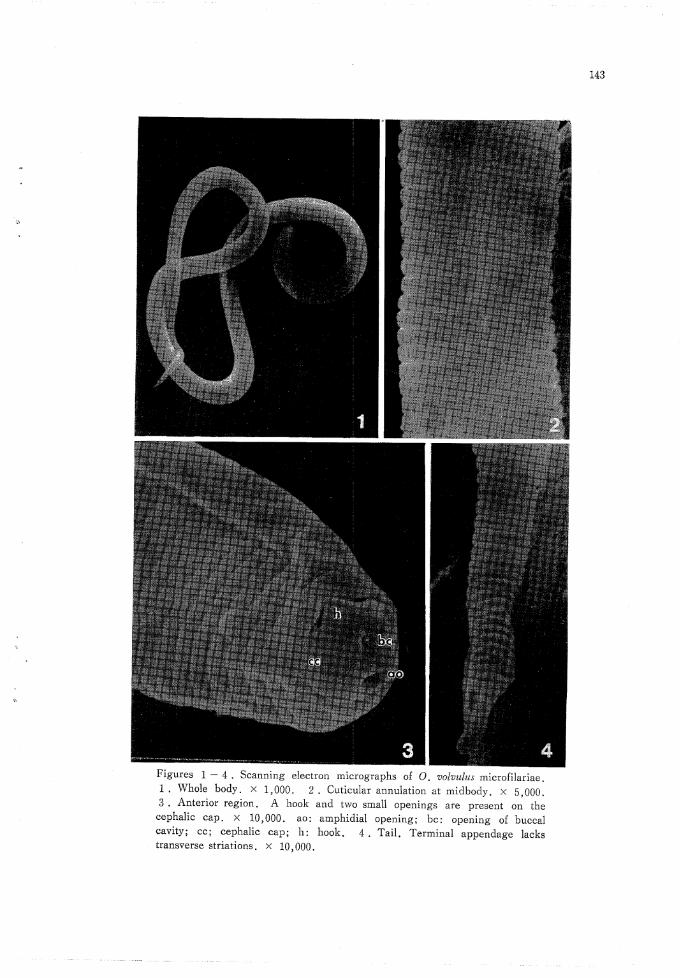
There are numerous transverse striations on the cuticular surface (Fig. 1). At the
midbody of microfilaria, the width of each annulation was 0.6-0.8^ (Fig. 2). The an-nulations numbered 325, 329, 334, 342 and 357;in 5 specimens which were positioned
for accurate observation. The anterior end of a microfilaria forms a round cephalic cap.
The cephalic cap bears a V-shaped hook, 0.5-0.6^ in length. There are two small
openings on the cephalic cap. One of them, 0.4-0.5/* in diameter, is present at the
site opposite to the hook and the other, 0.2jw in diameter, is at the center of the cap
(Fig. 3). The tail tapers gradually and ends in the terminal appendage, 2.0-2.5^ long,
which lacks the transverse striations (Fig. 4). Despite of the careful search, the open-
ings of the excretory and anal pores could not be observed,
143
à"
Figures 1 - 4. Scanning electron micrographs of O. volvulus microfilariae.1. Whole body. X 1,000. 2. Cuticular annulation at midbody. x 5,000.3. Anterior region. A hook and two small openings are present on the
cephalic cap. X 10,000. ao: amphidial opening; be: opening of buccalcavity; cc; cephalic cap; h: hook. 4. Tail. Terminal appendage lackstransverse striations. x 10,000.

144
DISCUSSION
The application of scanning electron microscopy for the differentiation of filarial
worms within the same genus have provided an additional basis for specific diagnosis
(Shoho and Uni, 1977 ; Wong and Brummer, 1978). Scanning electron microscopy, there-fore, has been used for the morphological studies on O. volvulus isolated from different
geographic regions (Martinez-Palomo and Martinez-Baez, 1977 ; Franz, 1980 ; Franz and
Schuz-Key, 1981).
Franz (1980) reported that there was no geographical difference in surface structure
between the adult worms isolated from Liberia, Upper Volta, Tanzania and Guatemala.
The present paper described the surface architecture of microfilariae of O. volvulus iso-
lated from the patients in Guatemala. Our results are comparable with the surface struc-
ture of microfilariae of O. volvulus from Mexico (Martinez-Palomo and Martinez-Baez,
1977) and from Liberia (Franz and Schulz-Key, 1981). The surface structure of Guate-malan microfilariae is identical with those of Mexican and Liberian microfilariae. How-
ever, the number of annulations of Mexican and Liberian microfilariae was not examined.
It might be interesting to compare the number of annulations of microfilariae isolated from
different geographic regions.The surface structures of microfilariae of Dirofilaria immitis, Brugia malayi and B.
pahangi have been published elsewhere (Aoki and Katamine, 1975; Aoki et a/., 1976).
B. malayi and B. pahangi microfilariae are sheathed. They differ markedly from micro-filariae of O. volvulus in having larger number of annulations, the spines on the cephalic
space, and the striations on the cuticular surface of the terminal appendage of the tail.D. immitis microfilariae are unsheathed. The cephalic cap has a hook and two small
openings. The tail has the appendage of 4--5j« long, which lacks the transverse stria-tions. The number of annulations ranges 296-321. It is interesting that 0. volvulus
microfilariae, as mentioned above, show striking resemblance to microfilariae of D. im-
mitis, despite of the fact that they inhabit the different tissues ; O. volvulus microfilariae
are present mainly in the skin, while D. immitis microfilariae are in the blood stream.O. volvulus microfilariae migrate the skin and invade frequently the various organs and
tissues other than the eyes. Sakamoto et al. (1983) reported that D. immitis microfilariae
can migrate the skin of a mouse and survive there for 3-4 weeks, when microfilariaewere inoculated subcutaneously. These findings may suggest a relation between the cu-
ticular structure and behaviour of microfilariae in the tissues, i.e., the unsheathed mi-
crofilariae with a hook on their cephalic cap probably migrate the tissues. It will be
interesting to study the surface structures of unsheathed microfilariae of other species ;
microfilariae of Dipetalonema streptocerca which inhabit the skin and those of D. perstans
which are in the blood stream.
We observed the two small openings on the cephalic cap of O. volvulus microfilaria.
The opening at the center of the cap probably corresponds to the opening of the buccal

145
cavity and the other is the opening of one of the amphidial channels, based on the trans-
mission electron microscopy of several species of microfilariae (Kozek, 1968, 1971 ; Mc-laren, 1969, 1972; Tongu, 1974; Martinez-palomo and Martinez-Baez, 1977). We failed
to observe the openings of anal and excretory pores, which were found on the microfilariae
of D. immitis (Aoki and Katamine, 1975). The anal pore and excretory pore might becovered by the dense plug as reported by Martinez-Palomo and Martinez-Baez (1977).
ACKNOWLEDGEMENT
The authors are indebted to Dr. H. A. Godoy B. of Servicio Nacional de Erradi-
cacion de la Malaria, Guatemala and his colleagues for their kind cooperation. This
study was supported by the Ministry of Public Health, Guatemala and the Japan Interna-tional Cooperation Agency, Japan.
REFERENCES
1) Aoki, Y. & Katamine, D. (1975): Scanning electron microscopic observations on Dirofilariaimmitis. Trop. Med. (Nagasaki), 17, 27-34.
2) Aoki, Y., Nakajima, Y. & Katamine, D. (1976): Studies on Malayan filariasis in Che-ju Is.,
Korea 3 Microfilarial surface architecture of Brugia malayi (Che-ju strain) in comparison with
that of Brugia pahangi. Japan. J. Trop. Med. Hyg., 4, 129-137.
3) Duke, B. O. L. (1967): Onchocerca-Simulium complexes. IV. Transmission of a variant of the
forest strain of Onchocerca volvulus. Ann. Trop. Med. Parasit., 61, 326-331.
4) Duke, B. O. L. (1970): Onchocerca-Simulium complexes. VI. Experimental studies on the trans-
mission of Venezuelan and West African strains of Onchocerca volvulus by Simulium metallicum
and S. exiguum in Venezuela. Ann. Trop. Med. Parasit., 64, 421-431.
5) Duke, B. O. L. (1976): Strains of Onchocerca volvulus and their pathogenicity. Tropenmed.Parasit. , 27 (Supplement l), 21-22.
6) Duke, B. O. L., Lewis, D. J. & Moore, P. J. (1966): Onchocerca-Simulium complexes I.
Transmission of forest and Sudan-savanna strains of Onchocerca volvulus from Cameroon by Simulium
damnosum from West African bioclimatic zones. Ann. Trop. Med. Parasit. , 60, 318-335.
7 ) Franz, M. (1980): Electron microscope study of the cuticle of male and female Onchocerca volvulus
from various geographic areas. Tropenmed. Parasit. , 31, 149-164.
8 ) Franz, M. & Schulz-Key, H. (1981): Scanning electron microscope studies on the anterior region
of the larvae of Onchocerca volvulus in the vector. Trans. Roy. Soc. Trop. Med. Hyg., 75,141-142.
9) Kozek, W. J. (1968): Unusual cilia in the microfilaria of Dirofilaria immitis. J. Parasit., 54,838-844.
10) Kozek, W. J. (1971): Ultrastructure of the microfilaria of Dirofilaria immitis. J. Parasit., 57,1052-1067.
ll) Laurence, B. R. & Simpson, M. G. (1968): Cephalic and pharyngeal structures in microfilariae

146
revealed by staining. J. Helminth., 42, 309-330.
12) DeLeon, J.R. & Duke, B. O. L. (1966): Experimental studies on the transmission of Guatemalan
and West African strains of Onchocerca volvulus by Simulium ochraceum, S. metallicum and S.
callidum. Trans. Roy. Soc. Trop. Med. Hyg., 60, 735-752.
13) Martinez-Palomo, A. & Martines-Baes, M. (1977): Ultrastructure of the microfilaria of Onchocerca
volvulus from Mexico. J. Parasit., 63, 1007-1018.
14) McLaren, D. J. (1969): Ciliary structures in the microfilaria of Loa loa. Trans. Roy. Soc. Trop.
Med. Hyg., 63, 290-291
15) McLaren, D. J. (1972): Ultrastructural studies on microfilariae (Nematoda: Filarioidea). Para-sitol., 65, 317-332.
16) Omar, M. S. (1978): Histochemical enzyme-staining patterns of Onchocerca volvulus microfilariae
and their occurrence in different onchocerciasis areas. Tropenmed. Parasit., 29, 462-472.
17) Omar, M. S., Prost, A. & Marshall, T. F. DE C. (1982): Histochemical enzyme variation in
Onchocerca volvulus microfilariae from rain-forest and Sudan-savanna areas of the onchocerciasis
control programme in West Africa. Bull. Wrld. Hlth, Organ., 60, 933-944.
18) Sakamoto, M., Kimura, E., Aoki, Y. & Nakajima, Y (1984): Subcutaneous and intraperitoneal
inoculation of Dirofilaria immitis microfilariae into mice. Jpn. J. Parasit., 33, 415-420.
19) Shoho, C. & Uni, S. (1977): Scanning electron microscopy (SEM) of some Setaria species(Filarioidea, Nematoda). Z. Parasitenkd., 53, 93-104.
20) Tongu, Y. (1974): Ultrastructural studies on the microfilaria of Brugia malayi. Acta Medica
Okayama, 28, 219-242.
21) Wong, M. M. & Brummer, M. E. G. (1978): Cuticular morphology offive species of Dirofilaria:
A scanning electron microscope study. J. Parasit., 64, 108-114.
グアテマラの回旋糸状虫仔虫の走査電子顕微鏡像
三浦,光政 坂本,信 青木,克己
Guillermo Zea F, Onofre Ochoa A
グアテマラで得た回旋糸状虫(Onchocerca volvulus)仔虫の体表微細構造を走査電子顕微鏡で
観察した.体表には多くの輪状溝が存在し,虫体は多くの体環状を示す.体環の数は325-357と
ほぼ一定である.頭端は半球状で,その一端より長さ0.5~0.6μの鉤が後方にのびる.また頭端
にはamphidial channelとbuccal cavityの開孔部と考えられる2つの小孔が存在する.尾部
では体環は徐々に小さくなり,尾端2.0~2.5μは棍棒状を呈し,その表面には輪状溝が存在しな
い.排泄孔,肛門孔の開口部は観察されなかった.オンコセルカ仔虫体表構造の虫体のstrainに
よる違いについて考察した.
熱帯医学 第27巻 第3号, 141-146見1985年9月