n-linked glycosylation and reticuloendotheliosis retrovirus envelope glycoprotein function
TRANSCRIPT

VIROLOGY 179, 648-657 (1990)
N-Linked Glycosylation and Reticuloendotheliosis Retrovirus Envelope Glycoprotein Function
ERIC L. DELWART AND ANTONITO T. PANGANIBAN’
McArdle Laboratory for Cancer Research, University of Wisconsin-Madison, Madison, Wisconsin 53706
Received April 12, 1990; accepted July 27, 1990
Different properties of the spleen necrosis virus (SNV) envelope glycoprotein were analyzed following biosynthesis in the presence of glycosylation inhibitors. Tunicamycin, which inhibits all asparagine N-linked glycosylation, prevented intracellular processing and translocation to the cell surface of the envelope protein. In contrast, castanospermine or deoxymannojirimycin, which block glycosidase trimming of the early high-mannose chains and subsequent complex type N-glycosylation, did not inhibit proteolytic cleavage or cellular translocation. The ability of unglycosylated and partially glycosylated envelope protein to bind the viral receptor was assayed using an infection interference assay. Tunicamycin abrogated SNV envelope glycoprotein-induced receptor interference, whereas the trimming glycosidase inhibitors had no effect on interference. Similarly, tunicamycin but not the glycosidase inhibitors reduced the titers of released virus 1 OO-fold. We conclude that carbohydrate trimming and complex N-glycosylation are not essential for envelope glycoprotein translocation, proteolytic cleavage, receptor binding, or infectivity, whereas cotranslational high-mannose N-glycosylation is essential for all of the SNV envelope glycoprotein properties tested. Syncytia forma- tion can be induced following transfection into Di 7 cells of an envelope glycoprotein expression plasmid. Unlike virus particle infectivity, cell fusion is strongly inhibited by the glycosidase inhibitors. o isso Academic press, IW.
INTRODUCTION
The envelope glycoprotein (env-gp) of spleen necro- sis virus (SNV), a member of the type C avian reticu- loendotheliosis virus group (REV) (Witter, 1984) shows a high degree of sequence similarity with the glycopro- teins of type D simian retroviruses (SRV) and other members of that interference group (Sommerfelt and Weiss, 1990). For example, when the amino acid se- quence of the SNV envelope gene (Kewalramani and Panganiban, unpublished data) is compared to that of the simian Mason-Pfizer retrovirus (MPMV) (Sonigo er a/., 1986) there is close alignment of potential N-glyco- sylation sites: seven of the eight SNV N-linked glycosyl- ation sites are found within three amino acids of MPMV N-glycosylation sites. Furthermore, 21 of the 23 cys- teine residues of the SNV env-gp are also found in the corresponding position in MPMV.
The mature, proteolytically processed SNV surface envelope glycoprotein is highly modified, owing almost half of its molecular weight to N-linked carbohydrate groups. Analogous to other systems N-linked glycosyl- ation begins concurrently with the translation of the viral envelope polypeptide chain into the endoplasmic reticulum (ER) (Hubbard and Ivatt, 1981; Pfeffer and Rothman, 1987). High-mannose sugar chains (Rob- bins et a/., 1977) are transferred to the canonical N-gly- cosylation acceptor sites [Asn-X-Ser(Thr)] on the na-
’ To whom requests for reprints should be addressed.
scent polypeptides (Rothman and Lodish, 1977; Glabe et al., 1980). The high-mannose N-linked glycans may then be trimmed during glycoprotein translocation to the cell surface by glycosidase enzymes residing in the ER and Golgi (Hubbard and Ivatt, 1981; Fuhrmann et a/., 1985). Glycosyl transferases then add different su- gar molecules to the correctly trimmed carbohydrate core, resulting in large, complex side groups with multi- ple possible sugar configurations (Fuhrmann et a/., 1985).
The importance of cotranslational glycosylation for the proper function of some polypeptides has been demonstrated using the drug tunicamycin, which pre- vents N-linked glycosylation by inhibiting the synthesis of the sugar donor group (Gibson et a/., 1980; Elbein, 1987). The effects of later post-translational glycosyla- tion modifications can be studied using transition state analogue inhibitors of the trimming glycosidases (Fuhr- mann et al., 1985). These inhibitors cause the carbo- hydrate side chains to remain in the untrimmed or par- tially trimmed high-mannose configurations which can be detected by their sensitivity to cleavage by endogly- cosidase H.
In a previous study evidence was presented that ex- pression of an SNV env-gp deletion mutant that is blocked in translocation in the ER could still confer re- sistance to superinfection (Delwart and Panganiban, 1989). The sugar groups of these mutant env-gp mole- cules were of the high-mannose, endo H-sensitive type, as expected of ER resident glycoproteins. This
0042-6822/90 53.00 Copyright 0 1990 by Academic Press, Inc. All rights of reproduction in any form reserved.
648
N-LINKED GLYCOSYLATION AND env-gp FUNCTION 649
observation led to the proposal that retrovirus-induced resistance to superinfection may be due to the com- plexing of the cellular viral receptor with partially N-gly- cosylated wt env-gp in the ER. In order to determine whether the normal envelope protein might similarly be able to interact with the receptor in the absence of complex glycosylation, several env-gp functions were examined following biosynthesis in the presence of various glycosylation inhibitors. In this communication results consistent with intracellular receptor binding are described, indicating that complex N-glycosylation is dispensable for wt SNV env-gp-receptor recognition. The role of cotranslational high-mannose glycosylation in other env-gp functions and the requirement for com- plex N-glycosylation in cells undergoing SNV env-gp- induced syncytia formation are also analyzed.
MATERIALS AND METHODS
Plasmids
pED75 is identical to pSW253 (Chen et al., 1981) (a provirus clone of Rev-A) except that it contains an SNV substitution between a unique Xhol site and an Avrll site adjacent to the polypurine tract. This fragment [de- rived from pPB101 (Bandyopadhyay and Temin, 1984)] encodes all of the SNV envelope sequence except three carboxy-terminal amino acids. pED75 encodes a Rev-A/SNV chimera virus, which is a better helper for JD2 14Hy than Rev-A (data not shown). pJD214Hy was kindly provided by I. Dougherty (Dougherty and Temin, 1986). To construct the env protein expression con- struct pED27 the Sacll-to-Sac1 envelope gene frag- ment of pPB101 was treated with Sl nuclease, and HindIll linkers were ligated. This fragment was inserted into the HindIll site of pRSVcat (Gorman et a/., 1982) to give pED23. The region of the DNA that encodes the 3’ untranslated portion of the mRNA was shortened by deleting a Pvull-to-Hpal fragment to give pED24. The M/&to-&t1 fragment carrying the Rous sarcoma virus long terminal repeat, envelope gene, and poly(A) addi- tion site from pED24 was treated with Klenow enzyme to generate a blunt-ended molecule and inserted into the similarly treatedxbal site of ~220.2 (kindly provided by Dr. B. Sugden) to give pED27. ~220.2 was derived from ~201 (Yates et a/., 1985) by insertion of a poly- linker sequence from pUC12 into the Narl site (J. L. Yates, personal communication). The orientation of the SNV env gene was the same as that of the EBNAl gene.
Cells, viruses, and assay for superinfection interference
The canine osteosarcoma cell line D17 (from the American Tissue Culture Collection, Rockville, MD),
SNV chronically infected D17 cells, and the previously described cell line 2.1 expressing the SNV env-gp were grown as previously described (Delwart and Pangani- ban, 1989). The REV stock was generated by cotrans- fecting chicken embryo fibroblast (CEF) by the poly- brene/dimethyl sulfoxide technique (Kawai and Nishi- zawa, 1984) with 5 pg of pJD214Hy and 0.5 pg of pED75. Culture supernatant was collected 5 days later and frozen in aliquots at -70”. The assay for superin- fection interference has been described previously (Delwart and Panganiban, 1989).
Glycosylation inhibitors
The same concentration of inhibitors was used in all experiments. Tunicamycin was from Sigma (T-7765) and was used at 1 pg/ml. Castanospermine and de- oxymannojirimycin (dMM) were both from Boehringer- Mannheim and used at 0.66 and 1 mn/l respectively. Castanospermine and deoxymannojirimycin toxicity was measured by treatment of D17 cells for 3 days followed by trypsin digestion and passage of treated cells into normal cell media. Cloning efficiency was not affected by pretreatment with the trimming glycosi- dase inhibitors under the conditions used here (data not shown).
lmmunofluorescence and immunoprecipitation
lmmunofluorescence and immunoprecipitation were performed as described previously (Delwart and Pan- ganiban, 1989). Surface immunofluorescence staining was done by fixing the cells on a glass coverslip with 3% paraformaldehyde (pH 7.4, preheated at 37’) for 10 min at room temperature. Antibody reaction and rinses were done in the presence of 100 mM glycine. The immunofluorescence signals seen in Figs. 2A-H are representative of the fluorescence seen in multiple mi- croscopic fields seen in two experiments. Little varia- tion is seen in the cellular distribution of the envelope protein between cells treated with the same inhibitor.
Syncytia formation
D17 cells were plated at a density of 1 O6 tells/60- mm-diameter plates on polystyrene tissue culture dishes marked with measuring grids. One day later 10 pg of pED27 or ~220.2 plasmid was introduced into cells by transfection using the polybrene/DMSO tech- nique (Kawai and Nishizawa, 1984). Trimming glycosi- dase inhibitors were added at the beginning of the 7-hr DNA/polybrene incubation period; after a 3.5-min 30% DMSO shock, media containing glycosylation inhibi- tors were added to the plates. Cells were incubated for 4 days and fixed with 0.5% violet blue in 50% metha- nol. Syncytia including at least four nuclei were counted by 156X magnification over an area of 2 cm’.

650 DELWART AND PANGANIBAN
A Deoxymann Castano Tunlcamycin ojlrlmycln spermine
A B A BA B Endo H + 1-l - I + I I
+, - 1 - ,+ +, - 1 - ,+
kibitor
A B
+ I- I-1 + kd
11 -22kd
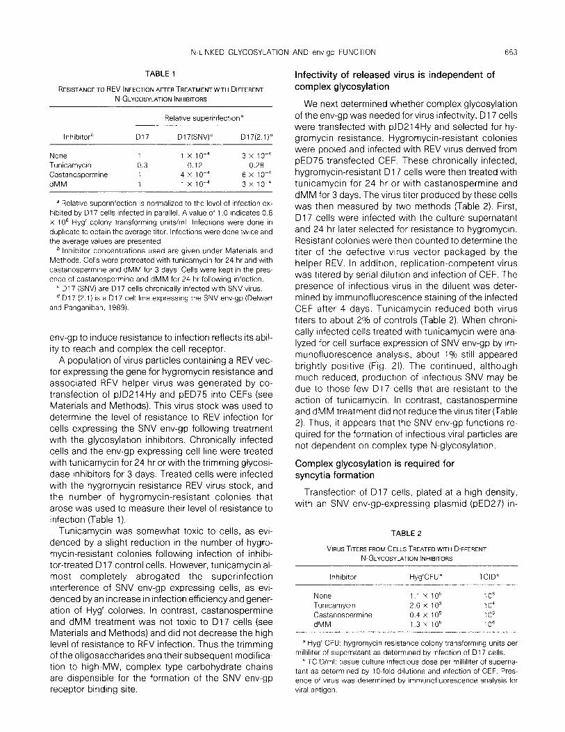
FIG. 1. Biosynthesis and immunoprecipitation of [35S]methionrne-labeled putative SNV SU and TM envelope proteins. Cells of the D17 2.1 cell line expressing SNV env-gp were treated with tunicamycin for 24 hr or castanospermine and deoxymannojirimycin for 3 days prior to the addition of label. Inhibitor concentrations are given under Materials and Methods. Radiolabeling was done in the continued presence of inhibitors. Lanes A: 1 O6 cells were labeled for 1 hr and immediately immunoprecipitated. Lanes B: Cells were labeled for 1 hr and chased with cold media for 4 hr before immunoprecipitation. Endo H: Endoglycosidase H digestion of immunoprecipitated samples was done by overnight incubation with 0.01 U. (A) Samples were boiled in loading buffer and run on a 7% polyactylamide gel. (B) Same as (A) but samples were run on a 10% polyactylamide gel for analysis of the TM protein. The same immunoprecipitated samples were loaded on gels (A) and (B). Only a small section of the autoradiograph is shown in (B) since the remainder was identical to that of (A).
The glycosidase inhibitors did not affect transfection efficiency as determined by the number of hygromycin- resistant D17 clones obtained after polybrene/DMSO transfection with pJD214Hy.
RESULTS
Complex N-glycosylation of the SNV envelope protein is not required for proteolytic cleavage
A D17 cell line expressing the SNV env-gp (clone 2.1) (Delwart and Panganiban, 1989) was pretreated with tunicamycin for 24 hr or with castanospermine and dMM for 3 days. In the continued presence of in- hibitors, cells were pulse-labeled for 1 hr with y5S]me- thionine and the label was then chased for 4 hr. Env-gp was immunoprecipitated using rabbit antisera raised against an Escherichia co/i synthesized envelope pro- tein antigen. In untreated cells the initial translation product after a 1 -hr label was the endo H-sensitive pre- cursor glycoprotein, gPr75 (Fig. 1A: no inhibitor A-,+) (Tsai et a/., 1986; Tsai and Orozslan, 1988; Delwart and Panganiban, 1989). After the 4-hr label chase the mature endo H-resistant surface gp90 (SU) and the transmembrane gp20 (TM) (Leis et al., 1988) could be detected by immunoprecipitation (Fig. 1A: no inhibitor B-,+, and Fig. 1B: no inhibitor). Glycoproteins gp90 and gp20 are the mature envelope product found on virus particles. Previous studies have shown that a short-lived, endo H-resistant gPr1 15 is the immediate precursor of gp90 and gp20. gPrl15 is derived from gPr75 following complex N-glycosylation and is endo H-resistant (Tsai and Orozslan, 1988; data not shown). The mature virion SU and TM glycoproteins are derived by proteolytic cleavage of gPr1 15 by a cellular endo- peptidase.
When all N-linked glycosylation was blocked with tunicamycin, an endo H-insensitive protein of the same MW as enzymatically deglycosylated gPr75 (Fig. 1A:
tunicamycin A-,+) was synthesized. No envelope- specific label was detected following the label chase, indicating that the stability of the unglycosylated enve- lope protein was reduced (Fig. 1A: tunicamycin B).
Treatment of D17 2.1 cells with castanospermine, an inhibitor of glucosidase I (Fuhrman et al., 1985) resulted in the biosynthesis of an endo H-sensitive env-gp with a MW larger than that synthesized in the same time period without the inhibitor (Fig. 1A: castan- ospermine A-). This larger MW presumably results from the inhibition of ER resident glucosidase I, which in untreated cells removes the terminal glucose resi- due from the initial high-mannose chains and renders other sugar residues accessible to further trimming. The MW of the initial translation product in cells treated with dMM was lower than that in cells treated with castanospermine (Fig. 1A: deoxymannojirimycin A-, castanospermine A-). This result is consistent with glucosidase trimming activity and the absence of sub- sequent complex glycosylation due to the inhibition of mannosidases with dMM.
Endo H deglycosylation of the major early translation products from untreated cells or from cells treated with either castanospermine or deoxymannojirimycin yielded products of the same MW as the unglycosyl- ated product synthesized in the presence of tunicamy- tin (Fig. 1A: tunicamycin A, no inhibitor A+, castano- spermine A+, deoxymannojirimycin A+). This result in- dicates that the differences in the major early translation products are due to different forms of endo H-sensitive high-mannose groups. The smaller “lad- der-like” bands are derived from endo H digestion of the chase products (Fig. 1 A: castanospermine B- and B+, deoxymannojirimycin B- and B+). Following the 4-hr label chase of castanospermine-treated cells, two envelope peptides of similar MW become more appar- ent (Fig. 1A: castanospermine B-). Like the normally synthesized gp90 (released by cleavage of gPr1 15)

N-LINKED GLYCOSYLATION AND env-gp FUNCTION 651
(Tsai and Orozslan, 1988) these two glycoproteins have a MW approximately 20 kDa smaller than the high-MW initial translation products synthesized in the presence of castanospermine. This change in MW could have resulted from the proteolytic cleavage of the untrimmed initial translation product to release the TM and SU. To determine whether TM proteins were generated in the presence of the glycosidase inhibi- tors, the immunoprecipitation products were exam- ined under gel conditions suitable for resolution of the TM peptides (Fig. 1 B). From castanospermine-treated cells a transmembrane glycoprotein doublet of approxi- mately 22 kDa could indeed be immunoprecipitated (Fig. 1 B: castanospermine). The env-gp chase pattern observed with dMM-treated cells was the same as that observed for castanospermine-treated cells. The ap- parent MWs of the SNV env-gps synthesized with dMM were smaller than those of the env-gps synthe- sized with castanospermine, as expected from the re- moval of the outer glucose residues from the high- mannose chains in the presence of dMM (Fig. 1A). The label chase patterns, the appearance of a major band approximately 20 kDa (the size of the TM) smaller than the major initial translation product, and the appear- ance of TM glycoproteins lead us to believe that pro- teolytic cleavage of SNV env-gp can take place in the absence of complex N-glycosylation.
After a labeling time of only 1 hr large levels of puta- tive SU and TM are detected in cells treated with glyco- sidase inhibitors, whereas at this time they are mostly absent from untreated cells (Fig. 1A: deoxymannojiri- mycin A-, castanospermine A-, no inhibitor, A-; Fig. 1 B: deoxymannojirimycin A, castanospermine A, no in- hibitor, A). This may indicate that translocation of the cleavage precursor to the cellular site of proteolytic processing takes place faster for the SNV env-gp-bear- ing high-mannose sugars than for the more heavily modified gPR1 15. It may also be that partially N-glyco- sylated env-gp is a better substrate for proteolytic cleavage.
The high-mannose chains found on the initial trans- lation products were all efficiently cleaved by endo H, yielding a single sharp band of the same MW (Fig. 1A: A+). This is unlike the endo H digestion pattern of the putative SU glycoprotein synthesized in the presence of trimming glycosidase inhibitors, which instead of yielding a single band produced a ladder-like series of bands (Fig. 1 A: deoxymannojirimycin B+, castano- spermine B+). Such a ladder-like series of glycopro- teins may represent modification of some of the un- trimmed (castanospermine) or partially trimmed (dMM) high-mannose chains that may occur during glycopro- tein translocation to the cell surface. Such aberrant oligosaccharide modification could account for the partial resistance to endo H digestion of the putative
SU. Nonetheless, the large reduction in the MW of the putative SNV SU proteins synthesized in the presence of either trimming glycosidase inhibitor (Fig. 1A: B-) versus the MW of gp90 attests to the efficiency of these inhibitors in preventing proper complex, high- MW N-glycosylation in D17 cells.
Cellular distribution of envelope glycoprotein in the presence of N-glycosylation inhibitors
Cells expressing SNV env-gp were treated with tuni- camycin for 24 hr or with castanospermine and dMM for 3 days. These cells were then analyzed by immuno- fluorescence labeling using anti-SNV envelope protein antibodies (Fig. 2). The distribution of fluorescence seen in permeabilized cells was largely unaffected by the trimming glycosidase inhibitors (Figs. 2A, E, G). As in untreated cells the env-gp antigen was found mostly in the Golgi and the plasma membrane (Delwar-t and Panganiban, 1989). In contrast, tunicamycin treatment resulted in the distribution of envelope protein in vesi- cles distributed through the cytoplasm with some con- centration in a Golgi-like perinuclear location (Fig. 2C).
We next examined the plasma membrane distribu- tion of the env-gp by immunofluorescence analysis of paraformaldehyde-fixed cells. Tunicamycin-treated cells were negative for envelope antigen on their sur- face (Fig. 2D) with the exception of a subpopulation comprising about 1% of the cells. These apparently tunicamycin-insensitive cells appeared brightly posi- tive (Fig. 21). Cells treated with the trimming glycosi- dase inhibitors were positive for surface env-gp but the fluorescence signal intensity was slightly weaker than that seen on untreated cells (Figs. 2B, F, H). This immu- nofluorescence analysis shows that tunicamycin treat- ment blocked the translocation of unglycosylated en- velope protein to the cell surface. On the other hand, blocking complex N-linked glycosylation did not abol- ish SNV env-gp translocation to the cell surface.
Complex glycosylation of the envelope glycoprotein is not required for superinfection interference
Chronically infected D17 cells and a cell line ex- pressing the SNV env-gp are resistant to infection by viruses of the same interference group (Delwat-t and Panganiban, 1989). This phenomenon presumably re- sults from the complexing of the cellular viral receptor with the endogenously synthesized SNV env-gp (Steck and Rubin, 1966a,b). This specific binding is thought to result in superinfection interference due to the reduc- tion of functional virus receptor on the cell surface. In this model the ability of endogenously synthesized

FIG. 2. lmmunofluorescence staining of the SNV envelope protein. Effect of glycosylation Inhibitors on cellular distribution of SNV envelope proteins and pED27-induced syncytium. (A-H) immunofluorescent staining of the 2.1 D17 cell line expressing SNV env-gp (magnification X586). Left-hand pictures and J show the staining pattern following permeabilization of cells with methanol; the right-hand prctures (except J) depict surface staining following paraformaldehyde membrane fixing. (A, B) No inhibitor; (C, D) tunrcamycin; (E, F) castanospermine; (G. H) dMM; (I) tunicamycin on cells chronically infected with SNV (magnification X 186); (J) D17 cell syncytrum induced by SNV env-gp expression.
652
N-LINKED GLYCOSYLATION AND env-gp FUNCTION 653
TABLE 1
RESISTANCE TO REV INFECTION AFTER TREATMENT WITH DIFFERENT N-GLYCOSYLATION INHIBITORS
Relative superinfectiona
Inhibitorb
None Tunicamycin Castanospermrne dMM
017 D17(SNV)’ D17(2.1)d
1 1 x 1o-4 3 x 1o-4 0.3 0.12 0.28 1 4 x 1om4 6 x 1O-4 1 1 x 1o-4 3 x 1o-4
’ Relatrve supennfection IS normalized to the level of infection ex- habited by D17 cells infected in parallel. A value of 1 .O indicates 0.8 X lo6 Hyg’ colony transforming units/ml. Infections were done in duplrcate to obtain the average titer. Infections were done twice and the average values are presented.
b Inhibitor concentrations used are given under Materials and Methods. Cells were pretreated wrth tunicamycin for 24 hr and with castanospermrne and dMM for 3 days. Cells were kept in the pres- ence of castanospermine and dMM for 24 hr following Infection.
’ D17 (SNV) are D17 cells chronically infected with SNV virus. d D17 (2.1) is a D17 cell line expressing the SNV env-gp (Delwart
and Panganrban, 1989).
env-gp to induce resistance to infection reflects its abil- ity to reach and complex the cell receptor.
A population of virus particles containing a REV vec- tor expressing the gene for hygromycin resistance and associated REV helper virus was generated by co- transfection of pJD214Hy and pED75 into CEFs (see Materials and Methods). This virus stock was used to determine the level of resistance to REV infection for cells expressing the SNV env-gp following treatment with the glycosylation inhibitors. Chronically infected cells and the env-gp expressing cell line were treated with tunicamycin for 24 hr or with the trimming glycosi- dase inhibitors for 3 days. Treated cells were infected with the hygromycin resistance REV virus stock, and the number of hygromycin-resistant colonies that arose was used to measure their level of resistance to infection (Table 1).
Tunicamycin was somewhat toxic to cells, as evi- denced by a slight reduction in the number of hygro- mycin-resistant colonies following infection of inhibi- tor-treated D17 control cells. However, tunicamycin al- most completely abrogated the superinfection interference of SNV env-gp expressing cells, as evi- denced by an increase in infection efficiency and gener- ation of Hyg’ colonies. In contrast, castanospermine and dMM treatment was not toxic to D17 cells (see Materials and Methods) and did not decrease the high level of resistance to REV infection. Thus the trimming of the oligosaccharides and their subsequent modifica- tion to high-MW, complex type carbohydrate chains are dispensible for the formation of the SNV env-gp receptor binding site.
infectivity of released virus is independent of complex glycosylation
We next determined whether complex glycosylation of the env-gp was needed for virus infectivity. D17 cells were transfected with pJD2 14Hy and selected for hy- gromycin resistance. Hygromycin-resistant colonies were pooled and infected with REV virus derived from pED75-transfected CEF. These chronically infected, hygromycin-resistant D17 cells were then treated with tunicamycin for 24 hr or with castanospermine and dMM for 3 days. The virus titer produced by these cells was then measured by two methods (Table 2). First, D17 cells were infected with the culture supernatant and 24 hr later selected for resistance to hygromycin. Resistant colonies were then counted to determine the titer of the defective virus vector packaged by the helper REV. In addition, replication-competent virus was titered by serial dilution and infection of CEF. The presence of infectious virus in the diluent was deter- mined by immunofluorescence staining of the infected CEF after 4 days. Tunicamycin reduced both virus titers to about 2% of controls (Table 2). When chroni- cally infected cells treated with tunicamycin were ana- lyzed for cell surface expression of SNV env-gp by im- munofluorescence analysis, about 1% still appeared brightly positive (Fig. 21). The continued, although much reduced, production of infectious SNV may be due to those few D17 cells that are resistant to the action of tunicamycin. In contrast, castanospermine and dMM treatment did not reduce the virus titer (Table 2). Thus, it appears that the SNV env-gp functions re- quired for the formation of infectious viral particles are not dependent on complex type N-glycosylation.
Complex glycosylation is required for syncytia formation
Transfection of D17 cells, plated at a high density, with an SNV env-gp-expressing plasmid (pED27) in-
TABLE 2
VIRUS TITERS FROM CELLS TREATED WITH DIFFERENT N-GLYCOSYLATION INHIBITORS
lnhrbrtor Hyg’CFU” TCIDb
None 1.1 x lo5 10” Tunicamycrn 2.6 x 10” 1 o4 Castanospermine 0.4 x lo5 10” dMM 1.3 x lo5 1 o6
a Hyg’ CFU: hygromycin resrstance colony transformrng units per milliliter of supernatant as determined by Infection of D 17 cells.
’ TCID/ml: tissue culture infectrous dose per mrlliliter of superna- tant as determrned by lo-fold dilutrons and infection of CEF. Pres- ence of vrrus was determined by tmmunofluorescence analysis for viral antrgen.

654 DELWART AND PANGANIBAN
TABLE 3
SYNCMIA FORMATION IN THE PRESENCE OF DIFFERENT N-GLYCOSYLATION INHIBITORS
SyncytiaB
Inhibitor*
None Castanospermine dMM control
Expt 1
193 4
20 3
Expt 2
83 16
2 3
Expt 3
112 15 11 10
a Syncytia formation described under Materials and Methods. ’ Inhibitor concentrations given under Materials and Methods.
duces the formation of multinucleated giant cells (Ta- ble 3). A syncytium labeled by immunofluorescence staining is shown in Fig. 2J. In a manner analogous to HIV-1 env-gp-induced cell fusion (Lifson et al., 1986; Sodroski et al., 1986) syncytia formation is presum- ably initiated by the intercellular recognition of the ret- roviral receptor by env-gp expressed on the surface of a neighboring cell. Such syncytia cannot be induced in preinfected cells or in cells already expressing SNV env-gp, as their viral receptors are already complexed by the endogenously synthesized env-gp (Delwart and Panganiban, 1989). Syncytia formation was attempted in the presence of the trimming glycosidase inhibitors. Tunicamycin was not tested because it proved too toxic over the time period required for the assay. Cas- tanospermine and dMM did not affect the transfection efficiency in D17 cells or cell survival during the 4 days required for the experiment (see Materials and Meth- ods). In the presence of these trimming inhibitors SNV env-gp-induced syncytia formation was strongly inhib- ited (Table 3). It therefore appears that SNV env-gp-in- duced cell fusion is dependent on proper complex N- glycosylation in cells participating in syncytia forma- tion.
DISCUSSION
N-linked oligosaccharides have different effects on glycoprotein function depending on the particular poly- peptide. Some glycoproteins are at least partially func- tional when N-glycosylation acceptor sites are re- moved by mutagenesis (Bedinger et a/., 1988) or when N-glycosylation is blocked by tunicamycin (Gibson et a/., 1980; Keating et a/., 1989). For other glycoproteins such as the influenza hemagglutinin, specific N-glyco- sylation sites can affect folding (Gething et al., 1986), antigenicity (Skehel et a/., 1984) and proteolytic cleav- age (Deshpande et a/., 1987). The primary role of N- glycosylation for many viral glycoproteins appears to be in assisting proper folding and in maintaining intra-
cellular solubility (Gibson et a/., 1980; Fennie and Lasky, 1989). Preventing N-glycosylation of various retroviral env-gps with tunicamycin can block their translocation to the cell surface (Bassin et al., 1982; Polonoff et a/., 1982; Rein eta/., 1982) reduce the yield of infectious virus (Schwarz et al., 1976) and abrogate superinfection interference (Bassin eta/., 1982; Rein et a/., 1982; Kai et a/., 1986).
Tunicamycin treatment reduced the stability of the SNV envelope protein and prevented its translocation to the cell surface. This could be due to increased pro- teolysis in the ER as a result of improper folding of the env-gp or because of a lack of protection from degrada- tive proteolysis by appended oligosaccharides. Im- proper oligomerization of the unglycosylated envelope protein may also, as for the influenza hemagglutinin (Gething et a/., 1986) prevent exit from the ER. Com- plexing of unglycosylated SNV envelope proteins with tunicamycin-induced, folding chaperone proteins in the ER could also prevent translocation to the cell sur- face (Flynn et al., 1989).
Superinfection interference is due to a block in retro- virus penetration (Steck and Rubin, 1966a,b) and is re- stricted to viruses belonging to the same immunologi- cal subgroup (Vogt and Ishizaki, 1966) implicating the viral env-gp of the preinfecting virus in the establish- ment of a receptor interference. Expression of REV retroviral env-gp is sufficient to induce receptor interfer- ence (Delwart and Panganiban, 1989; Federspiel eta/., 1989) and for HIV-1 env-gp expression results in down- regulation of the virus receptor from the cell surface (Stevenson et al., 1988). Superinfection interference prevents high levels of reinfection, a phenomenon as- sociated with cytopathic infections (Temin eta/., 1980; Mullins et a/., 1986; Temin, 1988).
The almost total abrogation of superinfection intet-fer- ence seen in chronically infected D17 cells following tunicamycin treatment indicates that the unglycosyl- ated SNV envelope protein can no longer sequester the cellular viral receptor. The susceptibility to REV in- fection induced by tunicamycin could be due to the inability of the unglycosylated envelope protein to translocate to the cell surface in order to complex the virus receptor at that location. However, since an SNV env-gp arrested in translocation within the ER still confers resistance to REV infection (Delwart and Pan- ganiban, 1989) and an intracellular form of the HIV-1 env-gp (gpl60) can be coimmunoprecipitated with the CD4 receptor (Hoxie et a/., 1986; Stevenson et a/., 1988) it is unlikely that the effect of tunicamycin on interference is caused simply by blocking env protein translocation. In further support of intracellular com- plexing a modified CD4 with an ER retention sequence was recently shown to bind the HIV-1 glycoprotein (Buonocore and Rose, 1990). Abrogation of interfer-
N-LINKED GLYCOSYLATION AND env-gp FUNCTION 655
ence by tunicamycin could also be due to the reduced half-life of the unglycosylated envelope protein. How- ever, direct binding studies have shown that the ungly- cosylated HIV-1 envelope protein synthesized in the presence of tunicamycin (Fennie and Lasky, 1989) or in E. co/i (Putney et al., 1986) could not bind the CD4 receptor. Similarly SNV envelope protein synthesized in E. co/i also did not block REV infection of D17 cells (data not shown). Therefore we favor the hypothesis that as for the HIV-l env-gp, EGF, and insulin receptors (Fennie and Lasky, 1989; Slieker et a/., 1986; Olson and Lane, 1987) the effect of tunicamycin on the SNV env protein is caused by the inability of the polypeptide synthesized in the absence of cotranslational glycosyl- ation to properly attain its ligand binding conformation.
The role of the trimming glycosidases in SNV replica- tion was investigated using inhibitors of glucosidase I and mannosidase I enzymes. Castanospermine in- hibits the removal of the distal glucose residue from the high-mannose group, Glc,Man,GlcNAc,, initially transferred to the growing polypeptides. dMM inhibits the mannosidase l-mediated trimming of the five outer mannose residues from the Man,GlcNAc, oligosaccha- ride side chains produced following glucosidase I and II trimming. In the presence of such inhibitors the label chase pattern of the SNV envelope gene products and the appearance of TM proteins indicate that correct proteolytic cleavage of env-gp is likely to be still taking place (Figs. 1A and B). The complex N-glycosylation normally found on the Rev-A cleavage precursor (Tsai and Orozslan, 1988) therefore appears dispensable for its processing by the host cell endopeptidase. Proteo- lytic cleavage of the HIV-l env-gp precursor synthe- sized in the presence of trimming inhibitors has also been demonstrated (Gruters et al., 1987; Montefiori et a/., 1988; Stein and Engleman, 1990). As with other retroviruses proteolytic cleavage is likely to be essen- tial for viral infectivity (Perez and Hunter, 1987; Freed and Risser, 1987; McCune et a/., 1988) by rendering accessible a highly hydrophobic peptide domain at the amino terminus of the TM glycoprotein required for membrane fusion. The normal infectivity of REV re- leased in the presence of the trimming inhibitors also indicates that correct proteolytic cleavage is likely to be taking place. As seen by immunofluorescence analy- sis, the ability of SNV glycoproteins bearing untrimmed oligosaccharides to translocate to the Golgi and the cell surface was not grossly affected, indicating that the high-MW complex form of the SNV env-gp carbo- hydrate chains is not essential as a signal for translo- cation.
As opposed to the effects seen with tunicamycin the resistance to REV infection of cells expressing SNV env-gp was not lowered by the trimming glycosidase inhibitors. Similarly the titers of REV released from in-
fected cells treated with the same inhibitors was not reduced. These results indicate that complex type oli- gosaccharide chain modification is dispensible for re- ceptor binding and viral infectivity in D17 cells. It is possible that the complex oligosaccharides found on SNV glycoproteins are necessary to assist in infecting other cell types or to escape proteolytic degradation and immune neutralization during infection of an ani- mal host (Huso et al., 1988).
Unlike SNV, HIV-l released in the presence of differ- ent trimming glycosidase inhibitors shows a reduction in tissue culture infectious dose per milliliter ranging from one to five orders of magnitude (Gruters et a/., 1987; Walker et al., 1987; Karpas et a/., 1988; Monte- fiori et al., 1988; Pal et al., 1989). Like the SNV env-gp, the HIV-1 env-gp bearing untrimmed glycans remains capable of binding its cellular receptor (Walker et al., 1987; Fennie and Lasky, 1989). This has led to the hypothesis that HIV-l infectivity is blocked when com- plex N-glycosylation is prevented by interfering with another env-gp function. The strong antisyncytial activ- ity of such inhibitors points toward a decrease in the HIV-l gpl20 membrane fusion activity (Gruters et al., 1987; Walker et al., 1987; Montefiori et a/., 1988; Pal et a/., 1989). Analogous to the influenza hemagglutinin, partially N-glycosylated HIV-1 env-gp may be unable to undergo a conformational change (Skehel et al., 1982; Wiley and Skehel, 1987) required to expose its hydro- phobic membrane fusion domain. Alternatively, in the absence of complex N-glycosylation the HIV-1 env-gp may be unable to bind a hypothetical fusion domain receptor.
Castanospermine and dMM also efficiently blocked SNV env-gp-induced syncytia formation. This block in syncytia formation was unexpected in light of the nor- mal infectivity of REV particles released from similarly treated cells. The pathway of SNV entry into D17 cells may occur through fusion of the virion’s membrane with the cell surface’s plasma membrane following re- ceptor binding (as is the case for some other retroviral systems, including the D-type simian retroviruses (McCune et al., 1990) and HIV-l (Stein et al., 1987)), rather than being induced by a low pH in the endocytic pathway (Marsh, 1984) since ammonium chloride and chloroquine did not affect the ability of D17 cells to be infected by SNV (data not shown). The mechanism of SNV env-gp-induced cell fusion is also apparently pH independent, as pED27-transfection-induced syncytia formation occurs at a physiological pH and lowering the medium’s pH does not induce syncytia in a culture of cells expressing SNV env-gp (data not shown). Syn- cytia inhibition was not due to a decrease in biosynthe- sis or proteolytic processing of the env-gp since the SU and TM proteolytic product levels are not decreased in treated cells (Figs. 1A and B). Syncitia inhibition may

656 DELWART AND PANGANIBAN
instead result from the slight decrease in the cell sur- face level of SNV env-gp in trimming glycosidase inhibi- tor-treated cells (Fig. 2). It may also be that other cell surface molecules need to be properly N-glycosylated for cells to come into close contact and fuse. As for HIV-1 a cell surface adhesion glycoprotein (Hildreth and Orentas, 1989; Valentin et a/., 1990) may play an auxiliary role in cell fusion. The inhibitory effect of trim- ming glycosidase on SNV env-gp-mediated cell-cell fusion while virus-cell membrane fusion required for virus entry remains apparently unaffected could reflect the lower density of viral glycoproteins on the cell sur- face than on the virion surface, where it is preferentially concentrated by interaction with viral core molecules during virus budding. The high viral glycoprotein con- centration on virions may therefore make virus entry less dependent on the full fusogenic potential of the SNV envelope glycoprotein than cell-to-cell fusion and this may account for the differential effects of trimming glycosidases inhibitors in the two assays. A direct ef- fect of the trimming inhibitors on the SNV receptor is also possible. The demonstration that the chimpanzee CD4 is unable to mediate cell-cell fusion with HIV-1 -in- fected human cells while still able to support HIV-1 infection (Camerini and Seed, 1990) may similarly re- flect different receptor requirements for the two kinds of membrane fusion events.
ACKNOWLEDGMENTS
We thank Kevin Schultz and Howard Temin for critical review of this manuscript and Betty Sheehan for proofreading. This work was supported by Research Grant CA22443-10 from the NIH, support from the Shaw Scholars Fund, and a junior faculty research award (JFRA-147) from the ACS.
REFERENCES
BANDYOPADHYAY, P. K., and TEMIN. H. M. (1984). Expression from an internal AUG codon of herpes simplex thymidine kinase gene in- serted in a retrovirus vector. Mol. Cell. Biol. 4, 743-748.
BASSIN, R. H., RUSCETI, S., ALI, I., HAAPALA, D. K., and REINS, A. (1982). Normal DBA/2 mouse cells synthesize a glycoprotein which interferes with MCF virus infection. Virology 123, 139-l 51,
BEDINGER, P., MORIARTY, A., VON BORSTEL, R. C., II, DONOVAN, N. J., STEIMER, K. S., and LITMAN, D. R. (1988). Internalization of the human immunodeficiency virus does not require the cytoplasmic domain of CD4. Nature (London) 334, 162-l 65.
BUONOCORE, L., and ROSE, J. K. (1990). Prevention of HIV-1 glycopro- tein transport by soluble CD4 retained in the endoplasmic reticu- lum. Nature (London) 345, 625-628.
CAMERINI, D., and SEED, B. (1990). A CD4 domain important for HIV-1 mediated syncytium formation lies outside the virus binding site. Cell 60, 747-754.
CHEN. I. S. Y., MAK, T. W., O’REAR, 1. 1.. and TEMIN, H. M. (1981). Characterization of reticuloendotheliosis virus strain T DNA and isolation of a novel variant of reticuloendotheliosis virus strain T by molecular cloning. J. Viral. 40, 800-81 1.
DELWART, E. L., and PANGANIBAN, A. T. (1989). Role of reticuloendo- theliosis virus envelope glycoprotein in superinfection interfer- ence. J. Virol. 63, 273-280.
DESHPANDE, K. L., FRIED, V. A., ANDO, M., and WEBSTER. R. G. (1987). Glycosylation affects cleavage of an H5N2 influenza virus hemag- glutinin and regulates virulence. Proc. Nat/. Acad. Sci. USA 84, 36-40.
DOUGHERTY, J. P., and TEMIN, H. M. (1986). High mutation rate of a spleen necrosis virus-based retrovirus vector. Mol. Cell. Biol. 6, 4387-4395.
ELBEIN, A. D. (1987). Inhibitors of the biosynthesis and processing of N-linked oligosaccharide chains. Annu. Rev. Biochem. 56, 497- 534.
FEDERSPIEL, M. J., CRIITENDEN, L. B., and HUGHES, S. H. (1989). Ex- pression of avian reticuloendotheliosis virus envelope confers host resistance. Virology 173, 167-l 77.
FENNIE. C., and LASKY, L. A. (1989). Model for intracellular folding of the human immunodeficiency virus type 1 gp120. J. Viral. 63, 639-646.
FLYNN, G. C., CHAPPELL, T. G., and ROTHMAN, J. E. (1989). Peptide binding and release by proteins implicated as catalysts of protein assembly. Science 245, 385-390.
FREED, E. 0.. and RISSER, R. (1987). The role of envelope glycoprotein processing in murine leukemia virus infection. /. Viral. 61, 2852- 2856.
FUHRMANN, U., BAUSE, E., and PLOEGH, H. (1985). Inhibitors of oligo- saccharide processing. Biochim. Biophys. Acta 825, 95-1 10.
GETHING, M.-J., MCCAMMON, K., and SAMBROOK. J. (1986). Expression of wild-type and mutant forms of influenza hemagglutinin: The role of folding in intracellular transport. Cell 46, 939-950.
GIBSON, R., KORNFELD, S., and SCHLESINGER, S. (1980). A roleforoligo- saccharides in glycoprotein biosynthesis. Trends Biochem. Sci. 5, 290-293.
GLABE, C. G., HANOVER, 1. A., and LENNARZ, W. J. (1980). Glycosyla- tion of ovalbumin nascent chains. J. Biol. Chem. 255, 9236-9242.
GORMAN, C. M., MERLINO, G. T., WILLINGHAM, M. C., PASTAN, I., and HOWARD, B. H. (1982). The Rous sarcoma virus long terminal re- peat is a strong promoter when introduced into a variety of eukar- yotic cells by DNA-mediated transfection. Proc. Nat/. Acad. SC;. USA 79, 6777-6781.
GRUTERS. R. A., NEEFJES, J. J., TERSMEITE, M., DE GOEDE, R. E. Y., TULP, A., HUISMAN, H. G., MIEDEMA, F., and PLOEGH, H. L. (1987). Interference with HIV-induced syncytium formation and viral infec- tivity by inhibitors of trimming glucosidase. Nature (London) 330, 74-77.
HILDRETH, J. E. K., and ORENTAS. R. J. (1989). Involvement of a leuko- cyte adhesion receptor (LFA-1) in HIV-induced syncytium forma- tion. Science 244, 1075-l 078.
HOXIE, J. A., ALPERS, 1. D., RACKOWSKI, J. L., HUEBNER. K., HAGGARPI, B. S., CEDARBAUM, A. J., and REED, J. C. (1986). Alterations in T4 (CD4) protein and mRNA synthesis in cells infected with HIV. Science 234, 1123-l 127.
HUBBARD, S. C., and IVATT, R. 1. (1981). Synthesis and processing of asparagine-linked oligosaccharides. Annu. Rev. Biochem. 50, 555-583.
Huso, D. L., NARAYAN, O., and HART, G. W. (1988). Sialic acids on the surface of caprine arthritis-encephalitis virus define the biological properties of the virus. J. Viral. 62, 1974-l 980.
K~I, K., SATO, H., and ODAKA, T. (1986). Relationship between the cellular resistance to Friend murine leukemia virus infection and the expression of murine leukemia virus-gp70-related glycoprotein on cell surface of BALBlc-Fv-4w’ mice. Virology 150, 509-5 12.
KARPAS, A., FLEET, G. W. J., DWEK, R. A., PETURSSON, S., NAMGOONG, S. K., RAMSDEN, N. G., JACOB, G. S.. and RADEMACHER, T. W. (1988). Aminosugar derivatives as potential anti-human immunodefi- ciency virus agents. Proc. Nat/. Acad. Sci. USA 85, 9229-9233.
KAWAI. S.. and NISHI~AWA, M. (1984). New procedure for DNA trans- fection with polycation and dimethyl sulfoxide. Mol. Cell. Biol. 4, 1172-1174.
N-LINKED GLYCOSYLATION AND env-gp FUNCTION 657
KEATING, M. T., HARRYMAN, C. C., and WILLIAMS, L. T. (1989). Platelet- derived growth factor receptor inducibility is acquired immediately after translation and does not require glycosylation. J. Biol. Chem. 264, 9129-9132.
LEIS, J., BALTIMORE, D., BISHOP, J. M.. COFFIN, J., FLEISSNER, E., GOFF, S. P., OROSZLAN, S., ROBINSON, H., SKALKA, A. M., TEMIN, H. M., and VOGT. V. (1988). Standardized and simplified nomenclature for proteins common to all retrovlruses. J. Viral. 62, 1808-l 809.
LIFSON, 1. D., REYES, G. R.. MCGRATH, M. S., STEIN. B. S., and ENGLE- MAN, E. G. (1986). AIDS retrovirus Induced cytopathology: Giant cell formation and involvement of CD4 antigen. Science 232, 1123-1127.
MARSH, M. (1984). The entry of enveloped viruses into cells by endo- cytosis. Biochem. J. 218, l-10.
MCCUNE, J. M., RABIN. L. B., FEINBERG. M. B., LIEBERMAN, M., KOSEK. 1. C., REYES, G. R., and WEISSMAN, I. L. (1988). Endoproteolytic cleavage of gpl60 is required for the activation of human immuno- deficiency virus. Cell 53, 55-67.
MONTEFIORI. D. C., ROBINSON, W. E., JR., and MITCHELL, W. M. (1988). Role of protein N-glycosylatlon in pathogenesis of human immuno- deficiency virus type 1. Proc. Nat/. Acad. Sci. USA 859248-9252.
MULLINS, 1. I., CHEN. C. S., and HOOVER, E. A. (1986). Disease-speci- fic and tissue specific production of unintegrated feline leukaemia virus variant DNA in feline AIDS. Nature (London) 319, 333-336.
OLSON, T. S., and LANE, M. D. (1987). Post-translational acquisition of insulin binding activity by the insulin proreceptor. J. Biol. Chem. 262, 6816-6822.
PAL, R., HOKE, G. M., and SARNGADHARAN, M. G. (1989). Role of oligo- saccharides in the processing and maturation of envelope glyco- proteins of human lmmunodeflciency virus type 1. Proc. Narl. Acad. Sci. USA 86, 3384-3388.
PEREZ, L. G.. and HUNTER. E. (1987). Mutations within the proteolytlc cleavage site of the Rous sarcoma virus glycoprotein that block processing to gp85 and gp37. J. viral. 61, 1609-l 614.
PFEFFER, S. R., and ROTHMAN, 1. E. (1987). Biosynthetic protein trans- port and sorting by the endoplasmic reticulum and Golgi. Annu. Rev. Biochem. 56, 829-852.
POLONOFF, E.. MACHIDA, C. A., and KASAT, D. (1982). Glycosylation and intracellular transport of membrane glycoprotelns encoded by murine leukemia viruses. J. Biol. Chem. 257, 14,023-14,028.
PUTNEY, S. D., MATHEWS, T. J., ROBEY. W. G.. LYNN, D. L., ROBERT- GUROFF, M., MUELLER, W. T.. LANGLOIS, A. J., GHRAYEB, J., PETE- WAY, S. R., JR., WEINHOLD. K. J., FISCHINGER. P. 1.. WONG-STAAL, F., GALLO, R. C., and BOLOGNESI, D. P. (1986). HTLV-III/LAV-neutraIIz- ing antibodies to an E. co/i-produced fragment of the virus enve- lope. Science 234, 1392-1395.
REIN, A., SCHULTZ, A. M., BADER, J. P., and BASSIN, R. H. (1982). Inhibitors of glycosylation reverse retroviral Interference. Virology 119, 185-192.
ROBBINS. P. W., HUBBARD, S. C., TURCO, S. J.. and WIRTH, D. F. (1977). Proposal for a common ollgosaccharide intermediate in the syn- thesis of membrane glycoproteins. Cell 12, 893-900.
ROTHMAN, E. J., and LODISH, H. F. (1977). Synchronised transmem- brane insertion and glycosylation of a nascent membrane protein. Nature (London) 269, 775-780.
SCHWARZ, R. T., ROHRSCHNEIDER, J. M., and SCHMIDT, M. F. G. (1976). Suppression of glycoprotein formation of Semliki Forest, influenza, and avlan sarcoma virus by tunicamycin. J. viral. 19, 782-791.
SKEHEL, 1. J., BAYLEY, P. M., BROWN, E. B., MARTIN. S. R., WATERFIELD, M. D., WHITE, 1. M., WILSON, I. A., and WILEY, D. C. (1982). Changes In the conformation of Influenza virus hemagglutinin at the pH optimum of vlrus-medlated membrane fusion. Proc. Nat/. Acad. Sci. USA 79, 968-972.
SKEHEL, J. J., STEVENS. D. J., DANIELS, R. S., DOUGLAS, A. R., KNOSSOW, M., WILSON, I. A., and WILEY, D. C. (1984). A carbohydrate side
chain on hemagglutinins of Hong Kong influenza viruses inhibits recognition by a monoclonal antibody. Proc. Nat/. Acad. Sci. USA 81, 1779-1783.
SLIEKER, L. J., MARTENSEN, T. M., and LANE, M. D. (1986). Synthesis of epidermal growth factor receptor in human A431 cells. J. Biol. Chem. 261, 15,233-l 5,241.
SODROSKI, J.. GOH, W. C., CRAIG, C., CAMPBELL, K., and HASELTINE, W. A. (1986). Role of the HTLV-III/LAV envelope in syncytium for- mation and cytopathlcity. Nature (London) 322, 470-474.
SOMMERFELT, M. A., and WEISS, R. A. (1990). Receptor interference groups of 20 retrovlruses plating on human cells. Virology 176, 58-69.
SONIGO, P., BARKER, C.. HUNTER, E., and WAIN-HOBSON, S. (1986). Nucleotide sequence of Mason-Pfizer monkey virus: An immuno- suppressive D-type retrovirus. Cell 45, 375-385.
STECK. F. T., and RUBIN, H. (1966a). The mechanism of interference between an avian leukosis virus and Rous sarcoma virus. I. Estab- lishment of Interference. virology 29, 628-643.
STECK, F. T., and RUBIN, H. (1966b). The mechanism of interference between an avian leukosis virus and Rous sarcoma virus. II. Early steps of Infection by RSV of cells under conditions of interference. Virology 29, 642-653.
STEIN, B. S., GOWDA, S. D., LIFSON, 1. D., PENHALLOW, R. C., BENSCH, K. G., and ENGLEMAN, E. G. (1987). pH-independent HIV entry into CD4-positive cells via virus envelope fusion to the plasma mem- brane. Cell 49, 659-668.
STEIN, B. S., and ENGLEMAN, E. G. (1990). Intracellular processing of the gpl60 HIV-l envelope precursor. /. Biol. Chem. 265, 2640- 2649
STEVENSON, M., MEIER, C., MANN, A. M., CHAPMAN, N., and WASIAK, A. (1988). Envelope glycoprotein of HIV induces interference and cytolysis resistance in CD4 positive cells: Mechanism for persls- tence in AIDS. Cell 53, 483-496.
TEMIN, H. M. (1988). Mechanisms of cell killlng/cytopathic effects by retroviruses. Rev. Infect. Es. 10, 399-405.
TEMIN, H. M., KESHET, E., and WELLER, S. K. (1980). Correlation of transient accumulation of linear unintegrated viral DNA and tran- sient cell killing by avian leukosis and retlculoendotheliosis VI- ruses. CSH Symp. Quant. Biol. 44, 773-778.
TSAI, W.-P., COPELAND, T. D., and OROSZLAN, S. (1986). Biosynthesis and chemical and Immunological characterization of avian reticu- loendotheliosis virus env gene-encoded proteins. Virology 155, 567-583.
TSAI, W.-P., and OROSZ~AN, S. (1988). Novel glycosylation pathways of retrovlral envelope proteins identified with avian reticuloendo- theliosis virus. J. Vkol. 62, 3167-3174.
VALENTIN, A., LUNDIN. K., PATARROYO, M., and ASJO. B. (1990). The leukocyte adhesion glycoproteln CD1 8 participates in HIV-1 -In- duced syncytia formation in monocytoid and T cells. J. lmmunol- ogy 144, 934-937.
VOGT, P. K., and ISHIZAKI, R. (1966). Patterns of viral interference In the avian leukosls and sarcoma complex. Virology 30, 368-374.
WALKER, 8. D., KOWALSKI. M., GOH, W. C., KOZARSKY, K., KRIEGER, M., ROSEN, C., ROHRSCHNEIDER, L.. HASELTINE, W., and SODROSKI, 1. (1987). InhibItIon of human lmmunodeflciency virus syncytium for- mation and virus replication by castanospermine. Proc. Nat/. Acad. So. USA 84, 8120-8124.
WILEY, D. C., and SKEHEL, J. 1. (1987). The structure and function of the hemagglutinln membrane glycoproteln of influenza virus. Annu. Rev. Blochem. 56, 365-394.
WITTER, R. L. (1984). In “Diseases of Poultry” (M. S. Hofstrad, H. J. Barnes, B. W. Calnek, W. M. Reid, and H. W. Yoder, Jr.. Eds.), pp. 406-417. Iowa State Univ. Press, Ames.
YATES, J. L., WARREN, N., and SUGDEN, B. (1985). Stable replication of plasmids derived from Epstein-Barr virus in various mammalian cells. Nature (London) 313, 8 12-8 15