mechanisms of pancreatic beta cell dysfunction in diabetes
TRANSCRIPT

Wayne State University
Wayne State University Dissertations
1-1-2016
Mechanisms Of Pancreatic Beta Cell DysfunctionIn DiabetesVaibhav SidaralaWayne State University,
Follow this and additional works at: https://digitalcommons.wayne.edu/oa_dissertations
Part of the Medicinal Chemistry and Pharmaceutics Commons
This Open Access Dissertation is brought to you for free and open access by DigitalCommons@WayneState. It has been accepted for inclusion inWayne State University Dissertations by an authorized administrator of DigitalCommons@WayneState.
Recommended CitationSidarala, Vaibhav, "Mechanisms Of Pancreatic Beta Cell Dysfunction In Diabetes" (2016). Wayne State University Dissertations. 1591.https://digitalcommons.wayne.edu/oa_dissertations/1591

MECHANISMS OF PANCREATIC BETA CELL DYSFUNCTION IN DIABETES
by
VAIBHAV SIDARALA
DISSERTATION
Submitted to the Graduate School
of Wayne State University,
Detroit, Michigan
in partial fulfillment of the requirements
for the degree of
DOCTOR OF PHILOSOPHY
2016
MAJOR: PHARMACEUTICAL SCIENCES
Approved By:
________________________________
Advisor Date
________________________________
________________________________
________________________________
ii
DEDICATION
This work is dedicated to my parents, Dr. Narasimha Rao Sidarala and
Dr. Meera Bai Sidarala; and my brother, Vasishth Sidarala.

iii
ACKNOWLEDGEMENTS
Firstly, I would like to express my sincere gratitude to my academic advisor, Dr.
Anjaneyulu Kowluru, who has been a constant source of inspiration, for guiding me during my
graduate studies. His teachings have had a huge positive impact on my attitude, and will
continue to motivate me, as I move forward in my career. I am truly grateful for being a part of
Dr. Kowluru’s laboratory, and for the opportunity to be trained under his guidance.
I am also grateful for being so fortunate to have worked with previous and present
members of Dr. Kowluru’s research laboratory, Dr. Rajakrishnan Veluthakal, Dr. Khadija Syeda,
Dr. Abiy Mussa Mohammed, Dr. Daleep Arora, Dr. Anil Chekuri and Dr. Anil Poudel. I am
thankful for their support during my PhD, and for reminding me that “Teamwork makes the
dream work”.
I am thankful to Dr. Fei Chen, Dr. Randall Commissaris and Dr. Timothy Hadden, for
making time and being on my dissertation committee. Our discussions have aided me in
critically analyzing scientific data and develop the skills needed for effectively presenting my
research findings. I am also very thankful to the Department of Pharmaceutical Sciences at
Wayne State University. A special thanks to Dr. George Corcoran, for his ever so encouraging
support.
I also thank my friends and family for their immense support, without which I would not
have made it this far. I am truly grateful to my parents for constantly reminding me that every
milestone we reach, marks the beginning of a new endeavor. My sincere gratitude to everyone
who has been a part of this incredible and enlightening journey.
iv
TABLE OF CONTENTS
Dedication ................................................................................................................................ ii
Acknowledgements ................................................................................................................. iii
List of Tables ........................................................................................................................... v
List of Figures ......................................................................................................................... vi
List of Abbreviations ............................................................................................................... viii
Chapter 1: Introduction ............................................................................................................. 1
Diabetes ................................................................................................................. 5
GTP-binding proteins .............................................................................................. 7
Stress-Activated protein kinases/Mitogen-Activated protein kinases (SAPKs/MAPKs) ................................................................................................... 14
p53 tumor suppressor ........................................................................................... 19
Hypothesis ............................................................................................................ 25
Chapter 2: Materials and Methods ......................................................................................... 28
Chapter 3: Glucotoxic Conditions Promote Rac1-Nox2-Induced Activation Of p38MAPK in Pancreatic β-cells ................................................................................................. 33
Exposure of INS-1 832/13 cells to glucotoxic conditions results in cell death ........ 35
gp91-ds-tat, an inhibitor of Nox2, markedly prevents Nox2 activation, ROS generation and p38MAPK phosphorylation under glucotoxic conditions ............... 36
Glucotoxic conditions promote phosphorylation of p38MAPK, which is dependent on Tiam1- and Vav2-mediated Rac1 activation ................................... 39
EHT1864, a novel inhibitor of Rac1, blocks activation and membrane association of Rac1 activation and insulin secretion upon physiological glucose stimulation in INS-1 832/13 β-cells ........................................................................ 41
Glucotoxicity-induced p38MAPK activation is significantly blocked by EHT1864 .. 45
2-bromopalmitate, an inhibitor of protein palmitoylation, markedly attenuates p38MAPK phosphorylation ................................................................................... 47
GGTI-2147, an inhibitor of protein geranylgeranylation, had no effect on p38MAPK phosphorylation ................................................................................... 48

v
Chapter 4: Glucotoxic Conditions Promote Rac1-p38MAPK-Dependent Activation Of p53 Tumor Suppressor. ............................................................................................... 50
p53 tumor suppressor is activated by serine-15 phosphorylation in INS-1 832/13 cells and rat islets exposed to glucotoxic conditions ............................................. 51
HG-induced p53 activation is significantly blocked in the presence of EHT1864, Simvastatin and GGTI2147 ................................................................................... 52
EHT1864 had no effect on HG-induced nuclear translocation of p53 .................... 54
p53 activation under HG conditions is markedly blocked in the presence of SB203580, a selective inhibitor of p38MAPK ........................................................ 56
ATM kinase, a known activator of p53, is also activated under HG conditions in a Rac1-dependent manner ................................................................................... 57
KU-55933, a selective inhibitor of ATM kinase, blocks HG-induced ATM kinase activation but has no effect on p53 activation ........................................................ 58
EHT1864, markedly blocks HG-induced β-cell death ............................................ 59
Chapter 5: Rac1-p38MAPK-p53 Signaling Axis in Pancreatic Islets from the ZDF Rat Model and Human Islets… ...................................................................................... ….…62
p38MAPK and p53 are activated in islets isolated from ZDF rats .......................... 63
Exposure of normal human islets to glucotoxic conditions activates p38MAPK and p53 ................................................................................................................ 65
Exposure to glucotoxic conditions promote nuclear localization of p53 in human islets ..................................................................................................................... 66
Chapter 6: Discussion ............................................................................................................ 68
Chapter 7: Conclusions and Future Directions ....................................................................... 78
Appendix A Sidarala et al. 2015…………………………………………...………… ..... ………….82
Appendix B Sidarala et al. 2015……………………………………...……… .... ………………….90
References ............................................................................................................................ 99
Abstract................................................................................................................................ 121
Autobiographical Statement ................................................................................................. 123

vi
LIST OF TABLES
Table 1-1: List of pharmacological inhibitors utilized to target Rac1, Nox2 and p38MAPK ..... 26
Table 6-1: Summary of effects of pharmacological inhibitors on HG-induced p38MAPK and p53 activation................................................................................................. 77
vii
LIST OF FIGURES
Figure 1-1: The pancreatic islet ................................................................................................ 2
Figure 1-2: The Insulin receptor ............................................................................................... 3
Figure 1-3: Glucose-stimulated insulin secretion ...................................................................... 4
Figure 1-4: Prevalence of Diabetes .......................................................................................... 6
Figure 1-5: Effects of acute and chronic exposure to glucose concentrations on the pancreatic β-cell ..................................................................................................... 7
Figure 1-6: Rac1 activation cycle ............................................................................................. 8
Figure 1-7: Pharmacological inhibitors of Rac1 ...................................................................... 10
Figure 1-8: Functinal assembly and activation of NADPH oxidase 2 ...................................... 13
Figure 1-9: The conventional MAPK signaling pathways ........................................................ 15
Figure 1-10: The structural and functional domains of the p53 tumor suppressor ................... 20
Figure 1-11: Several post-translational modifications induced by regulatory factors that modulate p53 function ........................................................................................ 22
Figure 1-12: Mechanisms of p53-induced apoptosis .............................................................. 24
Figure 1-13: Proposed working model for Rac1-Nox2-induced oxidative stress and activation of p38MAPK-p53 signaling pathway in pancreatic β-cells under glucotoxic conditions ............................................................................................................ 26
Figure 3-1: Exposure of INS-1 832/13 cells to glucotoxic conditions induces cell death ......... 35
Figure 3-2: gp91-ds-tat peptide a specific inhibitor of Nox2, but not its inactive analog, inhibits HG-induced Nox2 activation, ROS generation and p38MAPK phosphorylation in INS-1 832/13 cells ................................................................... 37
Figure 3-3: NSC23766 and Ehop-016, which selectively block Tiam1- and Vav2-mediated Rac1 activation, prevent HG-induced p38MAPK phosphorylation in INS-1 832/13 cells and rat pancreatic islets .......................................................... 40
Figure 3-4: EHT 1864 significantly inhibits glucose-induced Rac1 activation, insulin secretion and membrane association of Rac1 in INS-1 832/13 cells ..................... 43
Figure 3-5: EHT1864 attenuates HG-induced p38MAPK phosphorylation in INS-1 832/13 cells ...................................................................................................................... 46
Figure 3-6: 2-bromopalmitate, an inhibitor of protein palmitoylation, prevents HG-induced p38MAPK phosphorylation ................................................................................... 47
viii
Figure 3-7: GGTI-2147, an inhibitor of protein geranylgeranylation, exhibits no effect on HG-induced p38MAPK phosphorylation ................................................................ 48
Figure 3-8: Proposed model for Rac1-Nox2-mediated ROS generation and p38MAPK activation under glucotoxic conditions ................................................................... 49
Figure 4-1: Exposure of INS-1 832/13 cells and rodent pancreatic islets to glucotoxic conditions results in p53 phosphorylation ............................................................. 51
Figure 4-2: Glucotoxicity-induced p53 phosphorylation is attenuated in presence of EHT1864, Simvastatin and GGTI2147 .................................................................. 53
Figure 4-3: Exposure to HG conditions induced nuclear translocation of p53, which is not inhibited by EHT1864 ........................................................................................... 55
Figure 4-4: SB203580, a selective inhibitor of p38MAPK, attenuates HG-induced p53 phosphorylation .................................................................................................... 56
Figure 4-5: Exposure to HG conditions induces ATM kinase phosphorylation, which is inhibited by EHT1864 ........................................................................................... 57
Figure 4-6: KU55933, a selective inhibitor of ATM kinase, prevents HG-induced ATM kinase phosphorylation but has no effect on p53 phosphorylation ........................ 58
Figure 4-7: EHT1864 prevents HG-induced cell death in INS-1 832/13 cells .......................... 60
Figure 4-8: Proposed model for Rac1-Nox2-induced oxidative stress and activation of p38MAPK-p53 signaling axis leading to β-cell apoptosis ..................................... .61
Figure 5-1: Pancreatic islets from ZDF rats show elevated levels of phosphorylated p38MAPK and p53................................................................................................ 63
Figure 5-2: p38MAPK and p53 activation in human islets exposed to glucotoxic conditions ... 66
Figure 5-3: Glucotoxic conditions promote nuclear localization of p53 in human islets ........... 67
Figure 6-1: Our working model illustrating the involvement of Rac1-Nox2 signaling axis and associated oxidative stress in the activation of p38MAPK and p53, culminating in β-cell apoptosis .............................................................................. 71

ix
LIST OF ABBREVIATIONS
2-BP - 2-bromopalmitate
ATM kinase – Ataxia telangiectasia mutated kinase
Cdc42 – Cell division control protein 42
DCFDA – 2’,-7’-dichlorofluorescein diacetate
ECL – Electrochemiluminescence
ERK 1/2 – Extracellular regulated kinase 1/2
FPP – Farnesyl pyrophosphate
Ftase – Farnesyl transferase
GDP – Guanosine diphosphate
GEF – Guanine nucleotide exchange factor
GGPP – Geranygeranyl pyrophosphate
GGTI – Geranylgeranyl transferase inhibitor
GSIS – Glucose-stimulated insulin secretion
GGtase – Geranylgeranyl transferase
GTP – Guanosine triphosphate
HG – High glucose
JNK1/2 – c-jun N-terminal kinase 1/2
LG – Low glucose
LG-LS – Low glucose-low serum
T1DM – Type 1 diabetes mellitus
T2DM – Type 2 diabetes mellitus
Nox2 – NADPH oxidase 2
p38MAPK – p38 mitogen-activated protein kinase
Rac1 – Ras-related C3 botulinum toxin substrate 1

x
RhoA – Ras homolog gene family, member A
ROS – Reactive oxygen species
SAPK/MAPK – Stress activated protein kinase/mitogen activated protein kinase
Tiam1 – T-lymphoma invasion and metastasis 1
ZDF – Zucker Diabetic fatty rat
ZLC – Zucker lean control rat

1
CHAPTER 1: INTRODUCTION
The human body is composed of millions of cells which utilize glucose as their primary
source of energy. Depending on the energy demand and expenditure, glucose levels in the
bloodstream are regulated by several mechanisms including glycolysis, glycogenolysis and
gluconeogenesis. Glucose is metabolized via glycolysis and the TCA cycle, resulting in the
generation of ATP, available to the cell for its energy requirements. However, during fasting when
the blood glucose levels are low, glucose homeostasis is maintained by glycogen synthesis and
gluconeogenesis, occurring in various tissues of the body including skeletal muscle, fat tissue,
kidney, liver and brain. These metabolic processes are tightly regulated to maintain a normal
fasting blood glucose levels.
Islet of Langerhans constitute the endocrine part of the pancreas and are embedded in
the surrounding exocrine tissue. These clusters of cells are composed mainly of α-, β-, δ- and ε-
cells. α-cells constitute about ~20% of the total cell composition and secrete the endocrine
hormone, glucagon. β-cells, which constitute the majority of the cell population, secrete the
hormone insulin. Other hormones such as somatostatin and polypeptide are also secreted from
the pancreatic islet. The human pancreas are composed of nearly 3 million islets, with a distinct
morphology where the different endocrine cell types are scattered throughout islet (1). In contrast,
rodent islets are composed of insulin expressing β-cells clustered in the islet core, surrounded by
α- and δ-cells (2).
2
Insulin: Structure, Receptor and Functions:
Insulin, secreted by the pancreatic β-cells, is involved in several metabolic processes and
regulates nutrient uptake and utilization in the peripheral tissues. It is a 51-amino acid peptide
comprised of chains A and B, which are connected by disulphide bonds. The insulin gene product
preproinsulin mRNA is first translated and processed in the rough endoplasmic reticulum to
produce proinsulin. Proinsulin, which consists of the C-peptide connecting the A and B chains is
converted to mature insulin by converting enzymes which cleave the C-peptide in the Golgi. The
mature insulin and C-peptide are then packaged into secretory granules in the Golgi complex (4).
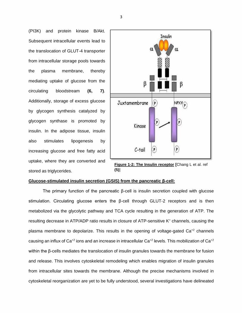
The insulin receptor is a tyrosine kinase receptor composed of two extracellular alpha and
two transmembrane beta subunits linked by disulfide bonds (5). The interaction of insulin with the
alpha sub-units of the insulin receptor induces a conformation change in the beta sub-units,
activating their intrinsic tyrosine activity leading to auto-phosphorylation of the receptor. This
results in the phosphorylation of insulin receptor substrate (IRS) proteins. IRS1 and IRS2, which
are the major isoforms present in the muscle cell, further activate phosphoinositide-3-kinase
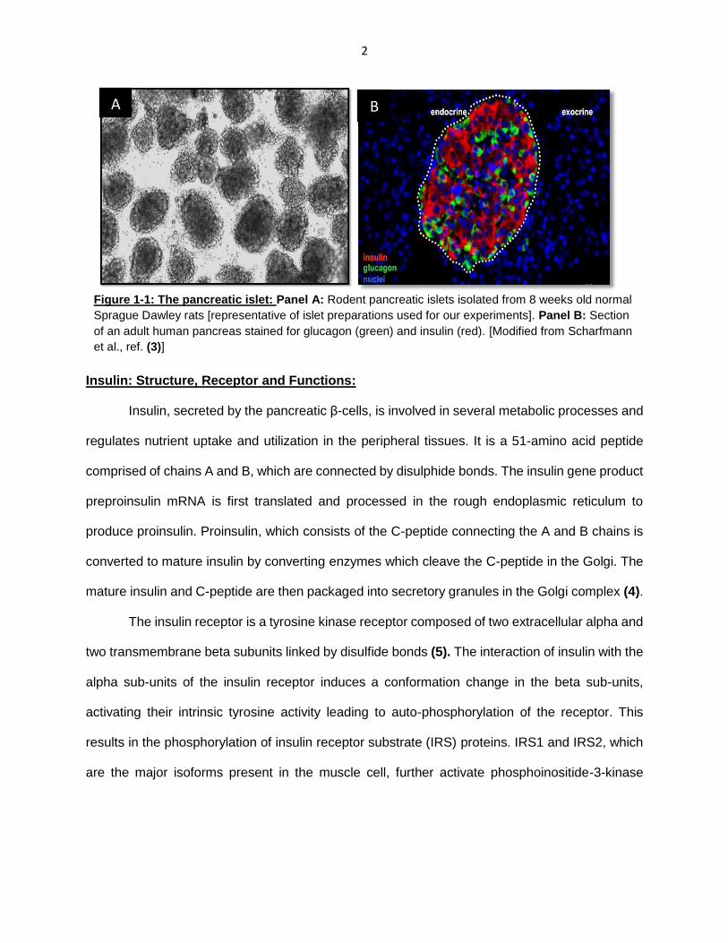
A B
Figure 1-1: The pancreatic islet: Panel A: Rodent pancreatic islets isolated from 8 weeks old normal
Sprague Dawley rats [representative of islet preparations used for our experiments]. Panel B: Section
of an adult human pancreas stained for glucagon (green) and insulin (red). [Modified from Scharfmann
et al., ref. (3)]
3
(PI3K) and protein kinase B/Akt.
Subsequent intracellular events lead to
the translocation of GLUT-4 transporter
from intracellular storage pools towards
the plasma membrane, thereby
mediating uptake of glucose from the
circulating bloodstream (6, 7).
Additionally, storage of excess glucose
by glycogen synthesis catalyzed by
glycogen synthase is promoted by
insulin. In the adipose tissue, insulin
also stimulates lipogenesis by
increasing glucose and free fatty acid
uptake, where they are converted and
stored as triglycerides.
Glucose-stimulated insulin secretion (GSIS) from the pancreatic β-cell:
The primary function of the pancreatic β-cell is insulin secretion coupled with glucose
stimulation. Circulating glucose enters the β-cell through GLUT-2 receptors and is then
metabolized via the glycolytic pathway and TCA cycle resulting in the generation of ATP. The
resulting decrease in ATP/ADP ratio results in closure of ATP-sensitive K+ channels, causing the
plasma membrane to depolarize. This results in the opening of voltage-gated Ca+2 channels
causing an influx of Ca+2 ions and an increase in intracellular Ca+2 levels. This mobilization of Ca+2
within the β-cells mediates the translocation of insulin granules towards the membrane for fusion
and release. This involves cytoskeletal remodeling which enables migration of insulin granules
from intracellular sites towards the membrane. Although the precise mechanisms involved in
cytoskeletal reorganization are yet to be fully understood, several investigations have delineated
Figure 1-2: The Insulin receptor [Chang L et al. ref
(5)]

4
the roles of small GTP-binding proteins in mediating F-actin reorganization and insulin granule
transport (8). The docking and fusion of insulin granules at membrane docking sites is also
mediated by several SNARE proteins such as syntaxin and vesicle-associated membrane protein.
Figure 1-3: Glucose-stimulated insulin secretion (GSIS) [Modified from Wang Z et al. ref (8)]
5
Diabetes
Diabetes is a serious pathological condition characterized by decreased glucose disposal
in the body, caused by insufficient insulin secretion from the pancreatic β-cell and decreased
insulin sensitivity in the peripheral tissues. According to the 2015 International Diabetes
Federation (IDF) atlas, approximately 415 million adults have been diagnosed with Diabetes
worldwide, and it is estimated that the number of cases will rise up to 642 million by the year 2040.
In addition, it is also reported that there are nearly 318 million adults with impaired glucose
tolerance and increased risk of developing diabetes. Diabetes is mainly of two types: Type 1
Diabetes (T1DM), also known as insulin-dependent diabetes mellitus (IDDM), and Type 2
Diabetes (T2DM), also referred to as non-insulin dependent diabetes mellitus (NIDDM). T1D is
characterized by insufficient insulin secretion caused by auto-immune destruction of the insulin-
secretin pancreatic β-cells. This is mediated by cell death induced by inflammatory cytokines
secreted by the immune cells, resulting in severe hyperglycemic conditions. Although the causes
for T1D still remain less understood, it has been suggested that genetic and/or environmental risk
factors contribute to disease development and progression (9). T2D, however, is characterized
by insulin resistance and decreased glucose tolerance in the peripheral tissues, resulting from
environmental and genetic risk factors. This results in prolonged exposure of pancreatic β-cells
to elevated levels of hyperglycemia, eventually leading to defects in insulin secretory response
and overt diabetic state. Elevated levels of circulating glucose can affect other tissues including
retina, kidney and cardiomyocytes and lead to serious diabetes-related complications.
6
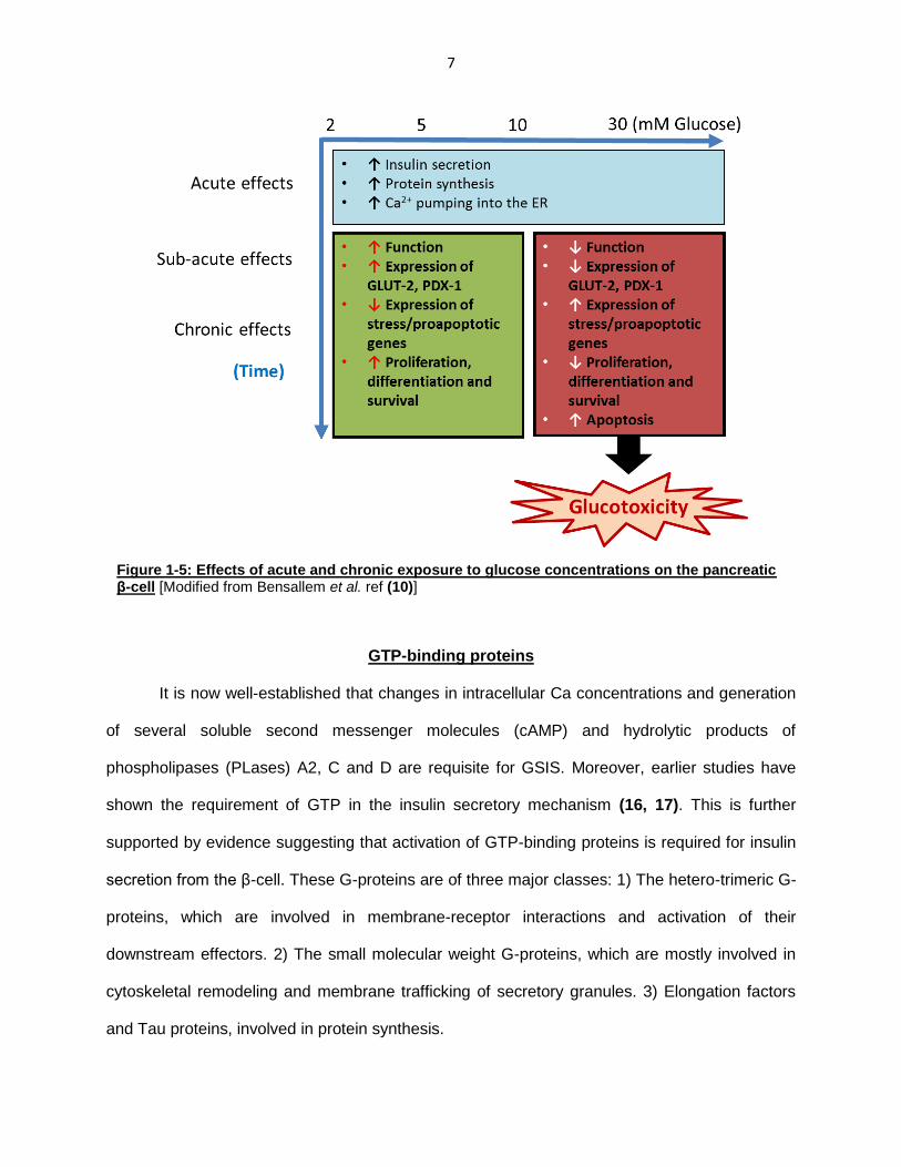
Pancreatic β-cell dysfunction:
Exposure of pancreatic β-cells to glucose results in initiation of several intracellular
metabolic pathways that mediate GSIS. As reviewed by Bensallem and associates, physiological
glucose concentrations play a major role in regulating β-cell function (10). Acute exposure to
stimulatory glucose concentrations has been shown to improve Ca+2 mobilization, protein
synthesis and β-cell function (11, 12). Furthermore, studies have indicated that prolonged
exposure to physiological glucose concentrations improves gene expression and is crucial for
maintaining optimal β-cell function and cell mass (13). However, prolonged exposure to elevated
levels of glucose exert toxic effects on the β-cell, causing decreased β-cell function and
proliferation. This condition, termed as glucotoxicity, results in decreased insulin secretory
response to the high metabolic demand and reduced β-cell mass (14, 15).
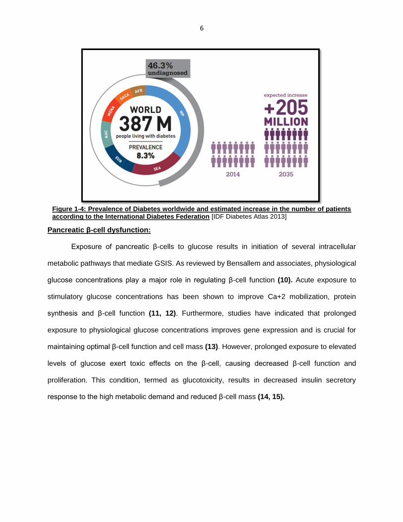
Figure 1-4: Prevalence of Diabetes worldwide and estimated increase in the number of patients according to the International Diabetes Federation [IDF Diabetes Atlas 2013]
7
GTP-binding proteins
It is now well-established that changes in intracellular Ca concentrations and generation
of several soluble second messenger molecules (cAMP) and hydrolytic products of
phospholipases (PLases) A2, C and D are requisite for GSIS. Moreover, earlier studies have
shown the requirement of GTP in the insulin secretory mechanism (16, 17). This is further
supported by evidence suggesting that activation of GTP-binding proteins is required for insulin
secretion from the β-cell. These G-proteins are of three major classes: 1) The hetero-trimeric G-
proteins, which are involved in membrane-receptor interactions and activation of their
downstream effectors. 2) The small molecular weight G-proteins, which are mostly involved in
cytoskeletal remodeling and membrane trafficking of secretory granules. 3) Elongation factors
and Tau proteins, involved in protein synthesis.
Figure 1-5: Effects of acute and chronic exposure to glucose concentrations on the pancreatic β-cell [Modified from Bensallem et al. ref (10)]

8
Small G-proteins:
Small G-proteins are categorized into five subfamilies with varying degrees of homology.
These include Ras, Rho, Rab, Arf and Ran GTPases which have been implicated in various
cellular functions including gene expression, cytoskeletal remodeling, vesicle trafficking,
nucleocytoplasmic transport (18). In the context of β-cell function, G-proteins belonging to the
Rho family including RhoA, Cdc42 and Rac1 have been implicated in cytoskeletal remodeling,
vesicle fusion and GSIS (8). Although the involvement of Rab sub-family GTPases that include
Rap1, Rab3A and Rab27 are yet to be fully understood, it has been suggested that these G-
proteins are involved in docking of insulin secretory granules at membrane docking sites (19).
These small G-proteins, which are inactive when bound to GDP, are activated by their interaction
with GTP. This process is mediated by several regulatory factors which include: 1) guanine
nucleotide exchange factors (GEFs), 2) GTPase-activating proteins (GAPs) and 3) GDP-
dissociation inhibitors. Under basal conditions, G-proteins are inactive (GDP-bound) and are
bound to GDI, which prevent their dissociation with GDP. However, upon stimulation, GDI
dissociate from the G-protein, thereby releasing GDP. G-proteins can then be activated by
interacting with GTP, which is mediated by GEFs. Similar to heterotrimeric G-proteins, small G-
proteins also possess a small degree of intrinsic GTPase activity. Therefore, when activated,
these GTPases shuttle back to their GDP-bound form by hydrolyzing GTP, and this is catalyzed
by GAPs.
Figure 1-6: Rac1 activation cycle [Modified from Kowluru A, ref (18)]

9
Post-translational modifications:
In addition to the GTP/GDP activation cycle, small G-proteins also undergo a series of
post-translational modifications at their C-terminal CAAX motifs (C-cysteine; A-amino acid; X-
terminal amino acid). These include: 1) Prenylation, 2) Carboxymethylation and 3)Palmitoyaltion.
Prenylation involves the attachment of a prenyl moiety at the cysteine residue of the CAAX
motif (20, 21). The prenyl moiety are derivates of mevalonic acid synthesized via the cholesterol
biosynthetic pathway, and include a 15-C farnesyl or a 20-C geranylgeranyl group. The
attachment to the cysteine residue is catalyzed by prenyl tranferases namely, farnesyl transferase
(FTase)-I and geranylgeranyl transferase (GGTase)-I and II. Following prenylation, the three
amino acids (-AAX) adjacent to the prenylated cysteine are cleaved by Ras-converting enzyme,
which exposes the carboxylate ion of the prenylated cysteine. This is followed by a
carboxymethylation that involves methylation of the carboxylate group by isoprenylcysteine-O-
carboxymethyl transferase (ICMT). Additionally, specific G-proteins also undergo palmitoylation
at other cysteine residues. These modifications have been shown to increase hydrophobicity of
the candidate G-proteins, that increases their localization in the membrane fraction of the cell
required for optimal interaction with their respective effector proteins.
Role in glucose stimulated insulin secretion (GSIS):
Earlier studies in the β-cell have shown that depletion of intracellular GTP pools impairs
physiological insulin secretion (16, 17). Further investigations, focusing on Rho family GTPases
including Rac1, have demonstrated that loss of function results in impairment in insulin secretory
response to ambient glucose concentrations. Studies by Asahara and associates have utilized a
β-cell specific Rac1 knockout model (22). They reported that these animals, when fed on a normal
diet showed decreased GSIS. Furthermore, when these animals were fed on high-fat diet, signs
of impaired glucose tolerance and GSIS were observed. Using pancreatic islets isolated from
these animals and INS-1 cell line, they reported significant defects in GSIS and F-actin remodeling

10
upon loss of Rac1 function. Furthermore, studies in our own laboratory, have shown that siRNA-
mediated depletion of Rac1 results in impaired GSIS (23).
Rac1 is activated by dissociating with RhoGDI and its interaction with GTP. Several GEFs
have been identified that regulated Rac1 function including Tiam1, Vav2, Trio (18, 23). Rac1 is
also known to undergo geranylgeranylation that increases its membrane association and
substrate specificity. Previous investigations in our laboratory have utilized pharmacological
agents to block Rac1 activity and demonstrated the role of Rac1 in physiological insulin secretion
and cytoskeletal remodeling in the β-cell (24, 25). The various pharmacological inhibitors used
are depicted in Figure 1-7.
NSC23766, synthesized by Gao and associates, blocks Tiam1-mediated Rac1 activation,
and has been extensively used in multiple cell types to block Rac1 function (26). Studies from our
laboratory, have utilized this compound and reported a significant drop in insulin secretory
response to stimulatory glucose (24). Ehop-016, synthesized by Vlaar and associates, disrupts
Vav2-mediated Rac1 activation. In a recent study from our laboratory, F-actin depolymerization
was visualized using GFP-tagged LifeAct plasmid and live-cell imaging (25). We reported that
Figure 1-7: Pharmacological inhibitors of Rac1 [Modified from Nagase M et al. ref (33)]

11
glucose stimulation results in depolymerization of F-actin filaments, which is inhibited in presence
of Ehop-016, thereby providing compelling evidence indicating the involvement of Rac1 in
cytoskeletal remodeling and insulin secretion.
Besides NSC23766 and Ehop-016, which target GEF-mediated Rac1 activation, we
utilized a novel inhibitor of Rac1, EHT1864, which blocks Rac1 function in a GEF-independent
manner (27). This compound, designed by Desire and associates, binds to Rac1 with a higher
affinity than GTP/GDP, thereby retaining Rac1 in an inert, inactive state (28). This compound has
been used in multiple cell types to examine the role of Rac1 in normal cell physiology and
pathology (29-33).
Additionally, previous published evidence have also shown that Rac1 geranylgeranylation
is critical for GSIS, since presence of GGTI-2147, a specific inhibitor of geranylgeranyl
transferase, blocked membrane association of Rac1 and insulin secretion in INS-1 832/13 cells
(34). Furthermore, when INS-1 832/13 cells were transfected with a dominant negative mutant of
the alpha subunit of FTase and GGTase-I, insulin secretion was significantly blocked. These data
have together confirmed the requisite role of prenylation of G-proteins including Rac1 in
physiological insulin secretion from the β-cell.
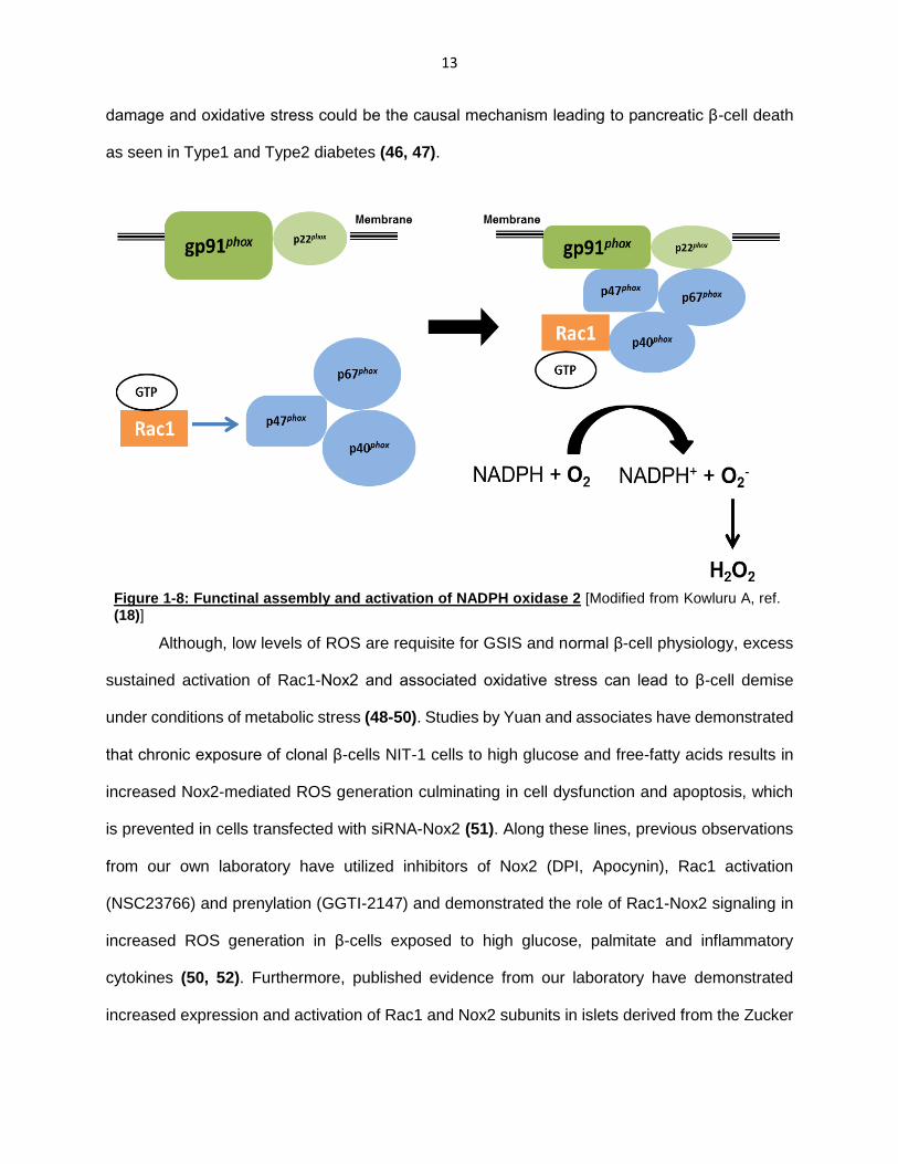
Phagocyte-like NADPH oxidase (Nox2) and ROS generation:
The NADPH oxidase is a family of membrane-associated enzymes that catalyze oxidation
of cytosolic NADPH and one electron reduction of molecular oxygen, resulting in the generation
of superoxide. This family of enzymes includes seven members: Nox1, Nox2, Nox3 Nox4, Nox5,
Duox1, Duox2 (35, 36), which are composed of different membrane and cytosolic protein
components. The phagocyte-like NADPH oxidase (Nox2) is composed of several membrane
(gp91phox and p22phox) and cytosolic (p47phox, p67phox, p40phox and Rac1) components.
Upon stimulation, the cytosolic components translocate to the membrane thereby, completing the
holoenzyme assembly and activation of the holoenzyme. The superoxide generated from
molecular oxygen is the major contributor of reactive oxygen species (ROS) and can be converted

12
to other ROS by superoxide dismutases. Pancreatic β-cells have known to be expressing Nox2
subunits, which represents a major source of extra-mitochondrial ROS (36, 37). Recent studies
have shown that a tonic increase in ROS in the pancreatic β-cell is required for GSIS. Although
the mechanism is still poorly understood, evidence suggests that ROS act as second messengers
and are known to regulate several downstream processes including mobilization of Ca and
glucose metabolism (38, 39). Interestingly, studies by Morgan et al. have demonstrated that
inhibition of Nox2 using DPI or by downregulating expression of p47phox using an anti-sense
oligonucleotide, resulted in impaired insulin secretion in rat pancreatic islets when perfused with
stimulatory glucose concentrations (40). Studies in our own laboratory, have shown that inhibition
of Nox2 (apocycin, DPI, siRNA p47phox) blocks glucose-induced ROS generation in insulin
secreting INS-1 832/13 cells. Furthermore, we demonstrated that glucose stimulation results in
increased Nox2 enzyme activity that is sensitive to apocynin. Additionally, studies using siRNA
and pharmacological approaches demonstrated that prenylation is critical for GSIS. In this
context, pharmacological inhibitors of prenylation (FTI-277 and GGTI-2147) blocked glucose-
induced ROS generation, implicating the role of Nox2-ROS signaling in GSIS (41).
Reactive oxygen species in pancreatic β-cell dysfunction:
Oxidative stress has been demonstrated to be a causal factor in the onset and
development of various disorders including diabetes and its complications (42-44). Earlier studies
by Lenzen et al. have examined the expression of various antioxidant enzymes in pancreatic islets
and compared with other tissues in albino mice (45). Their studies revealed that expression levels
of anti-oxidant enzymes such as glutathione peroxidase, Cu/Zn SOD and Mn SOD in the islet
were significantly lower compared to other tissues. Studies in islets isolated from ob/ob mice, in
which β-cells constitute about 90% of the total cell composition, have also shown low expression
of antioxidant enzymes. The investigators concluded that pancreatic β-cells possess far lower
levels of antioxidant compared to other endocrine and non-endocrine tissues. Several other
studies have strongly suggested that pancreatic β-cells are highly susceptible to oxidative
13
damage and oxidative stress could be the causal mechanism leading to pancreatic β-cell death
as seen in Type1 and Type2 diabetes (46, 47).
Although, low levels of ROS are requisite for GSIS and normal β-cell physiology, excess
sustained activation of Rac1-Nox2 and associated oxidative stress can lead to β-cell demise
under conditions of metabolic stress (48-50). Studies by Yuan and associates have demonstrated
that chronic exposure of clonal β-cells NIT-1 cells to high glucose and free-fatty acids results in
increased Nox2-mediated ROS generation culminating in cell dysfunction and apoptosis, which
is prevented in cells transfected with siRNA-Nox2 (51). Along these lines, previous observations
from our own laboratory have utilized inhibitors of Nox2 (DPI, Apocynin), Rac1 activation
(NSC23766) and prenylation (GGTI-2147) and demonstrated the role of Rac1-Nox2 signaling in
increased ROS generation in β-cells exposed to high glucose, palmitate and inflammatory
cytokines (50, 52). Furthermore, published evidence from our laboratory have demonstrated
increased expression and activation of Rac1 and Nox2 subunits in islets derived from the Zucker
Figure 1-8: Functinal assembly and activation of NADPH oxidase 2 [Modified from Kowluru A, ref. (18)]

14
diabetic fatty (ZDF) rat, a model for type 2 diabetes, in normal human islets exposed to
hyperglycemic conditions, and in islets from type2 diabetic human donors (49). Further studies in
INS-1 832/13 cells, ZDF islets and diabetic human islets have suggested significant increase in
stress kinase JNK1/2 activation and caspase-3 activation. Together, these studies provide
evidence suggesting that exposure of β-cells to diabetic conditions results in increased Rac1-
Nox2 activity and the associated oxidative stress causes activation of downstream apoptotic
stress kinase pathway, culminating in mitochondrial dysfunction and β-cell death.
Stress-activated protein kinases/Mitogen-activated protein kinases (SAPKs/MAPKs)
The mitogen-activated protein kinase family are serine-threonine protein kinases that
regulate and integrate multiple intracellular signaling processes. These include 14 MAPKs known
to be present in mammalian cells and are classifies into the conventional MAPKs and atypical
MAPKs. The most widely studied group of MAPKs are the conventional the extra-cellular-regulate
kinases (ERKs), c-jun NH2-terminal kinase (JNK) and p38MAPK isoforms. These conventional
MAPKs are regulated by distinct MAPK signaling cascades composed of three components: 1.
MAPK kinase kinase (MAPKKK); 2. MAPK kinase (MAPKK) and 3. MAPK.
In the presence of an extracellular stressor, MAPKKK are activated via interaction with a
small G-protein including Rac1 (53). MAPKKK then phosphorylate and activate MAPKK, which in
turn activate MAPKs. MAPKs are activated by phosphorylation on Threonine and Tyrosine
residues located in the conserved activation loop of the kinase domain. Once activated MAPKs
interact and phosphorylate several downstream kinases such as the MAPK-activated protein
kinases (MAPKAPK) such as RSK1-4 and MSK1/2 and transcription factors such as Elk-1, c-jun,
ATF3 and p53 (54). The activation of downstream signaling cascades by MAPKs mediates the
cellular responses to the stress stimuli including gene expression, mitosis, cell differentiation and
apoptosis. In addition to their essential role in a wide range of cellular functions, emerging
evidence implicates MAPK pathways in the pathogenesis of human diseases including diabetes,
cancer and neurodegenerative diseases (55).
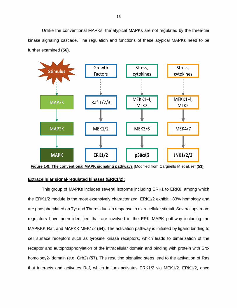
15
Unlike the conventional MAPKs, the atypical MAPKs are not regulated by the three-tier
kinase signaling cascade. The regulation and functions of these atypical MAPKs need to be
further examined (56).
Extracellular signal-regulated kinases (ERK1/2):
This group of MAPKs includes several isoforms including ERK1 to ERK8, among which
the ERK1/2 module is the most extensively characterized. ERK1/2 exhibit ~83% homology and
are phosphorylated on Tyr and Thr residues in response to extracellular stimuli. Several upstream
regulators have been identified that are involved in the ERK MAPK pathway including the
MAPKKK Raf, and MAPKK MEK1/2 (54). The activation pathway is initiated by ligand binding to
cell surface receptors such as tyrosine kinase receptors, which leads to dimerization of the
receptor and autophosphorylation of the intracellular domain and binding with protein with Src-
homology2- domain (e.g. Grb2) (57). The resulting signaling steps lead to the activation of Ras
that interacts and activates Raf, which in turn activates ERK1/2 via MEK1/2. ERK1/2, once
Figure 1-9: The conventional MAPK signaling pathways [Modified from Cargnello M et al. ref (53)]
16
activated, has been shown to localize and interact with several nuclear and cytosolic substrates.
The major functions of this module include cell cycle progression, cell growth and proliferation.
Several studies have implicated alterations in the ERK1/2 pathway in tumor development. For
example, in certain cancer cells, constitutive activation of ERK signaling induced by
overexpression of receptor tyrosine kinase or Ras/Raf mutations, has been associated with
cancer development (53, 57). Therefore, the ERK pathway is being extensively studied for drug
discovery and pharmacological inhibitors of Ras, Raf and MEK are currently under development
to prevent tumor progression.
c-jun NH2-terminal kinases (JNK):
The three isoforms of JNK identified in mammalian cells include JNK1/2/3, which share
~85% homology. While JNK1/2 are ubiquitously expressed in multiple cells, JNK3 seems to be
expressed in neuronal tissues, testis and cardiomyocytes (58). These are activated in response
to stress stimuli including inflammatory cytokines, oxidative stress, ionizing radiations and growth
factor deprivation. Activation is initiated by upstream MAPKKK including MLK1-4, MEK1-4 and
ASK1/2, which phosphorylate MAPKKs MKK4 and MKK7. MKK4/7 then activate JNKs by dual-
phosphorylation on Thr and Tyr residues within the conserved Thr-Pro-Tyr motif in the activation
loop. Once activated, JNK has been shown to phosphorylate and activate c-jun, promotes AP-1
complex formation and thereby mediates transcription of genes containing AP-1 binding sites
(59). It has also been reported that JNKs interact with other transcription factors including p53,
ATF-2, c-Myc, STAT3 (60). Mice lacking JNK1/2/3-encoding genes showed significant defects in
apoptosis and immune responses (60). In addition, inactivation of JNK1/2 has been associated
with decreased response to DNA-damaging agents and UV radiation in cancer cells, implying the
involvement of the JNK module in apoptosis (61). JNKs are also involved in cell metabolism,
immune responses, cytokine production and actin reorganization (62-64). The JNK signaling
pathway has been implicated in the pathologies of several neurological disorders including
Alzheimer’s and Parkinson’s disease (55).
17
p38 MAPK:
This group of MAPK comprise of four splice variants including p38α, p38β, which are
ubiquitously expressed in mammalian cells, p38ɣ and p38δ which are differentially expressed
(53). These are activated by several extraneous stress stimuli including oxidative stress, UV
radiation, inflammatory cytokines and hypoxia (65). Several upstream regulators including Rho
family GTPases Rac1 and Cdc42, GPCRs and adaptors proteins are known to initiate the
pathway, by recruiting MAPKKKs involved in p38MAPK activation. These MAPKKKs, which are
common for JNK and p38, phosphorylate MKK3/6 which are involved in the activation of
p38MAPK isoforms by dual phosphorylation at specific Thr-Gly-Tyr motifs in the activation loop.
Activated p38MAPK then interacts with several cytoplasmic and nuclear substrates which mediate
cellular responses including inflammation, cell cycle arrest, differentiation and apoptosis (65).
Functions of p38MAPK:
1. p38MAPK in cell cycle regulation: p38MAPK is activated in response to stress stimuli
such as DNA damage , oxidative stress and osmotic stress. In response to DNA damage,
p38MAPK has been shown to play a negative modulatory role in cell cycle progression at
G2/M transition by several mechanisms (66, 67). This is initiated by detection of DNA
damage by serine/threonine kinases that act as DNA damage sensors, including ATM and
ATR kinases. These sensor DNA repair proteins are known to interact with Tau proteins
that act as MAPKKK and activate MKK3/6, which in turn phosphorylate p38MAPK.
p38MAPK is known to interact and phosphorylate p53 transcription factor, which induces
the transcription of target genes namely p21 and Gadd45a, which are involved in cell cycle
arrest at G2/M phase (66). In addition, p38MAPK can also induce G1/S cell cycle
checkpoint in response to oxidative stress and hypoxia by several mechanisms including
expression of regulatory proteins such as p16INK4a and p19ARF (67).
2. p38MAPK in cell differentiation: Several studies have implicated the positive and
negative modulatory roles of p38MAPK in cellular differentiation. For example, it has been
18
suggested that p38MAPK induces cell cycle arrest in myoblasts which is followed by a
gradual increase in expression of muscle-specific genes, thereby, inducing myocyte
differentiation into myotubes (68). This is induced by increased activity of transcriptional
factors and chromatin remodeling. In addition, p38MAPK has also been implicated in
osteoclast differentiation initiated by RANKL signaling, by the activation of NFATc1 and
NF-κB transcription factors (69). Furthermore, studies in intestinal cells revealed that
p38MAPK is activated in intestinal cells induced to differentiate and inhibition of p38MAPK
blocks transcription of cell differentiation markers (70). These effects were found to be
coupled with the increased activity of CDX2/3 which are nuclear transcription factors,
induced by their interaction with p38MAPK. Apart from studies indicating the role of
p38MAPK in inducing cell differentiation, studies in adipocytes indicate inhibitory role of
p38MAPK in adipocyte differentiation (71, 72). Increased adipogenesis results in adipose
tissue expansion and obesity. Studies by Aouadi and associates have revealed that both
diet-induced and genetically obese mice exhibit decreased p38MAPK activity, resulting in
increased adipogenesis (71). Together, these observation suggest tissue-specific
regulatory role of p38MAPK in cellular differentiation.
3. p38MAPK in cell survival and apoptosis: Evidence from multiple cell types indicate that
p38MAPK exhibits cell-specific anti- and pro-apoptotic functions by interacting with its
diverse substrate proteins (53). Evidence from studies in certain cancer cells showed a
pro-survival effect of p38MAPK activation. For example, treatment of Jurkat cells and T
lymphocytes with p38MAPK inhibitor augmented the cytotoxic effect of 8-
methoxypsoralen and UV-radiation treatment, thereby increasing the efficacy of this
therapy (73). Furthermore, studies by Gutiérrez-Uzquiza et al. have revealed a novel
mechanism by which p38MAPK may play a role in cell survival in response to oxidative
stress, by mediating the transcription of anti-oxidant genes including superoxide
dismutases and catalase (74). Although some observations indicate anti-apoptotic

19
function of p38MAPK, a majority of studies have demonstrated its pro-apoptotic role.
Oxidative stress has been shown to be a major activator of p38MAPK in multiple cell types
including pancreatic β-cells (75), neurons (76) and cardiac myocytes (77). Furthermore,
treatment of cancer cell lines with All-trans-retinoic Acid activates p38MAPK in a Rac1-
dependent manner, which in turn activates MAPKAPK2 and mediates apoptosis (78).
Studies by Bulavin et al. have demonstrated that p38MAPK is involved in the activation of
p53 tumor suppressor pathway, where p38MAPK phosphorylates p53 at several residues
at it N-terminus (79). This causes p53 to dissociate from MDM2, a negative modulator of
p53 function, protecting p53 from proteosomal degradation and allowing its function as a
transcriptional factor. These events lead to the expression of pro-apoptotic target genes
of p53, thereby inducing cell death.
p53 tumor suppressor
p53 tumor suppressor is a transcription factor belonging to a unique family of proteins
which also includes p63 and p73 (80, 81). Encoded by the TP53 gene, p53 is commonly referred
to as the “guardian of the genome” and is involved in inducing anti-proliferative cellular responses
to DNA damage including cell cycle arrest, cellular senescence, DNA repair and apoptosis (82).
These functions are mediated by the binding of activated p53 to the DNA, thereby promoting the
transcription of several target genes (83). Evidence from mice lacking p53 demonstrated its critical
role as a tumor suppressor, since p53 -/- mice spontaneously develop neoplastic tumors (84).
TP53 was initially identified as an oncogene, since p53 was found to be overexpressed in most
tumor cells. However, later studies identified that these tumor cells expressed a missense mutant
of p53 and the oncogenic nature resulted from loss of p53 function (85, 86). Nearly 50 % of all
human cancer types exhibit mutation or loss of p53 function, where it is inactivated either by
mutation in TP53 gene, or mutations in genes encoding regulatory proteins that interact with p53
(87). Several reports in multiple cell types have provided insights into the post-translational

20
modifications involved in the p53 signaling pathway and have identified the regulatory proteins
involved in the modulation of p53 function (88-91).
Structure of p53:
TP53 gene, located on the small arm of chromosome 17, encodes a nuclear
phosphoprotein containing 393 amino acids composed of several structural and functional
domains (83, 89, 92). These are: 1) N-terminal transactivation domain (TAD) – divided into
subdomains TAD1 (1-40 amino acid residues), TAD2 (40-61 amino acid residues) and a proline-
rich region (61-94 amino acid residues); 2) Central DNA-binding domain (102-292 amino acid
residues) and 3) C-terminal region – containing a tetramerization domain (324-355 amino acid
residues), nuclear export and import signal sequences and a carboxyl-terminal regulatory domain
(363-393). The N-terminal TAD is mostly involved in transcriptional activation and interacts with
several regulatory factors including MDM2 and p300 (90, 91). The central DNA-binding domain
mediates site-specific binding of p53 to the DNA (93). The C-terminal domain containing the
tetramerization domain is required for the binding of p53 monomers to form tetramers that
possess a greater affinity for DNA-binding sites (94). Tetramerization also has been shown to
promote nuclear localization as the nuclear export sequence is masked in p53 tetramers (95).
The carboxy-terminal has also been implicated in regulating DNA binding and tranactivation of
p53 target genes (96, 97). p53 also consists of nuclear localization (NLS) and nuclear exclusion
sequences (NES) which regulate its nucleo-cytoplasmic shuttling (98, 99).
Figure 1-10: The structural and functional domains of the p53 tumor suppressor
[Modified from Joerger AC et al. ref (92).
21
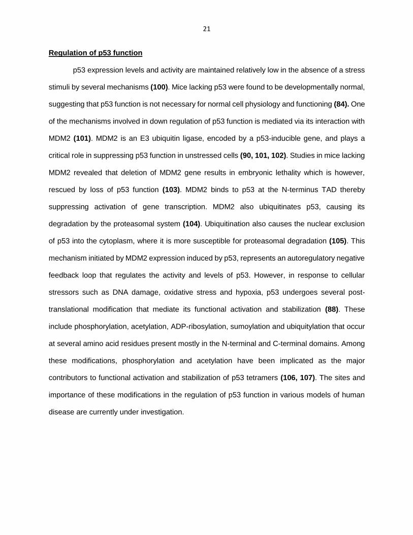
Regulation of p53 function
p53 expression levels and activity are maintained relatively low in the absence of a stress
stimuli by several mechanisms (100). Mice lacking p53 were found to be developmentally normal,
suggesting that p53 function is not necessary for normal cell physiology and functioning (84). One
of the mechanisms involved in down regulation of p53 function is mediated via its interaction with
MDM2 (101). MDM2 is an E3 ubiquitin ligase, encoded by a p53-inducible gene, and plays a
critical role in suppressing p53 function in unstressed cells (90, 101, 102). Studies in mice lacking
MDM2 revealed that deletion of MDM2 gene results in embryonic lethality which is however,
rescued by loss of p53 function (103). MDM2 binds to p53 at the N-terminus TAD thereby
suppressing activation of gene transcription. MDM2 also ubiquitinates p53, causing its
degradation by the proteasomal system (104). Ubiquitination also causes the nuclear exclusion
of p53 into the cytoplasm, where it is more susceptible for proteasomal degradation (105). This
mechanism initiated by MDM2 expression induced by p53, represents an autoregulatory negative
feedback loop that regulates the activity and levels of p53. However, in response to cellular
stressors such as DNA damage, oxidative stress and hypoxia, p53 undergoes several post-
translational modification that mediate its functional activation and stabilization (88). These
include phosphorylation, acetylation, ADP-ribosylation, sumoylation and ubiquitylation that occur
at several amino acid residues present mostly in the N-terminal and C-terminal domains. Among
these modifications, phosphorylation and acetylation have been implicated as the major
contributors to functional activation and stabilization of p53 tetramers (106, 107). The sites and
importance of these modifications in the regulation of p53 function in various models of human
disease are currently under investigation.
22
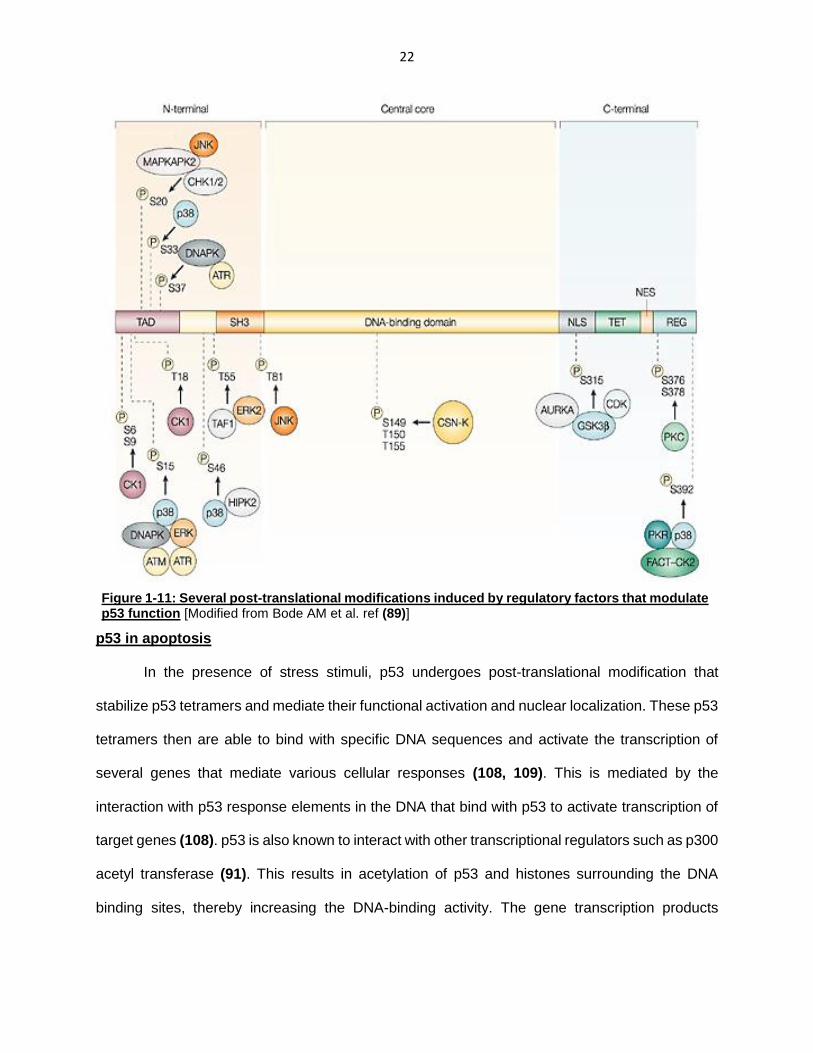
p53 in apoptosis
In the presence of stress stimuli, p53 undergoes post-translational modification that
stabilize p53 tetramers and mediate their functional activation and nuclear localization. These p53
tetramers then are able to bind with specific DNA sequences and activate the transcription of
several genes that mediate various cellular responses (108, 109). This is mediated by the
interaction with p53 response elements in the DNA that bind with p53 to activate transcription of
target genes (108). p53 is also known to interact with other transcriptional regulators such as p300
acetyl transferase (91). This results in acetylation of p53 and histones surrounding the DNA
binding sites, thereby increasing the DNA-binding activity. The gene transcription products
Figure 1-11: Several post-translational modifications induced by regulatory factors that modulate p53 function [Modified from Bode AM et al. ref (89)]
23
induced by p53 include p21, Gadd45, 14-3-3δ, Fas, Bax, PUMA and Noxa which are known to
mediate cell cycle arrest and apoptosis, in response to stress stimuli (110-114).
In the presence of stress stimuli, p53 is known to induce cell apoptosis by several
mechanisms including the extrinsic and the intrinsic apoptotic pathway, and by inducing genes
that suppress cell survival signaling (83). The extrinsic pathway involves the binding of specific
extracellular ligands (e.g., Fas ligand) to death receptors (e.g., Fas) which leads to the
accumulation of FADD and initiator caspases causing apoptosis (80). For example, the cytotoxic
effects of bleomycin in hepatoma cells is mediated by increased nuclear accumulation of p53 and
increased expression of Fas receptor (115). It has also been suggested that p53 mediates
membrane translocation of Fas receptor from the Golgi complex (116). Additionally, p53 is also
known to induce DR4 and DR5 death receptors which are involved in TNF-related apoptosis-
inducing ligand (TRAIL) (117, 118). Moreover, a majority of the p53-inducible genes are involved
in the intrinsic apoptotic. p53 activates the transcription of pro-apoptotic proteins belonging to the
Bcl2 family, which are modulators of mitochondrial membrane potential. Bcl2 family proteins can
be classified intro three subfamilies: 1) Anti-apoptotic Bcl2; 2) Pro-apoptotic Bax; and 3) Pro-
apoptotic BH3-only (Bcl2 homology-3) proteins. The pro-apoptotic Bax gene promoter region has
been shown to possess p53 binding sites. Bax increases mitochondrial membrane permeability
which releases cytochrome c into the cytoplasm and activates caspases and apoptosis. Similarly,
an important target gene for p53 is the p53-upregulated modulator of apoptosis (PUMA), which is
a BH3-only apoptotic protein. The PUMA gene, consisting of high affinity p53-binding sites,
encodes for two isoforms which are known to upregulate Bax activity thereby activating apoptosis
(113). Another BH3-only protein regulated by p53 is Noxa, which also promotes Bax activity and
apoptosis (114).

24
Figure 1-12: Mechanisms of p53-induced apoptosis [Vousden KH et al. ref (83)]

25
Hypothesis
Our preliminary data and above literature review indicate that excess generation of ROS
by Rac1-Nox2 enzyme complex causes oxidative stress, which leads to the activation of
downstream apoptotic stress kinases. Despite the above evidence suggesting the involvement of
Rac1-Nox2 in β-cell dysfunction under diabetic conditions, the downstream signaling pathways
need to be further elucidated. The central objective of my dissertation project is to examine the
role of Rac1 and Nox2 in the generation of oxidative stress, leading to the activation of pro-
apoptotic factors, resulting in pancreatic β-cell death under glucotoxic conditions. Using
pharmacological approaches, we propose to examine the regulation of stress kinase p38MAPK
and p53 pathways by Rac1-Nox2 enzyme complex in INS-1 832/13 cells, rodent and human
pancreatic islets. Furthermore, we will extend these studies in in vivo models of obesity, insulin
resistance and pancreatic β-cell dysfunction (Zucker diabetic fatty rat).
The proposed studies will test the hypothesis that (i) chronic exposure of pancreatic β-
cells to glucotoxic conditions leads to sustained activation of Rac1-Nox2 holoenzyme and the
resulting oxidative stress activate the p38MAPK and p53 signaling pathways, culminating in the
activation of apoptotic pathways and β-cell death; and (ii) therapeutic intervention of Rac1-Nox2
signaling cascade prevents pancreatic β-cell death, induced by glucotoxic conditions, and onset
of diabetes.
I will accomplish these goals by conducting studies under the following three specific aims:
Specific Aim 1: To determine if Rac1/Nox2 derived ROS leads to the activation of p38MAPK,
under glucotoxic conditions
Specific Aim 2: To demonstrate the role of p53 phosphorylation in mediating the effects of
Rac1/Nox2 under glucotoxic conditions
Specific Aim 3: To examine the Rac1-p38MAPK-p53 pathway in human pancreatic islets
exposed to glucotoxic conditions and in whole animal models of pancreatic β-cell dysfunction

26
Figure 1-13: Proposed working model for Rac1-Nox2-induced oxidative stress and activation of p38MAPK-p53 signaling pathway in pancreatic β-cells under glucotoxic conditions: We propose that chronic exposure of β-cells to elevated glucose concentrations leads to sustained activation of Rac1-Nox2 holoenzyme and oxidative stress, which in turn activates p38MAPK and p53 tumor suppressor. This leads to activation of p53-target gene transcription, which ultimately induce β-cell apoptosis.

27
Table 1-1: List of pharmacological inhibitors utilized to target Rac1, Nox2 and p38MAPK
Pharmacological agent
Target Mechanism of
action Functional
Consequence
gp91-ds-tat Nox2
Prevents association of p47phox with gp91phox
Inhibition of Nox2 holoenzyme assembly activation
NSC23766 Rac1 Blocks Tiam1-Rac1 interaction
Inhibition of Tiam1-mediated Rac1 activation
Ehop-016 Rac1 Blocks Vav2-Rac1 interaction
Inhibition of Vav2-mediated Rac1 activation
EHT1864 Rac1 Prevents GDP/GTP association of Rac1
Inhibition of Rac1 function
Simvastatin HMG CoA reductase
Inhibits the isoprenoid biosynthetic pathway
Decreased protein prenylation
GGTI2147 GGTase-I Blocks protein geranylgeranylation
Decreased protein geranygeranylation
2-bromopalmitate Protein acyl transferases
Blocks protein S-palmitoylation
Decreased protein palmitoylation
SB203580 p38MAPK Blocks kinase activity of p38MAPK
Inhibition of p38MAPK function

28
CHAPTER 2: MATERIALS AND METHODS
Chemicals and antibodies:
Rabbit polyclonal antibody for phospho-p38MAPK (Thr 180/Tyr 182) and total-p38MAPK
were obtained from Santa Cruz Biotechnology [Santa Cruz, CA]. Rabbit monoclonal antibody for
phospho-p53 and total-p53 was purchased from Cell Signaling Technology [Danvers, MA]. Mouse
monoclonal antibodies for Phospho-ATM (ser-1981) and total-ATM were obtained from Abcam
[Cambridge, MA]. NSC23766 and GGTI-2147 were obtained from Calbiochem [San Diego, CA].
EHT1864 was from R&D systems [Minneapolis, MN]. EHop-016 was kindly provided by Dr.
Cornelis Vlaar, University of Puerto Rico [San Juan, PR]. Scrambled gp91-ds-tat (inactive) and
active gp91-ds-tat were from Anaspec, Inc. [Fremont, CA]. ). IRDye® 800CW anti-rabbit and anti-
mouse secondary antibodies were obtained from LICOR [Lincoln, NE]. Glucose, 2-
Bromopalmitate (2-BP), 2’, 7’-dichlorofluorescein diacetate (DCFDA), N,-N’-dimethyl-9,-90-
bisacridiniumdinitrate (lucigenin) were purchased from Sigma–Aldrich [St. Louis, MO]. All other
reagents were obtained from Sigma (St. Louis, MO).
Kits:
Rat insulin ELISA kit was purchased from American Laboratory Products Co [Windham,
NH]. Rac1 activation G-LISA kit was from Cytoskeleton Inc. (Denver, CO). NE-PER® Nuclear and
Cytoplasmic Extraction Reagents were purchased from Thermo Scientific (Waltham, MA). Cell
Death Detection ELISA® were purchased from Sigma (St. Louis, MO). Dead Cell Apoptosis Kit
with Annexin V Alexa Fluor® 488 & Propidium Iodide (PI) was from ThermoFischer Scientific.
Insulin secreting INS-1 832/13 cells and culture conditions:
INS-1 832/13 cells were kindly provided by Dr. Chris Newgard, Duke University Medical
Center [Durham, NC]. Cells were cultured in RPMI-1640 medium containing 10 % fetal bovine
serum (FBS) supplemented with 100 IU penicillin and 100 IU/ml streptomycin, 1 mM sodium
pyruvate, 50 mM 2-mercaptoethanol and 10 mM HEPES (pH 7.4) at 37 ° C and 5 % CO2 in a
humidified incubator. Cultured cells were sub cloned twice weekly following trypsinization and

29
passages 53–61 were used for the studies. Cells were incubated in low glucose-low serum media
(LG-LS; 2.5 mM glucose; 2.5 % heat-inactivated FBS) overnight, prior to glucose treatments with
low glucose (LG; 2.5 mM) and high glucose (HG; 20 mM) for 24 hours in the absence or presence
of pharmacological agents as indicated in the text. At the end of the incubation time, cells were
rinsed with PBS and lysed using radio immunoprecipitation assay (RIPA) buffer supplemented
with containing protease inhibitor cocktail, 1 mM NaF, 1 mM PMSF and 1 mM Na3VO4.
Rodent and human islets and culture conditions:
Sprague-Dawley male rats (6 to 8 weeks old) were purchased from ENVIGO [Indianapolis,
IN]. Male Zucker Diabetic Fatty (ZDF) rats (9 to 11 weeks old) and their age-matched lean controls
(ZLC) were obtained from Charles River Laboratories [Wilmington, MA], and fed on Purina Diet
5008. All animals were maintained in a 12-h light/dark cycle with free access to water and food.
Hyperglycemia in the diabetic animals was confirmed by measuring blood glucose levels by tail
vein nick puncture using Freestyle glucometer from Abbott Diabetes Care, Inc [Alameda, CA]. All
animal protocols were reviewed and approved by Institutional Animal Care and Use Committee
at Wayne State University. Pancreatic islets from Sprague Dawley, ZDF and ZLC rats were
isolated using collagenase digestion method as described in (41, 49). Briefly, collagenase solution
(0.45 mg/ml) is injected into the common bile duct and inflated pancreata are excised. These were
then further digested in collagenase (0.9 mg/ml) at 37 ° C followed by density gradient purification
using Histopaque 1077. The isolated pancreatic islets were then incubated overnight in in RPMI-
1640 medium containing 10% heat-inactivated FBS supplemented with 100 IU/ ml penicillin and
100 IU/ml streptomycin, 1 mM sodium pyruvate and 10 mM HEPES [pH 7.4]. Pancreatic islets
isolated from ZLC and ZDF rats were rinsed in PBS and lysed in RIPA buffer supplemented with
protease inhibitor cocktail, 1 mM NaF, 1 mM PMSF and 1 mM Na3VO4. Islets isolated from normal
rats were further incubated in the presence of LG and HG for 24 hours in the absence or presence
of pharmacological inhibitors as indicated in the text. Islets were then rinsed in PBS and lysed in
RIPA buffer as described above. Human islets [~90-95% purity] from two normal [41-year-old

30
male and 63-year-old male] donors and culture media was purchased from Prodo Laboratories,
Inc. [Irvine, CA]. Islets were then treated with LG (5.8 mM) and HG (30 mM) for 24 hours,
harvested and lysed in RIPA buffer.
Glucose-stimulated insulin secretion studies:
For short-term insulin release assays, INS-1 832/13 cells were starved overnight in LG-
LS media and then incubated in Krebs-Ringer Bicarbonate buffer (KRB, pH 7.4) for 1 hour. Cells
were then stimulated with either 2.5 mM LG or 20 mM HG for 45 min at 37 ° C, and the insulin
released into the supernatant was quantified using a sandwich ELISA kit, according to the
manufacturer’s instructions. Briefly, 5 µL of the supernatants collected were loaded onto
microplates pre-coated with insulin monoclonal antibody. The microplates are then incubated at
room temperature on a shaker at 700-900 rpm. After washing, TMB substrate is added to the
microplate wells and incubated on the shaker at room temperature for another 15 minutes. The
reaction is then stopped using a Stop solution provided, and absorbance is read at 450 nm
wavelength (24, 25).
For long-term insulin release assays, following overnight starvation, cells were incubated
with glucose (2.5 mM, LG and 20 mM, HG) for 24 h. Cells were then pre-incubated in KRB buffer
and further stimulated with either LG or HG for 45 min at 37 ° C. The supernatants were then
collected and insulin released was quantified using the ELISA kit (119).
Quantification of ROS:
Following overnight starvation in LG-LS, INS-1 832/13 cells were incubated in the
presence of LG and HG in the absence and presence of gp91-ds-tat (2.5µM) or its inactive
scrambled peptide analog (2.5µM) for 24 hours. Cells were then rinsed in PBS, lysed and
homogenized in PBS supplemented with 1mM PMSF and 1mM EDTA. 20-30 ug of protein was
then incubated with 2 µM 2’,-7’-dichlorofluorescein diacetate (DCFDA) for 10-15 minutes. The
resulting fluorescence was then measured at 485nm and 530nm as excitation and emission
wavelengths (49, 120).

31
Nox2 activity assay:
INS-1 832/13 cells treated with LG and HG in the absence and presence of gp91-ds-tat
(2.5µM) or its inactive scrambled peptide analog (2.5µM) for 24 hours, were rinsed and
homogenized in PBS supplemented with 1mM PMSF and 1mM EDTA. 200-500ug protein was
then incubated with 20 µM lucigenin (N, N-dimethyl-9, 9’-biacridinium dinitrate) as electron
acceptor for 2 min followed by the addition of NADPH (100 μM). The resulting chemiluminescence
was measured and Nox2 activity was expressed as nmoles of NADPH oxidized/min/mg of protein.
Rac1 activation assay:
INS- 832/13 cells treated with LG and HG as indicated in the text were washed in PBS
and activated Rac1 was quantified using the GLISA kit according to the manufacturer’s
instructions (49). Briefly, cells were lysed in the lysis buffer provided and lysates were clarified by
centrifugation at 14,000 rpm for 1 min. Equal amounts of protein were loaded into the wells of a
Rac1-GTP affinity plate and incubated for 30 min at 4 ° C. The wells were then washed with
washing buffer and incubated with Rac1-specific primary antibody and HRP-conjugated
secondary antibody. This was followed by incubation with horseradish peroxidase-detection
reagent. The reaction was stopped using the stop buffer provided, and the absorbance was
measured at 490 nm.
Isolation of nuclear and non-nuclear fractions:
Following incubation with LG and HG, cells were harvested in PBS and cell fractionation
was done using NE-PER® Nuclear and Cytoplasmic Extraction Kit according to the
manufacturer’s instructions. Briefly, pelleted cells were suspended in Cytoplasmic Extraction
buffer-1 and incubated on ice for 10 min. After addition of buffer-2, cells were incubated on ice for
1 min and centrifuged at 16,000g for 5 min to pellet the nuclei. The supernatants were collected
as non-nuclear fractions. Nuclear proteins were then incubated with Nuclear Extraction buffer for
40 min and centrifuged at 16,000g for 10 min. The supernatants were then collected as nuclear
fractions.

32
Western Blotting:
Lysate protein (30-40 µg) were separated by SDS-PAGE. Proteins were then transferred
onto nitrocellulose membranes and blocked in 5 % non-fat dry milk solution in 1X TBST of 0.5 %
Casein in 0.2X PBST. The membranes were then incubated with primary antibody directed
towards the protein of interest in 5% milk or 0.1% Casein at room temperature for 1 hour or
overnight at 4 ° C. After washing, the membranes were probed with the corresponding secondary
antibodies. The antibody complexes were then detected using ECL detection kit (CareStream®
Imaging system or HyBlot CL® Autoradiography Film) or Odyssey® Imaging Systems. The band
intensities were quantified using CareStream® Molecular Imaging Software.
Cell Death Assays:
1. Dead Cell Apoptosis Kit with Annexin V & Propidium Iodide: INS-1 832/13 cells
incubated with LG or HG for 24 h were first rinsed with PBS. Cells werestained with
Annexin V/Propidium Iodide, according to Dead Cell Apoptosis Kit with Annexin V Alexa
Fluor® 488 & Propidium Iodide kit protocol, for 15 minutes. Cells were then visualized
under Olympus IX71 inverted fluorescence microscope using appropriate filters.
2. Cell Death Detection: Following incubation with LG and HG for 24 hours, cells were
washed with PBS and analyzed with Cell Death Detection ELISAplus according to the
manufacturer’s instructions. Briefly, cells were lysed with the lysis buffer provided with
the kit and centrifuged at 200g for 10 min. Supernatants were collected and incubated
in streptavidin-coated plates with immuno-reagent containing anti-histone-biotin and
anti-DNA-peroxidase for 2 h. Complexes were then detected photo-metrically using
ABTS as substrate. Absorbance was measured at 405nm wavelength (reference
wavelength at 490nm) and expressed as fold change over LG.

33
CHAPTER 3: GLUCOTOXIC CONDITIONS PROMOTE RAC1-NOX2-INDUCED ACTIVATION OF p38MAPK IN PANCREATIC β-CELLS
Portions of this work have been published [copies of the published manuscripts are
appended]
Sidarala V, Veluthakal R, Syeda K, Vlaar C, Newsholme P, Kowluru A. Phagocyte-
like NADPH oxidase (Nox2) promotes activation of p38MAPK in pancreatic β-cells
under glucotoxic conditions: Evidence for a requisite role of Ras-related C3
botulinum toxin substrate 1 (Rac1). Biochemical Pharmacology 2015; 95(4):301-
10.
Sidarala V, Veluthakal R, Syeda K, Kowluru A. EHT 1864, a small molecule
inhibitor of Ras-related C3 botulinum toxin substrate 1 (Rac1), attenuates glucose-
stimulated insulin secretion in pancreatic β-cells. Cell Signalling 2015; 27(6):1159-
67.
Glucose-stimulated insulin secretion (GSIS) is initiated by the entry of glucose into the
pancreatic β-cell, followed by a series of metabolic events, leading to translocation of insulin
granules towards the membrane for fusion and release. It is evident that small G-proteins such
as Rac1, Cdc42 and Arf6 play a critical role in cytoskeletal remodeling to mediate migration of
insulin-laden granules (8, 18). Studies in our own laboratory have demonstrated the requisite role
of Rac1 activation and prenylation in glucose-induced cytoskeletal remodeling and insulin
secretion (18, 23-25, 34). Inhibition of Tiam1, a guanine nucleotide exchange factor for Rac1, with
NSC23766, resulted in marked reduction in GSIS (24). We recently have utilized Ehop-016, which
targets Vav2-mediated Rac1 activation, and observed alterations in glucose-induced cytoskeletal
remodeling and reduction in insulin secretion (25). Similar effects were observed in the presence
of GGTI-2147, which blocks geranylgeranylation, demonstrating that Rac1 prenylation is also
requisite for GSIS (34). Furthermore, studies from several laboratories have reported the
34
involvement of phagocyte-like NADPH oxidase (Nox2) and physiological levels of ROS in
mediating GSIS. Studies by Leloup and associates have reported alterations in calcium
mobilization and decreased GSIS in rat pancreatic islets, in presence of anti-oxidants (38).
Additionally, studies by Morgan et al. have shown that inhibition of Nox2 function results in
reduced insulin secretory response (36).
Type 2 diabetes is characterized by insulin resistance in the peripheral tissues and
impaired insulin secretion from the pancreatic β-cell. Exposure of pancreatic β-cells to elevated
levels of glucose and free fatty acids (referred to as glucolipotoxicity) has been implicated to be
the cause of several complications of diabetes, including loss of β-cell function (14, 15). In the
context of β-cell dysfunction, several studies have reported increased activity of Rac1-Nox2
enzyme complex, resulting in excess ROS generation in models of diabetes (49, 50, 120). Studies
in the ZDF rat, a model for T2D, and human islets exposed to high glucose concentrations, have
indicated marked increase in Rac1-Nox2 activity and ROS generation. Since it has been reported
that pancreatic β-cells possess limited levels of anti-oxidant enzymes compared to other tissues,
oxidative stress has been suggested as the causal mechanism of β-cell dysfunction under
glucotoxic conditions (45). However, the downstream signaling pathways that mediate the
deleterious effects of oxidative stress need to be further examined.
Previous studies in our laboratory have implicated regulatory roles of stress kinases
JNK1/2 and ERK1/2 in the ZDF rat. Studies have also implicated activation of p38MAPK in β-cells
exposed to stress stimuli, culminating in the induction of apoptosis possibly mediated by p53
tumor suppressor (121). Herein, we investigated the involvement of p38MAPK, in Rac1-Nox2-
ROS signaling under glucotoxic conditions, resulting in β-cell dysfunction. We examined if
glucotoxic conditions promote activation of p38MAPK by dual-phosphorylation in INS-1 832/13
cells and normal rodent islets. Furthermore, we utilized several pharmacological inhibitors to
target the Rac1-Nox2 holoenzyme and observed their effects of p38MAPK activation.

35
Exposure of INS-1 832/13 cells to glucotoxic conditions results in cell death:
Unless stated otherwise, we have utilized 20mM glucose (HG) exposure for 24 h as our
model for glucotoxicity, and compared the effects to 2.5mM (LG) basal glucose concentration.
INS-1 832/13 cells, when exposed to HG for 24 h, showed increase cell death as indicated by
increased Annexin V/Propidium Iodide staining, compared to cells exposed to LG (Figure 3-1;
Panel A). Similarly, we quantified cell death using Cell Death Detection® kit as per the
manufacturer’s instructions, and observed a marked increase in cell death signal when cells were
exposed to HG for 24 h. Data shown in Figure 3-1 (Panel B) is representative of three independent
studies (* p < 0.05 vs 2.5mM glucose alone).
Figure 3-1: Exposure of INS-1 832/13
cells to glucotoxic conditions induces
cell death:
Panel A: Following overnight starvation,
INS-1 832/13 cells were incubated with
LG and HG for 24 h. Cells were then
washed and stained with Annexin V/
Propidium Iodide for 15 min. Cells were
then visualized under Olympus IX71
inverted fluorescence microscope. Panel
B: Following incubation with LG (2.5mM)
or HG (20mM) for 24 h, INS-1 832/13
cells were washed with PBS and
analyzed using Cell Death Detection
ELISAPlus kit according to manufacturer’s
instructions. Absorbance was measured
at 405nm and expressed as fold change
over basal LG. Data shown is
representative of three independent
studies (* p < 0.05 vs 2.5mM glucose
alone).

36
gp91-ds-tat, an inhibitor of Nox2, markedly prevents Nox2 activation, ROS generation and p38MAPK phosphorylation under glucotoxic conditions:
Previous studies by Rey et al. have utilized gp91-ds-tat, a novel peptide inhibitor of
NADPH oxidase assembly, to prevent vascular superoxide generation in mice (122). This
chimeric peptide interferes with the interaction of cytosolic p47phox and gp91phox in the
membrane, thereby, disrupting the holoenzyme assembly. Several studies have utilized this
inhibitor and its inactive analog, to study the role of Nox2 in models of various disorders (123-
127). At the outset, we utilized this inhibitor and measured its effects in pancreatic β-cells. We
incubated INS-1 832/13 cells with LG (2.5mM) and HG (20mM) for 24 hours and quantified Nox2
activation. Data in Figure 3-2 (Panel A) demonstrate a significant increase in Nox2 activation
under HG conditions. In addition, gp91-ds-tat, but not its inactive analog, significantly attenuated
HG-induced Nox2 activation. Furthermore, we observed a similar increase in ROS generation in
INS-1 832/13 cells incubated with HG, which was abrogated in the presence of gp91-ds-tat
(Figure 3-2; Panel B).
We, therefore, utilized this inhibitor to determine the involvement of Nox2-derived oxidative
stress in HG-induced p38MAPK phosphorylation. Data in Figure 3-2 (Panel C) indicate a marked
increase in p38MAPK phosphorylation under HG conditions. However, gp91-ds-tat, but not its
inactive analog, significantly attenuated HG-induced p38MAPK activation. Pooled data from
multiple experiments is shown in Panel D. Together, these observations suggest upstream
regulation by Nox2 activation in HG-induced p38MAPK activation, under glucotoxic conditions.

37
B)
A)
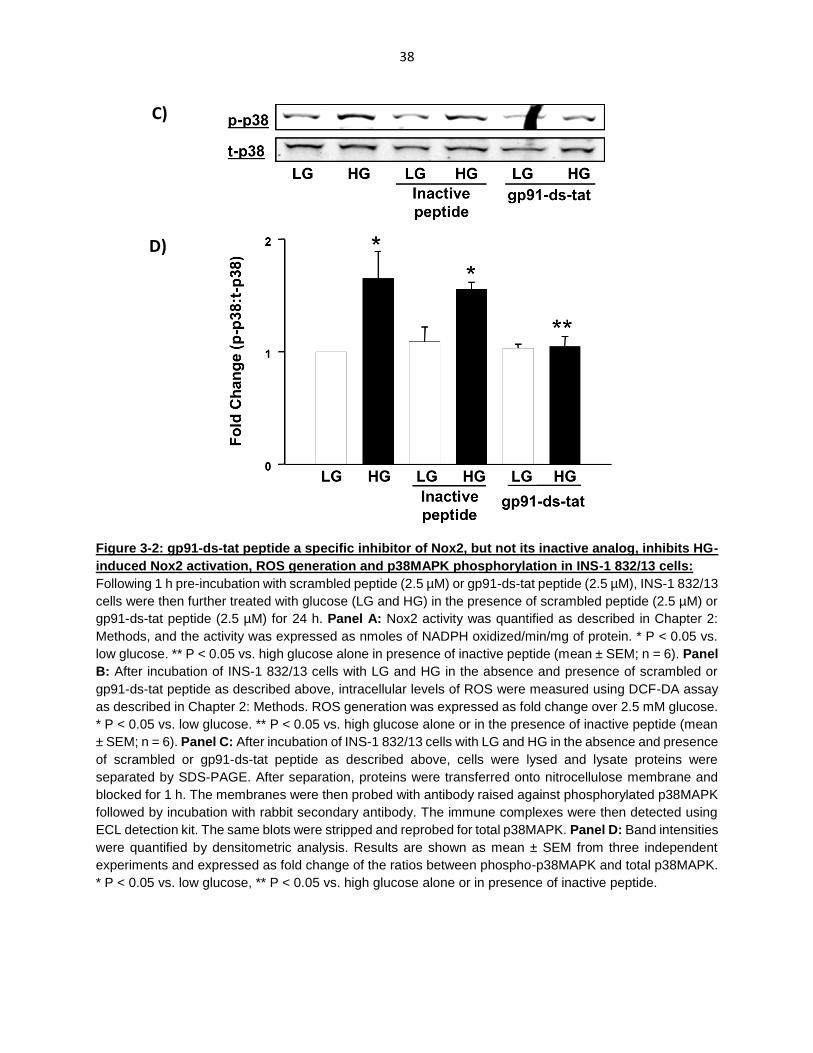
38
Figure 3-2: gp91-ds-tat peptide a specific inhibitor of Nox2, but not its inactive analog, inhibits HG-
induced Nox2 activation, ROS generation and p38MAPK phosphorylation in INS-1 832/13 cells:
Following 1 h pre-incubation with scrambled peptide (2.5 µM) or gp91-ds-tat peptide (2.5 µM), INS-1 832/13
cells were then further treated with glucose (LG and HG) in the presence of scrambled peptide (2.5 µM) or
gp91-ds-tat peptide (2.5 µM) for 24 h. Panel A: Nox2 activity was quantified as described in Chapter 2:
Methods, and the activity was expressed as nmoles of NADPH oxidized/min/mg of protein. * P < 0.05 vs.
low glucose. ** P < 0.05 vs. high glucose alone in presence of inactive peptide (mean ± SEM; n = 6). Panel
B: After incubation of INS-1 832/13 cells with LG and HG in the absence and presence of scrambled or
gp91-ds-tat peptide as described above, intracellular levels of ROS were measured using DCF-DA assay
as described in Chapter 2: Methods. ROS generation was expressed as fold change over 2.5 mM glucose.
* P < 0.05 vs. low glucose. ** P < 0.05 vs. high glucose alone or in the presence of inactive peptide (mean
± SEM; n = 6). Panel C: After incubation of INS-1 832/13 cells with LG and HG in the absence and presence
of scrambled or gp91-ds-tat peptide as described above, cells were lysed and lysate proteins were
separated by SDS-PAGE. After separation, proteins were transferred onto nitrocellulose membrane and
blocked for 1 h. The membranes were then probed with antibody raised against phosphorylated p38MAPK
followed by incubation with rabbit secondary antibody. The immune complexes were then detected using
ECL detection kit. The same blots were stripped and reprobed for total p38MAPK. Panel D: Band intensities
were quantified by densitometric analysis. Results are shown as mean ± SEM from three independent
experiments and expressed as fold change of the ratios between phospho-p38MAPK and total p38MAPK.
* P < 0.05 vs. low glucose, ** P < 0.05 vs. high glucose alone or in presence of inactive peptide.
C)
D)
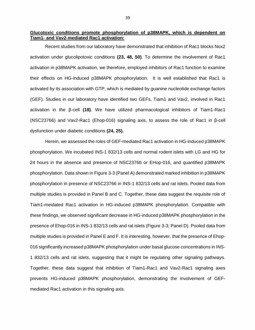
39
Glucotoxic conditions promote phosphorylation of p38MAPK, which is dependent on Tiam1- and Vav2-mediated Rac1 activation:
Recent studies from our laboratory have demonstrated that inhibition of Rac1 blocks Nox2
activation under glucolipotoxic conditions (23, 48, 50). To determine the involvement of Rac1
activation in p38MAPK activation, we therefore, employed inhibitors of Rac1 function to examine
their effects on HG-induced p38MAPK phosphorylation. It is well established that Rac1 is
activated by its association with GTP, which is mediated by guanine nucleotide exchange factors
(GEF). Studies in our laboratory have identified two GEFs, Tiam1 and Vav2, involved in Rac1
activation in the β-cell (18). We have utilized pharmacological inhibitors of Tiam1-Rac1
(NSC23766) and Vav2-Rac1 (Ehop-016) signaling axis, to assess the role of Rac1 in β-cell
dysfunction under diabetic conditions (24, 25).
Herein, we assessed the roles of GEF-mediated Rac1 activation in HG-induced p38MAPK
phosphorylation. We incubated INS-1 832/13 cells and normal rodent islets with LG and HG for
24 hours in the absence and presence of NSC23766 or EHop-016, and quantified p38MAPK
phosphorylation. Data shown in Figure 3-3 (Panel A) demonstrated marked inhibition in p38MAPK
phosphorylation in presence of NSC23766 in INS-1 832/13 cells and rat islets. Pooled data from
multiple studies is provided in Panel B and C. Together, these data suggest the requisite role of
Tiam1-meidated Rac1 activation in HG-induced p38MAPK phosphorylation. Compatible with
these findings, we observed significant decrease in HG-induced p38MAPK phosphorylation in the
presence of Ehop-016 in INS-1 832/13 cells and rat islets (Figure 3-3; Panel D). Pooled data from
multiple studies is provided in Panel E and F. It is interesting, however, that the presence of Ehop-
016 significantly increased p38MAPK phosphorylation under basal glucose concentrations in INS-
1 832/13 cells and rat islets, suggesting that it might be regulating other signaling pathways.
Together, these data suggest that inhibition of Tiam1-Rac1 and Vav2-Rac1 signaling axes
prevents HG-induced p38MAPK phosphorylation, demonstrating the involvement of GEF-
mediated Rac1 activation in this signaling axis.
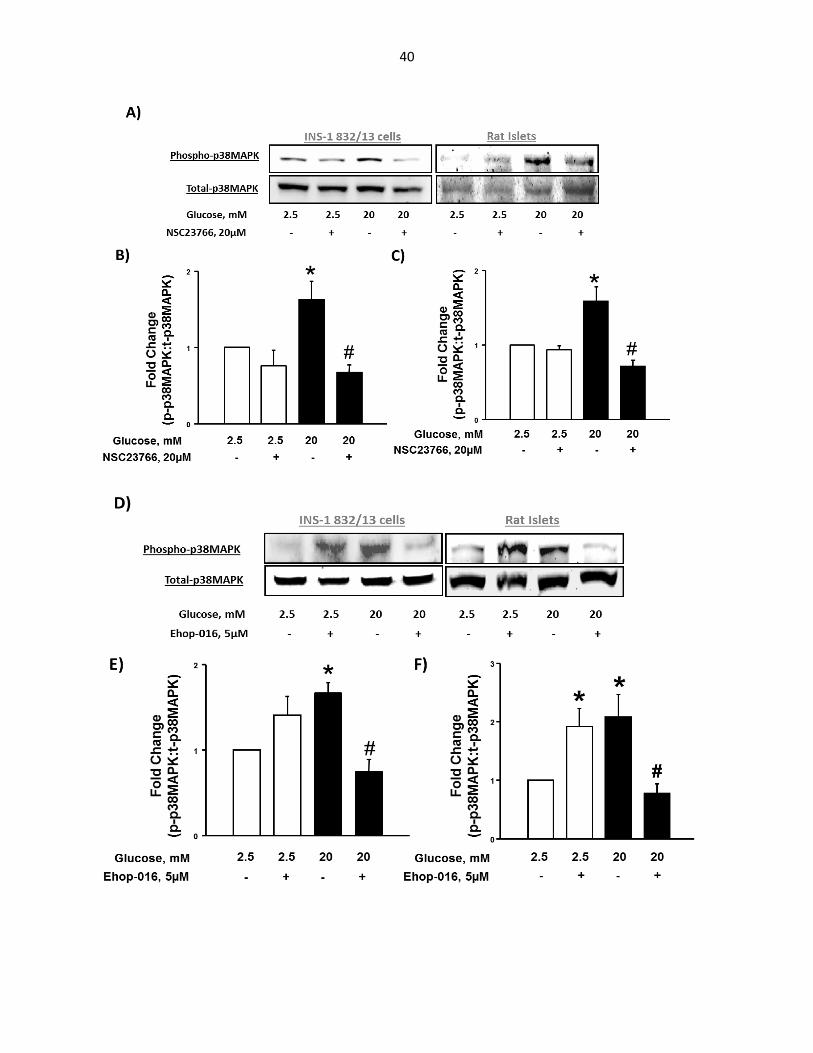
40

41
Figure 3-3: NSC23766 and Ehop-016, which selectively block Tiam1- and Vav2-mediated Rac1
activation, prevent HG-induced p38MAPK phosphorylation in INS-1 832/13 cells and rat pancreatic
islets:
Panel A: Rat pancreatic islets and INS-1 832/13 cells, pre-incubated overnight with NSC23766 (20 µM),
were further treated with glucose (LG and HG) in the absence or presence of NSC23766 (20 µM) for 24 h.
Cells were then lysed and proteins were resolved by SDS-PAGE. Separated proteins were transferred onto
nitrocellulose membranes. After blocking for 1 h, the membranes were then probed with phosphorylated
p38MAPK antibody followed by incubation with rabbit secondary antibody. The antibody complexes were
then detected by ECL detection method. The same blots were stripped and reprobed with antibody against
total p38MAPK. Panel B: Band intensities of phospho-p38MAPK and total-p38MAPK in INS-1 832/13 cells
were quantified by densitometric analysis. Results are shown as mean ± SEM from three independent
experiments and expressed as fold change of the ratios between phospho-p38MAPK and total-p38MAPK.
* P < 0.05 vs. low glucose, # P < 0.05 vs. high glucose. Panel C: Band intensities of phospho-p38MAPK
and total-p38MAPK in rat pancreatic islets were quantified by densitometric analysis. Results are shown as
mean ± SEM from three independent experiments and expressed as fold change of the ratios between
phospho-p38MAPK and total-p38MAPK. * P < 0.05 vs. low glucose, # P < 0.05 vs. high glucose. Panel D:
Rat pancreatic islets and INS-1 832/13 cells, pre-incubated overnight with Ehop-016 (5 µM), were further
treated with glucose (LG and HG) in the absence or presence of Ehop-016 (5 µM) for 24 h. Lysate proteins
were then resolved by SDS-PAGE, and analyzed by Western Blotting, as described above, for detecting
phosphorylated and total p38MAPK. Panel E: Band intensities of phospho-p38MAPK and total-p38MAPK
in INS-1 832/13 cells were quantified by densitometric analysis. Results are shown as mean ± SEM from
three independent experiments and expressed as fold change of the ratios between phospho-p38MAPK
and total p38MAPK. * P < 0.05 vs. low glucose, # P < 0.05 vs. high glucose. Panel F: Band intensities of
phosphor-p38MAPK and total-p38MAPK in rat pancreatic islets were quantified by densitometric analysis.
Results are shown as mean ± SEM from three independent experiments and expressed as fold change of
the ratios between phospho-p38MAPK and total p38MAPK. * P < 0.05 vs. low glucose, # P < 0.05 vs. high
glucose.
EHT1864, a novel inhibitor of Rac1, blocks activation and membrane association of Rac1 activation and insulin secretion upon physiological glucose stimulation in INS-1 832/13 β-cells:
Desire and associates have designed a novel small molecular weight inhibitor EHT1864,
which blocks Rac1 activation by direct interaction in a GEF-independent manner (28). Studies by
Shutes et al. have indicated that EHT1864 binds to Rac1 with a higher affinity than guanine
nucleotides (GDP/GTP) thereby displacing GDP/GTP from the Rac1 binding site, retaining Rac1
in an inert, inactive state (27, 128). Several studies have utilized this inhibitor to assess the roles
of Rac1 in cell physiology and disease (129-132). At the outset, we undertook the study to
examine the effects of EHT1864 on Rac1 function and glucose-stimulated insulin secretion under
physiological conditions. Following 1 h pre-incubation with EHT1864, we stimulated INS-1 832/13
cells with LG (2.5mM) and HG (20mM) for 20 min in the continuous absence or presence of

42
EHT1864 and quantified Rac1 activation. Data depicted in Figure 3-4 (Panel A) demonstrate a
marked increase in Rac1-GTP levels under stimulatory glucose concentration, which is
significantly blocked in the presence of EHT1864. It is noteworthy, however, that EHT1864
caused an increase in Rac1 activation under basal conditions, although such an increase was not
statistically significant (Bar1 vs Bar2).
We, next assessed the effects of Rac1 inhibition with EHT1864, on glucose-stimulated
insulin secretion. Following pre-incubation with EHT1864 for 1 h as indicated in Figure 3-4 (Panel
B), INS-1 832/13 cells were incubated with LG and HG for 30 in the continuous absence or
presence of EHT1864, and quantified GSIS. Data depicted in Figure (Panel B) indicate a marked
increase in insulin secretory response under stimulatory glucose concentration (20mM). We
noticed a modest inhibition in GSIS in presence of EHT1864 at 5 µM concentrations. However,
EHT1864 significantly blocked GSIS at 10uM concentrations. It is noteworthy, however, that 10
µM EHT1864 augmented insulin secretion under basal glucose concentrations. Similarly, as
depicted in Figure 3-4 (Panel C), we noticed a significant increase in insulin secretion at 5mM
glucose concentration in presence of 10 µM EHT1864. Together, these observations demonstrate
inhibitory effects of EHT1864 on Rac1 activation and GSIS in INS-1 832/13 cells.
Previous investigations have suggested that membrane targeting of small G-proteins
(Rac1 and Cdc42) is required for their optimal interaction with effector proteins and physiological
function. We therefore, examined if glucose-induced membrane association of Rac1 is affected
in presence of EHT1864. To address this, we first isolated cytosolic and membrane fractions of
INS-832/13 cells treated with LG and HG with or without EHT1864 for 20 min. The membrane
fractions were further processed by TritonX partitioning, to isolate hydrophilic and hydrophobic
compartments. The relative abundance of Rac1 in these fractions was determined by Western
Blotting and the purity of the fractions was confirmed by probing for their respective marker
proteins. Data depicted in Figure 3-4 (Panel D) indicate that Rac1 is localized in the cytosolic
fraction under basal conditions. However, cells treated with 20mM glucose showed marked

43
increase in Rac1 localized in the hydrophilic and hydrophobic fractions of the membrane.
Furthermore, the presence of EHT1864, prevented Rac1 translocation to the membrane fraction
induced by 20mM glucose concentration. A representative blot from multiple studies is provided
in Panel D. Collectively, these data demonstrate that EHT1864 prevents activation and membrane
targeting of Rac1, thereby inhibiting insulin secretion under stimulatory concentrations of glucose.
A)

44
B) C)
D)
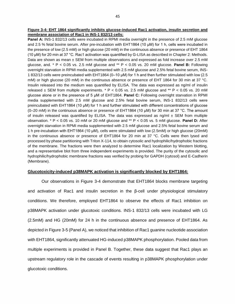
45
Figure 3-4: EHT 1864 significantly inhibits glucose-induced Rac1 activation, insulin secretion and
membrane association of Rac1 in INS-1 832/13 cells:
Panel A: INS-1 832/13 cells were incubated in RPMI media overnight in the presence of 2.5 mM glucose
and 2.5 % fetal bovine serum. After pre-incubation with EHT1864 (10 μM) for 1 h, cells were incubated in
the presence of low (2.5 mM) or high glucose (20 mM) in the continuous absence or presence of EHT 1864
(10 μM) for 20 min at 37 °C. Rac1 activation was quantified by G-LISA as described in Chapter 2: Methods.
Data are shown as mean ± SEM from multiple observations and expressed as fold increase over 2.5 mM
glucose, and. * P < 0.05 vs. 2.5 mM glucose and ** P < 0.05 vs. 20 mM glucose. Panel B: Following
overnight starvation in RPMI media supplemented with 2.5 mM glucose and 2.5% fetal bovine serum, INS-
1 832/13 cells were preincubated with EHT1864 (0–10 μM) for 1 h and then further stimulated with low (2.5
mM) or high glucose (20 mM) in the continuous absence or presence of EHT 1864 for 30 min at 37 °C.
Insulin released into the medium was quantified by ELISA. The data was expressed as ng/ml of insulin
released ± SEM from multiple experiments. * P < 0.05 vs. 2.5 mM glucose and ** P < 0.05 vs. 20 mM
glucose alone or in the presence of 5 μM of EHT1864. Panel C: Following overnight starvation in RPMI
media supplemented with 2.5 mM glucose and 2.5% fetal bovine serum, INS-1 832/13 cells were
preincubated with EHT1864 (10 μM) for 1 h and further stimulated with different concentrations of glucose
(0–20 mM) in the continuous absence or presence of EHT1864 (10 μM) for 30 min at 37 °C. The amount
of insulin released was quantified by ELISA. The data was expressed as ng/ml ± SEM from multiple
observation. * P < 0.05 vs. 10 mM or 20 mM glucose and ** P < 0.05 vs. 5 mM glucose. Panel D: After
overnight starvation in RPMI media supplemented with 2.5 mM glucose and 2.5% fetal bovine serum and
1 h pre-incubation with EHT1864 (10 μM), cells were stimulated with low (2.5mM) or high glucose (20mM)
in the continuous absence or presence of EHT1864 for 20 min at 37 °C. Cells were then lysed and
processed by phase partitioning with Triton X-114, to obtain cytosolic and hydrophilic/hydrophobic fractions
of the membrane. The fractions were then analyzed to determine Rac1 localization by Western blotting,
and a representative blot from three independent experiments is provided. The purity of the cytosolic and
hydrophilic/hydrophobic membrane fractions was verified by probing for GAPDH (cytosol) and E-Cadherin
(Membrane).
Glucotoxicity-induced p38MAPK activation is significantly blocked by EHT1864:
Our observations in Figure 3-4 demonstrate that EHT1864 blocks membrane targeting
and activation of Rac1 and insulin secretion in the β-cell under physiological stimulatory
conditions. We therefore, employed EHT1864 to observe the effects of Rac1 inhibition on
p38MAPK activation under glucotoxic conditions. INS-1 832/13 cells were incubated with LG
(2.5mM) and HG (20mM) for 24 h in the continuous absence and presence of EHT1864. As
depicted in Figure 3-5 (Panel A), we noticed that inhibition of Rac1 guanine nucleotide association
with EHT1864, significantly attenuated HG-induced p38MAPK phosphorylation. Pooled data from
multiple experiments is provided in Panel B. Together, these data suggest that Rac1 plays an
upstream regulatory role in the cascade of events resulting in p38MAPK phosphorylation under
glucotoxic conditions.

46
Figure 3-5: EHT1864 attenuates HG-induced p38MAPK phosphorylation in INS-1 832/13 cells:
Panel A: INS-1 832/13 cells were incubated with glucose (2.5 mM and 20 mM) for 24 h in the continuous
absence or presence of EHT1864 (10 µM). Cells were then lysed and lysate proteins were then separated
by SDS-PAGE, and transferred onto nitrocellulose membranes. The membranes were then blocked and
probed with antibody raised against phosphorylated p38MAPK, followed by incubation with rabbit
secondary antibody. The immune complexes were detected by ECL detection method. The saem blots
were then stripped and reprobed for total p38MAPK. Panel B: Band intensities were then quantified by
densitometric analysis. Results are shown as mean ± SEM from three independent experiments and
expressed as fold change of the ratios between phosphor-p38MAPK and total-p38MAPK. * P < 0.05 vs.
low glucose, # P < 0.05 vs. high glucose.
p-p38
t-p38
Glucose, mM 2.5 2.5 20 20
EHT 1864, 10µM - + - +
A)
B)
0
1
2
Fo
ld C
ha
ng
e (
p-p
38:t
-p3
8)
Glucose, mM 2.5 2.5 20 20
EHT 1864, 10µM - + - +
*
#
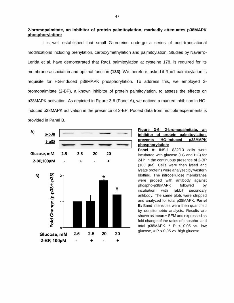
47
2-bromopalmitate, an inhibitor of protein palmitoylation, markedly attenuates p38MAPK phosphorylation:
It is well established that small G-proteins undergo a series of post-translational
modifications including prenylation, carboxymethylation and palmitoylation. Studies by Navarro-
Lerida et al. have demonstrated that Rac1 palmitoylation at cysteine 178, is required for its
membrane association and optimal function (133). We therefore, asked if Rac1 palmitoylation is
requisite for HG-induced p38MAPK phosphorylation. To address this, we employed 2-
bromopalmitate (2-BP), a known inhibitor of protein palmitoylation, to assess the effects on
p38MAPK activation. As depicted in Figure 3-6 (Panel A), we noticed a marked inhibition in HG-
induced p38MAPK activation in the presence of 2-BP. Pooled data from multiple experiments is
provided in Panel B.
Figure 3-6: 2-bromopalmitate, an
inhibitor of protein palmitoylation,
prevents HG-induced p38MAPK
phosphorylation:
Panel A: INS-1 832/13 cells were
incubated with glucose (LG and HG) for
24 h in the continuous presence of 2-BP
(100 µM). Cells were then lysed and
lysate proteins were analyzed by western
blotting. The nitrocellulose membranes
were probed with antibody against
phospho-p38MAPK followed by
incubation with rabbit secondary
antibody. The same blots were stripped
and analyzed for total p38MAPK. Panel
B: Band intensities were then quantified
by densitometric analysis. Results are
shown as mean ± SEM and expressed as
fold change of the ratios of phospho- and
total p38MAPK. * P < 0.05 vs. low
glucose, # P < 0.05 vs. high glucose.

48
GGTI-2147, an inhibitor of protein geranylgeranylation, had no effect on p38MAPK phosphorylation:
Previous experiments in our laboratory have utilized pharmacological inhibitors, dominant
negative mutants and siRNA approaches to demonstrate the role of post-translational
modifications of small G-proteins (e.g. Rac1) in their activity (34). Therefore, in the next set of
experiments, we determined if Rac1 geranylgeranylation is requisite for HG-induced p38MAPK
phosphorylation. We quantified p38MAPK phosphorylation following incubation of INS-1 832/13
cells with LG (2.5mM) and HG (20mM) for 24 h in the continuous presence or absence of GGTI-
2147, a known inhibitor of protein geranylgeranylation. As shown in Figure 3-7 (Panel A), we did
not notice any significant effects of GGTI-2147 on HG-induced p38MAPK activation. Pooled data
from multiple studies is provided in Panel B. These data together suggest that Rac1
geranylgeranylation of Rac1 may not be required for p38MAPK activation under glucotoxic
conditions.
Figure 3-7: GGTI-2147, an inhibitor of
protein geranylgeranylation, exhibits
no effect on HG-induced p38MAPK
phosphorylation:
Panel A: INS-1 832/13 cells were
incubated with glucose (LG and HG) for
24 h in the continuous presence of
GGTI-2147 (10 µM). Cells were then
lysed and lysate proteins were analyzed
by western blotting. The nitrocellulose
membranes were probed with antibody
against phospho-p38MAPK followed by
incubation with rabbit secondary
antibody. The same blots were stripped
and analyzed for total p38MAPK. Panel
B: Band intensities were then quantified
by densitometric analysis. Results are
shown as mean ± SEM and expressed
as fold change of the ratios of phospho-
and total p38MAPK. * P < 0.05 vs. low
glucose.

49
Collectively, evidence shown in figure 3-8 demonstrate that exposure of pancreatic β-cells
to glucotoxic conditions lead to activation of p38MAPK activation. Using pharmacological
inhibitors targeting Nox2 enzyme activity and Rac1 activation, we provide the first evidence
suggesting the involvement of Rac1-Nox2-derived oxidative stress in the signaling events leading
to p38MAPK activation.
Figure 3-8: Proposed model for Rac1-Nox2-mediated ROS generation and p38MAPK activation under glucotoxic conditions: Using pharmacological inhibitors of Rac1 (NSC23766, Ehop-016 and EHT1864), we have demonstrated that Rac1 activation is involved in HG-induced p38MAPK phosphorylation. Furthermore, gp91-ds-tat, but not is inactive analog, significantly attenuated HG-induced Nox2 activity, ROS generation and p38MAPK phosphorylation.
50
CHAPTER 4: GLUCOTOXIC CONDITIONS PROMOTE RAC1-p38MAPK-DEPENDENT ACTIVATION OF p53 TUMOR SUPPRESSOR
p53 tumor suppressor is a transcription factor and consists of several structural and
functional domains which regulate its function and stability. Evidence from multiple cell types have
implicated activation of the p53 pathway under the duress of oxidative stress and DNA damage,
thereby mediating cellular responses to the stress stimuli including cell cycle arrest, DNA repair
and apoptosis (83). Studies in multiple cell types have suggested activation of apoptotic genes
including Bax, Apaf1, PUMA, Noxa, by p53-mediated mechanisms (108). However, the
contributory role of p53 in pancreatic β-cell dysfunction under glucotoxic conditions remains poorly
understood. To address this, we herein examined the involvement of p53 in the Rac1-Nox2-
p38MAPK signaling cascade in β-cells exposed to glucotoxic conditions.
Several post-translational modification (e.g., Phosphorylation, Acetylation, Ubiquitination)
have been shown to regulate p53 functionality and protein levels in the cell (88). It has been
suggested that phosphorylation of p53 at serine-15 residue in the N-terminal transactivation
domain is critical for its stabilization and transcriptional activation (106). Studies have also
implicated that serine-15 phosphorylation regulates p53 interaction with MDM2 and CBP/p300
that modulate its stability and DNA binding affinity (90, 134). p38MAPK activation has been
suggested to result in phosphorylation and stabilization of p53 under specific stress stimuli in
multiple cell types (79, 135). Additionally, studies by Yoshida and associates have also indicated
the involvement of ATM kinase in p53 activation in Doxorubicin-induced cardiotoxicity. Using
pharmacological approaches, they have demonstrated the role of Rac1-mediated oxidative stress
in ATM kinase and p53 activation resulting in Doxorubicin-induced cardiomyopathy (136).
Furthermore, studies by Oleson et al. have also shown that ATM kinase mediates DNA repair in
pancreatic β-cells exposed to inflammatory cytokines, but upon extensive dsDNA breaks may be
responsible for activating apoptotic pathways primarily through p53-dependent mechanisms
(137). Using specific pharmacological inhibitors, we explored the regulatory roles of Rac1-Nox2

51
enzyme complex and upstream kinases including ATM kinase and p38MAPK in the p53 activation
pathway, ultimately resulting in β-cell death.
p53 tumor suppressor is activated by serine-15 phosphorylation in INS-1 832/13 cells and rat islets exposed to glucotoxic conditions:
Phosphorylation of p53 at serine-15 in N-terminus transactivation domain residue has
been associated with decreased binding to MDM2 (negative modulator) and increased interaction
with p300 (positive modulator), thereby increasing functional activation and stabilization of p53
(90, 134). Therefore, we first determined if exposure to glucotoxic conditions induced p53
activation by serine-15 phosphorylation. We incubated INS-1 832/13 cells and rat islets with LG
(2.5 mM) and HG (20 mM) for 24 hours and quantified p53 phosphorylation in these samples. As
shown in Figure 4-1, exposure to HG concentrations induced significant increase in p53
phosphorylation at serine-15 in INS-1 832/13 cells (Panel A) and rat islets (Panel C). Data from
multiple experiments is shown in Panel B and D.
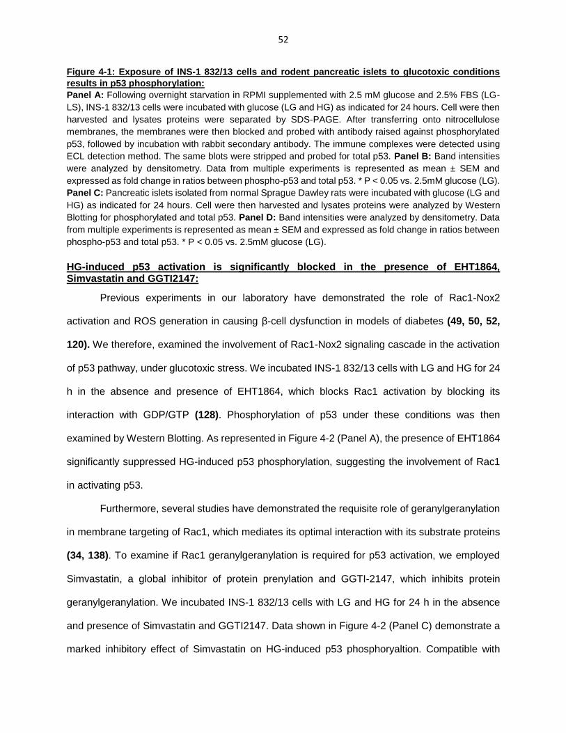
52
Figure 4-1: Exposure of INS-1 832/13 cells and rodent pancreatic islets to glucotoxic conditions
results in p53 phosphorylation:
Panel A: Following overnight starvation in RPMI supplemented with 2.5 mM glucose and 2.5% FBS (LG-
LS), INS-1 832/13 cells were incubated with glucose (LG and HG) as indicated for 24 hours. Cell were then
harvested and lysates proteins were separated by SDS-PAGE. After transferring onto nitrocellulose
membranes, the membranes were then blocked and probed with antibody raised against phosphorylated
p53, followed by incubation with rabbit secondary antibody. The immune complexes were detected using
ECL detection method. The same blots were stripped and probed for total p53. Panel B: Band intensities
were analyzed by densitometry. Data from multiple experiments is represented as mean ± SEM and
expressed as fold change in ratios between phospho-p53 and total p53. * P < 0.05 vs. 2.5mM glucose (LG).
Panel C: Pancreatic islets isolated from normal Sprague Dawley rats were incubated with glucose (LG and
HG) as indicated for 24 hours. Cell were then harvested and lysates proteins were analyzed by Western
Blotting for phosphorylated and total p53. Panel D: Band intensities were analyzed by densitometry. Data
from multiple experiments is represented as mean ± SEM and expressed as fold change in ratios between
phospho-p53 and total p53. * P < 0.05 vs. 2.5mM glucose (LG).
HG-induced p53 activation is significantly blocked in the presence of EHT1864, Simvastatin and GGTI2147:
Previous experiments in our laboratory have demonstrated the role of Rac1-Nox2
activation and ROS generation in causing β-cell dysfunction in models of diabetes (49, 50, 52,
120). We therefore, examined the involvement of Rac1-Nox2 signaling cascade in the activation
of p53 pathway, under glucotoxic stress. We incubated INS-1 832/13 cells with LG and HG for 24
h in the absence and presence of EHT1864, which blocks Rac1 activation by blocking its
interaction with GDP/GTP (128). Phosphorylation of p53 under these conditions was then
examined by Western Blotting. As represented in Figure 4-2 (Panel A), the presence of EHT1864
significantly suppressed HG-induced p53 phosphorylation, suggesting the involvement of Rac1
in activating p53.
Furthermore, several studies have demonstrated the requisite role of geranylgeranylation
in membrane targeting of Rac1, which mediates its optimal interaction with its substrate proteins
(34, 138). To examine if Rac1 geranylgeranylation is required for p53 activation, we employed
Simvastatin, a global inhibitor of protein prenylation and GGTI-2147, which inhibits protein
geranylgeranylation. We incubated INS-1 832/13 cells with LG and HG for 24 h in the absence
and presence of Simvastatin and GGTI2147. Data shown in Figure 4-2 (Panel C) demonstrate a
marked inhibitory effect of Simvastatin on HG-induced p53 phosphoryaltion. Compatible with

53
these findings, data shown in Figure 4-2 (Panel E) also demonstrate that GGT-2147 prevents p53
phosphorylation under HG conditions. Data pooled from multiple experiments is provided in Panel
D and F. Together, these findings implicate that Rac1 activation and prenylation are necessary
for HG-induced activation of the p53 pathway.
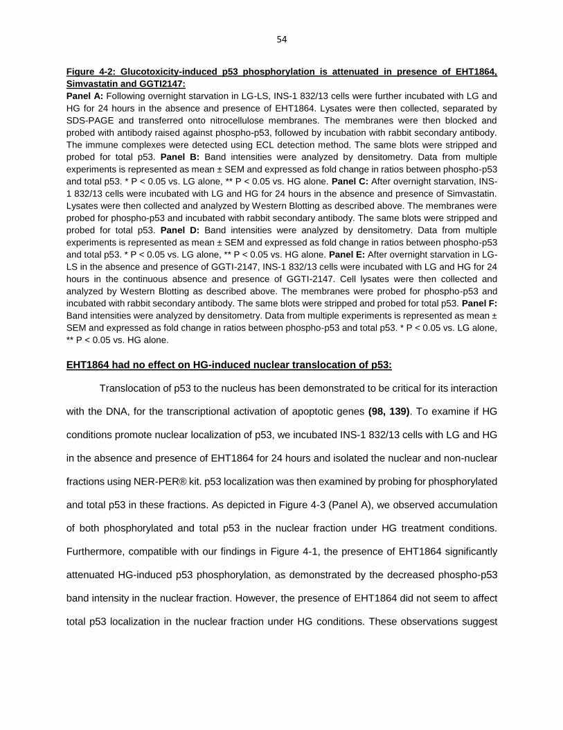
54
Figure 4-2: Glucotoxicity-induced p53 phosphorylation is attenuated in presence of EHT1864,
Simvastatin and GGTI2147:
Panel A: Following overnight starvation in LG-LS, INS-1 832/13 cells were further incubated with LG and
HG for 24 hours in the absence and presence of EHT1864. Lysates were then collected, separated by
SDS-PAGE and transferred onto nitrocellulose membranes. The membranes were then blocked and
probed with antibody raised against phospho-p53, followed by incubation with rabbit secondary antibody.
The immune complexes were detected using ECL detection method. The same blots were stripped and
probed for total p53. Panel B: Band intensities were analyzed by densitometry. Data from multiple
experiments is represented as mean ± SEM and expressed as fold change in ratios between phospho-p53
and total p53. * P < 0.05 vs. LG alone, ** P < 0.05 vs. HG alone. Panel C: After overnight starvation, INS-
1 832/13 cells were incubated with LG and HG for 24 hours in the absence and presence of Simvastatin.
Lysates were then collected and analyzed by Western Blotting as described above. The membranes were
probed for phospho-p53 and incubated with rabbit secondary antibody. The same blots were stripped and
probed for total p53. Panel D: Band intensities were analyzed by densitometry. Data from multiple
experiments is represented as mean ± SEM and expressed as fold change in ratios between phospho-p53
and total p53. * P < 0.05 vs. LG alone, ** P < 0.05 vs. HG alone. Panel E: After overnight starvation in LG-
LS in the absence and presence of GGTI-2147, INS-1 832/13 cells were incubated with LG and HG for 24
hours in the continuous absence and presence of GGTI-2147. Cell lysates were then collected and
analyzed by Western Blotting as described above. The membranes were probed for phospho-p53 and
incubated with rabbit secondary antibody. The same blots were stripped and probed for total p53. Panel F:
Band intensities were analyzed by densitometry. Data from multiple experiments is represented as mean ±
SEM and expressed as fold change in ratios between phospho-p53 and total p53. * P < 0.05 vs. LG alone,
** P < 0.05 vs. HG alone.
EHT1864 had no effect on HG-induced nuclear translocation of p53:
Translocation of p53 to the nucleus has been demonstrated to be critical for its interaction
with the DNA, for the transcriptional activation of apoptotic genes (98, 139). To examine if HG
conditions promote nuclear localization of p53, we incubated INS-1 832/13 cells with LG and HG
in the absence and presence of EHT1864 for 24 hours and isolated the nuclear and non-nuclear
fractions using NER-PER® kit. p53 localization was then examined by probing for phosphorylated
and total p53 in these fractions. As depicted in Figure 4-3 (Panel A), we observed accumulation
of both phosphorylated and total p53 in the nuclear fraction under HG treatment conditions.
Furthermore, compatible with our findings in Figure 4-1, the presence of EHT1864 significantly
attenuated HG-induced p53 phosphorylation, as demonstrated by the decreased phospho-p53
band intensity in the nuclear fraction. However, the presence of EHT1864 did not seem to affect
total p53 localization in the nuclear fraction under HG conditions. These observations suggest

55
that HG exposure induces p53 translocation to the nucleus, independent of phosphorylation at
serine-15 residue.
Figure 4-3: Exposure to HG conditions induced nuclear translocation of p53, which is not inhibited
by EHT1864:
Panel A: Following overnight incubation in LG-LS media, INS-1 832/13 cells were further incubated with
LG and HG for 24 h in the absence and presence of EHT1864. Cells were then rinsed with PBS and nuclear
and non-nuclear fractions were isolated using NE-PER® kit according to the manufacturer’s instructions.
The isolated fractions were then analyzed by Western Blotting for phosphorylated and total p53. The purity
of the fractions was analyzed by probing for nuclear Lamin B. Panel B: Band intensities for phospho-p53
and Lamin B were determined by densitometric analysis. Data from multiple studies is shown as mean ±
SEM and expressed as fold change in the ratios between phospho-p53 and Lamin B. * P < 0.05 vs. LG
alone or in the presence of EHT1864, ** P < 0.05 vs. HG alone. Panel C: Band intensities for total-p53 and
Lamin B were determined by densitometric analysis. Data from multiple studies is shown as mean ± SEM
and expressed as fold change in the ratios between total-p53 and Lamin B. * P < 0.05 vs. LG alone. NS:
not significant
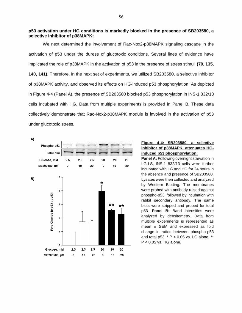
56
p53 activation under HG conditions is markedly blocked in the presence of SB203580, a selective inhibitor of p38MAPK:
We next determined the involvement of Rac-Nox2-p38MAPK signaling cascade in the
activation of p53 under the duress of glucotoxic conditions. Several lines of evidence have
implicated the role of p38MAPK in the activation of p53 in the presence of stress stimuli (79, 135,
140, 141). Therefore, in the next set of experiments, we utilized SB203580, a selective inhibitor
of p38MAPK activity, and observed its effects on HG-induced p53 phosphorylation. As depicted
in Figure 4-4 (Panel A), the presence of SB203580 blocked p53 phosphorylation in INS-1 832/13
cells incubated with HG. Data from multiple experiments is provided in Panel B. These data
collectively demonstrate that Rac-Nox2-p38MAPK module is involved in the activation of p53
under glucotoxic stress.
Figure 4-4: SB203580, a selective
inhibitor of p38MAPK, attenuates HG-
induced p53 phosphorylation:
Panel A: Following overnight starvation in
LG-LS, INS-1 832/13 cells were further
incubated with LG and HG for 24 hours in
the absence and presence of SB203580.
Lysates were then collected and analyzed
by Western Blotting. The membranes
were probed with antibody raised against
phospho-p53, followed by incubation with
rabbit secondary antibody. The same
blots were stripped and probed for total
p53. Panel B: Band intensities were
analyzed by densitometry. Data from
multiple experiments is represented as
mean ± SEM and expressed as fold
change in ratios between phospho-p53
and total p53. * P < 0.05 vs. LG alone, **
P < 0.05 vs. HG alone.
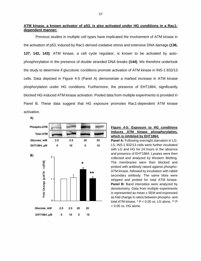
57
ATM kinase, a known activator of p53, is also activated under HG conditions in a Rac1-dependent manner:
Previous studies in multiple cell types have implicated the involvement of ATM kinase in
the activation of p53, induced by Rac1-derived oxidative stress and extensive DNA damage (136,
137, 142, 143). ATM kinase, a cell cycle regulator, is known to be activated by auto-
phosphorylation in the presence of double stranded DNA breaks (144). We therefore undertook
the study to determine if glucotoxic conditions promote activation of ATM kinase in INS-1 832/13
cells. Data depicted in Figure 4-5 (Panel A) demonstrate a marked increase in ATM kinase
phophorylation under HG conditions. Furthermore, the presence of EHT1864, significantly
blocked HG-induced ATM kinase activation. Pooled data from multiple experiments is provided in
Panel B. These data suggest that HG exposure promotes Rac1-dependent ATM kinase
activation.
Figure 4-5: Exposure to HG conditions
induces ATM kinase phosphorylation,
which is inhibited by EHT1864:
Panel A: Following overnight starvation in LG-
LS, INS-1 832/13 cells were further incubated
with LG and HG for 24 hours in the absence
and presence of EHT1864. Lysates were then
collected and analyzed by Western Blotting.
The membranes were then blocked and
probed with antibody raised against phospho-
ATM kinase, followed by incubation with rabbit
secondary antibody. The same blots were
stripped and probed for total ATM kinase.
Panel B: Band intensities were analyzed by
densitometry. Data from multiple experiments
is represented as mean ± SEM and expressed
as fold change in ratios between phospho- and
total ATM kinase. * P < 0.05 vs. LG alone, ** P
< 0.05 vs. HG alone.
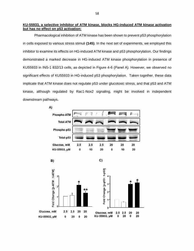
58
KU-55933, a selective inhibitor of ATM kinase, blocks HG-induced ATM kinase activation but has no effect on p53 activation:
Pharmacological inhibition of ATM kinase has been shown to prevent p53 phosphorylation
in cells exposed to various stress stimuli (145). In the next set of experiments, we employed this
inhibitor to examine its effects on HG-induced ATM kinase and p53 phosphorylation. Our findings
demonstrated a marked decrease in HG-induced ATM kinase phosphorylation in presence of
KU55933 in INS-1 832/13 cells, as depicted in Figure 4-6 (Panel A). However, we observed no
significant effects of KU55933 in HG-induced p53 phosphorylation. Taken together, these data
implicate that ATM kinase does not regulate p53 under glucotoxic stress, and that p53 and ATM
kinase, although regulated by Rac1-Nox2 signaling, might be involved in independent
downstream pathways.

59
Figure 4-6: KU55933, a selective inhibitor of ATM kinase, prevents HG-induced ATM kinase
phosphorylation but has no effect on p53 phosphorylation:
Panel A: Following overnight starvation, INS-1 832/13 cells were incubated with LG or HG in the absence
or presence of KU-55933 for 24 h. Cell lysate proteins were separated and analyzed by Western Blotting
for phosphorylated ATM kinase and p53. Blots were then stripped and re-probed for total ATM kinase and
p53. Panel B: Phospho-ATM kinase band intensities were quantified by densitometry and ratios were
calculated over total-ATM kinase. Data from multiple studies is shown as mean ± SEM and expressed as
fold change in ratios between phosphorylated and total ATM kinase. * P < 0.05 vs LG alone, ** P< 0.05 vs
HG alone. Panel C: Phospho-p53 band intensities were quantified by densitometry and ratios were
calculated over total-p53. Data from multiple studies is shown as mean ± SEM and expressed as fold
change in ratios between phosphorylated and total p53. * P < 0.05 vs LG alone.
EHT1864, markedly blocks HG-induced β-cell death:
Our findings have demonstrated that Rac1 activation leads to activation of p38MAPK and
p53 signaling pathway, resulting in β-cell death under glucotoxic stress. Therefore, in the last set
of experiments, we utilized EHT1864 to determine if pharmacological inhibition of Rac1 prevents
HG-induced β-cell death. We incubated INS-1 832/13 cells with LG and HG for 24 h in the
continuous absence and presence of EHT1864 and analyzed cell death using Cell Death
Detection® kit as per the manufacturer’s instructions. Our findings indicated a significant increase
in cell death signal under HG conditions as shown in Figure 4-7. We also observed a marked
decrease in HG-induced β-cell death in presence of EHT1864, demonstrating protective effects
of Rac1 inhibition under glucotoxic stress conditions.

60
Figure 4-7: EHT1864 prevents HG-induced cell death in INS-1 832/13 cells:
After overnight incubation in LG-LS media, INS-1 832/13 cells were incubated with LG and HG for 24 h in
the absence and presence of EHT1864. Cells were then washed with PBS and analyzed using Cell Death
Detection ELISAPlus kit according to the manufacturer’s instructions. Absorbance was measured at 405nm
and expressed as fold change over basal LG. Data from multiple studies is shown as mean ± SEM and
expressed as fold change over basal LG. * p < 0.05 vs LG alone, ** p < 0.05 vs HG alone, NS: Not
significant.
In conclusion, our findings in chapter 3 and 4 together demonstrate that exposure of
pancreatic betake cells to glucotoxic conditions results in increased Rac1-Nox2-derived oxidative
stress, leading to activation of p38MAPK-p53 signaling axis that culminates in the induction of
apoptotic pathways. Our findings have also indicated activation of ATM kinase under glucotoxic
conditions, which might be involved in other signaling pathways.

61
Figure 4-8: Proposed model for Rac1-Nox2-induced oxidative stress and activation of p38MAPK-
p53 signaling axis leading to β-cell apoptosis:
Our findings have implicated Rac1-Nox2 activation in p38MAPK activation under glucotoxic stress.
Furthermore, using pharmacological inhibitor of Rac1 (EHT1864) and p38MAPK (SB203580) we have
demonstrated that activation of Rac1-p38MAPK signaling module is involved in HG-induced p53
phosphorylation.

62
CHAPTER 5: RAC1-p38MAPK-p53 SIGNALING AXIS IN PANCREATIC ISLETS FROM THE ZDF RAT MODEL AND HUMAN ISLETS
Phagocyte-like NADPH oxidase (Nox2) is composed of several membrane and cytosolic
components including the small G-protein, Rac1. Rac1 activation has been demonstrated to play
a critical role in the functional assembly and activation of Nox2 holoenzyme complex (41, 50, 52).
Although there is ample evidence suggesting the positive regulatory role of Rac1-Nox2 signaling
in physiological ROS generation and glucose-stimulated insulin secretion, Nox2 has been shown
to play key contributory roles in the pathophysiology of beta-cell dysfunction leading to onset of
diabetes. Previous observations in our laboratory have demonstrated that increased Rac1-Nox2-
derived ROS generation results in metabolic dysfunction of β-cells when exposed to palmitate
(50). Studies have also implicated the involvement of Nox2-induced oxidative stress in models of
cytokine-induced beta-cell dysfunction (52, 120). Furthermore, exposure to glucotoxic conditions
resulted in increased Nox2 activation in INS-1 832/13 cells (146). The central objective of this
dissertation project is to demonstrate that exposure of pancreatic β-cells to glucotoxic conditions
leads to sustained activation of Rac1-Nox2 enzyme complex and the resulting oxidative stress
activates p38MAPK-p53 signaling cascade, ultimately causing β-cell death. As discussed in
chapters 3 and 4, our in vitro studies in INS-1 832/13 cells and rat islets exposed to glucotoxic
conditions demonstrates increased activation of p38MAPK and p53. To examine this pathway in
an in vivo model, we utilized the Zucker diabetic fatty (ZDF) rat, which has been extensively used
as a model for obesity, insulin resistance and pancreatic β-cell dysfunction (147).
ZDF rats possess a spontaneous mutation of the leptin receptor gene and initially develop
obesity. Consequently, these animals spontaneously develop diabetes induced by elevated
glucose and free fatty acid levels. Animals which possess a heterozygous leptin receptor mutation
(Zucker lean control; ZLC), however, do not exhibit signs of diabetes (148). Previous studies from
our laboratory have utilized this model to demonstrate the involvement of increased Rac1-Nox2
activity and ROS generation in mediating β-cell dysfunction. Additionally, exposure of human

63
pancreatic islets to glucotoxic conditions also resulted in increased Rac1 activation and ROS
generation. The downstream signaling pathways in these models, however, remain to be fully
understood. Herein, we examined the p38MAPK-p53 signaling axis in pancreatic islets isolated
from ZDF rats and in human islets exposed to glucotoxic concentrations.
p38MAPK and p53 are activated in islets isolated from ZDF rats:
We first examined p38MAPK phosphorylation in pancreatic islets isolated from male ZDF
rats and compared them to their age-matched ZLC rats. As evidenced in Figure 5-1 (Panel A),
the ZDF rats that developed overt diabetes and exhibited elevated blood glucose levels, also
showed increased phosphorylation of p38MAPK. Data pooled from four ZDF rats is shown in
Panel B. Furthermore, in a separate set of studies, we examined levels of phosphorylated p53 in
two ZDF rats. We noticed a marked increase in p53 phosphorylation in islets isolated from ZDF
rats compared to their age-matched controls, as depicted in Figure 5-1 (Panel C). Data pooled
from two ZDF and two ZLC rats is provided in Panel D.
A)
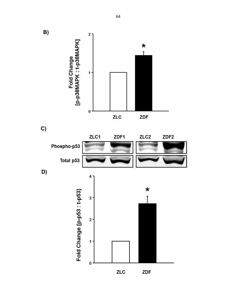
64
B)
C)
D)

65
Figure 5-1: Pancreatic islets from ZDF rats show elevated levels of phosphorylated p38MAPK and
p53:
Panel A: Pancreatic islets were isolated from ZDF rats and their age-matched controls by collagenase
digestion. After overnight incubation in RPMI media, islets were lysed and lysate proteins were separated
by SDS-PAGE. Proteins were then transferred onto nitrocellulose membranes and blocked for 1 h. The
membranes were then probed for phosphorylated p38MAPK followed by incubation with IR-Dye conjugated
rabbit secondary antibody. The immune complexes were detected by Odyssey® Imaging Systems. The
same blots were stripped and probed for total p38MAPK. Panel B: Band intensities were determined and
analyzed by densitometric analysis. Data shown as mean ± SEM are from four rats in each group and
expressed as fold change in the ratios between phospho- and total p38MAPK. * P < 0.05 vs ZLC rat islets.
Panel C: Pancreatic islets were isolated from ZLC and ZDF rats and analyzed by Western Blotting as
described above. The membranes were probed with antibody raised against phospho-p53 followed by
incubation with rabbit IR-Dye conjugated secondary antibody. The same blots were stripped and probed
for total p53. Panel D: Band intensities were determined and analyzed by densitometric analysis. Data
shown as mean ± variance from two rats in each group and expressed as fold change in the ratios between
phospho- and total p38MAPK.
Exposure of normal human islets to glucotoxic conditions activates p38MAPK and p53:
We next determined if exposure of pancreatic islets from normal human donors to
glucotoxic conditions promote p38MAPK and p53 phosphorylation. We incubated human islets to
LG (5.8 mM) and HG (30 mM) for 24 h and quantified p38MAPK and p53 phosphorylation. As
depicted in Figure 5-2 (Panel A), we noticed a marked increase in p38MAPK phosphorylation in
HG-treated islets (1.61 fold increase). Furthermore, our findings also indicated a 1.97 fold
increase in p53 phosphorylation in HG-treated islet samples. These data are compatible to our
findings in INS-1 832/13 cells, normal rat islets (Chapters 3 and 4) and ZDF rat islets (Figure 5-
1), together demonstrating increased activation of p38MAPK-p53 signaling pathway upon
exposure of β-cells to glucotoxic conditions.

66
Figure 5-2: p38MAPK and p53 activation in human islets exposed to glucotoxic conditions:
Pancreatic islets from non-diabetic human donors were incubated with LG (5.8 mM) and HG (30 mM) for
24 h. Islets were then lysed and analyzed by Western Blotting for p38MAPK (Panel A) and p53 (Panel B).
The membranes were probed for phospho-p38MAPK and phospho-p53, followed by incubation with anti-
rabbit secondary antibodies. The immune complexes were then detected by Odyssey® Imaging Systems.
The same blots were stripped and probed for total p38MAPK and p53. Data shown are expressed as fold
change in the ratios between phospho- over total p38MAPK and phospho- over total p53.
Exposure to glucotoxic conditions promote nuclear localization of p53 in human islets:
As described in Chapter 4, our studies in INS-1 832/13 cells treated with LG and HG for
24 h indicated accumulation of p53 in the nuclear fractions under HG conditions. We therefore,
incubated human pancreatic islets with LG (5.8 mM) and HG (30 mM) for 24 h and isolated nuclear
and non-nuclear fractions using NER-PER® kit. In line with our observations in INS-1 832/13
cells, we found increased localization of phospho- and total p53 in the nuclear fractions of HG-
treated samples (Figure 5-3).
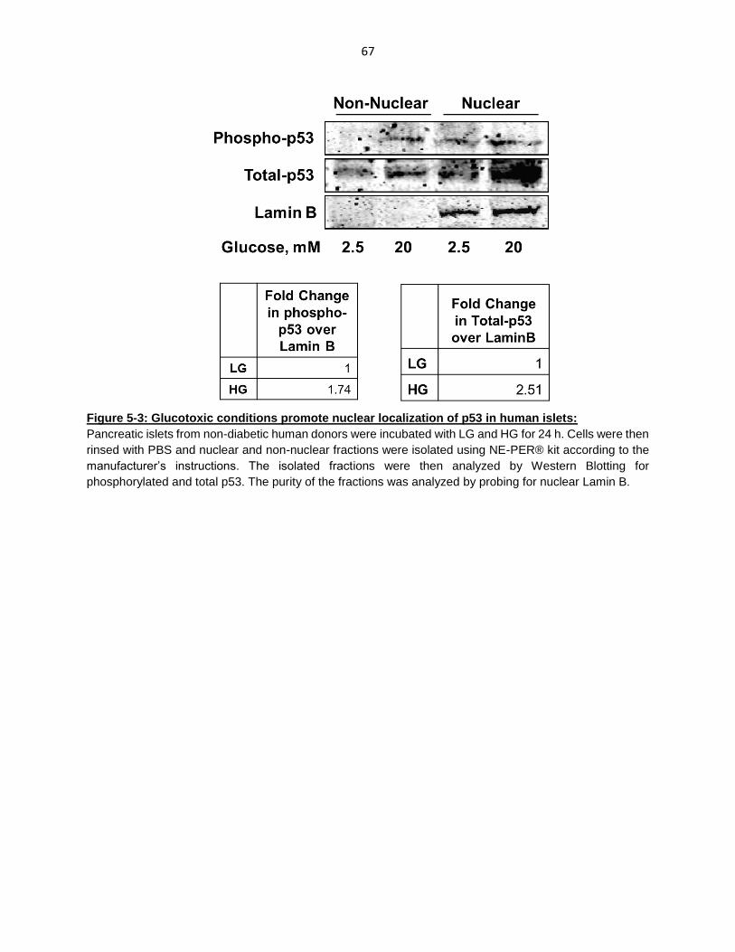
67
Figure 5-3: Glucotoxic conditions promote nuclear localization of p53 in human islets:
Pancreatic islets from non-diabetic human donors were incubated with LG and HG for 24 h. Cells were then
rinsed with PBS and nuclear and non-nuclear fractions were isolated using NE-PER® kit according to the
manufacturer’s instructions. The isolated fractions were then analyzed by Western Blotting for
phosphorylated and total p53. The purity of the fractions was analyzed by probing for nuclear Lamin B.

68
CHAPTER 6: DISCUSSION
Insulin resistance caused by various factors such as obesity, results in decreased glucose
uptake and utilization in the peripheral tissues. This is initially compensated by the pancreatic β-
cells, which secrete more insulin to normalize the elevated blood glucose levels. However, the
chronic exposure of β-cells to elevated glucose concentrations or glucotoxicity eventually leads
to decreased function and ultimately cell demise, culminating in the onset of diabetes. Recently,
we reported that exposure of INS-1 832/13 cells, rodent and human pancreatic islets to glucotoxic
conditions results in activation of executioner caspases and degradation of nuclear lamins (119,
149). In addition, studies have implicated the role of Rac1-Nox2 enzyme complex and associated
oxidative stress in the pathology of β-cell dysfunction (49, 50, 52). However, the precise signaling
mechanisms involved in mediating loss in glucose-stimulated insulin secretion (GSIS) and β-cell
death under glucotoxic conditions, remain to be poorly understood.
Apart from the negative modulatory roles in the induction of oxidative stress, several lines
of evidence have demonstrated the requisite role of Rac1 and Nox2 signaling in GSIS (18, 35).
Glucose enters the β-cell via GLUT-2 transporter, where it is metabolized to generate ATP. The
resulting increase in ATP/ADP ratio causes closure of ATP-sensitive K+ channels resulting in
membrane depolarization. This causes the opening of voltage-gated Ca+2 channels and increased
intracellular Ca+ concentration. In presence of Ca+, insulin-laden secretory granules are then
mobilized towards the membrane for fusion and release. This process of translocation of insulin
granules to the plasma membrane is mediated by vesicle-associated and t-SNARE proteins at
target docking sites, and requires cytoskeletal reorganization (8). Studies by Asahara and
associates have demonstrated the critical role of Rac1 in F-actin remodeling and GSIS, using a
beta-cell specific Rac1-/- mice model (22). Furthermore, previous studies from our laboratory
have also utilized several pharmacological agents to demonstrate that Rac1 activation is required
for GSIS. Using NSC23766, we demonstrated that inhibition of Tiam1, a GEF mediating Rac1
activation, results in marked reduction in GSIS in the presence of the inhibitor (24). We also
69
examined the effects of Ehop-016, a specific inhibitor of Vav2- mediated Rac1 activation, in
pancreatic β-cells (25). We observed alterations in F-actin remodeling and a significant decrease
in GSIS in the presence of Ehop-016, demonstrating the role of Rac1 in cytoskeletal
reorganization and insulin secretion.
Furthermore, several investigations have implicated the involvement of Nox2 and reactive
oxygen species (ROS) in GSIS (35, 36). Nox2 holoenzyme is composed of several membrane-
bound (gp91phox and p22phox) and cytosolic (p40phox, p47phox, p67phox and Rac1) components. Upon
stimulation, the cytosolic components translocate towards the membrane thereby mediating the
holoenzyme assembly. The functionally active Nox2 enzyme complex catalyzes the one electron
reduction of molecular oxygen to generate superoxide. Emerging evidence implicate that a tonic
increase in Rac1-Nox2-mediated ROS generation is necessary for nutrient-induced insulin
secretion (38, 39). Studies by Leloup and associates have demonstrated that co-provision of
antioxidants suppresses ROS generation induced by glucose, resulting in alterations in calcium
mobilization and decreased GSIS, in rodent pancreatic islets (38). Moreover, studies by Morgan
and associates have demonstrated the involvement of Nox2-derived ROS in GSIS in rodent
pancreatic islets (36).
As demonstrated by these studies, Rac1-Nox2 activity and associated generation of ROS
is required for physiological functioning of the β-cell. However, evidence from studies in pancreatic
β-cells, retinal endothelial cells and cardiomyocytes suggests the involvement of Rac1-Nox2
signaling axis in causing oxidative stress under diabetic conditions (48, 150, 151). Previous
studies in our laboratory have demonstrated that sustained activation of Rac1-Nox2 and ROS
generation in pancreatic β-cells exposed to glucolipotoxic conditions and inflammatory cytokines,
leads to loss in β-cell function and apoptosis (49, 50, 52). Our observations in the ZDF rat model
and type2 diabetic islets have also suggested that Rac1-Nox2-ROS signaling plays a key role in
causing β-cell dysfunction (49). However, the underlying mechanisms mediating the effects of
Rac1-Nox2-induced oxidative stress remain poorly understood. Using pharmacological

70
approaches to target the Rac1-Nox2 signaling cascade, my doctoral work focusses on identifying
the effector proteins activated by increased Rac1-Nox2-ROS signaling, that are involved in
activation of apoptotic factors leading to β-cell death.
Desire et al. have developed a small molecule inhibitor, EHT1864, which inhibits Rac1
activation in vivo (28). Further characterization by Shutes et al. demonstrated that the effect of
EHT1864 is independent of GEFs (Tiam1, Vav2) since they observed inhibition of cellular
transformation induced by constitutively active mutant of Rac1 (61L) (128). EHT1864 binds
directly to Rac1 with a greater affinity than guanine nucleotides (GDP/GTP) thereby retaining
Rac1 in an inert, inactive state by displacing pre-bound GDP/GTP. We first tested this Rac1
inhibitor to examine the role of Rac1 in physiological GSIS in INS-1 832/13 cells. Our results have
indicated a marked reduction in glucose-induced activation and membrane targeting of Rac1 in
the presence of EHT1864. Under these conditions, we also observed a marked reduction in GSIS.
Several studies in multiple cell types have utilized EHT1864 to understand Rac1 function in health
and disease (29-32). We have further utilized EHT1864, to deduce roles of Rac1-Nox2-signaling
axis in activating the downstream apoptotic pathways and β-cell death under glucotoxic
conditions.
To identify the downstream signaling proteins mediating the effects of Rac1-Nox2-induced
oxidative stress, we examined the activation status of stress-activated protein kinases
(SAPK/MAPK). Several lines of evidence have implicated the role of stress kinases, JNK1/2 and
p38MAPK in the activation of apoptotic pathways (55). Previous studies in our laboratory in ZDF
and human pancreatic islets, have coupled increased Rac1 activation and Nox2 subunit
expression with increased activation of JNK1/2 (49). Studies by Flores-Lopez and associates
have also suggested that p38MAPK is activated in β-cells exposed to glucotoxic conditions,
resulting in apoptosis (121). Additionally, studies in mice lacking an isoform of p38MAPK, showed
improved glucose tolerance as a result of improved insulin secretion from the β-cell (75). Our
findings have also demonstrated increased activation of p38MAPK in insulin-secreting INS-1

71
832/13 cells, normal rodent and human islets exposed to glucotoxic conditions, and in islets
isolated from ZDF rats.
Figure 6-1: Our working model illustrating the involvement of Rac1-Nox2 signaling axis and
associated oxidative stress in the activation of p38MAPK and p53, culminating in β-cell
apoptosis: We demonstrated that exposure of pancreatic β-cells to glucotoxic conditions leads to
sustained activation of Rac1-Nox2 enzyme complex and excess ROS generation. The resulting
oxidative stress leads to p38MAPK-mediated activation of p53, which, in turn, activates the transcription
of apoptotic proteins and causes β-cell death. Our finding also demonstrate the potential cytoprotective
effects of pharmacological inhibitors of Rac1 (EHT1864, NSC23766, Ehop-016), Nox2 (gp91-ds-tat)
and p38MAPK (SB203580) in the β-cell, exposed to glucotoxic conditions.

72
Based on this evidence, we examined if Rac1-Nox2 driven oxidative stress causes β-cell
dysfunction under glucotoxic conditions, mediated by p38 family of stress kinases. Several studies
including from those from our own laboratory, have utilized diphenyleneiodonium (DPI) and
Apocynin, which block Nox2 activation and ROS generation in models of cellular dysfunction
(152). However, it has been suggested that DPI also interacts with other flavin-dependent
enzymes including nitric oxide synthase and xanthine oxidase (153). In addition, studies have
also indicated that Apocynin has anti-oxidant properties, independent of its inhibitory effects on
Nox2 (154). Recently, Rey et al. have identified a Nox2-specifc peptide-based inhibitor, gp91-ds-
tat, which prevents the interaction of the cytosolic p47phox with gp91phox in the membrane (122),
thereby inhibiting Nox2 assembly and functional activation. Several studies have demonstrated
the specificity of gp91-ds-tat to Nox2, compared to its inactive scrambled analog (123-127). To
determine the role of Nox2 in p38MAPK activation under HG conditions in the β-cell, we first
utilized this peptide inhibitor and quantified HG-induced Nox2 activity and ROS generation. We
observed significant blockade in HG-induced Nox2 activation and ROS generation in the
presence of gp91-ds-tat, a specific inhibitor of Nox2, but not its inactive analog. We also
demonstrated that HG-induced p38MAPK phosphorylation is significantly blocked in the presence
of gp91-ds-tat, but not its inactive analog. Thus, our studies provide the first evidence linking HG-
induced Nox2 activation and ROS generation to p38MAPK activation.
Recent in vitro and in vivo studies have demonstrated that NSC23766, an inhibitor of
Tiam1-mediated Rac1 activation, prevents cellular dysfunction in pancreatic β-cells, retinal
endothelial cells and cardiomyocytes upon exposure to diabetic conditions (50, 150, 151). Our
studies with NSC23766 and Ehop-016 have also shown that inhibition of Tiam-Rac1 signaling
blocks HG-induced p38MAPK activation in INS-1 832/13 cells and rodent islets, indicating that
regulation of Rac1 function by Tiam1 and Vav2 are requisite for p38MAPK activation under HG
conditions. These observations are supported by a recent finding by Liu and associates where
they showed that while Vav2 mediates GTP loading of Rac1, Tiam1 functions as an adapter in a

73
VE-cadherin-p67phox-Par3 polarity complex that mediates localized activation of Rac1. They
concluded that Tiam1 and Vav2 play a dual regulatory role in inducing localized Rac1 activation
required for Nox2 activation and ROS generation (155). Moreover, the presence of EHT1864,
which disrupts Rac1 interaction with GTP/GDP in a GEF-independent manner, also attenuated
HG-induced p38MAPK phosphorylation. Together, these results demonstrate the involvement of
Rac1 in p38MAPK activation under HG conditions. It is noteworthy that our studies with Ehop-
016 showed activation of p38MAPK under basal LG conditions. These effects could be as a result
of activation of other signaling mechanisms in presence of Ehop-016. Studies by Montalvo-Ortiz
et al. suggest that other Rho-family G –proteins such as RhoA, which has been previously shown
to activate p38MAPK (156), are activated in presence of Ehop-016. Additional studies are needed
to understand these observations.
In the next set of studies, we examined the downstream effects of p38MAPK activation.
Indeed, studies in multiple cell types have documented the role of p38MAPK in cell cycle arrest
and apoptosis (53, 65). For example, evidence from multiple cell types have reported that
p38MAPK activates p53 tumor suppressor in response to stress stimuli to mediate apoptosis
(121). Previous studies by Karunakaran and associates have analyzed the effects of 1-methyl-4-
phenyl-1, 2, 3, 6-tetrahydropyridine (MPTP)-induced ROS generation on the regulation of
p38MAPK-p53 pathway. They observed that treatment of dopaminergic neurons with MPTP-
activated p38MAPK which in turn activated p53 and p53-dependent transcription of apoptotic Bax
and PUMA. Moreover, the presence of pharmacological inhibitors of p38MAPK and p53 protected
primary neurons from MPTP-induced cell death (157). Furthermore, earlier studies by Bulavin et
al. have also shown that p38MAPK activates p53 tumor suppressor by phosphorylation of serine-
33 and serine-46 residues in cancer cell lines upon UV radiation (79). These phosphorylation sites
are required for phosphorylation at serine-15, which has been implicated as the critical step in the
transcriptional activation and stabilization of p53 (88, 106). Loughery and associates have
suggested that serine-15 phosphorylation of p53 is essential for its interaction with transcriptional

74
proteins and target gene expression (106). They reported that substitution of serine-15 with
alanine results in decreased p53-dependent transcriptional activity which is rescued by
substitution with an aspartate that mimics serine residue. This is further supported by evidence
from multiple studies indicating that phosphorylation at serine-15 also modulated the interaction
of p53 with its regulators including Mdm2 and p300 (90, 91).
In the context of pancreatic β-cells, studies by Hoshino et al. have examined the role of
p53 in β-cell dysfunction in models of Type 1 and Type 2 diabetes (158). They observed that loss
of p53 function prevented onset of diabetes in streptozotocin-induced type1 and db/db mouse
model for type 2 diabetes. Additionally, a proteomic study in type2 diabetic human islets indicated
significant increase in apoptotic signaling pathways including the p53 apoptotic pathway (159).
Despite these findings, the regulatory factors involved in the activation of p53 in β-cells under
diabetic conditions, remain to be fully understood. Recently, Flores-Lopez and associates have
reported that p38MAPK co-localizes with p53 under HG conditions and mediates p53
phosphorylation, resulting in β-cell apoptosis (121).
We therefore, tested if p53 is activated by Rac1-Nox2-p38MAPK signaling axis in β-cells
exposed to HG conditions by quantifying phosphorylation at serine-15 residue. Our observations
have indicated increased serine-15 phosphorylation of p53 in INS-1 832/13 cells and human islets
exposed to HG conditions, and in diabetic ZDF rats compared to their age-matched ZLC control
animals. However, the presence of Rac1 inhibitor (EHT1864) and p38MAPK inhibitor (SB203580)
markedly attenuated HG-induced p53 phosphorylation in INS-1 832/13 cells, suggesting the
involvement of Rac1-p38MAPK signaling in p53 activation. Furthermore, we isolated nuclear and
non-nuclear fractions from INS-1 832/13 cells incubated with LG and HG. We observed increased
accumulation of p53 in the nucleus under HG conditions, which is requisite for its interaction with
specific DNA sites to promote the transcription of apoptotic genes (139). However, our results
show that the presence of EHT1864 had no significant effect on nuclear accumulation of p53,
indicating that nuclear localization is regulated by other mechanisms but not phosphorylation (98).

75
Previous studies from our own laboratory, have shown that post-translational prenylation
of Rac1 (geranylgeranylation) is requisite for its localization in the membrane fraction, thereby,
facilitating its interaction with downstream substrates. Pharmacological and siRNA-mediated
inhibition of prenylation resulted in decreased GSIS, demonstrating the critical role of these
modifications in β-cell function (34). The presence of FTI-277 and GGTI-2147, inhibitors of protein
farnesylation and geranylgeranylation, significantly attenuated Nox2-induced ROS generation
under stimulatory glucose concentration, suggesting the importance of protein prenylation (Rac1)
in Nox2-mediated ROS generation (41). Studies from our laboratory have also reported that
increased Nox2 activation in presence of inflammatory cytokines, is attenuated in the presence
of 2-bromopalmitate, suggesting that Rac1 palmitoylation is also requisite for Nox2-induced
oxidative stress (120). Therefore, we next asked the question if post-translational modifications
of Rac1 are required for p38MAPK and p53 activation. We observed a significant inhibition in
p38MAPK activation under HG conditions in presence 2-bromopalmitate (2-BP), a specific
inhibitor of protein palmitoylation (160).
Additionally, we tested the effects of GGTI-2147, which inhibits geranylgeranylation, on
HG-induced p38MAPK phosphorylation. We observed no effects of GGTI-2147 on HG-induced
p38MAPK activation implying that Rac1 geranylgeranylation is not necessary for p38MAPK
activation. However, our studies with Simvastatin, a global inhibitor of protein prenylation, and
GGTI2147, suggested a marked decrease in HG-induced p53 phosphorylation. It has been
suggested that bisphosphonates, which inhibit prenylation by blocking mevalonate pathway,
activate p38MAPK before eliciting their effects on G-protein prenylation, independent of Rac1
activation (161). Further studies are required to understand the effects of GGTI-2147 on HG-
induced p38MAPK phosphorylation.
As a logical extension of these studies, we tested if inhibition of Rac1 with EHT1864 had
any effect on glucotoxicity-induced β-cell death. Our observations showed a significant decrease
in cell death in INS-1 832/13 cells exposed to glucotoxic conditions. Together, our observations

76
in INS-1 832/13 cells, rodent and human pancreatic islets have demonstrated that sustained
activation of Rac1-Nox2 signaling module and associated oxidative stress, results in p38MAPK-
dependent activation of p53, culminating in β-cell death, under glucotoxic conditions.
We also examined the involvement of ATM kinase, a known regulator of p53, in the Rac1-
p38MAPK-p53 signaling cascade. Evidence from multiple studies have shown that ATM kinase
is activated upon DNA damage, mainly by auto-phosphorylation (144). Studies by Oleson et al.
have implicated that ATM kinase is activated in pancreatic β-cells exposed to inflammatory
cytokines and mediated DNA damage response (DDR). However, upon excessive DNA damage,
ATM kinase activates cellular apoptotic mechanism mainly by p53-dependent mechanisms (137).
Furthermore, Yoshida et al. have reported that ATM kinase activates p53 upon DNA damage
caused by Doxorubicin, which was prevented in presence of inhibitor of protein prenylation
(pitavastatin) and Rac1 (NSC23766) (136). We therefore, asked if ATM kinase is activated in β-
cells exposed to HG conditions. We observed a marked increase in activation of ATM kinase,
when INS-1 832/13 cells were exposed to HG conditions, which was significantly blocked upon
Rac1 inhibition by EHT1864. To determine the role of ATM kinase in the regulation of p53, we
utilized KU55933, a known inhibitor of ATM kinase activity, to examine the effects on HG-induced
signaling events. Our observations indicated a marked reduction in ATM kinase activation under
HG conditions. However, the presence of KU55933 had no significant effect on p53 activation,
suggesting that ATM kinase, although regulated by Rac1 signaling, might be involved in other
signaling pathways (83). Since multiple studies, including those in ATM deficient mice, have
indicated a unique role of ATM kinase in glucose homeostasis, it is possible that it is involved in
DNA damage response mechanisms activated as a result of Rac1-induced oxidative stress.
Further studies are required to understand the regulatory role of ATM kinase in β-cells under
glucotoxic stress.

77
Pharmacological agent
Effect p38MAPK
phosphorylation p53
phosphorylation
EHT1864 ↓ Rac1 activity Inhibition Inhibition
gp91-ds-tat ↓ Nox2 activity Inhibition Not determined
NSC23766 ↓ Rac1 activity Inhibition Not determined
Ehop-016 ↓ Rac1 activity Inhibition Not determined
Simvastatin ↓ Protein
prenylation Not determined Inhibition
GGTI2147 ↓ Protein
geranylgeranylation No effect Inhibition
2-bromopalmitate ↓ Protein
palmitoylation Inhibition Not determined
SB203580 ↓ p38MAPK activity Not determined Inhibition
Table 6-1: Summary of effects of pharmacological inhibitors on HG-induced p38MAPK and p53
activation

78
CHAPTER 7: CONCLUSIONS AND FUTURE DIRECTIONS
The central objective of my Ph.D. dissertation project is to assess the role of sustained
activation of Rac1 and Nox2 holoenzyme in induction of oxidative stress, resulting in the activation
of p38MAPK and p53 signaling axis, culminating in pancreatic β-cell dysfunction. Previous
observations in our laboratory have demonstrated contributory roles of increased Rac1-Nox2
activity and ROS generation in mediating metabolic dysfunction of the β-cell in models of diabetes
(49, 50, 52). As a logical extension to these studies, this project focuses on examining the
downstream apoptotic pathways, activated by Rac1-Nox2-derived oxidative stress. These data
can be summarized as follows:
1. Exposure of pancreatic β-cells (INS-1 832/13 cells, rodent and human islets) to glucotoxic
conditions results in activation of pro-apoptotic p38MAPK and p53 signaling pathway.
Pancreatic islets isolated from Zucker diabetic fatty (ZDF) rat, a model for type 2 diabetes,
also exhibit increased activation of p38MAPK and p53.
2. gp91-ds-tat, but not its inactive analog, attenuated HG-induced p38MAPK
phosphorylation, suggesting that HG-induced Nox2 activation and ROS generation are
upstream to p38MAPK activation.
3. NSC23766 and Ehop-016, which target Tiam1-Rac1 and Vav2-Rac1 interaction
respectively, attenuated HG-induced p38MAPK phosphorylation. Additionally, EHT1864
which blocks Rac1 activation in a GEF-independent manner, blocked p38MAPK
phosphorylation. These findings implicate the contributory role of Rac1 activation in HG-
induced phosphorylation of p38MAPK.
4. Our findings also suggested that HG-induced p53 phosphorylation is also regulated by
Rac1, which is attenuated in presence of the Rac1 inhibitor, EHT1864. However,
EHT1864 did not block p53 translocation to the nucleus suggesting that HG-induced p53
nuclear localization is independent of its activation step.
79
5. Rac1 prenylation is also requisite for HG-induced p53 activation in INS-1 832/13 cells,
since the presence of GGTI-2147, which blocks geranylgeranylation, and Simvastatin, a
global inhibitor of protein prenylation, prevented p53 phosphorylation.
6. SB203580, a selective inhibitor for p38MAPK, blocked HG-induced p53 phosphorylation,
confirming the involvement of p38MAPK in activating p53 apoptotic pathway, under
glucotoxic stress.
7. EHT1864, which blocked HG-induced p38MAPK and p53 activation, also prevented β-cell
death, confirming the role of Rac1-p38MAPK-p53 signaling cascade in β-cell apoptosis
under glucotoxic conditions.
8. Our findings also indicated marked activation of ATM kinase, a known activator of p53 and
a cell cycle regulator, under HG exposure conditions, which was attenuated in the
presence of EHT1864 and ATM kinase inhibitor, KU55933. However, KU55933 exerted
no effect on HG-induced p53 phosphorylation, suggesting that ATM kinase might be
involved in other regulatory pathways.
Based on these findings, I conclude that chronic exposure of pancreatic β-cells to elevated
glucose concentrations lead to sustained activation of Rac1-Nox2 enzyme complex and excess
generation of intracellular ROS. The resulting oxidative stress leads to nuclear accumulation and
p38MAPK-dependent phosphorylation of p53 tumor suppressor. These events, in turn, lead to
transcription of several apoptotic genes mediated by p53, culminating in activation of apoptotic
pathways and loss of β-cell mass. These studies provide the first evidence suggesting that
therapeutic intervention of this signaling pathway prevents β-cell dysfunction under the duress of
hyperglycemic conditions.
80
Future Directions:
These finding have provided valuable insights into the role of Rac1-Nox2 holoenzyme and
associated oxidative stress in mediating β-cell dysfunction under glucotoxic stress. However,
studies are needed to further verify these findings in other invivo models of T2DM and human
patients, and also fill several knowledge gaps. Some of these are listed below:
We have utilized the ZDF rat, as an animal model for T2DM, to verify our findings.
However, since the ZDF rat develops diabetes as a result of genetic manipulations, we
need to examine this pathway in a model for diabetes induced by diet/environment, as
commonly seen in human populations. This can be addressed by utilizing the high-fat
(45% kilocalories from fat) fed C57BL/6 mice model for diabetes to examine the activity
levels of Rac1-Nox2 and p38MAPK-p53 signaling axes in the pancreatic islet.
Furthermore, studies in pancreatic islets from Type2 diabetic patients would provide
valuable evidence demonstrating the role of Rac1-p38MAPK-p53 module in human
diabetic patients.
Our findings have implicated that Rac1 prenylation is requisite for HG-induced p53
phosphorylation but not p38MAPK. Although, there is some evidence implicating the
effects of prenylation inhibitors on p38MAPK activation, independent of their effects on
Rac1, the involvement of Rac1 prenylation in mediating HG-induced effects needs to
further examined. This can be addressed by utilizing Rac1 C189A (non-prenylatable) and
V12Rac1 SAAX (non-prenylatable, constitutively active) mutants of Rac1.
Our studies have also suggested a possible role of ATM kinase in HG-induced effects.
Activation of ATM kinase, which also seems to be regulated by Rac1, might be involved
in other signaling mechanisms including DNA repair pathways. Studies by Oleson et al.
have implicated ATM kinase activation in H2AX phosphorylation leading to DNA repair
response in β-cells exposed to inflammatory cytokines (137). These mechanisms need to
be examined in β-cells exposed to glucotoxic conditions.
81
Lastly, we need to investigate if the Rac1-p38MAPK-p53 signlaing module is involved in
β-cell dysfunction induced by exposure to saturated fatty acids (Palmitate) and
glucolipotoxic conditions. Previous studies in our laboratory have shown increased Rac1-
Nox2 activity and oxidative stress in β-cells exposed to palmitate (50). Additional studies
are need to examine activation of p38MAPK and p53, regulated by Rac1-Nox2 enzyme
complex, under glucolipotoxic conditions.

82
APPENDIX A

83
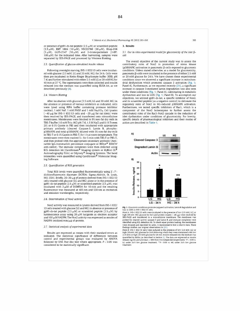
84

85

86

87
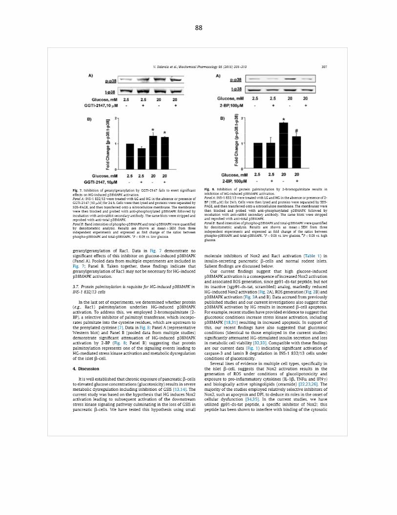
88
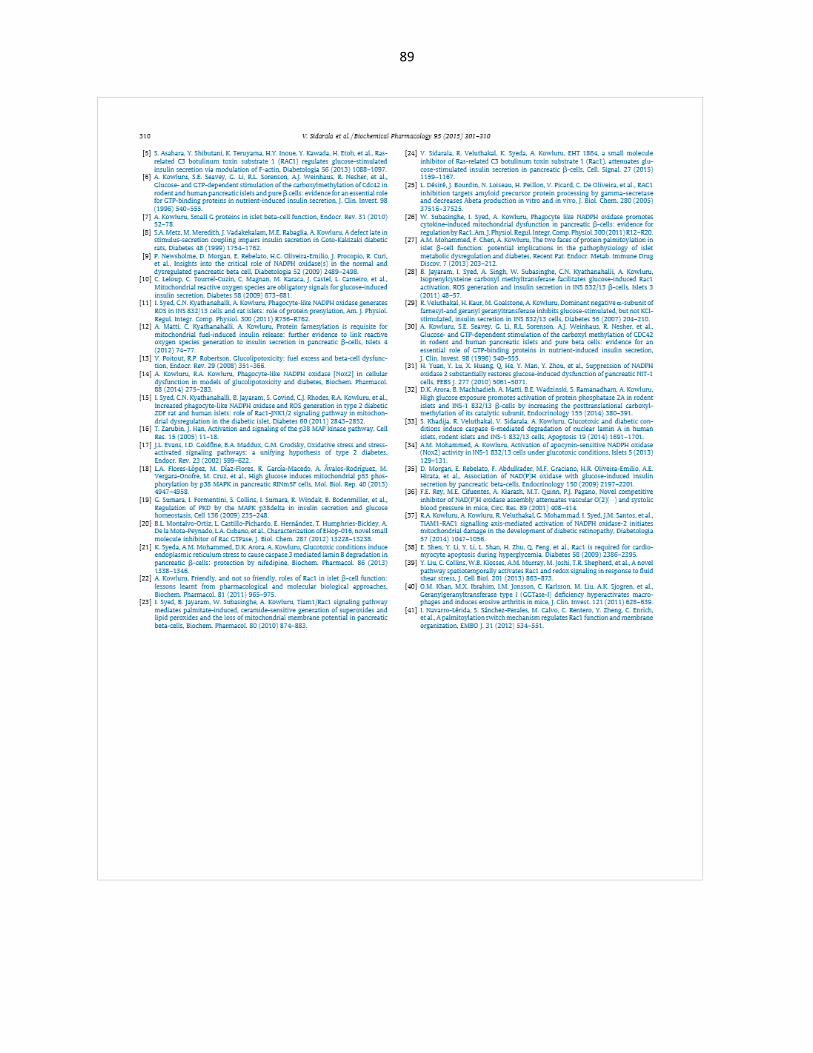
89
90
APPENDIX B

91

92
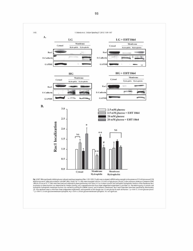
93

94
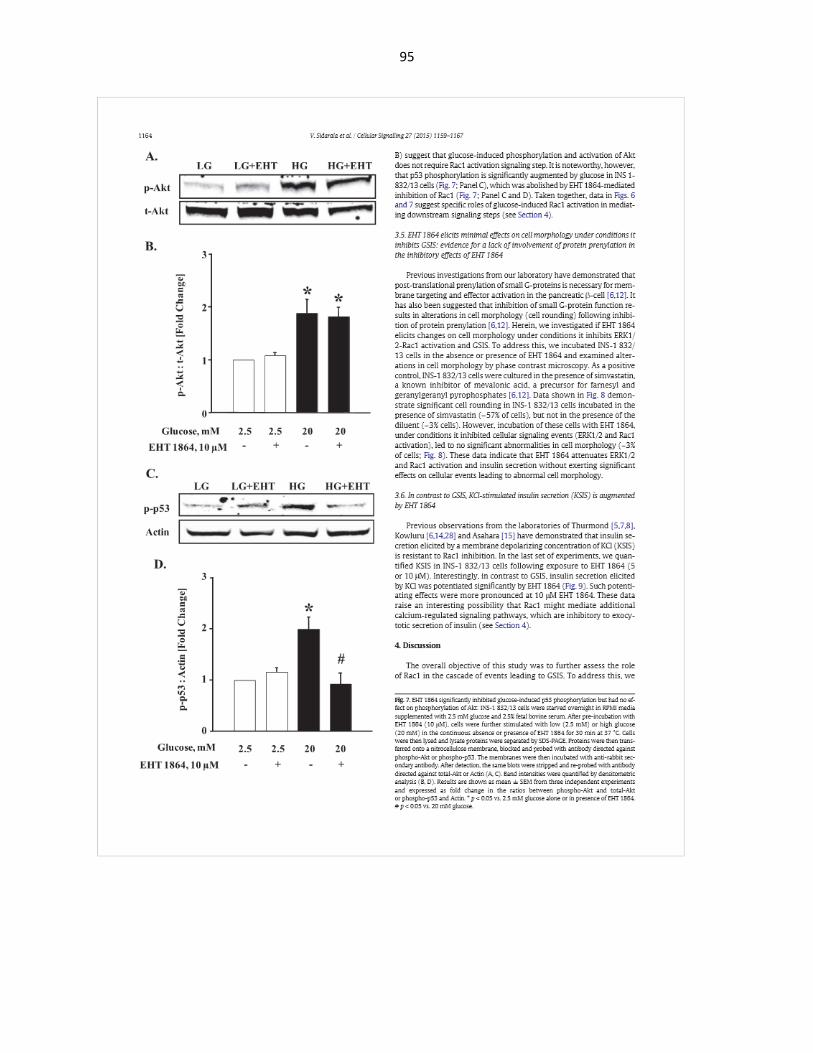
95

96
97

98

99
REFERENCES
1. Ionescu-Tirgoviste C, Gagniuc PA, Gubceac E, Mardare L, Popescu I, Dima S, Militaru
M. A 3D map of the islet routes throughout the healthy human pancreas. Sci Rep.
2015;5:14634. doi: 10.1038/srep14634. PubMed PMID: 26417671; PMCID: 4586491.
2. Cabrera O, Berman DM, Kenyon NS, Ricordi C, Berggren PO, Caicedo A. The unique
cytoarchitecture of human pancreatic islets has implications for islet cell function. Proc
Natl Acad Sci U S A. 2006;103(7):2334-9. doi: 10.1073/pnas.0510790103. PubMed
PMID: 16461897; PMCID: 1413730.
3. Scharfmann R, Xiao X, Heimberg H, Mallet J, Ravassard P. Beta cells within single
human islets originate from multiple progenitors. PLoS One. 2008;3(10):e3559. doi:
10.1371/journal.pone.0003559. PubMed PMID: 18958289; PMCID: 2571119.
4. Weiss M, Steiner DF, Philipson LH. Insulin Biosynthesis, Secretion, Structure, and
Structure-Activity Relationships. In: De Groot LJ, Beck-Peccoz P, Chrousos G, Dungan
K, Grossman A, Hershman JM, Koch C, McLachlan R, New M, Rebar R, Singer F, Vinik
A, Weickert MO, editors. Endotext. South Dartmouth (MA)2000.
5. Chang L, Chiang SH, Saltiel AR. Insulin signaling and the regulation of glucose transport.
Mol Med. 2004;10(7-12):65-71. doi: 10.2119/2005-00029.Saltiel. PubMed PMID:
16307172; PMCID: 1431367.
6. Lee J, Pilch PF. The insulin receptor: structure, function, and signaling. Am J Physiol.
1994;266(2 Pt 1):C319-34. PubMed PMID: 8141246.
7. Kanzaki M. Insulin receptor signals regulating GLUT4 translocation and actin dynamics.
Endocr J. 2006;53(3):267-93. PubMed PMID: 16702775.
8. Wang Z, Thurmond DC. Mechanisms of biphasic insulin-granule exocytosis - roles of the
cytoskeleton, small GTPases and SNARE proteins. J Cell Sci. 2009;122(Pt 7):893-903.
doi: 10.1242/jcs.034355. PubMed PMID: 19295123; PMCID: 2720925.
100
9. von Herrath M, Sanda S, Herold K. Type 1 diabetes as a relapsing-remitting disease? Nat
Rev Immunol. 2007;7(12):988-94. doi: 10.1038/nri2192. PubMed PMID: 17982429.
10. Bensellam M, Laybutt DR, Jonas JC. The molecular mechanisms of pancreatic beta-cell
glucotoxicity: recent findings and future research directions. Mol Cell Endocrinol.
2012;364(1-2):1-27. doi: 10.1016/j.mce.2012.08.003. PubMed PMID: 22885162.
11. Skelly RH, Schuppin GT, Ishihara H, Oka Y, Rhodes CJ. Glucose-regulated translational
control of proinsulin biosynthesis with that of the proinsulin endopeptidases PC2 and PC3
in the insulin-producing MIN6 cell line. Diabetes. 1996;45(1):37-43. PubMed PMID:
8522057.
12. Guest PC, Bailyes EM, Hutton JC. Endoplasmic reticulum Ca2+ is important for the
proteolytic processing and intracellular transport of proinsulin in the pancreatic beta-cell.
Biochem J. 1997;323 ( Pt 2):445-50. PubMed PMID: 9163336; PMCID: 1218339.
13. Flamez D, Berger V, Kruhoffer M, Orntoft T, Pipeleers D, Schuit FC. Critical role for
cataplerosis via citrate in glucose-regulated insulin release. Diabetes. 2002;51(7):2018-
24. PubMed PMID: 12086928.
14. Poitout V, Robertson RP. Glucolipotoxicity: fuel excess and beta-cell dysfunction. Endocr
Rev. 2008;29(3):351-66. doi: 10.1210/er.2007-0023. PubMed PMID: 18048763; PMCID:
2528858.
15. Unger RH, Grundy S. Hyperglycaemia as an inducer as well as a consequence of
impaired islet cell function and insulin resistance: implications for the management of
diabetes. Diabetologia. 1985;28(3):119-21. PubMed PMID: 3888754.
16. Metz SA, Rabaglia ME, Pintar TJ. Selective inhibitors of GTP synthesis impede
exocytotic insulin release from intact rat islets. J Biol Chem. 1992;267(18):12517-27.
PubMed PMID: 1352288.
17. Metz SA, Meredith M, Rabaglia ME, Kowluru A. Small elevations of glucose
concentration redirect and amplify the synthesis of guanosine 5'-triphosphate in rat islets.

101
J Clin Invest. 1993;92(2):872-82. doi: 10.1172/JCI116662. PubMed PMID: 8349822;
PMCID: 294926.
18. Kowluru A. Small G proteins in islet beta-cell function. Endocr Rev. 2010;31(1):52-78. doi:
10.1210/er.2009-0022. PubMed PMID: 19890090; PMCID: 2852207.
19. Arora DK, Syed I, Machhadieh B, McKenna CE, Kowluru A. Rab-geranylgeranyl
transferase regulates glucose-stimulated insulin secretion from pancreatic beta cells.
Islets. 2012;4(5):354-8. doi: 10.4161/isl.22538. PubMed PMID: 23114750; PMCID:
3524143.
20. Kowluru A, Kowluru RA. Protein prenylation in islet beta-cell function in health and
diabetes: Putting the pieces of the puzzle together. Biochem Pharmacol. 2015;98(3):363-
70. doi: 10.1016/j.bcp.2015.07.004. PubMed PMID: 26215874.
21. Zhang FL, Casey PJ. Protein prenylation: molecular mechanisms and functional
consequences. Annu Rev Biochem. 1996;65:241-69. doi:
10.1146/annurev.bi.65.070196.001325. PubMed PMID: 8811180.
22. Asahara S, Shibutani Y, Teruyama K, Inoue HY, Kawada Y, Etoh H, Matsuda T, Kimura-
Koyanagi M, Hashimoto N, Sakahara M, Fujimoto W, Takahashi H, Ueda S, Hosooka T,
Satoh T, Inoue H, Matsumoto M, Aiba A, Kasuga M, Kido Y. Ras-related C3 botulinum
toxin substrate 1 (RAC1) regulates glucose-stimulated insulin secretion via modulation of
F-actin. Diabetologia. 2013;56(5):1088-97. doi: 10.1007/s00125-013-2849-5. PubMed
PMID: 23412604; PMCID: 3622740.
23. Kowluru A. Friendly, and not so friendly, roles of Rac1 in islet beta-cell function: lessons
learnt from pharmacological and molecular biological approaches. Biochem Pharmacol.
2011;81(8):965-75. doi: 10.1016/j.bcp.2011.01.013. PubMed PMID: 21300027; PMCID:
3073707.
24. Veluthakal R, Madathilparambil SV, McDonald P, Olson LK, Kowluru A. Regulatory roles
for Tiam1, a guanine nucleotide exchange factor for Rac1, in glucose-stimulated insulin
102
secretion in pancreatic beta-cells. Biochem Pharmacol. 2009;77(1):101-13. doi:
10.1016/j.bcp.2008.09.021. PubMed PMID: 18930714; PMCID: 2605786.
25. Veluthakal R, Tunduguru R, Arora DK, Sidarala V, Syeda K, Vlaar CP, Thurmond DC,
Kowluru A. VAV2, a guanine nucleotide exchange factor for Rac1, regulates glucose-
stimulated insulin secretion in pancreatic beta cells. Diabetologia. 2015;58(11):2573-81.
doi: 10.1007/s00125-015-3707-4. PubMed PMID: 26224100; PMCID: 4591202.
26. Gao Y, Dickerson JB, Guo F, Zheng J, Zheng Y. Rational design and characterization of
a Rac GTPase-specific small molecule inhibitor. Proc Natl Acad Sci U S A.
2004;101(20):7618-23. doi: 10.1073/pnas.0307512101. PubMed PMID: 15128949;
PMCID: 419655.
27. Onesto C, Shutes A, Picard V, Schweighoffer F, Der CJ. Characterization of EHT 1864, a
novel small molecule inhibitor of Rac family small GTPases. Methods Enzymol.
2008;439:111-29. doi: 10.1016/S0076-6879(07)00409-0. PubMed PMID: 18374160.
28. Desire L, Bourdin J, Loiseau N, Peillon H, Picard V, De Oliveira C, Bachelot F, Leblond B,
Taverne T, Beausoleil E, Lacombe S, Drouin D, Schweighoffer F. RAC1 inhibition targets
amyloid precursor protein processing by gamma-secretase and decreases Abeta
production in vitro and in vivo. J Biol Chem. 2005;280(45):37516-25. doi:
10.1074/jbc.M507913200. PubMed PMID: 16150730.
29. Rosenblatt AE, Garcia MI, Lyons L, Xie Y, Maiorino C, Desire L, Slingerland J, Burnstein
KL. Inhibition of the Rho GTPase, Rac1, decreases estrogen receptor levels and is a
novel therapeutic strategy in breast cancer. Endocr Relat Cancer. 2011;18(2):207-19. doi:
10.1677/ERC-10-0049. PubMed PMID: 21118977; PMCID: 3644524.
30. Martinez LA, Tejada-Simon MV. Pharmacological inactivation of the small GTPase Rac1
impairs long-term plasticity in the mouse hippocampus. Neuropharmacology. 2011;61(1-
2):305-12. doi: 10.1016/j.neuropharm.2011.04.017. PubMed PMID: 21569781; PMCID:
3106418.

103
31. Katz E, Sims AH, Sproul D, Caldwell H, Dixon MJ, Meehan RR, Harrison DJ. Targeting of
Rac GTPases blocks the spread of intact human breast cancer. Oncotarget.
2012;3(6):608-19. doi: 10.18632/oncotarget.520. PubMed PMID: 22689141; PMCID:
3442288.
32. Stefanini L, Boulaftali Y, Ouellette TD, Holinstat M, Desire L, Leblond B, Andre P, Conley
PB, Bergmeier W. Rap1-Rac1 circuits potentiate platelet activation. Arterioscler Thromb
Vasc Biol. 2012;32(2):434-41. doi: 10.1161/ATVBAHA.111.239194. PubMed PMID:
22075250; PMCID: 3262085.
33. Nagase M, Fujita T. Role of Rac1-mineralocorticoid-receptor signalling in renal and
cardiac disease. Nat Rev Nephrol. 2013;9(2):86-98. doi: 10.1038/nrneph.2012.282.
PubMed PMID: 23296296.
34. Veluthakal R, Kaur H, Goalstone M, Kowluru A. Dominant-negative alpha-subunit of
farnesyl- and geranyltransferase inhibits glucose-stimulated, but not KCl-stimulated,
insulin secretion in INS 832/13 cells. Diabetes. 2007;56(1):204-10. doi: 10.2337/db06-
0668. PubMed PMID: 17192483.
35. Newsholme P, Morgan D, Rebelato E, Oliveira-Emilio HC, Procopio J, Curi R, Carpinelli
A. Insights into the critical role of NADPH oxidase(s) in the normal and dysregulated
pancreatic beta cell. Diabetologia. 2009;52(12):2489-98. doi: 10.1007/s00125-009-1536-
z. PubMed PMID: 19809798.
36. Morgan D, Rebelato E, Abdulkader F, Graciano MF, Oliveira-Emilio HR, Hirata AE,
Rocha MS, Bordin S, Curi R, Carpinelli AR. Association of NAD(P)H oxidase with
glucose-induced insulin secretion by pancreatic beta-cells. Endocrinology.
2009;150(5):2197-201. doi: 10.1210/en.2008-1149. PubMed PMID: 19147679.
37. Oliveira HR, Verlengia R, Carvalho CR, Britto LR, Curi R, Carpinelli AR. Pancreatic beta-
cells express phagocyte-like NAD(P)H oxidase. Diabetes. 2003;52(6):1457-63. PubMed
PMID: 12765957.
104
38. Leloup C, Tourrel-Cuzin C, Magnan C, Karaca M, Castel J, Carneiro L, Colombani AL,
Ktorza A, Casteilla L, Penicaud L. Mitochondrial reactive oxygen species are obligatory
signals for glucose-induced insulin secretion. Diabetes. 2009;58(3):673-81. doi:
10.2337/db07-1056. PubMed PMID: 19073765; PMCID: 2646066.
39. Pi J, Collins S. Reactive oxygen species and uncoupling protein 2 in pancreatic beta-cell
function. Diabetes Obes Metab. 2010;12 Suppl 2:141-8. doi: 10.1111/j.1463-
1326.2010.01269.x. PubMed PMID: 21029311.
40. Morgan D, Oliveira-Emilio HR, Keane D, Hirata AE, Santos da Rocha M, Bordin S, Curi
R, Newsholme P, Carpinelli AR. Glucose, palmitate and pro-inflammatory cytokines
modulate production and activity of a phagocyte-like NADPH oxidase in rat pancreatic
islets and a clonal beta cell line. Diabetologia. 2007;50(2):359-69. doi: 10.1007/s00125-
006-0462-6. PubMed PMID: 17151863.
41. Syed I, Kyathanahalli CN, Kowluru A. Phagocyte-like NADPH oxidase generates ROS in
INS 832/13 cells and rat islets: role of protein prenylation. Am J Physiol Regul Integr
Comp Physiol. 2011;300(3):R756-62. doi: 10.1152/ajpregu.00786.2010. PubMed PMID:
21228337; PMCID: 3064276.
42. Giacco F, Brownlee M. Oxidative stress and diabetic complications. Circ Res.
2010;107(9):1058-70. doi: 10.1161/CIRCRESAHA.110.223545. PubMed PMID:
21030723; PMCID: 2996922.
43. Maritim AC, Sanders RA, Watkins JB, 3rd. Diabetes, oxidative stress, and antioxidants: a
review. J Biochem Mol Toxicol. 2003;17(1):24-38. doi: 10.1002/jbt.10058. PubMed PMID:
12616644.
44. Spector A. Review: Oxidative stress and disease. J Ocul Pharmacol Ther.
2000;16(2):193-201. doi: 10.1089/jop.2000.16.193. PubMed PMID: 10803430.

105
45. Lenzen S, Drinkgern J, Tiedge M. Low antioxidant enzyme gene expression in pancreatic
islets compared with various other mouse tissues. Free Radic Biol Med. 1996;20(3):463-
6. PubMed PMID: 8720919.
46. Drews G, Krippeit-Drews P, Dufer M. Oxidative stress and beta-cell dysfunction. Pflugers
Arch. 2010;460(4):703-18. doi: 10.1007/s00424-010-0862-9. PubMed PMID: 20652307.
47. Cnop M, Welsh N, Jonas JC, Jorns A, Lenzen S, Eizirik DL. Mechanisms of pancreatic
beta-cell death in type 1 and type 2 diabetes: many differences, few similarities. Diabetes.
2005;54 Suppl 2:S97-107. PubMed PMID: 16306347.
48. Kowluru A, Kowluru RA. Phagocyte-like NADPH oxidase [Nox2] in cellular dysfunction in
models of glucolipotoxicity and diabetes. Biochem Pharmacol. 2014;88(3):275-83. doi:
10.1016/j.bcp.2014.01.017. PubMed PMID: 24462914.
49. Syed I, Kyathanahalli CN, Jayaram B, Govind S, Rhodes CJ, Kowluru RA, Kowluru A.
Increased phagocyte-like NADPH oxidase and ROS generation in type 2 diabetic ZDF rat
and human islets: role of Rac1-JNK1/2 signaling pathway in mitochondrial dysregulation
in the diabetic islet. Diabetes. 2011;60(11):2843-52. doi: 10.2337/db11-0809. PubMed
PMID: 21911753; PMCID: 3198065.
50. Syed I, Jayaram B, Subasinghe W, Kowluru A. Tiam1/Rac1 signaling pathway mediates
palmitate-induced, ceramide-sensitive generation of superoxides and lipid peroxides and
the loss of mitochondrial membrane potential in pancreatic beta-cells. Biochem
Pharmacol. 2010;80(6):874-83. doi: 10.1016/j.bcp.2010.05.006. PubMed PMID:
20493824; PMCID: 2919057.
51. Yuan H, Lu Y, Huang X, He Q, Man Y, Zhou Y, Wang S, Li J. Suppression of NADPH
oxidase 2 substantially restores glucose-induced dysfunction of pancreatic NIT-1 cells.
FEBS J. 2010;277(24):5061-71. doi: 10.1111/j.1742-4658.2010.07911.x. PubMed PMID:
21073655.

106
52. Subasinghe W, Syed I, Kowluru A. Phagocyte-like NADPH oxidase promotes cytokine-
induced mitochondrial dysfunction in pancreatic beta-cells: evidence for regulation by
Rac1. Am J Physiol Regul Integr Comp Physiol. 2011;300(1):R12-20. doi:
10.1152/ajpregu.00421.2010. PubMed PMID: 20943855; PMCID: 3023281.
53. Cargnello M, Roux PP. Activation and function of the MAPKs and their substrates, the
MAPK-activated protein kinases. Microbiol Mol Biol Rev. 2011;75(1):50-83. doi:
10.1128/MMBR.00031-10. PubMed PMID: 21372320; PMCID: 3063353.
54. Pearson G, Robinson F, Beers Gibson T, Xu BE, Karandikar M, Berman K, Cobb MH.
Mitogen-activated protein (MAP) kinase pathways: regulation and physiological functions.
Endocr Rev. 2001;22(2):153-83. doi: 10.1210/edrv.22.2.0428. PubMed PMID: 11294822.
55. Kim EK, Choi EJ. Pathological roles of MAPK signaling pathways in human diseases.
Biochim Biophys Acta. 2010;1802(4):396-405. doi: 10.1016/j.bbadis.2009.12.009.
PubMed PMID: 20079433.
56. Coulombe P, Meloche S. Atypical mitogen-activated protein kinases: structure, regulation
and functions. Biochim Biophys Acta. 2007;1773(8):1376-87. doi:
10.1016/j.bbamcr.2006.11.001. PubMed PMID: 17161475.
57. Dhillon AS, Hagan S, Rath O, Kolch W. MAP kinase signalling pathways in cancer.
Oncogene. 2007;26(22):3279-90. doi: 10.1038/sj.onc.1210421. PubMed PMID:
17496922.
58. Weston CR, Davis RJ. The JNK signal transduction pathway. Curr Opin Genet Dev.
2002;12(1):14-21. PubMed PMID: 11790549.
59. Raman M, Chen W, Cobb MH. Differential regulation and properties of MAPKs.
Oncogene. 2007;26(22):3100-12. doi: 10.1038/sj.onc.1210392. PubMed PMID:
17496909.

107
60. Leppa S, Bohmann D. Diverse functions of JNK signaling and c-Jun in stress response
and apoptosis. Oncogene. 1999;18(45):6158-62. doi: 10.1038/sj.onc.1203173. PubMed
PMID: 10557107.
61. Picco V, Pages G. Linking JNK Activity to the DNA Damage Response. Genes Cancer.
2013;4(9-10):360-8. doi: 10.1177/1947601913486347. PubMed PMID: 24349633;
PMCID: 3863338.
62. Vallerie SN, Hotamisligil GS. The role of JNK proteins in metabolism. Sci Transl Med.
2010;2(60):60rv5. doi: 10.1126/scitranslmed.3001007. PubMed PMID: 21123811.
63. Dong C, Yang DD, Tournier C, Whitmarsh AJ, Xu J, Davis RJ, Flavell RA. JNK is required
for effector T-cell function but not for T-cell activation. Nature. 2000;405(6782):91-4. doi:
10.1038/35011091. PubMed PMID: 10811224.
64. Ma AD, Metjian A, Bagrodia S, Taylor S, Abrams CS. Cytoskeletal reorganization by G
protein-coupled receptors is dependent on phosphoinositide 3-kinase gamma, a Rac
guanosine exchange factor, and Rac. Mol Cell Biol. 1998;18(8):4744-51. PubMed PMID:
9671484; PMCID: 109060.
65. Zarubin T, Han J. Activation and signaling of the p38 MAP kinase pathway. Cell Res.
2005;15(1):11-8. doi: 10.1038/sj.cr.7290257. PubMed PMID: 15686620.
66. Ambrosino C, Nebreda AR. Cell cycle regulation by p38 MAP kinases. Biol Cell.
2001;93(1-2):47-51. PubMed PMID: 11730321.
67. Thornton TM, Rincon M. Non-classical p38 map kinase functions: cell cycle checkpoints
and survival. Int J Biol Sci. 2009;5(1):44-51. PubMed PMID: 19159010; PMCID: 2610339.
68. Cuenda A, Rousseau S. p38 MAP-kinases pathway regulation, function and role in
human diseases. Biochim Biophys Acta. 2007;1773(8):1358-75. doi:
10.1016/j.bbamcr.2007.03.010. PubMed PMID: 17481747.
69. Huang H, Ryu J, Ha J, Chang EJ, Kim HJ, Kim HM, Kitamura T, Lee ZH, Kim HH.
Osteoclast differentiation requires TAK1 and MKK6 for NFATc1 induction and NF-kappaB

108
transactivation by RANKL. Cell Death Differ. 2006;13(11):1879-91. doi:
10.1038/sj.cdd.4401882. PubMed PMID: 16498455.
70. Houde M, Laprise P, Jean D, Blais M, Asselin C, Rivard N. Intestinal epithelial cell
differentiation involves activation of p38 mitogen-activated protein kinase that regulates
the homeobox transcription factor CDX2. J Biol Chem. 2001;276(24):21885-94. doi:
10.1074/jbc.M100236200. PubMed PMID: 11283019.
71. Aouadi M, Laurent K, Prot M, Le Marchand-Brustel Y, Binetruy B, Bost F. Inhibition of
p38MAPK increases adipogenesis from embryonic to adult stages. Diabetes.
2006;55(2):281-9. PubMed PMID: 16443758.
72. Bost F, Aouadi M, Caron L, Binetruy B. The role of MAPKs in adipocyte differentiation
and obesity. Biochimie. 2005;87(1):51-6. doi: 10.1016/j.biochi.2004.10.018. PubMed
PMID: 15733737.
73. Cappellini A, Tazzari PL, Mantovani I, Billi AM, Tassi C, Ricci F, Conte R, Martelli AM.
Antiapoptotic role of p38 mitogen activated protein kinase in Jurkat T cells and normal
human T lymphocytes treated with 8-methoxypsoralen and ultraviolet-A radiation.
Apoptosis. 2005;10(1):141-52. doi: 10.1007/s10495-005-6069-4. PubMed PMID:
15711930.
74. Gutierrez-Uzquiza A, Arechederra M, Bragado P, Aguirre-Ghiso JA, Porras A. p38alpha
mediates cell survival in response to oxidative stress via induction of antioxidant genes:
effect on the p70S6K pathway. J Biol Chem. 2012;287(4):2632-42. doi:
10.1074/jbc.M111.323709. PubMed PMID: 22139847; PMCID: 3268422.
75. Sumara G, Formentini I, Collins S, Sumara I, Windak R, Bodenmiller B, Ramracheya R,
Caille D, Jiang H, Platt KA, Meda P, Aebersold R, Rorsman P, Ricci R. Regulation of
PKD by the MAPK p38delta in insulin secretion and glucose homeostasis. Cell.
2009;136(2):235-48. doi: 10.1016/j.cell.2008.11.018. PubMed PMID: 19135240; PMCID:
2638021.

109
76. De Zutter GS, Davis RJ. Pro-apoptotic gene expression mediated by the p38 mitogen-
activated protein kinase signal transduction pathway. Proc Natl Acad Sci U S A.
2001;98(11):6168-73. doi: 10.1073/pnas.111027698. PubMed PMID: 11344273; PMCID:
33440.
77. Saurin AT, Martin JL, Heads RJ, Foley C, Mockridge JW, Wright MJ, Wang Y, Marber
MS. The role of differential activation of p38-mitogen-activated protein kinase in
preconditioned ventricular myocytes. FASEB J. 2000;14(14):2237-46. doi: 10.1096/fj.99-
0671com. PubMed PMID: 11053245.
78. Alsayed Y, Uddin S, Mahmud N, Lekmine F, Kalvakolanu DV, Minucci S, Bokoch G,
Platanias LC. Activation of Rac1 and the p38 mitogen-activated protein kinase pathway in
response to all-trans-retinoic acid. J Biol Chem. 2001;276(6):4012-9. doi:
10.1074/jbc.M007431200. PubMed PMID: 11060298.
79. Bulavin DV, Saito S, Hollander MC, Sakaguchi K, Anderson CW, Appella E, Fornace AJ,
Jr. Phosphorylation of human p53 by p38 kinase coordinates N-terminal phosphorylation
and apoptosis in response to UV radiation. EMBO J. 1999;18(23):6845-54. doi:
10.1093/emboj/18.23.6845. PubMed PMID: 10581258; PMCID: 1171747.
80. Meulmeester E, Jochemsen AG. p53: a guide to apoptosis. Curr Cancer Drug Targets.
2008;8(2):87-97. PubMed PMID: 18336191.
81. Batinac T, Gruber F, Lipozencic J, Zamolo-Koncar G, Stasic A, Brajac I. Protein p53--
structure, function, and possible therapeutic implications. Acta Dermatovenerol Croat.
2003;11(4):225-30. PubMed PMID: 14670223.
82. Lane DP. Cancer. p53, guardian of the genome. Nature. 1992;358(6381):15-6. doi:
10.1038/358015a0. PubMed PMID: 1614522.
83. Vousden KH, Lu X. Live or let die: the cell's response to p53. Nat Rev Cancer.
2002;2(8):594-604. doi: 10.1038/nrc864. PubMed PMID: 12154352.

110
84. Donehower LA, Harvey M, Slagle BL, McArthur MJ, Montgomery CA, Jr., Butel JS,
Bradley A. Mice deficient for p53 are developmentally normal but susceptible to
spontaneous tumours. Nature. 1992;356(6366):215-21. doi: 10.1038/356215a0. PubMed
PMID: 1552940.
85. Dippold WG, Jay G, DeLeo AB, Khoury G, Old LJ. p53 transformation-related protein:
detection by monoclonal antibody in mouse and human cells. Proc Natl Acad Sci U S A.
1981;78(3):1695-9. PubMed PMID: 6940183; PMCID: 319199.
86. Finlay CA, Hinds PW, Levine AJ. The p53 proto-oncogene can act as a suppressor of
transformation. Cell. 1989;57(7):1083-93. PubMed PMID: 2525423.
87. Vogelstein B, Lane D, Levine AJ. Surfing the p53 network. Nature. 2000;408(6810):307-
10. doi: 10.1038/35042675. PubMed PMID: 11099028.
88. Meek DW, Anderson CW. Posttranslational modification of p53: cooperative integrators of
function. Cold Spring Harb Perspect Biol. 2009;1(6):a000950. doi:
10.1101/cshperspect.a000950. PubMed PMID: 20457558; PMCID: 2882125.
89. Bode AM, Dong Z. Post-translational modification of p53 in tumorigenesis. Nat Rev
Cancer. 2004;4(10):793-805. doi: 10.1038/nrc1455. PubMed PMID: 15510160.
90. Moll UM, Petrenko O. The MDM2-p53 interaction. Mol Cancer Res. 2003;1(14):1001-8.
PubMed PMID: 14707283.
91. Wang YH, Tsay YG, Tan BC, Lo WY, Lee SC. Identification and characterization of a
novel p300-mediated p53 acetylation site, lysine 305. J Biol Chem. 2003;278(28):25568-
76. doi: 10.1074/jbc.M212574200. PubMed PMID: 12724314.
92. Joerger AC, Fersht AR. The tumor suppressor p53: from structures to drug discovery.
Cold Spring Harb Perspect Biol. 2010;2(6):a000919. doi: 10.1101/cshperspect.a000919.
PubMed PMID: 20516128; PMCID: 2869527.

111
93. Kern SE, Kinzler KW, Bruskin A, Jarosz D, Friedman P, Prives C, Vogelstein B.
Identification of p53 as a sequence-specific DNA-binding protein. Science.
1991;252(5013):1708-11. PubMed PMID: 2047879.
94. Chene P. The role of tetramerization in p53 function. Oncogene. 2001;20(21):2611-7. doi:
10.1038/sj.onc.1204373. PubMed PMID: 11420672.
95. Stommel JM, Marchenko ND, Jimenez GS, Moll UM, Hope TJ, Wahl GM. A leucine-rich
nuclear export signal in the p53 tetramerization domain: regulation of subcellular
localization and p53 activity by NES masking. EMBO J. 1999;18(6):1660-72. doi:
10.1093/emboj/18.6.1660. PubMed PMID: 10075936; PMCID: 1171253.
96. Kim H, Kim K, Choi J, Heo K, Baek HJ, Roeder RG, An W. p53 requires an intact C-
terminal domain for DNA binding and transactivation. J Mol Biol. 2012;415(5):843-54. doi:
10.1016/j.jmb.2011.12.001. PubMed PMID: 22178617; PMCID: 3267882.
97. Ahn J, Prives C. The C-terminus of p53: the more you learn the less you know. Nat Struct
Biol. 2001;8(9):730-2. doi: 10.1038/nsb0901-730. PubMed PMID: 11524665.
98. Liang SH, Clarke MF. The nuclear import of p53 is determined by the presence of a basic
domain and its relative position to the nuclear localization signal. Oncogene.
1999;18(12):2163-6. doi: 10.1038/sj.onc.1202350. PubMed PMID: 10321742.
99. O'Brate A, Giannakakou P. The importance of p53 location: nuclear or cytoplasmic zip
code? Drug Resist Updat. 2003;6(6):313-22. PubMed PMID: 14744495.
100. Oren M, Damalas A, Gottlieb T, Michael D, Taplick J, Leal JF, Maya R, Moas M, Seger R,
Taya Y, Ben-Ze'Ev A. Regulation of p53: intricate loops and delicate balances. Ann N Y
Acad Sci. 2002;973:374-83. PubMed PMID: 12485897.
101. Prives C, Hall PA. The p53 pathway. J Pathol. 1999;187(1):112-26. doi:
10.1002/(SICI)1096-9896(199901)187:1<112::AID-PATH250>3.0.CO;2-3. PubMed
PMID: 10341712.
112
102. Barak Y, Juven T, Haffner R, Oren M. mdm2 expression is induced by wild type p53
activity. EMBO J. 1993;12(2):461-8. PubMed PMID: 8440237; PMCID: 413229.
103. Jones SN, Roe AE, Donehower LA, Bradley A. Rescue of embryonic lethality in Mdm2-
deficient mice by absence of p53. Nature. 1995;378(6553):206-8. doi: 10.1038/378206a0.
PubMed PMID: 7477327.
104. Kubbutat MH, Jones SN, Vousden KH. Regulation of p53 stability by Mdm2. Nature.
1997;387(6630):299-303. doi: 10.1038/387299a0. PubMed PMID: 9153396.
105. Haupt Y, Maya R, Kazaz A, Oren M. Mdm2 promotes the rapid degradation of p53.
Nature. 1997;387(6630):296-9. doi: 10.1038/387296a0. PubMed PMID: 9153395.
106. Loughery J, Cox M, Smith LM, Meek DW. Critical role for p53-serine 15 phosphorylation
in stimulating transactivation at p53-responsive promoters. Nucleic Acids Res.
2014;42(12):7666-80. doi: 10.1093/nar/gku501. PubMed PMID: 24928858; PMCID:
4081099.
107. Reed SM, Quelle DE. p53 Acetylation: Regulation and Consequences. Cancers (Basel).
2014;7(1):30-69. doi: 10.3390/cancers7010030. PubMed PMID: 25545885; PMCID:
4381250.
108. Riley T, Sontag E, Chen P, Levine A. Transcriptional control of human p53-regulated
genes. Nat Rev Mol Cell Biol. 2008;9(5):402-12. doi: 10.1038/nrm2395. PubMed PMID:
18431400.
109. Mirza A, Wu Q, Wang L, McClanahan T, Bishop WR, Gheyas F, Ding W, Hutchins B,
Hockenberry T, Kirschmeier P, Greene JR, Liu S. Global transcriptional program of p53
target genes during the process of apoptosis and cell cycle progression. Oncogene.
2003;22(23):3645-54. doi: 10.1038/sj.onc.1206477. PubMed PMID: 12789273.
110. He G, Siddik ZH, Huang Z, Wang R, Koomen J, Kobayashi R, Khokhar AR, Kuang J.
Induction of p21 by p53 following DNA damage inhibits both Cdk4 and Cdk2 activities.

113
Oncogene. 2005;24(18):2929-43. doi: 10.1038/sj.onc.1208474. PubMed PMID:
15735718.
111. Carrier F, Smith ML, Bae I, Kilpatrick KE, Lansing TJ, Chen CY, Engelstein M, Friend SH,
Henner WD, Gilmer TM, et al. Characterization of human Gadd45, a p53-regulated
protein. J Biol Chem. 1994;269(51):32672-7. PubMed PMID: 7798274.
112. Muller M, Wilder S, Bannasch D, Israeli D, Lehlbach K, Li-Weber M, Friedman SL, Galle
PR, Stremmel W, Oren M, Krammer PH. p53 activates the CD95 (APO-1/Fas) gene in
response to DNA damage by anticancer drugs. J Exp Med. 1998;188(11):2033-45.
PubMed PMID: 9841917; PMCID: 2212386.
113. Nakano K, Vousden KH. PUMA, a novel proapoptotic gene, is induced by p53. Mol Cell.
2001;7(3):683-94. PubMed PMID: 11463392.
114. Oda E, Ohki R, Murasawa H, Nemoto J, Shibue T, Yamashita T, Tokino T, Taniguchi T,
Tanaka N. Noxa, a BH3-only member of the Bcl-2 family and candidate mediator of p53-
induced apoptosis. Science. 2000;288(5468):1053-8. PubMed PMID: 10807576.
115. Muller M, Strand S, Hug H, Heinemann EM, Walczak H, Hofmann WJ, Stremmel W,
Krammer PH, Galle PR. Drug-induced apoptosis in hepatoma cells is mediated by the
CD95 (APO-1/Fas) receptor/ligand system and involves activation of wild-type p53. J Clin
Invest. 1997;99(3):403-13. doi: 10.1172/JCI119174. PubMed PMID: 9022073; PMCID:
507813.
116. Bennett M, Macdonald K, Chan SW, Luzio JP, Simari R, Weissberg P. Cell surface
trafficking of Fas: a rapid mechanism of p53-mediated apoptosis. Science.
1998;282(5387):290-3. PubMed PMID: 9765154.
117. Wu GS, Burns TF, McDonald ER, 3rd, Meng RD, Kao G, Muschel R, Yen T, el-Deiry WS.
Induction of the TRAIL receptor KILLER/DR5 in p53-dependent apoptosis but not growth
arrest. Oncogene. 1999;18(47):6411-8. doi: 10.1038/sj.onc.1203025. PubMed PMID:
10597242.

114
118. Liu X, Yue P, Khuri FR, Sun SY. p53 upregulates death receptor 4 expression through an
intronic p53 binding site. Cancer Res. 2004;64(15):5078-83. doi: 10.1158/0008-
5472.CAN-04-1195. PubMed PMID: 15289308.
119. Khadija S, Veluthakal R, Sidarala V, Kowluru A. Glucotoxic and diabetic conditions
induce caspase 6-mediated degradation of nuclear lamin A in human islets, rodent islets
and INS-1 832/13 cells. Apoptosis. 2014;19(12):1691-701. doi: 10.1007/s10495-014-
1038-4. PubMed PMID: 25292013; PMCID: 4225171.
120. Mohammed AM, Syeda K, Hadden T, Kowluru A. Upregulation of phagocyte-like NADPH
oxidase by cytokines in pancreatic beta-cells: attenuation of oxidative and nitrosative
stress by 2-bromopalmitate. Biochem Pharmacol. 2013;85(1):109-14. doi:
10.1016/j.bcp.2012.09.024. PubMed PMID: 23092759; PMCID: 3529999.
121. Flores-Lopez LA, Diaz-Flores M, Garcia-Macedo R, Avalos-Rodriguez A, Vergara-Onofre
M, Cruz M, Contreras-Ramos A, Konigsberg M, Ortega-Camarillo C. High glucose
induces mitochondrial p53 phosphorylation by p38 MAPK in pancreatic RINm5F cells.
Mol Biol Rep. 2013;40(8):4947-58. doi: 10.1007/s11033-013-2595-2. PubMed PMID:
23657598.
122. Rey FE, Cifuentes ME, Kiarash A, Quinn MT, Pagano PJ. Novel competitive inhibitor of
NAD(P)H oxidase assembly attenuates vascular O(2)(-) and systolic blood pressure in
mice. Circ Res. 2001;89(5):408-14. PubMed PMID: 11532901.
123. Park L, Anrather J, Zhou P, Frys K, Pitstick R, Younkin S, Carlson GA, Iadecola C.
NADPH-oxidase-derived reactive oxygen species mediate the cerebrovascular
dysfunction induced by the amyloid beta peptide. J Neurosci. 2005;25(7):1769-77. doi:
10.1523/JNEUROSCI.5207-04.2005. PubMed PMID: 15716413.
124. Park L, Anrather J, Girouard H, Zhou P, Iadecola C. Nox2-derived reactive oxygen
species mediate neurovascular dysregulation in the aging mouse brain. J Cereb Blood

115
Flow Metab. 2007;27(12):1908-18. doi: 10.1038/sj.jcbfm.9600491. PubMed PMID:
17429347.
125. Gupte RS, Floyd BC, Kozicky M, George S, Ungvari ZI, Neito V, Wolin MS, Gupte SA.
Synergistic activation of glucose-6-phosphate dehydrogenase and NAD(P)H oxidase by
Src kinase elevates superoxide in type 2 diabetic, Zucker fa/fa, rat liver. Free Radic Biol
Med. 2009;47(3):219-28. doi: 10.1016/j.freeradbiomed.2009.01.028. PubMed PMID:
19230846; PMCID: 2700195.
126. Lee S, Paudel O, Jiang Y, Yang XR, Sham JS. CD38 mediates angiotensin II-induced
intracellular Ca(2+) release in rat pulmonary arterial smooth muscle cells. Am J Respir
Cell Mol Biol. 2015;52(3):332-41. doi: 10.1165/rcmb.2014-0141OC. PubMed PMID:
25078456; PMCID: 4370261.
127. Williams HC, Griendling KK. NADPH oxidase inhibitors: new antihypertensive agents? J
Cardiovasc Pharmacol. 2007;50(1):9-16. doi: 10.1097/FJC.0b013e318063e820. PubMed
PMID: 17666910.
128. Shutes A, Onesto C, Picard V, Leblond B, Schweighoffer F, Der CJ. Specificity and
mechanism of action of EHT 1864, a novel small molecule inhibitor of Rac family small
GTPases. J Biol Chem. 2007;282(49):35666-78. doi: 10.1074/jbc.M703571200. PubMed
PMID: 17932039.
129. Babelova A, Jansen F, Sander K, Lohn M, Schafer L, Fork C, Ruetten H, Plettenburg O,
Stark H, Daniel C, Amann K, Pavenstadt H, Jung O, Brandes RP. Activation of Rac-1 and
RhoA contributes to podocyte injury in chronic kidney disease. PLoS One.
2013;8(11):e80328. doi: 10.1371/journal.pone.0080328. PubMed PMID: 24244677;
PMCID: 3820652.
130. Wang Y, Kunit T, Ciotkowska A, Rutz B, Schreiber A, Strittmatter F, Waidelich R, Liu C,
Stief CG, Gratzke C, Hennenberg M. Inhibition of prostate smooth muscle contraction
and prostate stromal cell growth by the inhibitors of Rac, NSC23766 and EHT1864. Br J
116
Pharmacol. 2015;172(11):2905-17. doi: 10.1111/bph.13099. PubMed PMID: 25631101;
PMCID: 4439884.
131. Yoshida S, Ishizawa K, Ayuzawa N, Ueda K, Takeuchi M, Kawarazaki W, Fujita T,
Nagase M. Local mineralocorticoid receptor activation and the role of Rac1 in obesity-
related diabetic kidney disease. Nephron Exp Nephrol. 2014;126(1):16-24. doi:
10.1159/000358758. PubMed PMID: 24603367.
132. Stanley AC, Wong CX, Micaroni M, Venturato J, Khromykh T, Stow JL, Lacy P. The Rho
GTPase Rac1 is required for recycling endosome-mediated secretion of TNF in
macrophages. Immunol Cell Biol. 2014;92(3):275-86. doi: 10.1038/icb.2013.90. PubMed
PMID: 24343664.
133. Navarro-Lerida I, Sanchez-Perales S, Calvo M, Rentero C, Zheng Y, Enrich C, Del Pozo
MA. A palmitoylation switch mechanism regulates Rac1 function and membrane
organization. EMBO J. 2012;31(3):534-51. doi: 10.1038/emboj.2011.446. PubMed PMID:
22157745; PMCID: 3273400.
134. Lambert PF, Kashanchi F, Radonovich MF, Shiekhattar R, Brady JN. Phosphorylation of
p53 serine 15 increases interaction with CBP. J Biol Chem. 1998;273(49):33048-53.
PubMed PMID: 9830059.
135. Sanchez-Prieto R, Rojas JM, Taya Y, Gutkind JS. A role for the p38 mitogen-acitvated
protein kinase pathway in the transcriptional activation of p53 on genotoxic stress by
chemotherapeutic agents. Cancer Res. 2000;60(9):2464-72. PubMed PMID: 10811125.
136. Yoshida M, Shiojima I, Ikeda H, Komuro I. Chronic doxorubicin cardiotoxicity is mediated
by oxidative DNA damage-ATM-p53-apoptosis pathway and attenuated by pitavastatin
through the inhibition of Rac1 activity. J Mol Cell Cardiol. 2009;47(5):698-705. doi:
10.1016/j.yjmcc.2009.07.024. PubMed PMID: 19660469.
137. Oleson BJ, Broniowska KA, Schreiber KH, Tarakanova VL, Corbett JA. Nitric oxide
induces ataxia telangiectasia mutated (ATM) protein-dependent gammaH2AX protein

117
formation in pancreatic beta cells. J Biol Chem. 2014;289(16):11454-64. doi:
10.1074/jbc.M113.531228. PubMed PMID: 24610783; PMCID: 4036281.
138. Henneman L, Schneiders MS, Turkenburg M, Waterham HR. Compromized
geranylgeranylation of RhoA and Rac1 in mevalonate kinase deficiency. J Inherit Metab
Dis. 2010;33(5):625-32. doi: 10.1007/s10545-010-9173-8. PubMed PMID: 20814828;
PMCID: 2946549.
139. Marine JC. p53 stabilization: the importance of nuclear import. Cell Death Differ.
2010;17(2):191-2. doi: 10.1038/cdd.2009.183. PubMed PMID: 20062067.
140. Cardaci S, Filomeni G, Rotilio G, Ciriolo MR. p38(MAPK)/p53 signalling axis mediates
neuronal apoptosis in response to tetrahydrobiopterin-induced oxidative stress and
glucose uptake inhibition: implication for neurodegeneration. Biochem J.
2010;430(3):439-51. doi: 10.1042/BJ20100503. PubMed PMID: 20590525.
141. Mayr M, Hu Y, Hainaut H, Xu Q. Mechanical stress-induced DNA damage and rac-
p38MAPK signal pathways mediate p53-dependent apoptosis in vascular smooth muscle
cells. FASEB J. 2002;16(11):1423-5. doi: 10.1096/fj.02-0042fje. PubMed PMID:
12205035.
142. Cheng Q, Chen J. Mechanism of p53 stabilization by ATM after DNA damage. Cell Cycle.
2010;9(3):472-8. doi: 10.4161/cc.9.3.10556. PubMed PMID: 20081365; PMCID:
2977994.
143. Saito S, Goodarzi AA, Higashimoto Y, Noda Y, Lees-Miller SP, Appella E, Anderson CW.
ATM mediates phosphorylation at multiple p53 sites, including Ser(46), in response to
ionizing radiation. J Biol Chem. 2002;277(15):12491-4. doi: 10.1074/jbc.C200093200.
PubMed PMID: 11875057.
144. Bakkenist CJ, Kastan MB. DNA damage activates ATM through intermolecular
autophosphorylation and dimer dissociation. Nature. 2003;421(6922):499-506. doi:
10.1038/nature01368. PubMed PMID: 12556884.
118
145. Hickson I, Zhao Y, Richardson CJ, Green SJ, Martin NM, Orr AI, Reaper PM, Jackson
SP, Curtin NJ, Smith GC. Identification and characterization of a novel and specific
inhibitor of the ataxia-telangiectasia mutated kinase ATM. Cancer Res.
2004;64(24):9152-9. doi: 10.1158/0008-5472.CAN-04-2727. PubMed PMID: 15604286.
146. Mohammed AM, Kowluru A. Activation of apocynin-sensitive NADPH oxidase (Nox2)
activity in INS-1 832/13 cells under glucotoxic conditions. Islets. 2013;5(3):129-31. doi:
10.4161/isl.25058. PubMed PMID: 23695780.
147. Shiota M, Printz RL. Diabetes in Zucker diabetic fatty rat. Methods Mol Biol.
2012;933:103-23. doi: 10.1007/978-1-62703-068-7_8. PubMed PMID: 22893404.
148. Harmon JS, Gleason CE, Tanaka Y, Poitout V, Robertson RP. Antecedent
hyperglycemia, not hyperlipidemia, is associated with increased islet triacylglycerol
content and decreased insulin gene mRNA level in Zucker diabetic fatty rats. Diabetes.
2001;50(11):2481-6. PubMed PMID: 11679425.
149. Syeda K, Mohammed AM, Arora DK, Kowluru A. Glucotoxic conditions induce
endoplasmic reticulum stress to cause caspase 3 mediated lamin B degradation in
pancreatic beta-cells: protection by nifedipine. Biochem Pharmacol. 2013;86(9):1338-46.
doi: 10.1016/j.bcp.2013.08.023. PubMed PMID: 23994168; PMCID: 3832847.
150. Kowluru RA, Kowluru A, Veluthakal R, Mohammad G, Syed I, Santos JM, Mishra M.
TIAM1-RAC1 signalling axis-mediated activation of NADPH oxidase-2 initiates
mitochondrial damage in the development of diabetic retinopathy. Diabetologia.
2014;57(5):1047-56. doi: 10.1007/s00125-014-3194-z. PubMed PMID: 24554007.
151. Shen E, Li Y, Li Y, Shan L, Zhu H, Feng Q, Arnold JM, Peng T. Rac1 is required for
cardiomyocyte apoptosis during hyperglycemia. Diabetes. 2009;58(10):2386-95. doi:
10.2337/db08-0617. PubMed PMID: 19592621; PMCID: 2750234.
152. Olukman M, Orhan CE, Celenk FG, Ulker S. Apocynin restores endothelial dysfunction in
streptozotocin diabetic rats through regulation of nitric oxide synthase and NADPH

119
oxidase expressions. J Diabetes Complications. 2010;24(6):415-23. doi:
10.1016/j.jdiacomp.2010.02.001. PubMed PMID: 20226688.
153. Cifuentes-Pagano E, Saha J, Csanyi G, Ghouleh IA, Sahoo S, Rodriguez A, Wipf P,
Pagano PJ, Skoda EM. Bridged tetrahydroisoquinolines as selective NADPH oxidase 2
(Nox2) inhibitors. Medchemcomm. 2013;4(7):1085-92. doi: 10.1039/C3MD00061C.
PubMed PMID: 24466406; PMCID: 3897123.
154. Bedard K, Krause KH. The NOX family of ROS-generating NADPH oxidases: physiology
and pathophysiology. Physiol Rev. 2007;87(1):245-313. doi:
10.1152/physrev.00044.2005. PubMed PMID: 17237347.
155. Liu Y, Collins C, Kiosses WB, Murray AM, Joshi M, Shepherd TR, Fuentes EJ, Tzima E.
A novel pathway spatiotemporally activates Rac1 and redox signaling in response to fluid
shear stress. J Cell Biol. 2013;201(6):863-73. doi: 10.1083/jcb.201207115. PubMed
PMID: 23733346; PMCID: 3678169.
156. Montalvo-Ortiz BL, Castillo-Pichardo L, Hernandez E, Humphries-Bickley T, De la Mota-
Peynado A, Cubano LA, Vlaar CP, Dharmawardhane S. Characterization of EHop-016,
novel small molecule inhibitor of Rac GTPase. J Biol Chem. 2012;287(16):13228-38. doi:
10.1074/jbc.M111.334524. PubMed PMID: 22383527; PMCID: 3339933.
157. Karunakaran S, Saeed U, Mishra M, Valli RK, Joshi SD, Meka DP, Seth P, Ravindranath
V. Selective activation of p38 mitogen-activated protein kinase in dopaminergic neurons
of substantia nigra leads to nuclear translocation of p53 in 1-methyl-4-phenyl-1,2,3,6-
tetrahydropyridine-treated mice. J Neurosci. 2008;28(47):12500-9. doi:
10.1523/JNEUROSCI.4511-08.2008. PubMed PMID: 19020042.
158. Hoshino A, Ariyoshi M, Okawa Y, Kaimoto S, Uchihashi M, Fukai K, Iwai-Kanai E, Ikeda
K, Ueyama T, Ogata T, Matoba S. Inhibition of p53 preserves Parkin-mediated mitophagy
and pancreatic beta-cell function in diabetes. Proc Natl Acad Sci U S A.
120
2014;111(8):3116-21. doi: 10.1073/pnas.1318951111. PubMed PMID: 24516131;
PMCID: 3939874.
159. Nyblom HK, Bugliani M, Fung E, Boggi U, Zubarev R, Marchetti P, Bergsten P. Apoptotic,
regenerative, and immune-related signaling in human islets from type 2 diabetes
individuals. J Proteome Res. 2009;8(12):5650-6. doi: 10.1021/pr9006816. PubMed PMID:
19852514.
160. Davda D, El Azzouny MA, Tom CT, Hernandez JL, Majmudar JD, Kennedy RT, Martin
BR. Profiling targets of the irreversible palmitoylation inhibitor 2-bromopalmitate. ACS
Chem Biol. 2013;8(9):1912-7. doi: 10.1021/cb400380s. PubMed PMID: 23844586;
PMCID: 3892994.
161. Merrell MA, Wakchoure S, Lehenkari PP, Harris KW, Selander KS. Inhibition of the
mevalonate pathway and activation of p38 MAP kinase are independently regulated by
nitrogen-containing bisphosphonates in breast cancer cells. Eur J Pharmacol.
2007;570(1-3):27-37. doi: 10.1016/j.ejphar.2007.05.075. PubMed PMID: 17640631.

121
ABSTRACT
MECHANISMS OF PANCREATIC BETA CELL DYSFUNCTION IN DIABETES
by
VAIBHAV SIDARALA
August 2016
Advisor: Dr. Anjaneyulu Kowluru
Major: Pharmaceutical Sciences
Degree: Doctor of Philosophy
Diabetes is a serious medical condition characterized by decreased insulin secretion from
pancreatic β-cells and decreased insulin sensitivity in the peripheral tissues, resulting in elevated
levels of blood glucose. According to the International Diabetes Federation, about 387 million
cases have been reported worldwide in the year 2013 and it is estimated that about 500 million
people would be affected by 2050. Type 2 diabetes, which accounts for about 90% of the total
number of cases, is caused by decreased insulin sensitivity in the peripheral tissues and
decreased glucose-stimulated insulin secretion from the pancreatic β-cells. The underlying
mechanisms involved in β-cell dysfunction under hyperglycemic conditions are currently under
investigation. Previous studies in our laboratory have implicated the role of Rac1-Nox2-induced
oxidative stress in pancreatic β-cell dysfunction in models of impaired insulin secretion and T2DM.
Studies in pancreatic islets derived from the ZDF rat and human diabetic donors have revealed
increased activation of Rac1 and Nox2 subunits in these models. Further investigations have
suggested the involvement of stress kinases in the activation of downstream apoptotic pathways,
leading to β-cell death. Therefore, the primary objective of my dissertation project is to determine
the role of Rac1-Nox2-derived oxidative stress in the activation of p38MAPK and p53 tumor
suppressor, culminating in β-cell death under glucotoxic conditions.
122
Our studies have revealed that exposure of clonal β-cells (INS-1 832/13 cells) and rodent
islets to glucotoxic conditions, results in the activation of p38MAPK and p53. We first, examined
the regulatory role of Rac1-Nox2 holoenzyme in the activation of p38MAPK. We utilized
pharmacological agents which target Rac1 and Nox2 function by various mechanisms and
observed that inhibition of Rac1-Nox2 holoenzyme prevented HG-induced activation of
p38MAPK. Since, it is well established that p38MAPK induces apoptosis via p53-dependent
mechanisms, we next examined the regulation of p53 under these conditions. We observed that
HG-induced p53 phosphorylation was significantly blocked in the presence of inhibitors of Rac1
[EHT1864] and p38MAPK [SB203580]. Additionally, co-provision of Simvastatin, a global inhibitor
of protein prenylation, and GGTI-2147, an inhibitor of geranylgeranylation, blocked HG-induced
p53 phosphorylation, indicating that Rac1 prenylation is requisite for these signaling events.
Furthermore, using cell death detection assay, we observed that inhibition of Rac1 [EHT1864]
prevented HG-induced β-cell death. In our next set of studies, we verified our in vitro findings in
INS-1 832/13 cells and rodent islets using human islets and islets derived from the ZDF rat. We
observed increased activation of p38MAPK-p53 signaling axis in these models, thereby
demonstrating the role of Rac1-p38MAPK-p53 signaling pathway in β-cell apoptosis under
glucotoxic stress. In conclusion, we were able to demonstrate that sustained activation of Rac1-
Nox2 enzyme complex leads to excess ROS generation, and the resulting oxidative stress
activates downstream p38MAPK-p53 signaling axis, which in turn, promotes activation of
apoptotic genes, ultimately resulting in β-cell death. Our studies provide evidence that therapeutic
intervention of this signaling pathway could be used as a tool for the prevention of β-cell
dysfunction and the onset of type 2 diabetes.
123
AUTOBIOGRAPHICAL STATEMENT
Education
PhD in pharmaceutical sciences, Eugene Applebaum College of Pharmacy and Health
Sciences, Wayne State University, Detroit, MI, USA
Bachelor’s in Pharmacy, Al-Ameen College of Pharmacy, Rajiv Gandhi University Of
Health Sciences, Bangalore, India
Awards
2016 George C. Fuller Endowed Scholarship, Wayne State University
Graduate Research Assistantship 2015-2016
Publications
NSC23766, a known inhibitor of Tiam1-Rac1 signaling module, prevents the onset of
Type 1 diabetes in the NOD mouse model. Rajakrishnan Veluthakal, Vaibhav Sidarala
and Anjaneyulu Kowluru. Cell Physiol Biochem 2016 [Accepted]
VAV2, a guanine nucleotide exchange factor for Rac1, regulates glucose-stimulated
insulin secretion in pancreatic beta cells. Veluthakal R, Tunduguru R, Arora DK,
Sidarala V, Syeda K, Vlaar CP, Thurmond DC, Kowluru A. Diabetologia 2015;
58(11):2573-81. PMID: 26224100
Phagocyte-like NADPH oxidase (Nox2) promotes activation of p38MAPK in pancreatic
β-cells under glucotoxic conditions: Evidence for a requisite role of Ras-related C3
botulinum toxin substrate 1 (Rac1). Sidarala V, Veluthakal R, Syeda K, Vlaar C,
Newsholme P, Kowluru A. Biochemical Pharmacology 2015; 95(4):301-10. PMID:
25881746
EHT 1864, a small molecule inhibitor of Ras-related C3 botulinum toxin substrate 1
(Rac1), attenuates glucose-stimulated insulin secretion in pancreatic β-cells. Sidarala V,
Veluthakal R, Syeda K, Kowluru A. Cell Signalling 2015; 27(6):1159-67. PMID:
25725286
Glucotoxic and diabetic conditions induce caspase 6-mediated degradation of nuclear
lamin A in human islets, rodent islets and INS-1 832/13 cells. Khadija S, Veluthakal R,
Sidarala V, Kowluru A. Apoptosis 2014; 19(12):1691-701. PMID: 25292013