a survival kit for pancreatic beta cells: stem cell factor
TRANSCRIPT
REVIEW
A survival Kit for pancreatic beta cells: stem cell factor and c-Kitreceptor tyrosine kinase
Zhi-Chao Feng & Matthew Riopel & Alex Popell &Rennian Wang
Received: 17 October 2014 /Accepted: 8 January 2015 /Published online: 3 February 2015# Springer-Verlag Berlin Heidelberg 2015
Abstract The interactions between c-Kit and its ligand, stemcell factor (SCF), play an important role in haematopoiesis,pigmentation and gametogenesis. c-Kit is also found in thepancreas, and recent studies have revealed that c-Kit marks asubpopulation of highly proliferative pancreatic endocrinecells that may harbour islet precursors. c-Kit governs andmaintains pancreatic endocrine cell maturation and functionvia multiple signalling pathways. In this review we addressthe importance of c-Kit signalling within the pancreas, includ-ing its profound role in islet morphogenesis, islet vascularisa-tion, and beta cell survival and function. We also discuss theimpact of c-Kit signalling in pancreatic disease and the use ofc-Kit as a potential target for the development of cell-basedand novel drug therapies in the treatment of diabetes.
Keywords Beta cell growth and function . c-Kit . Review .
Stem cell factor
Abbreviationsc-KitWv/+ Heterozygous c-Kit Wv mutant micec-KitβTg Transgenic mice with beta cell-specific
c-KIT overexpressionESC Embryonic stem cellFasR Fas receptorGSK3β Glycogen synthase kinase 3β
GWAS Genome-wide association studiesHFD High-fat dietIR Insulin receptorMAPK Mitogen-activated protein kinaseNGN3 Neurogenin 3PDGFR Platelet-derived growth factor receptorPDX-1 Pancreatic and duodenal homeobox 1PI3K Phosphoinositide 3-kinaseSCF Stem cell factor (s- soluble m- membrane)SFK Src family of tyrosine kinasessiRNA Small interfering RNASNP Single nucleotide polymorphismSOCS Suppressor of cytokine signallingSTZ StreptozotocinVEGF-A Vascular endothelial growth factor-A
Introduction
Diabetes mellitus, characterised primarily by a disturbance inglucose homeostasis, is widely recognised as a global epidemic.Type 1 and type 2 diabetes mellitus constitute the two maintypes of diabetes. Type 1 diabetes mellitus is associated withabsolute insulin deficiency as a consequence of autoimmune-mediated destruction of beta cells in the pancreas [1]. In con-trast, type 2 diabetesmellitus is characterised by a failure of betacells to compensate for systemic insulin resistance [1]. In bothdiabetic states, beta cells are exposed to a hyperglycaemic en-vironment that results in the progressive deterioration of betacell function and the induction of beta cell apoptosis. Of note,insulin resistance is neither necessary nor sufficient to causediabetes, whereas beta cell dysfunction is the primary causeof both types of diabetes mellitus. It follows that a major aimfor diabetes research is to determine how to restore and preservebeta cell function. Therefore, understanding the factors thatgovern beta cell expansion and survival in the pancreas is
Z.<C. Feng :M. Riopel :A. Popell :R. Wang (*)Children’s Health Research Institute, Victoria Research Laboratories,Room A5-140, 800 Commissioners Road East, London,ON, Canada N6C 2V5e-mail: [email protected]
Z.<C. Feng :M. Riopel :A. Popell :R. WangDepartment of Physiology and Pharmacology, Western University,London, ON, Canada
R. WangDepartment of Medicine, Western University, London, ON, Canada
Diabetologia (2015) 58:654–665DOI 10.1007/s00125-015-3504-0
essential. One such factor currently under examination is c-Kit,a receptor tyrosine kinase, and its ligand, stem cell factor (SCF).The binding of SCF to c-Kit results in its activation, whichmediates survival, migration and proliferation in multiple celltypes, including pancreatic beta cells. This review provides abrief overview of SCF–c-Kit biochemistry and their role inorgan development. The current understanding of c-Kit in thedeveloping pancreas, especially with regard to islet formation inboth humans and rodents, is summarised. Furthermore, we dis-cuss c-Kit as a marker and maintenance factor for pancreaticstem/progenitor cells, providing new insights into the signaltransduction machinery by which c-Kit regulates beta cell sur-vival and function under normal and diabetic physiologicalconditions.
c-Kit and SCF
Structure of c-Kit and SCF
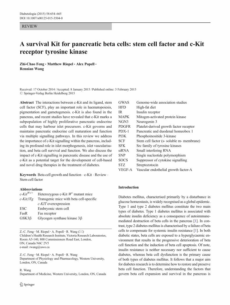
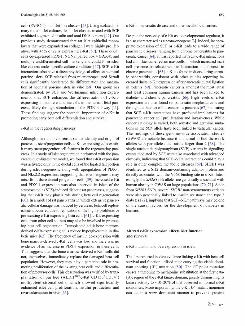
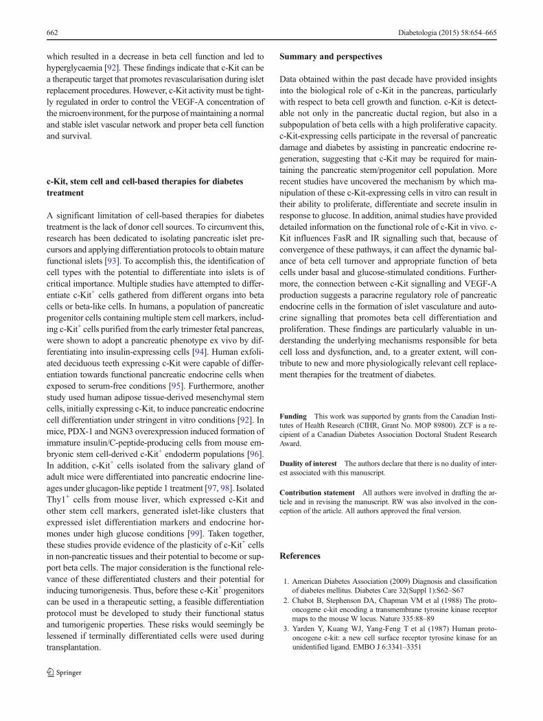
c-Kit is a cellular homologue of the v-Kit oncogene. It is amember of the type III group of receptor tyrosine kinases,encoded by the W locus on chromosome 4 (4q11-21) inhumans, and on chromosome 5 in mice [2, 3]. Structurally,c-Kit is closely related to platelet-derived growth factor recep-tor (PDGFR), sharing 63% homology with the tyrosine kinasesequences and 53% homology with the amino terminus of thekinase domain [4]. Similar to PDGFR, c-Kit consists of anextracellular region comprising five Ig-like domains, a singlemembrane-spanning region and a cytoplasmic region
containing a hydrophilic kinase insert domain (Fig. 1a). Thefirst three domains form the ligand-binding pocket, while thefourth and fifth domains play a critical role in c-Kit monomerpositioning and dimerisation [5]. A similar ligand-bindingmechanism has been proposed for PDGFR. Like PDGFR,the intracellular portion of c-Kit consists of a juxtamembranedomain with an ATP-binding region, a phosphotransferaseregion split into two domains by a kinase insert and aCOOH-terminal tail. Most of the phosphorylation sites arelocated in the cytoplasmic region and are important for trans-ducing intracellular activation signals.
SCF is a product of the Sl locus, mapped to chromosome12 in humans and chromosome 10 in mice [6]. There are sixknown SCF transcripts in humans and four in mice, but twoalternative transcripts are predominantly synthesised in thepancreas, as membrane-bound forms of 220 or 248 (mSCF)amino acids (Fig. 1b). The protein structure of both mSCFforms includes an extracellular domain, a transmembranedomain and an intracellular region. In humans, SCF 248 con-tains a proteolytic cleavage site, possibly accelerating the pro-duction of soluble SCF (sSCF) 165 by post-transcriptionalmodification. However, both SCF 220 and 248 can be cleavedin mice to generate monomeric sSCF (Fig. 1b) [7].Dimerisation of mSCF makes it more biologically active thanits monomeric counterpart [8]. mSCF also results in morepersistent c-Kit activation and prolonged receptor lifespan,whereas sSCF induces relatively transient receptor activationand enhances receptor degradation [9]. SCF is produced andreleased by various cells, including fibroblasts, keratinocytesand endothelial cells [10, 11]. It has also been reported that
Extracellular region(five Ig-like domains)
Ligand binding domain
Dimerisation domain
Transmembrane region
Juxtamembrane domain - Y568- Y570
Kinase domain
- Y703- Y721- Y730
Kinase domain
- Y747
- Y936
Kinase insert domain Cytoplasmic region
5
4
3
2
1
COOH
- Y823
- Y900
Activation loop
COOH COOH
Transmembrane region
Cleavage site Exon 6Exon 7
220 248
165sSCF
NH2NH2
NH2
NH2
COOH
mSCF
a b
Fig. 1 Structure of c-Kit and SCF. (a) c-Kit has extracellular, transmem-brane and intracellular regions. The extracellular region consists of fiveIg-like domains. The transmembrane region keeps c-Kit attached to thecell membrane. The intracellular region contains two kinase domains split
into two parts by the kinase insert domain [4, 5, 10–13]. (b) sSCF isgenerated by cleavage of mSCF (220 or 248) [6–9, 14, 15]. Primaryproteolytic cleavage sites are indicated by the arrows
Diabetologia (2015) 58:654–665 655
both Scf (also known as Kitl) mRNA and protein expressionoccur in adult mouse islets [12].
SCF and c-Kit signalling pathways
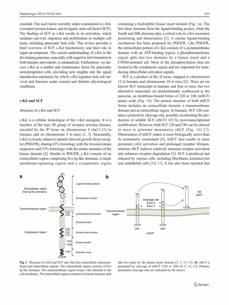
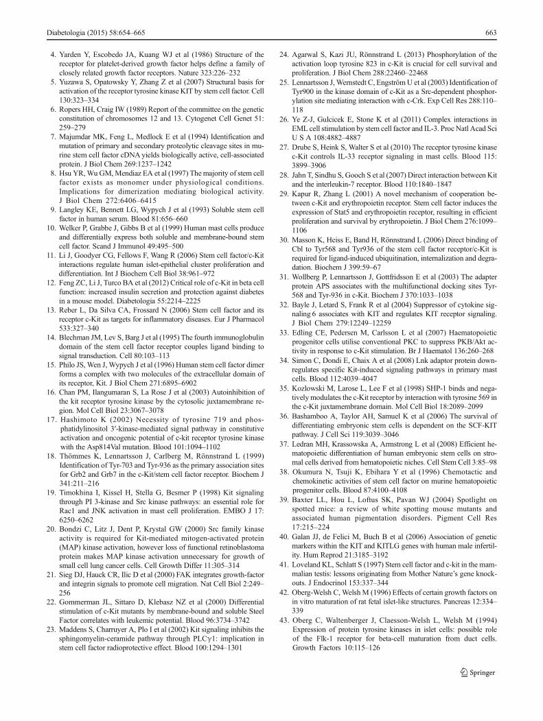
Receptor activation c-Kit exists as a monomer and is stimu-lated to form homodimers following binding of SCF to thefirst three Ig-like domains of c-Kit [13]. The subsequent inter-action between Ig-like domains 4 and 5 in adjacent c-Kitmonomers works to further stabilise c-Kit dimerisation,allowing correct positioning and efficient trans-phosphoryla-tion of its cytoplasmic kinase domains [14, 15]. Thejuxtamembrane domain plays a critical role in regulatingc-Kit activity [16] (Fig. 1a). SCF stimulation promotes therelease of the juxtamembrane domain from the activationloop, enabling catalytic function of the receptor tyrosinekinase to transduce its downstream signal [5].
Molecular signal transduction Nine tyrosine phosphorylationsites have been identified on c-Kit (Fig. 1a). Trans-phosphorylation occurs on tyrosine kinase residues, whichact as docking sites for signalling kinase molecules containinga Src homology 2 domain and a phosphotyrosine-binding
domain (Fig. 2). c-Kit phosphorylation at Y721 activates thephosphoinositide 3-kinase (PI3K) pathway, resulting inenhanced cell survival and proliferation [17]. Phosphorylationof c-Kit at Y703 and Y936 activates the mitogen-activatedprotein kinase (MAPK) pathway [18], which affects genetranscription, cell differentiation and proliferation [13]. c-Kitcan be phosphorylated at Y568, Y570 and Y936 to enhanceinteraction with the SH2 domain of the Src family of tyrosinekinases (SFK). Activation of this kinase family is associatedwith cell proliferation and survival via Akt phosphorylation.Also, one of the mechanisms of activation of the Janus ki-nase–signal transducers and activators of transcription(JAK–STAT) pathway requires the action of both SFK andPI3K [13, 19, 20]. There are also reports suggesting thatSFK directly phosphorylates focal adhesion kinase, which isimportant for cell migration [21]. The phospholipase C-γpathway can interact with the tyrosine kinase residue Y730of c-Kit [22], and has been found to play an important role insuppressing cell apoptosis [23]. The phosphorylation of Y823is a ligand-activated event required for sustaining phosphory-lation of downstream signalling molecules (e.g. PI3K,MAPK) of c-Kit. Mutation of Y823 leads to increased c-Kitinternalisation and degradation, suggesting a role for this site
PP
PP
sSC
F
mS
CF
c-Kit
c-Kit
SC
F
P P
GRB2
ShcA
Ras Sos
Raf-1
MEK1/2
MAPK
Akt
BADP
mTOR
SrcLyn
JAK
STAT
PLCγ SHP-1PI3K
c-Cbl
SOCS
PKC
ReceptordownregulationPhospho-site:
Y568/570, Y936
Proliferation,differentiation
Phospho-site: Y568/570,Y730
Proliferation, survival,adhesion
Phospho-site: Y721, Y900
Gene transcription,proliferation
Phospho-site: Y568/570,Y703, Y936.
DAG
IP3
Fig. 2 c-Kit signalling pathways. Binding of SCF triggers c-Kit down-stream signalling pathways, including the MAPK (blue), PI3K (green),phospholipase C-γ (PLCγ) and Janus kinase–signal transducers and ac-tivators of transcription (JAK–STAT) pathways (orange). Activation ofthese pathways is implicated in numerous cellular processes, such as genetranscription, cellular proliferation, differentiation, survival and adhesion[16–27]. Src homology region 2 domain-containing phosphatase-1 (SHP-
1), Casitas b-lineage lymphoma (c-Cbl), SOCS and the protein kinase Csignals (brown) are involved c-Kit downregulation. BAD, Bcl-2-associ-ated death promoter; DAG, diacylglycerol; GRB2, growth factor recep-tor-bound protein 2; MEK1/2, mitogen-activated protein kinase/extracel-lular-regulated kinase kinase 1/2; mTOR, mechanistic target ofrapamycin
656 Diabetologia (2015) 58:654–665
in stabilising the active kinase conformation [24]. CrkII is anadaptor protein of the Crk family, which can be phosphorylatedby SCF and can interact with Y900 in c-Kit. However, thefunctional consequence of Crk binding with Y900 in c-Kitvia the p85 subunit of PI3K is not clear [25], but may providepossible link to the c-Jun N-terminal kinase (JNK) pathway.c-Kit signalling pathways are not simple linear reactions, but,rather, integrated inputs fromdifferent pathways that determinethe biological consequences in different cellular contexts. Forinstance, c-Kit crosstalks with the erythropoietin receptor orinterleukin receptors to recruit common downstream signallingmolecules, creating a method for modulating diverse physio-logical responses [26–29].
Receptor downregulation Tight regulation of c-Kit signallingis crucial for maintaining proper cellular function. c-Kit sig-nalling can be attenuated through several routes, includingreceptor internalisation, tyrosine dephosphorylation and ki-nase domain inactivation by serine phosphorylation. It hasbeen shown that c-Kit internalisation can be initiated by theassociation of the E3 ubiquitin ligase, Casitas b-lineage lym-phoma (c-Cbl) at Y568 and Y936 on c-Kit [30, 31]. Thesignalling proteins, suppressor of cytokine signalling isoforms1 (SOCS1) and 6 (SOCS6), can also induce c-Kit endocytosisand its subsequent lysosomal degradation [32]. Furthermore,protein kinase C signalling may exert negative feedback on c-Kit activity through serine phosphorylation at S741 and S746in the receptor, which results in shedding of the c-Kit extra-cellular domain from the cell surface [13, 33]. Lymphocyte-specific adaptor protein Lnk also plays an important role inc-Kit downregulation. Lnk preferentially binds to thejuxtamembrane region of c-Kit, suppressing the intrinsicc-Kit catalytic activity [34]. In addition, c-Kit signalling canbe negatively regulated by Src homology region 2 domain-containing phosphatase-1 (SHP-1) through receptor tyrosinedephosphorylation [35].
c-Kit function in organ development
A strong correlation between c-Kit expression and thepluripotency of embryonic stem cells (ESCs) indicates thatc-Kit is a critical factor for the differentiation and survival ofthese cells. c-Kit-null murine ESCs die when induced to dif-ferentiate, and apoptosis also occurs upon differentiation ofnormal ESCs treated with a c-Kit-neutralising antibody [36].Similarly, combinations of SCF and other growth factors havebeen reported to promote efficient haematopoietic differentia-tion from human ESCs [37], and other reports indicate thatSCF-c-Kit interactions are essential for haematopoietic stemcell proliferation, survival and adhesion via the PI3K pathway[38]. Indeed, investigation of the physiological roles of SCFand c-Kit in mice with various naturally occurring mutationsin theW and Sl loci, revealed that SCF-c-Kit interactions play
a crucial functional role in a wide variety of tissues.c-Kit signalling plays an indispensable role in melanocytesurvival and pigmentation, demonstrated by a lack of hairpigmentation, associated with loss of melanocytes, in W orSl mutant mice [39]. SCF-c-Kit interactions are importantfor gametogenesis, as evidenced in men by an SCF mutationthat causes idiopathic male infertility [40]. Furthermore, Wand Slmutant mice display increased germ cell apoptosis, witha corresponding degree of sterility [41]. More importantly,accumulating evidence suggests that c-Kit expression is alsolinked to the development of the endocrine pancreas, as well asto beta cell survival and function [11, 12, 42–53].
c-Kit expression and function in the pancreas
c-Kit in the developing rodent pancreas
c-Kit expression was first detected in RINm5F rat insulinomacell lines and fetal rat islets (Table 1) [43]. Subsequent immu-nohistochemistry studies revealed c-Kit localisation to pancre-atic ducts [42], and c-Kit (also known as Kit) mRNA wasdetectable by embryonic day 13 in the fetal rat pancreas[48]. One cell lineage tracing study utilising lacZ transgenicmurine embryos showed that c-Kit expression was restrictedin a subpopulation of endocrine and epithelial cells [46]. Theabundance of c-Kit expression in early rodent pancreatic de-velopment indicates that it may be involved in maintaining theendocrine cell precursor pool in fetal rodents. We investigatedc-Kit expression in the rodent pancreas during the transitionfrom prenatal to postnatal life at our laboratory. c-Kit waslocalised to the ductal region and in 40% of beta cells atembryonic day 18, but c-Kit+ cells progressively declined inthe pancreas postnatally [47]. These observations suggest thatdecreased c-Kit expression may correlate with islet endocrinecell maturation. Two distinct putative stem cell-like popula-tions expressing either c-Kit or Sca-1, another stem cell anti-gen, in the developing rodent pancreas were recentlycharacterised. Flow cytometry analyses revealed that the iso-lated c-Kit+ cell population co-expressed markers related toislet differentiation, including pancreatic and duodenal ho-meobox 1 (PDX-1) and neurogenin 3 (NGN3), but isolatedSca-1+ cells lacked expression of these markers [54]. Thesefindings imply that c-Kit can be used as a marker to identify aputative islet precursor cell population in the developingrodent endocrine pancreas.
Few studies have focused on the effects of c-Kit mutationswith regard to pancreatic endocrine morphogenesis. Onestudy reported that islets were still present in the absence ofc-Kit in WlacZ/WlacZ embryos, although few morphometricdetails were examined, and there was no indication of func-tional status [55]. These data raise two possibilities: (1) c-Kit
Diabetologia (2015) 58:654–665 657
is not critical for the determination and specification of isletformation, but is more involved in islet survival and function;or, more likely, (2) there are redundant pathways, such asPDGFR signalling, compensating for the loss of c-Kit duringpancreatic development in rodents.
c-Kit in the developing human pancreas
The developing human pancreas and multiple human pancre-atic cancer cell lines express both c-Kit and SCF (Table 1) [11,49, 53, 56]. c-Kit+ cells were detected at the earliest stage ofhuman pancreatic development, as single endocrine cells dif-ferentiated from ducts [49]. The expression of c-Kit wasrestricted to ductal regions and adjoining neogenic islet clus-ters, whereas SCF expression was scattered throughout thedeveloping human pancreas [11]. The majority of c-Kit+ cellsdisplayed the ductal cell marker, cytokeratin 19, and transcrip-tion factors associated with islet differentiation, including
PDX-1, sex determining region Y-box 9 (SOX9), NGN3,and homeobox protein Nkx6.1, between 8 and 12 weeks ofhuman fetal age. However, as age progressed to 19–21 weeks,co-expression between c-Kit and these markers declined [11].These results coincide with the findings of studies in rodents,indicating that cell populations expressing c-Kit may: (1) rep-resent endocrine precursors participating in islet neogenesis;and/or (2) serve as an instructive signal, directing islet differ-entiation and proliferation in the developing human pancreas.
SCF–c-Kit in islet differentiation
SCF–c-Kit interactions mediate beta cell differentiation andproliferation and have been demonstrated across multiple spe-cies in vitro. Stimulation of c-Kit activity promoted gene tran-scription and proliferation in INS-1 rat insulinoma cells [12,46, 51]. Furthermore, treatment by exogenous SCF induceddifferentiation of human pancreatic carcinoma, epithelial-like
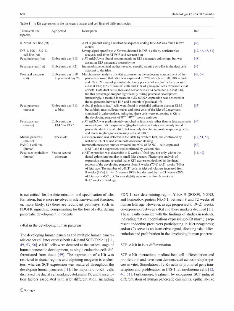
Table 1 c-Kit expression in the pancreatic tissues and cell lines of different species
Tissue/cell line(species)
Age period Description Ref.
RINm5F cell line (rat) – A PCR product using a nucleotide sequence coding for c-Kit was found in twoclones
[43]
INS-1, INS-1 832-13cell line (rat)
– Strong signal specific to c-Kit was detected in INS-1 cells by northern blotanalysis, real-time RT-PCR and western blot
[12, 46, 48, 51]
Fetal pancreas (rat) Embryonic day E13 c-Kit mRNAwas found predominantly in E13 pancreatic epithelium, but wasabsent in E13 pancreatic mesenchyme
[48]
Fetal pancreas (rat) Embryonic day E21 Immunohistochemical studies revealed specific staining of c-Kit in the duct cellsadjacent to the islets
[42]
Postnatal pancreas(rat)
Embryonic day E18to postnatal day 28
Morphometric analysis of c-Kit expression in the endocrine compartment of thepancreas showed that c-Kit was expressed in 25% of cells at E18, 10% at birth,and 3% at 28 days of postnatal life. Forty per cent of insulin+ cells expressedc-Kit at E18; 10% of insulin+ cells and 11% of glucagon+ cells expressed c-Kitat birth. Both duct cells (16%) and acinar cells (5%) contained c-Kit at E18,but this percentage dropped significantly during postnatal development.Interestingly, a twofold increase in c-Kit mRNA expression was observed inthe rat pancreas between E18 and 1 month of postnatal life
[47, 57]
Fetal pancreas(mouse)
Embryonic day E13to birth
A few β-galactosidase+ cells were found in epithelial collector ducts at E12.5,but at birth, most collector tubes and most cells of the islet of Langerhanscontained β-galactosidase, indicating these cells were expressing c-Kit inthe developing pancreas of WlacZ/WlacZ mouse embryos
[55]
Fetal pancreas(mouse)
Embryonic dayE14.5 to E18.5
c-Kit mRNAwas predominately enriched in fetal islets rather than in fetal pancreaticmesenchyme. c-Kit expression (β-galactosidase activity) was mainly found inpancreatic duct cells at E14.5, but was only detected in insulin-expressing cells,and rarely in glucagon-expressing cells, at E18.5
[46]
Mature pancreas(mouse)
8 weeks old c-Kit expression was detected in the islets by western blot, and confirmed byreal-time RT-PCR and immunofluorescence staining
[12, 51, 52]
PANC-1 cell line(human)
– Immunofluorescence studies revealed that 97% of PANC-1 cells expressedc-KIT, and the expression was confirmed by western blot
[53]
Fetal islet–epithelium(human)
First to secondtrimesters
c-KIT expression was detectable at 8 weeks of fetal age, not only within theductal epithelium but also in small islet clusters. Phenotypic analysis ofexpression patterns revealed that c-KIT expression declined in the ductalregions of the developing pancreas from 8 weeks (70%) to 21 weeks (30%)of fetal age. The number of c-KIT+ cells in islet cell clusters increased from8 weeks (18%) to 14–16 weeks (30%), but declined by 19–21 weeks (20%)of fetal age. c-KIT mRNAwas slightly increased at 14–16 weeks vs.8–12 weeks of fetal age
[11, 49]
658 Diabetologia (2015) 58:654–665
cells (PANC-1) into islet-like clusters [53]. Using isolated pri-mary rodent islet cultures, fetal islet clusters treated with SCFexhibited augmented insulin and total DNA content [42]. Ourprevious study demonstrated that rat islet epithelial mono-layers that were expanded on collagen I were highly prolifer-ative, with 45% of cells expressing c-Kit [57]. These c-Kit+
cells co-expressed PDX-1, NGN3, paired box 4 (PAX4), andmultiple undifferentiated cell markers, and could form islet-like clusters under specific culture conditions [57]. SCF–c-Kitinteractions also have a direct physiological effect on neonatalporcine islets. SCF released from microencapsulated Sertolicells significantly accelerated the differentiation and matura-tion of neonatal porcine islets in vitro [58]. Our group hasdemonstrated, by SCF and Wortmannin inhibition experi-ments, that SCF enhances the differentiation of c-Kit-expressing immature endocrine cells in the human fetal pan-creas, likely through stimulation of the PI3K pathway [11].These findings suggest the potential importance of c-Kit inpromoting early beta cell differentiation and survival.
c-Kit in the regenerating pancreas
Although there is no consensus on the identity and origin ofpancreatic stem/progenitor cells, c-Kit-expressing cells exhib-it many stem/progenitor cell features in the regenerating pan-creas. In a study of islet regeneration performed with the pan-creatic duct-ligated rat model, we found that c-Kit expressionwas activated only in the ductal cells of the ligated tail portionduring islet neogenesis, along with upregulation of PDX-1and Nkx2.2 expression, suggesting that islet neogenesis mayarise from these ductal progenitor cells [59]. Increased c-Kitand PDX-1 expression was also observed in islets of thestreptozotocin (STZ)-induced diabetic rat pancreases, suggest-ing that c-Kit may play a role during beta cell regeneration[60]. In a model of rat pancreatitis in which extensive pancre-atic cellular damagewas induced by cerulean, beta cell replen-ishment occurred due to replication of the highly proliferativepre-existing c-Kit-expressing beta cells [61]. c-Kit-expressingcells from other cell sources may also be involved in promot-ing beta cell regeneration. Transplanted adult bone marrow-derived c-Kit-expressing cells reduce hyperglycaemia in dia-betic mice [62]. The frequency of insulin co-expression withbone marrow-derived c-Kit+ cells was low, and there was noevidence of an increase in PDX-1 expression in these cells.This suggests that the bone marrow-derived c-Kit+ cells didnot, themselves, immediately replace the damaged beta cellpopulation. However, they may play a paracrine role in pro-moting proliferation of the existing beta cells and differentia-tion of precursor cells. This observation was verified by trans-plantation of purified (ALDHhighc-Kit+CD133+CD34+)multipotent stromal cells, which showed significantlyenhanced islet cell proliferation, insulin production andrevascularisation in vivo [63].
c-Kit in pancreatic disease and other metabolic disorders
Despite the necessity of c-Kit as a developmental regulator, itis also characterised as a proto-oncogene [3]. Indeed, inappro-priate expression of SCF or c-Kit leads to a wide range ofpancreatic diseases, ranging from chronic pancreatitis to pan-creatic cancer [64]. It was reported that SCF-c-Kit interactionshad an influential effect onmast cells, in which increased mastcell presence correlated with inflammation and fibrosis inchronic pancreatitis [65]. c-Kit is found in ducts during chron-ic pancreatitis, consistent with other studies reporting in-creased ductal c-Kit expression after pancreatic ductal ligationin rodents [59]. Pancreatic cancer is amongst the most lethaland least common human cancers and has been linked todiabetes and chronic pancreatitis [66]. High levels of c-Kitexpression are also found on pancreatic neoplastic cells andthroughout the duct of the cancerous pancreas [67], indicatingthat SCF–c-Kit interactions have profound implications forpancreatic cancer cell proliferation and invasiveness. Whilecancer aetiology is varied, both somatic and germline muta-tions in the SCF allele have been linked to testicular cancer.The findings of these genome-wide association studies(GWAS) are notable because it is unusual to find these riskalleles with per-allele odds ratios larger than 2 [68]. Thesingle nucleotide polymorphism (SNP) variants in signallingevents mediated by SCF were also associated with advancedcirrhosis, indicating that SCF–c-Kit interactions could play arole in other complex metabolic diseases [69]. SH2B1 wasidentified as a SH2 domain-containing adaptor protein anddirectly associates with the Y568 binding site in c-Kit. Inter-estingly, the SH2B1 risk alleles are genetically associated withhuman obesity in GWAS on large populations [70, 71]. Asidefrom SH2B1 SNPs, several SH2B1 non-synonymous variantswere also genetically linked to insulin resistance and type 2diabetes [72], implying that SCF–c-Kit pathways may be oneof the causal factors for the development of diabetes inhumans.
Altered c-Kit expression affects islet functionand survival
c-Kit mutation and overexpression in islets
The first reported in vivo evidence linking c-Kit with beta cellsurvival and function utilised mice carrying the viable domi-nant spotting (Wv) mutation [50]. The Wv point mutationcauses a threonine to methionine substitution at the first cata-lytic region of the c-Kit kinase domain, greatly diminishing itskinase activity to ~10–20% of that observed in normal c-Kitmonomers. More importantly, the c-Kit Wv mutant monomercan act in a trans-dominant manner to prevent tyrosine
Diabetologia (2015) 58:654–665 659
phosphorylation of normal c-Kit by dimerisation, reflected bya ~60% reduction in c-Kit transduction activity [73]. Pheno-typic analysis of mice heterozygous for the Wv mutation(c-KitWv/+) showed impaired glucose tolerance and a markedinsulin secretory defect by 8 weeks of age [50]. The develop-ment of a diabetic phenotype, particularly in male mice, wasdue to a severe loss of beta cell mass and reduced proliferativecapacity. Interestingly, these changes were not observable infemale mice until 40 weeks of age, indicating that the delay ofdiabetes was possibly due to differences between sexes in sexhormone function [50]. Furthermore, downregulation of thePI3K–Akt–glycogen synthase kinase 3β (GSK3β) pathway,which connects c-Kit activity to physiological changes in betacells of c-KitWv/+ mice, was demonstrated [52]. Inhibition ofactivated GSK3β with 1-azakenpaullone, a GSK3β inhibitor,was shown to rescue c-KitWv/+ mice from defective glucosemetabolism and increase islet cyclin D1 and PDX-1 expres-sion [52]. This observation suggests that dysregulation of theAkt–GSK3β pathway, downstream of c-Kit, is responsible forthe onset of diabetes in c-KitWv/+ mice.
The most convincing evidence supporting the importanceof the impact of c-Kit on beta cell function and survival wasdetermined using a transgenic mouse model with overexpres-sion of the human c-KIT gene specifically in beta cells direct-ed by the rat insulin promoter (c-KitβTgmice) [12]. Our groupshowed that c-KitβTgmice displayed improved glucose toler-ance, which was associated with expansion of beta cell massas a result of increased beta cell proliferation. Overexpressionof c-KIT also led to a profound effect on insulin secretion inresponse to glucose challenge, and insulin content in beta cellsof these mice was increased compared with that in their con-trol littermates [12]. Increased c-Kit activity upregulated theAkt–GSK3β–cyclin D1 pathway, which was associated withenhanced expression of islet-specific transcription factors,including PDX-1 andMafA [12]. Notably, c-KitβTgmice alsocounteracted the detrimental effects of a 4 week high-fat diet(HFD) challenge, in which beta cell function and mass weremaintained. Furthermore, overexpression of c-KIT in betacells of c-KitWv/+ mice could partially reverse the diabeticphenotype, demonstrating that c-Kit has a beneficial effecton beta cells and directly influences beta cell health [12].
c-Kit and Fas receptor interplay in islets
Accumulating evidence has demonstrated that Fas receptor(FasR) signalling contributes to cell apoptosis, which isaccompanied by the absence of c-Kit signalling in melano-cytes [74], gametes [75], oocytes [76] and haematopoietic stemcells [77]. FasR is known as apoptosis antigen 1, CD95, ortumour necrosis factor receptor superfamily member 6. Fasligand binding and subsequent activation of FasR leads toprogrammed cell death in many systems via activation of thecaspase 8-mediated downstream cell death machinery. We
recently reported that deficient c-Kit signalling resulted inincreased beta cell death in c-KitWv/+ mice, which was associ-ated with upregulation of p53 levels and induction of FasRactivity in c-KitWv/+ mouse islets [51] (Fig. 3). It is clear thatp53 is an important checkpoint protein that promotes cellcycle arrest [78]. Several lines of evidence have indicated thatFasR expression can be upregulated by p53 activation throughpromotion of FasR gene transcription and trafficking of FasRto the cell surface via the Golgi apparatus [79, 80]. The inter-play between c-Kit and FasR signalling in beta cell survivalwas further verified by a double mutant mouse model(c-KitWv/+ mice with the lymphoproliferation [lpr] mutation,which disrupts expression of the cell surface FasR). Lack offunctional FasR in c-KitWv/+ mice reduced the extent of betacell apoptosis via downregulation of the caspase 8-mediatedextrinsic apoptotic pathway, and enhanced FLICE-like inhib-itor protein/nuclear factor κB pro-survival signalling [51].These in vivo results were further supported by in vitro obser-vations using INS-1 cells, whereby c-Kit activation negativelymodulated both p53 and FasR in a PI3K-dependent manner. Inparticular, knockdown of c-Kit in INS-1 cells led to increasedp53 and FasR levels, reversed by treatment with a p53 inhibitoror Fas small interfering (si)RNA, ultimately resulting in areduction in apoptosis [51]. These data imply that a balancebetween c-Kit and FasR signalling is instrumental in maintain-ing proper beta cell mass turnover.
c-Kit and insulin receptor crosstalk in islets
It has been documented that c-Kit is able to crosstalk withmany growth factor and cytokine receptors [26–28, 81]. Forexample, erythropoietin and SCF have synergistic effects onerythropoiesis. In fact, erythropoietin appears to stimulate c-Kit dimerisation and tyrosine phosphorylation in human he-matopoietic cell lines [81]. Recently, treatment with recombi-nant human erythropoietin was shown to provide protectionagainst the development of diabetes in STZ-induced anddb/db mice. This was associated with a significant up regula-tion of c-Kit and vascular endothelial growth factor-A(VEGF-A) protein expression [82]. It is well established thatinsulin signalling pathways can participate in crosstalk withother receptors [83, 84]. Insulin, secreted from beta cells inresponse to glucose stimulation, activates the beta cell insulinreceptor (IR) and the downstream signalling transducer, IRS.This, in turn, triggers the PI3K–Akt pathway, which is animportant mediator of beta cell proliferation, function and sur-vival. Overexpression of c-KIT leads to increased IR proteinexpression and tyrosine phosphorylation of IRS1 and -2, andtreatment of INS-1 cells with exogenous SCF leads to en-hanced co-localisation of c-Kit and IR, as revealed by doubleimmunofluorescence [12]. The effect of c-Kit and IR signal-ling was suppressed by wortmannin (PI3K inhibitor) or c-KitsiRNA, suggesting an interaction between these two upstream
660 Diabetologia (2015) 58:654–665
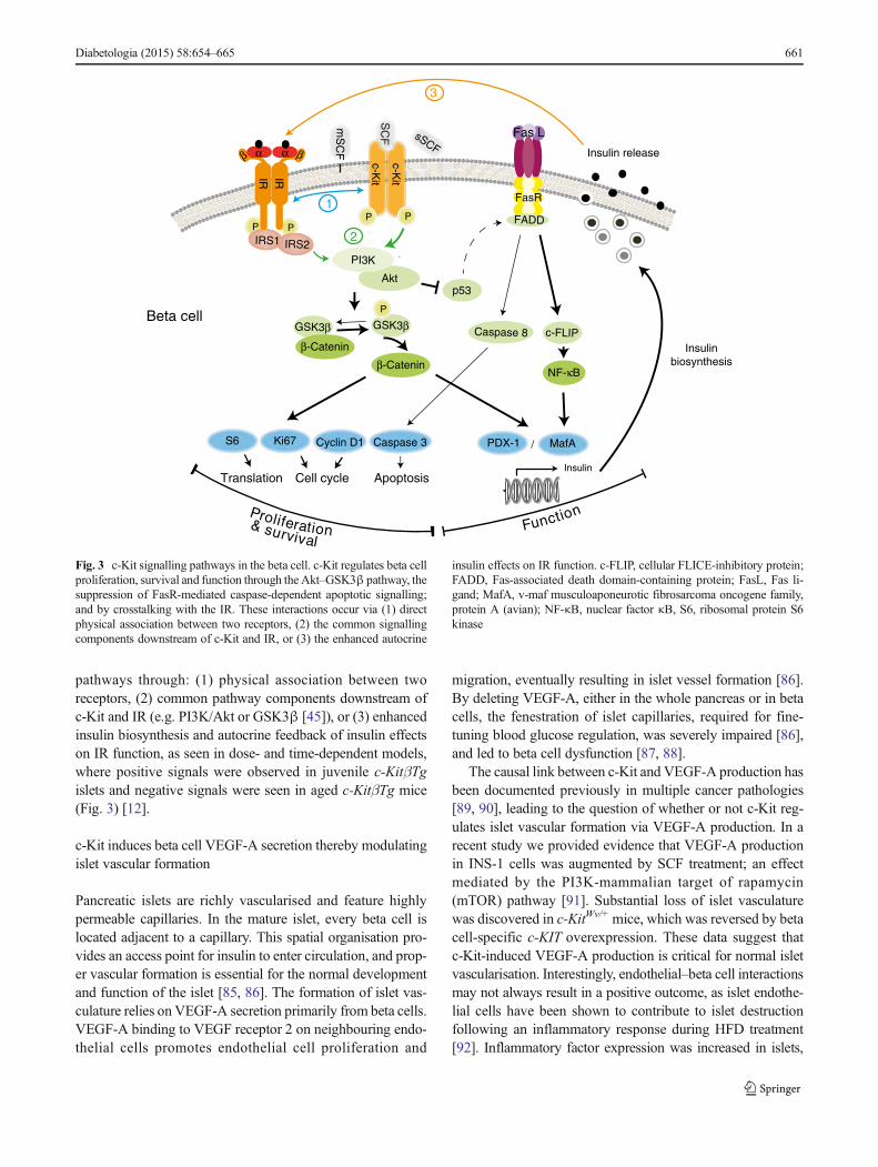
pathways through: (1) physical association between tworeceptors, (2) common pathway components downstream ofc-Kit and IR (e.g. PI3K/Akt or GSK3β [45]), or (3) enhancedinsulin biosynthesis and autocrine feedback of insulin effectson IR function, as seen in dose- and time-dependent models,where positive signals were observed in juvenile c-KitβTgislets and negative signals were seen in aged c-KitβTg mice(Fig. 3) [12].
c-Kit induces beta cell VEGF-A secretion thereby modulatingislet vascular formation
Pancreatic islets are richly vascularised and feature highlypermeable capillaries. In the mature islet, every beta cell islocated adjacent to a capillary. This spatial organisation pro-vides an access point for insulin to enter circulation, and prop-er vascular formation is essential for the normal developmentand function of the islet [85, 86]. The formation of islet vas-culature relies on VEGF-A secretion primarily from beta cells.VEGF-A binding to VEGF receptor 2 on neighbouring endo-thelial cells promotes endothelial cell proliferation and
migration, eventually resulting in islet vessel formation [86].By deleting VEGF-A, either in the whole pancreas or in betacells, the fenestration of islet capillaries, required for fine-tuning blood glucose regulation, was severely impaired [86],and led to beta cell dysfunction [87, 88].
The causal link between c-Kit and VEGF-A production hasbeen documented previously in multiple cancer pathologies[89, 90], leading to the question of whether or not c-Kit reg-ulates islet vascular formation via VEGF-A production. In arecent study we provided evidence that VEGF-A productionin INS-1 cells was augmented by SCF treatment; an effectmediated by the PI3K-mammalian target of rapamycin(mTOR) pathway [91]. Substantial loss of islet vasculaturewas discovered in c-KitWv/+mice, which was reversed by betacell-specific c-KIT overexpression. These data suggest thatc-Kit-induced VEGF-A production is critical for normal isletvascularisation. Interestingly, endothelial–beta cell interactionsmay not always result in a positive outcome, as islet endothe-lial cells have been shown to contribute to islet destructionfollowing an inflammatory response during HFD treatment[92]. Inflammatory factor expression was increased in islets,
P
/
sSCF
c-Kit
c-Kit
SC
F
P P
Akt
Proliferation
GSK3β
β-Catenin
Cell cycleTranslation
PI3K
Insulin release
P
IRS2
mS
CF
IRS1
Function
Cyclin D1S6 PDX-1 MafA
β βαα
RI
Beta cellGSK3β
β-Catenin
P
2
3
Ki67
FasR
FADD
p53
& survival
Caspase 3
Apoptosis
Caspase 8 c-FLIP
NF-κB
Fas L
Insulinbiosynthesis
1
Insulin
RI
Fig. 3 c-Kit signalling pathways in the beta cell. c-Kit regulates beta cellproliferation, survival and function through the Akt–GSK3β pathway, thesuppression of FasR-mediated caspase-dependent apoptotic signalling;and by crosstalking with the IR. These interactions occur via (1) directphysical association between two receptors, (2) the common signallingcomponents downstream of c-Kit and IR, or (3) the enhanced autocrine
insulin effects on IR function. c-FLIP, cellular FLICE-inhibitory protein;FADD, Fas-associated death domain-containing protein; FasL, Fas li-gand; MafA, v-maf musculoaponeurotic fibrosarcoma oncogene family,protein A (avian); NF-κB, nuclear factor κB, S6, ribosomal protein S6kinase
Diabetologia (2015) 58:654–665 661
which resulted in a decrease in beta cell function and led tohyperglycaemia [92]. These findings indicate that c-Kit can bea therapeutic target that promotes revascularisation during isletreplacement procedures. However, c-Kit activity must be tight-ly regulated in order to control the VEGF-A concentration ofthe microenvironment, for the purpose ofmaintaining a normaland stable islet vascular network and proper beta cell functionand survival.
c-Kit, stem cell and cell-based therapies for diabetestreatment
A significant limitation of cell-based therapies for diabetestreatment is the lack of donor cell sources. To circumvent this,research has been dedicated to isolating pancreatic islet pre-cursors and applying differentiation protocols to obtainmaturefunctional islets [93]. To accomplish this, the identification ofcell types with the potential to differentiate into islets is ofcritical importance. Multiple studies have attempted to differ-entiate c-Kit+ cells gathered from different organs into betacells or beta-like cells. In humans, a population of pancreaticprogenitor cells containing multiple stem cell markers, includ-ing c-Kit+ cells purified from the early trimester fetal pancreas,were shown to adopt a pancreatic phenotype ex vivo by dif-ferentiating into insulin-expressing cells [94]. Human exfoli-ated deciduous teeth expressing c-Kit were capable of differ-entiation towards functional pancreatic endocrine cells whenexposed to serum-free conditions [95]. Furthermore, anotherstudy used human adipose tissue-derived mesenchymal stemcells, initially expressing c-Kit, to induce pancreatic endocrinecell differentiation under stringent in vitro conditions [92]. Inmice, PDX-1 and NGN3 overexpression induced formation ofimmature insulin/C-peptide-producing cells from mouse em-bryonic stem cell-derived c-Kit+ endoderm populations [96].In addition, c-Kit+ cells isolated from the salivary gland ofadult mice were differentiated into pancreatic endocrine line-ages under glucagon-like peptide 1 treatment [97, 98]. IsolatedThy1+ cells from mouse liver, which expressed c-Kit andother stem cell markers, generated islet-like clusters thatexpressed islet differentiation markers and endocrine hor-mones under high glucose conditions [99]. Taken together,these studies provide evidence of the plasticity of c-Kit+ cellsin non-pancreatic tissues and their potential to become or sup-port beta cells. The major consideration is the functional rele-vance of these differentiated clusters and their potential forinducing tumorigenesis. Thus, before these c-Kit+ progenitorscan be used in a therapeutic setting, a feasible differentiationprotocol must be developed to study their functional statusand tumorigenic properties. These risks would seemingly belessened if terminally differentiated cells were used duringtransplantation.
Summary and perspectives
Data obtained within the past decade have provided insightsinto the biological role of c-Kit in the pancreas, particularlywith respect to beta cell growth and function. c-Kit is detect-able not only in the pancreatic ductal region, but also in asubpopulation of beta cells with a high proliferative capacity.c-Kit-expressing cells participate in the reversal of pancreaticdamage and diabetes by assisting in pancreatic endocrine re-generation, suggesting that c-Kit may be required for main-taining the pancreatic stem/progenitor cell population. Morerecent studies have uncovered the mechanism by which ma-nipulation of these c-Kit-expressing cells in vitro can result intheir ability to proliferate, differentiate and secrete insulin inresponse to glucose. In addition, animal studies have provideddetailed information on the functional role of c-Kit in vivo. c-Kit influences FasR and IR signalling such that, because ofconvergence of these pathways, it can affect the dynamic bal-ance of beta cell turnover and appropriate function of betacells under basal and glucose-stimulated conditions. Further-more, the connection between c-Kit signalling and VEGF-Aproduction suggests a paracrine regulatory role of pancreaticendocrine cells in the formation of islet vasculature and auto-crine signalling that promotes beta cell differentiation andproliferation. These findings are particularly valuable in un-derstanding the underlying mechanisms responsible for betacell loss and dysfunction, and, to a greater extent, will con-tribute to new and more physiologically relevant cell replace-ment therapies for the treatment of diabetes.
Funding This work was supported by grants from the Canadian Insti-tutes of Health Research (CIHR, Grant No. MOP 89800). ZCF is a re-cipient of a Canadian Diabetes Association Doctoral Student ResearchAward.
Duality of interest The authors declare that there is no duality of inter-est associated with this manuscript.
Contribution statement All authors were involved in drafting the ar-ticle and in revising the manuscript. RW was also involved in the con-ception of the article. All authors approved the final version.
References
1. American Diabetes Association (2009) Diagnosis and classificationof diabetes mellitus. Diabetes Care 32(Suppl 1):S62–S67
2. Chabot B, Stephenson DA, Chapman VM et al (1988) The proto-oncogene c-kit encoding a transmembrane tyrosine kinase receptormaps to the mouse W locus. Nature 335:88–89
3. Yarden Y, Kuang WJ, Yang-Feng T et al (1987) Human proto-oncogene c-kit: a new cell surface receptor tyrosine kinase for anunidentified ligand. EMBO J 6:3341–3351
662 Diabetologia (2015) 58:654–665
4. Yarden Y, Escobedo JA, Kuang WJ et al (1986) Structure of thereceptor for platelet-derived growth factor helps define a family ofclosely related growth factor receptors. Nature 323:226–232
5. Yuzawa S, Opatowsky Y, Zhang Z et al (2007) Structural basis foractivation of the receptor tyrosine kinase KIT by stem cell factor. Cell130:323–334
6. Ropers HH, Craig IW (1989) Report of the committee on the geneticconstitution of chromosomes 12 and 13. Cytogenet Cell Genet 51:259–279
7. Majumdar MK, Feng L, Medlock E et al (1994) Identification andmutation of primary and secondary proteolytic cleavage sites in mu-rine stem cell factor cDNAyields biologically active, cell-associatedprotein. J Biol Chem 269:1237–1242
8. Hsu YR,WuGM,Mendiaz EA et al (1997) The majority of stem cellfactor exists as monomer under physiological conditions.Implications for dimerization mediating biological activity.J Biol Chem 272:6406–6415
9. Langley KE, Bennett LG, Wypych J et al (1993) Soluble stem cellfactor in human serum. Blood 81:656–660
10. Welker P, Grabbe J, Gibbs B et al (1999) Human mast cells produceand differentially express both soluble and membrane-bound stemcell factor. Scand J Immunol 49:495–500
11. Li J, Goodyer CG, Fellows F, Wang R (2006) Stem cell factor/c-Kitinteractions regulate human islet-epithelial cluster proliferation anddifferentiation. Int J Biochem Cell Biol 38:961–972
12. Feng ZC, Li J, Turco BA et al (2012) Critical role of c-Kit in beta cellfunction: increased insulin secretion and protection against diabetesin a mouse model. Diabetologia 55:2214–2225
13. Reber L, Da Silva CA, Frossard N (2006) Stem cell factor and itsreceptor c-Kit as targets for inflammatory diseases. Eur J Pharmacol533:327–340
14. Blechman JM, Lev S, Barg J et al (1995) The fourth immunoglobulindomain of the stem cell factor receptor couples ligand binding tosignal transduction. Cell 80:103–113
15. Philo JS, Wen J, Wypych J et al (1996) Human stem cell factor dimerforms a complex with two molecules of the extracellular domain ofits receptor, Kit. J Biol Chem 271:6895–6902
16. Chan PM, Ilangumaran S, La Rose J et al (2003) Autoinhibition ofthe kit receptor tyrosine kinase by the cytosolic juxtamembrane re-gion. Mol Cell Biol 23:3067–3078
17. Hashimoto K (2002) Necessity of tyrosine 719 and phos-phatidylinositol 3′-kinase-mediated signal pathway in constitutiveactivation and oncogenic potential of c-kit receptor tyrosine kinasewith the Asp814Val mutation. Blood 101:1094–1102
18. Thömmes K, Lennartsson J, Carlberg M, Rönnstrand L (1999)Identification of Tyr-703 and Tyr-936 as the primary association sitesfor Grb2 and Grb7 in the c-Kit/stem cell factor receptor. Biochem J341:211–216
19. Timokhina I, Kissel H, Stella G, Besmer P (1998) Kit signalingthrough PI 3-kinase and Src kinase pathways: an essential role forRac1 and JNK activation in mast cell proliferation. EMBO J 17:6250–6262
20. Bondzi C, Litz J, Dent P, Krystal GW (2000) Src family kinaseactivity is required for Kit-mediated mitogen-activated protein(MAP) kinase activation, however loss of functional retinoblastomaprotein makes MAP kinase activation unnecessary for growth ofsmall cell lung cancer cells. Cell Growth Differ 11:305–314
21. Sieg DJ, Hauck CR, Ilic D et al (2000) FAK integrates growth-factorand integrin signals to promote cell migration. Nat Cell Biol 2:249–256
22. Gommerman JL, Sittaro D, Klebasz NZ et al (2000) Differentialstimulation of c-Kit mutants by membrane-bound and soluble SteelFactor correlates with leukemic potential. Blood 96:3734–3742
23. Maddens S, Charruyer A, Plo I et al (2002) Kit signaling inhibits thesphingomyelin-ceramide pathway through PLCγ1: implication instem cell factor radioprotective effect. Blood 100:1294–1301
24. Agarwal S, Kazi JU, Rönnstrand L (2013) Phosphorylation of theactivation loop tyrosine 823 in c-Kit is crucial for cell survival andproliferation. J Biol Chem 288:22460–22468
25. Lennartsson J,Wernstedt C, EngströmU et al (2003) Identification ofTyr900 in the kinase domain of c-Kit as a Src-dependent phosphor-ylation site mediating interaction with c-Crk. Exp Cell Res 288:110–118
26. Ye Z-J, Gulcicek E, Stone K et al (2011) Complex interactions inEML cell stimulation by stem cell factor and IL-3. Proc Natl Acad SciU S A 108:4882–4887
27. Drube S, Heink S, Walter S et al (2010) The receptor tyrosine kinasec-Kit controls IL-33 receptor signaling in mast cells. Blood 115:3899–3906
28. Jahn T, Sindhu S, Gooch S et al (2007) Direct interaction between Kitand the interleukin-7 receptor. Blood 110:1840–1847
29. Kapur R, Zhang L (2001) A novel mechanism of cooperation be-tween c-Kit and erythropoietin receptor. Stem cell factor induces theexpression of Stat5 and erythropoietin receptor, resulting in efficientproliferation and survival by erythropoietin. J Biol Chem 276:1099–1106
30. Masson K, Heiss E, Band H, Rönnstrand L (2006) Direct binding ofCbl to Tyr568 and Tyr936 of the stem cell factor receptor/c-Kit isrequired for ligand-induced ubiquitination, internalization and degra-dation. Biochem J 399:59–67
31. Wollberg P, Lennartsson J, Gottfridsson E et al (2003) The adapterprotein APS associates with the multifunctional docking sites Tyr-568 and Tyr-936 in c-Kit. Biochem J 370:1033–1038
32. Bayle J, Letard S, Frank R et al (2004) Suppressor of cytokine sig-naling 6 associates with KIT and regulates KIT receptor signaling.J Biol Chem 279:12249–12259
33. Edling CE, Pedersen M, Carlsson L et al (2007) Haematopoieticprogenitor cells utilise conventional PKC to suppress PKB/Akt ac-tivity in response to c-Kit stimulation. Br J Haematol 136:260–268
34. Simon C, Dondi E, Chaix A et al (2008) Lnk adaptor protein down-regulates specific Kit-induced signaling pathways in primary mastcells. Blood 112:4039–4047
35. Kozlowski M, Larose L, Lee F et al (1998) SHP-1 binds and nega-tively modulates the c-Kit receptor by interactionwith tyrosine 569 inthe c-Kit juxtamembrane domain. Mol Cell Biol 18:2089–2099
36. Bashamboo A, Taylor AH, Samuel K et al (2006) The survival ofdifferentiating embryonic stem cells is dependent on the SCF-KITpathway. J Cell Sci 119:3039–3046
37. Ledran MH, Krassowska A, Armstrong L et al (2008) Efficient he-matopoietic differentiation of human embryonic stem cells on stro-mal cells derived from hematopoietic niches. Cell Stem Cell 3:85–98
38. Okumura N, Tsuji K, Ebihara Y et al (1996) Chemotactic andchemokinetic activities of stem cell factor on murine hematopoieticprogenitor cells. Blood 87:4100–4108
39. Baxter LL, Hou L, Loftus SK, Pavan WJ (2004) Spotlight onspotted mice: a review of white spotting mouse mutants andassociated human pigmentation disorders. Pigment Cell Res17:215–224
40. Galan JJ, de Felici M, Buch B et al (2006) Association of geneticmarkers within the KIT and KITLG genes with human male infertil-ity. Hum Reprod 21:3185–3192
41. Loveland KL, Schlatt S (1997) Stem cell factor and c-kit in the mam-malian testis: lessons originating from Mother Nature’s gene knock-outs. J Endocrinol 153:337–344
42. Oberg-Welsh C, WelshM (1996) Effects of certain growth factors onin vitro maturation of rat fetal islet-like structures. Pancreas 12:334–339
43. Oberg C, Waltenberger J, Claesson-Welsh L, Welsh M (1994)Expression of protein tyrosine kinases in islet cells: possible roleof the Flk-1 receptor for beta-cell maturation from duct cells.Growth Factors 10:115–126
Diabetologia (2015) 58:654–665 663
44. Welsh M, Annerén C, Lindholm C et al (2000) Role of tyrosinekinase signaling for beta-cell replication and survival. Ups J Med Sci105:7–15
45. WelshM (2012) The platelet-derived growth factor (PDGF) family oftyrosine kinase receptors: a Kit to fix the beta cell? Diabetologia 55:2092–2095
46. Rachdi L, El Ghazi L, Bernex F et al (2001) Expression of the recep-tor tyrosine kinase KIT in mature beta-cells and in the pancreas indevelopment. Diabetes 50:2021–2028
47. Yashpal NK, Li J, Wang R (2004) Characterization of c-Kit andnestin expression during islet cell development in the prenatal andpostnatal rat pancreas. Dev Dyn 229:813–825
48. LeBras S, Czernichow P, Scharfmann R (1998) A search for tyrosinekinase receptors expressed in the rat embryonic pancreas.Diabetologia 41:1474–1481
49. Li J, Quirt J, Do HQ et al (2007) Expression of c-Kit receptor tyrosinekinase and effect on beta-cell development in the human fetal pan-creas. Am J Physiol Endocrinol Metab 293:E475–E483
50. Krishnamurthy M, Ayazi F, Li J et al (2007) c-Kit in early onset ofdiabetes: a morphological and functional analysis of pancreatic beta-cells in c-KitW-v mutant mice. Endocrinology 148:5520–5530
51. Feng Z-C, Riopel M, Li J et al (2013) Downregulation of Fas activityrescues early onset of diabetes in c-KitWv/+ mice. Am J PhysiolEndocrinol Metab 304:E557–E565
52. Feng Z-C, Donnelly L, Li J et al (2012) Inhibition of Gsk3β activityimproves β-cell function in c-KitWv/+ male mice. Lab Investig 92:543–555
53. Wu Y, Li J, Saleem S et al (2010) c-Kit and stem cell factor regulatePANC-1 cell differentiation into insulin- and glucagon-producingcells. Lab Investig 90:1373–1384
54. Ma F, Chen F, Chi Y et al (2012) Isolation of pancreatic progenitorcells with the surface marker of hematopoietic stem cells. Int JEndocrinol 2012:948683
55. Bernex F, de Sepulveda P, Kress C et al (1996) Spatial and temporalpatterns of c-kit-expressing cells in WlacZ/+ and WlacZ/WlacZ mouseembryos. Development 122:3023–3033
56. Yasuda A, Sawai H, Takahashi H et al (2006) The stem cell factor/c-kit receptor pathway enhances proliferation and invasion of pancre-atic cancer cells. Mol Cancer 5:46
57. Wang R, Li J, Yashpal N (2004) Phenotypic analysis of c-Kit expres-sion in epithelial monolayers derived from postnatal rat pancreaticislets. J Endocrinol 182:113–122
58. Mancuso F, Calvitti M, Luca G et al (2010) Acceleration of function-al maturation and differentiation of neonatal porcine islet cell mono-layers shortly in vitro cocultured with microencapsulated sertoli cells.Stem Cells Int 2010:587213
59. Peters K, Panienka R, Li J et al (2005) Expression of stem cellmarkers and transcription factors during the remodeling of the ratpancreas after duct ligation. Virchows Arch 446:56–63
60. Tiemann K, Panienka R, Klöppel G (2007) Expression of transcrip-tion factors and precursor cell markers during regeneration of betacells in pancreata of rats treated with streptozotocin. Virchows Arch450:261–266
61. Gong J, Zhang G, Tian F, Wang Y (2012) Islet-derived stem cellsfrom adult rats participate in the repair of islet damage. J Mol Histol43:745–750
62. Hess D, Li L, Martin M et al (2003) Bone marrow-derived stem cellsinitiate pancreatic regeneration. Nat Biotechnol 21:763–770
63. Bell GI, Putman DM, Hughes-Large JM, Hess DA (2012)Intrapancreatic delivery of human umbilical cord blood aldehydedehydrogenase-producing cells promotes islet regeneration.Diabetologia 55:1755–1760
64. Raimondi S, Lowenfels AB, Morselli-Labate AM et al (2010)Pancreatic cancer in chronic pancreatitis; aetiology, incidence, andearly detection. Best Pract Res Clin Gastroenterol 24:349–358
65. Esposito I, Friess H, Kappeler A et al (2001) Mast cell distributionand activation in chronic pancreatitis. Hum Pathol 32:1174–1183
66. Raimondi S, Maisonneuve P, Lowenfels AB (2009) Epidemiology ofpancreatic cancer: an overview. Nat Rev Gastroenterol Hepatol 6:699–708
67. Esposito I, Kleeff J, Bischoff SC et al (2002) The stem cell factor-c-kit system and mast cells in human pancreatic cancer. Lab Investig82:1481–1492
68. Kanetsky PA, Mitra N, Vardhanabhuti S et al (2009) Common vari-ation in KITLG and at 5q31.3 predisposes to testicular germ cellcancer. Nat Genet 41:811–815
69. Chen Q-R, Braun R, Hu Yet al (2013) Multi-SNP analysis of GWASdata identifies pathways associated with nonalcoholic fatty liver dis-ease. PLoS One 8:e65982
70. Willer CJ, Speliotes EK, Loos RJF et al (2009) Six new loci associ-ated with body mass index highlight a neuronal influence on bodyweight regulation. Nat Genet 41:25–34
71. Thorleifsson G, Walters GB, Gudbjartsson DF et al (2009) Genome-wide association yields new sequence variants at seven loci that as-sociate with measures of obesity. Nat Genet 41:18–24
72. Doche ME, Bochukova EG, Su H-W et al (2012) Human SH2B1mutations are associated with maladaptive behaviors and obesity.J Clin Invest 122:4732–4736
73. Nocka K, Majumder S, Chabot B et al (1989) Expression of c-kitgene products in known cellular targets ofWmutations in normal andW mutant mice—evidence for an impaired c-kit kinase in mutantmice. Genes Dev 3:816–826
74. Sharov AA, Li G-Z, Palkina TN et al (2003) Fas and c-kit are in-volved in the control of hair follicle melanocyte apoptosis and migra-tion in chemotherapy-induced hair loss. J Invest Dermatol 120:27–35
75. Sakata S, Sakamaki K,WatanabeK et al (2003) Involvement of deathreceptor Fas in germ cell degeneration in gonads of Kit-deficientWv/Wv mutant mice. Cell Death Differ 10:676–686
76. Moniruzzaman M, Sakamaki K, Akazawa Y, Miyano T (2007)Oocyte growth and follicular development in KIT-deficient Fas-knockout mice. Reproduction 133:117–125
77. Mori T, AndoK, Tanaka K et al (1997) Fas-mediated apoptosis of thehematopoietic progenitor cells in mice infected with murine cyto-megalovirus. Blood 89:3565–3573
78. Rocha S, Martin AM, Meek DW, Perkins ND (2003) p53 repressescyclin D1 transcription through down regulation of Bcl-3 and induc-ing increased association of the p52 NF-kappaB subunit with histonedeacetylase 1. Mol Cell Biol 23:4713–4727
79. Bennett M, Macdonald K, Chan SW et al (1998) Cell surface traf-ficking of Fas: a rapid mechanism of p53-mediated apoptosis.Science 282:290–293
80. Müller M,Wilder S, Bannasch D et al (1998) p53 activates the CD95(APO-1/Fas) gene in response to DNA damage by anticancerdrugs. J Exp Med 188:2033–2045
81. Kapur R, Cooper R, Zhang L, Williams DA (2001) Cross-talk be-tween α4β1/α5β1 and c-Kit results in opposing effect on growth andsurvival of hematopoietic cells via the activation of focal adhesionkinase, mitogen-activated protein kinase, and Akt signaling path-ways. Blood 97:1975–1981
82. Choi D, Schroer SA, Lu SY et al (2010) Erythropoietin protectsagainst diabetes through direct effects on pancreatic beta cells.J Exp Med 207:2831–2842
83. Ricort JM, Tanti JF, van Obberghen E, Le Marchand-Brustel Y(1997) Cross-talk between the platelet-derived growth factor andthe insulin signaling pathways in 3T3-L1 adipocytes. J Biol Chem272:19814–19818
84. Li J, DeFea K, Roth RA (1999) Modulation of insulin receptorsubstrate-1 tyrosine phosphorylation by an Akt/phosphatidylinositol3-kinase pathway. J Biol Chem 274:9351–9356
85. Lammert E, Cleaver O, Melton D (2001) Induction of pancreaticdifferentiation by signals from blood vessels. Science 294:564–567
664 Diabetologia (2015) 58:654–665
86. Lammert E, Gu G, McLaughlin M et al (2003) Role of VEGF-A invascularization of pancreatic islets. Curr Biol 13:1070–1074
87. Brissova M, Shostak A, Shiota M et al (2006) Pancreatic islet pro-duction of vascular endothelial growth factor–a is essential for isletvascularization, revascularization, and function. Diabetes 55:2974–2985
88. Iwashita N, Uchida T, Choi JB et al (2007) Impaired insulin secretionin vivo but enhanced insulin secretion from isolated islets in pancre-atic beta cell-specific vascular endothelial growth factor-A knock-outmice. Diabetologia 50:380–389
89. Litz J, Krystal GW (2006) Imatinib inhibits c-Kit-induced hypoxia-inducible factor-1alpha activity and vascular endothelial growth fac-tor expression in small cell lung cancer cells. Mol Cancer Ther 5:1415–1422
90. Zhang N, Richter A, Suriawinata J et al (2004) Elevated vascularendothelial growth factor production in islets improves islet graftvascularization. Diabetes 53:963–970
91. Feng ZC, Li J, Silverstein J, Popell A, Yee SP, Wang R (2013) Long-term overexpression of c-Kit negatively impacts beta-cell functionand survival via capillary and inflammatory recruitment in the islet.Diabetologia 56(Suppl 1):S222, Abstract
92. Agudo J, Ayuso E, Jimenez V et al (2012) Vascular endothelialgrowth factor-mediated islet hypervascularization and inflammation
contribute to progressive reduction of β-cell mass. Diabetes 61:2851–2861
93. Pagliuca FW, Millman JR, Gürtler M et al (2014) Generation offunctional human pancreatic β cells in vitro. Cell 159:428–439
94. Suen PM, Zou C, Zhang YA et al (2008) PDZ-domain containing-2(PDZD2) is a novel factor that affects the growth and differentiationof human fetal pancreatic progenitor cells. Int J Biochem Cell Biol40:789–803
95. Ishkitiev N, Yaegaki K, Kozhuharova A et al (2013) Pancreatic dif-ferentiation of human dental pulp CD117+ stem cells. Regen Med 8:597–612
96. Kubo A, Stull R, Takeuchi M et al (2011) Pdx1 and Ngn3 overex-pression enhances pancreatic differentiation of mouse ES cell-derived endoderm population. PLoS One 6:e24058
97. Matsumoto S, Okumura K, Ogata A et al (2007) Isolation oftissue progenitor cells from duct-ligated salivary glands ofswine. Cloning Stem Cells 9:176–190
98. Baek H, Noh YH, Lee JH et al (2014) Autonomous isolation, long-term culture and differentiation potential of adult salivary gland-derived stem/progenitor cells. J Tissue Eng Regen Med 8:717–727
99. Yang L, Li S, Hatch H et al (2002) In vitro trans-differentiation ofadult hepatic stem cells into pancreatic endocrine hormone-producing cells. Proc Natl Acad Sci U S A 99:8078–8083
Diabetologia (2015) 58:654–665 665