role of nadph oxidase in beta cell dysfunction - springer · role of nadph oxidase in beta cell...
TRANSCRIPT

Role of NADPH Oxidase in Beta Cell Dysfunction
Jessica R. Weavera and David A. Taylor-Fishwickb*aDepartment of Microbiology and Molecular Cell Biology, Eastern Virginia Medical School, Norfolk, VA, USAbDepartment of Microbiology and Molecular Cell Biology, Department of Medicine, Eastern Virginia Medical School,Strelitz Diabetes Center, Norfolk, VA, USA
Abstract
Dysfunction of pancreatic beta cells and loss of beta cell mass is a major factor in the development ofdiabetes. Currently there is no cure for diabetes, and available therapies do not focus on halting orreversing the loss of beta cell function. New strategies to preserve beta cells in diabetes are needed.Conferring protection to the beta cells against the effects of sustained intracellular reactive oxygenspecies (ROS) presents a novel approach to preserve beta cell mass. Important contributors toincreases in intercellular ROS in beta cells are nicotinamide adenine dinucleotide phosphate(NADPH) oxidase enzymes. Discussed in this review are the roles of NADPH oxidases in thebeta cell, their contribution to beta cell dysfunction, and new emerging selective inhibitors ofNADPH oxidase.
Keywords
Beta cell; Diabetes; Inhibitors; Islet; NADPH oxidase; NOX; Proinflammatory cytokines; ROS
Introduction
An underlying feature of diabetes is a loss of functional beta cell capacity. The therapeutic potentialto cure diabetes by restoring functional beta cell mass has been best illustrated in islet transplantationstudies. These proof-of-principle efforts reversed insulin dependence. As a therapeutic strategy, islettransplantation is not currently viable (Taylor-Fishwick et al. 2008). However, these studies validateefforts to seek approaches to preserve and protect beta cell mass in diabetes. Fundamental to this isa better appreciation of key events that drive beta cell failure. Sensitivity of beta cells to reactiveoxygen species and oxidative stress is well recognized (Lenzen 2008). Relative to other cells,pancreatic beta cells are vulnerable to sustained elevation in intracellular reactive species (Lenzen2008). This is due, in part, to the low activity of free-radical detoxifying enzymes such as catalase,superoxide dismutase, and glutathione peroxidase in beta cells (Grankvist et al. 1981; Lenzenet al. 1996; Tiedge et al. 1997; Modak et al. 2007). Islets also exhibit poor ability to rectify oxidativedamage to DNA (Modak et al. 2007). Consequently, beta cells are easily overwhelmed by elevatedreactive oxygen species (ROS) and enter a state of oxidative stress (Lenzen 2008). Under oxidativestress conditions, ROS in addition to oxidizing proteins, lipids, and DNA also activate stress-sensitive second messengers such as p38-mitogen-activated protein kinase (MAPK), c-JunN-terminal kinase (JNK), and protein kinase C (PKC) (Koya and King 1998; Purves et al. 2001).
*Email: [email protected]
Islets of Langerhans, 2. ed.DOI 10.1007/978-94-007-6884-0_46-3# Springer Science+Business Media Dordrecht 2014
Page 1 of 29

For example, a consequence of JNK activation is translocation of the homeodomain transcriptionfactor pdx-1 from the nucleus to the cytoplasm. Pdx-1 is a key transactivator of the insulin gene in thenucleus (Ohneda et al. 2000). An outcome of cytoplasmic translocation of pdx-1 is defective insulinexpression that contributes to beta cell dysfunction. As pdx-1 transactivates its own expression, theimpact of a cytoplasmic relocation for pdx-1 is amplified (Kawamori et al. 2003). It seems a paradoxthen that while beta cells are susceptible to sustained elevations in ROS, transient increases in ROSare required for function and the generation of intracellular signaling (Goldstein et al. 2005; Piet al. 2007; Newsholme et al. 2009). The source of intracellular ROS in beta cells is a fertile area ofresearch and reflects recognition for the key role elevated ROS can play in beta cell pathophysiology.Intracellular events such as mitochondrial stress and/or endoplasmic reticulum stress do contributeto elevated intracellular ROS in the beta cells (reviewed in Newsholme et al. (2009), Volchuk andRon (2010)). Peroxisome metabolism of fatty acids elevates cellular ROS, and this ROS elevationhas been linked to beta cell lipotoxicity and dysfunction (Gehrmann et al. 2010; Elsner et al. 2011;Fransen et al. 2012). More recently, recognition has been made for an important contribution of theROS-generating NADPH oxidase enzyme family in beta cell pathophysiology. Reviewed in thischapter are the roles that NADPH oxidases have in the beta cell with an emphasis on contributions tobeta cell dysfunction. Initially, the family of NADPH oxidase enzymes is introduced witha description of common features for this family in terms of structure, function, and expression.Specific features of each member of the family are considered relative to structure, function,expression, and regulation. The contribution of NADPH oxidase enzymes to beta cell functionand dysfunction is considered by describing the members and subunits that are expressed andfunctional in beta cells. The activity of these NADPH oxidase enzymes is regulated by stimuliknown to be elevated in diabetes. The consequence of stimulating NADPH oxidase activity isrelevant for both beta cell physiology and beta cell pathophysiology. The role of NADPH oxidasesin insulin secretion, beta cell signaling, and induced beta cell dysfunction is explored. Newlyrecognized are the contributions of intracellular ROS to discrete activation of second messengers.Thus, action of NADPH oxidase in terms of influencing the beta cell ranges from subtle control tofrank cell destruction. Lastly, major advances in the development of new selective inhibitors ofNADPH oxidases are reviewed. These inhibitors represent candidate agents to advance the field andpropose new therapeutic strategies to preserve and protect beta cell mass in diabetes.
NADPH Oxidase Family
NADPH oxidases are multi-protein complexes that generate reactive oxygen species, includingsuperoxide (O2
•�) and hydroxyl radical (•OH). Characteristically, the family shares a commonbasic structure but differ by their membrane localization and protein components (illustrated inFig. 1).
There are seven identified members of the NADPH oxidase family. In humans, all seven enzymesare expressed. NADPH oxidases contain the following characteristic elements: (1) a COOHterminus, (2) six conserved transmembrane domains, (3) a flavin adenine dinucleotide (FAD)-binding site, (4) a NADPH-binding site at the COOH terminus, and (5) generally two heme-binding histidines in the third transmembrane domain and two in the fifth transmembrane domain,although the location of these sites vary (Rotrosen et al. 1992; Cross et al. 1995; Finegold et al. 1996;Biberstine-Kinkade et al. 2001; Lambeth et al. 2007). Some family members have additionaldomains, such as EF hands, or an additional NH2-terminal transmembrane domain. As a class,NADPH oxidases are members of the flavocytochrome family. Functional enzymes require
Islets of Langerhans, 2. ed.DOI 10.1007/978-94-007-6884-0_46-3# Springer Science+Business Media Dordrecht 2014
Page 2 of 29

association with protein subunits to form complexes. Lambeth et al. reviewed subunit and domaininteraction in NADPH oxidase enzymes (Lambeth et al. 2007).
Functionally, NADPH oxidase complexes traverse membranes and convert oxygen to a reactiveradical via electron transfer across plasma or organelle membranes. NADPH serves as the singleelectron donor. NADPH donates a single electron to FAD which is located in the cytoplasmic end ofthe NOX enzyme (Nisimoto et al. 1999). The electron is passed between heme groups beforereacting with oxygen to create superoxide (Finegold et al. 1996). Superoxide can then be convertedto hydrogen peroxide (H2O2), a principle stable product.
NADPH oxidase family members are found primarily in eukaryotic cells of animals, plants, andfungi. Members of the NADPH oxidase family are implicated in numerous biological functions inanimals. For example, phagocyte NADPH oxidase plays a critical role in host immune defense byactivating the respiratory burst associated with neutrophil-mediated innate immunity (Radaet al. 2008). NADPH oxidases function in thyroid hormone synthesis and help regulate inner earfunction (Caillou et al. 2001; Paffenholz et al. 2004; Ris-Stalpers 2006). Maintenance of the vascularsystem, hypertensive tone, and vascular wall function are influenced by NADPH oxidase activity(reviewed in Manea (2010)). Indeed, reflective of the central influence of ROS in many biologicalprocesses, NADPH oxidases are associated with physiological or pathophysiological processes ofmost mammalian organ systems (reviewed (Bedard and Krause 2007; Bedard et al. 2007; Lambethet al. 2007)). Additionally, an elevation of intracellular ROS and association of NADPH oxidaseactivity are implicated in numerous diseases involving these mammalian organ systems. In plants,NADPH oxidase plays a role in plant defenses and development (reviewed in Marino et al. (2012))(Torres and Dangl 2005; Gapper and Dolan 2006; Sagi and Fluhr 2006). NADPH oxidase regulatesroot growth by cell expansion (Foreman et al. 2003). Fungi express several different isoforms ofNOXs (reviewed in Tudzynski et al. (2012)). Filamentous fungi express two subfamilies of NADPHoxidase that are similar to mammalian phagocyte NADPH oxidase. Homologous subunits to othermammalian NADPH oxidases have also been reported in fungi (Aguirre et al. 2005). The NADPH
NOX-1 NOX-2 NOX-3
NOX-4 NOX-5
= transmembranedomain
= p40phox = EF-hand
= p22phox = NADPH/FAD = p67phox/NOXAI
= p47phox/NOXOI= peroxidasedomain
= Rac
= Ca2+
DUOX-1/-2
Fig. 1 NADPH oxidase (NOX) family members and their structures. Each NOX member contains six conservedtransmembrane domains and a NADPH/FAD site at the COOH terminus. Additional structures needed for an activecomplex for some of the NOXs include p22phox, p67phox/NOXA1, p47phox, Rac, p47phox, and p40phox, NOX-5 andDUOX-1/-2 have an EF-hand where Ca2+ binds for activation. DUOX-1/-2 have an extra-transmembrane domain withan extracellular peroxidase domain
Islets of Langerhans, 2. ed.DOI 10.1007/978-94-007-6884-0_46-3# Springer Science+Business Media Dordrecht 2014
Page 3 of 29

oxidase subfamilies present in fungi play a role in growth and development including germination(Lara-Ortiz et al. 2003; Malagnac et al. 2004; Cano-Dominguez et al. 2008). Evidence to suggestthat fungal NADPH oxidases may play a role in pathogenesis of cellulose degradation has also beenreported (Brun et al. 2009).
NADPH Oxidase Family Members
Phagocyte NADPH oxidase, named for its expression in phagocytes, especially neutrophils, was thefirst identified NADPH oxidase and plays a critical role in the respiratory burst response associatedwith innate immune defense (Babior et al. 1973, 2002). Subsequent gene discoveries have identifieda family of genes/proteins that are distinct enzymes but homologous to the catalytic core subunit ofphagocyte NADPH oxidase. This family of NADPH oxidases is termed NOX/DUOX with thenomenclature of the seven members being termed NOX-1, NOX-2, NOX-3, NOX-4, NOX-5,DUOX-1, and DUOX-2. Under this nomenclature the core catalytic subunit of phagocyteNADPH oxidase (previously termed gp91phox) is called NOX-2. A brief overview of each familymember is provided starting with the archetypal phagocyte NADPH oxidase (NOX-2) beforeconsideration of the role of NADPH oxidase in beta cells (Table 1).
NOX-2The structure of the functional NOX-2 enzyme is made up of five protein subunits that are located inthe plasma membrane or translocated to the plasma membrane from the cytosol. Based upon theoriginal name, phagocyte NADPH oxidase, the naming convention of subunits is gpXphox, wheregpX indicates glycoprotein of “X” molecular weight. Activation of phagocyte NADPH oxidase(NOX-2) occurs through a complex series of protein interactions. The core catalytic component ofNADPH oxidase, gp91phox, is stabilized in the membrane by p22phox. The adaptor protein p47phox isactivated by phosphorylation. Recruitment of p47phox facilitates addition to the complex of p40phox,p67phox, and Rac (small GTP-binding protein). The latter two appear to regulate catalysis (Aboet al. 1991; Ando et al. 1992; Wientjes et al. 1993; Heyworth et al. 1994; Hordijk 2006; Orientet al. 2007; Guichard et al. 2008).
NOX-2 in phagocytes functions in the host defense by providing a respiratory burst response thatserves to kill bacteria enclosed within the phagosome through production of hypochlorous acid.NOX-2 also activates inflammatory and immune responses (Rada et al. 2008). Genetic defects inNOX-2 activity arising from mutations in the genes encoding gp91phox, p22phox, p47phox, andp67phox are associated with chronic granulomatous disease (Heyworth et al. 2003). Generation ofROS by NOX-2 has also been shown to activate pathways involved in angiogenesis (as reviewed inUshio-Fukai (2006)).
In addition to phagocytes, NOX-2 expression has been reported in pancreas, B lymphocytes,neurons, cardiomyocytes, endothelium, skeletal muscle, hepatocytes, smooth muscle, and hemato-poietic stem cells (Bedard and Krause 2007).
NOX-2 is regulated by angiotensin II (AngII), endothelin-1, growth factors such as vascularendothelial growth factor (VEGF), cytokines such as tumor necrosis factor alpha (TNF-a), inter-feron gamma (IFN-g), lipopolysaccharide, mechanical forces, and hyperlipidemia (Cassatellaet al. 1990; Dworakowski et al. 2008).
Islets of Langerhans, 2. ed.DOI 10.1007/978-94-007-6884-0_46-3# Springer Science+Business Media Dordrecht 2014
Page 4 of 29
NOX-1The first homolog of the NOX-2 catalytic subunit (p91phox) to be described was NOX-1. Similar tofunctional NOX-2, the functional NOX-1 enzyme is a multi-protein complex. The subunits that bind
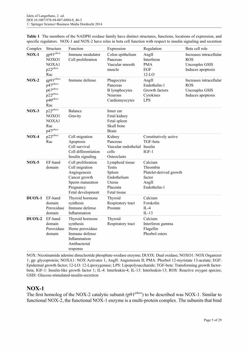
Table 1 The members of the NADPH oxidase family have distinct structures, functions, locations of expression, andspecific regulators. NOX-1 and NOX-2 have roles in beta cell function with respect to insulin signaling and secretion
Complex Structure Function Expression Regulation Beta cell role
NOX-1 gp91phox
NOXO1NOXA1p22phox
Rac
Immune modulatorCell proliferation
Colon epitheliumPancreasVascular smoothmuscle
AngIIInterferonPMAEGF12-LO
Increases intracellularROSUncouples GSISInduces apoptosis
NOX-2 gp91phox
p47phox
p67phox
p22phox
p40phox
Rac
Immune defense PhagocytesPancreasB lymphocytesNeuronsCardiomyocytes
AngIIEndothelin-1Growth factorsCytokinesLPS
Increases intracellularROSUncouples GSISInduces apoptosis
NOX-3 p22phox
NOXO1NOXA1Racp47phox
BalanceGravity
Inner earFetal kidneyFetal spleenSkull boneBrain
NOX-4 p22phox
RacCell migrationApoptosisCell survivalCell differentiationInsulin signaling
KidneyPancreasVascular endothelialcellsOsteoclasts
Constitutively activeTGF-betaInsulinIGF-1
NOX-5 EF-handdomain
Cell proliferationCell migrationAngiogenesisCancer growthSperm maturationPregnancyFetal development
Lymphoid tissueTestisSpleenEndotheliumUterusPlacentaFetal tissue
CalciumThrombinPlatelet-derived growthfactorAngIIEndothelin-1
DUOX-1 EF-handdomainPeroxidasedomain
Thyroid hormonesynthesisImmune defenseInflammation
ThyroidRespiratory tractProstate
CalciumForskolinIL-4IL-13
DUOX-2 EF-handdomainPeroxidasedomain
Thyroid hormonesynthesisHeme peroxidaseImmune defenseInflammationAntibacterialresponse
ThyroidRespiratory tract
CalciumInterferon gammaFlagellinPhorbol esters
NOX: Nicotinamide adenine dinucleotide phosphate-oxidase enzyme; DUOX: Dual oxidase; NOXO1: NOXOrganizer1; gp: glycoprotein; NOXA1: NOX Activator 1; AngII: Angiotensin II; PMA: Phorbol 12-myristate 13-acetate; EGF:Epidermal growth factor; 12-LO: 12-Lipoxygenase; LPS: Lipopolysaccharide; TGF-beta: Transforming growth factor-beta; IGF-1: Insulin-like growth factor 1; IL-4: Interleukin-4; IL-13: Interleukin-13; ROS: Reactive oxygen species;GSIS: Glucose-stimulated-insulin-secretion
Islets of Langerhans, 2. ed.DOI 10.1007/978-94-007-6884-0_46-3# Springer Science+Business Media Dordrecht 2014
Page 5 of 29

NOX-1 are NOXO1 (NOX organizer 1) and NOXA1 (NOX activator 1), which while being distinctproteins are homologs of p47phox and p67phox, respectively (Takeya et al. 2003; Uchizonoet al. 2006). For a functional complex, active NOX-1 requires the NOX-1 catalytic core bound tothe membrane subunit p22phox, NOXO1, and NOXA1 along with the GTPase Rac subunit. NOXO1is predominantly associated with membrane-bound p22phox (Sumimoto 2008). NOXA1 and Ractranslocate to the membrane, in a phosphorylation-dependent process, and are required for NOX-1activation (Miyano et al. 2006). Transfection experiments have shown that NOX-1 can use thep47phox and p67phox subunits of NOX-2, raising the possibility of a dynamic interaction amongstNOX isotypes where subunits are interchangeable between the different NOX family members(Banfi et al. 2003).
NOX-1 activity is associated in immune defense in inflammatory bowel disease and cancer(Rokutan et al. 2006, 2008). Studies have linked NOX-1 activity to colon cancer by affecting cellproliferation of colon carcinoma cell lines and controlling cell migration of colon adenocarcinomacells (de Carvalho et al. 2008; Sadok et al. 2008). NOX-1 also functions in the vasculature byregulating smooth muscle growth, migration (cell movement), and blood pressure (Cave et al. 2006;Gavazzi et al. 2006; Garrido and Griendling 2009). NOX-1 helps regulate neuronal differentiationby negatively affecting excessive neurite outgrowth and influences pain sensitivity during inflam-mation (Ibi et al. 2006, 2008).
NOX-1 is expressed in the colon epithelium and at lower levels in the pancreas, vascular smoothmuscle, endothelium, uterus, placenta, prostate, osteoclasts, and retinal pericytes (Nisimotoet al. 2008). Patterns of expression for NOX-1 are related to species. For example, the expressionof NOX-1 is found in rodent stomach but not in human stomach (Kawahara et al. 2005; Kusumotoet al. 2005; Rokutan et al. 2008).
Regulators of NOX-1 include AngII, IFN-g, protein kinase C (PKC) activation (PMA:4b-phorbol 12-myristate 13-acetate), and epidermal growth factor receptor ligation (Suhet al. 1999; Lassegue et al. 2001; Wingler et al. 2001; Katsuyama et al. 2002; Seshiah et al. 2002;Touyz et al. 2002; Geiszt et al. 2003a; Takeya et al. 2003; Fan et al. 2005). In the liver cell line FaO,NOX-1 regulation involves an autoregulatory feedforward loop involving second messenger acti-vation of Src kinase and extracellular-signal-regulated kinase (ERK) (Fan et al. 2005; Adachiet al. 2008; Sancho and Fabregat 2010).
NOX-3Activity of NOX-3 is dependent on p22phox. Additional cytosolic subunits NOXO1, NOXA1,p47phox, or Rac are not needed for basal NOX-3 activity. The activity of NOX-3 is howeversignificantly increased following association with cytosolic subunits (Banfi et al. 2004a; Chenget al. 2004; Ueyama et al. 2006).
A major site for NOX-3 expression is the inner ear where its function is to assist biogenesis ofotoconia and regulation of balance and gravity (Paffenholz et al. 2004). Expression of NOX-3 hasadditionally been described in fetal kidney, fetal spleen, skull bone, brain, and lung endothelial cellswhere it is associated with the development of emphysema (Banfi et al. 2004a; Zhang et al. 2006;Bedard and Krause 2007).
NOX-4NOX-4 activity requires association with the p22phox subunit, but unlike NOX-2, other subunits arenot essential (Martyn et al. 2006). It is unresolved whether the Rac subunit is required for NOX-4activity (Gorin et al. 2003). Unlike the other NOX family members that initially produce superoxide,NOX-4 produces H2O2 (Martyn et al. 2006; Serrander et al. 2007).
Islets of Langerhans, 2. ed.DOI 10.1007/978-94-007-6884-0_46-3# Springer Science+Business Media Dordrecht 2014
Page 6 of 29

NOX-4 function has been associated with cell migration, apoptosis, cell survival, cell differen-tiation, insulin signaling, cell migration, the unfolded protein response, and differentiation(Mahadev et al. 2004; Pedruzzi et al. 2004; Vaquero et al. 2004; Cucoranu et al. 2005; Liet al. 2006; Meng et al. 2008; Pendyala et al. 2009; Santos et al. 2009). It is a major source ofoxidative stress in the failing heart (Kuroda et al. 2010).
NOX-4 is expressed mainly in the kidney but is also detected in vascular endothelial cells,osteoclasts, endothelium, smooth muscle, hematopoietic stem cells, fibroblasts, keratinocytes,melanoma cells, neurons, pancreas, and adipocytes (Geiszt et al. 2000; Cheng et al. 2001; Shioseet al. 2001; Ago et al. 2004; Bedard and Krause 2007).
NOX-4 is constitutively active and levels of mRNA directly correlate with enzyme function(Serrander et al. 2007). The activity of NOX-4 can be upregulated by tumor growth factor beta(TGF-b) in cardiac fibroblasts, pulmonary artery smooth muscle cells, and lungs (Cucoranuet al. 2005; Sturrock et al. 2006, 2007). Insulin activates NOX-4 in adipocytes, and it can beactivated by insulin-like growth factor-1 (IGF-1) in vascular smooth muscle cells (VSMCs)(Mahadev et al. 2004; Meng et al. 2008; Schroder et al. 2009).
NOX-5Distinct to the other NOX enzymes, activation of NOX-5 is calcium dependent. At its aminoterminal, NOX-5 has a calmodulin-like domain that has four calcium-binding EF-hand domains(Lambeth 2007). NOX-5 does not require p22phox or other phox subunits for activity. Several splicevariant forms of NOX-5 exist (Banfi et al. 2001, 2004b; BelAiba et al. 2007; Pandey et al. 2012).
NOX-5 activity has functional relevance in endothelial cell proliferation, migration, and angio-genesis (BelAiba et al. 2007; Jay et al. 2008; Schulz and Munzel 2008). NOX-5 activity is linked tocancer, including prostate cancer growth, esophageal adenocarcinoma, breast cancer, and hairy cellleukemia (Brar et al. 2003; Kamiguti et al. 2005; Fu et al. 2006; Kumar et al. 2008; Juhaszet al. 2009). NOX-5 exerts a role in fetal development and sperm maturation (reviewed Bedardet al. (2012)).
NOX-5 is expressed mainly in the lymphoid tissue, testis, and spleen (Banfi et al. 2001; Chenget al. 2001). Expression of NOX-5 in endothelium, smooth muscle, pancreas, placenta, ovary,uterus, stomach, certain prostate cancers, and various fetal tissues has been reported (Banfiet al. 2001; Cheng et al. 2001; Bedard and Krause 2007; BelAiba et al. 2007).
Calcium, thrombin, platelet-derived growth factor (PDGF), AngII, and endothelin-1 are factorsreported to regulate NOX-5 (Montezano et al. 2010, 2011). IFN-g has also been shown to activateNOX-5 in smooth muscle cells possibly through the release of intracellular calcium stores (Maneaet al. 2012).
DUOX-1 and DUOX-2DUOX-1 and DUOX-2 do not require subunits to form an active enzyme complex. Like NOX-5,calcium is required to activate to each DUOX enzyme (Selemidis et al. 2008; Rigutto et al. 2009).Structurally, DUOX enzymes differ to NOX enzymes by having an additional transmembranedomain at the amino terminal and an extracellular peroxidase domain (De Deken et al. 2000).DUOX-1/DUOX-2 locate to the plasma membrane from the ER with the help of maturation factorsDUOXA1 and DUOXA2 (Grasberger and Refetoff 2006; Morand et al. 2009).
DUOX-1 and DUOX-2 are highly expressed in the thyroid and directly produce H2O2 that is usedduring thyroid hormone T4 synthesis (Dupuy et al. 1989, 1999; De Deken et al. 2000; Caillouet al. 2001; Ris-Stalpers 2006). While current knowledge suggests the major role for DUOX is in thesynthesis of thyroid hormone, expression of DUOX has been described in other sites including the
Islets of Langerhans, 2. ed.DOI 10.1007/978-94-007-6884-0_46-3# Springer Science+Business Media Dordrecht 2014
Page 7 of 29

respiratory tract, prostate, testis, pancreas, colon, and heart (Edens et al. 2001; Geiszt et al. 2003b;Harper et al. 2005; Allaoui et al. 2009; Gattas et al. 2009).
In addition to elevation in calcium, activation of DUOXs can be regulated by forskolin, interleu-kin (IL)-4, IL-13, IFN-g, phorbol esters, and insulin (Morand et al. 2003; Harper et al. 2005, 2006;Rigutto et al. 2009).
NADPH Oxidases and Beta Cells
Select NOX family members are expressed in pancreatic beta cells. NOX-1, NOX-2, NOX-4,NOXO1 (homolog of p47phox), NOXA1 (homolog of p67phox), and p40phox have been describedin a variety of pancreatic and islet studies, including isolated rat beta cells (Oliveira et al. 2003;Nakayama et al. 2005; Lupi et al. 2006; Shao et al. 2006; Uchizono et al. 2006; Rebelato et al. 2012).Expression of NOX-5, DUOX-1 and DUOX-2, as determined by RT-PCR, has additionally beendescribed in the pancreas, though the functional relevance of their expression has yet to bedetermined (Cheng et al. 2001; Edens et al. 2001). Historically, expression of NADPH oxidasefamily members in beta cells has been associated with regulation of glucose-stimulated insulinsecretion (Morgan et al. 2007; Pi et al. 2007; Morgan et al. 2009). More recently, activity of NOXenzymes has been linked to beta cell dysfunction. Beta cell damage likely arises from generation ofintracellular ROS stimulating redox signaling pathways and, more chronically, oxidative stress. Inthe following sections, the roles of the specific NOXs expressed in the beta cell are discussed.
NOX and Glucose-Stimulated Insulin SecretionSeveral key observations have linked NADPH oxidase activity with regulation of insulin secretion.The product of NADPH oxidase activity, generation of H2O2, is required for insulin secretion(Pi et al. 2007). Elevated glucose leads to an increase in H2O2 generation, thus linking NADPHoxidase activity to regulation of insulin secretion (Morgan et al. 2007, 2009). Inhibition of NADPHoxidase by the general inhibitor, diphenyleneiodonium (DPI), led to a decrease in H2O2 productionand also impaired insulin secretion (Imoto et al. 2008). Antisense-mediated decrease in the expres-sion of p47phox, an important subunit for the active NOX enzyme, reduced glucose-stimulatedinsulin secretion (Morgan et al. 2009). Activation and translocation of p47phox is required for anactive NADPH complex. Translocation of proteins to membranes or subunits is supported sinceinhibitors of protein prenylation or protein farnesyltransferase significantly decrease NOX-2-induced ROS generation and decrease glucose-stimulated insulin secretion (Syed et al. 2011b;Matti et al. 2012). These results suggest NOX activity is necessary for the transient increase inROS that is needed for glucose-stimulated insulin secretion.
Which NOX family isotypes are involved in insulin secretion and what exactly their role isremains an unanswered question. A major limitation to resolving this question has been the lack ofisoform-specific inhibitors of the NOX enzymes.
NOX and Beta Cell DysfunctionAssociated with a diabetic state is an increase in the serum levels of proinflammatory cytokines, free-fatty acids (FFA), and glucose. These serum mediators have been shown to elevate the expressionand activity of NADPH oxidases (Morgan et al. 2007). Additionally, deposition of fibrillar humanislet amyloid polypeptide (IAPP) in the beta cell line RIN5mF cells increases NADPH oxidaseactivity and intracellular lipid peroxidation (Janciauskiene and Ahren 2000). Accumulation ofamyloid in islets is a pathogenic state associated with type 2 diabetes (Marzban and Verchere
Islets of Langerhans, 2. ed.DOI 10.1007/978-94-007-6884-0_46-3# Springer Science+Business Media Dordrecht 2014
Page 8 of 29

2004). In a small sample group, we showed that NOX-1 expression is elevated in islets from humantype 2 diabetic donors (Weaver et al. 2012). Animal models of type 2 diabetes have also reported anincrease in NOXs. For example, the role of NOX-2 in beta cell dysfunction has been explored in theZucker diabetic fatty rat (Syed et al. 2011a). The Zucker diabetic fatty (ZDF) rat is a model of type2 diabetes where rats become obese and develop hyperinsulinemia, hyperglycemia, and beta celldysfunction. Examination of islets isolated from ZDF rats showed an increase in intracellular ROSlevels that corresponded with elevated expression of the NOX enzyme subunits p47phox, gp91phox,and Rac1 (Syed et al. 2011a). Exposure of islets from Wistar rats to the free-fatty acid palmitateresulted in elevation of p47phox protein and an increase in the mRNA levels of p22phox, gp91phox,p47phox, proinsulin, and the G protein-coupled protein receptor 40, a signaling receptor that activatesthe phospholipase C signaling pathway (Graciano et al. 2011). With global inhibition of NADPHoxidase activity, beta cells are protected from the effects of cytokine or FFA treatment (Michalskaet al. 2010).
Advanced glycation end products (AGE) contribute to oxidative stress and the development ofdiabetes (Kaneto et al. 1996; Hofmann et al. 2002; Peppa et al. 2003; Cai et al. 2008; Zhaoet al. 2009; Coughlan et al. 2011). AGEs form when carbohydrates, such as glucose, reactnonenzymatically (e.g., glycation and oxidation) with amino groups. Binding of AGE to its receptor,RAGE (receptor for advanced glycation end products), generates ROS leading to oxidative stress inbeta cells. Evidence suggests that AGE may be increasing ROS generation through NADPHoxidase. In isolated rat islets, NADPH-dependent superoxide generation in homogenates increasedafter treatment with high glucose plus glycolaldehyde (Costal et al. 2013); addition of the NADPHoxidase inhibitor DPI decreased superoxide production. VAS2870, a NADPH oxidase inhibitor, alsodecreased intracellular superoxide production in islets treated with glucose plus glycolaldehyde(Costal et al. 2013). The increase in superoxide preceded apoptosis. Pancreatic beta cell linesincluding INS-1, MIN6, and BTC-6 cells and isolated primary rat islets treated with AGE showedan increase in ROS generation that was followed by apoptosis (Lim et al. 2008). INS-1 beta cellsexposed to varying concentrations of AGE showed a time-dependent increase in intracellular ROSand apoptosis that was dependent upon NADPH oxidase (Lin et al. 2012). Since superoxideproduction in vascular smooth muscle cells exposed to AGE has been linked to an increase in thetranscription of NOX-1, a similar pathway linking AGE and NADPH oxidase-1 in beta cells mayoccur (San Martin et al. 2007).
NOX-2 and Beta Cell Function
(a) NOX-2 and Regulation of Insulin SecretionOf the NOX family members expressed in beta cells, NOX-2 has most closely been associated
with regulation of insulin secretion. In an attempt to address critical roles of NOX enzymes ininsulin secretion and discern isotype function, Li et al. performed studies using knockout mice,an approach that uses genetic gene depletion. Knockout mice that were genetically depleted ofthe core catalytic unit of NOX-1, NOX-2, or NOX-4 were evaluated to determine the role of eachNADPH oxidase member in physiological insulin secretion. In an unanticipated outcome, noneof the knockout mice showed a decrease in insulin secretion when challenged for glucose-stimulated insulin secretion (Li et al. 2012). This raises the question of whether the NADPHoxidase family is necessary for physiological glucose-stimulated insulin secretion. These resultsdiffer from a previous report in isolated rat islets. When treated with the general NADPH oxidaseinhibitor, diphenyleneiodonium (DPI) chloride, a decrease in glucose-stimulated insulin secre-tion was observed (Uchizono et al. 2006). The discrepancy between the outcomes of the two
Islets of Langerhans, 2. ed.DOI 10.1007/978-94-007-6884-0_46-3# Springer Science+Business Media Dordrecht 2014
Page 9 of 29

studies has been explained by the nonspecific inhibitory action of DPI. As a nonspecific inhibitorof flavoenzymes, DPI inhibits numerous flavoproteins in addition to NOX enzymes. However,glucose-stimulated insulin secretion was reduced in isolated rat islets following depletion ofp47phox using antisense oligonucleotide (Morgan et al. 2009). This effect was not seen withcontrol (scrambled) oligonucleotide. The p47phox subunit is a required element for activity incertain NOX isoforms. This experimental approach is not subject to the arguments ofnonspecificity attributed to the use of the chemical inhibitor of flavoenzymes, DPI. Depletionof p47phox was associated with a reduction in intracellular ROS (H2O2) production supportinga functional decrease in NOX activity. The results for inhibition of glucose-stimulated insulinsecretion reported by Morgan et al. using depletion of p47phox were matched with theirevaluation of DPI. The role of NOX enzymes in physiological insulin secretion may not yetbe resolved. Clarity will be better achieved with discovery of selective isoform-specific inhib-itors of NOX enzymes. An additional consideration is the possibility of the cross-use of isotypesubunits between the NOX enzymes ensuring homeostatic regulation. It is possible that adap-tation in genetic deletion (transgenic knockout) studies may occur or be overcome by functionalcompensation. This was addressed in the studies by Li et al., which looked in eachNOX-knockout mouse for a homeostatic upregulation in remaining NOX enzymes. None wasreported nor was physiological insulin secretion affected following a transient knockdown ofNOX-2 (Li et al. 2012). Li et al. reported that NOX-2 played a role in insulin secretion throughcyclic adenosine monophosphate (cAMP)/protein kinase A (PKA) signaling. In isolated wild-type islets, an increase in NOX-2 activity and generation of intracellular ROS results in negativemodulation of the insulin secretory response and reduction in adenylate cyclase/cAMP/PKAsignaling. Overall, these results show a link between NOX-2 and insulin regulation, though themechanism may be more complex than first assumed.
(b) NOX-2 and Regulation of ROS GenerationImportantly, the beta cell has to traverse a delicate balance in ROS generation. Acutely,
a transient increase in ROS is a physiological requirement for insulin secretion. However,chronic sustained ROS generation negatively regulates insulin secretion and promotes betacell dysfunction (Morgan et al. 2007; Pi et al. 2007; Morgan et al. 2009). The activity of NADPHoxidase, whose primary enzymatic function is the production of ROS, is a logical candidate fora sustained chronic increase in intracellular ROS in the beta cell. In addition to direct activationof NOX, mitochondrial activity has been related to NOX-2 activity in beta cells (Syedet al. 2011a, b;Matti et al. 2012). Glucose or specificmitochondrial fuels (monomethyl succinateand a-ketoisocaproate) lead to an increase in NOX-2 activity and generation of ROS. In both theisolated rat islets and the homogeneous INS-1 beta cell line, stimulation of NOX-2 by hyper-glycemia and hyperlipidemia results in ROS generation and activation of the JNK1/2 signalingpathway. Activation of the JNK pathway precedes mitochondrial dysfunction and an increase incaspase-3 activity (Syed et al. 2011a). NOX enzymes can be upregulated in response topathogenic stimuli, including proinflammatory cytokines, elevated FFAs, and high glucose.These serum factors are recognized as promoting beta cell dysfunction, presumably by sustainedactivation of NAPDH oxidase and elevation of ROS.
Islets of Langerhans, 2. ed.DOI 10.1007/978-94-007-6884-0_46-3# Springer Science+Business Media Dordrecht 2014
Page 10 of 29

(c) NOX-2 and Beta Cell Dysfunction/SurvivalIn NOX-2-deficient mice, protection to beta cell destruction associated with streptozotocin
(STZ) exposure was observed (Xiang et al. 2010). NOX-2 expression in the beta cell line INS832/13 is increased in response to hyperglycemic conditions, a condition known to cause betacell dysfunction (Mohammed and Kowluru 2013). Upon exposure of beta cells to FFA orlow-density lipoprotein, NOX-2 activity was linked to beta cell dysfunction (Yuanet al. 2010b; Jiao et al. 2012). Decreased glucose-stimulated insulin secretion and apoptosisoccur in pancreatic NIT-1 cells following exposure to very low-density lipoprotein (VLDL).This beta cell dysfunction is co-associated with an increase in NOX-2 generated ROS anda decrease in expression and secretion of insulin (Jiao et al. 2012). NIT-1 cells showed onlyexpression of NOX-2 and its subunits, and no expression of any of the other NADPH oxidasefamily members (Yuan et al. 2010a, b). In contrast, when NIT-1 cells were treated with VLDLplus siRNA-NOX-2, beta cell function was preserved. Similar results were found when NIT-1cells were treated with palmitate or oleate (Yuan et al. 2010b). FFAs induced beta cell dysfunc-tion and increased apoptosis through elevated ROS generation that arises from an increase inNOX-2 activity. When siRNA-NOX-2 was used to knock down NOX-2 protein, FFA-treatedcells responded like control (untreated) cells with preserved beta cell function and negligibleapoptosis.
NOX-1 and Beta Cell Function
(a) NOX-1 and InflammationOur own studies have explored the regulation of NOX enzyme expression in beta cells
following stimulation with inflammatory cytokines. The cocktail combination of inflammatorycytokines used (TNF-a, IL-1b, IFN-g) is widely reported to induce beta cell dysfunction. Wehave shown that acute treatment of primary human islets, mouse islets, or homogeneous murinebeta cell lines with this inflammatory cytokine cocktail (termed PICs) induces gene expression,leads to a loss of glucose-stimulated insulin secretion, and induces apoptosis (Weaver et al. 2012;Weaver and Taylor-Fishwick 2013). In these model systems, expression of NOX-1 is selectivelyupregulated relative to other NOX isotypes, following stimulation with PICs (Weaveret al. 2012). The upregulation of NOX-1 co-associates with elevated intracellular ROS, loss ofglucose-stimulated insulin secretion, and induction of beta cell apoptosis (Weaver et al. 2012;Weaver and Taylor-Fishwick 2013). Significantly, inhibition of NOX-1 protected beta cells fromthe damaging effects resulting from PIC stimulation, suggesting NOX-1 may be an importanttarget for beta cell preservation in diabetes (Weaver and Taylor-Fishwick 2013).
(b) NOX-1 and 12-LipoxygenaseIn terms of intracellular regulators of NOX-1 expression, the lipid-metabolizing enzyme,
12-lipoxygenase (12-LO), has been shown to induce NOX-1 expression in beta cells (Weaveret al. 2012). 12-LO and one of its major bioactive lipid products, 12-HETE, are key mediators ofbeta cell dysfunction and inflammation (as detailed in chapter X entitled “Inflammatory path-ways linked to beta cell demise and diabetes” by Imai et al.). The 12-LO pathway is associatedwith inflammation and activation of the transcription factor STAT4 (signal transducer andactivator of transcription 4) and cytokines IL-12 and IFN-g. The significance of the 12-LOpathway for diabetes has been shown in several mouse models. Mice with a deletion in 12-LOare resistant to diabetes induced by low-dose streptozotocin (Bleich et al. 1999). The non-obese
Islets of Langerhans, 2. ed.DOI 10.1007/978-94-007-6884-0_46-3# Springer Science+Business Media Dordrecht 2014
Page 11 of 29

diabetic (NOD) mouse model of type 1 diabetes was also protected from spontaneous diabetesdevelopment when 12-LOwas deleted (McDuffie et al. 2008). When beta cells were treated with12-HETE, a product of 12-LO activity, or proinflammatory cytokines, there was an induction ofNOX-1 expression (Weaver et al. 2012). Conversely, selective inhibition of 12-LO activity byselective small molecules (Kenyon et al. 2011) reduced proinflammatory cytokine-inducedNOX-1 expression (Weaver et al. 2012). These data integrate inflammation with induction of12-LO activity and NOX-1 expression in a pathway regulating beta cell dysfunction.
(c) NOX-1 and Feedforward Regulation in Beta CellsOur studies have additionally provided evidence for a feedforward regulation of NOX-1 in
beta cells. An autoregulatory feedback loop amplifies NOX-1 upregulation in beta cells. Thiswork parallels similar observations in a liver cell line (Sancho and Fabregat 2010). In the betacell, where relatively limited defense mechanisms exist to counter a sustained increase in ROS,oxidative stress arising from feedforward regulation of NOX-1 could be a significant event.Identifying and inhibiting such regulation could prove important in developing new strategiesfor preservation and protection of functional beta cell mass in diabetes. Our studies demonstratedthat induced expression of NOX-1 by PIC stimulation of beta cells was abrogated with inhibitorsof NADPH oxidase activity. Assuming NOX activity is subsequent to induced gene expression,the simplest explanation of the data is that NOX activity upregulates NOX-1 gene expression.The resultant elevation of intracellular ROS was implicated. General antioxidants, whichneutralize cellular ROS, inhibited NOX-1 expression induced by PIC stimulation. In contrast,pro-oxidants that directly elevate cellular ROS in the absence of other stimuli induced NOX-1expression. Redox-sensitive signaling pathways (discussed below) were shown to mediate thisfeedforward regulation of NOX-1 in beta cells. It will be interesting to evaluate if feedforwardregulation of NADPH oxidase in beta cells is a phenomenon restricted to NOX-1 or has a morebroad relevance to other NOX enzymes functional in beta cells. Selective inhibition of NOX-1 orkey redox signaling events arising from NOX-1 activity in beta cells may offer therapeuticopportunities.
In summary, NADPH oxidases play important roles in regulation of beta cell biology. This is interms of both regulation of physiological insulin secretion and mediation of beta cell pathophysi-ology. The latter may result in beta cell dysfunction arising from uncontrolled oxidative stress ormore discrete modulation of function mediated by redox signaling initiated events. In terms ofdiabetes, NADPH oxidase activity is stimulated by several diabetes-associated stimuli.
NOX-4 and Insulin SignalingNOX- 4 activity has additionally been implicated in the regulation of insulin signaling (Mahadevet al. 2004). NOX-4 is expressed in insulin-sensitive adipose cells (Mahadev et al. 2004). It has beenpreviously shown that 3T3-L1 adipocytes produce H2O2 in response to insulin (Krieger-Brauer andKather 1995). Dominant negative deletion constructs (missing either the NADPH-binding domainor the FAD/NADPH domains of NOX-4) were expressed in differentiated 3T3-L1 adipocyte cells.These cells expressing a deregulated/mutated NOX-4 showed a decrease in the generation of H2O2
when stimulated with insulin and a decrease in tyrosine phosphorylation of both the insulin receptor
Islets of Langerhans, 2. ed.DOI 10.1007/978-94-007-6884-0_46-3# Springer Science+Business Media Dordrecht 2014
Page 12 of 29
and insulin receptor substrate-1 (IRS-1) (Mahadev et al. 2004). Intracellular events associated withactivation of the insulin signaling pathway, such as activation of ERK1/2 and glucose uptake, werealso inhibited in the cells expressing the NOX-4 mutants. These studies highlight a link betweenNOX-4, ROS generation, and changes in insulin signaling (Mahadev et al. 2004).
Redox Signaling
A consequence of NADPH oxidase activation is production of ROS. Sustained increase in ROSresults in oxidative stress that can be destructive to cells, resulting in dysfunction and cell death.Transient increases in ROS are, however, part of normal physiological processes. There is increasingrecognition that intracellular signaling pathways are sensitive to changes in redox levels (Goldsteinet al. 2005). Thus, in addition to global oxidative stress, NADPH oxidase activity is likely to alsoplay an important role in regulation of discrete signaling pathways, kinase activation in particular.Upregulation of ROS, especially superoxide and hydrogen peroxide, can lead to activation ofspecific signaling pathways. Several signaling pathways are regulated by changes in intracellularROS (reviewed in Goldstein et al. (2005), Mittler et al. (2011)). As NADPH oxidase is upregulated,there is an increase in ROS generation. For example, overexpression of NOX-1 in NIH 3T3 cellsresulted in elevation in intracellular ROS and activation of signaling kinases JNK and ERK1/2(Go et al. 2004). Adjustments to intracellular ROS levels likely lead to conformational changes inkinases and access to phosphorylation sites. Modification of kinase phosphorylation can result ineither activation or inhibition of subsequent signals in a specific pathway. Known signalingpathways regulated by cellular redox state include kinases in the mitogen-activated protein kinase(MAPK) family, ERK, JNK, p38 kinase, and the Src-kinase family (Giannoni et al. 2005). Sometranscription factors, such as NF-kB, AP-1, Nrf2, and c-Jun, are also sensitive to redox signaling.Elevated ROS produced from NADPH oxidase activity also inactivate phosphatases, includingprotein-tyrosine phosphatase 1B (PTP1B) (Mahadev et al. 2004).
NOX-1 and Second Messenger Src-Kinase SignalingThe expression of NOX-1 in the INS-1 beta cell line is regulated through a feedforward loop inwhich NOX-mediated ROS generation affects second messengers resulting in a signal to upregulateNOX-1 protein expression. The second messengers involved in this pathway include activation ofSrc kinase (Weaver and Taylor-Fishwick 2013). When INS-1 cells were treated withproinflammatory cytokines, elevation in intracellular ROS and NADPH oxidase activity led to anincrease in NOX-1 expression. This feedforward regulation was blocked by the selective Src-kinaseinhibitor, PP2. Importantly, PP3, the structural chemical analog of PP2 that is inactive for Src-kinaseinhibition, did not block the upregulation of NOX-1. Signaling pathways associated with NOX-1activation include p38MAPK, Akt, and Src kinase (Gianni et al. 2008; Sancho and Fabregat 2010).Both p38MAPK and Akt are downstream mediators of NOX-1-activated ROS elevation in vascularsmooth muscle cells (Lassegue et al. 2001).
NOX-2 and AngII SignalingIn addition to NOX-2 activation of cAMP/PKA in beta cells, stimulation of NOX-2 by AngII alsomediates JNK and Janus kinase (JAK)/STAT activation (Alves et al. 2012; Li et al. 2012). Alveset al. found that when rat islets were stimulated with AngII, NOX-2 was activated and ROS wasgenerated. This elevation in intracellular ROS led to phosphorylation of JAK/STAT and JNKproteins (Alves et al. 2012). Islets isolated from the type 2 diabetic-like animal model, ZDF rat,
Islets of Langerhans, 2. ed.DOI 10.1007/978-94-007-6884-0_46-3# Springer Science+Business Media Dordrecht 2014
Page 13 of 29

have increased expression and phosphorylation of the NADPH oxidase p47phox subunit. They alsohave increased expression of the gp91phox subunit and increased activation of Rac (Syedet al. 2011a). Increase in NOX subunit expression and phosphorylation was correlated withactivation of JNK1/2 and a decrease in activation of ERK1/2 in isolated ZDF islets. This studytherefore linked the increase in NOX-2 subunits with second messenger activation and beta celldysfunction (Syed et al. 2011a). Supportive data was additionally presented in the INS-1 rat beta cellline. Upon treatment with high glucose or the FFA palmitate, JNK1/2 phosphorylation increased,ERK1/2 phosphorylation decreased, and caspase-3 was active (Syed et al. 2011a). Both high glucoseand palmitate are effective activators of NADPH oxidase in beta cells. Studies in human islets haveprovided similar results. Islets isolated from human type 2 diabetic donors show an increase in Racexpression, JNK1/2 activation, and caspase-3 degradation, results that were analogous to the ZDFmodel. Exposure of human islets to high glucose activated Rac (Syed et al. 2011a). Collectively,these data indicate that exposure of beta cells to stimuli (high glucose, cytokines, FFAs) results in anNADPH oxidase-mediated activation of second messengers. Activation of these second messengersis associated with beta cell dysfunction.
NOX-4 and Renal SignalingRedox signaling involving NOX-4 activity is an important contributor to renal dysfunction. Highglucose leads to an upregulation of NOX-4 and an associated increase in ROS generation in the type2 diabetic-like mouse model db/db. Elevated ROS results in phosphorylation of p38MAPK andincreased expression of TGF-b1/2 and fibronectin (Sedeek et al. 2010). NOX-4 generated ROS inadipose cells inhibits PTP1B by blocking its catalytic activity and affecting insulin signaling(Mahadev et al. 2004).
Redox Signaling and Activators of TranscriptionIn terms of signaling mediators, several transcription factors are associated with NAPDH oxidaseactivation. Both NOX-1 and NOX-2 were shown to activate NF-kB in vascular smooth muscle cellsand MCF-7 cells (human mammary epithelial cells). Generation of H2O2 by endosomal NOX-2facilitates formation of an active TNF receptor 1 (TNFR1) complex which is required for NF-kBactivation. These are redox-dependent events (Li et al. 2009). Similarly, in vascular smooth musclecells, ligation of receptors for IL-1b or TNF-a triggers receptor-ligand internalization into anendosomal compartment containing NOX-1. The elevation in ROS that is mediated by NOX-1results in NF-kB activity (Miller et al. 2007). AngII activates the transcription factors NF-kB andAP-1 in arterial smooth muscle cells. This redox signaling-mediated event is dependent on NOX-1activity and leads to cell migration and proliferation (Valente et al. 2012).
Adenoviral expression of NOX-4 results in GATA-4 gene transcription in pluripotent progenitorcells (Murray et al. 2013). Signaling is mediated via ROS generation and activation of c-Jun. NOX-4is implicated in the regulation of Smad2/3 and differentiation of cardiac fibroblasts intomyofibroblasts. TGF-b activates the transcription factors Smad2/3 (Cucoranu et al. 2005). Theknockdown of NOX-4 with siRNA blocked TGF-b-stimulated Smad2/3, highlighting the key roleof NOX-4 in this signaling pathway. These studies, described in cardiomyocytes, may haverelevance to beta cells. TGF-b signaling activates Smad3 in beta cells, which regulates insulingene transcription (Lin et al. 2009). Smad3-deficient mice developed moderate hyperinsulinemiaand mild hypoglycemia (Lin et al. 2009).
The contribution of redox signaling to beta cell function has likely been underappreciated.NADPH oxidase family members NOX-1, NOX-2, and NOX-4 play significant roles in elevatingintracellular ROS and therefore regulating redox signaling events in the beta cell. NOX enzymes
Islets of Langerhans, 2. ed.DOI 10.1007/978-94-007-6884-0_46-3# Springer Science+Business Media Dordrecht 2014
Page 14 of 29

clearly regulate signaling outcomes in non-beta cell systems. Whether this influence on cellregulation by NADPH oxidase isotypes is cell type-specific or translates also to regulation of thebeta cell will be revealed with further study. The activation of specific kinases, phosphatases, andtranscription factors can be dependent upon changes in intracellular ROS. Induction of thesepathways has influence on major physiological and pathophysiological responses by the beta cell.Mapping redox signaling responses in the beta cell will help characterize the relative importance andtherapeutic potential of redox signaling events in terms of beta cell survival and preservation.
NOX Inhibition
Early identified inhibitors of NADPH oxidase activity have helped in defining the importantcontribution of NADPH oxidase enzymes to biological processes and disease. The resultingidentification of pathophysiology associated with NADPH oxidase and elevated NADPH oxidaseproducts has stimulated the search for improved inhibitors. In addition to enhanced efficacy, thefocus has been to identify inhibitors that are selective to different NOX isoforms, therebyestablishing the relative contribution of each NOX enzyme to disease processes and facilitatingdevelopment of a therapeutic strategy to control, treat, or reverse the disease.
Historically accepted inhibitors of NADPH oxidase include apocynin and diphenyleneiodonium(DPI). Apocynin was first identified in the 1800s from plant root and was recognizedand widely used as a NADPH oxidase inhibitor from the mid-1900s. Apocynin (40-hydroxy-30-methoxyacetophenone) also known as acetovanillone is a naturally occurring methoxy-substituted catechol. It is not considered selective for NOX subunits. Apocynin is a pro-drug,being converted to an active form by peroxidase. Marketed as an inhibitor of NADPH oxidasewith limited adverse effects in vivo, apocynin is reported to exhibit off-target effects (Lafeberet al. 1999). Significantly, peroxide-deficient cells are sensitive to apocynin inhibition, and ROSproduction in non-phagocytes has been associated with apocynin treatment (Vejrazka et al. 2005).Apocynin demonstrated antioxidant effects in endothelial and vascular smooth muscle cells(Heumuller et al. 2008). Reported mechanisms of action for apocynin include a block in themembrane translocation of p67phox and p47phox, sequestration of H2O2, and interaction of anapocynin radical with NOX thiol groups (Stolk et al. 1994; Johnson et al. 2002; Ximeneset al. 2007). Diphenyleneiodonium (DPI) is an inhibitor of flavoprotein dehydrogenases and hasbeen extensively described in the research literature as an inhibitor of NADPH oxidase activity. DPIinhibits electron transporters in flavoenzymes. Thus, in addition to inhibition of NADPH oxidase,DPI also inhibits other flavin-dependent enzymes including nitric oxide synthase, xanthine oxidase,NADPH dehydrogenase, glucose phosphate dehydrogenase, mitochondrial complex I, and cyto-chrome P-450 reductase. Other nonspecific NOX inhibitors have been reported including AEBSF(pefabloc, 4-(2-aminoethyl)-benzenesulfonyl fluoride), which is an irreversible serine proteaseinhibitor and blocks complex assembly by inhibiting binding of p47phox (Diatchuk et al. 1997).
Discovery of endogenous peptide inhibitors of NOX revealed candidate peptide sequences thatcould inhibit enzyme complex assembly by blocking targetable interacting domains (Kleinberget al. 1990; Nauseef et al. 1993; DeLeo et al. 1995; Uhlinger et al. 1995; Shi et al. 1996). Exploitationof peptide inhibition has prompted efforts to design peptides with selectivity to the NOX enzymeisoforms. Peptide construct gp91ds-tat combines a peptide sequence from the NOX-2 B-loop thatinteracts with the p47phox subunit and a nine amino acid ds-tat sequence. While designed to bespecific, concern of inhibition of other NOX enzymes has been raised. This was driven by thesequence homology between NOX-2 and, to a lesser extent, NOX-4 (Jackson et al. 2010).
Islets of Langerhans, 2. ed.DOI 10.1007/978-94-007-6884-0_46-3# Springer Science+Business Media Dordrecht 2014
Page 15 of 29

Reconstitution studies that express enzyme components in an endogenously devoid cell howeversupport specificity for NOX-2 (Csanyi et al. 2011). This 18 amino acid peptide (which has later beentermed NOX-2ds-tat) inhibited angiotensin II-mediated ROS in vascular smooth muscle cells andattenuated vascular superoxide and systolic blood pressure in mice (Rey et al. 2001; Yanget al. 2005). The concept of peptide-based inhibition of complex assembly remains attractive dueto the ability to target specific regions offering the potential for specificity and fewer off-targeteffects. Reviewed by Dahan and Pick is a detailed consideration of parameters to consider inpeptide-based inhibition of NOX (Dahan and Pick 2012). Other peptide regions of NOX-2 havebeen targeted along with peptide regions in p22phox, p47phox, and accessory molecules (Rotrosenet al. 1990; Nauseef et al. 1993; Dahan et al. 2002). While offering the prospect of specificity,peptide-based therapeutics are limited by poor oral bioavailability, being readily inactivated in thegastrointestinal tract. It is possible that this limitation could be overcome by alternative deliveryroutes, including injection, patches, or inhalation, as has been explored for insulin administration.Further, advances in nanoparticle encapsulation and gene therapy/DNA therapeutics could helpfacilitate delivery of peptide-based strategies.
Current limitations in bioavailability of peptide approaches to selectively inhibit NOX enzymespaired with the emerging realization of the therapeutic potential of inhibitors targeted to NOXisotypes have renewed investigation of small molecular weight compound inhibitors. In recentyears, significant investment in high-throughput screening of small molecule libraries has resulted innew emergent inhibitors with promising selectivity profiles. Initial evidenced inhibitors of neutro-phil NADPH oxidase were VAS2870 and its second generation relative VAS3947. Both arose fromhigh-throughput screens. These compounds display micromolar potency for NOX inhibition andhave widely reported efficacy in assays dependent on NOX-induced ROS. Unlike earlier com-pounds, these molecules offer selectivity to NADPH oxidase and do not have activity against otherflavoproteins, e.g., xanthine oxidase (ten Freyhaus et al. 2006; Wind et al. 2010). Although screenedagainst neutrophil NADPH oxidase (NOX-2), the efficacy of these compounds in a variety of assayssuggests they are selective pan-NOX inhibitors and lack isoform selectivity for individual NOXenzymes. Other high-throughput screening approaches have used NOX isoform-specific assays.These approaches have identified new compounds that preferentially inhibit NOX isoforms. Screen-ing for ROS production in a cell-free assay of human NOX-4 identified a series of first-in-classpyrazolopyridine dione inhibitors with nanomolar potency (Laleu et al. 2010). Subsequent enhance-ment in chemical structure led to identification of GKT136901 and GKT137831, orally active potentinhibitors. The pyrazolopyridine dione inhibitors, while screened for NOX-4, also exhibitequipotent activity for NOX-1 inhibition. The lead compound has demonstrated a high degree ofpotency in several in vitro and in vivo assays (Laleu et al. 2010). Further, GKT136901 is 20-fold lesspotent at inhibiting NOX-2 and thus can be considered as a selective dual NOX-1/NOX-4 inhibitor.As an alternative approach, a cell-based high-throughput screen of NOX-1 expressing colon cancercell line identified a chemical hit in a distinct chemical class. Studies of the structure-activityrelationship of the trifluoromethyl-phenothiazine hit led to characterization of ML171(2-acetylphenothiazine), a nanomolar inhibitor of NOX-1. Moreover, ML171 displays isoformselectivity for NOX-1 inhibition, having a potency for NOX-1 inhibition 20-fold lower thanNOX-2, NOX-3, and NOX-4. Inhibition of NOX-1 by ML171 is evidenced in human colon cancercells that only express NOX-1 plus NOXO1 and NOXA1. Overexpression of NOX-1 reversesML171 inhibition supporting a selective inhibitory profile for ML171 (Gianni et al. 2010).
Identification of NADPH oxidase and ROS efficacy in pathways promoting pathogenesis in thebeta cell fosters the potential utility of selective NOX inhibition as a strategy to preserve beta cellmass in diabetes. The progress of a range of new selective inhibitors, either small molecule, peptide,
Islets of Langerhans, 2. ed.DOI 10.1007/978-94-007-6884-0_46-3# Springer Science+Business Media Dordrecht 2014
Page 16 of 29
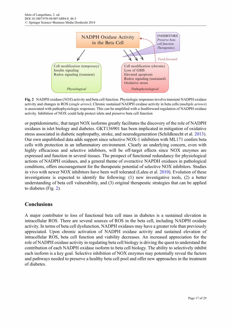
or peptidomimetic, that target NOX isoforms greatly facilitates the discovery of the role of NADPHoxidases in islet biology and diabetes. GKT136901 has been implicated in mitigation of oxidativestress associated in diabetic nephropathy, stroke, and neurodegeneration (Schildknecht et al. 2013).Our own unpublished data adds support since selective NOX-1 inhibition with ML171 confers betacells with protection in an inflammatory environment. Clearly an underlying concern, even withhighly efficacious and selective inhibitors, will be off-target effects since NOX enzymes areexpressed and function in several tissues. The prospect of functional redundancy for physiologicalactions of NADPH oxidases, and a general theme of overactive NAPDH oxidases in pathologicalconditions, offers encouragement for the therapeutic potential of selective NOX inhibitors. Studiesin vivo with newer NOX inhibitors have been well tolerated (Laleu et al. 2010). Evolution of theseinvestigations is expected to identify the following: (1) new investigative tools, (2) a betterunderstanding of beta cell vulnerability, and (3) original therapeutic strategies that can be appliedto diabetes (Fig. 2).
Conclusions
A major contributor to loss of functional beta cell mass in diabetes is a sustained elevation inintracellular ROS. There are several sources of ROS in the beta cell, including NADPH oxidaseactivity. In terms of beta cell dysfunction, NADPH oxidases may have a greater role than previouslyappreciated. Upon chronic activation of NADPH oxidase activity and sustained elevation ofintracellular ROS, beta cell function and viability decreases. An increased appreciation for therole of NADPH oxidase activity in regulating beta cell biology is driving the quest to understand thecontribution of each NADPH oxidase isoform to beta cell biology. The ability to selectively inhibiteach isoform is a key goal. Selective inhibition of NOX enzymes may potentially reveal the factorsand pathways needed to preserve a healthy beta cell pool and offer new approaches in the treatmentof diabetes.
NADPH Oxidase Activityin the Beta Cell
Cell modification (temporary)Insulin signalingRedox signaling (transient)
Cell modification (chronic)Loss of GSISElevated apoptosisRedox signaling (sustained)Oxidative stress
Physiological Pathophysiological
Feed-forward
INHIBITORSPreserve betacell function.Therapeutics.
Fig. 2 NADPH oxidase (NOX) activity and beta cell function. Physiologic responses involve transient NADPH oxidaseactivity and changes in ROS (single arrow). Chronic sustained NADPH oxidase activity in beta cells (multiple arrows)is associated with pathophysiologic responses. This can be amplified with a feedforward regulation of NADPH oxidaseactivity. Inhibition of NOX could help protect islets and preserve beta cell function
Islets of Langerhans, 2. ed.DOI 10.1007/978-94-007-6884-0_46-3# Springer Science+Business Media Dordrecht 2014
Page 17 of 29

Cross-References
▶ Inflammatory Pathways Linked to Beta Cell Demise in Diabetes
References
Abo A, Pick E et al (1991) Activation of the NADPH oxidase involves the small GTP-bindingprotein p21rac1. Nature 353(6345):668–670
Adachi Y, Shibai Y et al (2008) Oncogenic Ras upregulates NADPH oxidase 1 gene expressionthrough MEK-ERK-dependent phosphorylation of GATA-6. Oncogene 27(36):4921–4932
Ago T, Kitazono T et al (2004) Nox4 as the major catalytic component of an endothelial NAD(P)Hoxidase. Circulation 109(2):227–233
Aguirre J, Rios-Momberg M et al (2005) Reactive oxygen species and development in microbialeukaryotes. Trends Microbiol 13(3):111–118
Allaoui A, Botteaux A et al (2009) Dual oxidases and hydrogen peroxide in a complex dialoguebetween host mucosae and bacteria. Trends Mol Med 15(12):571–579
Alves ES, Haidar AA et al (2012) Angiotensin II-induced JNK activation is mediated by NAD(P)Hoxidase in isolated rat pancreatic islets. Regul Pept 175(1–3):1–6
Ando S, Kaibuchi K et al (1992) Post-translational processing of rac p21s is important both for theirinteraction with the GDP/GTP exchange proteins and for their activation of NADPH oxidase.J Biol Chem 267(36):25709–25713
Babior BM, Kipnes RS et al (1973) Biological defense mechanisms. The production by leukocytesof superoxide, a potential bactericidal agent. J Clin Invest 52(3):741–744
Babior BM, Lambeth JD et al (2002) The neutrophil NADPH oxidase. Arch Biochem Biophys397(2):342–344
Banfi B, Molnar G et al (2001) A Ca(2+)-activated NADPH oxidase in testis, spleen, and lymphnodes. J Biol Chem 276(40):37594–37601
Banfi B, Clark RA et al (2003) Two novel proteins activate superoxide generation by the NADPHoxidase NOX1. J Biol Chem 278(6):3510–3513
Banfi B, Malgrange B et al (2004a) NOX3, a superoxide-generating NADPH oxidase of the innerear. J Biol Chem 279(44):46065–46072
Banfi B, Tirone F et al (2004b) Mechanism of Ca2+ activation of the NADPH oxidase 5 (NOX5).J Biol Chem 279(18):18583–18591
Bedard K, Krause KH (2007) The NOX family of ROS-generating NADPH oxidases: physiologyand pathophysiology. Physiol Rev 87(1):245–313
Bedard K, Lardy B et al (2007) NOX family NADPH oxidases: not just in mammals. Biochimie89(9):1107–1112
Bedard K, Jaquet Vet al (2012) NOX5: from basic biology to signaling and disease. Free Radic BiolMed 52(4):725–734
BelAiba RS, Djordjevic T et al (2007) NOX5 variants are functionally active in endothelial cells.Free Radic Biol Med 42(4):446–459
Biberstine-Kinkade KJ, DeLeo FR et al (2001) Heme-ligating histidines in flavocytochrome b(558):identification of specific histidines in gp91(phox). J Biol Chem 276(33):31105–31112
Bleich D, Chen S et al (1999) Resistance to type 1 diabetes induction in 12-lipoxygenase knockoutmice. J Clin Invest 103(10):1431–1436
Islets of Langerhans, 2. ed.DOI 10.1007/978-94-007-6884-0_46-3# Springer Science+Business Media Dordrecht 2014
Page 18 of 29

Brar SS, Corbin Z et al (2003) NOX5 NAD(P)H oxidase regulates growth and apoptosis in DU145 prostate cancer cells. Am J Physiol Cell Physiol 285(2):C353–C369
Brun S, Malagnac F et al (2009) Functions and regulation of the Nox family in the filamentousfungus Podospora anserina: a new role in cellulose degradation. Mol Microbiol 74(2):480–496
Cai W, He JC et al (2008) Oral glycotoxins determine the effects of calorie restriction on oxidantstress, age-related diseases, and lifespan. Am J Pathol 173(2):327–336
Caillou B, Dupuy C et al (2001) Expression of reduced nicotinamide adenine dinucleotide phos-phate oxidase (ThoX, LNOX, Duox) genes and proteins in human thyroid tissues. J ClinEndocrinol Metab 86(7):3351–3358
Cano-Dominguez N, Alvarez-Delfin K et al (2008) NADPH oxidases NOX-1 and NOX-2 requirethe regulatory subunit NOR-1 to control cell differentiation and growth in Neurospora crassa.Eukaryot Cell 7(8):1352–1361
Cassatella MA, Bazzoni F et al (1990) Molecular basis of interferon-gamma and lipopolysaccharideenhancement of phagocyte respiratory burst capability. Studies on the gene expression of severalNADPH oxidase components. J Biol Chem 265(33):20241–20246
Cave AC, Brewer AC et al (2006) NADPH oxidases in cardiovascular health and disease. AntioxidRedox Signal 8(5–6):691–728
Cheng G, Cao Z et al (2001) Homologs of gp91phox: cloning and tissue expression of Nox3, Nox4,and Nox5. Gene 269(1–2):131–140
Cheng G, Ritsick D et al (2004) Nox3 regulation by NOXO1, p47phox, and p67phox. J Biol Chem279(33):34250–34255
Costal F, Oliveira E et al (2013) Dual effect of advanced glycation end products in pancreatic isletapoptosis. Diabetes Metab Res Rev 29(4):296–307
Coughlan MT, Yap FYet al (2011) Advanced glycation end products are direct modulators of beta-cell function. Diabetes 60(10):2523–2532
Cross AR, Rae J et al (1995) Cytochrome b-245 of the neutrophil superoxide-generating systemcontains two nonidentical hemes. Potentiometric studies of a mutant form of gp91phox. J BiolChem 270(29):17075–17077
Csanyi G, Cifuentes-Pagano E et al (2011) Nox2 B-loop peptide, Nox2ds, specifically inhibits theNADPH oxidase Nox2. Free Radic Biol Med 51(6):1116–1125
Cucoranu I, Clempus R et al (2005) NAD(P)H oxidase 4 mediates transforming growth factor-beta1-induced differentiation of cardiac fibroblasts into myofibroblasts. Circ Res 97(9):900–907
Dahan I, Pick E (2012) Strategies for identifying synthetic peptides to act as inhibitors of NADPHoxidases, or “all that you did and did not want to know about Nox inhibitory peptides”. Cell MolLife Sci 69(14):2283–2305
Dahan I, Issaeva I et al (2002) Mapping of functional domains in the p22(phox) subunit offlavocytochrome b(559) participating in the assembly of the NADPH oxidase complex by“peptide walking”. J Biol Chem 277(10):8421–8432
de Carvalho DD, Sadok A et al (2008) Nox1 downstream of 12-lipoxygenase controls cellproliferation but not cell spreading of colon cancer cells. Int J Cancer 122(8):1757–1764
De Deken X,Wang D et al (2000) Cloning of two human thyroid cDNAs encoding newmembers ofthe NADPH oxidase family. J Biol Chem 275(30):23227–23233
DeLeo FR, Nauseef WM et al (1995) A domain of p47phox that interacts with human neutrophilflavocytochrome b558. J Biol Chem 270(44):26246–26251
Diatchuk V, Lotan O et al (1997) Inhibition of NADPH oxidase activation by 4-(2-aminoethyl)-benzenesulfonyl fluoride and related compounds. J Biol Chem 272(20):13292–13301
Islets of Langerhans, 2. ed.DOI 10.1007/978-94-007-6884-0_46-3# Springer Science+Business Media Dordrecht 2014
Page 19 of 29

Dupuy C, Kaniewski J et al (1989) NADPH-dependent H2O2 generation catalyzed by thyroidplasma membranes. Studies with electron scavengers. Eur J Biochem 185(3):597–603
Dupuy C, Ohayon R et al (1999) Purification of a novel flavoprotein involved in the thyroid NADPHoxidase. Cloning of the porcine and human cdnas. J Biol Chem 274(52):37265–37269
Dworakowski R, Alom-Ruiz SP et al (2008) NADPH oxidase-derived reactive oxygen species in theregulation of endothelial phenotype. Pharmacol Rep 60(1):21–28
Edens WA, Sharling L et al (2001) Tyrosine cross-linking of extracellular matrix is catalyzed byDuox, a multidomain oxidase/peroxidase with homology to the phagocyte oxidase subunitgp91phox. J Cell Biol 154(4):879–891
Elsner M, Gehrmann W et al (2011) Peroxisome-generated hydrogen peroxide as important medi-ator of lipotoxicity in insulin-producing cells. Diabetes 60(1):200–208
Fan C, Katsuyama M et al (2005) Transactivation of the EGF receptor and a PI3 kinase-ATF-1pathway is involved in the upregulation of NOX1, a catalytic subunit of NADPH oxidase. FEBSLett 579(5):1301–1305
Finegold AA, Shatwell KP et al (1996) Intramembrane bis-heme motif for transmembrane electrontransport conserved in a yeast iron reductase and the human NADPH oxidase. J Biol Chem271(49):31021–31024
Foreman J, Demidchik V et al (2003) Reactive oxygen species produced by NADPH oxidaseregulate plant cell growth. Nature 422(6930):442–446
Fransen M, Nordgren M et al (2012) Role of peroxisomes in ROS/RNS-metabolism: implicationsfor human disease. Biochim Biophys Acta 1822(9):1363–1373
Fu X, Beer DG et al (2006) cAMP-response element-binding protein mediates acid-inducedNADPH oxidase NOX5-S expression in Barrett esophageal adenocarcinoma cells. J Biol Chem281(29):20368–20382
Gapper C, Dolan L (2006) Control of plant development by reactive oxygen species. Plant Physiol141(2):341–345
Garrido AM, Griendling KK (2009) NADPH oxidases and angiotensin II receptor signaling. MolCell Endocrinol 302(2):148–158
Gattas MV, Forteza R et al (2009) Oxidative epithelial host defense is regulated by infectious andinflammatory stimuli. Free Radic Biol Med 47(10):1450–1458
Gavazzi G, Banfi B et al (2006) Decreased blood pressure in NOX1-deficient mice. FEBS Lett580(2):497–504
Gehrmann W, Elsner M et al (2010) Role of metabolically generated reactive oxygen species forlipotoxicity in pancreatic beta-cells. Diabetes Obes Metab 12(Suppl 2):149–158
Geiszt M, Kopp JB et al (2000) Identification of renox, an NAD(P)H oxidase in kidney. Proc NatlAcad Sci U S A 97(14):8010–8014
Geiszt M, Lekstrom K et al (2003a) NAD(P)H oxidase 1, a product of differentiated colon epithelialcells, can partially replace glycoprotein 91phox in the regulated production of superoxide byphagocytes. J Immunol 171(1):299–306
Geiszt M,Witta J et al (2003b) Dual oxidases represent novel hydrogen peroxide sources supportingmucosal surface host defense. FASEB J 17(11):1502–1504
Gianni D, Bohl B et al (2008) The involvement of the tyrosine kinase c-Src in the regulation ofreactive oxygen species generation mediated by NADPH oxidase-1. Mol Biol Cell19(7):2984–2994
Gianni D, Taulet N et al (2010) A novel and specific NADPH oxidase-1 (Nox1) small-moleculeinhibitor blocks the formation of functional invadopodia in human colon cancer cells. ACS ChemBiol 5(10):981–993
Islets of Langerhans, 2. ed.DOI 10.1007/978-94-007-6884-0_46-3# Springer Science+Business Media Dordrecht 2014
Page 20 of 29

Giannoni E, Buricchi F et al (2005) Intracellular reactive oxygen species activate Src tyrosine kinaseduring cell adhesion and anchorage-dependent cell growth. Mol Cell Biol 25(15):6391–6403
Go YM, Gipp JJ et al (2004) H2O2-dependent activation of GCLC-ARE4 reporter occurs bymitogen-activated protein kinase pathways without oxidation of cellular glutathione orthioredoxin-1. J Biol Chem 279(7):5837–5845
Goldstein BJ, Mahadev K et al (2005) Redox paradox: insulin action is facilitated by insulin-stimulated reactive oxygen species with multiple potential signaling targets. Diabetes54(2):311–321
Gorin Y, Ricono JM et al (2003) Nox4 mediates angiotensin II-induced activation of Akt/proteinkinase B in mesangial cells. Am J Physiol Renal Physiol 285(2):F219–F229
Graciano MF, Santos LR et al (2011) NAD(P)H oxidase participates in the palmitate-inducedsuperoxide production and insulin secretion by rat pancreatic islets. J Cell Physiol226(4):1110–1117
Grankvist K, Marklund SL et al (1981) CuZn-superoxide dismutase, Mn-superoxide dismutase,catalase and glutathione peroxidase in pancreatic islets and other tissues in the mouse. BiochemJ 199(2):393–398
Grasberger H, Refetoff S (2006) Identification of the maturation factor for dual oxidase. Evolution ofan eukaryotic operon equivalent. J Biol Chem 281(27):18269–18272
Guichard C, Moreau R et al (2008) NOX family NADPH oxidases in liver and in pancreatic islets:a role in the metabolic syndrome and diabetes? Biochem Soc Trans 36(Pt 5):920–929
Harper RW, Xu C et al (2005) Differential regulation of dual NADPH oxidases/peroxidases, Duox1and Duox2, by Th1 and Th2 cytokines in respiratory tract epithelium. FEBS Lett579(21):4911–4917
Harper RW, Xu C et al (2006) Duox2 exhibits potent heme peroxidase activity in human respiratorytract epithelium. FEBS Lett 580(22):5150–5154
Heumuller S, Wind S et al (2008) Apocynin is not an inhibitor of vascular NADPH oxidases but anantioxidant. Hypertension 51(2):211–217
Heyworth PG, Bohl BP et al (1994) Rac translocates independently of the neutrophil NADPHoxidase components p47phox and p67phox. Evidence for its interaction with flavocytochromeb558. J Biol Chem 269(49):30749–30752
Heyworth PG, Cross AR et al (2003) Chronic granulomatous disease. Curr Opin Immunol15(5):578–584
Hofmann SM, Dong HJ et al (2002) Improved insulin sensitivity is associated with restricted intakeof dietary glycoxidation products in the db/db mouse. Diabetes 51(7):2082–2089
Hordijk PL (2006) Regulation of NADPH oxidases: the role of Rac proteins. Circ Res98(4):453–462
Ibi M, Katsuyama M et al (2006) NOX1/NADPH oxidase negatively regulates nerve growth factor-induced neurite outgrowth. Free Radic Biol Med 40(10):1785–1795
Ibi M, Matsuno K et al (2008) Reactive oxygen species derived from NOX1/NADPH oxidaseenhance inflammatory pain. J Neurosci 28(38):9486–9494
Imoto H, Sasaki N et al (2008) Impaired insulin secretion by diphenyleneiodium associated withperturbation of cytosolic Ca2+ dynamics in pancreatic beta-cells. Endocrinology149(11):5391–5400
Jackson HM, Kawahara Tet al (2010) Nox4 B-loop creates an interface between the transmembraneand dehydrogenase domains. J Biol Chem 285(14):10281–10290
Islets of Langerhans, 2. ed.DOI 10.1007/978-94-007-6884-0_46-3# Springer Science+Business Media Dordrecht 2014
Page 21 of 29

Janciauskiene S, Ahren B (2000) Fibrillar islet amyloid polypeptide differentially affects oxidativemechanisms and lipoprotein uptake in correlation with cytotoxicity in two insulin-producing celllines. Biochem Biophys Res Commun 267(2):619–625
Jay DB, Papaharalambus CA et al (2008) Nox5 mediates PDGF-induced proliferation in humanaortic smooth muscle cells. Free Radic Biol Med 45(3):329–335
Jiao J, Dou L et al (2012) NADPH oxidase 2 plays a critical role in dysfunction and apoptosis ofpancreatic beta-cells induced by very low-density lipoprotein. Mol Cell Biochem370(1–2):103–113
Johnson DK, Schillinger KJ et al (2002) Inhibition of NADPH oxidase activation in endothelial cellsby ortho-methoxy-substituted catechols. Endothelium 9(3):191–203
Juhasz A, Ge Y et al (2009) Expression of NADPH oxidase homologues and accessory genes inhuman cancer cell lines, tumours and adjacent normal tissues. Free Radic Res 43(6):523–532
Kamiguti AS, Serrander L et al (2005) Expression and activity of NOX5 in the circulating malignantB cells of hairy cell leukemia. J Immunol 175(12):8424–8430
Kaneto H, Fujii J et al (1996) Reducing sugars trigger oxidative modification and apoptosis inpancreatic beta-cells by provoking oxidative stress through the glycation reaction. BiochemJ 320(Pt 3):855–863
Katsuyama M, Fan C et al (2002) NADPH oxidase is involved in prostaglandin F2alpha-inducedhypertrophy of vascular smooth muscle cells: induction of NOX1 by PGF2alpha. J Biol Chem277(16):13438–13442
Kawahara T, Kohjima M et al (2005) Helicobacter pylori lipopolysaccharide activates Rac1 andtranscription of NADPH oxidase Nox1 and its organizer NOXO1 in guinea pig gastric mucosalcells. Am J Physiol Cell Physiol 288(2):C450–C457
Kawamori D, Kajimoto Yet al (2003) Oxidative stress induces nucleo-cytoplasmic translocation ofpancreatic transcription factor PDX-1 through activation of c-Jun NH(2)-terminal kinase. Diabe-tes 52(12):2896–2904
Kenyon V, Rai G et al (2011) Discovery of potent and selective inhibitors of human platelet-type 12-lipoxygenase. J Med Chem 54(15):5485–5497
Kleinberg ME, Malech HL et al (1990) The phagocyte 47-kilodalton cytosolic oxidase protein is anearly reactant in activation of the respiratory burst. J Biol Chem 265(26):15577–15583
Koya D, King GL (1998) Protein kinase C activation and the development of diabetic complications.Diabetes 47(6):859–866
Krieger-Brauer HI, Kather H (1995) Antagonistic effects of different members of the fibroblast andplatelet-derived growth factor families on adipose conversion and NADPH-dependent H2O2generation in 3 T3 L1-cells. Biochem J 307(Pt 2):549–556
Kumar B, Koul S et al (2008) Oxidative stress is inherent in prostate cancer cells and is required foraggressive phenotype. Cancer Res 68(6):1777–1785
Kuroda J, Ago T et al (2010) NADPH oxidase 4 (Nox4) is a major source of oxidative stress in thefailing heart. Proc Natl Acad Sci U S A 107(35):15565–15570
Kusumoto K, Kawahara T et al (2005) Ecabet sodium inhibits Helicobacter pylorilipopolysaccharide-induced activation of NADPH oxidase 1 or apoptosis of guinea pig gastricmucosal cells. Am J Physiol Gastrointest Liver Physiol 288(2):G300–G307
Lafeber FP, Beukelman CJ et al (1999) Apocynin, a plant-derived, cartilage-saving drug, might beuseful in the treatment of rheumatoid arthritis. Rheumatology 38(11):1088–1093
Laleu B, Gaggini F et al (2010) First in class, potent, and orally bioavailable NADPH oxidaseisoform 4 (Nox4) inhibitors for the treatment of idiopathic pulmonary fibrosis. J Med Chem53(21):7715–7730
Islets of Langerhans, 2. ed.DOI 10.1007/978-94-007-6884-0_46-3# Springer Science+Business Media Dordrecht 2014
Page 22 of 29

Lambeth JD (2007) Nox enzymes, ROS, and chronic disease: an example of antagonistic pleiotropy.Free Radic Biol Med 43(3):332–347
Lambeth JD, Kawahara T et al (2007) Regulation of Nox and Duox enzymatic activity andexpression. Free Radic Biol Med 43(3):319–331
Lara-Ortiz T, Riveros-Rosas H et al (2003) Reactive oxygen species generated by microbialNADPH oxidase NoxA regulate sexual development in Aspergillus nidulans. Mol Microbiol50(4):1241–1255
Lassegue B, Sorescu D et al (2001) Novel gp91(phox) homologues in vascular smooth muscle cells :nox1 mediates angiotensin II-induced superoxide formation and redox-sensitive signaling path-ways. Circ Res 88(9):888–894
Lenzen S (2008) Oxidative stress: the vulnerable beta-cell. Biochem Soc Trans 36(Pt 3):343–347Lenzen S, Drinkgern J et al (1996) Low antioxidant enzyme gene expression in pancreatic islets
compared with various other mouse tissues. Free Radic Biol Med 20(3):463–466Li J, Stouffs M et al (2006) The NADPH oxidase NOX4 drives cardiac differentiation: role in
regulating cardiac transcription factors and MAP kinase activation. Mol Biol Cell17(9):3978–3988
Li Q, Spencer NY et al (2009) Endosomal Nox2 facilitates redox-dependent induction ofNF-kappaB by TNF-alpha. Antioxid Redox Signal 11(6):1249–1263
Li N, Li B et al (2012) NADPH oxidase NOX2 defines a new antagonistic role for reactive oxygenspecies and cAMP/PKA in the regulation of insulin secretion. Diabetes 61(11):2842–2850
Lim M, Park L et al (2008) Induction of apoptosis of Beta cells of the pancreas by advancedglycation end-products, important mediators of chronic complications of diabetes mellitus. AnnN YAcad Sci 1150:311–315
Lin HM, Lee JH et al (2009) Transforming growth factor-beta/Smad3 signaling regulates insulingene transcription and pancreatic islet beta-cell function. J Biol Chem 284(18):12246–12257
Lin N, Zhang H et al (2012) Advanced glycation end-products induce injury to pancreatic beta cellsthrough oxidative stress. Diabetes Metab 38(3):250–257
Lupi R, Del Guerra S et al (2006) The direct effects of the angiotensin-converting enzyme inhibitors,zofenoprilat and enalaprilat, on isolated human pancreatic islets. Eur J Endocrinol154(2):355–361
Mahadev K, Motoshima H et al (2004) The NAD(P)H oxidase homolog Nox4 modulates insulin-stimulated generation of H2O2 and plays an integral role in insulin signal transduction. Mol CellBiol 24(5):1844–1854
Malagnac F, Lalucque H et al (2004) Two NADPH oxidase isoforms are required for sexualreproduction and ascospore germination in the filamentous fungus Podospora anserina. FungalGenet Biol 41(11):982–997
Manea A (2010) NADPH oxidase-derived reactive oxygen species: involvement in vascularphysiology and pathology. Cell Tissue Res 342(3):325–339
Manea A, Manea SA et al (2012) Positive regulation of NADPH oxidase 5 by proinflammatory-related mechanisms in human aortic smooth muscle cells. Free Radic Biol Med 52(9):1497–1507
Marino D, Dunand C et al (2012) A burst of plant NADPH oxidases. Trends Plant Sci 17(1):9–15Martyn KD, Frederick LM et al (2006) Functional analysis of Nox4 reveals unique characteristics
compared to other NADPH oxidases. Cell Signal 18(1):69–82Marzban L, Verchere CB (2004) The role of islet amyloid polypeptide in type 2 diabetes. Can
J Diabetes 28(4):39–47
Islets of Langerhans, 2. ed.DOI 10.1007/978-94-007-6884-0_46-3# Springer Science+Business Media Dordrecht 2014
Page 23 of 29

Matti A, Kyathanahalli C et al (2012) Protein farnesylation is requisite for mitochondrial fuel-induced insulin release: further evidence to link reactive oxygen species generation to insulinsecretion in pancreatic beta-cells. Islets 4(1):74–77
McDuffie M, Maybee NA et al (2008) Nonobese diabetic (NOD) mice congenic for a targeteddeletion of 12/15-lipoxygenase are protected from autoimmune diabetes. Diabetes 57(1):199–208
Meng D, Lv DD et al (2008) Insulin-like growth factor-I induces reactive oxygen species productionand cell migration through Nox4 and Rac1 in vascular smooth muscle cells. Cardiovasc Res80(2):299–308
Michalska M, Wolf G et al (2010) Effects of pharmacological inhibition of NADPH oxidase oriNOS on pro-inflammatory cytokine, palmitic acid or H2O2-induced mouse islet or clonalpancreatic beta-cell dysfunction. Biosci Rep 30(6):445–453
Miller FJ Jr, Filali M et al (2007) Cytokine activation of nuclear factor kappa B in vascular smoothmuscle cells requires signaling endosomes containing Nox1 and ClC-3. Circ Res 101(7):663–671
Mittler R, Vanderauwera S et al (2011) ROS signaling: the new wave? Trends Plant Sci16(6):300–309
Miyano K, Ueno N et al (2006) Direct involvement of the small GTPase Rac in activation of thesuperoxide-producing NADPH oxidase Nox1. J Biol Chem 281(31):21857–21868
Modak MA, Datar SP et al (2007) Differential susceptibility of chick and mouse islets tostreptozotocin and its co-relation with islet antioxidant status. J Comp Physiol B 177(2):247–257
Mohammed AM, Kowluru A (2013) Activation of apocynin-sensitive NADPH oxidase (Nox2)activity in INS-1 832/13 cells under glucotoxic conditions. Islets 5(3):129–131
Montezano AC, Burger D et al (2010) Nicotinamide adenine dinucleotide phosphate reducedoxidase 5 (Nox5) regulation by angiotensin II and endothelin-1 is mediated via calcium/calmodulin-dependent, rac-1-independent pathways in human endothelial cells. Circ Res106(8):1363–1373
Montezano AC, Burger D et al (2011) Novel Nox homologues in the vasculature: focusing on Nox4and Nox5. Clin Sci 120(4):131–141
Morand S, Dos Santos OF et al (2003) Identification of a truncated dual oxidase 2 (DUOX2)messenger ribonucleic acid (mRNA) in two rat thyroid cell lines. Insulin and forskolin regulationof DUOX2 mRNA levels in FRTL-5 cells and porcine thyrocytes. Endocrinology144(2):567–574
Morand S, Ueyama T et al (2009) Duox maturation factors form cell surface complexes with Duoxaffecting the specificity of reactive oxygen species generation. FASEB J 23(4):1205–1218
Morgan D, Oliveira-Emilio HR et al (2007) Glucose, palmitate and pro-inflammatory cytokinesmodulate production and activity of a phagocyte-like NADPH oxidase in rat pancreatic islets anda clonal beta cell line. Diabetologia 50(2):359–369
Morgan D, Rebelato E et al (2009) Association of NAD(P)H oxidase with glucose-induced insulinsecretion by pancreatic beta-cells. Endocrinology 150(5):2197–2201
Murray TV, Smyrnias I et al (2013) NADPH oxidase 4 regulates cardiomyocyte differentiation viaredox activation of c-Jun protein and the cis-regulation of GATA-4 gene transcription. J BiolChem 288(22):15745–15759
Nakayama M, Inoguchi T et al (2005) Increased expression of NAD(P)H oxidase in islets of animalmodels of Type 2 diabetes and its improvement by an AT1 receptor antagonist. Biochem BiophysRes Commun 332(4):927–933
Nauseef WM, McCormick S et al (1993) Functional domain in an arginine-rich carboxyl-terminalregion of p47phox. J Biol Chem 268(31):23646–23651
Islets of Langerhans, 2. ed.DOI 10.1007/978-94-007-6884-0_46-3# Springer Science+Business Media Dordrecht 2014
Page 24 of 29

Newsholme P, Morgan D et al (2009) Insights into the critical role of NADPH oxidase(s) in thenormal and dysregulated pancreatic beta cell. Diabetologia 52(12):2489–2498
Nisimoto Y, Motalebi S et al (1999) The p67(phox) activation domain regulates electron flow fromNADPH to flavin in flavocytochrome b(558). J Biol Chem 274(33):22999–23005
Nisimoto Y, Tsubouchi R et al (2008) Activation of NADPH oxidase 1 in tumour colon epithelialcells. Biochem J 415(1):57–65
Ohneda K, Mirmira RG et al (2000) The homeodomain of PDX-1 mediates multiple protein-proteininteractions in the formation of a transcriptional activation complex on the insulin promoter. MolCell Biol 20(3):900–911
Oliveira HR, Verlengia R et al (2003) Pancreatic beta-cells express phagocyte-like NAD(P)Hoxidase. Diabetes 52(6):1457–1463
Orient A, Donko A et al (2007) Novel sources of reactive oxygen species in the human body.Nephrol Dial Transplant 22(5):1281–1288
Paffenholz R, Bergstrom RA et al (2004) Vestibular defects in head-tilt mice result frommutations inNox3, encoding an NADPH oxidase. Genes Dev 18(5):486–491
Pandey D, Patel A et al (2012) Expression and functional significance of NADPH oxidase 5 (Nox5)and its splice variants in human blood vessels. Am J Physiol Heart Circ Physiol 302(10):H1919–H1928
Pedruzzi E, Guichard C et al (2004) NAD(P)H oxidase Nox-4 mediates 7-ketocholesterol-inducedendoplasmic reticulum stress and apoptosis in human aortic smooth muscle cells. Mol Cell Biol24(24):10703–10717
Pendyala S, Gorshkova IA et al (2009) Role of Nox4 and Nox2 in hyperoxia-induced reactiveoxygen species generation and migration of human lung endothelial cells. Antioxid Redox Signal11(4):747–764
Peppa M, He C et al (2003) Fetal or neonatal low-glycotoxin environment prevents autoimmunediabetes in NOD mice. Diabetes 52(6):1441–1448
Pi J, Bai Y et al (2007) Reactive oxygen species as a signal in glucose-stimulated insulin secretion.Diabetes 56(7):1783–1791
Purves T, Middlemas A et al (2001) A role for mitogen-activated protein kinases in the etiology ofdiabetic neuropathy. FASEB J 15(13):2508–2514
Rada B, Hably C et al (2008) Role of Nox2 in elimination of microorganisms. Semin Immunopathol30(3):237–253
Rebelato E, Mares-Guia TR et al (2012) Expression of NADPH oxidase in human pancreatic islets.Life Sci 91(7–8):244–249
Rey FE, Cifuentes ME et al (2001) Novel competitive inhibitor of NAD(P)H oxidase assemblyattenuates vascular O(2)(�) and systolic blood pressure in mice. Circ Res 89(5):408–414
Rigutto S, Hoste C et al (2009) Activation of dual oxidases Duox1 and Duox2: differentialregulation mediated by camp-dependent protein kinase and protein kinase C-dependent phos-phorylation. J Biol Chem 284(11):6725–6734
Ris-Stalpers C (2006) Physiology and pathophysiology of the DUOXes. Antioxid Redox Signal8(9–10):1563–1572
Rokutan K, Kawahara Tet al (2006) NADPH oxidases in the gastrointestinal tract: a potential role ofNox1 in innate immune response and carcinogenesis. Antioxid Redox Signal 8(9–10):1573–1582
Rokutan K, Kawahara T et al (2008) Nox enzymes and oxidative stress in the immunopathology ofthe gastrointestinal tract. Semin Immunopathol 30(3):315–327
Rotrosen D, Kleinberg ME et al (1990) Evidence for a functional cytoplasmic domain of phagocyteoxidase cytochrome b558. J Biol Chem 265(15):8745–8750
Islets of Langerhans, 2. ed.DOI 10.1007/978-94-007-6884-0_46-3# Springer Science+Business Media Dordrecht 2014
Page 25 of 29

Rotrosen D, Yeung CL et al (1992) Cytochrome b558: the flavin-binding component of thephagocyte NADPH oxidase. Science 256(5062):1459–1462
Sadok A, Bourgarel-Rey V et al (2008) Nox1-dependent superoxide production controls colonadenocarcinoma cell migration. Biochim Biophys Acta 1783(1):23–33
Sagi M, Fluhr R (2006) Production of reactive oxygen species by plant NADPH oxidases. PlantPhysiol 141(2):336–340
San Martin A, Foncea R et al (2007) Nox1-based NADPH oxidase-derived superoxide is requiredfor VSMC activation by advanced glycation end-products. Free Radic Biol Med42(11):1671–1679
Sancho P, Fabregat I (2010) NADPH oxidase NOX1 controls autocrine growth of liver tumor cellsthrough up-regulation of the epidermal growth factor receptor pathway. J Biol Chem285(32):24815–24824
Santos CX, Tanaka LY et al (2009) Mechanisms and implications of reactive oxygen speciesgeneration during the unfolded protein response: roles of endoplasmic reticulum oxidoreductases,mitochondrial electron transport, and NADPH oxidase. Antioxid Redox Signal11(10):2409–2427
Schildknecht S, Weber A et al (2013) The NOX1/4 inhibitor GKT136901 as selective and directscavenger of peroxynitrite. Curr Med Chem 21:365–376
Schroder K, Wandzioch K et al (2009) Nox4 acts as a switch between differentiation and prolifer-ation in preadipocytes. Arterioscler Thromb Vasc Biol 29(2):239–245
Schulz E, Munzel T (2008) NOX5, a new “radical” player in human atherosclerosis? J Am CollCardiol 52(22):1810–1812
Sedeek M, Callera G et al (2010) Critical role of Nox4-based NADPH oxidase in glucose-inducedoxidative stress in the kidney: implications in type 2 diabetic nephropathy. Am J Physiol RenalPhysiol 299(6):F1348–F1358
Selemidis S, Sobey CG et al (2008) NADPH oxidases in the vasculature: molecular features, roles indisease and pharmacological inhibition. Pharmacol Ther 120(3):254–291
Serrander L, Cartier L et al (2007) NOX4 activity is determined by mRNA levels and revealsa unique pattern of ROS generation. Biochem J 406(1):105–114
Seshiah PN, Weber DS et al (2002) Angiotensin II stimulation of NAD(P)H oxidase activity:upstream mediators. Circ Res 91(5):406–413
Shao J, Iwashita N et al (2006) Beneficial effects of candesartan, an angiotensin II type 1 receptorblocker, on beta-cell function and morphology in db/db mice. Biochem Biophys Res Commun344(4):1224–1233
Shi J, Ross CR et al (1996) PR-39, a proline-rich antibacterial peptide that inhibits phagocyteNADPH oxidase activity by binding to Src homology 3 domains of p47 phox. Proc Natl Acad SciU S A 93(12):6014–6018
Shiose A, Kuroda J et al (2001) A novel superoxide-producing NAD(P)H oxidase in kidney. J BiolChem 276(2):1417–1423
Stolk J, Hiltermann TJ et al (1994) Characteristics of the inhibition of NADPH oxidase activation inneutrophils by apocynin, a methoxy-substituted catechol. Am J Respir Cell Mol Biol11(1):95–102
Sturrock A, Cahill B et al (2006) Transforming growth factor-beta1 induces Nox4 NAD(P)Hoxidase and reactive oxygen species-dependent proliferation in human pulmonary artery smoothmuscle cells. Am J Physiol Lung Cell Mol Physiol 290(4):L661–L673
Islets of Langerhans, 2. ed.DOI 10.1007/978-94-007-6884-0_46-3# Springer Science+Business Media Dordrecht 2014
Page 26 of 29

Sturrock A, Huecksteadt TP et al (2007) Nox4 mediates TGF-beta1-induced retinoblastoma proteinphosphorylation, proliferation, and hypertrophy in human airway smooth muscle cells. AmJ Physiol Lung Cell Mol Physiol 292(6):L1543–L1555
Suh YA, Arnold RS et al (1999) Cell transformation by the superoxide-generating oxidase Mox1.Nature 401(6748):79–82
Sumimoto H (2008) Structure, regulation and evolution of Nox-family NADPH oxidases thatproduce reactive oxygen species. FEBS J 275(13):3249–3277
Syed I, Kyathanahalli CN et al (2011a) Increased phagocyte-like NADPH oxidase and ROSgeneration in type 2 diabetic ZDF rat and human islets: role of Rac1-JNK1/2 signaling pathwayin mitochondrial dysregulation in the diabetic islet. Diabetes 60(11):2843–2852
Syed I, Kyathanahalli CN et al (2011b) Phagocyte-like NADPH oxidase generates ROS in INS832/13 cells and rat islets: role of protein prenylation. Am J Physiol Regul Integr Comp Physiol300(3):R756–R762
Takeya R, Ueno N et al (2003) Novel human homologues of p47phox and p67phox participate inactivation of superoxide-producing NADPH oxidases. J Biol Chem 278(27):25234–25246
Taylor-Fishwick DA, Pittenger GL et al (2008) Transplantation and beyond. Drug Dev Res69:165–176
ten Freyhaus H, Huntgeburth M et al (2006) Novel Nox inhibitor VAS2870 attenuates PDGF-dependent smooth muscle cell chemotaxis, but not proliferation. Cardiovasc Res 71(2):331–341
Tiedge M, Lortz S et al (1997) Relation between antioxidant enzyme gene expression andantioxidative defense status of insulin-producing cells. Diabetes 46(11):1733–1742
Torres MA, Dangl JL (2005) Functions of the respiratory burst oxidase in biotic interactions, abioticstress and development. Curr Opin Plant Biol 8(4):397–403
Touyz RM, Chen X et al (2002) Expression of a functionally active gp91phox-containingneutrophil-type NAD(P)H oxidase in smooth muscle cells from human resistance arteries:regulation by angiotensin II. Circ Res 90(11):1205–1213
Tudzynski P, Heller J et al (2012) Reactive oxygen species generation in fungal development andpathogenesis. Curr Opin Microbiol 15(6):653–659
Uchizono Y, Takeya R et al (2006) Expression of isoforms of NADPH oxidase components in ratpancreatic islets. Life Sci 80(2):133–139
Ueyama T, Geiszt M et al (2006) Involvement of Rac1 in activation of multicomponent Nox1- andNox3-based NADPH oxidases. Mol Cell Biol 26(6):2160–2174
Uhlinger DJ, Tyagi SR et al (1995) On the mechanism of inhibition of the neutrophil respiratoryburst oxidase by a peptide from the C-terminus of the large subunit of cytochrome b558.Biochemistry 34(2):524–527
Ushio-Fukai M (2006) Redox signaling in angiogenesis: role of NADPH oxidase. Cardiovasc Res71(2):226–235
Valente AJ, Yoshida T et al (2012) Angiotensin II enhances AT1-Nox1 binding and stimulatesarterial smooth muscle cell migration and proliferation through AT1, Nox1, and interleukin-18.Am J Physiol Heart Circ Physiol 303(3):H282–H296
Vaquero EC, Edderkaoui M et al (2004) Reactive oxygen species produced by NAD(P)H oxidaseinhibit apoptosis in pancreatic cancer cells. J Biol Chem 279(33):34643–34654
Vejrazka M, Micek R et al (2005) Apocynin inhibits NADPH oxidase in phagocytes but stimulatesROS production in non-phagocytic cells. Biochim Biophys Acta 1722(2):143–147
Volchuk A, Ron D (2010) The endoplasmic reticulum stress response in the pancreatic beta-cell.Diabetes Obes Metab 12(Suppl 2):48–57
Islets of Langerhans, 2. ed.DOI 10.1007/978-94-007-6884-0_46-3# Springer Science+Business Media Dordrecht 2014
Page 27 of 29

Weaver JR, Taylor-Fishwick DA (2013) Regulation of NOX-1 expression in beta cells: a positivefeedback loop involving the Src-kinase signaling pathway. Mol Cell Endocrinol 369(1–2):35–41
Weaver JR, Holman TR et al (2012) Integration of pro-inflammatory cytokines, 12-lipoxygenaseand NOX-1 in pancreatic islet beta cell dysfunction. Mol Cell Endocrinol 358(1):88–95
Wientjes FB, Hsuan JJ et al (1993) p40phox, a third cytosolic component of the activation complexof the NADPH oxidase to contain src homology 3 domains. Biochem J 296(Pt 3):557–561
Wind S, Beuerlein K et al (2010) Comparative pharmacology of chemically distinct NADPHoxidase inhibitors. Br J Pharmacol 161(4):885–898
Wingler K, Wunsch S et al (2001) Upregulation of the vascular NAD(P)H-oxidase isoforms Nox1and Nox4 by the renin-angiotensin system in vitro and in vivo. Free Radic Biol Med31(11):1456–1464
Xiang FL, Lu X et al (2010) NOX2 deficiency protects against streptozotocin-induced beta-celldestruction and development of diabetes in mice. Diabetes 59(10):2603–2611
Ximenes VF, Kanegae MP et al (2007) The oxidation of apocynin catalyzed by myeloperoxidase:proposal for NADPH oxidase inhibition. Arch Biochem Biophys 457(2):134–141
Yang M, Foster E et al (2005) Insulin-stimulated NAD(P)H oxidase activity increases migration ofcultured vascular smooth muscle cells. Am J Hypertens 18(10):1329–1334
Yuan H, Lu Yet al (2010a) Suppression of NADPH oxidase 2 substantially restores glucose-induceddysfunction of pancreatic NIT-1 cells. FEBS J 277(24):5061–5071
Yuan H, Zhang X et al (2010b) NADPH oxidase 2-derived reactive oxygen species mediate FFAs-induced dysfunction and apoptosis of beta-cells via JNK, p38 MAPK and p53 pathways. PLoSOne 5(12):e15726
Zhang X, Shan P et al (2006) Toll-like receptor 4 deficiency causes pulmonary emphysema. J ClinInvest 116(11):3050–3059
Zhao Z, Zhao C et al (2009) Advanced glycation end products inhibit glucose-stimulated insulinsecretion through nitric oxide-dependent inhibition of cytochrome c oxidase and adenosinetriphosphate synthesis. Endocrinology 150(6):2569–2576
Islets of Langerhans, 2. ed.DOI 10.1007/978-94-007-6884-0_46-3# Springer Science+Business Media Dordrecht 2014
Page 28 of 29

Index Terms:
Beta cell 2Diabetes 11Inhibitors 15Islet 8, 13NADPH oxidase 2NOX 4Pro-inflammatory cytokines 10Reactive oxygen species (ROS) 2, 9–10, 13
Islets of Langerhans, 2. ed.DOI 10.1007/978-94-007-6884-0_46-3# Springer Science+Business Media Dordrecht 2014
Page 29 of 29