mechanism of lymphocyte-mediated cytotoxicity
TRANSCRIPT

Ann. Rev.lmmunol. 1985.3: 31-58
MECHANISM OF
LYMPHOCYTE-MEDIATED
CYTOTOXICITY!
Pierre A. Henkart
Immunology Branch, National Cancer Institute, National Institutes of Health, Bethesda, Maryland 20205
INTRODUCTION
Studies in many laboratories over the past 20 years have revealed that there are two well-characterized classes of lymphocytes that have cytotoxic activity: the cytotoxic T lymphocytes (CTL) and the large granular lymphocytes (LGL), which are responsible for antibody-dependent cellmediated cytotoxicity (ADCC) and for natural killer (NK). Most studies of cytotoxic mechanisms have focused on one or the other of these distinct cell types, which have clear differences in the way they recognize target cells. In my judgment, however, the results of studies of both types of lymphocytemediated cytotoxicity indicate that they share basic features, which will be described in this review. In particular, three recent developments have helped stimulate the view that LGL and CTL share a basically similar lytic mechanism: (a) Various lines of evidence indicate that after target-cell binding is complete, the NK lytic process is similar to that previously demonstrated for CTL. (b) Researchers, in several laboratories, have found that cloned CTL can develop an NK-like pattern of target recognition after culture. (c) Isolated cytoplasmic granules from LGL and from CTL, which appear to be plausible mediators of the lytic effects of the cells, have generally similar cytolytic properties.
In this review I will survey the recent studies of events in effector and target cells that occur after the formation of a stable effector-target adhesion and will consider various possibilities for defining the nature of
1 The US government has the right to retain a nonexclusive, royalty-free license in and to any copyright covering this paper.
31
Ann
u. R
ev. I
mm
unol
. 198
5.3:
31-5
8. D
ownl
oade
d fr
om w
ww
.ann
ualr
evie
ws.
org
by M
CG
ILL
UN
IVE
RSI
TY
LIB
RA
RIE
S on
03/
12/1
3. F
or p
erso
nal u
se o
nly.
32 HENKART
the lethal damage inflicted on the target cell. The most plausible current hypothesis for the mechanism of lymphocyte cytotoxicity is that target cell binding to a membrane receptor induces a secretory process in the effector cell in which the contents of cytoplasmic granules are released by local exocytosis between the effector cell and its bound target (1). This could be termed the granule exocytosis model. It has become more attractive with the recent demonstration of the cytolytic activities of cytoplasmic granules purified from CTL and LGL; these will be described below.
Needless to say, students of cytotoxic lymphocyte mechanisms may not all agree that NK and CTL operate by a common mechanism and that cytoplasmic granules are the source of the lethal damage. A number of recent reviews expressing other viewpoints are recommended (2-5), although their authors were not aware ofthe granule lytic activity when these articles were written. Other excellent, recent reviews on various aspects of lymphocyte cytotoxic mechanisms should also be consulted (6-9).
Stages of the Lytic Process
During the 1970s it became clear from work done in several laboratories that the CTL lytic process can be broken down into three basic stages, which can be studied independently: (a) a specific binding, which can be measured by counting individual effector-target pairs or conjugates; (b) the calcium-dependent programming for lysis or the lethal hit; and (c) killer cell independent lysis (KCIL), where the target cell dies of the lethal damage (10-12). Recent work with CTL has shed new light on the binding phase (13-15), and single cell assays of target cell lysis have provided some new insights ( 16), but the basic mechanism of the lethal hit remains controversial (5).
The more recent studies of the mechanism of NK-cell-mediated cytotoxicity have tended to use the techniques and concepts developed by researchers working on CTL, and it has been possible to directly compare the properties of the different stages of cytotoxicity of NK cells and CTL. Initial studies ( 17) using these general approaches found that the formation of NK-target conjugates, unlike CTL-target conjugates, was not inhibited by low temperature and drugs blocking energy metabolism. However, as will be discussed below, many studies now show parallels between the postbinding stages of LGL- and CTL-mediated cytotoxicity; and the NK programming and KCIL stages have been defined ( 18).
The Properties of Cultured Cytolytic Lymphocytes
A major recent technical advance, which holds great promise for the study of cytotoxic lymphocyte mechanisms, has been the use of IL2 and other lymphokines to allow the continuous growth and cloning of cytotoxic lymphocytes (19). Both CTL and NK cells have been cloned and can retain
Ann
u. R
ev. I
mm
unol
. 198
5.3:
31-5
8. D
ownl
oade
d fr
om w
ww
.ann
ualr
evie
ws.
org
by M
CG
ILL
UN
IVE
RSI
TY
LIB
RA
RIE
S on
03/
12/1
3. F
or p
erso
nal u
se o
nly.

CYTOTOXIC LYMPHOCYTE MECHANISM 33
the lytic specificities expected from studies of lymphocyte populations (20, 21). In some cases, such cultured cytotoxic lymphocytes have unexpected patterns of target cell specificities, including some which appear to be different from any previously described (22, 23). Most surprisingly, it has been shown that cloned CTL can become convincingly NK-like in their target cell recognition, morphology, and surface markers (24, 25). Furthermore, some cloned cytotoxic T cells have both CTL- and NK-like specificities (26, 27). While these findings do not provide compelling evidence that LGL- and CTL-mediated cytotoxicities share a common basic lytic mechanism, this is clearly the simplest hypothesis. Nevertheless, it seems possible that along with the recognition elements, some components of the postrecognition lytic pathways may differ between CTL and NK cells.
EFFECTOR CELL ANTIGENS IN POSTBINDING EVENTS
Cytotoxic T Lymphocytes Studies of the blocking of CTL-target recognition by monoclonal antibodies have recently revealed the importance of several molecular interactions beyond those directly involving the antigen receptor. These interactions involve the Lyt 2/T8 antigens (28, 29), the L3T4/T4 antigens (30), and the LFA antigens; the latter are also involved in NK-target binding (7, 3 1). All of these appear to act by enhancing effector-target binding when the receptor-antigen interaction does not have a high affinity. A detailed model to explain the relationship of these interactions to the triggering of the lytic pathway is not yet available, and those studies will not be reviewed here. Other antibodies also block NK-target recognition. Antibodies blocking the lytic process at post binding steps have not been found as frequently, but they could potentially reveal accessible components required during and after the triggering of the effector cell.
Antibodies to the human T-cell antigen known as T3 have been shown to block CTL-mediated target cell lysis at a post binding step, but they do not inhibit effector-target adhesion (32, 33). This blocking occurred with F(ab')2 or F(ab') fragments (34) and could be overcome with Con A (33). When individual CTL clones were examined (35), only 70% were blocked by antiT3. Resistant clones showed T3 expression and modulation comparable to the inhibitable clones, and there was no direct correlation between their ability to be blocked by anti-T3 and by antibodies to T8 and T4 (35). Leeuwenberg et al (36) discovered that anti-T3 will induce nonspecific cytotoxicity when CTL are mixed with irrelevant target cells in the presence of any of the anti-T3 antibodies, but not when mixed with antibodies against other effector-surface antigens. This effect was not observed with
Ann
u. R
ev. I
mm
unol
. 198
5.3:
31-5
8. D
ownl
oade
d fr
om w
ww
.ann
ualr
evie
ws.
org
by M
CG
ILL
UN
IVE
RSI
TY
LIB
RA
RIE
S on
03/
12/1
3. F
or p
erso
nal u
se o
nly.
34 HENKART
normal blood lymphocytes but was found with MLR-activated CTL and cloned CTL. While many functional questions remain unanswered, these studies suggested that anti-T3 may be able to trigger the CTL lytic pathway while bypassing the normal step of specific antigen recognition.
These observations are particularly interesting in light of other recently described effects of anti-T3 on T-cell function and of the relationship of T3 to the antigen receptor. The antibody is a potent mitogen (37, 38) and stimulates lymphokine secretion (39), but these effects appear to require adherent cells (40) or a second signal provided by PMA (41). High concentrations block several T-cell functions in addition to cytotoxicity (42). Reinherz and his collaborators have provided evidence that the T3 antigen can be associated with the T-cell antigen receptor through cocapping (43), coprecipitation (44), and stoichiometric coexpression (45) of the two surface proteins. In addition to the T-cell receptor polypeptides, anti-T3 antibodies precipitate a major protein chain of 20 kd plus other bands of 20--28 kd (46, 47), several of which penetrate the lipid bilayer (48).
The above findings support the hypothesis that T3 can act as a trigger to stimulate biochemical pathways, leading to functional changes following recognition by the antigen receptor. One insight into the nature of this triggering comes from the findings of Weiss et al (49), who showed that antiT3 antibodies can cause a three- to four-fold increase in the intracellular free calcium concentration in a T-leukemia-celliine that produces IL-2. It will be interesting to see if such calcium increases are also observed with CTL.
Researchers have shown that other antibodies block CTL function at a postbinding stage. A rat-antimouse CTL antiserum known as rat* (8, 50--52) inhibits the CTL lytic process at a post binding and postprogramming step. The blocking antibody can be absorbed by several sources of "activated" CTL, but not by resting CTL or other lymphocytes. Unfortunately, follow-up studies have provided few insights into the nature of this inhibition. Antibodies against a lymphotoxin preparation have also been reported to block CTL function at a postbinding step (53).
Large Granular Lymphocytes
In the case of NK cells, blocking of the lytic function by antibodies is complicated by the LGL Fc receptors, which can mediate a postbinding inhibition of the lytic function (54). Nevertheless, Targan & Newman (55) showed that a monoclonal antibody against T200 inhibited the lytic activity of human NK cells against some but not most target cells. The antibody did not block conjugate formation, but it did act prior to the calcium-dependent programming stage. Other monoclonal antibodies
Ann
u. R
ev. I
mm
unol
. 198
5.3:
31-5
8. D
ownl
oade
d fr
om w
ww
.ann
ualr
evie
ws.
org
by M
CG
ILL
UN
IVE
RSI
TY
LIB
RA
RIE
S on
03/
12/1
3. F
or p
erso
nal u
se o
nly.

CYTOTOXIC LYMPHOCYTE MECHANISM 35
against T200 block NK conjugates (56). Postbinding inhibition of NK cytotoxicity has also been reported by other monoclonal antibodies against surface determinants (57, 58), xeno-antibodies against poorly defined antigens (52, 53, 59), and antigranule antibodies (see below).
EFFECTOR-CELL RESPONSES TO TARGET-CELL BINDING
Cytoplasmic Granules in Cytotoxic Lymphocytes Granules have been observed in the cytoplasm of all types of cytotoxic lymphocytes in most electron microscopy (EM) studies. These granules are membrane bound and generally contain material that is darkly stained by osmium when seen under the electron microscope. A relatively recent advance in lymphocyte classification has been the definition ofLGL, which are characterized by their low density and by the presence of cytoplasmic granules (60). In human LGL many of these granules contain striking paracrystalline tubular arrays (61 , 62), which are not seen in LGL granules of other species. Researchers sometimes overlooked the fact that cytoplasmic granules have been consistently observed in CTL, starting with the earliest EM studies of CTL-target conjugates (63-66). These granules do not seem to be reliably detected by light microscopy in CTL that have been generated in vivo or in short-term cultures, but EM studies of cloned CTL have revealed striking numbers of such granules (67, 68). Grossi et al (69) recently reported an impressive correlation between the presence of cytoplasmic granules and lytic activity. They found that 15 human MLRderived T-cell clones with CTL, NK-, or K-cell activity all contained cytoplasmic granules, while 10 other MLR-derived clones that were not cytolytic did not have granules detectable by EM. Other correlations between the quality or quantity of granules and cytotoxic capacity have been noted in studies with NK and ADCC effectors (7(}-73). However, in human clones having NK activity, Schneider et al (74) found that most cytolytically active cells had fewer granules than the slower growing, modestly active clones.
Histochemical studies make it clear that most CTL and LGL cytoplasmic granules contain lysosomal enzymes (75-77). The cytoplasmic granules of cytotoxic LGL leukemia cells from rats have recently been purified (78). Henkart et al found that these granules contain an extremely potent membrane-active cytolytic agent, to be described below. Although, as expected, they contained a variety of lysosomal enzymes, these apparently were not related to the cytolytic activity (79). Biochemical markers characteristic of mast cell and neutrophil granules such as histamine or neutral proteases were not present. The granules contained protein
Ann
u. R
ev. I
mm
unol
. 198
5.3:
31-5
8. D
ownl
oade
d fr
om w
ww
.ann
ualr
evie
ws.
org
by M
CG
ILL
UN
IVE
RSI
TY
LIB
RA
RIE
S on
03/
12/1
3. F
or p
erso
nal u
se o
nly.

36 HENKART
primarily with small amounts of sugar. Five major granule proteins were seen on sodium dodecyl sulfate (SDS) gels; since their sizes (62, 58, 30, 29, and 28 kd) did not correspond to terminal complement proteins, it was argued that the lytic properties of these granules probably could not be attributed to complement (78).
Reynolds et al (80) have shown that antibodies against the purified LGL tumor granules are reactive with a number of the granule proteins and are capable of inhibiting the lytic activity of the granule cytolysin. Furthermore, they demonstrated that F(ab'h fragments of such antibodies specifically block both LG L-mediated NK activity and ADCC activity at a postbinding stage of the lytic process. As predicted by the granule exocytosis model, these results provide direct evidence that one or more granule components are required for LG L-mediated cytotoxic processes to occur, since such antibodies could not have access to the granules in the absence of secretion.
Effector Cell Cytoplasmic Rearrangement
Morphological studies of killing by CTL, NK, and K cells have revealed that the effector cell cytoplasm undergoes a pronounced rearrangement after target binding, so that most of the cytoplasmic organelles become localized between the nucleus and the target cell. Thiernesse et al (75) first observed this for CTL and showed that after incubation for 15 min at 37°, a great majority of the granules and Golgi apparatuses of effector cells in conjugates were located toward the target cell. Both of these observations were confirmed by Bykovskaja et al (65, 81 ), who described a timedependent rearrangement of the CTL cytoplasm during the 30 min following target cell binding. The changes described include a reorientation of the above-mentioned organelles, a broadening of the region of membrane contact between the two cells, and an enlargement of the Golgi. Geiger et al (82) also described the asymmetric distribution of CTL cytoplasm in conjugates using immunofluorescent labeling of the cytoskeletal protein tubulin. They found that the polarized tubulin distribution is observed immediately after conjugate formation, and assumed that the initial binding of the target cell occurs preferentially via receptors asymmetrically located over the microtubule organizing center. However, immunofluorescent localization of actin in CTL conjugates (83) showed a capping-like redistribution of this cytoskeletal protein during conjugate formation; immediately afterwards, actin is preferentially localized towards the target cell, in contrast to its symmetrical distribution in free CTL. Thus it is clear that at least some of the cytoskeletal rearrangement in CTL occurs at room temperature conditions of conjugate formation.
Human K cells binding to plastic surfaces coated with Ag-Ab complexes
Ann
u. R
ev. I
mm
unol
. 198
5.3:
31-5
8. D
ownl
oade
d fr
om w
ww
.ann
ualr
evie
ws.
org
by M
CG
ILL
UN
IVE
RSI
TY
LIB
RA
RIE
S on
03/
12/1
3. F
or p
erso
nal u
se o
nly.

CYTOTOXIC LYMPHOCYTE MECHANISM 37
undergo a striking and rapid cytoplasmic rearrangement (84). This process, which requires energy and divalent ions, is characterized by a flattening and elongation of the previously spherical cells and can be inhibited by a variety of drugs. Using a flat surface to mimic a target cell allowed us to visualize cytoplasmic rearrangements including uropod formation that is not readily observed with cell targets but is suggestive of those reported in CTL (81).
In NK cells a cytoplasmic rearrangement similar to that in CTL has been described. Carpen et al (85) showed that the Golgi apparatus in human NK cells is localized between the nucleus and the bound target in NK conjugates but not when LGL were bound to nonNK targets. These authors also described a time-dependent capping-like rearrangement of the actin in 50% of the NK cells bound to good NK targets but not in NK cells bound to NK-resistant targets (86). As with CTL (83), myosin rearrangement did not occur. Kupfer et al (87) provided the further evidence for cytoplasmic rearrangement induced by target cells in a cloned mouse NK cell. Using immunofluorescence techniques, they showed that the NK cell microtubule organizing center, as well as its Golgi apparatus, becomes rearranged towards the target cell after binding.
Another feature of the effector lymphocyte-target cell interaction found with all types of cytotoxic lymphocytes is an often pronounced interdigitation of the cytoplasmic processes and membranes of the two cells (64, 75, 85, 88-90). The functional consequences of this phenomenon are unclear, but interdigitation may provide a means of maximizing the contact area between effector and target cells, corresponding to the cell spreading seen with LGL on immobilized Ig complexes.
The divalent ion requirements of the cytoplasmic rearrangements have not been well defined for any of the systems studied. If calcium is required, the cytoplasmic rearrangement would be considered part of the programming for lysis stage of the lytic process; if not, it would be considered a later part of the adhesion stage.
A Secretory Process as Part of the Lytic Mechanism
Zagury and his collaborators (91 , 92) have provided morphological evidence that a secretory process is part of the cytoplasmic rearrangement in CTL. They showed that acid phosphatase, originally found in the densely staining lysosomal granules of the CTL, is deposited at effector-target junctions after the cytoplasmic rearrangement. Bykoskaja et al (65) observed the deposition of material into the intercellular space between effector and target cells. This material had osmophilic staining properties similar to the granule contents, leading to an interpretation of secretion (65, 93).
In the case ofLGL-mediated cytotoxicity, there is strong morphological
Ann
u. R
ev. I
mm
unol
. 198
5.3:
31-5
8. D
ownl
oade
d fr
om w
ww
.ann
ualr
evie
ws.
org
by M
CG
ILL
UN
IVE
RSI
TY
LIB
RA
RIE
S on
03/
12/1
3. F
or p
erso
nal u
se o
nly.

38 HENKART
evidence for a secretory process involving the cytoplasmic granules. Henkart & Henkart (1) described the fusion of NK effector cell granules with each other and the release of granule contents into the space between effector and target cells after incubation at 37° of NK conjugates. The exocytosed material contained cylindrical structures with an inner diameter of 15 nm that were previously identified on target membranes after LGL-mediated killing (see below). Podack & Dennert (94) confirmed these observations using cloned mouse NK cells. With the identification of LGL granules as the source of these structures (79), there is a complete line of morphological evidence for granule exocytosis and the transfer of secreted material to target membranes during the cytotoxic process. Other morphological evidence for a secretory process involving NK cell granules includes the observation of empty granules after the lytic process (77) and the localization of the granule enzyme aryl sulfatase at the NK-target contact region (95).
Another line of evidence for granule exocytosis in the NK lytic process has come from Neighbour et aI's studies with NK effector cells pretreated with strontium. Such cells bound normally to NK targets but were unable to kill them (96). Incubation ofLGL with Sr+2 resulted in a time-dependent loss of cytoplasmic granules, and this correlated with the loss of lytic capacity (97).
As documented in the above morphological studies, the secretory process in cytotoxic lymphocytes is unusual in that it occurs in a polarized fashion that develops rapidly with the cytoplasmic rearrangement. While many secretory processes in other cells are asymmetrical due to a permanent polarization of the cell, other examples of rapidly developing, polarized secretory processes occur in phagocytic cells (98) and in mast cells presented with a surface-bound stimulus (99).
In addition to these morphological observations, a number of other types of evidence imply the involvement of a secretory process in lymphocyte cytotoxicity. The calcium dependence of CTL cytotoxicity originally suggested this ( 100), since an increase in cytoplasmic free calcium appears to be required for most secretory processes triggered by membrane receptors, and this is often provided by a calcium influx (101). The demonstration that the calcium requirement occurs during the lethal hit stage of both the CTL and NK lytic process ( 18, 102), has lent further support for a secretory mechanism. The quantitative and qualitative aspects of the calcium requirement, as well as antagonist actions, appear similar to other secretory systems (103). It is tempting to speculate that the increase in the cytoplasmic calcium level in CTL could be provided by the T3 antigen complex, which may trigger a calcium influx in CTL as well as in the IL2 secreting cells described (49). Such a mechanism could easily account for the rapid kinetics
Ann
u. R
ev. I
mm
unol
. 198
5.3:
31-5
8. D
ownl
oade
d fr
om w
ww
.ann
ualr
evie
ws.
org
by M
CG
ILL
UN
IVE
RSI
TY
LIB
RA
RIE
S on
03/
12/1
3. F
or p
erso
nal u
se o
nly.

CYTOTOXIC LYMPHOCYTE MECHANISM 39
of the programming for lysis stage of the lytic process. The monovalent ion requirements of CTL lytic activity have been interpreted as suggesting a secretory process ( 104). Furthermore, Russell & Dubos (105) recently demonstrated a calcium-dependent increase in CTL potassium permeability following target cell recognition. These findings strongly suggest the existence of calcium-activated potassium channels, which are known to play a regulatory role in other secretory cells ( 106). However, Fukushima et al ( 107) did not find this type of potassium channel when they conducted patch electrode measurements of CTL membrane properties.
Studies ofthe inhibition oflymphocyte-mediated cytotoxicity by various drugs also supported the possibility of a secretory process, particularly at the programming for lysis phase. These include chloroquine's inhibition of CTL and NK killing ( 17, 1 08), monensin's inhibition of all types of lymphocyte cytotoxicity (25, 68, 109, 1 10), and the actions of inhibitors of proteases and energy metabolism which are both parallel in cytotoxicity and in mast cell degranulation (109, 1 1 1).
The demonstration of the potent lytic activity of granule cytolysins in CTL as well as LGL (described below) calls for a serious evaluation of the arguments raised for and against the presence of a secretory process in the delivery of the lethal hit by CTL and LGL. Some reports of EM studies of the NK lytic process have failed to find evidence of granule fusion with the plasma membrane (25, 67), and other EM studies of the CTL- and LGLmediated cytolytic process have not mentioned exocytosis of granules. Such negative findings are hard to evaluate in light of the small number of granules undergoing rapid exocytosis during the lytic process (1, 92).
The lack of innocent bystander killing by CTL (1 12) does not argue against secretion either of an insoluble, nonspecific lytic agent such as the granule cytolysin (79) or of a soluble lytic agent with target cell specificity. Both CTL and NK cells can clearly kill multiple targets and thus are not themselves destroyed when the target cell is lethally injured (1 1 3, 1 14). However, studies of the action of one CTL on another show that these cells do not possess an immunity to their own lytic effects; thus the lytic process is unidirectional (115-117). While these experiments argue against some models of cytotoxicity involving the secretion of nonspecific cytolysins, this kind of model can generally be accommodated if it is postulated that the stimulus mediated by the membrane receptor triggers secretion and also a temporary resistance to the lytic substance being secreted ( 1 1 5). A complex series of biochemical reactions occurs during a secretory process ( 1 18), and in cytotoxic lymphocytes similar reactions are inferred from drug experiments. While it seems plausible that some of these could result in the required temporary resistance, this has not been experimentally demonstrated.
Ann
u. R
ev. I
mm
unol
. 198
5.3:
31-5
8. D
ownl
oade
d fr
om w
ww
.ann
ualr
evie
ws.
org
by M
CG
ILL
UN
IVE
RSI
TY
LIB
RA
RIE
S on
03/
12/1
3. F
or p
erso
nal u
se o
nly.
40 HENKART
ADCC against erythrocytes was found in two systems (119, 120) to require magnesium but not calcium, which the authors interpreted as evidence against a stimulus-secretion mechanism. In the case of mouse spleen effector cells in such ADCC reactions, subsequent studies have shown that cells of the monocyte-macrophage lineage can be the major effectors (121) and that LGL-mediated NK and ADCC activity can be detected only after substantial enrichment (122). With human blood effector cells, ADCC against erythrocytes can be mediated by various effector cells, including monocytes (123), LGL (60), or non-LGL T cells (73).
It seems likely that the calcium-independent ADCC against erythrocyte targets was mediated by monocytes; and the calcium-dependent ADCC against tumor targets (120), was mediated by LGL. The mechanism of monocyte-mediated ADCC against red cell targets has not been extensively investigated. However, some secretory processes do occur without calcium in the external medium (101, 124).
The mouse mutation known as beige and the human Chediak-Higashi syndrome reportedly do not affect CTL function, but they do cause defects in the postbinding steps of LGL cytolytic functions (125-128). Since these conditions are also characterized by lysosomes of abnormal appearance
(129), one interpretation of these results is that the LGL mechanism involves a lysosomal granule exocytosis but that the CTL do not use this pathway. However, a defect in CTL function in beige mice has been reported (130); and since the basic defect in these mutants is unknown, a definitive argument regarding granule exocytosis cannot be made.
The studies reviewed in this section show that there is considerable morphological evidence for granule exocytosis in LGL- and CTL-mediated cytotoxicity. Such a secretory process is compatible with the ionic requirements of lymphocyte cytotoxicity and with the actions of a number of drugs. The arguments against the presence of such a secretory process in the cytotoxic mechanism do not appear to be strong.
TARGET CELL DAMAGE
Membrane Damage
The first evidence that cytotoxic lymphocytes inflict membrane damage on their target cells came from studies of the release of various markers from labeled target cells. Henney (131) found that small markers such as 86Rb and A TP were released from target cells before 51Cr and DNA. These results were confirmed with other small markers (132, 133) and were extended to killing by K cells (134). Martz (135) showed that the increase in 86Rb permeability occurred concomitantly with the programming for lysis phase of the CTL-mediated lytic process. These results indicated early
Ann
u. R
ev. I
mm
unol
. 198
5.3:
31-5
8. D
ownl
oade
d fr
om w
ww
.ann
ualr
evie
ws.
org
by M
CG
ILL
UN
IVE
RSI
TY
LIB
RA
RIE
S on
03/
12/1
3. F
or p
erso
nal u
se o
nly.

CYTOTOXIC LYMPHOCYTE MECHANISM 41
release of small ions and suggested that colloid osmotic lysis is the mechanism of target cell death, as occurs with killing by complement (see below).
An alternative approach to demonstrating membrane damage is to study the increases in permeability among artificial or isolated membranes. My collaborators and I have used this approach with ADCC, since the recognition requirements for killing are simpler in this system than in other lymphocyte cytotoxic systems. Using electrical measurements, we found large increases in ionic permeability in artificial planar lipid bilayers under conditions where target cell lysis would occur ( 136). Membrane pore formation was suggested by the discrete electrical conductance increases. Several groups used Ii po somes as ADCC targets, but the specific marker release was either small or negative ( 137-139). However, substantial marker release from liposomes by CTL showing H-2 specificity has been reported (140). Such marker release required the presence of undefined proteins from human eye muscle that were incorporated into the liposomes along with the appropriate H-2 antigens. CTL apparently fail to recognize subcellular target antigens even when the appropriate antigenic determinants are recognizable by antibody ( 13, 14, 141). However, researchers recently reported (142) specific binding of cloned CTL to a H-2K--containing lipid monolayer. Furthermore, fusion of appropriate H-2--containing membranes into nonrecognized host target cells renders them susceptible to lysis by CTL (141 , 143).
Intact natural membranes such as resealed red cell ghosts are excellent ADCC targets, and a study of the release of various sized markers by Simone & Henkart provided strong evidence for pore formation in the target membrane ( 144). Remarkably, the maximal pore size suggested by marker sieving was about 15 nm, or considerably larger than the complement-induced pores in these membranes (144). Dourmashkin et aI's (145) examination of such target membranes using electron microscopy with negative staining revealed cylindrical structures with 15 nm internal diameters. These pore-like structures were generally similar in appearance to those made by complement, but the dimensions were clearly larger. Podack & Dennert (94) found similar structures of two sizes on target membranes after the lysis of erythrocytes by cloned NK cells in the presence of Con A. They showed that these structures are short cylinders that are stable to detergent extraction and gel filtration.
When we purified cytoplasmic granules from rat LGL tumor cells (78), we found that the granules have a potent lytic effect on a wide variety of cells (79). Granule-mediated lysis was rapid, requiring less than 5 minutes at room temperature, which potentially makes this process even more rapid than the KCIL stage of NK-mediated lysis ( 18). Cytolysis by granules had
Ann
u. R
ev. I
mm
unol
. 198
5.3:
31-5
8. D
ownl
oade
d fr
om w
ww
.ann
ualr
evie
ws.
org
by M
CG
ILL
UN
IVE
RSI
TY
LIB
RA
RIE
S on
03/
12/1
3. F
or p
erso
nal u
se o
nly.
42 HENKART
the same divalent ion requirements as the CTL lethal hit (> 10-4 M calcium or > 1 0 -3 M strontium). However, the lytic activity was not stable in the presence of calcium, decaying within minutes, and showed the heat lability and Pronase sensitivity characteristic of proteins. Although it was largely insoluble at physiological ionic strength, the lytic activity could be solubilized by high salt (79), and this soluble activity was termed a granule cytolysin. Granules isolated from a variety of noncytotoxic lymphoid cells did not have detectable cytolytic activity (79).
Purified LGL-tumor granules induced the deposition of membraneassociated cylindrical structures on target membranes in a rapid process requiring calcium, which suggests that the lytic activity was associated with pore formation (79). Evidence that LGL granule cytolysin can insert such pores into lipid bilayers came from studies with lips os orne targets ( 146), from which the cytolysin induced a rapid, calcium-dependent release of the hydrophilic marker, carboxyfluorescein. Electron microscopy of liposomes exposed to LGL granule cytolysin in the presence of calcium showed they contained the 15 nm cylindrical structures and that penetration of the negative stain into the liposomes was correlated with the insertion of these structures (146). These studies also showed that target-membrane proteins do not participate in pore formation.
A membrane insertion model is also compatible with studies demonstrating that a variety of lipids and lipid-like molecules inhibit LGL granule cytolysin activity (c. Vue, C. R. Reynolds, P. J. Millard, P. A. Henkart, manuscript in preparation). These results suggested that an amphipathic species is formed during the action of the cytolysin and that this species will interact with any nearby hydrophobic moiety. Electrical measurements of the action of LGL granule cytolysin on planar lipid membranes (Blumenthal et aI, unpublished observations) revealed a behavior very similar to that seen with human PBL on antibody-coated bilayers ( 136).
In many respects, CTL appear to parallel the behavior described above for LGL during the cytotoxic process. Dennert & Podack (68) have described the deposition of two classes of cylindrical structures on the target membrane during cytolysis by cloned CTL. One class was approximately 15 nm, like that described for LGL; and a second class was smaller, with 5-7 nm inner diameters. The appearance of these structures was correlated with cytotoxicity in mixtures of different effector and target cells, and they appeared to originate from the cytoplasmic granules of the effector cell (68).
Granules isolated from cloned CTL apparently possess cytolytic properties that are generally similar to those described for LGL tumor granules (147, 148, 194). These include rapid kinetics, calcium dependence, and inactivation in the presence of calcium. As in the LGL system, the addition of
Ann
u. R
ev. I
mm
unol
. 198
5.3:
31-5
8. D
ownl
oade
d fr
om w
ww
.ann
ualr
evie
ws.
org
by M
CG
ILL
UN
IVE
RSI
TY
LIB
RA
RIE
S on
03/
12/1
3. F
or p
erso
nal u
se o
nly.

CYTOTOXIC LYMPHOCYTE MECHANISM 43
calcium to the isolated granules gives rise to the cylindrical structures seen in the EM. There are a number of similarities between these structures and those formed by polymers of the complement protein C9 (148). Granules from cloned CTL will release markers from liposomes in a rapid and calcium-dependent manner (P. Fredrickse, R. Blumenthal, J. Bluestone, P. Henkart, manuscript in preparation). The cytolytic properties of CTL granules differ in minor ways from those of LGL granules. Cytolytic activity can be detected in granules prepared from primary in vitro allostimulated spleen cells (147). Although most noncytotoxic T-cell clones do not yield lytic granules, some exceptions have been found (147); the implications of this finding are not yet clear.
At present, there is no single experiment showing unequivocally that membrane damage caused by granule cytolysins is required for target cell death to be induced by cytotoxic lymphocytes. However, the results outlined above make it clear that (a) LGL do induce the formation of large membrane pores in target membranes, as seen functionally and in the EM; (b) pore structures are assembled from the material in LGL and CTL cytoplasmic granules; (c) pore structures are transferred to target cell membranes during LGL- and CTL-mediated cytotoxicity; and (d) antigranule antibodies that block granule cytolysin activity also block LGLmediated cytotoxicity at a postbinding step. Antibodies to a homogeneous cytolysin preparation may provide a definitive demonstration of the role of cytolysin in lymphocyte-mediated cytotoxicity.
The Role of Colloid Osmotic Lysis in Target Cell Death
Colloid osmotic lysis occurs when the natural permeability barrier to small ions provided by the lipid bilayer becomes compromised so that a significant equilibration of small ions takes place across the membrane. The macromolecules inside the cell now exert an unbalanced osmotic pressure across the membrane, causing the membranes to rupture and release the cytoplasmic contents (149). The colloid osmotic lytic mechanism predicts an early selective increase in membrane permeability to small ions, as measured by the loss of intracellular K + or other ions. As indicated above, this phenomenon is well documented for lymphocyte-mediated cytotoxicity. The most compelling evidence for this mechanism is the ability of high external concentrations of macromolecules to prevent membrane rupture by balancing the osmotic forces across the membrane after the initial increase in ionic permeability; this has been clearly demonstrated for complement-induced injury (150). Similar experiments for CTL injury to target cells pose greater difficulties because of viscosity problems. Such
Ann
u. R
ev. I
mm
unol
. 198
5.3:
31-5
8. D
ownl
oade
d fr
om w
ww
.ann
ualr
evie
ws.
org
by M
CG
ILL
UN
IVE
RSI
TY
LIB
RA
RIE
S on
03/
12/1
3. F
or p
erso
nal u
se o
nly.

44 HENKART
inhibition has been reported ( 15 1 , 1 52), but these results are controversial (153). Taken as a whole, the evidence in favor of colloid osmotic death for cytotoxic lymphocyte targets is suggestive but hardly definitive.
A number of studies of target cell death, principally by CTL, have challenged the hypothesis that such deaths are entirely due to colloid osmotic lysis after membrane damage. The first such studies were Sanderson's time-lapse cinematographic analyses of complement, CTL, and ADCC killing of tumor cells (154, 155). He showed that tumor targets 'attacked by CTL and ADCC effectors often undergo a series of dramatic cytoplasmic bulging movements termed zeiosis or membrane bleb bing. Zeiosis was observed after effector-target contact but well before the loss of cytoplasmic contents made the target cells phase-dark in appearance. These observations have been subsequently confirmed in other studies of cell-mediated cytotoxicity using time-lapse cinematography. Sanderson showed that the same target cells attacked by antibody and complement did not experience zeiosis, leading to the conclusion that the mechanism of target cell death induced by cytotoxic lymphocytes must be fundamentally different from that induced by complement. Since researchers generally accept that complement acts by colloid osmotic lysis, this finding suggested that such a mechanism does not entirely explain target cell death induced by cytotoxic lymphocytes.
Further evidence that the morphological events of target cell death are distinct from those seen with complement and other toxins has been summarized by Wyllie et al ( 1 56). These morphological observations have been difficult to interpret on a mechanistic level, but they do point out that there must be differences in the mechanisms involved in various types of cell death.
Using a biochemical approach, Russell and his collaborators have clearly shown that there is a rapid hydrolysis of target cell nuClear DNA during CTL-mediated lysis, In a series of careful studies they demonstrated that this DNA breakdown did not occur when the same target cells were lysed by complement or by hypotonic lysis, but only when different types of CTL were used (157). DNA breakdown began shortly after the calcium-requiring lethal hit phase began ( 158). Only about 10 minutes were required to digest it to a size that could escape from the detergent-treated nuCleus, although target cell lysis as measured by chromium release required a considerably longer period ( 159). Since these events occurred so rapidly and were not observed with agents known to cause colloid osmotic lysis, they postulated that activation of a DNAse activity in the target nuCleus was involved (4). That the CTL were the source of this DNAse seemed unlikely, since it did not seem possible that enough enzyme could be delivered to the target cell interior without a cytoplasmic connection, and such connections were not
Ann
u. R
ev. I
mm
unol
. 198
5.3:
31-5
8. D
ownl
oade
d fr
om w
ww
.ann
ualr
evie
ws.
org
by M
CG
ILL
UN
IVE
RSI
TY
LIB
RA
RIE
S on
03/
12/1
3. F
or p
erso
nal u
se o
nly.

CYTOTOXIC LYMPHOCYTE MECHANISM 45
detected when careful experiments were performed (1 60). Thus, they proposed that CTL kill the target cell by triggering an internal disintegration process (4, 16 1 ). The rapid cleavage of target cell nuclear DNA has been demonstrated for NK and K cell cytotoxicity, in addition to that by CTL (162). The authors of this study found that the nuclear breakdown was a characteristic of mouse, but not human, target cells.
When mast cells were used as the target cells for C:rL, Martz et al did not observe any prelytic degranulation or the consequent serotonin release (103). Since others had reported that these cells degranulate when exposed to antibody and complement, they argued that the lethal damage was significantly different in these two systems.
Thus, various lines of evidence indicate that membrane damage followed by colloid osmotic lysis cannot readily explain all of the observed types of target cell damage; however, they do not rule out damage to the target cell membrane as a primary cause of target cell death.
POSSIBLE MEDIATORS OF TARGET CELL DAMAGE
Granule Components As outlined above, the current evidence for membrane damage by granule cytolysin in LGL-mediated cytotoxicity is strong. For CTL the evidence is less conclusive, since there is no reported blocking of CTL cytotoxicity by anti granule antibodies. The granule excytosis model allows for the possibility that other granule components may play a role in target cell damage after the implantation of a pore by the cytolysin. Granule lysosomal enzymes are potentially able to play this role and have been suggested as participants ( 17, 92, 95). However, they do not appear to cause detectable target cell death on their own (79). It would be particularly revealing if cytotoxic lymphocyte granules were shown to contain a DNAse-whether lysosomal or of some other character-and it is possible that other granule components may account for yet other types of target cell damage.
Reactive Oxygen Intermediates
Roder and his colleagues have presented evidence that NK cells release reactive oxygen intermediates after target cell binding ( 163-165). The finding that NK activity ( 166, 1 67) and ADCC (168) are normal in patients with chronic granulomatous disease argues strongly against the hypothesis that reactive oxygen intermediates play a role in the LGL-mediated lethal hit. The results of Nathan et al ( 169), showed that this type of mechanism is
Ann
u. R
ev. I
mm
unol
. 198
5.3:
31-5
8. D
ownl
oade
d fr
om w
ww
.ann
ualr
evie
ws.
org
by M
CG
ILL
UN
IVE
RSI
TY
LIB
RA
RIE
S on
03/
12/1
3. F
or p
erso
nal u
se o
nly.

46 HENKART
unlikely for CTL. Contaminating monocytes may have been responsible for the positive findings with NK cells ( 167). See note, p. 56.
Soluble Lytic Factors
Lymphotoxins are soluble factors found in the culture medium oflymphoid cell lines or of lymphocytes stimulated by antigens or mitogens. They have cytostatic and/or cytolytic effects on certain cells grown in vitro, particularly the fibroblast line L-929. The possibility that lymphotoxins are responsible for target cell damage in lytic processes of CTL and LGL requiring cell contact has long been debated, but some of the described properties of lymphotoxins have been difficult to reconcile with this role ( 170). The slow kinetics of lymphotoxin-induced target cell death, which requires a minimum of 5 hours to detect ( 171 ), stands in contrast to the rapid (within minutes) target cell death when it is mediated by cytotoxic lymphocytes ( 18, 1 72). Also, the specificity of a-Iymphotoxin-induced cytolysis does not correspond to the antigenic specificity of the stimulus that gives rise to the lymphotoxin, so the lack of innocent bystander lysis is difficult to explain. Hiserodt et al ( 171) described an unstable, complex lymphotoxin that demonstrated the same allospecificity as its cell source, but their findings do not appear to have been confirmed subsequently. Several correlations between lymphotoxin action and cell-mediated cytotoxicity have been cited as evidence in favor of a role for lymphotoxin in such cytotoxicity ( 173), and it has been reported that antibodies against lymphotoxin preparations block CTL (50, 174) and NK (175) lytic functions. Other antilymphotoxin antibodies, however, fail to block CTL function ( 176, 1 77). The availability of cloned recombinant lymphotoxin should permit the production of defined antibodies and allow its biochemical definition.
Wright & Bonavida ( 178, 179) have described a lymphotoxin-like, soluble cytotoxic factor termed NK cytotoxic factor or NKCF, which is released into the medium by mouse and human lymphocytes after contact with tumor cells or mitogens. The hallmark of this factor is its target cell specificity, which correlates with that of the NK cells from which it is derived; particularly impressive in this regard is the correlation of cytotoxicity by NKCF and by NK cells on NK-resistant YAC tumor variants ( 180). While its NK-like specificity appears to distinguish NKCF from classical lymphotoxins, a direct comparison has not been reported. For both mouse and human systems, NKCF production requires LGL in the stimulated lymphocyte popUlation ( 179, 1 8 1 , 1 82), but similar cells have recently been reported to produce lymphotoxin ( 183). NKCF can be detected in lymphocyte lysates, but it requires a 4-48 hr culture period with
Ann
u. R
ev. I
mm
unol
. 198
5.3:
31-5
8. D
ownl
oade
d fr
om w
ww
.ann
ualr
evie
ws.
org
by M
CG
ILL
UN
IVE
RSI
TY
LIB
RA
RIE
S on
03/
12/1
3. F
or p
erso
nal u
se o
nly.
CYTOTOXIC LYMPHOCYTE MECHANISM 47
the stimulus for it to be detectable in the medium (184). While one laboratory has reported that NKCF production was only elicited by tumor cells infected by mycoplasma (185), Wright & Bonavida (186) have reported that NKCF production is elicited by a variety of stimuli, including mycoplasma-free cultured cells that are not NK targets.
Based on a series of correlations between NK cell-mediated lysis and NKCF lysis and between the conditions required to induce NKCF and the recognition event in NK killing, it has been proposed (187, 1 88) that NKCF is responsible for the lethal damage inflicted on target cells by NK cells. As is the case for lymphotoxins, the chief difficulty in accommodating NKCF into a model of NK -mediated cytolytic action is its slow kinetics. NKCF cytolytic activity can be detected after 16-20 hours of incubation with target cells, but target cell lysis increases until 40-70 hours (179, 1 82, 189). It has been argued that purer and more concentrated forms ofNKCF may kill more rapidly or that a focusing mechanism operates to increase its local concentration (1 88). Another problem for the NKCF model ofNK killing is the unclear relationship between NK-target binding and the postulated NKCF secretion, which can be triggered by many stimuli (186). Some of the correlations reported between NKCF and NK killing could be explained if NKCF were stored in LGL granules and exocytosed after target contact, but this would not mean that NKCF mediates the lethal hit inflicted by NK cells. Biochemical characterization of NKCF is necessary before its hypothesized role in NK cell killing and its possible relationships to lymphotoxin and LGL granule cytolysin can be tested rigorously.
Mediators Not Requiring Secretion
Largely because no plausible lytic mediators had been described, several proposals have emerged suggesting that target cell lysis occurs via physical forces or other mechanisms that would not involve a transfer of molecules from effector to target (2, 4, 5). In some EM studies, breaks in the target membrane have been seen after incubation of CTL-target pairs (190), leading to suggestions of membrane damage by shear forces. Critical experiments addressing these mechanisms are hard to design and have not been carried out. Careful experiments did not reveal any transfer of molecules by cytoplasmic junctions between effector and target cells (160).
Some investigators have considered the possibility that the plasma membrane of cytotoxic lymphocytes is the source of the lethal damage inflicted on target cells ( 191 , 192). Recent experiments in which reconstituted material from CTL transferred cytolytic capacity to noncytologic cells by membrane fusion were interpreted as support for this concept ( 193). However, these experiments need to be repeated using more
Ann
u. R
ev. I
mm
unol
. 198
5.3:
31-5
8. D
ownl
oade
d fr
om w
ww
.ann
ualr
evie
ws.
org
by M
CG
ILL
UN
IVE
RSI
TY
LIB
RA
RIE
S on
03/
12/1
3. F
or p
erso
nal u
se o
nly.
48 HENKART
defined reconstitution systems and recipient cells that clearly do not have cytolysin-containing granules before we can conclude that CTL plasma membranes are responsible for their lytic activity.
CONCLUSIONS
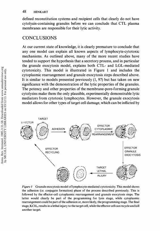
At our current state of knowledge, it is clearly premature to conclude that any one model can explain all known aspects of lymphocyte-cytotoxic mechanisms. As outlined above, many of the more recent studies have tended to support the hypothesis that a secretory process, and in particular the granule exocytosis model, explains both CTL- and LGL-mediated cytotoxicity. This model is illustrated in Figure 1 and includes the cytoplasmic rearrangement and granule exocytosis steps described above. It is similar to models presented previously ( 1 , 97) but has taken on new significance with the demonstration of the lytic properties of the granules. The potency and other properties of the membrane-pore-forming granule cytolysins make them the only plausible, experimentally demonstrable lytic mediators from cytotoxic lymphocytes. However, the granule exocytosis model allows for other types of target cell damage, which can be inflicted by
TARGET
ADHESION •
EFFECTOR RECYCLING
..
EFFECTOR CYTOPLASMIC
• REARRANGEMENT
TARGET LETHAL
INJURY
EFFECTOR GRANULE EXOCYTOSIS
Figure 1 Granule exocytosis model oflymphocyte-mediated cytotoxicity. This model shows the adhesion (i.e. conjugate formation) phase of the process described previously. This is followed by the effector-cell cytoplasmic rearrangement and granule exocytosis steps. The latter would clearly be part of the programming for lysis stage, while cytoplasmic rearrangement could be part of the adhesion or, more likely, the programming stage. The final stage, KCIL, results in a lethal injury to the target cell, while the effector cell can recycle and kill another target.
Ann
u. R
ev. I
mm
unol
. 198
5.3:
31-5
8. D
ownl
oade
d fr
om w
ww
.ann
ualr
evie
ws.
org
by M
CG
ILL
UN
IVE
RSI
TY
LIB
RA
RIE
S on
03/
12/1
3. F
or p
erso
nal u
se o
nly.

CYTOTOXIC LYMPHOCYTE MECHANISM 49
other granule components after the pores are implanted in the target membrane. As outlined above, the granule exocytosis model is supported by many different lines of evidence, and the arguments against this type of mechanism do not appear to be strong. There are several issues that must be addressed experimentally before for the granule exocytosis model can be accepted completely. Among these are the kinetics of target cell death induced by the granule cytolysins vs by cytotoxic lymphocytes and the question of why the killer cells do not die after granule exocytosis. Critical experiments that explore these questions and characterize cytotoxic lymphocyte granules in more detail are underway in several laboratories.
The concept of a secretory mechanism, such as the granule exocytosis model, for lymphocyte-mediated cytotoxicity is satisfying in a broader context because it then becomes possible to regard lymphocyte effector mechanisms in general as being secretory. The secretion of immunoglobulin by plasma cells is clearly the end result of the differentiation of B lymphocytes. In a similar way, one can view the differentiation of T lymphocytes as a specialization to secrete lymphokines or other biologically active factors. While some of these may have an antigenic specificity, others may be molecularly nonspecific but act locally. For functions such as cytotoxicity where rapid secretion is advantageous, such mediators may be stored intracellularly in granules. In other cases, such as antibody secretion by plasma cells, there may be no significant storage. The current rapid pace of molecular biology and the availability of clones and hybridomas makes the prospect of defining such secretory products an exciting one for future study.
ACKNOWLEDGMENTS
I thank Dr. Maryanna Henkart for many stimulating discussions and for helpful suggestions in the preparation of this manuscript. I gratefully acknowledge the helpful comments of Dr. Jeff Bluestone, Dr. William Munger, Dr. Louise Ramm, and Dr. Cho Yue.
Literature Cited
1. Henkart, M. P., Henkart, P. A. 1982. Lymphocyte mediated cytolysis as a secretory phenomenon. Adv. Exp. Med. BioI. 146:227
2. Sanderson, C. J. 1981. The mechanism of lymphocyte-mediated cytotoxicity. Bioi. Rev. Cambridge Phi/os. Soc. 56 : 1 53
3. Berke, G., Clark, W. R. 1982. T lymphocyte-mediated cytolysis-A comprehensive theory. I. The mechan-
ism of CTL-mediated cytolysis. Adv. Exp. Med. Bioi. 1 46 : 57
4. Russell, 1. H. 1 983. Internal disintegration model of cytotoxic Iymphocyteinduced target damage. Immunol. Rev. 72 : 97
5. Berke, G. 1983. Cytotoxic T lymphocytes-How do they function? Immunol. Rev. 72 : 1
6. Henney, C. S., Gillis, S. 1984. CelJmediated cytotoxicity. In Fundamental
Ann
u. R
ev. I
mm
unol
. 198
5.3:
31-5
8. D
ownl
oade
d fr
om w
ww
.ann
ualr
evie
ws.
org
by M
CG
ILL
UN
IVE
RSI
TY
LIB
RA
RIE
S on
03/
12/1
3. F
or p
erso
nal u
se o
nly.
50 HENKART
Immunology, ed. W. E. Paul, p. 669. New York : Raven
7. Martz, E., Heagy, W., Gromkowski, S. H. 1983. The mechanism of CTLmediated killing : Monoclonal antibody analysis of the roles of killer and target-cell membrane proteins. Immunol. Rev. 72 : 73
8. Bonavida, B., Bradley, T., Fan, J., Hiserodt, J., Elfros, R., Wexler, H. 1983. Molecular interactions in T-cellmediated cytotoxicity. Immunol. Rev. 72 : 1 19
9 . Deleted in proof 10. Martz, E. 1977. Mechanisms of specific
tumor cell lysis by alloimmune T lymphocytes : Resolution and characterization of discrete steps in the cellular interaction. Contemp. Top. Immunobiol. 7 : 301
1 1 . Golstein, P., Smith, E . T . 1977. Mechanism of T-cell-mediated cytolysis : The lethal hit stage. Contemp. Top. Immunobiol. 7 : 273
12. Berke, G. 1980. Interaction of cytotoxic T lymphocytes and target cells. Prog. Allergy 27 : 69
1 3. Balk, S. P., Walker, J., Mescher, M. F. 198 1 . Kinetics of cytolytic T lymphocyte binding to target cells in suspension. J. Immunol. 1 26 : 2177
14. Balk, S. P., Mescher, M. F. 1981. Specific reversal of cytolytic T celltarget cell Cunctional binding is induced by free target cells. J. Immunol. 1 27 : 5 1
1 5. Bongrand, P., Golstein, P . 1983. Reproducible dissociation of cellular aggregates with a wide range of calibrated shear forces : Application to cytolytic lymphocyte-target cell conjugates. J. Immunol. Methods 58 : 209
16. Bonavida, B., Bradley, T. P., Grimm, E. A. 1983. The single-cell assay in cellmediated cytotoxicity. Immunol. Today 4 : 196
17. Roder, J. c., Argov, S., Klein, M., Peterson, c., Kiessling, R., Anderson, K., Hanson, M. 1980. Target-effector cell interaction in the natural killer system. V. Energy requirements, membrane integrity, and the possible involvement of lysosomal enzymes. Immunology 40 : 107
18. Hiserodt, 1. c., Britvan, L. c., Targan, S. R. 1982. Characterization oC the cytolytic reaction mechanism of the human natural killer (NK) lymphocyte : Resolution into binding, programming and killer cell-independent steps. J. Immunol. 129 : 1 782
19. Nabholz, M., MacDonald, H. R. 1983. Cytolytic T lymphocytes. Ann. Rev. Immunol. 1 : 273
20. Taswell, c., MacDonald, H. R., Cerottini, 1. C. 1980. Clonal analysis oC cytolytic T lymphocyte specificity. I. Phenotypically distinct sets of clones as the cellular basis of cross-reactivity to alloantigens. J. Exp. Med. 1 5 1 : 1 372
21 . Allavena, P., Ortaldo, J. R. 1984. Characteristics of human NK clones : Target specificity and phenotype. J. Immunol. 1 32 : 2363
22. Grimm, E. A., Mazumder, A., Zhang, H. Z., Rosenberg, S. A. 1982. Lymphokineactivated killer cell phenomenon : Lysis of natural killer-resistant fresh solid tumor cells by interleukin 2-activated autologous human peripheral blood lymphocytes. J. Exp. Med. 155 : 1823
23. Simon, M. M., Weltzien, H. U., Buhring, H. 1., Eichmann, K. 1984. Aged murine killer T-cell clones acquire specific cytotoxicity for P81 5 mastocytoma cells. Nature 308 : 367
24. Brooks, C. G., Urdal, D. L., Henney, C. S. 1983. Lymphokine-driven "differentiation" of cytotoxic T cell clones into cells with NK -like specificity : Correlations with display of membrane macromolecules. I mmunol. Rev. 72 : 43
25. Acha-Orbea, H., Groscurth, P., Lang, R., Stitz, L., Hengartner, H. 1983. Characterization of cloned cytotoxic lymphocytes with NK-like activity. J. Immunol. 130 : 1 952
26. Binz, H., Fenner, M., Frei, D., Wigzell, H. 1983. Two independent receptors allow selective target lysis by T cell clones. J. Exp. Med. 1 57 : 1252
27. Moretta, A., Pantaleo, G., Mingari, M. c., Melioli, G., Moretta, L., Cerottini, 1.-c. 1984. Assignment of human natural killer (NK)--like cells to the T cell lineage : Single allospecific T cell clones lyse specific or NK-sesntive target cells via distinct recognition structures. Eur. J. lmmunol. 14 : 1 2 1
28. Fan, J., Ahmed, A., Bonavida, B . 1980. Studies on the induction and expression of T-cell mediated immunity. X. Inhibition by Lyt 2,3 antisera of cytotoxic T lymphocyte mediated antigenspecific and non-specific cytotoxicity : Evidence for the blocking ofthe binding between T lymphocytes. J. Immunol. 1 25 : 2444
29. MacDonald, H. R., Glasebrook, A. L., Cerottini, J. C. 1 982. Clonal heterogeneity in the functional requirement for Lyt-2/3 molecules on cytolytic T lymphocytes : Analysis by antibody blocking and selective trypsinization. J. Exp. Med. 1 56 : 1 7 1 1
30. Biddison, W. E., Rao, P. E., Talle, M. A., Goldstein, G., Shaw, S. 1984. Possible
Ann
u. R
ev. I
mm
unol
. 198
5.3:
31-5
8. D
ownl
oade
d fr
om w
ww
.ann
ualr
evie
ws.
org
by M
CG
ILL
UN
IVE
RSI
TY
LIB
RA
RIE
S on
03/
12/1
3. F
or p
erso
nal u
se o
nly.

CYTOTOXIC LYMPHOCYTE MECHANISM 51
involvement of the T4 molecule in T cell recognition of class II HLA antigens : Evidence from studies of CTL-target cell binding. J. Exp. Med. 159 : 783
3 1 . Krensky, A. M., Sanchez-Madrid, F., Robbins, E., Nagy, 1. A., Springer, T. A., Burakoff, S. J. 1983. The functional significance, distribution, and structure of LFA-l, LFA-2, and LFA-3 : Cell surface antigens associated with CTLtarget interactions. J. lmmunol. 131 : 611
32. Landegren, u., Ramstedt, U., Axberg, I., Ullberg, M., Jondal, M., Wigzell, H. 1982. Selective inhibition of human T cell cytotoxicity at levels of target recognition or initiation of lysis by monoclonal OKT3 and Leu-2a antibodies. J. Exp. Med. 155 : 1579
33. Tsoukas, C. D., Carson, D. A., Fong, S., Vaughan, 1. H. 1982. Molecular interactions in human T cell-mediated cytotoxicity to EBV. II. Monoclonal antibody OKT3 inhibits a post killertarget recognition/adhesion step. J. Immunol. 129 : 1421
34. Tsoukas, C. K., Fox, R. I., Carson, D. A., Fong, S., Vaughan, J. H. 1982. Molecular interactions in human T cell mediated cytotoxicity to Epstein-Barr virus. I. Blocking of effector cell function by monoclonal antibody OKT 3. Cell. lmmunol. 63 : 1 1 3
35. Moretta, A., Pantaleo, G., Mingari, M . c., Moretta, L., Cerottini, J.-c. 1984. Clonal heterogeneity in the requirement for T3, T4, and T8 molecules in human cytolytic T lymphocyte function. J. Exp. Med. 1 59 : 92 1
36. Leeuwenberg, J . F. M., Spits, H., Tax, W. J. M., Capel, P. J. A. 1984. Induction of non-specific cytotoxicity by monoclonal anti-T3 antibodies. J. lmmunol. Submitted for publication
37. Wauwe, J. P., DeMey, J. R., Goossens, J. G. 1980. OKT3 : A monoclonal antihuman T lymphocyte antibody with potent mitogenic properties. J. lmmunol. 1 24 : 2708
38. Chang, T. W., Kung, P. C., Gingras, S. P., Goldstein, G. 1981. Does the OKT3 monoclonal antibody react with an antigen-recognition structure on human T cells? Proc. Nat!. Acad. Sci. USA 78 : 1 805
39. VonWussow, P., Platsoucas, C. D., Wiranowska-Stewart, M., Stewart, W. E. II. 1981. Human gamma-interferon production by leukocytes induced with monoclonal antibodies recognizing T cells. J. lmmunol. 1 27 : 1 197
40. Tax, W. J. M., Willems, H. W., Reekers, P. P. M., Capel, P. 1. A., Koene, R. A. P.
1983. Polymorphism in mitogenic effect of IgG 1 monoclonal antibodies against T3 antigen on human T cells. Nature 304 : 445
41. Weiss, A., Wiskocil, R. L., Stobo, J. D. 1984. The role of T3 surface molecules in the activation of human cells : A twostimulus requirement for IL 2 production reflects events occurring at a pre-translational level. J. lmmunol. 1 33 : 123
42. Reinherz, E. L., Hussey, R. E., Schlossman, S. F. 1980. A monoclonal antibody blocking human T cell function. Eur. J. lmmunoI. 1 O : 758
43. Meuer, S. c., Fitzgerald, K. A., Hussey, R. E., Hodgdon, J. c., Schlossman, S. F., Reinherz, E. L. 1983. Clonotypic structures involved in antigen specific human T cell function : Relationship to the T3 molecular complex. J. Exp. M ed. 1 57 : 705
44. Reinherz, E. L., Meuer, S. c., Fitzgerald, K. A., Hussey, R. E., Hodgdon, J. c., Acuto, 0., Schlossman, S. F. 1983. Comparison ofT3 associated 49- and 43-kilodalton cell surface molecules on individual human T cell clones : Evidence for peptide variability in T cell receptor structures. Proc. N atl. Acad. Sci. USA 80 : 41 04
45. Meuer, S. c., Acuto, 0., Hussey, R. E., Hodgdon, 1. c., Fitzgerald, K. A., Schlossman, S. F., Reinherz, E. L. 1983. Evidence for the T3-associated 90K heterodimer as the T-cell antigen receptor. Nature 303 : 808
46. Borst, J., Alexander, S., Elder, J., Terhorst, C. 1983. The T3 complex on human T lymphocytes involves four structurally distinct glycoproteins. J. Bioi. Chem. 258 : 5135
47. Bergmen, Y., Stewart, S., Levy, S., Levy, R. 1983. Biosynthesis, glycosylation and in vitro translation of the human T cell antigen Leu 4. J. Immunol. 131 : 1876
48. Kanellopoulous, J. M., Wigglesworth, N. M., Owen, M. J., Crumpton, M. J. 1982. Biosynthesis and molecular nature of the T3 antigen of human T lymphocytes. EMBO J. 2 : 1 807
49. Weiss, A., Imboden, J., Shoback, D., Stobo, J. 1984. Role of T3 surface molecules in human T cell activation : T3 dependent activation results in a rise in cytoplasmic free calcium. Proc. Natl. Acad. Sci. USA 81 : 4169
50. Hiserodt, 1. c., Bonavida, B. 198 1 . Studies o n the induction and expression of T cell-mediated immunity. XI. Inhibition of the "lethal hit" in T cellmediated cytotoxicity by heterologous
Ann
u. R
ev. I
mm
unol
. 198
5.3:
31-5
8. D
ownl
oade
d fr
om w
ww
.ann
ualr
evie
ws.
org
by M
CG
ILL
UN
IVE
RSI
TY
LIB
RA
RIE
S on
03/
12/1
3. F
or p
erso
nal u
se o
nly.

52 HENKART
rat antiserum made against alloimmune cytotoxic T lymphocytes. J. lmmunol. 126 : 256
5 1 . Neville, M. E., Hiserodt, J. C. 1982. Inhibition of human antibodydependent cellular cytotoxicity, cellmediated cytotoxicity, and natural killing by a xenogeneic antiserum prepared against "activated" alloimmune human lymphocytes. J. lmmunol. 128 : 1246
52. Devlin, J. J., Phaneuf, 1. D., Granger, G. A. 1982. Inhibition of human lectindependent cell-mediated cytotoxicity, natural killer-like cytotoxicity, and cytotoxic T-lymphocyte mediated cytolysis by xenoantisera raised against concanavalin A stimulated human lymphocytes. Cell Immunol. 68 : 64
53. Ware, C. F., Granger, G. A. 1 98 1 . . Mechanisms of lymphocyte-mediated cytotoxicity. III. Characterization of the mechanism of inhibition of the human alloimmune Iymphocytemediated cytotoxic reaction by polyspecific anti-lymphocyte sera in vitro. J. lmmuno/. 1 26 : 1 934
54. Merrill, 1. E., Ullberg, M., Jondal, M. 1981 . Influence of IgG and IgM receptor triggering on human natural killer cell cytotoxicity measured on the level of the single etTector cell. Eur. J. lmmunol. 1 1 : 536
55. Targan, S. R., Newman, W. 1983. Definition of a "trigger" stage in the NK cytolytic reaction sequence by a monoclonal antibody to the glycoprotein T-200. J. lmmunol. 1 3 1 : 1 149
56. Sparrow, R. L., McKenzie, I. F. C. 1983. A function for human T200 in natural killer cytolysis. Transplantation 36 : 166
57. Burns, G. F., Triglia, T., Bartlett, P. F., Mackay, I. R. 1 983. Human natural killer cells, activated lymphocyte killer cells, and monocytes possess similar cytotoxic mechanisms. Proc. Natl. Acad. Sci. USA 80 : 7606
58. Nieminen, P., Paasivuo, R., Saksela, E. 1 982. EtTect of a monoclonal anti-large granular lymphocyte antibody on the human NK activity. J. lmmunol. 128 : 1097
59. Kahle, R., Hiserodt, 1. c., Bonavida, B. 1983. Characterization of antibody mediated inhibition of natural killer (NK) cytotoxicity : Evidence for blocking of both recognition and lethal hit stages of cytolysis. Cell. lmmunol. 80 : 97
60. Timonen, T., Ortaldo, J. R., Herberman, R. B. 1981. Characteristics of human large granular lymphocytes and relationship to natural killer and K cells. J. Exp. Med. 1 53 : 569
61 . Payne, C. M., Glasser, L., Fiederlein, R., Lindberg, R. 1983. New ultrastructural observations : Parallel tubular arrays in human T gamma lymphoid cells. J. Immunol. Methods 65 : 307
62. Kanayama, Y., Hiraoka, A., Machii, T., Tamaki, T., Kanakura, Y., Y onezawa, T., Tarui, S., Kitani, T. 1983. Ultrastructure of normal human T cell subpopulations : Parallel tubular arrays in T gamma lymphocytes and clustered dense bodies in T.u lymphocytes. Acta H aematol. 70 : 220
63. Zagury, D., Bernard, J., Thierness, N., Feldman, M., Berke, G. 1975. Isolation and characterization of individual functionally reactive cytotoxic T lymphocytes : Conjugation, killing and recycling at the single cell level. Eur. J. Immunol. 5 : 8 1 8
64. Sanderson, C . J., Glaueret, A . M. 1977. The mechanism of T cell mediated cytotoxicity. V. Morphological studies by electron microscopy. Proe. R. Soc. London Ser. B 198 : 3 1 5
65. Bykovskaja, S . N., Rytenko, A . N., Rauschenbach, M. 0., Bykovsky, A. F. 1978. Ultrastructural alteration of cytolytic T lymphocytes following their interaction with target cells. II. Morphogenesis of secretory granules and intracellular vacuoles. Cell. Immuno/. 40 : 175
66. Matter, A. 1979. Microcinematographic and electron microscopic analysis of target cell lysis induced by cytotoxic T lymphocytes. Immunology 36 : 179
67. Hackett, C. J., Sullivan, K., Lin, Y. L. 1982. Ultrastructure of an influenza virus-specific cytotoxic T cell clone and its interaction with P815 and macrophage targets. Cell. I mmunol. 68 : 276
68. Dennert, G., Podack, E. R. 1983. Cytolysis by H-2 specific T killer cells : Assembly of tubular complexes on target membranes. J. Exp. Med. 1 57 : 1483
69. Grossi, C. E., Zicca, A., Cadoni, A., Mingari, M. c., Moretta, A., Moretta, L. 1983. Ultrastructural characteristics of human T cell clones with various cytolytic activities. Eur. J. Immunol. 13 : 670
70. Itoh, K., Suzuki, R., Umezu, Y., Hanaumi, K., Kumagai, K. 1982. Studies of murine large granular lymphocytes. II. Tissue, strain, and age distributions of LGL and LAL. J. Immunol. 129 : 395
7 1 . Nocera, A., Montesoro, E., Balbo, P., Ferrarini, M., Leprini, A., Zicca, A., Grossi, C. E. 1983. Complement recep-
Ann
u. R
ev. I
mm
unol
. 198
5.3:
31-5
8. D
ownl
oade
d fr
om w
ww
.ann
ualr
evie
ws.
org
by M
CG
ILL
UN
IVE
RSI
TY
LIB
RA
RIE
S on
03/
12/1
3. F
or p
erso
nal u
se o
nly.

CYTOTOXIC LYMPHOCYTE MECHANISM 53
tor distinguishes between two subsets of large granular lymphocytes with different natural killer activity and cytochemical and ultrastructural features. Scand. J. Immunol. 1 8 : 345
72. Kay, N. E., Zarling, 1. M. 1984. Impaired natural killer activity in patients with chronic lymphocytic leukemia is associated with a deficiency of azurophilic cytoplasmic granules in putative NK cells. Blood 63 : 305
73. Wahlin, B., Perlmann, P. 1983. Characterization of human K cells by surface antigens and morphology at the single cell level. J. I mmunol. 131 : 2340
74. Schneider, E. M., Pawelec, G. P., Liangru, S., Wernet, P. 1984. A novel type of human T cell clone with highly potent natural killer-like cytotoxicity divorced from large granular lymphocyte morphology. J. Immunol. 133 : 173
75. Thiernesse, N., David, A., Bernard, J., Jeanesson, P., Zagury, D. 1977. Activite phosphatasique acide de la cellule T cytolytique au cours du processus de cytolyse. C. R. Acad. Sci. Ser. D 285 : 7 1 3
76. Grossi, C . E., Cadoni, A., Zicca, A., Leprini, A., Ferrarini, M. 1982. Large granular lymphocytes in human peripheral blood : Ultrastructural and cytochemical characterization of the granules. Blood 59 : 277
77. Frey, T., Petty, H. R., McConnell, H. M. 1982. Electron microscopic study of natural killer cell-tumor cell conjugates. Proc. Natl. Acad. Sci. USA 79 : 53 1 7
78. Millard, P . J., Henkart, M . P., Reynolds, C. W., Henkart, P. A. 1984. Purification and properties of cytoplasmic granules from cytotoxic rat LGL tumors. J. Immunol. 1 32 : 3197
79. Henkart, P. A., Millard, P. J., Reynolds, C. W., Henkart, M. P. 1984. Cytolytic activity of purified cytoplasmic granules from cytotoxic rat LGL tumors. J. Exp. Med. 160 : 75
80. Reynolds, C. W., Reichardt, D., Henkart, M., Millard, P., Henkart, P. 1 984. Functional characterization of antibodies against purified cytoplasmic granules from rat LGL tumors. Submitted for publication
81 . Bykovskaja, S. N., Rytenko, A. N., Rauschenbach, M. 0., Mykovsky, A. F. 1978. Ultrastructural alteration of cytolytic t lymphocytes following their interaction with target cells. I. Hypertrophy and change of orientation of the Golgi apparatus. Cell. Immunol. 40 : 164
82. Geiger, B., Rosen, D., Berke, R. 1982.
Spatial relationships of microtubuleorganizing centers and the contact area of cytotoxic T lymphocytes and target cells. J. Cell Bioi. 95 : 137
83. Ryser, J. E. , Rungger-Brandle, E., Chaponnier, C., Gabbiani, G., Vassalli, P. 1982. The area of attachment of cytolytic T lymphocytes to their target cells shows high motility and polarization of actin, but not myosin. J. Immunol. 128 : 1 1 59
84. Alexander, E., Henkart, P. 1976. The adherence of human Fc receptorbearing lymphocytes to antigenantibody complexes. II. Morphological alterations induced by the substrate. J. Exp. Med. 143 : 329
85. Carpen, 0., Virtanen, I., Saksela, E. 1982. Ultrastructure of the human natural killer cells : Nature of the cytolytic contacts in relation to cellular secretion. J. Immunol. 128 : 2691
86. Carpen, 0., Virtanen, I., Letho, V.-P., Saksela, E. 1983. Polarization of NK cell cytoskeleton upon conjugation with sensitive targets. J. Immunol. 1 3 1 : 2695
87. Kupfer, A., Dennert, G., Singer, S. J. 1983. Polarization of the Golgi apparatus and the microtubule-organizing center within cloned natural killer cells bound to their targets. Proc. N atl. Acad. Sci. USA 80 : 7224
88. Ryser, J. E., Sordat, B., Cerottini, J. C., Brunner, K. T. 1977. Mechanism of target cell lysis by cytolytic T lymphocytes. I. Characterization of specific lymphocyte-target cell conjugates separated by velocity sedimentation. Eur. J. Immunol. 7 : 1 1
89. Barber, T. A., Alter, B . J . 1 978. Ultrastructure of effector-target cell interaction in secondary cell-mediated Iympholysis. Scand. J. Immunol. 7 : 57
90. Glauert, A. M., Sanderson, C. J. 1979. The mechanism of k-cell (antibodydependent) mediated cytotoxicity. J. Cell Sci. 35 : 355
91 . David, A., Bernard, J., Thiernesse, N., Nicolas, G., Cerottini, J. C., Zagury, D. 1979. Le processus d'exocytose Iysosomale localisee est-il responsable de l'action cytolytique des lymphocytes T ttiers? C. R. Acad. Sci. Ser. D 288 : 441
92. Zagury, D. 1982. Direct analysis of individual killer T cells : Susceptibility of target cells to lysis and secretion of hydrolytic enzymes by CTL. Adv. Exp. Med. BioI. 146 : 149
93. Bykovskaja, S. N., Sergeev, A. V., Rauschenbach, M. 0., Bykovsky, A. F. 1980. The ultrastructure of the tubular
Ann
u. R
ev. I
mm
unol
. 198
5.3:
31-5
8. D
ownl
oade
d fr
om w
ww
.ann
ualr
evie
ws.
org
by M
CG
ILL
UN
IVE
RSI
TY
LIB
RA
RIE
S on
03/
12/1
3. F
or p
erso
nal u
se o
nly.

54 HENKART
complex in the cytoplasm of cytolytic T lymphocytes. Scand. J. Immunol. 1 1 : 261
94. Podack, E. R., Dennert, G. 1983. Assembly of two types of tubules with putative cytolytic function by cloned natural killer cells. Nature 302 : 442
95. Zucker-Franklin, D., Grusky, G., Yang, J.-S. 1983. Arylsulfatase in natural killer cells : Its possible role in cytotoxicity. Proc. Natl Acad. Sci. USA 80 : 6977
96. Neighbour, P. A., Huberman, H. S., Kress, Y. 1982. Human large granular lymphocytes and natural killing : Ultrastructural studies of strontiuminduced degranulation. Eur. J. Immunol. 1 2 : 588
97. Neighbour, P. A. Huberman, H. S. 1982. Sr+2-induced inhibition of human natural killer (NK) cell-mediated cytotoxicity. J. Immunol. 1 28 : 1236
98. Henson, P. M. 1971 . The immunologic release of constituents from neutrophil granules. II. Mechanisms of release during phagocytosis, and adherence to nonphagocytosable surfaces. J. Immunol. 107 : 1 547
99. Lawson, D., Fewtrell, C., Raff, M. C. 1978. Localized mast cell degranulation induced by concanavalin A-Sepharose beads. J. Cell Bioi. 79 : 394
100. Henney, C. S. 1973. On the mechanism ofT-cell mediated cytolysis. Transplant. Rev. 17 : 37
101 . Douglas, W. W. 1978. Stimulussecretion coupling : Variations on the theme of calcium-activated exocytosis involving cellular and extracellular sources of calcium. Ciba Found. Symp. 54 : 61
102. Plaut, M., Bubbers, J. E., Henney, C. S. 1976. Studies on the mechanism of lymphocyte-mediated cytolysis. VII. Two stages in the T cell-mediated lytic cycle with distinct cation requirements. J. Immunol. 1 1 6 : 150
103. Martz, E., Parker, W. L., Gately, M. K., Tsoukas, C. D. 1982. The role of calcium in the lethal hit of T lymphocytemediated cytolysis. Adv. Exp. M ed. BioI. 146 : 12 1
104. Russell, J . H., Dobos, C . R . 198 1 . The role of monovalent cations in the interaction between the cytotoxic T lymphocyte and its target. Eur. J. Immunol. 1 1 : 840
105. Russell, 1. H., Dobos, C. R. 1983. Accelerated 86Rb + (K +) release from the cytotoxic T lymphocyte is a physiologic event associated with delivery of the lethal hit. J. Immunol. 1 3 1 : 1 138
106. Petersen, O. H., Maruyama, Y. 1 984.
Calcium-activated potassium channels and their role in secretion. Nature 307 : 693
107. Fukushima, Y., Hagiwara, S., Henkart, M. 1984. Potassium current in clonal cytotoxic T lymphocytes from the mouse. J. Physiol. 351 : 645
108. Brondz, 8. D., Snegirova, A. E., Rassulin, Y. A., Shambo rant, O. G. 1973. Modification of in vitro immune lymphocyte-target cell interaction by some biologically active drugs. Immunochemistry 10 : 175
109. Carpen, 0., Virtanen, I. , Saksela, E. 1981 . The cytotoxic activity of human natural killer cells requires an intact secretory apparatus. Cell. Immunol. 58 : 97
1 10. Pederson, B., Norrild, 8., Krebs, H. J. 1982. The effect of monensin on the antibody-dependent cell-mediated killing. Cell. Immunol. 72 : 208
1 1 1 . Quan, P. c., Ishizake, T., Bloom, B. 1982. Studies on the mechanism of NK cell lysis. J. Immunol. 128 : 1786
1 12. Wei, W. Z., Lindquist, R. R. 1981 . Alloimmune cytolytic lymphocyte activity : Triggering and expression of killing mechanisms in cytolytic T lymphocytes. J. Immunol. 126 : 5 13
1 13. Martz, E., Benacerraf, B. 1976. Multiple target cell killing by cytolytic Tlymphocytes and the mechanism of cytotoxicity. Transplantation 21 : 5
1 14. Ullberg, M., Jondal, M. 1981 . Recycling and target binding capacity of human natural killer cells. J. Exp. Med. 1 53 : 6 15
1 15. Golstein, P . 1974. Sensitivity of cytotoxic T cells to T-cell mediated cytotoxicity. Nature 252 : 8 1
1 16. Fishelson, Z., Berke, G . 1978. T lymphocyte cytolysis : Dissociation of the binding from the lytic mechanism ofthe effector cells. J. Immunol. 120 : 1 121
1 1 7. Kuppers, R. c., Henney, C . S . 1977. Studies on the mechanism of lymphocyte-mediated cytolysis. IX. Relationships between antigen recognition and lytic expression in killer T cells. J. Immunol. 1 18 : 71
1 18. Ishizaka, T., Ishizaka, K. 1984. Activation of mast cells for mediator release through IgE receptors. Prog. Allergy 34 : 188
1 19. Golstein, P. , Gomperts, B. D. 1975. Non-T cell-mediated cytolysis of antibody-coated sheep and red blood cells requires Mg+2 but not Ca + 2 : An argument against a conventional "stimulus-secretion" mechanism for cytolysis. J. Immunol. 1 1 4 : 1264
1 20. Golstein, P., Fewtrell, C. 1975.
Ann
u. R
ev. I
mm
unol
. 198
5.3:
31-5
8. D
ownl
oade
d fr
om w
ww
.ann
ualr
evie
ws.
org
by M
CG
ILL
UN
IVE
RSI
TY
LIB
RA
RIE
S on
03/
12/1
3. F
or p
erso
nal u
se o
nly.

CYTOTOXIC LYMPHOCYTE MECHANISM 55
Functional fractionation of human cytotoxic cells through differences in their cation requirements. Nature 255 : 491
1 2 1 . Taka, M., Hinuma, S., Abo, T., Kumagai, K. 1980. Murine antibodydependent cell-mediated cytotoxicity : Failure to detect effector cells equivalent to human K cells. J. Immunol. 124 : 1929
122. Kumagai, K., Itoh, K., Suzuki, R., Hinuma, S., Saitoh, F. 1 982. Studies of murine large granular lymphocytes. I. Identification as effector cells in NK and K cytotoxicities. J. Immunol. 129 : 388
123. Nelson, D. L., Poplack, D. G., Holiman, B. J., Henkart, P. A. 1979. ADCC against human erythrocyte target cells : Role of the anti-target cell antibodies in determining lymphocyte killer activity. Clin. Exp. Immunol. 35 : 447
1 24. Shoback, D. M., Thatcher, J., Leombruno, R., Brown, E. M. 1984. Relationship between parathyroid hormone secretion and cytosolic calcium concentration in dispersed bovine thyroid cells. Proc. Natl. Acad Sci. USA 8 1 : 3 1 1 3
125. Roder, 1 . C. 1979. The beige mutation in the mouse, I. A stem cell predetermined impairment in natural killer cell function. J. Immunol. 123 : 2 1 68
126. Roder, J. c., Lohmann-Matthes, M. L., Domzig, W., Wigzell, H. 1979. The beige mutation in the mouse. II. Selectivity of the natural killer cell defect. J. lmmunol. 123 : 2 1 74
127. Haliotis, T., Roder, J., Klein, M., Ortaldo, 1., Fauci, A. S., Herberman, R. B. 1980. Chediak-Higashi gene in humans. I. Impairment of natural killer gene in humans. J. Exp. Med. 1 5 1 : 1039
128. Klein, M., Roder, 1., Haliotis, T., Korec, S., Jett, J., Herberman, R. B., Katz, P., Fauci, A. S. 1980. Chediak-Higashi gene in humans. II. The selectivity ofthe defect in natural killer and antibodydependent cell-mediated cytotoxicity function. J. Exp. Med. 1 5 1 : 1049
129. Windhorst, D. B., Padgett, G. 1973. The Chediak-Higashi syndrome and the homologous trait in animals. J. Invest. Dermatol. 60 : 529
1 30. Saxena, R. K., Saxena, Q. B., Adler, W. H. 1982. Defective T-cell response in beige mutant mice. Nature 295 : 240
1 3 1. Henney, C. S. 1973. Studies on the mechanism of lymphocyte-mediated cytolysis. II. The use of various target cell markers to study cytolytic events. J. Immunol. 1 10 : 73
1 32. Martz, E., Burakoff, S. J., Benacerraf, B. 1 974. Interruption of the sequential
release of small and large molecules from tumor cells by low temperature during cytolysis mediated by immune T cells or complement. Proc. Natl. Acad. Sci. USA 7 1 : 177
133. Sanderson, C. J. 1976. The mechanism of T cell mediated cytotoxicity. I. The release of different cell components. Proc. R. Soc. London Ser. B 192 : 221
134. Sanderson, C. J., Thomas, J. A. 1977. The mechanism of K cell (antibodydependent) cell mediated cytotoxicity. I. The release of different cell components. Proc. R. Soc. London Ser. B 197 : 407
135. Martz, E. 1976. Early steps in specific tumor cell lysis by sensitized mouse T lymphocytes. II. Electrolyte permeability increase in the target cell membrane concomitant with programming for lysis. J. lmmunol. 1 1 7 : 1023
136. Henkart, P., Blumenthal, R. 1975. The interaction of lymphocytes with lipid bilayer bilayer membranes : A model for the lymphocyte-mediated lysis of target cells. Proc. Natl. Acad. Sci. USA 72 : 2789
137. Ozato, K., Ziegler, H. K., Henney, C. S. 1978. Liposomes as model membrane systems for immune attack. II. The interaction of complement and K cell populations with immobilized liposomes. J. Immunol. 1 2 1 : 1 383
138. Juy, D., Billecocq, A., Faure, M., Bona, C. 1977. Damage of Iiposomes in antibody-dependent cell-mediated cytotoxicity. Scand. J. Immunol. 6 : 607
1 39. Geiger, B., Schreiber, A. D. 1979. The use of antibody-coated liposomes as a target cell model for antibodydependent cell-mediated cytotoxicity. Clin. Exp. Immunol. 35 : 149
140. Hollander, N., Mehdi, S. Q., Weissman, I. L., McConnell, H. M., Kriss, J. P. 1979. Allogeneic cytolysis of reconstituted membrane vesicles. Proc. Natl. Acad. Sci. USA 76 : 4042
141 . Baird, B., Henkart, P. 1983. Enhancement of the recognition by cytotoxic T lymphocytes (CTL) of target membrane antigens after fusion with whole cells. Cell. Immunol. 75 : 3 1 2
142. Nakanishi, M. , Brian, A . A., McConnell, H. M. 1983. Binding of cytotoxic T lymphocytes to supported lipid monolayers containing trypsinized H-2Kk. Mol. Immunol. 20 : 1227
143. Schick, B., Jakobovits, A., Sharon, N., Berke, G. 1983. Specific lysis of antigenically irrelevant cells by cytotoxic T lymphocytes upon insertion of appropriate antigens into the target cell plasma membranes. Transplantation 36 : 84
Ann
u. R
ev. I
mm
unol
. 198
5.3:
31-5
8. D
ownl
oade
d fr
om w
ww
.ann
ualr
evie
ws.
org
by M
CG
ILL
UN
IVE
RSI
TY
LIB
RA
RIE
S on
03/
12/1
3. F
or p
erso
nal u
se o
nly.
56 HENKART
144. Simone, C. B., Henkart, P. A. 1980. Permeability changes induced in erythrocyte ghost targets by antibodydependent cytotoxic effector cells : Evidence for membrane pores. J. Immunol. 124 : 952
145. Dourmashkin, R. R., Deteix, P., Simone, C. B., Henkart, P. A. 1980. Electron microscopic demonstration of lesions on target cell membranes associated with antibody-dependent cytotoxicity. Clin. Exp. Immunol. 43 : 554
146. Blumenthal, R., Millard, P. 1., Henkart, M. P., Reynolds, C. W., Henkart, P. A. 1984. Liposomes as targets for granule cytolysin from cytotoxic LGL tumors. Proc. Nat/. Acad. Sci. USA 8 1 : 5551
147. Henkart, P., Millard, P., Vue, c., Fredrickse, P., Blumenthal, R., Bluestone, J., Reynolds, C. W. Henkart, M. P. 1984. Biochemical and functional properties of LGL and CTL cytoplasmic granules. Adv. Exp. Med. BioI. In press
148. Podack, E. R., Konigsberg, P. 1., AchaOrbea, H., Pircher, H., Hengartner, H. 1984. Cytolytic T-cell granules : Biochemical properties and functional specificity. Adv. Exp. M ed. Bioi. In press
149. Mayer, M. M. 1982. Membrane attack by complement (with comments on cellmediated cytotoxicity). Adv. Exp. Med. BioI. 146 : 1 93
1 50. Green, H., Fleischer, R. A., Barrow, P., Goldberg, B. 1 959. The cytotoxic action of immune gamma globulin and complement on Krebs ascites tumor cells. II. Chemical studies. J. Exp. Med. 1 09 : 5 1 1
1 5 1 . Henney, C. S . 1 974. Estimation of the size of a T-cell-induced lytic lesion. Nature 249 : 456
1 52. Ferluga, J., Allison, A. C. 1974. Observations on the mechanism by which T lymphocytes exert cytotoxic effects. Nature 250 : 673
1 53. Kurt, E. A., Lindquist, R. R. 1981 . Failure of dextran to osmotically protect CTL-"hit" target cells. Transplant. Proc. 8 : 1 178
1 54. Sanderson, C. J., Thomas, J. A. 1977. The mechanism of K cell (antibodydependent) cell mediated cytotoxicity. II. Characteristics of the effector cell and morphological changes in the target cell. Proc. R. Soc. London Ser. B 197 : 4 1 7
1 55. Sanderson, C . J . 1976. The mechanism of T cell mediated cytotoxicity. II. Morphological studies of cell death by time-lapse microcinematography. Proc. R. Soc. London Ser. B 192 : 241
1 56. Wyllie, A. H., Kerr, 1. F. R., Currie, A. R. 1980. Cell death : The significance of apoptosis. Int. Rev. Cytol. 68 : 25 1
1 57. Russell, 1. H., Masakowski, V. R., Dobos, C. R. 1980. Mechanisms of immune lysis. I. Physiological distinction between target cell death mediated by cytotoxic T lymphocytes and antibody plus complement. J. Immunol. 1 24 : 1 100
158. Russell, J. H., Masakowski, V., Rucinsky, T., Phillips, G. 1982. Mechanisms of immune lysis. III. Characterization of the nature and kinetics of the cytotoxic T Iymphocyteinduced nuclear lesion in the target. J. Immunol. 128 : 2087
1 59. Russell, J. H., Dobos, C. R. 1980. Mechanisms of immune lysis. II. CTLinduced nuclear disintegration of the target begins within minutes of cell contact. J. Immunol. 1 25 : 1 256
160. Sanderson, C. J., Hall, P. J., Thomas, J. A. 1977. The mechanism of T cell mediated cytotoxicity. IV: Studies on communicating junctions between cells in contact. Proc. R. Soc. London Ser. B 1 96 : 73
161 . Duke, R. C., Chervenak, R., Cohen, J. 1. 1983. Endogenous endonucleaseinduced DNA fragmentation : An early event in cell-mediated cytolysis. Proc. Natl. Acad. Sci. USA 80 : 6361
1 62. Sears, D. W., Christiaansen, J. E. 1984. Mechanism of rapid tumor lysis by human ADCC : Mediation by monoclonal antibodies and fragmentation of target cell DNA. Adv. Exp. M ed. BioI. In press
163. Roder, J, C" Helfand, S. L., Werkmeister, J., McGarry, R., Beaumont, T. J., Duwe, A. 1982. Oxygen intermediates are triggered early in the cytolytic pathway of human NK cells. Nature 298 : 569
1 64. Helfand, S. L., Werkmeister, J., Roder, J. C. 1982. Chemiluminescence response of human natural killer cells. I . The relationship between target cell binding, chemiluminescence, and cytolysis. J. Exp. Med. 1 56 : 492
1 65. Werkmeister, J., Helfand, S., Roder, J., Pross, H. 1983. The chemiluminescence response of human natural killer cells. II. Association of a decreased response with low natural killer activity. Eur. J. Immunol. 1 3 : 5 1 4
166. Tanaka, Y., Sakane, T., Usui, T . 1981. Natural killer (NK) activity in patients with chronic granulomatous disease. Hiroshima J. Med. Sci. 30 : 1 3 5
167. Kay, H. D., Smith, D. L., Sullivan, G., Mandell, G. L., Donowitz, G. R. 1983.
Ann
u. R
ev. I
mm
unol
. 198
5.3:
31-5
8. D
ownl
oade
d fr
om w
ww
.ann
ualr
evie
ws.
org
by M
CG
ILL
UN
IVE
RSI
TY
LIB
RA
RIE
S on
03/
12/1
3. F
or p
erso
nal u
se o
nly.

CYTOTOXIC LYMPHOCYTE MECHANISM 57
Evidence for a nonoxidative mechanism of human natural killer (NK) cell cytotoxicity by using mononuclear effector cells from healthy donors and from patients with chronic granulomatous disease J. Immunol. 1 3 1 : 1784
168. Katz, P., Simone, C. B., Henkart, P. A., Fauci, A. S. 1980. Mechanisms of antibody-dependent cellular cytotoxicity : The use of effector cells from chronic granulomatous disease patients as investigative probes. J. Clin. Invest. 65 : 55
169. Nathan, C. F., Mercer-Smith, J. A., Desantis, N. M., Pall andino, M. A. 1982. Role of oxygen in T cell-mediated cytolysis. J. Immunol. 129 : 2164
170. Henney, C. S. 1977. T cell-mediated cytolysis : An overview of some current issues. Contemp. Top. Immunobiol. 7 : 245
171 . Hiserodt, J. c., Tiangco, G. J., Granger, G. A. 1979. The L T system in experimental animals. IV. Rapid specific lysis of 51 Cr labelled allogeneic target cells by highly unstable high mw lymphotoxin-receptor complex(es) released in vitro by activated alloimmune murine T lymphocytes. J. Immunol. 123 : 332
172. Martz, E. 1975. Early steps in specific tumor cell lysis by sensitized mouse T lymphocytes. I. Resolution and characterizations. J. Immunol. 1 1 5 : 261
173. Granger, G. A., Hiserodt, J. c., Yamamoto, R. S., Ross, M. W. 1980. LT molecules form a subunit system of cell toxins. In Biochemical Characterization of Lymphokines, ed. A. L. de Weck, F. Kristensen, M. Landy, pp. 279-83. New York : Academic
174. Leopardi, E., Rosenau, W. 1982. Human T cell mediated cytotoxicity : Role of subsets and neutralization of cytotoxicity by anti-alpha-lymphotoxin serum. Cell. Immunol. 70 : 148
175. Weitzen, M. L., Innins, E., Yamamoto, R. S., Granger, G. A. 1983. Inhibition of human NK-induced cell lysis and soluble cell-lytic molecules with antihuman LT antisera and various saccharides. Cell. Immunol. 77 : 42
176. Gately, M. K., Mayer, M. M., Henney, C. S. 1976. Effect of anti-lymphotoxin on cell-mediated cytotoxicity : Evidence for two pathways, one involving Iymphotoxin and the other requiring intimate contact between the plasma membranes of killer and target cells. Cell. Immunol. 27 : 82
177. Ware, C. F., Granger, G. A. 1981. Mechanisms of lymphocyte-mediated cytotoxicity. I. The effects of anti-
human lymphotoxin antisera on the cytolysis of allogeneic B cell lines by MLC-sensitized human lymphocytes in vitro. J. Immunol. 126 : 1919
178. Wright, S. c., Bonavida, B. 1981. Selective lysis of NK-sensitive target cells by a soluble mediator released from murine cells and human peripheral blood lymphocytes. J. Immunol. 126 : 1516
179. Wright, S. c., Bonavida, B. 1982. Studies on the mechanism of natural killer (NK) cell-mediated cytotoxicity (CMC). I. Release of cytotoxic factors specific for NK-sensitive target cells (NKCF) during co-culture of NK effector cells with NK target cells. J. Immunol. 129 : 433
180. Wright, S. c., Bonavida, B. 1983. YAC-1 variant clones selected for resistance to natural killer cytotoxic factors are also resistant to natural killer cellmediated cytotoxicity. Proc. Natl. Acad. Sci. USA 80 : 1688
181 . Wright, S. c., Weitzen, M. L., Kahle, R., Granger, G. A., Bonavida, B. 1983. Studies on the mechanism of natural killer cytotoxicity. II. Coculture of human PBL with NK-sensitive or resistant cell lines stimulates release of natural killer cytotoxic factors (NKCF) selectively cytotoxic to NK-sensitive target cells. J. Immunol. 130 : 2479
182. Farram, E., Targan, S. R. 1983. Identification of human natural killer soluble cytotoxic factor(s) (NKCF) derived from NK-enriched lymphocyte populations : Specificity of generation and killing. J. Immunol. 130 : 1252
183. Leopardi, E., Rosenau, W. 1984. Production of alpha-lymphotoxin by human T-cell subsets. Cell. Immunol. 83 : 73
184. Wright, S. C., Bonavida, B. 1983. Studies on the mechanism of natural killer cytotoxicity. III. Activation of NK cells by interferon augments the lytic activity of released natural killer cytotoxic factors (NKCF). J. Immunol. 130 : 2960
185. Wayner, E. A., Brooks, C. G. 1984. Induction of NKCF-like activity in mixed lymphocyte-tumor cell culture : Direct involvement of mycoplasma infection of tumor cells. J. Immunol. 1 3 2 : 2135
186. Wright, S. C., Bonavida, B. 1984. Studies on the mechanism of natural killer cell mediated cytotoxicity. V. Lack of NK specificity at the level of induction of natural killer cytotoxic factors in cultures of human, murine, or rate effector cells stimulated with NK
Ann
u. R
ev. I
mm
unol
. 198
5.3:
31-5
8. D
ownl
oade
d fr
om w
ww
.ann
ualr
evie
ws.
org
by M
CG
ILL
UN
IVE
RSI
TY
LIB
RA
RIE
S on
03/
12/1
3. F
or p
erso
nal u
se o
nly.

58 HENKART
sensitive or resistant mycoplasma free cell lines. J. Immunol. In press
1 87. Wright, S. c., Bonavida, B. 1983. Studies on the mechanism of natural killer cell-mediated cytotoxicity. IV. Interferon-induced inhibition of NK target cell susceptibility to lysis is due to a defect in their ability to stimulate release of natural killer cytotoxic factors. J. Immunol. 1 30 : 2965
188. Hiserodt, l. C., Britvan, L., Targan, S. R. 1983. Studies on the mechanism of the human natural killer cell lethal hit : Analysis of the mechanism of protease inhibition of the lethal hit. J. Immunol. 1 3 1 : 2705
189. Hiserodt, 1. c., Britvan, L. 1., Targan, S. R. 1982. Differential effects of various pharmacologic agents on the cytologic reaction mechanism of the human natural killer lymphocyte : Further resolution of programming for lysis and KCIL into discrete stages. J. Immunol. 129 : 2266
190. Grimm, E., Price, Z., Bonavida, B. 1979. Studies on the induction and expression
of T cell-mediated immunity. VIII. Effector-target junctions and target cell membrane disruption during cytolysis. Cell. Immunol. 46 : 77
191 . Ferluga, 1., Allison, A. S. 1975. Cytotoxicity of isolated plasma membranes from lymph node cells. Nature 255 : 708
192. Kahn-Perles, B., Golstein, P. 1978. CeJl membrane cytolysis by membranes from non-cytolytic cells. Eur. J. Immunol. 8 : 71
193. Harris, D. T., MacDonald, H. R., Cerottini, l.-C. 1984. Direct transfer of antigen-specific cytolytic activity to noncytolytic cells upon fusion with liposomes from cytolytic T cell clones. J. Exp. A1ed. 159 : 261
194. Pohajdak, B., Gomez, 1. L., Wilkins, 1. A., Greenberg, A. H. 1984. Tumor activated NK cells trigger monocyte oxidative metabolism. J. Immunol. In press
195. Podack, E. R., Konigsberg, P. 1. 1984. Cytolytic T cell granules. Isolation, biochemical and functional characterization. J. Exp. Med. 160 : 695
NOTE ADDED IN PROOF Pohajdak et al ( 194) have recently shown that NKsensitive target cells rapidly induce purified LGL to release a soluble factor that triggers the respiratory burst in monocytes. These results may provide an explanation for the chemiluminescence reported to be associated with NK cytolysis (163-165) if monocyte depletion was incomplete in the original experiments.
Ann
u. R
ev. I
mm
unol
. 198
5.3:
31-5
8. D
ownl
oade
d fr
om w
ww
.ann
ualr
evie
ws.
org
by M
CG
ILL
UN
IVE
RSI
TY
LIB
RA
RIE
S on
03/
12/1
3. F
or p
erso
nal u
se o
nly.