lrnrnune--mediated cytotoxicity in inflammatory bowel disease
TRANSCRIPT

IBO: PATHOGENESIS
lrnrnune--mediated cytotoxicity in inflammatory bowel disease
STEPHAN RT ARGAN, MD, RICI IARD L DEEM, MS, FFRl,US SI IANAJ IAN, MD
ABSTRACT: Thirty years of research on the role of immune-mediated cytotoxic activity in the tissue in Jury of inflammatory bowel disease (IBO) has yielded only inconclusive data on the relevance of cytotoxic mechanisms. Two hypolheses have heen advanced. One is that the destruction of target cells 1s mediated by direct recognition of target antigens by cytotoxic cells which in tum triggers lysis. Another hypothesis is that lysis occurs via an indirect bystander mechanism in which cells do not recognize a specific antigen on the target, hut upon nonspecific activation release cytokines which are capable of lysing the target. The authors have investigated both hypotheses and studied the role of cytotoxic T cclb and cytotoxic r ctors released from acttvated T cells in the destruction of epithelial cells. Elevated levels of cytotoxic T cells were found in penpheral blood lymphocytes of patients with IBO, particularly Crohn's disease. The cytotoxic T cells were contained within the Leu 7+, CDS+ T cell subset and were detected using anci-CD3-triggered lys1s. Increased cytotoxic T cell activity was also present within inflamed mucosa of patients with both Crohn's disease and ulcerative colitis. The specific targets of lhis activity have yet to be determined. Acnvation of mucosa! T cells by antibodies to lhc T cell receptor (anli-CD3) m organ culture of normal fetal jejunum induces an enteropathy characterized by villous atrophy and crypt cell hyperplasia. This same change is seen near aphthous ulcers in patients w1th Crohn's disease. Soluble cytokines produced by T cells might be responsible for the mucosa! damage observed in these two models of mucosa! injury. The goal of this study was to establish in vitro if cytotoxic cytokines can be released by triggering isolated colonic T cells, and what cytokine interactions are required for killing of colonic epithelial cells. These results demonstrate that human lamina propria lymphocyte T cells, triggered by addition of anti-CD3 and target cells, produce tumour necrosis factor-alpha and interferon-gamma, both of which are required for optimal killing of HT-29. Simultaneous release of these cytokmes in the vicinity of epithelial cells during immune responses could play an important role in mucosa! damage in IBO. The development of animal models and long term cultures of epithelial cells will allow many advances in research of the role of immune-mediated cytotoxicity in IBO. Can J Gastroenterol l 990;4(7):278-284 (pour resume, voir page 279)
Key Words: Cytotoxicity, Etiology , Immunology, Inflammatory bowel disease
University of Calif omia-Los Angeles School of Medicine, D1t1ision of Gastroen terology, Los Anl{eles, Califorrua, USA
Correspondence and reprints; Dr Stephan R Targan, UCLA School of Medicine, D1viswn of Gastroenterology, 900 Veteran Avenue, Los Angeles, CA 90024, U<;A Telephone ( 213) 206-3580
TIIE UNDER! YIN<, EVENTS THAT
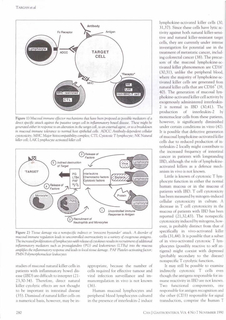
trigger or perpetuate mucnsal damage in Crohn 's disease and ulcerative colitis have not been defined. As yet, the target of the immune attac.k has not been determined, a lthough the epi thelia l cell is thought to be the likely candida te. The role of immune cytn· toxic mechanisms 111 epithe lial mJury has been a focus of investigat ion for almost 30 years. Two pathways of immune-mediated killing have been proposed. The direct pathway depicted in Figure I , or 'autoimmune' damage, 1,
based on the recognition of a select antigen on the target tissue by any of several effector mechanisms within the immune system. The mechanism of target killing could be either complementmediated lysis or anugen-triggereJ lysis. Cells could recognize antigens m
conJunction with ma1or histocompatibiltty antigens, such as cytotox ic T cells, or through direct antigen recognition hy a variety of effector cell types, ie, T cells, na tural killer cells, killer cells which become functional only after exposure to mterleukin-2, or lymphokine-activatcd killer cells.
A second pathway, also known as the 'innocent hystander' attack is illustrated in Figure 2. This pathway 1s
based on a defect in immunoregulauon a llowmg a cascade of uncontrolled immunologic events. There is no specific recognition of antigen on a target cell, but the release of 'cytotoxic' soluble
278 CAN J GASTROENTEROL VOL 4 No 7 NOVEMBER 1990

Cytotoxicite anticorps dependante clans les maladies inflammatoires de l'intestin
RESUME: Trente annees de recherche consacrees au role de l'activire cytotoxique anticorps dependante clans les lesions de la muqueuse caracteristiques des maladies inflammatoires de l'intestin (Mil) n'ont fourni que des donnees peu concluantes sur la rertinence des mecanismes cytotoxiques. Deux hypotheses ont ete proposees: que la destruction des cellules cibles est provoquee par la reconnaissance directe des antigcnes cibles par les cellules cytotoxiques qui declenchent a leur tour la lyse; et que la lyse survient par le biais d'un mecanisme indirect ou les cellules ne reconnaissent pas d'antigene specifique mais, apres activation non specifique, liberent !es cytokines en mesure de lyser la cible. Les auteurs ont erudie les deux hypotheses et examine le role des cellules T cytotoxiques et des facteurs cytotoxiques excretes par !es cellules T accivees lors de la destruction des cellules epitheliales. Ils ont note des concentrations elevees de cellules T cytotoxiques dans les lymphocytes du sang peripherique des patients porteurs de Mil et de maladie de Crohn surtout. Les cellules T cytotoxiques se trouvaient clans le sous-groupe de cellules T: Leu 7 + et CD8+, et ont ete deteccees utilisant la lyse declenchee par l'anti-CD3. L'accivite accrue des cellules T cytotoxiques etait egalement presence au sein de la muqueuse des patients porteurs de maladie de Crohn et de col ice ulcereuse. Les cibles specifiques de cette activite sonc a determiner. L'activacion des cellules T de la muqueuse par les anticorps et envers le recepteur de la cellule T (anti-CD3) clans la culture d'organe provenant de jejenum foetal normal provoque une enteropathie caracterisee par une atrophic villeuse et une hyperplasie des cellules crypciques. Cette meme modification est visible pres des ulceres aphteux chez les patients atteints <le maladie de Crohn. Les cytokines solubles provenanc des cellules T pourraient ecre responsables des Lesions observees dans Les deux modeles etudies. L'etude visait a determiner in vitro si !'activation des celluLes T coliques peut entrainer la Liberation des cytokines cycocoxiques et quelles sont les interactions des cytokines necessaires a la destruction des cellules epitheliales coliques. Selon !es resuluus, !es celluLes T humaines, activees par L'ajouc d'anti-CD3 et de cellules cibles, liberent le facteur de necrose tumorale alpha et !'interferon-gamma, tous deux necessaires pour la destruction optimale du HT-29. La liberation simultance de ces deux cytokines a proximite des cellules epitheliales au cours des reponses immunitaires pourrait jouer un role important clans !es lesions muqueuses des Mil. L'elaboration de modeLes animaux et de cultures a long terme des cellules epitheliaLes contribuera aux nombreux progres des recherches portant sur le role de la cycocoxicice ancicorps-dcpendante dans les MIi.
factors from cells. T cells reaching co multiple or select antigens in an uncontrolled fashion in the vicinity of the epithelial cell could cause lysis of such targets directly, as well as recruitment of other cell types and secretion of other factors.
Several investigators have reported lymphocyte-mediated cytotoxicity against epithelial cells in both Crohn's disease and ulcerative colitis (1-1 l) . Lymphocyte-mediated cyromxicity hc1s been shown using peripheral blood lymphocytes and intestinal mononuclear cells. Antibody-dependent cellular cytotox1ctty, in which lymphocytes are triggered by antibody directed against an antigen on epithelial cells, is the apparent
mechanism (3-8, 11 ). The cytotoxic reaction occurs only with colonic, and not small howel, epithelial cells - even in patients with Crohn's disease limited to the small intestine. Therefore, not all lesions of Crohn's disec1se can be characterized by this example. Interpretation of these findings is complicated due co potential limitations of some of the experiments, as freshly isolated epithelial cells are labile and unrel iahle as target cells ( 12). When cells are isolated from inflamed tissue, the problem may be compounded and comparisons inherently biased.
Erythrocytes coated with epithelial antigens have also been used to study epithelial autosensitization (13-14). When injected with adjuvant, soluble
CAN J GASTROENTEROL VOL 4 No 7 NOVEMBER 1990
Cytotoxlclty In IBD
epithelial cell antigen complexes isolated from rat and murine colon and intestine induced organ-specific inflammation (15). These antigens, bound to indicator cells (chicken erythrocytes), were found to be lysed by peripheral blood and lamina propria lymphocytes from patients with Crohn's disease and ulcerative colitis, but not in samples from control subjects ( l3). Erythrocytes labelled with purified kidney epithelial antigens were resistant co lysis; therefore, the cytotoxic reaction was specific for gutassociated epithelial antigens. Direct T cell cytotoxicity appeared to be involved when intestinal lamina propria lymphocytes were tested, although the mechanism of lysis was antibody-dependent cellular cytotoxicity when peripheral blood lymphocytes were the effector cells ( 13 ). The low levels of lysis observed, the unusual cytotoxicity curve, and the requirement in classical cytotoxic T cell killing that the target antigen be presented to the effector cell in association with the major histocompatibility complex ( MHC) ( 16, 17) render the significance of these findings uncerta in; reactions against epithelial antigens on the surface of chicken erythrocytes cannot be MHCrestricted. Several studies have highlighted antibody-dependent cellular cytotoxicity (8, 11, l 2) , yet wherher isolated intestinal mononuclear celb can mediate this cytoroxicity is controversial ( 18-22). The influence of enzymatic isolation procedures on lymphocyte and Fe receptor function might account for the difference in results (23-25).
Certain tumour and virally infected cells appear to be targets of natural killer cell cytotoxic activity. The peripheral blood of patients with Crohn's disease and ulcerative colitis has been shown to have decreased natural killer cell function, probably secondary to the inflammatory process (26-28). (le is known that plasma from the mesenteric vein of patients wiLh intestinal inflammation inhibits natural killer cells 129].) Because of the low numbers and low levels of activity in unfractionated cell preparations from normal or inflamed intesunes,
279
TARGANetal
Fe Receptor
\ Antibody
~--,::::::::::: Antigen
TARGET CELL
Figure 1) Mucosal immune effecwr mechanisms that hatie been proposed as possible mediators of a direct specific atlilCk against the putauve target cell in inflammatory bowel disease. These might be generated either in response to an altermion in the target cell, to an external agent, or to a breakdown m mucosal immune tolerance co normal host epithelinl cells. ADCC Annbody-dependenc cellular cycotoxicity ; MHC Ma1or hmocom/xwbihty complex; CTL Cytotoxic T lymphocyte, NK Natural killer cell; LAK Lymphocyte activated killer cell
RBsponse to Antigen
© RBCruitmBnt of NButrophils and MonocytBs
Figure 2) Tissue damage vin a nonspecific indirect or 'innocent bystander' arlilCk. A disorder of mucosa! 1mm1me regulation leads to uncontrolled overreact1v1ty to a variety of exogenous anagens. The increased /rroliferauon of lymphocytes with release of cywkines results in recruitment of additional mflammatory medtators such as prostaglandins (PG) and leukomenes (LTB4) mto the mucosa amplifies the m.flammatory response and leads w local tissue damage. PAF Platelec-ac11vating factor; PMN Polymorphonuclear leukocytes
studies of mucosa! natural killer cells in patients with inflammatory bowel disease (IBO) are difficult to interpret (21-23,30-34 ). Therefore, direct natural killer cytolytic effects are not thought to be important in intcrnnal disease (35 ). Dismissal of natural killer cells on a numerical baMs, however, may be in-
appropriate, because the number of cells required for effective tumour and viral infection surveillance and immunoregulation in vivo is not known (36).
Human mucosa! lymphocytes and peripheral blood lymphocytes cultured in the presence of interleukin-2 induce
lymphokine-activated killer cells (30, 31,3 7). Since these cells have lytic activity against both natural killer-sensitive and natural killer-resistant target cells, they are currently under intense investigation for potential use in the Lreatment of metastatic cancer, including colorectal cancer (38). The precursors of the mucosa! lymphokine-activated killer phenomenon are CD16' (30,31 ), unlike the peripheral blood, where the majority of lymphokine-activated killer cells arc generated from natural killer cells that are CD16+ (39, 40). The generation of mucosa! lymphokine-activated killer cell activity by exogenously administered interleukin-2 is normal in IBO (30,4l). The production of interleukin-2 hy mononuclear cells from these patients, however, is significantly diminished under certain conditions in vitro ( 42). Ir is possible that defective generation of mucosa[ lymphokine-activated killer cells due to reduced production of interleukin-2 locally might contribute to the increased frequency of mtestmal cancer m patients with longstanding IBO, although the role of lymphokineactivated killers as a defence mechanism m vivo is not known.
Little is known of cytotoxic T lymphocyte function in either the normal human mucosa or in the mucosa of patients with IBO. T cell cytotoxicity has been measured hy mitogen-induceJ cellular cyt0tox1city in culture. A decrease in T cell cytotoxicity in the mucosa of patients with IBO has been reported (21,32,43). The nonspecific cytocox1city induced by micogens, however, 1s probably distinct from chat of specifically in vivo-activated killer cells (31.44 ). It is possible that a subset of in vivo-activated cytotoxic T lymphocytes (possibly reactive to self anttgens) might coexist with depressed (probably secondary to the disease) norn,pecific T cytolyt1c function.
It may still be possible to examine indirectly cytotoxic T cells even though the antigens responsible for immune reactivity in LBD are not known. Two functional components, one responsible for antigen recogn1uon and the other (CD3) responsible for signal transducnon, comprise the human T
280 CAN J GASTROENTEROL VOL 4 NL) 7 NOVEMRER 1990

cell antigen receptor ( 17,4 5). Subsequent stimulation in vitro with monoclonal antibodies to the CDJ component of this receptor will trigger the cytolytic activity of the cell, if a cytotoxic T cell has already been primed by' antigen in v ivo. Therefore, this technique might be a useful indirect method for measuring and identifying in vivo-primed cytotoxic T lymphocytes when the antigen to
which they are reacting is not known (46). Using this method, the author has recently demonstrated the lytic function of freshly isolated cytotoxic T lymphocytes from uninflamed intestinal mucosa (47). He has shown that antiCD3-triggered peripheral blood lymphocytes have greater cytotoxic activity in patients with IBO than in normal subjects (48). The author has recently demonstrated that such cytotoxic T cell activity is also increased in the mucosa of patients with Crohn's disease and in many intestinal samples from patients with ulcerative colitis (49). The clonality of these cytotoxic T cells, as well as the targets against which they are d irected, are currently under investigation.
The innocent bystan<ler hypothesis implies the existence of some defect in the regulation of mucosa[ immunity leading to an uncontrolled response co a variety of exogenous antigens ( virus, bacteria or protein). Such an event would lead to the generation of effector cells within the efferent mucosa[ compartment or lamina propria, leading to release of cytokines and chemotactic factors an<l resulting in the recruitment of acute inflammatory cells (ie, neutrophils, eosinophils and mast cells). The arrival of these auxiliary effectors is associated with release of various additional mediators such as arachidonate metabolites and oxygen radicals which , in conjunction with cytokines, are locally injurious in a nonspecific fashion. This model hypothesizes a central role forcycokines in orchestrating epithelial damage.
Lymphocytes in the lamina propria halve been shown to be different from lymphocytes in the peripheral blood in regard to phenotype ( 4 7) and level of activation (50). Since these lamina
propria cells are constantly exposed to foreign antigens, it is important that response to challenge be thorough and yet restricted enough so that normal tissue is not damaged during an immune response. When this response becomes uncontrolled, mucosa[ damage occurs, such as char seen in IBO (12), celiac disease (51) and other inflammatory diseases of the intestine. A series of recent observations provides support for the central role of T cells and associated cycokines in nonspecific injury to epithelial cells as a possible mechanism of tissue injury in IBO. Organ culture studies of human fetal jejunum have elegantly shown that T cell activation using monoclonal antiboJies to the COJ component of the T cell receptor led to a marked enteropathy consisting of villous atrophy and crypt cell hyperplasia ( 52). Villous atrophy and crypt cell hyperplasia were also demonstrated in the mucosa adjacent to aphthous ulcerations in Crohn's disease (53 ).
Many different cytokines are produced by various immunocytes during an immune response. These cyrokines not only react with ocher cells of the immune system, but also affect the functions of nonimmune cell types, such as epithelial cells ( 54,5 5). It has been demonstrated chat there is increased MHC class II expression on epithelial cells overlying inflamed areas in IBO tissue samples (54). It has also been demonstrated in vitro that recombinant cytokines, interferon-gamma and tumour necrosis factor (TNF)alpha can induce such expression (54,55).
The author has recently performed studies to examine how increased numbers of 'activated' mucosa! T cells might contribute to damage, and whether such cyrokines play a role. Mucosa! T cells were polyclonally activated by incubation with anti-CDJ monoclonal antibody in combination with crosslinking via an Fe receptor-expressing target cell (K562). This treatment resulted in secretion of cytokines that were cytostatic to the human epithelial colon line HT-29. Subsequent experiments demonstrated chat these supematants were also
CAN J 0ASTR0ENTER0L VOL 4 No 7 NOVEMBER 1990
Cytotoxlclty In IBD
cytotoxic to the murine fibroblast line, L929 and HT-29. Little or no spontaneous secretion of these cyrokines occurred and incubation of lamina propria lymphocytes with anti-CD3 alone resulted in less secretion of cytotoxic/cyrosraric cyrokines compared to incubation with anti-COJ and K562 targets. These results suggest that crosslinking of the COJ receptor is probably required for optimal triggering of cytokine release.
Specific anticytokine monoclonal antibodies were used co determine what cyrokines were produced by the triggered lamina propria lymphocytes. TNF-beta, which one might expect to be produced by cytotoxic T cells, is either present in low quantities or not at all, as shown by the inability of antiTNF-beta to neutralize supernatant cycoroxicicy against L929. However, either anti-TNF-alpha or anti-interferon-gamma inhibited HF-29 cytotoxicity induced by lamina propria lymphocyte supcrnatants, demonstrating that both cycokines were secreted by the lamina propria lymphocytes, and both were required for optimal killing of HT-29. Further evidence was seen in experiments using recombinant TNFalpha and interferon-gamma, which a lso demonstrated chat both cytokines were required for optimal killingofHT-29.
TNF-alpha and interferon-gamma have been shown to be capable effector molecules in immune destruction of self tissues (56-58). Individually or in combination, TNF-alpha and interferon-gamma destroyed isolated betaislet cells in vitro (58). In addition, TNF-alpha has been identified in lesions from brain tissues of multiple sclerosis patients (59). Normal colonic epithelial cells may or may not have the same sensitivity to these cycokines as colonic cell lines. The ability of interferon-gamma to up-regulate a variety of cellular receptors including those for TNF-alpha suggests one mechanism responsible for synergism between these two cytokines (60). Sensitivity of targets to TNF-alpha or interferongamma may be related to expression of certain antigens or receptors on their cell surfaces or effects of viral products
281
TARGANeta/
(D More Senstive to Triggering @ Less Sensitive to Down ® Target Cell More
© Environment (LPS, Peptides)
@ Mitogens
Regulatory Signals Sensitive to Cytoklnes @ Cytoklnes
----- ~(IL-4, TGF-B, IL-?)
® Arachidonic Acid Metabolites (PGE 2 , LTB4 )
• @ Viral Infections
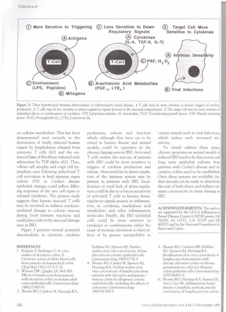
Figure 3) Three hy/>0thesized immune abnormalities in inflammatory hawel disease. 1 T cells may be more sensitive w known trrggen of C'ltokmt production. 2 T celL~ may be less sensmve w down-regulatory srgnals present m the mucosa/ comparonent. 3 The target cell may he more semruw ro rndw,dual effects or combmation.1 of cytokines. LPS L1po/>0lysaccharides; IL Interleukin; TGF Transforming growth factor; l'AF Platelet-acuvarmg faccor; PGE2 Prostaglandrn E2; L T84 Leukotricne 84
on cellular metabolism. This has been demonstrated most recently in the destruction of virally infecLed human targets by lymphokines released from cytotoxic T cells ( 61) and the enhanced I ~s of fibroblasts infected with adenovirus by TNF-alpha (62). Thus, villous cell atrophy and crypt cell hyperplas ia seen following polyclonal T cell acttvation in fetal Jejunum organ culture (53) or Crohn's disease epiLhelial change:. could reflecL d iffering responses of the two cell types co released cycokines. The present study suggests that human mucosa! T cells may be involved in indirect cytokinemediated damage to colonic mucosa during local immune reactions and could play a role in the mucosa! damage seen in IBO.
Figure 3 portrays several potential abnonnalit1es in cytotoxic cycokine
REFERENCES I. Perlman P, Broberger 0. In vitro
\tu<lies of ulcerative colitis. 11. Cytotoxic action of whrte blocxl cells from patients on human feral colon. J Exp Med I 963;117:717-33.
2. Watson OW, Quigley JA, Role RH. Effects of lymphocym, from patients with ulceratrve colrtrs on human adult colon epithelial cells. Gastroenterology 1966;5 I :985-93.
3. Shorter RG, Cardoza M, I luizinga KA,
282
production, release and function which, although they have yet to be tested in human disease and animal mo<leb, could be operative in the chronic damage seen in IBO. Activated T cells within the mucosa of patients with IBD coulJ be more sensitive to triggers of cytokine production and release. Abnormalities in down-regulation of the immune system may be present in patients with IBO. Insufficiency or total lack of down-regulation could be due to a loss in sensitivity of activated T cells to known downregulatory signals present in inflammation, 1e, cytokines, arachidonic acid metabolites and other inflammatory molecules. Finally, the l'BD epithelial cells could be more sensitive to cytokines or combinations either because of intrinsic alteration to their effects or by greater susceptibility to
ReMine SG, Spencer RJ. Further studies of rn vitro cytorox1crty of lymphocytes for colonic epithelial cells. Gastroenterology l 969;57:30-5.
4. Shorter RG, Cardoz M, Spencer RJ, Hu1zmga KA. Further studies of rn vitro cytoroxicity of lymphocytes from patients wrth ulcerative and granulomatous colitis for allogene1c colonic epithelial cells, including the effects of colectomy. Gastrocnterology 1969;56: 304-9.
certain stimuli such as viral infections, which induce \uch increased sens1tiv1Ly.
To dearly addres.~ these issue\ chronic sponLaneous animal models of induced IBO need co be discovered, and long term epithelial cultures from patients with Crohn's disease and ulcerative colitis need to be established. Once these systems are available, further inroads can be made to determine the role of both direct and indirect un, mune cytotox1city in tissue damage in
IBO.
ACKNOWLEDGEMENTS: The author1 are supported by the UCLA Inflammatory Bowel Disease Center (USPHS grants AM 36200, Al 15332, CA 37205 an<l DK 40057) and by the Natrona! Foun<l.1tion for lleim and Colitis.
5. Shorter RG, Cardoza MR, RcMmc SG, Spencer RJ, Hurzmga KA. Modification of rn vitro cytotoxrury of lymphocytes from patrcncs with chronic ulcerative colitis or chronic granulmnatous nilitr\ for allogenic colon epithelial cells. Gastmcnccrology l 970;58:692-8.
6. Shorter RG, I lu1zinga KA, Spencer RJ, Aas J, Guy SK. Inflammatory bowel dbcasc. Cyrophil1c amiho<ly and the cytotox1city nf lymphocyte~ for c1>lonrc
CAN J GA~TROENTEROL VOL 4 No 7 NlWEMBER 1990

Cytotoxlclty In IBD
cells in vitro. Dig Dis Sci 1971; 16:6 7 3- macory bowel disease. Isolation and 31. Shanahan F, Brogan M, Targan S. 80. preliminary characterization. Dig Dis Human mucosal cytotoxic effector
7. Shorter RO, Huizinga KA, Spencer RJ. Sci 1979;24: 705-1 7. cells. Gastrocntcrology 1987;92: 1951-7. A working hypothesis for the etiology 20. Chiba M, Shorter RG, Thayer WR, 32. Falchuk ZM, Barnhard E, Machado I. and pathogenesis of nonspecific inflam- Bartnik W, ReMinc S. K-cell activity Human colonic mononuclear cells: matory bowel disease. Dig Dis Sci in lamina propria lymphocytes from Studies of cytotoxic function. Gut !972;17:1024-32. the human colon. Dig Dis Sci 1981 ;22:290-4.
8. StoboJD, Tomasi TB, Huizinga KA, l 979;24:817-22. 33. T argan SR, Bnrvan L, Kendall R, Spencer RJ, Shorter'RG. In vitro 21. MacDermott RP, Franklin GO, Vimadalal S. Soll A. Isolation of spon-studies of inflammatory bowel disease: Jenkins KM, Kodner [J, Nash GS, taneous and interferon inducible Surface receptors of the mononuclear Weinrieb I]. Human intestinal natural killer like cells from human cells required to lyse allogeneic colonic mononuclear cells. I. Investigation of colonic mucosa: Lysis of lymphoid and epithelial cells. Gastroenterology antibody-dependent, lectm-imluced, aurologous epithelial target cells. Clin 1976;70:171-6. and spontaneous cell-mediated Exp lmmunol 1983;54:14-22.
9. Watson OW, Bolt RJ. Immune cytotoxic capabilities. Gastroenterology 34. Becken WL, Gundel M. St Andre-mechanisms in ulcerative colitis. In: 1980;78:47-56. Ukena S, McAuliffc T. In vitro cellu-Glass OBJ, ed. Progress in 22. Bland PW, Britton DC, Richens ER, lar cytotoxicity for a human colon Gastroenterology, Vol I. New York: Pledger JV. Peripheral, mucosa[, and cancer cell line by mucosal mono-Grune and Stratton, ! 968:391-411. tumor infiltrating components of eel- nuclear cells of patients with colon
JO. Kemler BJ, Alpert JE. Inflammatory lular immunity in cancer of the large cancer and other disorders. bowel disease: A study of cell-med iated bowel. Gut !981;22:744-51. Cancer 1985;55:1024-9. cytotoxicity for isolated human colonic 23. Bland PW, Richens ER, Britton DC, 35. Kett K, Rognum TO, Brandtzacg P. epithelial cells. Gut 1980;2 l :353-9. Lloyd JV. Isolation and purification of Mucosal subclass distribution of im-
11. Shorter RO, McGill DB, Bahn RC. human large bowel mucosa! lymphoid munoglohulin G-producing cells is dif-Cytotoxicity of mononuclear cells for cells: Effect of separation technique on fcrcnt m ulcerative colitis and Crohn's autologous colonic epithelial cells in functional characterbtics. Gut disease of the colon. Gastroenterology colonic diseases. Gastroentcrology 1979;20: 1037 -46. I 987;83:919-24. 1984;86: 13-22. 24. Chiba M, Bartnik W, ReMinc SG, }6. Gibson PR. NK cells, IBD and cancer.
12. Shanahan F. Inflammatory bowel dis- Thayer WR, Shorter RG. Human Gastroenterology 1986;90: 1314-5. ease. In: Targan SR, moderator. colonic intraepithelial and lamina 37. Hogan PG, Hapcl AJ, Doc WF. Lym-Immunology of Intestinal Diseases. propria lymphocytes: Cycocoxicity in phokine activated and natural killer Ann Intern Med 1987; 106:853-70. vitro and the potential effects of isola- cell activity 111 human mtestmal
13. Aronson RA, Cook SL, Roche JK. t1on methw on their functional proper- mucosa. J lmmunol 1985;135:1731-8. Sensitization to epithelial antigens in tics. Gut 1981 ;22: 177-86. 38. Rosenberg SA, Lotze MT, Muul LM, ,chronic~ ucosal inflammatory disease. 25. Gibson PR. Hermanowicz A, Verhaar et al. A progress report on the treat-I. Purification characterization and im- HJJ, Ferguson DJP, Lopez Bernal A, ment of 157 patients wirh advanced mune reactivity of murine epithelial Jewell DP. Isolation of intestinal cancer using lymphokine activated cell-associated components (ECAC). mononuclear cells: Factors released killer cells and mterleukin-2 ofh1gh J lmmunol 1983;131:2796-804. which may affect lymphocyte viability dose alone. N Engl J Med
14. RocheJK, Fiocchi C, Youngman K. and function. Gut l 985;26:60-8. 1987; 316:889-97. Sensitization to epithelial antigens m 26. Auer 10, Zeimer E, Sommer H. Im- 39. ltoh K, Tilden AR, Kumaga1 K, Ralch chronic mucosa! inflammatory disease. mune status in Crohn's d isease. V. CM. Leu 11 + lymphncytcs with Characterization of human intestinal Decreased m virro natural killer ce ll natural killer (NK) act1v1ty arc prccur-mucosa-derived mononuclear cells activity in peripheral blood. Clin Exp sms of recombinant interleukm-2 (rlL-reactive with purified epithelial cell- lmmunol 1980;42:41 -9. 2)-induced activated killer (AK) cells. associated components in vitro. J Clin 27. Bceken WL, MacPherson BR, Gundel J lmmunol 1985;134:802-7. Invest 1985; 75:522-30. RM, St Andre-Ukena S, Wood SG, 40. Phillips JH, L:inicr LL. Dissection of
15. RocheJK, Cook SR, Day ED. Cellular Sylvester DL. Depressed spontaneous the lymphokine activated killer cytotoxicity and gastrointestinal in- cell -mediated cyromx1c1ty in Crohn's phenomenon. Relative contribution of flammation in inbred rats: Induction disease. Clin Exp lmmunol penpheral blood natural killer cells and with gut organ-specific antigens. 1983;51:351 -8. T lymphocytes to cytolysis. J Exp Med Immunology 1981 ;44:489-97. 28. Ginsburg CH, Dambrauskas JT, Ault 1986; 164:814-25.
16. Zinkemagel RM, Doheny PC. H-2 KA, Falchuk ZM. Impaired natural 41. Kasugami K, Youngman KR, West compatibility requirement for T cell killer cell activity in patients with in- GA, Fiocch1 C. lntcsrinal immune mediated lysis of target cells infected flammatory bowel disease: Evidence for reactivity to mterleukin 2 differs with lymphocytic choriomcningitis a quantitative defect. Gastrocnterology among Crohn's disease, ulcerative virus. Different cytotoxic T cell ! 983;85:846-51. col it1s, and controls. Gastrocntcrolob'Y ,pecificities are associated with scruc- 29. Gibson PR, Verhaar HJJ, Sellby WS, 1989;97: 1-9. tures coded in H-2K or 11-20. J Exp Jewell DP. The mononuclear ce lls of 42. Fiocchi C, Hilfiker ML, Youngman Med 1975;141:1427-36. human mesenrcric blood, intestinal KR, Docrder NC, Finke JI l. Inter-
17. Acuto 0, Reinherz EL. The human T mucosa and mesenteric lymph nodes: leukin-2 activity of human intestinal cell receptor. Structure and function. Compartmentalization ofNK cells. mucosa I mononuclear cells. Decreased N EnglJ Med 1985;312:1100-11. Clin Exp lmmunol I 984;56:445-52. levels of mflammatt>ry bowel disease.
18. Clancy R, Pucci A. Absence of K cells 30. Fic)Cchi C, Tubbs RR, Youngman KR. Gastrocncerology l 984;86:734-42. in the human gut mucosa. Gut Human intestinal mucosal mono- 43. MacDcrmott RP, Bragdon MJ, Kodner 1978;19:273-6. nuclear cells exhibit lymphokine-ac- IJ, Bertovich MJ . Deficient cell-
19. Fiocchi C, Battisto JR, Fanner RG. nvared cell activity. Gastroenterology mediated cytotoxic1ty and hyporcspon-Gut mucosal lymphocytes m mflam- I 985;44:625-37. s1veness to 111terferon and m1mgc111c
CAN J GASTROENTEROL VOL 4 No 7 NOVEMBER 1990 283
TARGANetal
leccin activation by inflammatory bowel disease peripheral blood and intestinal mononuclear cells. Gastroenterology 1986;90:6- l l.
44. Shanahan F, Brogan M, Newman W, Targan S. K562 killing by Kand KL-2 responsive NK and T cells involves different post binding trigger mechanisms. J lmmunol 1986;137:723-6.
45. Weiss A, lmboded J, Hardy K, Manger B, T erhorsc C, Stobo J. The role of the T3/ancigen receptor complex in T-cell activation. Annu Rev lmmunol 1986;4:59 3-619.
46. Phillips J H, Lanier LL Lectin-dependent and anci-C03 induced cyrocoxicicy are preferentially mediated by peripheral blood cytotoxic T lymphocytes expressing Leu 7 antigen. J lmmunol 1986;l36:I579-85.
47. Shanahan F, Deem R, Nayersina R, Leman B, T argan S. Human mucosa! T cell cycotoxicity. Gascroenterology l 988;94:960-7.
48. Shanahan F, Leman B, Deem R, Niederlehner A, Brogan M, Targan S. Enhanced periphecal blood T cell cytocoxicicy in inflammatory bowel disease. Gastroenterology l 989;9:55-64.
49. Duerr R, Deem R, Landers C, Niederlehner A, Targan S, Shanahan F. Analysis of elevated mucosa! and peripheral cytotoxic T cell function in IBO. Gascroencerology 1989;96:A 132.
284
50. Pallone F, Fais S, Squarcia 0, Biancone L, Boirivant M. Activation of peripheral blood and intestinal lamina propria lymphocytes in Crohn's disease. In vivo state activation and in vitro response to stimulation as defined by the expression of early activation antigens. Gue l 987;28:745-53.
5 I. KagnoffNM, Austin RK, HubercJJ, BenardinJE, Kadarda DD. Celiac sprue: Correlation with murine T cell responses to wheat gliadin components. J Exp Med 1984;160:1544-57.
52. MacDonald TT, Spencer J. Evidence that activated mucosal T cells play role in the pathogenesis of enteropathy in human small intestine. J Exp Med 1988;167: 1341-9.
53. Encrican JH, Busuttil A, Ferguson A. Are the focal microscopic jejuna! lesions in Crohn's disease produced by a T cell mediated immune response? Scand J Gastroemerol l 987;22:1071 -5.
54. Selby WS, Janossy G, Mason DY, Jewell DP. Expression of HLA-DR antigens by colonic epithelium in inflammatory bowel disease. Clin Exp lmmunol I 983;53:614-8.
55. McDonald GB, Jewell OP. Class H antigen (HLA-DR) expression by intestinal epithelial cells in inflammatory bowel diseases of the colon. J Clin Pathol 1987 ;40:3 12-7.
56. Thompson CB, Lindsten T, Ledbetter
JA, et al. CD28 activation pathway regulates production of multiple T-cellderived lymphokines/cytokines. Proc Natl Acad Sci USA 1989;86:1333.
57. Sung SSSJ, Bjorndahl JM, Wang CY, Kao HT, Fu SM. Production of tumor necrosis facror/cachectin by human T cell lines and peripheral blood T lymphocytes stimulated by phorbol myriscace acetate and anci-C03 antibody. J Exp Med 1988;167:937.
58. Campbell IL, Iscaro A, Harrison LC. IFN-gamma and tumor necrosis factor· alpha cytotoxicicy co murine islet of Langerhans. J lmmunol 1988; 141 :2325-9.
59. Hofman FM, Hinton DR, Johnson K, Merrill JE. Tumor necrosis factor identified in multiple sclerosis brain. J Exp Med 1989; 170:607-12.
60. Pay CV, Kenmotsu N, Schoon RA, Leibson PH.Tumor necrosis factor and lymphotoxin secretion by human natural killer cells leads co antiviral cytotoxicity. J lmmunol 1988; 141: 1989-95.
61. Rosenblum MG, Donato NJ. Tumour necrosis factor alpha: A multifaceted peptide hormone. Cric Rev lmmunol 1989;9:21-44.
62. Duerksen-Hughes P, Wold WSM, Gooding LR. Adcnovirus El A render, infected cells sens1t1ve to cytolysis by tumor necrosis factor. J lmmunol 1989; 143:4193-200.
CAN J GASTROENTEROL VOL 4 No 7 NOVEMBER 1990

Submit your manuscripts athttp://www.hindawi.com
Stem CellsInternational
Hindawi Publishing Corporationhttp://www.hindawi.com Volume 2014
Hindawi Publishing Corporationhttp://www.hindawi.com Volume 2014
MEDIATORSINFLAMMATION
of
Hindawi Publishing Corporationhttp://www.hindawi.com Volume 2014
Behavioural Neurology
EndocrinologyInternational Journal of
Hindawi Publishing Corporationhttp://www.hindawi.com Volume 2014
Hindawi Publishing Corporationhttp://www.hindawi.com Volume 2014
Disease Markers
Hindawi Publishing Corporationhttp://www.hindawi.com Volume 2014
BioMed Research International
OncologyJournal of
Hindawi Publishing Corporationhttp://www.hindawi.com Volume 2014
Hindawi Publishing Corporationhttp://www.hindawi.com Volume 2014
Oxidative Medicine and Cellular Longevity
Hindawi Publishing Corporationhttp://www.hindawi.com Volume 2014
PPAR Research
The Scientific World JournalHindawi Publishing Corporation http://www.hindawi.com Volume 2014
Immunology ResearchHindawi Publishing Corporationhttp://www.hindawi.com Volume 2014
Journal of
ObesityJournal of
Hindawi Publishing Corporationhttp://www.hindawi.com Volume 2014
Hindawi Publishing Corporationhttp://www.hindawi.com Volume 2014
Computational and Mathematical Methods in Medicine
OphthalmologyJournal of
Hindawi Publishing Corporationhttp://www.hindawi.com Volume 2014
Diabetes ResearchJournal of
Hindawi Publishing Corporationhttp://www.hindawi.com Volume 2014
Hindawi Publishing Corporationhttp://www.hindawi.com Volume 2014
Research and TreatmentAIDS
Hindawi Publishing Corporationhttp://www.hindawi.com Volume 2014
Gastroenterology Research and Practice
Hindawi Publishing Corporationhttp://www.hindawi.com Volume 2014
Parkinson’s Disease
Evidence-Based Complementary and Alternative Medicine
Volume 2014Hindawi Publishing Corporationhttp://www.hindawi.com