igg-mediated cytotoxicity - proceedings of the national academy of
TRANSCRIPT

Proc. Natl. Acad. Sci. USAVol. 93, pp. 6796-6801, June 1996Medical Sciences
Expression of calbindin-D28K in motoneuron hybrid cells afterretroviral infection with calbindin-D28K cDNA preventsamyotrophic lateral sclerosis IgG-mediated cytotoxicity
(calcium-binding proteins/motoneuron degeneration)
BAO-KUAN Ho*, MARIA E. ALEXIANU*, Luis V. COLOM*, A. HABIB MOHAMED*, FERNANDO SERRANOt,AND STANLEY H. APPEL*-Departments of *Neurology and tMolecular and Human Genetics, Baylor College of Medicine, Houston, TX 77030
Communicated by Salih J. Wakil, Baylor College of Medicine, Houston, TX, February 27, 1996 (received for review October 30, 1995)
ABSTRACT Calbindin-D28K and/or parvalbumin appearto influence the selective vulnerability of motoneurons inamyotrophic lateral sclerosis (ALS). Their immunoreactivityis undetectable in motoneurons readily damaged in humanALS, and in differentiated motoneuron hybrid cells [ventralspinal cord (VSC 4.1 cells)] that undergo calcium-dependentapoptotic cell death in the presence ofALS immunoglobulins.To provide additional evidence for the role of calcium-bindingproteins in motoneuron vulnerability, VSC 4.1 cells wereinfected with a retrovirus carrying calbindin-D28K cDNAunder the control of the promoter of the phosphoglyceratekinase gene. Differentiated calbindin-D28K cDNA-infectedcells expressed high calbindin-D28K and demonstrated in-creased resistance to ALS IgG-mediated toxicity. Treatmentwith calbindin-D28K antisense oligodeoxynucleotides, whichsignificantly decreased calbindin-D28K expression, renderedthese cells vulnerable again to ALS IgG toxicity.
Despite extensive investigations of superoxide dismutase mu-tations in familial amyotrophic lateral sclerosis (ALS) andexcitotoxicity and autoimmunity in sporadic ALS, our under-standing of the factors dictating selective vulnerability ofmotoneurons are incompletely understood (1-5). Calbindin-D28K and/or parvalbumin have been implicated in ALS patho-genesis since immunoreactivity for these calcium bindingproteins is absent in neurons early and severely affected inALS such as ventral horn motoneurons and is present inneurons late or infrequently affected such as oculomotorneurons or Onufs neurons (6, 7). In addition, motoneuronhybrid cells that are selectively killed by ALS IgG have little orabsent immunoreactivity for calbindin-D28K and parvalbuminwhereas cells not affected by ALS IgG, including undifferen-tiated motoneuron hybrid cells and the parent neuroblastomaN18TG2 cells, have high levels of calbindin-D28K and parval-bumin (7).We report herein that retroviral infection of motoneuron
hybrid cells with calbindin-D28K cDNA induces increasedcalbindin-D28K expression that is maintained after cAMPdifferentiation and prevents ALS IgG-induced toxicity. Con-versely, inhibition of calbindin-D28K expression by treatment ofcalbindin-D28K-infected ventral spinal cord (VSC) 4.1 cells(I-VSC 4.1 cells) with calbindin-D28K antisense oligode-oxynucleotides restores vulnerability to ALS IgG-mediatedtoxicity.
MATERIALS AND METHODSConstruction of pStMCS-PCalb for Retrovirus Packaging.
The calbindin-D28K cDNA was cloned from rat brain as
follows: total RNA was extracted from rat brain by theguanidinium/cesium chloride method (8). Poly(A)+ RNA waspurified by oligo(dT) cellulose (Collaborative Research, Bed-ford, MA) affinity chromatography. The entire coding se-quence (786 bp) of calbindin-D28K was synthesized from thepoly(A)+ RNA by reverse transcriptase (GIBCO/BRL)-polymerase chain reaction (PCR, Perkin-Elmer/Cetus) andcloned into the BamHI site of plasmid pGEM-3Z (Promega).The sequences of the oligomers for PCR are TTCGGATCC-ATGGCAGAATCCCACCTGCA for the 5' forward primerand AAAGGATCCTAGTTGTCCCCAGCAGAGAGAATfor the 3' reverse primer (the oligomers were synthesized at theDepartment of Cell Biology, Baylor College of Medicine). Aclone containing the correct complete calbindin-D28K se-quence was identified by dideoxynucleotide sequencing (9).The calbindin-D28K cDNA was obtained from the clone
pGEM-calbindin by BamHI digestion and cloned into theSmaI site of pPGKbpA (derived from pPGKneo) after fill-ing-in of ends by treatment with the Klenow fragment ofDNApolymerase I (Promega). As shown in Fig. 1, this enabledinsertion of the calbindin-D28K cDNA downstream of the PGKpromoter/enhancer sequence and upstream of a polyadeny-lylation signal of the growth hormone gene. Orientation of thecDNA was determined by digestion with EcoRI. The resultingconstruct was digested with XhoI, treated with Klenow poly-merase, and subsequently, digested with HindIII. The resultingmixture of fragments were ligated into Stul- and HindlIl-digested vector pStMCS. The plasmid pStMCS (kindly pro-vided by John Belmont, Baylor College of Medicine, Houston,TX) is a retroviral vector, containing Moloney murine leuke-mia virus long terminal repeats. The orientation of the calbi-ndin expression cassette is opposite to that of the long terminalrepeat sequences in the final pStMCS-PGK-calbindin (pSt-MCS-PCalb) plasmid (Fig. 1).
Production of Recombinant Retrovirus and Infection ofVSC 4.1 Cells with Calbindin-D28K-Expressing Retrovirus.The plasmid pPGKneo and the ecotropic virus packing cellline GP+E86 were provided by John Belmont. Cells werecultured in Dulbecco's modified Eagle's medium (DMEM)with 10% fetal calf serum in 10% C02/90% air.Twenty micrograms of pStMCS-PCalb and 1 ,g of pPGK-
neo were cotransfected into 3.5 x 10⁶ ecotropic packing GP +E86 cells by electroporation (Invitrogen electroporator II) atoofl, 1000 AF, and 280 V. Transfection efficiency varied from7 to more than 200 neomycin-resistant clones per electropo-ration. Several hundred neomycin (GIBCO/BRL, 4 ,ug/ml)-resistant clones were obtained. The recombinant virus pro-
Abbreviations: ALS, amyotrophic lateral sclerosis; ODN, oligode-oxynucleotides; PGK, phosphoglycerate kinase; VSC, ventral spinalcord.*To whom reprint requests should be addressed at: Department ofNeurology, Baylor College of Medicine, 6501 Fannin, NB302, Hous-ton, TX 77030.
6796
The publication costs of this article were defrayed in part by page chargepayment. This article must therefore be hereby marked "advertisement" inaccordance with 18 U.S.C. §1734 solely to indicate this fact.

Proc. Natl. Acad. Sci. USA 93 (1996) 6797
FIG. 1. Construction of retroviral vector for expression of thecalbindin-D28K gene under the regulation of the promoter of thephosphoglycerate kinase (PGK) gene.
duced by transient expression of the infected GP + E86 cellswas harvested after 48 h from conditioned medium by filtra-tion through 0.45-,um (pore size) Millipore filters. As this virusdoes not carry a selectable marker, we amplified the dilutedsupernatant by ping-pong infection in a mixture of 50% GP +envAml2 and 50% GP + E86 cells and titrated the virus byscoring the wells positive to retroviral sequences by PCR. TheVSC 4.1 cells were infected by exposure to virus with a titer of106 virus particles per ml for 4 h in the presence of Polybreneat 4 ,Lg/ml (to neutralize cell surface charge). Among themultiple PGK-calbindin retrovirus-infected cell lines, we havechosen the line named I-VSC 4.1, since after differentiationwith dibutyryl cAMP and aphidocolin, the level of calbindinimmunoreactivity did not decrease as it does in noninfectedVSC 4.1 cells. We have used the I-VSC 4.1 cells in allsubsequent experiments.
Evidence of Calbindin-D28K cDNA and mRNA in VSC 4.1and I-VSC 4.1 Cells. Southern blot analysis. Genomic DNAfrom VSC 4.1 and I-VSC 4.1 cells were extracted by phenol/chloroform, followed by RNase treatment. For Southern blotanalysis, gDNA was digested overnight at 37°C with HindIIIand XbaI and separated on an 1% agarose gel. The DNA wasdenatured and transferred to nitrocellulose membrane. Afterfixing and blocking, the filter was probed with 32p calbindin-D28K cDNA, labeled by nick-translation, overnight at 65°C in6x SSC/1 x Denhardt's solution. Washes were carried out atroom temperature for four 20-min periods in 2x SSC and for30 min at 65°C in 0.5 x SSC. Autoradiography was performedovernight at -70°C.Northern blot analysis. The mRNA of the VSC 4.1 and I-VSC
4.1 cells was isolated as mentioned before, but with one more
cycle of oligo(dT) affinity chromatography. For Northern blotanalysis, 15 ,ug of mRNA was electrophoresed on a 1.2%formaldehyde agarose gel and transferred to a nylon filter. Thefilter was probed with 32p calbindin-D28K cDNA at 42°C in 50%formamide/6x SSPE. Filters were washed for four 30-minperiods at room temperature in 2x SSC and for one 30-minperiod in 0.5x SSC at 60°C. The autoradiography exposuretime was 2 days at -70°C.
In situ hybridization. The presence of calbindin-D28K mRNAin the motoneuron hybrid cells with or without introduction ofthe calbindin-D28K gene was also documented by nonradioac-tive in situ hybridization (10, 11) using fluorescein-labeledoligodeoxynucleotide (ODN) probes (Genosys, The Wood-lands, TX) and the oligocolor kit from Amersham. The ODNprobes used in our assay consisted of 33 nt complementary to
the coding region for amino acids 31-41 of the calbindin-D28Kgene sequence (12). The 5'-3' sequence of the antisense ODNused was CTGGATCAAGTTCTGCAGCTCCTTTCCT-TCCAG. The hybridization was performed by the instructionsin the Amersham kit. The in situ buffer provided by Amershamcontained 50% formamide, 2x SSC, 1 x Denhardt's solution,herring testes DNA (300 gxg/ml), and an enhancement com-pound. Fifty microliters of hybridization solution with a finalprobe concentration of 50 ng/ml was applied on each coverslipand the incubation was performed for 5 h at 37°C. The slideswere washed in lx SSC/0.1% SDS at room temperature andthen in 0.2x SSC/0.1% SDS at 37°C. Signal was detected witha biotinylated anti-fluorescein antibody (Molecular Probes)followed by avidin-biotin complex (Vector Laboratories) and3,3'-diaminobenzidine (Sigma).
Control experiments were carried out to assure the speci-ficity of the probes and the appropriateness of the hybridiza-tion conditions and detection methods as follows: To controlfor the specificity of the effect of calbindin-D28K expression, weinvestigated the levels of mRNA for a cytoskeletal protein(heavy-chain neurofilament) (13) and for another calcium-binding protein normally expressed in VSC 4.1 cells (parval-bumin) (14). Experimental procedures performed with fluo-rescein-labeled sense ODN, complementary to the antisenseprobes for calbindin-D28K, parvalbumin, and neurofilament,respectively, or experiments without the hybridization buffer,or omitting the antibody against fluorescein resulted in com-plete absence of any signal.
Cell Culture Conditions and Assessment of the EffectsProduced by Calbindin-D28K Gene Transfection. General cellgrowth, cAMP, and aphidocolin differentiation conditions aswell as the preparation for survival/cytotoxicity assays andimmunohistochemical experiments were performed as de-scribed in our previous studies (7, 15, 16).To reverse the effect of calbindin-D28K gene transfection,
the I-VSC 4.1 cells were treated after differentiation with 20,iM calbindin-D28K antisense oligodeoxynucleotides (Geno-sys) for 24 h before further treatments or procedures. Theantisense ODN sequence used was 5'-CAGGTGTTCTGC-CAT-3', which starts from the putative translation initiationcodon according to the published calbindin-D28K gene se-quence (12). In all experiments, sister cultures were treatedwith a complementary calbindin-D28K sense ODN probe at 20,uM. Neither the antisense probe nor the sense probe demon-strated any toxic effects on the survival of motoneuron hybridcells. Furthermore, cultures treated with calbindin-D28K senseODN did not show changes in immunoreactivity for calbindin-D28K. These results are consistent with other reports usingODN to modulate specific protein expression in neuronal-typetissue culture experiments (17-19).The quantitation of calbindin-D28K in the motoneuron hy-
brid cells was determined by a sensitive immuno-sandwichassay as described by Zhu et al. (20). Briefly, the monoclonalanti-calbindin-D28K antibody (Sigma) was adsorbed onto sili-cone tube pieces; the same antibody was chemically crosslinkedto j3-galactosidase used as a reporter enzyme (21). The bivalentantigen was sandwiched between the antibody adsorbed to thesilicone piece and the antibody crosslinked to /3-galactosidase(22). The recorded enzyme activity measures the level ofantigen after deducting the background activity obtained inthe absence of antigen. The optical density values obtainedwithout cell extract or purified calbindin-D28K were taken asbackground values in each experiment and were deductedfrom the values obtained for our specific experimental probes.
Electrophysiological recordings were made on the stage ofa World Precision Instruments (WPI) inverted microscope.The extracellular fluid contained 40 mM BaCl2, 107.5 mMtetraethylammonium chloride, and 10 mM Hepes. Tetrodo-toxin (1 mM) was added to inhibit sodium currents. Patchrecordings were performed with low-resistance (1-2 MW)
Mvedical Sciences: Ho et al.
Proc. Natl. Acad. Sci. USA 93 (1996)
pipettes pulled from borosilicate glass (WPI), using a Flaming/Brown puller (P.80/PC, Sutter). Whole-cell patch recordingswere made from the cells by using patch electrodes filled witha solution containing 120 mM cesium methanesulfonate, 4.5mM MgCl2, 9 mM glucose, 9 mM EGTA, 9 mM Hepes, and4.5 mM ATP. Patch electrodes and extracellular solutions wereadjusted to pH 7.3. Signals were recorded using a patch-clampamplifier (PC-SOlA, Warner) and a laboratory computer(486/66) equipped with a Labmaster A/D converter. Voltagepulses of 50- or 500-ms duration were applied from a holdingpotential (Vh) of -70 mV, at frequencies of 0.1-0.3 s-1. Sealresistances and capacitative transients were monitored duringexperiments by applying small (5 mV) voltage pulses super-imposed on the command potential. Current-voltage (I-V)relationships were obtained by plotting the peak currentevoked by a voltage pulse against the pulse potential. Digitalleak subtraction was carried out prior to current-voltageanalysis by scaling and subtraction of responses to small 5-10mV hyperpolarizing pulses.
For toxicity studies uninfected and infected differentiatedmotoneuron hybrid cells, in triplicate wells for each immuno-globulin used in each experiment, were treated within 24 hafter plating with IgG at 0.2-0.5 mg/ml purified from patientswith ALS, as reported (23). The sixALS IgGs used in this studywere chosen at random from our stock of immunoglobulins.The mean age of the patients was 63 ± 11 years, and all patientsfulfilled clinical and laboratory criteria for ALS. Cell survivalwas assessed after incubation for 48 h with ALS IgG bycounting five contiguous 1-mm2 fields predetermined andmarked after plating at day zero. This direct counting assay hasbeen well correlated with vital staining using fluoresceindiacetate and propidium iodide and with the lactate dehydro-genase release assay (15).
RESULTSExpression of Calbindin-D2UK cDNA. VSC 4.1 cells were
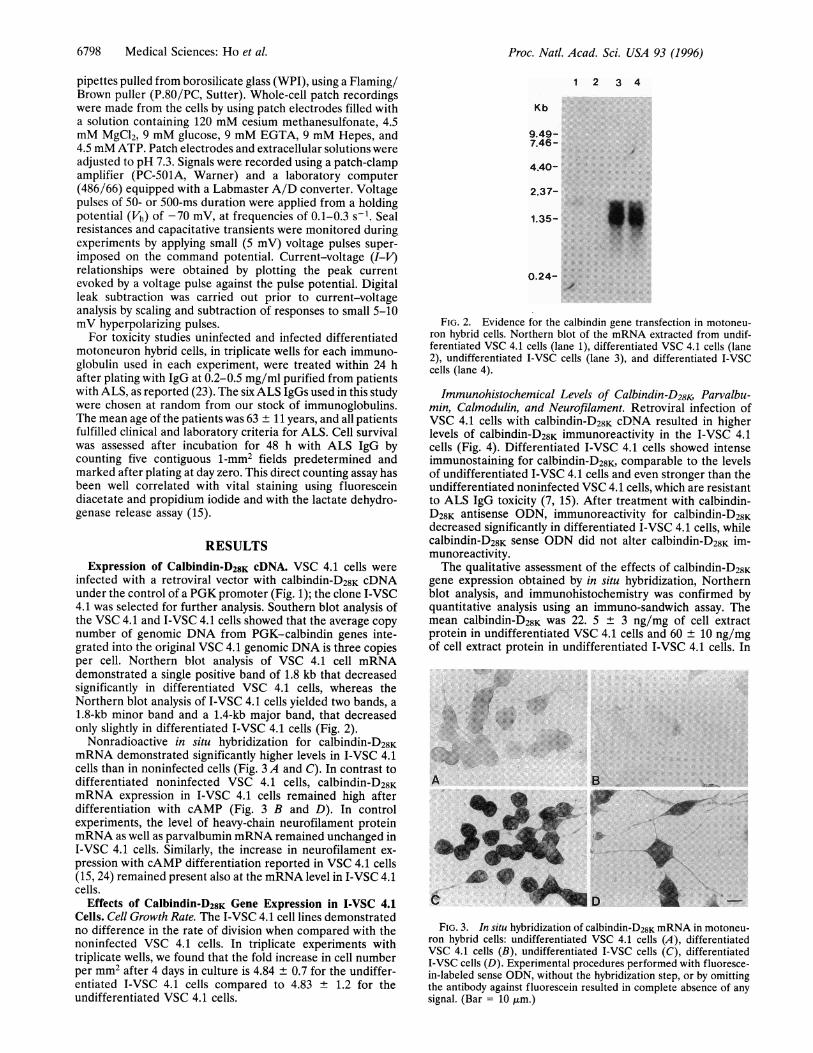
infected with a retroviral vector with calbindin-D28K cDNAunder the control of a PGK promoter (Fig. 1); the clone I-VSC4.1 was selected for further analysis. Southern blot analysis ofthe VSC 4.1 and I-VSC 4.1 cells showed that the average copynumber of genomic DNA from PGK-calbindin genes inte-grated into the original VSC 4.1 genomic DNA is three copiesper cell. Northern blot analysis of VSC 4.1 cell mRNAdemonstrated a single positive band of 1.8 kb that decreasedsignificantly in differentiated VSC 4.1 cells, whereas theNorthern blot analysis of I-VSC 4.1 cells yielded two bands, a1.8-kb minor band and a 1.4-kb major band, that decreasedonly slightly in differentiated I-VSC 4.1 cells (Fig. 2).
Nonradioactive in situ hybridization for calbindin-D28KmRNA demonstrated significantly higher levels in I-VSC 4.1cells than in noninfected cells (Fig. 3 A and C). In contrast todifferentiated noninfected VSC 4.1 cells, calbindin-D28KmRNA expression in I-VSC 4.1 cells remained high afterdifferentiation with cAMP (Fig. 3 B and D). In controlexperiments, the level of heavy-chain neurofilament proteinmRNA as well as parvalbumin mRNA remained unchanged inI-VSC 4.1 cells. Similarly, the increase in neurofilament ex-pression with cAMP differentiation reported in VSC 4.1 cells(15, 24) remained present also at the mRNA level in I-VSC 4.1cells.
Effects of Calbindin-D28K Gene Expression in I-VSC 4.1Cells. Cell Growth Rate. The I-VSC 4.1 cell lines demonstratedno difference in the rate of division when compared with thenoninfected VSC 4.1 cells. In triplicate experiments withtriplicate wells, we found that the fold increase in cell numberper mm2 after 4 days in culture is 4.84 ± 0.7 for the undiffer-entiated I-VSC 4.1 cells compared to 4.83 ± 1.2 for theundifferentiated VSC 4.1 cells.
1 2 3 4
Kb
9.49-7.46-
:/
4.40-
2.37-
1.35-
0.24-
FIG. 2. Evidence for the calbindin gene transfection in motoneu-ron hybrid cells. Northern blot of the mRNA extracted from undif-ferentiated VSC 4.1 cells (lane 1), differentiated VSC 4.1 cells (lane2), undifferentiated I-VSC cells (lane 3), and differentiated I-VSCcells (lane 4).
Immunohistochemical Levels of Calbindin-D28K Parvalbu-min, Calmodulin, and Neurofilament. Retroviral infection ofVSC 4.1 cells with calbindin-D28K cDNA resulted in higherlevels of calbindin-D28K immunoreactivity in the I-VSC 4.1cells (Fig. 4). Differentiated I-VSC 4.1 cells showed intenseimmunostaining for calbindin-D28K, comparable to the levelsof undifferentiated I-VSC 4.1 cells and even stronger than theundifferentiated noninfected VSC 4.1 cells, which are resistantto ALS IgG toxicity (7, 15). After treatment with calbindin-D28K antisense ODN, immunoreactivity for calbindin-D28Kdecreased significantly in differentiated I-VSC 4.1 cells, whilecalbindin-D28K sense ODN did not alter calbindin-D28K im-munoreactivity.The qualitative assessment of the effects of calbindin-D28K
gene expression obtained by in situ hybridization, Northernblot analysis, and immunohistochemistry was confirmed byquantitative analysis using an immuno-sandwich assay. Themean calbindin-D28K was 22. 5 ± 3 ng/mg of cell extractprotein in undifferentiated VSC 4.1 cells and 60 ± 10 ng/mgof cell extract protein in undifferentiated I-VSC 4.1 cells. In
... ..... ... .... ... :.
A ~~~~B
FIG. 3. In situ hybridization of calbindin-D28K mRNA in motoneu-ron hybrid cells: undifferentiated VSC 4.1 cells (A), differentiatedVSC 4.1 cells (B), undifferentiated I-VSC cells (C), differentiatedI-VSC cells (D). Experimental procedures performed with fluoresce-in-labeled sense ODN, without the hybridization step, or by omittingthe antibody against fluorescein resulted in -complete absence of anysignal. (Bar = 10 Aim.)
6798 Medical Sciences: Ho et al.

Proc. Natl. Acad. Sci. USA 93 (1996) 6799
M...i5 ...'.........
-,1, ,S ..
FIG. 4. Immunohistochemical expression of calbindin-D28K in un-
differentiated VSC 4.1 cells (A), undifferentiated I-VSC cells (B),differentiated VSC 4.1 cells (C), differentiated I-VSC cells (D), and
differentiated I-VSC cells treated with calbindin-D28K antisense oli-godeoxynucleotide (E). Specificity of the monoclonal anti-calbindin-D28K antibody used in these experiments has been described (7, 25, 26).(Bar = 15 ,tm.)
VSC 4.1 cells, cAMP differentiation induced a 54.3 ± 4%decrease in calbindin-D28K expression (Fig. 5), while retroviralinfection resulted in almost three times (283.1 ± 9%) more
calbindin-D28K protein expression in infected cells comparedto the undifferentiated noninfected VSC 4.1 cells. Further-more, incubation of differentiated I-VSC 4.1 cells with calbi-ndin-D28K antisense ODN induced a 55.7 ± 11% decrease incalbindin-D28K protein expression compared with the undif-ferentiated I-VSC 4.1 cells and a 40 ± 5% decrease comparedto differentiated I-VSC 4.1 cells. Thus, both immunohisto-chemical methods and quantitative immunoassays yielded thesame pattern of calbindin-D28K protein expression in differentcell types.
Immunoreactivity for other cytoplasmic proteins with struc-tural or functional roles in motoneuron cells, such as neuro-filaments or parvalbumin, and calmodulin remained un-
changed after calbindin-D28K gene transfection (data notshown). Furthermore, the cAMP-inducible levels of cholineacetyltransferase activity [evaluated using Fonnum's method
Az
0
uz
C/OU.
LLW
0
8':
4"-
2":
B
cuesroro tigd cobs ca ibn dh s ons (OON
FIG. 5. Quantitative immunoassay for calbindin-D28K expressionshows that differentiated VSC 4.1 cells have significantly decreasedlevels of calbindin-D28K protein (A), whereas in calbindin-D28K ret-rovirus-infected cells, a significant decrease in calbindin-D28K isachieved only with calbindin-D28K antisense ODN treatment (B).Plotted values represent the ratio of calbindin-D28K content (in ng ofcalbindin per mg of soluble cell extract) in differentiated versusundifferentiated cells. Raw values (in ng) were calculated by convert-ing the fluorescence intensity of ,B-galactosidase activity in the im-muno-sandwich assay using the standard graph generated in thepresence of purified rat kidney calbindin-D28K as control antigen.
ALS PATIENTS 1 - 6=II VSC 4.1ce1Is; I-VSC 4.1 cdIs;F:2 I-VSC 4.1 cells + cbirdin ansemODN
FIG. 6. Quantitative effect of immunoglobulins from patients withALS on differentiated motoneuron hybrid cells with different levels ofcalbindin-D28K expressiQn. Data represent changes in cell numberafter a 2-day IgG exposure, expressed as a percentage of total cellnumber in untreated cultures. Each bar represents the mean ± SD forcell survival in described cell populations, averaging data obtainedfrom triplicate coverslips for each experiment with each IgG. Cellcounts for each coverslip were obtained from five to seven contiguous1-mm2 microscopic fields.
(27)] in undifferentiated VSC 4.1 cells (15) were also notinfluenced by the calbindin-D28K gene transfection (data notshown).
Effect of Calbindin Transfection on Calcium Currents. Cal-cium currents in differentiated VSC 4.1 and I-VSC 4.1 cellswere measured using 40 mM barium as the charge carrier, asused in our studies of the motoneuron hybrid cells (28). Sincecell size and shape were variable in both VSC 4.1 and I-VSC4.1 cell lines differentiated with cAMP, we only recordedcurrents from well differentiated cells with a somatic diameterof 30-40 ,um to minimize differences due to cell size andshape. In both cell lines, inward currents activated near apotential of -40 mM, displayed peak amplitudes at potentialsof +20 mV, and exhibited rapid voltage-dependent inactiva-tion. No significant differences were found between the am-plitudes and voltage range of activation of calcium currents inVCS 4.1 and I-VSC 4.1 cell lines (data not shown). Furtherstudies using calcium as the charge carrier are currently inprogress.
Susceptibility to ALS IgG Toxicity. Differentiated I-VSC 4.1cells expressing high levels of calbindin-D28K were significantlyresistant to toxicity of ALS IgG (Fig. 6), which had beenpreviously demonstrated to induce cell death of differentiatedVSC 4.1 cells (7, 15). After 48 h, the mean survival ofdifferentiated I-VSC 4.1 cells incubated with ALS IgG was98.1 + 13.1% compared to PBS-treated cultures. The sameIgGs induced a 42.7 + 13.2% cell loss in differentiated VSC 4.1cells with very low to absent levels of immunoreactive calbi-ndin-D28K (7). Treatment of differentiated I-VSC cells for 24 hwith 20 ,uM calbindin antisense ODN produced a markedlyincreased ALS IgG-mediated toxicity concomitant with asignificant decrease in immunoreactivity for calbindin (Figs. 4and 6). The mean loss of cells was 33.9 ± 15.2% in differen-tiated I-VSC cells treated with calbindin-D28K antisense ODN.Statistical analysis of the ALS IgG-mediated toxicity by usingone-way ANOVA shows a significance ofP < 0.001 when thesurvival of differentiated I-VSC cells is compared with thesurvival of differentiated motoneuron cells with low levels ofcalbindin-D28K expression (either differentiated VSC cells ordifferentiated I-VSC cells treated with calbindin-D28K anti-sense ODN). There is no significant difference between thetwo differentiated cell populations in which the levels ofcalbindin-D28K expression is very low or even absent.
DISCUSSIONThe present findings demonstrate that calbindin-D28K appearsto influence the susceptibility of motoneuron hybrid cells to
Medical Sciences: Ho et al.
T

Proc. Natl. Acad. Sci. USA 93 (1996)
ALS IgG-mediated toxicity. Differentiated motoneuron hy-brid cells infected with retrovirus carrying calbindin-D28KcDNA express high levels of calbindin-D28K and are resistantto ALS IgG-mediated toxicity. Treatment of these differenti-ated infected cells with calbindin-D28K antisense ODN signif-icantly decreased the expression of calbindin-D28K and en-hanced the sensitivity to ALS IgG-mediated toxicity. We havepreviously documented that ALS IgG can selectively kill cellsthat have very low to absent levels of calcium-binding proteins(calbindin-D28K or parvalbumin) (7) through a mechanism thatis calcium-dependent and possibly mediated through voltage-gated calcium channels (15). ALS IgG can also increasecalcium currents in motoneuron hybrid cells (28) and inducesa transient increase in intracellular calcium (24) that correlateswith subsequent apoptotic cell death (16).The data obtained with our motoneuron cell line correlate
well with the findings in human central nervous system wheremotoneurons that degenerate early in ALS have no calbindin-D28K and/or parvalbumin expression whereas motoneuronsthat are infrequently or only very late affected have significantimmunoreactivity for these calcium-binding proteins (6, 7).The importance of calcium homeostasis in the pathogenesis ofmotoneuron injury in ALS is supported by the in vivo passivetransfer experiments (29), which document that injection ofALS immunoglobulins induces an increase in synaptic vesiclesat motor end plates and in terminals synapsing on motoneuronperikarya as well as an increase in calcium-containing precip-itates in synaptic terminals and in the cell body of motoneuronsthat lack calcium-binding proteins. In contrast, other neuronalpopulations such as dorsal horn neurons and Purkinje cells,known to be rich in calcium-binding proteins, do not show suchalterations.Our prior studies documented that the interaction of ALS
IgG with neuronal-type calcium channels induces a transientincrease in intracellular calcium, which leads to apoptotic celldeath (15, 16, 24). Such data suggest an important role forcalcium channels in this process. However, these neuronal-type(N-type, P-type, Q-type) calcium channels are not confined tomotoneurons but are present on other types of neurons andcannot, per se, explain the selective motoneuron vulnerability.For example, ALS IgG increases P-type calcium currents inisolated Purkinje cells and in P-type channel protein recon-structed in lipid bilayers (30), but Purkinje cells are notaffected in ALS. The explanation may reside in the fact thatPurkinje cells possess high levels of calbindin-D28K and par-valbumin (6, 7, 31). Furthermore, increased calcium current aswell as increased calcium entry due to ALS IgG was also notedin neuroblastoma N18TG2 cells (24), but these cells possesshigh levels of calbindin-D28K and are not vulnerable to ALSIgG (7). Thus, the ability of ALS IgG to interact with calciumchannels and increase calcium entry may not be sufficientperse to induce cell death. Intracellular factors, such as calbindin-D28K, could also play a critical role in cell vulnerability.Why calbindin-D28K may be neuroprotective has not been
fully elucidated (32). The capacity to buffer intracellularcalcium is a commonly cited function for calbindin-D28K, butin differentiated VSC 4.1 cells as well as in adult mammalianmotoneurons, calmodulin, another calcium-binding protein, ismarkedly increased and yet provides no neuroprotective effect.In a calbindin-D28K-transfected pituitary cell line, calciumlevels were, in fact, increased, but calcium influx throughvoltage-gated T- and L-type calcium channels was reduced(33). Thus the effects of calbindin-D28K are not merely theconsequence of calcium buffering, just as the functions ofcalmodulin are not merely the consequence of calcium buff-ering. Further evidence comes from experiments showing thatin cells injected with calcium-binding proteins, calbindin-D2SKas well as parvalbumin significantly reduced the peak ofintracellular calcium recorded for a calcium influx of anequivalent charge density in control cells (34). In these exper-
iments, calbindin-D28K not only caused an 8-fold decrease inthe rate of rise in calcium but also altered the kinetics ofcalcium decay, suggesting a more complex role for calbindin-D28K in the regulation of calcium-dependent aspects of neu-ronal functions. Mattson et al. (35) have also demonstratedthat calbindin-D28K containing hippocampal neurons are bet-ter able to handle increased intracellular calcium than calbi-ndin-D28K-negative neurons.
In agreement with these studies, we have used laser scanningconfocal microscopy and fluo3 to quantitate intracellularcalcium after exposure to ALS IgG and have noted a relativelyspecific transient increase in intracellular calcium lasting lessthan 3 min (24). The peak intracellular calcium levels afteraddition of ALS IgG to our differentiated motoneuron hybridline (with absent calbindin-D28K and parvalbumin) were 690 ±70 nM. In the differentiated parental cell line (with high levelsof calbindin-D28K and parvalbumin), the number of cellsdemonstrating calcium transients was reduced to less than20%, and in these cells the peak intracellular calcium was lessthan 250 nM. Baseline calcium was the same in high and lowcalbindin-D28K and parvalbumin-containing cells. Further, thecytotoxicity of ALS IgG clearly correlated with the intracel-lular calcium transients (24).The role of calbindin-D28K in protecting against calcium-
mediated cell death is of considerable importance since cal-cium may play an important role in apoptosis-associated events(36). Stable expression of proteins that buffer calcium fluxes,in particular calbindin-D28K, can effectively suppress deathinduced by different agents in apoptosis-susceptible cells (37).The same report (37) suggests that calbindin-D28K may exert itsprotective effect subsequent to transcriptional activation,which would be in accord with other data suggesting an activerole for calbindin-D28K in altering key enzymatic processes (35,38, 39). In agreement with these findings is our demonstrationthat ALS IgG leads to apoptotic compromise of motoneuronhybrid cells by a calcium-dependent process (16, 24).
Thus, calbindin-D28K appears to play a meaningful role incalcium homeostasis and contributes significantly to selectiveneuronal vulnerability. The relative lack of calbindin-D28K inmotoneurons may increase susceptibility to neurodegenerativeprocesses in ALS triggered by excitotoxic damage (3, 4),antibody-induced increases in intracellular calcium (5, 15, 16,29), oxidative injury (1, 2), alteration of trophic support (40),or disruption of the neuronal cytoarchitecture (41). Regardlessof which process initiates motoneuron injury, the ability toincrease calcium-binding proteins might possibly delay mo-toneuron injury and provide therapeutic benefit in ALS.
We thank Dr. J. Belmont for kindly providing the plasmid pPGK andvirus packaging cell line, Dr. P. Patel for her critical review of themanuscript, and Dr. Y. Harati for use of his Nikon Microphot-FXAmicroscope. We are grateful to Dr. R.G. Smith, K. Henry, Dr. W-J.Xie, and W-H. Lang for asistance throughout this study. This researchwas supported by grants from the Muscular Dystrophy Association,The M. H. "Jack" Wagner Memorial Fund, Cephalon, Inc., andNational Institutes of Health Grants NS31886 and AG08664.
1. Rosen, D. R., Siddique, T., Patterson, D., Figlewicz, D. A., Sapp,P., et al. (1993) Nature (London) 362, 59-62.
2. Deng, H.-X., Hentati, A., Tainer, J. A., Iqbal, Z., Cayabyab, A.,Hung, W.-Y., Getzoff, E. D., Hu, P., Herfeldt, B., Roos, R. P.,Warner, C., Deng, G., Soriano, E., Smyth, C., Parge, H. E.,Ahmed, A., Roses, A. D., Hallewell, R. A., Pericak-Vance, M. A.& Siddique, T. (1993) Science 261, 1047-1051.
3. Rothstein, J. D., Martin, L. J. & Kuncl, R. W. (1992) N. Engl.J. Med. 326, 1464-1468.
4. Rothstein, J. D., Jin, L., Dykes-Hoberg, M. & Kuncl, R. W.(1993) Proc. Natl. Acad. Sci. USA 90, 6591-6595.
5. Appel, S. H., Smith, R. G., Alexianu, M. E., Engelhardt, J. I.,Mosier, D. R., Colom, L. V. & Stefani, E. (1994)Ann. N.Y Acad.Sci. 747, 183-195.
6800 Medical Sciences: Ho et al.

Proc. Natl. Acad. Sci. USA 93 (1996) 6801
6. Ince, P., Stout, N., Shaw, P., Slade, J., Hunziker, W., Heizmann,C. W. & Baimbridge, K. G. (1993) Neuropathol. Appl. Neurobiol.19, 291-299.
7. Alexianu, M. E., Ho, B.-K., Mohamed, A. H., LaBella, V., Smith,R. G. & Appel, S. H. (1994) Ann. Neurol. 36, 846-858.
8. Glisin, V., Crkvenjakov, R. & Byus, C. (1974) Biochemistry 13,2633.
9. Sanger, F., Nicklen, S. & Coulson, A. R. (1977) Proc. Natl. Acad.Sci. USA 74, 5463-5467.
10. Guitteny, A.-F., Fouque, B., Mougin, C., Teoule, R. & Bloch, B.(1988) J. Histochem. Cytochem. 36, 563-571.
11. Normand, E. & Bloch, B. (1991) J. Histochem. Cytochem. 39,1575-1578.
12. Hunziker, W. & Schrickel, S. (1988) Mol. Endocrinol. 2,465-473.13. Julien, J. J., Meyer, D., Flavell, D., Hurst, J. & Grosveld, F.
(1986) Mol. Brain Res. 1, 243-250.14. Epstein, P., Means, A. R. & Berchtold, M. W. (1986) J. Bio.
Chem. 261, 5886-5891.15. Smith, R. G., Alexianu, M. E., Crawford, G., Nyormoi, 0. &
Appel, S. H. (1994) Proc. Natl. Acad. Sci. USA 91, 3393-3397.16. Alexianu, M. E., Mohamed, A. H., Smith, R. G., Colom, L. V. &
Appel, S. H. (1994) J. Neurochem. 63, 2365-2368.17. Caceres, A. & Kosik, K. S.(1990) Nature (London) 343, 461-463.18. Caceres, A., Mautino, J. & Kosik, K. S.(1992) Neuron 9,607-618.19. Ferreira, A., Kosik, K. S., Greengard, P. & Han, H.-Q. (1994)
Science 264, 977-979.20. Zhu, Y., Takashi, M., Miyake, K. & Kato, K. (1991) Clin. Chim.
Acta 201, 183-192.21. Yoshitake, S., lamagawa, M., Ishikawa, E., Niitsu, Y., Urush-
izaki, I., Nishiura, M., Kanazawa, R., Kurosaki, H., Tachibana, S.,Nakazawa, N. & Ogawa, H. (1982) J. Biochem. (Tokyo) 92,1413-1424.
22. Kato, K., Hamaguchi, Y., Okawa, S., Ishikawa, E., Kobayashi, K.& Katunuma, N. (1977) J. Biochem. (Tokyo) 81, 1557-1566.
23. Smith, R. G., Hamilton, S., Hofmann, F., Schneider, T., Nas-tainczyk, W., Birnbaumer, L., Stefani, E. & Appel, S. H. (1992)N. Engl. J. Med. 327, 1721-1728.
24. Colom, L. V., Alexianu, M. E., Smith, R. G. & Appel, S. H.(1994) Soc. Neurosci. Abstr. 1647.
25. Celio, M. R., Baier, W., Scharer, L., Gregersen, H. J., de Viragh,P. A. & Norman, A. W. (1990) Cell Calcium 11, 599-602.
26. Celio, M. R., Baier, W., Scharer, L., de Viragh, P. A. & Gerday,CH. (1988) Cell Calcium 9, 81-86.
27. Fonnum, F. (1975) J. Neurochem. 24, 407-409.28. Mosier, D. R., Baldelli, P., Delbono, O., Smith, R. G., Alexianu,
M. E., Appel, S. H. & Stefani, E. (1995)Ann. Neurol. 37,102-109.29. Engelhardt, J. I., Siklos, L., Komuves, L., Smith, R. G. & Appel,
S. H. (1995) Synapse 20, 185-199.30. Llinas, R., Sugimori, M., Cherksey, B., Smith, R. G., Delbono, O.,
Stefani, E. & Appel, S. H. (1993) Proc. Natl. Acad. Sci. USA 90,11743-11747.
31. Hillman, D., Chen, S., Aung, T. T., Cherksey, B., Sugimori, M. &Llinas, R. (1991) Proc. Natl. Acad. Sci. USA 88, 7076-7080.
32. Clapham, D. E. (1995) Cell 80, 259-268.33. Lledo, P.-M., Somasundaram, B., Morton, A. J., Emson, P. C. &
Mason, W. T. (1992) Neuron 9, 943-954.34. Chard, P. S., Bleakman, D., Christakos, S., Fullmer, C. & Miller,
R. (1993) J. Physiol. (London) 472, 341-357.35. Mattson, M. P., Rychlik, B., Chu, C. & Christakos, S. (1991)
Neuron 6, 41-51.36. Barr, P. J. & Tomei, L. D. (1994) BioTechniques 12, 487-493.37. Dowd, D. R., MacDonald, P. N., Komm, B. S., Haussler, M. R. &
Miesfeld, R. L. (1992) Mol. Endocrinol. 6, 1843-1848.38. Reisner, P. D., Christakos, S. & Vanaman, T. C. (1992) FEBS
Lett. 297, 127-131.39. Wasserman, R. H., Chandler, J. S., Meyer, S. A., Smith, C. A.,
Brindak, M. E., Fullmer, C. S., Penniston, J. T. & Kumar, R.(1992) J. Nutr. 122, Suppl., 662-671.
40. Masu, Y., Wolf, E., Holtmann, B., Sendtner, M., Brem, G. &Jhoenen, H. (1993) Nature (London) 365, 27-32.
41. Collard, J.-F., Cote, F. & Jullien, J.-P. (1995) Nature (London)375, 61-64.
Medical Sciences: Ho et aL