maryland - home | tedco
TRANSCRIPT
MARYLAND Stem Cell Research Fund
AnnualReport
2020

1
MSCR Commission
Year in Review
9 - 11
12 - 18
19 - 20
21 - 23
24 - 25
Launch Grant Awards . . . . . . . . . . . . . . . . . . . . . .
Discovery Grant Awards . . . . . . . . . . . . . . . . . . . . .
Validation Grant Awards . . . . . . . . . . . . . . . . . . . . .
Commercialization Grant Awards . . . . . . . . . . . . . . . . . Clinical Grant Awards . . . . . . . . . . . . . . . . . . . . . .
Post-Doctoral Fellowship Grant Awards . . . . . . . . . . . . . .
2
3 - 8
Contents
26 - 29

Maryland Stem Cell Research Commission
Diane Ho� mann, M.S., J.D.
Appointed by the University System of Maryland
Professor of Law, Director Law & Health Care Program, University of Maryland
School of Law
Rabbi Avram Reisner, Ph.D.
Appointed by the GovernorRabbi of Congregation Chevrei Tzedek,
Baltimore, Maryland
Appointed by Johns Hopkins UniversityAssistant Director for Science Programs,
Johns Hopkins Berman Institute of Bioethics; Associate Professor, Dept. of Pediatrics,
Johns Hopkins School of Medicine
Debra Mathews, Ph.D., M.A. - Chair
Appointed by the University System of Maryland
Associate Professor; Biological Sciences University of Maryland,
Baltimore County
Rachel Brewster, Ph.D.
David Mosser, Ph.D.
Appointed by the University System of Maryland
Department of Cell Biology and Molecular Genetics, University of
Maryland, College Park.
Ira Schwartz, Esq.
Attorney General‘s DesigneeGeneral Counsel, MD Technology
Development Corporation
Curt Van Tassell, Ph.D. - Vice Chair
Appointed by the Speaker of the House of Delegates
Research Geneticist, USDA-ARS, Beltsville, MD
Appointed by the GovernorHealth Advisory Board and Institutional
Review Board, Johns Hopkins Bloomberg School of Public Health; Embryonic Stem Cell
Research Oversight Committee, Johns Hopkins School of Medicine
Margaret Conn Himelfarb
Linda Powers, J.D.
Appointed by the President of the Senate
Managing Director of Toucan Capital, Early & Active Supporter of Biotech
Companies
Bowen Weisheit, Esq.
Appointed by the GovernorAttorney, President of the
Ensign C. Maryland Kelly, Jr. Memorial Foundation
Appointed by Johns Hopkins UniversityAssociate Professor; Biochemistry and
Molecular Biology, Johns Hopkins Bloomberg School of Public Health; Johns Hopkins School of Medicine
Scott Bailey, Ph.D.
Appointed by Johns Hopkins UniversityProfessor of Pediatrics McKusick-Nathans
Institute of Genetic Medicine
Haig Kazazian, Jr., M.D.
2
1. Informa/Alliance for Regenerative Medicine
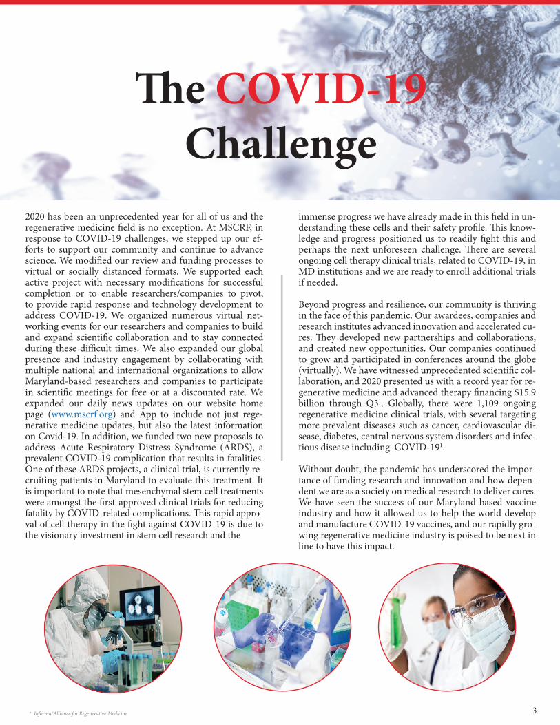
2020 has been an unprecedented year for all of us and the regenerative medicine � eld is no exception. At MSCRF, in response to COVID-19 challenges, we stepped up our ef-forts to support our community and continue to advance science. We modi� ed our review and funding processes to virtual or socially distanced formats. We supported each active project with necessary modi� cations for successful completion or to enable researchers/companies to pivot, to provide rapid response and technology development to address COVID-19. We organized numerous virtual net-working events for our researchers and companies to build and expand scienti� c collaboration and to stay connected during these di� cult times. We also expanded our global presence and industry engagement by collaborating with multiple national and international organizations to allow Maryland-based researchers and companies to participate in scienti� c meetings for free or at a discounted rate. We expanded our daily news updates on our website home page (www.mscrf.org) and App to include not just rege-nerative medicine updates, but also the latest information on Covid-19. In addition, we funded two new proposals to address Acute Respiratory Distress Syndrome (ARDS), a prevalent COVID-19 complication that results in fatalities. One of these ARDS projects, a clinical trial, is currently re-cruiting patients in Maryland to evaluate this treatment. It is important to note that mesenchymal stem cell treatments were amongst the � rst-approved clinical trials for reducing fatality by COVID-related complications. � is rapid appro-val of cell therapy in the � ght against COVID-19 is due to the visionary investment in stem cell research and the
immense progress we have already made in this � eld in un-derstanding these cells and their safety pro� le. � is know-ledge and progress positioned us to readily � ght this and perhaps the next unforeseen challenge. � ere are several ongoing cell therapy clinical trials, related to COVID-19, in MD institutions and we are ready to enroll additional trials if needed.
Beyond progress and resilience, our community is thriving in the face of this pandemic. Our awardees, companies and research institutes advanced innovation and accelerated cu-res. � ey developed new partnerships and collaborations, and created new opportunities. Our companies continued to grow and participated in conferences around the globe (virtually). We have witnessed unprecedented scienti� c col-laboration, and 2020 presented us with a record year for re-generative medicine and advanced therapy � nancing $15.9 billion through Q31. Globally, there were 1,109 ongoing regenerative medicine clinical trials, with several targeting more prevalent diseases such as cancer, cardiovascular di-sease, diabetes, central nervous system disorders and infec-tious disease including COVID-191.
Without doubt, the pandemic has underscored the impor-tance of funding research and innovation and how depen-dent we are as a society on medical research to deliver cures. We have seen the success of our Maryland-based vaccine industry and how it allowed us to help the world develop and manufacture COVID-19 vaccines, and our rapidly gro-wing regenerative medicine industry is poised to be next in line to have this impact.
� e COVID-19Challenge
3

Creating a NewIndustry in Maryland
To advance the research and discoveries into the market we must enable an industry to commercialize these technologies, manufacture products and make them accessible to patients. We work with our faculty and companies to help them suc-ceed and advance their technologies. Our e� orts have stimu-lated the creation and growth of this industry in Maryland. We create value by providing critical support for early-stage high-risk/high-reward scienti� c discoveries, de-risking these innovations with key milestone-based grants, enabling
company formation and novel product development, support-ing each project and scientist with guidance and resources to meet their unique needs, and creating a collaborative commu-nity to accelerate cures. Regenerative medicine is thriving in Maryland, our research continues to accelerate at a rapid pace, and our companies continue to grow. Some of the companies in our portfolio are highlighted below. For more information on all our portfolio companies, visithttps://www.mscrf.org/portfolio-companies.
RoosterBio, Inc. following their Series B raise last year, had the � rst patient dosed under a newly awarded Investi-gational New Drug (IND) application using their CliniControl™ products, in addition to product launches, CRADA, partnerships and interna-tional expansion.
LifeSprout, Inc. founded with technology licensed from Johns Hopkins University, closed a $28.5 million Series A � nancing. � e company will be using proceeds to support clinical development of novel therapeutic products from its Regenerative Matrix platform.
Vita � erapeutics, Inc., a Johns Hopkins University spinout, announced that it received orphan drug designation (ODD) from the U.S. Food and Drug Administration (FDA) for VTA-110, a novel regen-erative therapy for the treatment of Duchenne Muscular Dystrophy (DMD).
NeoProgen, Inc., a University of Maryland Baltimore company, announced a strategic partnership with Aspire Health Science for development and manufacturing of their cell-based therapy.
In addition, two of our portfolio companies (Vita � erapeutics, Inc. and � eradaptive Inc.) made the Maryland Future 20 List, selected by the Department of Commerce and announced by the Governor. � e selection was based on a variety of factors, including innovation, future growth potential, the company’s Maryland story, and “wow” factor. You can read more about the new companies that we funded this year in the Commercialization section of this report.
4
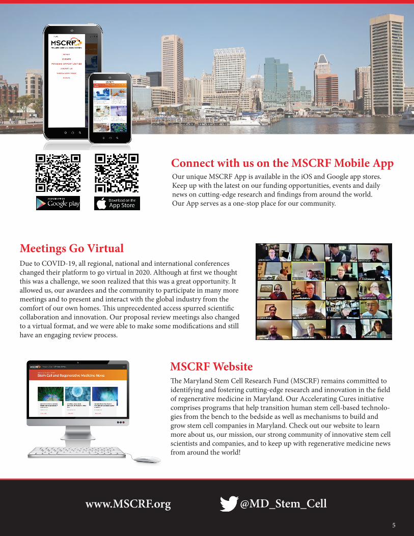
Connect with us on the MSCRF Mobile AppOur unique MSCRF App is available in the iOS and Google app stores. Keep up with the latest on our funding opportunities, events and daily news on cutting-edge research and � ndings from around the world. Our App serves as a one-stop place for our community.
Due to COVID-19, all regional, national and international conferences changed their platform to go virtual in 2020. Although at � rst we thought this was a challenge, we soon realized that this was a great opportunity. It allowed us, our awardees and the community to participate in many more meetings and to present and interact with the global industry from the comfort of our own homes. � is unprecedented access spurred scienti� c collaboration and innovation. Our proposal review meetings also changed to a virtual format, and we were able to make some modi� cations and still have an engaging review process.
Meetings Go Virtual
� e Maryland Stem Cell Research Fund (MSCRF) remains committed to identifying and fostering cutting-edge research and innovation in the � eld of regenerative medicine in Maryland. Our Accelerating Cures initiative comprises programs that help transition human stem cell-based technolo-gies from the bench to the bedside as well as mechanisms to build and grow stem cell companies in Maryland. Check out our website to learn more about us, our mission, our strong community of innovative stem cell scientists and companies, and to keep up with regenerative medicine news from around the world!
MSCRF Website
www.MSCRF.org @MD_Stem_Cell
5
Post-Doctoral Fellowship
Commercialization ClinicalValidationDiscoveryLaunch
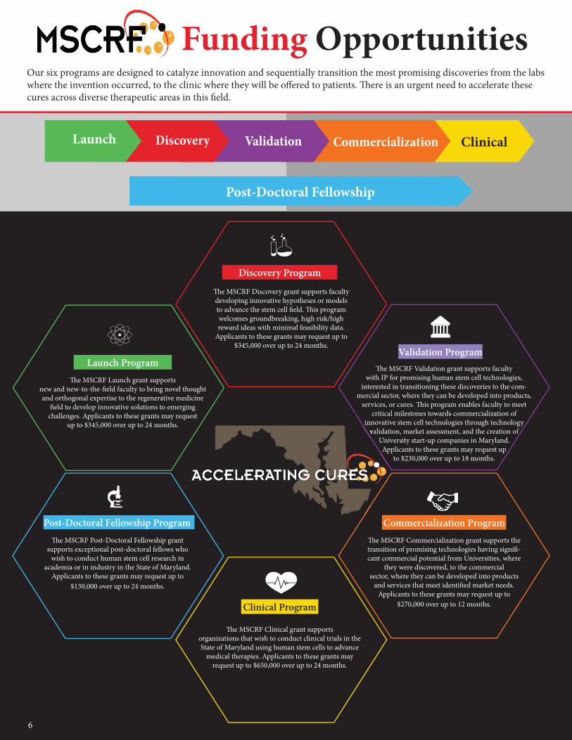
Our six programs are designed to catalyze innovation and sequentially transition the most promising discoveries from the labs where the invention occurred, to the clinic where they will be o� ered to patients. � ere is an urgent need to accelerate these cures across diverse therapeutic areas in this � eld.
� e MSCRF Launch grant supports new and new-to-the-� eld faculty to bring novel thought and orthogonal expertise to the regenerative medicine
� eld to develop innovative solutions to emerging challenges. Applicants to these grants may request
up to $345,000 over up to 24 months.
Launch Program
� e MSCRF Discovery grant supports faculty developing innovative hypotheses or models to advance the stem cell � eld. � is program welcomes groundbreaking, high risk/high reward ideas with minimal feasibility data.
Applicants to these grants may request up to $345,000 over up to 24 months.
Discovery Program
� e MSCRF Validation grant supports faculty with IP for promising human stem cell technologies,
interested in transitioning these discoveries to the com-mercial sector, where they can be developed into products,
services, or cures. � is program enables faculty to meet critical milestones towards commercialization of
innovative stem cell technologies through technology validation, market assessment, and the creation of
University start-up companies in Maryland. Applicants to these grants may request up
to $230,000 over up to 18 months.
Validation Program
� e MSCRF Commercialization grant supports the transition of promising technologies having signi� -cant commercial potential from Universities, where
they were discovered, to the commercial sector, where they can be developed into products
and services that meet identi� ed market needs. Applicants to these grants may request up to
$270,000 over up to 12 months.
Commercialization Program
� e MSCRF Clinical grant supports organizations that wish to conduct clinical trials in the State of Maryland using human stem cells to advance
medical therapies. Applicants to these grants may request up to $650,000 over up to 24 months.
Clinical Program
� e MSCRF Post-Doctoral Fellowship grant supports exceptional post-doctoral fellows who
wish to conduct human stem cell research in academia or in industry in the State of Maryland.
Applicants to these grants may request up to $130,000 over up to 24 months.
Post-Doctoral Fellowship Program
innovative stem cell technologies through technology validation, market assessment, and the creation of
Applicants to these grants may request up
Funding Opportunities
6
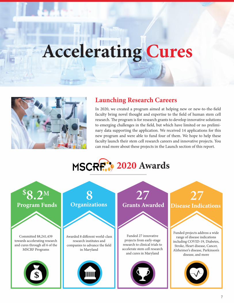
In 2020, we created a program aimed at helping new or new-to-the-� eld faculty bring novel thought and expertise to the � eld of human stem cell research. � e program is for research grants to develop innovative solutions to emerging challenges in the � eld, but which have limited or no prelimi-nary data supporting the application. We received 14 applications for this new program and were able to fund four of them. We hope to help these faculty launch their stem cell research careers and innovative projects. You can read more about these projects in the Launch section of this report.
8Organizations
27Grants Awarded
Awarded 8 di� erent world-class research institutes and
companies to advance the � eld in Maryland
Funded 27 innovative projects from early-stage
research to clinical trials to accelerate stem cell research
and cures in Maryland
$8.2M
Program Funds
Committed $8,241,439 towards accelerating research and cures through all 6 of the
MSCRF Programs
2020 Awards
Accelerating Cures
Launching Research Careers
27Disease Indications
Funded projects address a wide range of disease indications
including COVID-19, Diabetes, Stroke, Heart disease, Cancer,
Alzheimer’s disease, Parkinson’s disease, and more
7
Advancing Medical Research & Treatments in Maryland
2. Centers for Disease Control and Prevention/National Center for Health Statistics
8
During calendar year 2020, we had six active programs. We received 87 applications in two separate funding cycles, and we funded 27 new awards with $8,241,439 in 8 di� erent re-search institutes and companies. We supported projects ad-dressing a wide range of disease indications including psy-chiatric, neurological and neurodegenerative diseases, heart and blood disorders, autoimmune conditions, aging-related disorders as well as research advancing an understanding of human development and genetics. We identify and fund the most innovative research that may provide new treatments for several prevalent, chronic diseases as well as for rare diseases that then serve as a platform to expand therapies to other areas. As an indication of the scienti� c collabora-tion that we have driven, we received several collaborative proposals, including partnerships between for-pro� t Mary-land companies and academic institutions as well as proj-ects between various Maryland research institutions. Such synergistic partnerships help us accelerate discoveries to-wards products and clinical trials so they can reach patients sooner.
� e Maryland Stem Cell Research Fund is also an economic engine for the State, creating jobs and generating new rev-enue. MSCRF grants support researchers, physicians, lab
technicians, and students as well as scientists and executives at stem cell companies. We are creating the next generation of workforce to grow the regenerative medicine industry and advance therapies.
Many of the research grants we have funded to date study stem cells in the context of speci� c diseases including heart disease, cancer, stroke, chronic lower respiratory disease, diabetes, Alzheimer’s disease and � u/pneumonia. It is im-portant to note that these conditions account for the top 10 leading causes of death in Maryland2. Resulting therapies improve the health of many Marylanders and also reduce health care costs for the state. State funding has already al-lowed Maryland scientists and companies to advance the use of stem cells all the way to the clinic. It is imperative that we continue to advance research and treatments for all diseases a� ecting Marylanders with the added under-standing that several of the listed pre-existing conditions cause greater risk of serious illness from COVID-19 and the knowledge that the virus targets multiple organs and systems in our bodies, not just the lungs. Despite the im-mense progress we’ve made, our work is far from over and we, at MSCRF, continue to bring the stem cell community together with a sense of urgency and a common purpose to advance biological science and improve human health.

Grant
Awards
2020annual Report
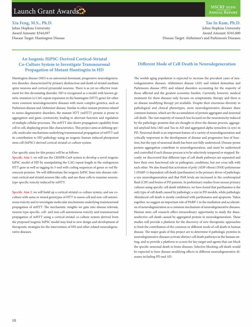
Tae In Kam, Ph.D.Johns Hopkins UniversityAward Amount: $345,000
Disease Target: Alzheimer’s and Parkinson’s Diseases
Xia Feng, M.S., Ph.D.Johns Hopkins UniversityAward Amount: $344,697Disease Target: Huntington Disease
Huntington disease (HD) is an autosomal dominant, progressive neurodegenera-tive disorder, characterized by primary dysfunction and death of striatal medium spiny neurons and cortical pyramidal neurons. � ere is as yet no e� ective treat-ment for this devastating disorder. HD is recognized as a model with known ge-netic mutation (a CAG repeat expansion in the huntingtin (HTT) gene) for other more common neurodegenerative diseases with more complex genetics, such as Parkinson disease and Alzheimer disease. Similar to other mutant proteins related to neuro-degenerative disorders, the mutant HTT (mHTT) protein is prone to aggregation and gains cytotoxicity, leading to aberrant function and regulation of multiple cellular processes. � e mHTT also shows propagation capability from cell to cell, displaying prion-like characteristics. � is project aims at de� ning spe-ci� c molecular mechanisms underlying transneuronal propagation of mHTT and its contribution to HD pathology using an isogenic human induced pluripotent stem cell (hiPSC)-derived cortical-striatal co-culture system.
Our speci� c aims for this project will be as follows:Speci� c Aim 1: we will use the CRISPR-Cas9 system to develop a novel isogenic hiPSC model of HD by manipulating the CAG repeat length in the endogenous HTT gene as well as tagging its 5-end with coding sequences of green or red � u-orescent proteins. We will di� erentiate the isogenic hiPSC lines into disease-rele-vant cortical and striatal neuron-like cells, and use these cells to examine neuron-type-speci� c toxicity induced by mHTT.
Speci� c Aim 2: we will build up a cortical-striatal co-culture system, and use co-culture with same or mixed genotypes of HTT to assess cell and non-cell autono-mous toxicity and to investigate molecular mechanisms underlying transneuronal propagation of mHTT. � e mechanistic insights we gain into disease-relevant, neuron-type-speci� c, cell- and non-cell-autonomous toxicity and transneuronal propagation of mHTT using a cortical-striatal co-culture system derived from the proposed isogenic hiPSC model may lead to new design and development of therapeutic strategies for the intervention of HD and other related neurodegene-rative diseases.
An Isogenic HiPSC-Derived Cortical-Striatal Co-Culture System to Investigate Transneuronal
Propagation of Mutant Huntingtin in HD
� e worlds aging population is expected to increase the prevalent cases of neu-rodegenerative diseases. Alzheimers disease (AD) and related dementias and Parkinsons disease (PD) and related disorders accounting for the majority of those a� icted and the greatest economic burden. Currently, however, medical treatment for these diseases only focuses on symptomatic therapy and there is no disease modifying therapy yet available. Despite their enormous diversity in pathological and clinical phenotypes, most neurodegenerative diseases share common features, which are the accumulation of protein aggregates and neuronal cell death. � e vast majority of research has focused on the neurotoxicity induced by the pathologic proteins that are thought to drive the disease process, aggrega-ted amyloid-beta (Ab) and Tau in AD and aggregated alpha-synuclein (a-syn) in PD. Neuronal death is an important feature of a variety of neurodegeneration and critically important in the development of disease and progressive loss of func-tion, but the type of neuronal death has been not fully understood. Disease-prone protein aggregation contribute to neurodegeneration, and must be understood and controlled if each disease process is to be selectively tempered or stopped. Re-cently we discovered that di� erent type of cell death pathways are separated and have their own functional role in pathogenic conditions, but not cross talk with each other. We also found that activation of poly (ADP-ribose) (PAR) polymerase 1 (PARP-1) dependent cell death (parthanatos) is the primary driver of pathologic a-syn neurodegeneration and that PAR levels are increased in the cerebrospinal � uid (CSF) and brains of PD patients. In preliminary studies from mouse primary cultures using speci� c cell death inhibitors, we have found that parthanatos is the only type of cell death caused by pathologic a-syn in PD models, while pathologic Abinduced cell death is mostly combined with parthanatos and apoptosis. Taken together, we suggest an important role of PARP-1 in the mediation and accelerati-on of neurodegeneration as a common mechanism of neurodegenerative diseases. Human stem cell research o� ers extraordinary opportunity to study the disea-seselective cell death caused by aggregated protein in neurodegeneration. � ese studies will provide a platform for the discovery of new therapeutic approaches to limit the contribution of the common or di� erent mode of cell death in human disease. � e major goals of this project are to determine if pathologic proteins in neurodegenerative diseases activate distinct cell death pathways in the human set-ting, and to provide a platform to screen for key target and agents that can block the speci� c neuronal death in brain diseases. Selective blocking cell death would be expected to have disease modifying e� ects in di� erent neurodegenerative di-seases including PD and AD.
Di� erent Mode of Cell Death in Neurodegeneration
MSCRF 2020:Annual ReportLaunch Grant AwardsLaunch Grant Awards
10
Sashank Reddy, M.D., Ph.D.Johns Hopkins UniversityAward Amount: $345,000
Disease Target: Initial - Craniofacial Microsomia
Basic biomedical research and drug discovery work has relied on in vitro cellular models to represent normal and disease biology since the 1950s. Historically, such cell models have been con� gured as 2-dimensional cultures where cells subsist in a planal layer on the bottom of a plastic tissue culture vessel. It is now becoming quite evident at both a molecular and functional level that 2D cell culture models fail to accurately embody the normal in vivo microenvironment where variation in tissue ultrastructure, cell-cell communication, and metabolic conditions exist. Recent advances to create more biologically-relevant cell models have focused on use of primary cells (rather than transformed cell lines) together with methods to form a 3D architecture; most notable of these advances has been organoid technology using expanded adult stem cells to form di� erentiated, structured cell aggregates of de� ned tissue types. Organoid technology has experienced robust commercial interest from life science companies selling DIY reagents, biorepo-sitories distributing patient-speci� c organoid models, and (limited) biopharma groups using organoid cell models for drug discovery. However, a major issue with the current organoid technology is that the cells face their apical surface inwards to the lumen of the organoid rather than the normal outward orientation. Propagenix has recently discovered and � led provisional patents on a scalable me-thod to generate epithelial cell organoids positioned in their biologically normal orientation, termed apical surface-outward (ASO) organoids. Having epithelial organoids with a normal cellular orientation such that their apical surface points outwards enables an entire new set of research interrogations including cell-virus and cell-bacteria interactions, drug transport, and protein secretion. ASO orga-noids also have features that have potential importance for diagnostic and the-rapeutic use. � is Commercialization Grant proposal is focused on generating application data that will drive commercial success of our ASO organoid techno-logy in multiple markets.
Human iPSC-Derived EGFR+ Functional Schwann Cells to Enhance Nerve Regeneration and Improve
Functional Outcomes in VCA
Vascularized composite allotransplantation (VCA) holds much promise to im-prove the quality of life for our civilian who su� er from devastating injuries such as severe and irreplaceable tissue loss or amputations. Transplantation is cur-rently the only treatment option to fully restore missing limbs with functional and anatomical equivalents by replacing like-with-like tissue. Recent advances in microsurgical techniques and immunosuppressive protocols have enabled wider application of VCA with highly encouraging immunologic and aesthetic results. However, the overall success of VCA is dictated by the pace and quality of nerve regeneration. Following transplantation, the recipients peripheral nerve axons must regenerate into the gra� so as to innervate the transplanted muscle and skin. � is process allows the recipient to establish motor control over and receive sensory input from the gra� . Without adequate innervation, a transplanted gra� remains inanimate and insensate and provides little if any bene� t to the recipient. Over time, a lack of innervation will result in progressive, permanent atrophy within the gra� , rendering it useless. � e importance of adequate gra� innerva-tion applies to all types of VCA; in upper extremity transplantation, meaningful hand function is dependent on the recipients motor and sensory axons reaching the intrinsic muscles and distal skin of the transplanted hand; in facial transplan-tation, gra� innervation is necessary for everything from facial expression to pre-venting drooling. Substantial advances have been made in the enhancement of nerve regeneration across gaps through the use of conduits and acellular nerve gra� s. However, very few therapeutic approaches have been successfully studied in primary end-to-end nerve repairs, which is the preferred and most common-ly used method of repair in VCA. � erefore, the objective of this proposal is to develop a novel stem cell-based therapy utilizing human iPSC-derived Schwann cells to enhance nerve regeneration and improve functional outcomes in recon-structive transplantation. In VCA, the slow pace and the requirement for nerve regeneration over long distances to regain full motor and sensory function is still prohibitive to expand the indication for VCA to full arm or lower extremity trans-plantation. � e overall hypothesis of this proposal is that human iPSC-Schwann cells will signi� cantly increase the pace and degree of nerve regeneration a� er end-to-end injuries in VCA and thus improve functional outcomes. In experi-mental approaches, we plan to expand human Schwann cells without losing my-elination capability and to investigate the interaction of donor and host Schwann cell toward remyelination. Moreover, we propose to demonstrate enhanced func-tional recovery following peripheral nerve transection and repair following syn-geneic forelimb transplantation as well as allogeneic transplantation to investigate the neuro-regenerative potential of Schwann cells in a setting that recapitulates the clinical scenario of VCA using unrelated cadaveric donor.
Byoung Chol Oh, Ph.D.Johns Hopkins UniversityAward Amount: $299,765Disease Target: Peripheral Nerve Injury, Hand Transplantation
Regenerative Cell � erapies for So� Tissue Restoration
MSCRF 2020:Annual ReportLaunch Grant Awards
11
Grant
Awards
2020annual Report
Je� Bulte, M.S., Ph.D.Johns Hopkins UniversityAward Amount: $339,297
Disease Target: Stroke
� e use of stem cells and progenitors for cell replacement therapy and tissue resto-ration is a sought-a� er approach for correction of damaged tissue. When such therapies are pursued in patients, it would be highly desirable to have non-invasive cell tracking techniques available that can report longitudinally on the local and whole-body distribution of transplanted cells, in order to better understand their fate in vivo and in particular to optimize dosing and delivery as part of perso-nalized therapies. In this proposal, we aim to develop magnetic particle imaging (MPI) in conjunction with magnetic resonance imaging (MRI) to answer several basic questions associated with the e� cacy and safety of cell therapy. � ese are two complementary techniques, where MPI can report on the local and whole-body distribution of administered cells in an absolute quantitative manner, whereas MRI provides supporting anatomical information and can report on real-time ho-ming and immediate retention of cells. We propose to use human glial-restricted progenitor cells (hGRPs) as an example therapeutic cell type, as these cells have been extensively used in several pre-clinical animal models, have been approved for clinical use, and we have ample experience using them. We have chosen an ischemic stroke model (middle cerebral artery occlusion or MCAO) as an example of a target disease, in which loss of axonal myelin is one of the key events leading to permanent debilitation. � is MPI/MRI two-pronged approach of imaging the same cell (labeled with superparamagnetic iron oxide particles or SPIO for both MPI and MRI) will be applied for intraarterially injected hGRPs with and without VLA-4 transfection where VLA-4/VCAM-1 docking has been shown to result in diapedesis and increased targeting to the brain parenchyma. If successful, this ex-ample MPI application of tracking (transfected) hGRPs in stroke may encourage the use of MPI to interrogate the fate of other cells and other disease scenarios in vivo.
Kenneth Boheler, Ph.D.Johns Hopkins UniversityAward Amount: $345,000Disease Target: Vascular Diseases
Human pluripotent (i.e., embryonic and induced) stem cell (hPSC) derived va-scular smooth muscle cells (vSMCs) are of considerable interest for the study of human vascular development, modeling of vascular diseases, and regenerative medicine. While signi� cant advances in the di� erentiation and puri� cation of hPSC-derived vSMCs have been achieved, the lack of speci� c protein or genetic markers that distinguish both among phenotypically distinct vSMC cell subtypes in mixed cultures and between vSMCs and non-SMCs is a major limitation to pro-gress in this � eld. � is limitation is particularly problematic for studies involving polymorphic hiPSC lines, where interline variabilities a� ect both the quality and composition of in vitro di� erentiated vSMC cultures and the phenotypes of these cells. One proven method to overcome issues of heterogeneity is through immu-nophenotyping and isolation of marker-de� ned cell types by � uorescence-acti-vated cell sorting (FACS). Currently, known surface proteins suitable for sorting vSMCs with de� ned functional phenotypes are extremely limited. To discover new and improved marker panels, we have labelled, captured and identi� ed cell surface glycoproteins using state of the art chemoproteomic and mass spectrometry (MS) techniques. � ese pilot experiments have led to the identi� cation of 13 targets that appear to be restricted to vSMCs (when compared with the cell surface protein atlas (CSPA)). Additional targets have been putatively identi� ed that are restricted to early, synthetic vSMCs. Based on our existing data, we propose to develop com-mercially (while maintaining IP) two sets of monoclonal antibodies (mAbs) for vSMC restricted targets. � e MS output, which experimentally con� rms protein orientation and identi� es cell surface accessible epitopes present in the extracel-lular (EC) space, will be used to guide the development of the new mAbs. We also propose to test and validate markers for the selective sorting of synthetic versus contractile vSMCs and for assessing interline variability. For this, we propose to examine the surfaceomes of both synthetic and contractile vSMCs generated from 6-10 hPSC lines (normal and diseased) and from primary human aorta SMCs. � is broad-based assessment of vSMCs is now feasible because we have establis-hed in the laboratory a new state-of-the-art micro-Cell Surface Capture (mCSC) approach, which requires 10-fold fewer cells than our original protocol. As marker panels are established and Abs are validated for sorting vSMCs with de� ned phe-notypes, molecular, biochemical and bioengineering approaches will be used to validate the phenotypes of sorted cells across 2-3 hPSC lines. At the completion of this study, the improved isolation and characterization of human vSMCs with de� ned phenotypes (synthetic versus contractile) will positively impact the � eld and overcome the current lack of a speci� c protein or genetic marker that distin-guish vSMC cell types in mixed cultures. Once published, the data and reagents generated in this study will not only improve the isolation and characterization of vSMCs from heterogeneous cultures for basic science, the reagents produced may be apt for drug repurposing and for regenerative medicine approaches involving hPSC-derived vSMCs.
Chemoproteomic Immunophenotyping of Human Pluripotent Stem Cell Derived Vascular
Smooth Muscle Cells
Developing MPI for Tracer-Based, Non-Radioactive, and Quantitative Whole-Body Imaging of Cell Delivery
MSCRF 2020:Annual ReportDiscovery Grant Awards
13
Alan Friedman, M.D.Johns Hopkins UniversityAward Amount: $345,000Disease Target: Cancer
Yingli Fu, M.S., Ph.D.Johns Hopkins UniversityAward Amount: $345,000
Disease Target: Diabetes
Multiple solid tumors grow slower in mice lacking NF-kB p50, and adoptive transfer of immature myeloid cells lacking p50, a� er a dose of myelo-depleting 5-� uorouracil, slows the growth of murine prostate cancer, pancreatic ductal carcinoma, and neuro-blastoma. We have developed methods for e� cient p50 gene editing in human marrow myeloid progenitors and for their expansion in serum-free media. As a more feasible and cost-e� ective means to obtain su� cient numbers of p50-IMC for clinical transla-tion, including repeated cycles of treatment, we herein propose to develop methods for p50 gene editing of human iPSC and their optimal di� erentiation and expansion into p50-IMC. In doing so we will utilize stromal-activated, myeloid progenitor-derived iPSC that have minimal epigenetic abnormalities, will convert such lines to the „nave“ state using LIF-3i if this proves advantageous, will compare lines for o� -target edits and their functional consequences, and will evaluate their ability to form M1-polarized tumor myeloid cells in tumor-bearing immune-de� cient mice. In addition, we will evaluate the utility of heterologous p50-IMC in murine models, with the view towards using „o� -the-shelf “ iPSC-derived p50-IMC should heterologous p50-IMC reduce tumor growth as e� ectively as autologous p50-IMC. Upon completion of these studies we anticipate initiating clinical trials evaluating the safety and e� cacy of iPSC-derived p50-IMC in patients with a broad range of malignancies. As we observe cooperati-on between p50-IMC and anti-PD-1 antibody in the treatment of murine pancreatic ductal carcinoma, we also intend to explore addition of T cell checkpoint inhibitory antibodies to p50-IMC in the clinical context.
Immunotherapy has emerged over the past decade as a key weapon against cancer. We have developed a novel immunotherapy utilizing immature myeloid cells lacking NF-kB p50 (p50-IMC), which activates T cells to inhibit tumor growth. p50-IMC have the potential to be e� ective in patients with a broad range of malignancies. � e goal of this proposal is to develop iPSC as an e� ective source of p50-IMC. While p50-IMC can be derived from patient marrow or blood, we anticipate that gene editing of autologous iPSC will more e� ectively provide su� cient p50-IMC for multiple rounds of therapy. We also propose murine studies to determine the e� cacy of heterologous p50-IMC in an e� ort to validate use of „o� -the-shelf“ iPSC-derived p50-IMC, which would be even more cost-e� ective and convenient to provide patients. As we � nd cooperation between p50-IMC and T cell checkpoint inhibition in our murine pancreatic cancer model, these two immunotherapies may cooperatively activate anti-tumor T cell acti-vity, as we anticipate evaluating also in clinical trials.
Human iPSC-Derived NF-kB p50-De� cient Myeloid Cell Immunotherapy for Cancer
Type I diabetes mellitus (T1D) is a devastating T-cell mediated autoimmune disease that results in the destruction of insulin-producing beta cells in the pancreas and subsequent hyperglycemia, for which no cure exists. Current treatment with exo-genous insulin injections, although lifesaving, cannot replicate the level of feedback control a� orded by naturally occurring functional beta cells, and is ine� cient to prevent chronic complications. � us, novel therapies to provide long-term and dy-namic glucose control via minimally invasive beta cell delivery would signi� cantly improve T1D health care. � e discovery of human induced pluripotent stem cells (hiPSCs) and their ability to di� erentiate into functional beta cells o� ers signi� cant potential to overcome the shortage of human beta cells, while immunoisolating 3D constructs that are amenable for injection can be engineered using biocompatible materials to deliver hiPSC-derived beta cells. � e goal of the present proposal is to develop an o� -the-shelf, injectable, hiPSC-derived beta cell therapeutic for image-guided T1D treatment using novel bioprinted 3D constructs. As such, we propose an integrated approach in which insulin-responsive, functional beta cells derived from hiPSCs will be printed with biocompatible materials to create an imaging-visi-ble, immunoprotective matrix that can be delivered via minimally invasive, routine delivery routes, such as s.c. or i.p.
Speci� c Aims (1): To test the hypothesis that per� uorinated, � brin-alginate mi-crocapsules can be printed for single hiPSC-beta cells using a custom-made, fully automated, piezoelectric bioprinting system. In this aim, we will establish the most promising bioprinting formulations for imaging-guided hiPSC-derived beta cell de-livery to enhance cell viability and imaging visibility.
Speci� c Aim (2): To test the hypothesis that bioprinted � brin-alginate microcapsu-les improve single beta cell survival and function over conventional alginate micro-capsules in vitro and in vivo and enables image-guided, minimally invasive delivery and longitudinal tracking. In this aim, we will determine the e� ect of bioprinting on beta cell function in vitro and analyze the kinetics of glucose normalization by bioprinted hiPSC-beta cells in diabetic animals. � e proposed study brings a unique combination of expertise in type I diabetes management, stem cell encapsulation, imaging-guided delivery, biomaterials and 3D bioprinting to address the critical challenges in engineering and delivering beta cell-based therapeutics. Successful completion of the proposed activities will have vast rami� cation for hiPSC-derived beta cell therapy by creating a o� -the-shelf cellular product that can be precisely delivered via minimally invasive s.c. or i.p. injections for T1D treatment, which is aligned with the mission of Maryland Stem Cell Research Fund.
Engineering Stem Cell Microenvironment for Image-Guided � erapeutic Intervention
MSCRF 2020:Annual ReportDiscovery Grant Awards
14
Xiaofeng Jia, B.M., Ph.D.University of Maryland, Baltimore
Award Amount: $345,000 Disease Target: Peripheral Nerve Injury
Approximately 360,000 people su� er from upper extremity peripheral nerve injury (PNI) in the United States of America each year. PNI o� en results in poor functio-nal recovery and subsequently impaired quality of life for the patient. Currently, many approaches to improve peripheral nerve regeneration have not surpassed the gold standard‘ autogra� procedures. However, autogra� s are not ideal due to limited gra� source availability and the morbidity of a second surgical incision. Supported by the 2013 and 2018 MSCRF awards, our previous studies have shown that human neural crest stem cells (NCSCs) promote sciatic nerve regeneration with axonal regrowth and myelin formation. Despite signi� cant progress, tumou-rigenicity and immunogenicity have long been a challenge for stem cell therapy and hinder clinical applications. Increasing evidence suggests that exosome therapy represents a novel cell-free treatment with compelling advantages over whole cell therapy by their parent cells. Our preliminary data showed exosomes from NC-SCs improve recovery a� er nerve crush injury. Objectives/Hypothesis: With the long-term goal to develop and advance the exosome-based therapeutic approach to ultimately improve the patients quality of life, we will test the hypotheses: (1) NCSC exosome transplantation will improve nerve regeneration in vivo, possibly through facilitating di� erentiation into Schwann cells (SCs) and/or upregulation of growth factors; (2) Compared to NCSC transplantation, exosome therapy will lessen the immune responses with similar therapeutic e� ect; (3) microRNAs (miRNAs) is the key molecular target in exosomes to promote peripheral nerve regeneration, and modulation of miRNA cargo can further improve outcomes a� er nerve injury. Speci� c Aim 1: Establish NCSC derived exosomes therapy to improve outcomes a� er PNI. Speci� c Aim 2: Compare the immune responses and therapeutic e� ects between exosome and NCSC therapies. Speci� c Aim 3: Further enhance therapeu-tic e� ects of exosome therapy by modulating miRNA cargo to promote periphe-ral nerve regeneration. Study Design: � is proposal will investigate the e� ect of NCSC derived exosome therapy in peripheral nerve crush injury and nerve defect repair animal models via histopathological, electrophysiological and functional outcome measurements. Innovation and signi� cance: For the � rst time, we will not only develop exosome therapy but also further enhance the therapeutic e� ect by modulating miRNA cargo, the signaling vehicle, to promote nerve regeneration a� er PNI. We are the � rst to target immunogenicity and autotomy by comparing the rodents treated with exosomes and their parent cells. We have developed a novel cell modi� cation technology with less invasive sources of stem cells and a more desirable source of SCs for transplant therapies. Our project combines the expert knowledge of interdisciplinary investigators to execute this clinically mo-tivated, translational experimental design with systematic in vivo measurements. � e proposed research aims to provide a novel cell-based therapeutic approach to promote nerve regeneration. � e speci� c design and motivation of our technolo-gy and protocols allow for straightforward translation to a clinical setting. Further development will enable commercialization towards a point of service product. It will develop optimal treatment protocols for clinicians treating patients with nerve injury. Success in this project would greatly enhance the surgical repair of nerve injuries with better functional outcomes.
Muthukumar Gunasekaran, Ph.D.University of Maryland, BaltimoreAward Amount: $345,000Disease Target: Myocardial Infarction
Stem cell transplantation is an e� ective treatment option for myocardial infarc-tion (MI). However, identi� cation of potential stem cell type, which rescues the infarcted myocardium is still under investigation. Further, immune response to transplanted stem cells reduces its regenerative potential in stem cell therapy. Recent � ndings from our group demonstrated that neonatal cardiac progenitor cells (nCPCs) showed superior e� cacy in repairing the injured heart compared to other stem cells types mesenchymal stem cells (MSCs), cardiosphere derived cells (CDCs) and umbilical cord (blood) in an ischemic immune de� cient rodent MI model. However, the therapeutic e� ects of nCPCs to evade the immune response by molecular signaling pathways, essential for cardiac repair remains uncharacte-rized and critical for its clinical success. Our preliminary data supports that CPCs evade from the immune response by novel mechanism. Controlling CD47 levels in CPCs, critical for evading the innate immune surveillance by binding to sig-nal regulatory protein- (SIRP) on macrophages which inhibits the macrophage phagocytic activity. � erefore, we hypothesize that CD47 to be the key molecule upregulated in nCPCs, and responsible for their immune evasion leading to incre-ased cell retention and restoration of cardiac function in an immune-competent rat MI model. Furthermore, HSF1 and microRNAs such as miR-133a and miR-34a regulate CD47 expression in CPCs. Our hypotheses will be achieved by, 1). Determine HSF1-CD47 axis mediated immune evasion by CPCs 2). Demonstrate miR-133a and miR-34a regulates CD47 expression in CPCs. � is study is the � rst step towards optimization of nCPCs for allogeneic stem cell therapy in clinical settings for its superior cardiac regeneration e� cacy in the infarcted myocardium compared to other stem cells. In the future, we will contend that nCPCs will be utilized for therapeutic application or used as an adjunct to surgical intervention for patients with myocardial infarction to provide game-changing outcomes and thus change their life expectancy. We believe our focus on nCPCs will lead to in-novative avenues to identify novel mechanism of immune evasion, hence paving yet another milestones in regenerative medicine.
A Novel Mechanism of Immune Evasion by Human Neonatal Cardiac Progenitor Cells
Developing Human iPSC-Derived Exosome � erapy to Improve Recovery a� er Peripheral Nerve Injury
MSCRF 2020:Annual ReportDiscovery Grant Awards
15

Tami Kingsbury, Ph.D.University of Maryland, BaltimoreAward Amount: $345,000Disease Target: Red Blood Cell Disorders
Hanseok Ko, M.S., Ph.D.Johns Hopkins UniversityAward Amount: $345,000
Disease Target: Parkinson’s Disease
Defects in erythropoiesis lead to anemias and blood malignancies. Elucidating novel mechanisms regulating red blood cell (RBC) formation from hematopoietic stem-progenitor cells (HSPCs) has the potential to provide new approaches to stimulate erythropoiesis in patients to reduce transfusion dependency and to support the de-velopment of improved methods for in vitro expansion of RBCs for clinical applica-tions. We recently demonstrated that the developmental transcription factor SIX1 is a positive regulator of human erythropoiesis. Overexpression of SIX1 in primary peri-pheral blood HPSCs or CD34 positive TF1 erythroleukemia cells used to model early stage erythropoiesis triggered erythroid di� erentiation. Conversely, CRISPR mediated SIX1 knockout reduced the ability of erythropoietin (EPO) to stimulate erythropoie-sis. SIX1 BioID proximity labeling performed in TF1 cells revealed GATA1 was part of the SIX1 proximal interactome. Further work demonstrated that SIX1 associates with GATA1 and stimulates GATA1 mediated gene expression. Consistent with the-se observations, we showed that SIX1-stimulated erythropoiesis required the master erythroid transcription factor GATA1, but not the EPO receptor. � us, SIX1-GATA1 interaction is a novel regulatory mechanism functioning in human hematopoietic cells that may provide a method to stimulate erythropoiesis downstream of ine� ective EPO signaling. � e work outlined in this proposal seeks to address gaps in our knowledge needed to translate this exciting discovery into novel approaches to treat red blood cell disorders or enhance red blood cell formation in vitro for utilization in the clinic or development of drug delivery sytems.
Aim 1: We will determine the cellular mechanisms by which SIX1 increases erythro-poiesis and cell stages impacted by SIX1 manipulation using gain and loss of function approaches combined with analysis of cell proliferation, cell death, and cell lineage speci� cation, as determined by immunophenotyping, single cell RNA sequencing and colony formation assays. SIX1 mutant alleles selectively disrupting interaction with its co-activator EYA or co-repressor TLE, will determine whether SIX1 gene activa-tion and/or repression mediates erythroid e� ects. SIX1 mutants incapable of binding GATA1 will determine reveal GATA1 dependent vs independent functions of SIX1 in erythropoiesis. Importantly, we will extend our initial � ndings on early stage erythro-poiesis to late stage erythropoiesis to determine the consequences of SIX1 manipula-tion of terminal erythroid di� erentiation, assessing red blood cell yields, blood group antigen expression, hemoglobin pro� les and immunomodulatory function.
Aim 2: We will perform CRISPR knockout screening to functionally identify additio-nal members of the SIX1 proximal interactome required for SIX1 or EPO-stimulated erythropoiesis. In addition to GATA1, and multiple known GATA1 interacting factors, the SIX1 interactome contains 56 factors not previously implicated in erythropoiesis. Novel erythropoietic factors identi� ed within the SIX1 interactome will provide new molecular insight into normal and defective human erythropoiesis and reveal additio-nal novel targets for erythropoiesis stimulating agents.
Leveraging Novel SIX1 Transcription Factor Network Interactions to Stimulate Human Erythropoiesis
Parkinsons disease (PD) pathophysiology is a complex cascade of protein interac-tions and molecular pathway activation that is associated with pathologic -synuclein (-syn) folding and subsequent neuronal cell death. � ere is growing recognition that one of the critical steps in neurodegeneration in PD is neuroin� ammation, charac-terized by activation of microglia and astrocytes. Recently, we described a subtype of reactive astrocytes, A1 astrocytes, that we observed in various human neurode-generative diseases including PD. We found that activation of microglia leads to the conversion of bene� cial A2 astrocytes into toxic A1 type astrocyte via secretion of IL-1alpha, TNF-alpha, and C1q (reported as A1 astrocyte inducer). Building on the-se � ndings in primary murine neuronal cultures, we discovered that -syn preformed � brils (PFF), which mimic pathologic -syn in PD, activate microglia to induce A1 as-trocyte formation and neuronal death in vitro. We observed evidence for microglial activation and A1 astrocyte formation that correlates with neurobehavioral de� cits in mouse models of PD. However, the mechanisms underlying the activated micro-glia/astrocyte axis in the mediation and acceleration of the neurodegeneration in PD remains unclear. As such, an important role for the activated microglia/astrocyte axis in PD needs to be studied in great depth. Importantly, our preliminary study in-dicates that RIPK2 (receptor-interacting serine/threonine-protein kinase 2) is active in microglia in PD brain and genetic depletion and pharmacological inhibition of RIPK2 inhibit the microglia activation, A1 astrocytes conversion and neuronal to-xicity due to -syn aggregates in murine cells suggesting RIPK2s role in the activated microglia/astrocyte axis in PD pathogenesis.
Speci� c Aim 1: We will further extend and con� rm the � ndings in human cells using control and PD iPSC lines derived human microglia with or without RIPK2 depletion, astrocytes, and dopaminergic (DA) neurons that may a� ord us a more predictive model of human brain. Furthermore, we will identify and characterize the down-stream pathway associated with RIPK2 activation in neuroin� ammation due to -syn aggregates by employing proximity-dependent biotin identi� cation (BioID) of RIPK2 from control and PD iPSC lines derived microglia.
Speci� c Aim 2: We will explore the role of a newly devolved RIPK2 inhibitor, CMPD0673 with highly potent and blood brain barrier (BBB) permeable on the microglia activation, A1 astrocytes conversion and neuronal toxicity due to -syn aggregates in control and PD iPSC lines derived human microglia, astrocyte and do-paminergic neurons. � is study enables us to uncover the role of RIPK2 activation on microglia activation, A1astrocytes conversion and DA neurodegeneration due to -syn aggregates in human cells. Also, this investigation will also provide new insights into the pathogenesis in PD and leads to promising novel therapeutic agent for the treatment of PD and related -synucleinopathies.
Targeting an Activated Microglia/Astrocyte Axis for Reducing Neurodegeneration
MSCRF 2020:Annual ReportDiscovery Grant Awards
16
Linda Resar, M.D.Johns Hopkins UniversityAward Amount: $345,000
Disease Target: Osteoporosis and Aging
� e goal of our collaborative project is to develop stem cell technology to enhan-ce bone formation for patients with fractures caused by bone loss or osteoporosis with aging. Our focus is High Mobility Group A (HMGA1) chromatin remodeling proteins. � e Resar laboratory pioneered studies demonstrating that HMGA1 pro-teins are key epigenetic regulators in adult stem cells while Drs. Wan & Xian are international leaders in mesenchymal stem cell (MSC) biology and bone formation (osteogenesis). With aging, there is a global loss of stem cell function, including decreases in the regenerative capacity of MSC within the bone marrow. Bone loss (osteoporosis or OP) occurs because bone resorbed by osteoclasts is not fully resto-red with bone deposited by osteoblasts. Since osteoblasts require constant replenish-ment from multipotent bone marrow MSCs, an insu� cient supply of osteoblasts from MSCs is central to age-related OP. Importantly, systemic infusion of MSCs does not improve osteogenesis due to poor delivery and impaired di� erentiation to bone. Here, we focus on the role of HMGA1 as a key factor in osteogenic poten-tial of MSCs. Rigorous prior research indicating that HMGA1 is a critical factor for adult stem cell (ASC) function includes the following published work and new, unpublished � ndings: 1) HMGA1 is a key epigenetic regulator that maintains ASC number and function within intestinal epithelium (Nature Comm, 2017; Cancer Res, 2018). 2) HMGA1 acts as a molecular switch by � ipping on Wnt signaling genes required for stem cell function and homeostasis, including genes important in bone formation. 3) More recently, we discovered that mice de� cient in Hmga1 develop premature aging with accelerated kyphosis and osteoporosis. 4) RNA se-quencing (RNAseq) data from mouse embryonic � broblasts (MEFs) indicates that Hmga1 represses senescence genes (Cdkn2A) while activating genes required for osteoblast di� erentiation. 5) Lrp6 encodes a Wnt co-receptor essential for osteoge-nesis by maintaining MSC survival and osteoblast di� erentiation (Bone Res, 2014 & 2018). 6) Cellular senescence depletes the MSC pool, thus impairing osteoge-nesis (Nature Comm, 2018). 7) HMGA1 levels decrease in diverse tissue-speci� c stem cells with aging in both mice and humans. Together, these intriguing results support the following hypotheses: 1) HMGA1 is required for bone forming poten-tial from MSC, 2) HMGA1 de� ciency in MSC with advancing age promotes bone loss by disrupting key epigenetic networks, and, 3) Maintaining HMGA1 function will rejuvenate MSCs, improve bone formation, and restore bone mass by enhan-cing MSC osteogenic potential and antagonizing senescence. Speci� c Aims: To test this, we propose to harness our unique human cell-based and murine models with genetic manipulations in HMGA1 in the following Aims: 1) To dissect the function of HMGA1 in MSC maintenance, osteogenic potential, and bone mass, and, 2) To develop stem cell technology for bone formation from MSC using CRISPR-based activation (CRISPRa) to enhance HMGA1 transcriptional networks and epigenetic reprogramming. Impact: � ese innovative studies will not only provide insight into MSC function, bone formation, and aging, but could also reveal new therapies to foster osteogenesis, treat fractures, and even prevent bone loss and osteoporosis with aging.
Vassilis Koliatsos, M.D.Johns Hopkins UniversityAward Amount: $345,000Disease Target: TBI, AD, PD
Axonal pathology and axonal transport impairments are key pathogenic mecha-nisms in several neurological disorders including traumatic brain injury, peri-pheral neuropathies, glaucoma, and select neurodegenerative diseases. Together, these conditions a� ect a substantial portion of the US population, impair quality of life, have high mortality and, to a large extent, have no satisfactory treatments. In all previous disorders, axons undergo a series of molecular and cellular changes that have been best characterized in simple axotomy models and are known as Wallerian degeneration. Our proposal is inspired by recent exciting work in our laboratory in models of traumatic brain injury and engages novel technologies developed by our collaborators to create an in vitro platform of axonopathy/Walle-rian degeneration in human neurons useful for both mechanistic studies and drug discovery. � is axonopathy platform is a signi� cant addition to in vivo models be-cause it obviates daunting challenges related to the intricate anatomy of axons and complex in� ammatory and other tissue responses to injury and, in addition, utili-zes human nerve cells. Here we compartmentalize neuronal cell bodies and axons and allow room for the potential addition of astrocytes, oligodendrocytes and microglial cells to study pathology at a desired level of complexity. Perhaps more importantly, the proposed platform paves the way for future use of induced pluri-potent stem cells from patients where mechanisms and treatments of disease can be explored at a higher level of precision, at the population and individual level. Aim 1, we establish the platform by combining compartmentalized micro� uidic device technology that ensures the separation of axons from cell bodies and a rapid neuronal di� erentiation technology that allows the e� cient conversion of human embryonic stem cells to neurons inside the devices with minimal perikaryal conta-mination of the axonal compartment. Axons are then subjected to di� erent insults to address di� erent patterns of axonal degeneration applicable to diverse neuro-logical diseases. In particular we establish models of mechanical injury (axotomy and graded crush injury) relating to TBI and spinal cord injury; neurotoxic inju-ry (treatment with the tubulin polymerization inhibitor vincristine) pertinent to chemotherapy-induced peripheral neuropathy; and treatment with hydrogen per-oxide that models oxidative stress-related axonal degeneration. Following injury, axons are analyzed with unbiased methodologies to assess and quantify patterns of degeneration. Aim 2, we use the humanized axonopathy platform to explore select mechanisms of Wallerian degeneration based on our promising published and pilot work. We speci� cally focus on two molecular pathways, one centered on Sterile alpha and heat/armadillo motifs-containing protein 1 (SARM1) and the other related to the activation of Mitogen-activated protein kinases (MAPKs). � is work will not only yield important information about mechanisms, but also sharpen molecular targeting for drug discovery. In Aim 3, we follow on molecular leads from work in Aim 2 and block SARM1- and MAPK-associated signals in the platform with small molecules with which we have considerable experience. � is work will serve to con� rm the molecular � ndings from Aim 2 but also validate the platform as a bioassay system for screening novel compounds that may eventually be used as drugs for traumatic and other axonopathies. We are already in talks with the Hopkins Drug Discovery Program for future screening of small-mole-cule libraries and further translational work using our platform. In conclusion, we propose the establishment and characterization of a novel human axonopathy platform designed as a versatile tool to model a variety of axonal degenerative conditions, explore mechanisms, and develop drugs.
A Stem Cell-Derived Human Neuron Model To Explore Treatments of Axonopathies
Developing Stem Cell � erapy for Bone Loss and Fractures with Aging
MSCRF 2020:Annual ReportDiscovery Grant Awards
17
Je� rey Rothstein, M.D., Ph.D.Johns Hopkins UniversityAward Amount: $345,000Disease Target: Dementia, ALS, Alzheimers, FTD
Elias Zambidis, M.D., Ph.D.Johns Hopkins UniversityAward Amount: $345,000
Disease Target: Diabetes, Vascular Disease
� e motor neuron disease Amyotrophic Lateral Sclerosis (ALS) and the second most common form of dementia, Frontotemporal Dementia (FTD), comprise a spectrum of fatal neurodegenerative diseases. Although clinically two distinct diseases, multiple genetic loci including an intronic GGGGCC (G4C2) hexanucleotide repeat expansion (HRE) in the C9orf72 gene, have been linked to ALS and FTD. � e C9orf72 HRE is the most common cause of both familial and sporadic ALS accounting for ~40% and ~8% of patients respectively. Overall, only about 10% of ALS cases are familial with the remaining 90% being sporadic in nature. Despite >20 genetic causes of ALS having been identi� ed, the molecular mechanisms underlying disease pathogenesis remain poorly understood. Defects in nucleocytoplasmic transport and the nuclear pore complex itself have recently emerged as a prominent pathomechanism underlying multiple neurode-generative diseases including C9orf72 ALS/FTD, sALS, Alzheimers Disease, and Hun-tingtons Disease. However, little is known about the nature of the injury to the nuclear pore complex and its individual nucleoporin components themselves. Using induced pluripotent stem cell derived spinal neurons and postmortem human tissue, we have now amassed substantial data that loss of the transmembrane nucleoporin POM121 from the nuclear envelope and nuclear pore complexes initiates a pathological cascade impacting nuclear pore complex composition, function, and cellular survival. Notably, loss of POM121 is mediated by pathologic G4C2 repeat RNA and not DPRs or loss of C9ORF72 protein. Given that POM121 is not mislocalized or aggregated in the cyto-plasm, we hypothesized that POM121 and subsequently altered nucleoporins are aber-rantly degraded in the early stages of C9orf72 ALS/FTD pathogenesis. Indeed, our new preliminary data suggests that the degradation of POM121 is initiated by nuclear accu-mulation of CHMP7. Nuclear CHMP7 has previously been shown to activate ESCRT-III mediated degradation of nuclear pore complexes and nuclear envelope components during nuclear pore surveillance and homeostasis. Our preliminary data suggest that the G4C2 repeat RNA mediated aberrant activation of this pathway is responsible for nuclear pore injury in C9orf72 ALS/FTD, making CHMP7 an attractive therapeutic target in neurodegeneration. Prevention of CHMP7mediated nuclear injury could pro-ve to be a powerful therapy for thee earliest event in ALS and FTD. Importantly- this research program can only be carried out properly using human iPS derived neurons.
CHMP7 in the Initiation of Nuclear Pore Injury in C9orf72 ALS/FTD and Sporadic ALS
Human vasculature arises during development from proli� c embryonic vascular progenitors (VP) that di� erentiate into endothelial progenitor cells (EPC) and pe-ricytes. In the adult, circulating EPC and pericytes are rare, limited in their mul-ti-potency and expansion, and functionally defective in diabetes. Our group � rst established that proli� c CD31+CD146+CXCR4+ embryonic VP could be genera-ted from patient-speci� c conventional hiPSC and utilized to repair damaged adult retinal blood vessels in a murine ischemia/reperfusion (I/R) model (1-3). We also reported a new class of tankyrase/PARP inhibitor-regulated nave hiPSC (N-hiPSC) with improved epigenetic plasticity (4-6). � ese novel N-hiPSC displayed enhan-ced multi-lineage di� erentiation potential, elimination of interline variability, and erasure of donor epigenetic memory bias. � e therapeutic potential of these novel N-hiPSC has not been evaluated by any group. However, we recently reported that the functionality of embryonic VP generated from both normal and diseased con-ventional hiPSC were signi� cantly improved following reversion to this tankyrase/PARP inhibitor-regulated nave epiblast-like state (7). Conventional, primed diabetic hiPSC (DhiPSC) were reprogrammed from type-1 diabetic donor � broblasts and stably reverted to nave DhiPSC (N-DhiPSC) Nave diabetic VP (N-DVP) di� eren-tiated from N-DhiPSC expanded more e� ciently, possessed more stable genomic integrity, and displayed higher in vitro vascular functionality than primed diabetic VP (DVP) generated from isogenic conventional DhiPSC. Moreover, N-DVP survi-ved, migrated, and engra� ed in vivo into the deep vasculature of the neural retinal layers with signi� cantly higher e� ciencies than isogenic primed DVP in a murine model of ischemic retinopathy. Analyses of CpG DNA methylation and histone con-� gurations at developmental promoters of N-hiPSC revealed tight lineage-speci� c gene expression and a de-repressed nave epiblast-like epigenetic state. Although nave VP (N-VP) with improved functionalities may have wide impact for vascu-lar regenerative medicine, their broad clinical application via patient-speci� c ap-proaches faces important challenges. For example, the labor and cost of screening individual hiPSC lines for high-quality clones makes patient-speci� c therapies in-accessible in a cost-conscious health care system. As an alternative, global e� orts have begun to develop HLA-homozygous iPSC banks, including from inventories of clinical-grade HLA-typed cord blood (CB) banks. However, clinical bone mar-row transplantation (BMT) provides important paradigms to facilitate such hiPSC bank therapies. For example, the existing infrastructure of BMT routinely leverages partially HLA-matched, or haplo-identical HLA-matched hematopoietic stem cells, along with post-transplant systemic immune suppression to cure a multitude of he-matopoietic disorders. We (8,9) and others (10-16) have proposed the adoption of tolerance induction paradigms to broaden hiPSC therapies to a wider number of individuals via use of HLA-de� ned Universal Donor hiPSC lines. We propose that HLA-homozygous Universal donor N-hiPSC with improved, versatile multi-lineage di� erentiation potential, and more e� cient erasure of disease-associated epigene-tic lesions can signi� cantly advance the goals of regenerative medicine. Herein, we advance these concepts by testing the pre-clinical functionality of N-hiPSC-derived N-VP for rescuing vision loss in a humanized ischemic retinopathy animal model, using a patient-speci� c approach. Secondly, we begin pilot e� orts to test the feasi-bility of generating clinical-grade, HLA-de� ned Universal Donor tankyrase/PARP inhibitor-regulated N-hiPSC (UTIRN-iPSC) from existing blood bank repositories for future expanded application of these improved human stem cells.
Universal Donor Naive Vascular Progenitors for Regenerative Medicine
MSCRF 2020:Annual ReportDiscovery Grant Awards
18
Grant
Awards
2020annual Report

Chulan Kwon, M.S., Ph.D.Johns Hopkins UniversityAward Amount: $230,000Disease Target: Alzheimer’s Disease
We have developed a technology that can quickly mature human iPSC (hiPSCs)-deri-ved cells in vivo through bioincubation. As a next step, we will validate this technology for use as in vivo model to serve the market of preclinical Alzheimer’s Disease (AD) research. AD is the most common cause of dementia and billions of dollars are spent on AD therapeutics and diagnostics research annually. However, the lack of faithful prec-linical models for AD has been a long-standing roadblock for developing treatments for the disease and no disease-modifying therapy has been developed. Mouse models, while widely used in preclinical AD research, are poor predictors of whether an expe-rimental treatment will be successful in clinical trials, largely because the evolutionary divergence between mice and humans, especially in the central nervous system. Using the bioincubation technology, we will create novel mouse models that carry mature hu-man neurons derived from AD patient hiPSCs, providing a faithful preclinical animal model for AD treatment development.
Engra� ment of Human iPSC Derived Neural Progenitors in Mouse for Developing Treatments for Alzheimers Disease
MSCRF 2020:Annual ReportValidation Grant Awards
20
� e bioincubation technology is based on engra� ing hiPSC-derived progenitor cells inside the corresponding organs of a neonatal animal host. In this process, the host organ promotes maturation in hiPSC-derived progenitor cells as it goes through post-embryonic developmental stages, typically in a much-accelerated manner compared to humans. � us, phenotypes of late-onset diseases such as AD are expected to be reca-pitulated much quicker in an animal host using patient-derived hiPSCs. In the propo-sed MSCRF project, we will adapt the bioincubation technology to engra� mice with human AD patient-derived cells in the brain and validate their use as novel preclinical AD models. Speci� cally, we will optimize methods to engra� hiPSC-derived neural progenitor cells (NPCs) into the neonatal mouse brain, reproducibly generate adult mice carrying patient hiPSC-derived neural tissue and use these models to con� rm and characterize speci� c AD-related tissue-level phenotypes. Successful completion of the proposed project will enable future manufacture process development for generation of these novel preclinical animal models and support the commercialization of this tech-nology as a drug development platform serving the AD preclinical research market.
Grant
Awards
2020annual Report
Douglas Falk, M.S.Vita � erapeutics, Inc.Award Amount: $300,000Disease Target: Limb-Girdle Muscular Dystrophy
Muscular dystrophy is a group of 30+ diseases that is characterized by loss of muscle mass referred to as muscle wasting. A common form of muscular dystrophy is Limb-girdle muscular dystrophy (LGMD), which is a diverse subgroup of genetic disorders that usually manifests in the proximal muscles around the hips and shoulders. � ere are approximately 4000 patients in the US alone and approximately 80-100 new pati-ents identi� ed every year. � e onset and progression of LGMD varies and can mani-fest during adulthood as well as during childhood. In most cases, childhood onset of LGMD progress more rapidly and is associated with early disability such as di� culty climbing stairs and walking resulting in many patients being wheelchair-bound. � us, LGMD is related to a poor quality of life and currently no curable treatments exist. Consequently, developing e� ective treatments for these degenerative disorders are in high demand. At Vita � erapeutics, we are developing an allogeneic long-term mu-scle stem cell-based therapy to repair and replace damaged muscle tissue. We have demonstrated that transplanted human muscle stem cells behave like endogenous sa-tellite cells as they engra� , repair and reside in a quiescent state in the basal lamina between the myo� bers to regulate homeostasis and provide long-term regeneration in mouse injury- and genetic muscular dystrophy models. � e current proposal aims to validate the technology in a pig model and to determine optimal dosing needed for e� ective repair of large muscle groups, an essential step for commercialization of this technology. To do this, we have set the following speci� c goals with quanti� able miles-tones: To determine dosing, engra� ment and regeneration by quantitative analyses of (1) myo� ber fusion, (2) human dystrophin levels, (3) numbers of human satellite cells residing under the sarcolemma in the basal lamina (niche) between the myo� bers, and (4) in� ammatory signaling and leucocyte in� ltration following treatment.
Satellite Stem Cell � erapy for Limb-Girdle Muscular Dystrophy
Amir Saberi, Ph.D.Domicell, Inc.
Award Amount: $268,180Disease Target: Acute Respiratory Distress Syndrome
COVID-19 in� icts high mortality in patients who develop severe pneumonia and acute respiratory distress syndrome (ARDS). A hallmark of ARDS is rampant ma-ladaptive in� ammation, the so-called “cytokine storm”, resulting in hyperin� amm-atory vascular injury, multi-organ failure, and o� en death. Stem cell therapy is being investigated in clinical trials to treat ARDS; in particular, mesenchymal stem cells (MSCs) are thought to be able to modulate maladaptive in� ammation to a large extent through release of secreted factors. Sustained, optimized delivery of stem cell secreted factors to the lungs and other organs remains a major hurdle, however, lar-gely due to rapid elimination of administered cells following delivery to the patient, leaving little time (typically minutes to hours) to exert bene� cial e� ects. Domicell’s Stem Cell Implantable Bioreactor (SCIB) is a novel cell delivery platform that over- comes these limitations and enables sustained delivery of therapeutic factors pro-duced and released on demand by stem cells housed in a protected environment in the patient. � e SCIB’s proprietary selectively permeable cell chamber protects contained cells from washout and immune clearance while allowing sustained de-livery of bene� cial secreted therapeutic factors tailored to the stage of tissue injury and in� ammation. SCIB-based MSC therapy was safe and showed e� cacy in limi-ting adverse heart remodeling and in� ammation a� er myocardial infarction (“heart attack”) in a large animal model. In light of the COVID-19 pandemic, and the exag-gerated in� ammation and ARDS seen in critically ill COVID-19 patients, we seek to assess the e� cacy of SCIB-based MSC therapy to suppress maladaptive in� am-mation and lung injury in a clinically relevant sheep model of ARDS. � e results, leveraged with the SCIB’s existing large animal safety data, will pave the way for rapid translation to early-phase clinical trials to bring SCIB technology to critically ill COVID-19 patients, and those with other conditions complicated by ARDS and cytokine storm.
Intravascular Bioreactor Delivery of Anti-In� ammatory Paracrine Factors to Treat Acute
Respiratory Distress Syndrome
MSCRF 2020:Annual ReportCommercialization Grant Awards
22
MSCRF 2020:Annual ReportCommercialization Grant Awards
23
Bhanu Telugu, Ph.D.RenOVAte Biosciences, Inc.
Award Amount: $269,500Disease Target: Hepatic Insu� ciency
In the United States alone, more than 123,000 men, women and children currently need lifesaving organ transplants (https://optn.transplant.hrsa.gov/ ). Every 10 mi-nutes another name is added to the national organ transplant waiting list. Sadly, an average of 22 people die each day due to lack of available life-saving organ, with the numbers expected to increase every year. � e same is true for patients on liver trans-plantation waitlist. In the United States, there are an average of 12,000 patients wai-ting for a liver transplantation at any given time. � e ability to generate exogenous organs in pig for transplantation into humans (xenotransplantation) is considered as one of the sources to bridge this shortfall. Pig is already being used for xenotrans-planation studies as the size of the animal, organs and physiology are similar to hu-mans. Several tissues from pigs (heart valves, bladder, cornea, etc) are already being used or in advanced stages of product development for transplantation into humans. � e main goal of our Company is to generate organs of endodermal origin, in this case liver from donor progenitor cells called extraembryonic endodermal cells or XEN cells established from patient- speci� c stem cells using pig as a bio-incubator. Following technical validation, this will provide a pathway for revenue generation by providing “on-demand” source of transplantation ready hepatic cells for cellular therapies and will plug-in into the associated technologies such as organ-on chips, 3D- printing, and pharmaceutical applications in the short-term. In the long-term, the goal is to generate immune-compatible transplantation ready solid organs for transplantation.
Generating Human Liver in Pigs: Meeting a Growing Unmet Need
� ere exist closely related alternative methodologies including the humanization of livers in FRG (FAH-de� cient mice)11, 12; uPA-transgenic mice13, etc., where hu-man hepatoblasts are transplanted into murine liver14, 15. Following transplanta-tion, the human hepatocytes populate the liver and the associated death of mouse hepatocytes allow for 70% of the liver being humanized. Similar e� orts are currently underway in pigs. Conceptually, our approach will allow for 100% of the hepato-cytes to be of human origin. Other competing technologies including cellular the-rapies16-18, extra-corporal devices (Bio-artici� cal liver, etc)19-21, 3-D printing22, etc., do exist that o� er a more humane and safer alternative. However, the current bottleneck for these technologies is the availability of unlimited supply of on-de-mand “functional” human hepatocytes. Competing technologies, such as iPS cell- based di� erentiation into hepatocytes have not matured yet and cannot replicate the full- spectrum of hepatocyte functionality, and supply of primary hepatocytes from cadaver are in short supply. In summary, our technology and approach are concep-tually and technologically innovative and will bridge a critical gap in the availability of primary human hepatocytes for transplantation.
Grant
Awards
2020annual Report
Luis Garza, M.D., Ph.D.Johns Hopkins UniversityAward Amount: $750,000Disease Target: Amputees with Limb Loss
Anthony Oliva, Ph.D.Longeveron, LLC.
Award Amount: $650,000Disease Target: Acute Respiratory Distress syndrome (ARDS)
Stem cell therapy holds great promise in medicine, but faces hurdles to de� ne basic parameters such as ideal schedule, ideal dose, evidence of long-term engra� ment, and evidence of long-term tissue alteration. � e skin is an ideal model system to test these variables given the accessibility and low morbidity for intervention, but the re-sults hopefully will be generalizable to the � eld of regenerative medicine. To begin to address these questions in the pursuit of a de� ned clinical goal, we have been using a � broblast cell therapy to try to help the more than 2 million amputees in the US. Many improvements have been made in prosthetics design, but amputees still underuse prosthetics or abandon their use altogether. To enhance prosthetic use, we propose to imbue normal palmo-plantar (VOLAR) skin features such as pressure adaptation to the non-volar stump site of amputees. Our solution hinges on the ability of mesenchyme (� broblasts) to in� uence epithelial (keratinocyte) gene expression. Since 2013, we have been working under a CBER IND to test how ectopic volar � broblasts can alter skin function in normal healthy volunteers. With MSCRF Clinical program funding, our ongoing results a� er injecting volar � broblasts versus vehicle in each of more than 30 normal human subjects nonvolar skin show promising and signi� cant changes. We � rst tested for histologic endpoints known to be greater in native volar skin to see if these were enhanced in injected non-volar skin. Greater KRT9 expression, higher epidermal thickness, larger keratinocyte cytoplasmic size, and longer collagen length are markers of volar skin. We � nd that these are ectopically increased in nonvolar skin a� er volar � broblast injection (n=31, p<0.03; n=31, p<0.005; n=29, p<0.007; n=11 p<0.04 respec-tively), even a� er 5 months. Excitingly, in scRNA seq data we can also see a persistent ectopic cell population a� er 5 months. Finally, both sc and bulk RNA seq demonstrates gene ontology categories of Organ Development (n=3, p<3*10-4) and Morphogenesis (n=3, p<8*10-6). In physical tests with a durometer, we � nd increased physical � rmness at the site of volar � broblast injections. � e long-term engra� ment of these cells and tissue changes of our model create a robust platform to test concepts of stem cell therapy towards the development of a new therapeutic. � e above successful data motivate us to apply for a second round of MSCRF funding for a phase 2 study in actual amputees. For matching funds, we are already awarded a phase 2 grant from the Department of Defense. However, we are now concurrently applying to the NIH for an RMIP grant (Regenerative Medicine Innovation Project) that also itself requires matching funds. To build on those grants, we propose to add more subjects where we inject the entire stump rather than a small test area, and also include novel noninvasive techniques to test the success of our therapy, in an e� ort to validate these evaluation tools for a larger phase 3 study.
Autologous Volar Fibroblast Injection Into the Stump Site of Amputees
Acute respiratory distress syndrome (ARDS) is a devastating consequence of infec-tion with SARS- CoV-2, which has led to the coronavirus disease 2019 (COVID-19) pandemic with a high mortality rate [1-4]. Symptoms onset can take over a week a� er exposure, with rapid progression to ARDS therea� er [5, 6]. Such severely af-fected patients can have resting respiratory rates exceeding 30 breaths per minute, C reactive protein (CRP) levels >30 mg/L (reference range <3mg/L), and blood oxygen saturation below 93% [5]. Computed tomography (CT) typically reveals rapidly de-veloping subpleural ground-glass opacities (GGOs) in these patients, with poten-tial development of long-term � brosis [3, 5, 7-9]. Acute in� ammation resulting in a cytokine storm is a primary factor in COVID-19-induced ARDS, and results in disruption of the pulmonary endothelial and epithelial barriers [10, 11]. ARDS can also result from other infectious agents via similar mechanisms, including seasonal in� uenza virus. � ere are no approved treatments or preventions for COVID-19, several promising treatments have failed in clinical trials, and a potential vaccine is likely a year away. As an RNA virus, there is strong potential for SARS-CoV-2 to mutate into new strains for which vaccines under development may be ine� ecti-ve. Furthermore, the most highly vulnerable patients to COVID-19 o� en respond poorly to vaccines. � us, there is an urgent need to develop symptomatic treatments that can be of general bene� t to highly vulnerable patients to COVID-19, or whate-ver the next pandemic may be. Allogeneic mesenchymal stem cells (MSCs) present a highly attractive therapeutic candidate for ARDS due to their pleiotropic mecha-nisms of action. MSCs have powerful anti-in� ammatory properties without leading to toxic immunosuppression, and thus can potentially treat virally-induced cytokine storms and detrimental in� ammation to the pulmonary endothelial and epithelial barriers. MSCs also have potential to reduce � brosis, improve immune function, and promote intrinsic regenerative responses [12-20]. Longeveron Mesenchymal Stem Cells (LMSCs) are a proprietary formulation of allogeneic MSCs backed by over a decade of pre-clinical and early clinical research, and currently under evaluation in several Phase 1 and 2 clinical trials. We will conduct a Phase IIa trial to evaluate the safety and e� cacy of LMSCs to treat ARDS due to COVID-19 or in� uenza virus infection. � e premise of this study is that LMSCs can be a disease- modifying in-tervention by decreasing ARDS severity and duration, and improve both short- and long- term clinically outcomes, including survival. � e rigor of this study comes from its double-blinding, placebo-control, appropriate powering and stopping ru-les, and evaluation of endpoints in multiple domains. � is study is under an FDA-cleared IND and CMC section , is IRB- approved, and is poised to begin patient enrollment immediately.
� e RECOVER Trial: Longeveron Mesenchymal Stem Cells (LMSCs) for ARDS due
to COVID-19 and Flu
MSCRF 2020:Annual ReportClinical Grant AwardsClinical Grant Awards
25

Grant
Awards
2020annual Report

Ji Young (Julie) Choi, Ph.D.University of Maryland, College ParkMentor: John Fisher, Ph.D.Award Amount: $130,000Disease Target: Osteoarthritis
Laura D’Ignazio, Ph.D.Lieber Institute for Brain Development
Mentor: Jennifer Erwin, Ph.D.Award Amount: $130,000
Disease Target: X-linked Dystonia Parkinsonism
Cellular senescence is a critical mechanism that contributes to age-related cellular phe-notypes and diseases. Physiological aging in human is linked to cellular aging which is associated with oxidative stress, genetic instability, telomere shortening, and mito-chondrial dysfunction. � is proposal investigates the bioengineering approach to con-vey mechanistic study on age-related changes due to cellular aging and senescence in human joint tissue that lead to degenerative bone disease such as osteoarthritis (OA). Although past studies noted that accumulation of senescent cells in cartilage causes early onset of OA in response to joint injury or aging, less is known about the interac-tion of senescent cells with neighboring cells within the local and distant tissues that forms articular cartilage and synovial joint.
Here, we propose two major aims: Speci� c Aim 1: Develop stem cell-based platform to recreate a biomimetic multilayer cellular niches of the healthy human joint.
Speci� c Aim 2: Investigate the e� ect of senescent hMSC- chondrocytes (CHD) on neighboring synoviocytes (HFLS) and hMSC-osteoblasts/osteocytes (OB/OC) within the 3D joint tissue model of age-related OA. � e study explores the threshold level of senescent CHD that negatively in� uence the di� erentiation capacity of local stem cells into osteogenic lineage, and develop detrimental cellular function in HFLS within in vitro OA joint model.
To understand the cellular interactions between senescent CHD and adjacent cells, our initial goal is to design 3D printed system to co-culture gradient of three di� e-rent cell types that are encapsulated in hydrogel construct for tri-culture bioreactor (TCB). � e impact of senescent cells will be evaluated by several cell-based assays to assess the expression of in� ammatory chemokines and cytokines produced by HFLS, and analyze osteogenesis potential by transcriptional & protein markers. Overall, this study will demonstrate the mechanistic role of senescent cells in altering cellular mic-roenvironment of human joint, and potential advancement in identifying the targeted therapeutic strategy for age-related OA.
Engineering the Human Joint Microenvironment to Understand the Mechanism
of Cellular Senescence and Stem Cell Aging
X-linked Dystonia-Parkinsonism (XDP) is a fatal adult-onset Mendelian neurodege-nerative disorder. Neuropathological analyses of XDP postmortem brain samples de-tected a progressive loss of the medium spiny neurons (MSNs) within the striatum, but the reasons underlying this degeneration remains unknown. Conventional genetics analysis identi� ed a founder haplotype consisting of seven variants mapping on the X chromosome, including a SINE-VNTR-Alu (SVA) retrotransposon insertion. � ese variants cluster within or around the non-coding region of human TAF1 gene. TAF1 (TATA- binding-protein (TBP)-associated factor 1) is a subunit of the TFIID com-plex, essential component of the transcriptional machinery. Although XDP neurode-generation causes are not clearly de� ned yet, defective TAF1 RNA splicing, aberrant intron retention, and transcriptional alterations driven by the presence of the SVA in TAF1 intronic region are associated with cellular models of the disease. However, these molecular associations are only present in XDP � broblasts, blood RNA and neural progenitors derived from induced pluripotent stem cells (iPSC). Unfortunately, all in vitro models based on monolayer neuronal cultures failed to identify molecular fea-tures associated with the disease in neurons. � erefore, any potential therapeutic target remains unknown. Here, we propose that a three- dimensional (3D) cellular model of XDP iPSCs di� erentiated into striatal brain organoids will better mimic the complexi-ty of XDP brains, shedding light on speci� c pathological mechanisms a� ecting XDP MSNs. We posit that the SVA insertion into the intronic region of TAF1 modi� es the methylation status of the region, altering TAF1 transcription or splicing. We hypothe-size that XDP SVA hexamer arrays form toxic RNA aggregates in neuronal cells. Since neuroin� ammation is a common feature across neurodegenerative disorders, we will explore the activation of key in� ammatory mediators in neuronal cells and astrocytes in 3D striatal cellular models. We aim to decipher their involvement in the detrimental activation of in� ammation, and consequent neurodegeneration mechanisms associa-ted with XDP pathology.
Speci� c Aim 1: We will determine the expression of TAF1 isoforms in XDP iPSC-derived striatal organoid models.
Speci� c Aim 2: We will investigate the contribution of SVA to the etiology of the di-sease, by evaluating how SVA insertion changes the methylation status of the region in vicinity to TAF1 gene, and by monitoring the accumulation of toxic SVA-derived transcripts or single-stranded DNA in the di� erent XDP 3D cellular models.
Speci� c Aim 3: Furthermore, we will assess the contribution of neuronal and glia cells in the induction of neuroin� ammation mechanisms, likely associated to the neuronal damage found in XDP MSNs.
Dissecting the Pathological Molecular Mechanisms Underlying X -Linked Dystonia-Parkinsonism (XDP)
with Patient iPSC-Derived Striatal Organoids
MSCRF 2020:Annual ReportPost-Doctoral Fellowship Grant AwardsPost-Doctoral Fellowship Grant Awards
27
Su Bin Lim, Ph.D.Johns Hopkins University
Mentor: Valina Dawson, Ph.D.Award Amount: $130,000
Disease Target: Parkinson’s Disease
Parkinson’s Disease (PD) is prion-like disorder characterized by the spread of pathologic α-Synuclien (α-syn) from cell to cell. It is thus clinically pertinent to identify modulators of α-syn transmission and aggregation as potential disease-modifying or neuroprotective targets. Yet, the mechanism underlying α-syn PFFs triggered aggregation of endogenous α-syn - and the e� ect of PD-causing muta-tions on this process - is still largely unexplored particularly at the single-cell or single-nuclei resolution. Consequently, it remains unclear whether such genetic variants can be targeted for therapeutic interventions.
While most PD is idiopathic, genetic models of PD have provided deep insights into the more common sporadic form of the disease, uncovering novel candidate biomarkers. Mutations in the leucine-rich repeat kinase 2 (LRRK2) gene are the most common cause of late onset PD, with similar clinical features and neuroche-mical changes in idiopathic PD including α-syn pathology.
In this proposal, I aim to address the aforementioned gaps by leveraging in vit-ro model of PFF-induced α-syn aggregation and single nuclei RNA-sequencing (HiF snRNA-seq), coupled with the newly established concept of RNA velocity, using human-induced pluripotent stem cells (hiPS)-derived dopamine neurons from PD patients with LRRK2 mutations. Since the role of G2019S mutation in LRRK2 on α-syn uptake, transport, aggregation and neurodegeneration are being elucidated, and the deletion of LRRK2 has been reported to rescue α-syn PFF-induced pathology in mouse models and human neurons, it is hypothesized that LRRK2 modi� es α-syn pathology, with potential clinical implications for LRRK2 inhibitors, which are currently in clinical trials.
Unlike bulk-cell pro� ling, sc/snRNA-seq analyses enable a more holistic interro-gation of distinct cell subpopulations and cellular trajectories through pseudotime analysis, revealing temporal and evolutionary processes de� ning PD. Here, the concept of ‘RNA velocity’ will be applied to HiF snRNA-seq data, in combination with subclustering, to provide insights into the transcriptional dynamics of di� e-rent cellular states and PFF- induced α-syn pathology related to LRRK2 mutations.Supported by exciting preliminary data, and through high-level cross-disciplinary collaborations, I aim to achieve a deep molecular understanding of the e� ect of PD- causing mutation on α-syn PFF-induced pathology at the single-nuclei level. � e proposed research will shed light on how early biological changes in transcrip-tomic pro� les in� uence later disease phenotype through the study of molecular trajectories and transition status. In addition to unbiased classi� cation of cell types and states, the study will enable construction of systems biology models that pre-dict the behavior of degenerating cells during dynamic processes.
Dipankar Dutta, Ph.D.University of Maryland, College ParkMentor: Steven Jay, Ph.D.Award Amount: $130,000Disease Target: Spinal Cord Injury
Stem cell transplantation has shown promise in animal models of spinal cord in-jury (SCI) where transplant-mediated regulation of the signaling milieu in host tissues (rather than direct integration) is a key regenerative mechanism. Extracel-lular vesicles (EVs) – nanoscale particles secreted by nearly all cell types – have thus gained acute interest due to their bioactive cargo that retain the potential to recapitulate the regenerative the properties of their parent stem cells, while mitiga-ting some concerns related to cell therapies . Speci� cally, EVs derived from mesen-chymal stem cells (MSCs) have been linked to anti-in� ammatory and angiogenic e� ects in many applications, including SCI. However, they have not been shown to directly induce robust remyelination and axonal plasticity in the CNS, critical determinants of long-term prognosis a� er SCI. � e overarching hypothesis for this proposal states that EVs derived from oligodendrocyte precursor cells (OPCs) and their di� erentiated oligodendrocyte (OL) phenotypes, the only myelinating cells of the CNS, have CNS-speci� c therapeutic potency for SCI. Although OPC di� erentiation to give rise to mature (myelinating) OLs is well understood, OPC/OL-derived EVs as a function of parent cell phenotype is not known. � e � rst aim of this proposal will isolate EVs from de� ned populations of induced-pluripotent stem cell (iPSC)-derived OPC/OLs and characterize their CNS-speci� c therapeu-tic bioactivity relative to EVs isolated from MSC- and iPSC-derived neural pro-genitor/stem cells (NPSCs). � e second aim of this proposal will develop a novel platform for sustained EV release from � brin hydrogels to probe the e� cacy of EV controlled release in an in vivo model of rodent SCI. OPC/OLs play critical roles in diverse CNS injury/disease conditions (stroke, multiple sclerosis, Alzheimer’s disease, etc.). OPC biology, di� erentiation and heterogeneity is well characterized both in vitro and in vivo from the perspective of cell morphology, function, ge-nomics and proteomics. � e dynamic gene regulatory mechanisms that underlie OPC di� erentiation have also received signi� cant attention. Moreover, studies regarding EV biogenesis and modulating EV bioactivity have seen an exponen-tial rise in interest over the previous 15 years spurred by the potential of cell-free therapies. However, OPC/OL-derived EVs are scarcely characterized in literature even though the oligodendroglial lineage of cells play critical roles in diverse CNS injury/disease conditions (multiple sclerosis, stroke, Alzheimer’s disease, etc.). Completion of the aims set forth in the current proposal will signi� cantly enhance the mission of Maryland Stem Cell Research fund because this proposal will:
Aim (1): Contribute to a better understanding of whether known phenotypic changes in iPSC-derived OPC/OLs at di� erent stages of di� erentiation plays a role in the bioactivity of their EVs.
Aim (2): Characterize a novel bifunctional fusion protein (expected to function in a modular fashion with EVs from any cell type) to achieve EV sustained release for diverse applications.
Aim (3): Determine the therapeutic e� cacy of sustained bioavailability (relative to bolus injections) of EVs with CNS-speci� c regenerative potency in a in vivo rodent model of SCI.
Engineering Potency and Delivery of Oligodendrocyte-derived Extracellular Vesicles
for Spinal Cord Injury
RNA Velocity of Single Nuclei in Parkinson Disease
MSCRF 2020:Annual ReportPost-Doctoral Fellowship Grant AwardsPost-Doctoral Fellowship Grant Awards
28
29
Seong Hyun Park, Ph.D.Johns Hopkins University
Mentor: Gabsang Lee, Ph.D.Award Amount: $130,000
Disease Target: Charcot–Marie Tooth Diseases
Human iPSC-Derived Myelinating Schwann Cell-Based Disease Modeling and E� cient Di� erentiation Study
Charcot-Marie-Tooth (CMT) disease is the most common and incurable genetic dis-order of the peripheral nervous system (PNS), disrupting myelin structures and lea-ding to permanent neuron loss, signi� cant pain, and morbidity. While there is some capacity for remyelination and regeneration of the PNS, recovery is o� en incomplete, and patients are le� with life-long debilitating symptoms. � e current rodent models of CMT have provided valuable information, but there are no humanized models using patients’ genetic mutations and no e� ective treatments to prevent demyelination or to enhance remyelination, partly because of an incomplete understanding of the genetic and molecular details of PNS remyelination and a lack of a robust human Schwann cell model. � ese barriers to our understanding of myelination in the PNS can be overcome with new approaches to derive human Schwann cells and in vitro myelination. � ere-fore, we propose to establish a new cellular model for CMTs, using human induced pluripotent stem cells. Our “CMT in a dish” model will be bene� cial to identify of each CMT disease cell types, develop new pharmacological drugs and to realize future cell replacement therapy. In this proposal, we will enhance the e� ciency of the in vitro my-elination of human Schwann cell culture system and identify the molecular pathology of di� erentiated CMT hiPSCs.
Aim 1: In silico analysis of CMT diseases (ISAC) for molecular phenotyping of CM-T1A, 1B and 1X in an hiPSC-based Schwann cell culture model: We will di� eren-tiate multiple hiPSC lines of CMT1A, CMT type 1B (CMT1B), and CMT type 1X (CMT1X) with the myelinating culture and identify the transcriptional phenotypes in single cell levels.
Aim 2: Enhance human Schwann cell myelination using pharmacological invention: We will optimize our current in vitro myelination protocol (in a 96-well plate format) via a compound screening with a small molecule library containing ~160 chemical modi� ers of multiple signaling pathways.
Our study will enable us to develop a hiPSC-based platform for a high throughput screening, a new humanized and patient-speci� c myelination model of CMT, and a new strategy to isolate competent human Schwann cells for pharmacological rescue and cell replacement therapy in future.
MSCRF 2020:Annual ReportPost-Doctoral Fellowship Grant AwardsPost-Doctoral Fellowship Grant Awards