management after neoadjuvant chemotherapy in a benets of a
TRANSCRIPT

Page 1/13
Bene�ts of a standardized protocol for axillarymanagement after neoadjuvant chemotherapy in asingle center.Marina de Paula Canal ( [email protected] )
ACCamargo Cancer Center https://orcid.org/0000-0002-6052-6245Caroline gomes de almeida Rocha
ACCamargo Cancer CenterCynthia Ap. Bueno de Toledo Osório
ACCamargo Cancer CenterAlmir Bitencourt
ACCamargo Cancer CenterMarina Sonagli
ACCamargo Cancer CenterMonique Celeste Tavares
ACCamargo Cancer CenterSolange Moraes Sanches
ACCamargo Cancer CenterFabiana Baroni Alves Makdissi
ACCamargo Cancer Center
Research Article
Keywords: Axillary lymph node, neoadjuvant chemotherapy, downstaging, standardized protocol, clipmarker
Posted Date: April 28th, 2021
DOI: https://doi.org/10.21203/rs.3.rs-395622/v1
License: This work is licensed under a Creative Commons Attribution 4.0 International License. Read Full License

Page 2/13
AbstractPurpose: Axillary lymph node status is one of the most important prognostic factors in breast cancer.Neoadjuvant chemotherapy (NACT) has been indicated for locally advanced tumors, and for HER2-positive and triple negative tumors larger than 2.0 and 1.0 cm, respectively, regardless of axillary status.NACT allows clinicians to assess the biological responses of the tumor, predicting prognosis andpermitting more conservative surgeries in breast and axilla.
Objective: evaluate the use of a standardized protocol after NACT to enable, sentinel node dissection(SLND) in patients with axillary downstaging, avoiding axillary dissection (ALND).
Methods: retrospective cohort study of data collected from medical records of patients who underwentNACT in a single center, from January 2014 to December 2018. 471 patients were included in this study:303 patients before and 165 patients after the implementation of this protocol. This protocol comprises aplacement of a metal clip in biopsied lymph node, in patients with up to 2 clinically lymph nodes. AfterNACT, if a radiologic complete response were achieved, SLND was performed using blue die andtecnecion99. ALDN were avoided in patients whose clipped sentinel node were negative for metastasis.Trial registration number 2786/19, october,19, 2019, retrospectively registered.
Results: Protocol avoided ALDN in 18% of selected patients (p< 0.05). Also, patients with triple negativeand HER2 positive tumors underwent SLND most frequently, when compared to Luminal tumors (p0.03).This results showed that the proposed protocol can improve SLND in selected patients whenaxillary targeted dissection cannot be performed.
HighlightsAxillary lymph node status is one of the most important prognostic factors in breast cancer.
Neoadjuvant chemotherapy has been indicated for locally advanced tumors, and for HER2-positiveand triple negative tumors larger than 2.0 and 1.0 cm, respectively, regardless of axillary status.
A standardized protocol was created with the objective of reducing the number of false-negativeresults of sentinel lymph node dissection and minimizing technical limitations of target axillarydissection.
1. IntroductionAxillary lymph node status is one of the most important prognostic factors in breast cancer. For locallyadvanced tumors, neoadjuvant chemotherapy (NACT)[1] shows no difference in overall survival (OS)andspeci�c-cancer survival(SCS) when compared to adjuvant chemotherapy [2], but it favors higher rates oflumpectomy of the breast and axilla [3]. In addition, the current indication for NACT in breast cancer

Page 3/13
allows for in vivo evaluation of the tumor for systemic treatment, which has an important prognosticvalue for certain subtypes, such as triple negative and HER2-positive.
Axillary lymph node dissection (ALND) is related to an increased risk of adverse events, such aslymphedema (14%), limitation of upper limb mobility (28%), and neuropathic pain (31%)[4]. Nevertheless,it remains the gold standard for treatment of axillary lymph node tumors, even after complete clinical /radiological response to NACT[1].
The three main clinical, prospective, randomized studies that assessed axillary management afterNACT(ACOSOG Z1071, SENTINA arm C, and SN FNAC)[5-7] mainly included cT1, cT2, and cN1 patientswho underwent sentinel lymph node dissection (SLND), followed by ALND, after NACT. The detection ofsentinel lymph nodes (SLNs) was possible in 80–92.7% ofcN+ patients who had a clinical response. Thefalse negative rate (FNR) ranged from 12.3% to 14.2%. However, when 3 more SLNs were removed theFNR dropped to 4.9–9.1% (using technetium 99 detection method) and 8.6–10.8% (using blue dye).
The strategy of target axillary dissection has been adopted as a strategy to reduce the FNR in cN+patients. In this technique, the target lymph node is marked with a metal clip at the time of biopsy, beforeNACT and up to �ve days before surgery. An additional ultrasound (US) is then performed, during whichthe clipped lymph node receives radioactive ‘seeds’ [8] and technetium 99 (Tc99) and blue dye, ensuringaccurate SLND. With this technique, FNRs as low as 2% have been observed, and in 77% of cases themarked lymph node corresponded to the SLN [9]. However, target axillary dissection has not beenuniversally adopted because of di�culties related to pre- and intra- operative localization of previouslymarked lymph nodes which had shown complete response to NACT[1]. In view of this, to minimizethe FNR in relation to axillary dissection, we have developed a standardized protocol at our institution forclipping positive lymph nodes prior to NACT combined with post-NACT axillary management. Theobjective of this study is to show that it is possible to prevent ALND in clinically negative patients afterNACT, using a protocol that is easily accessible to doctors and patients from other centers.
2. Materials And MethodsThis was a retrospective cohort study of data collected from medical records of patients who underwentNACT in a single center, from January 2014 to December 2018. This study was approved by institutionalethics review board. Data collected included molecular subtype of the tumor, clinical stage (T or N) priorto NACT, type of surgery (mastectomy or lumpectomy), and type of axillary dissection (ALND, SLND, orSLND followed by ALND). Patients were included if they had 1) been diagnosed with invasivebreast carcinoma and were undergoing NACT, 2)undergone biopsy or slide review at the institution, and 3)received all treatment (chemotherapy and surgery) at the institution. Patients were excluded if they 1)were male, or 2) had in�ammatory, metastatic, recurrent, or bilateral tumors. Patients wereexamined before and after the institution of a standardized protocol for axillary treatment after NACT.There were three possible protocols, depending on the lymph node status of each patient: axilla clinicallynegative (Fig 1 a); up to two clinically positive lymph nodes prior to NACT, which were clipped (Fig 1 b); up
Page 4/13
to two clinically positive lymph nodes prior to NACT, which were not clipped (Fig 1 c). In short, ALND wasperformed in patients with two or more clinically positive lymph nodes prior to NACT, patients withpersistent disease after NACT (clinical or radiological), and patients whose initial stage was T4 orin�ammatory and had no SLN migration.This was followed by SLND in patients with up to two clinicallypositive lymph nodes prior to NACT, and patients who had had a complete clinical and radiologicalresponse after NACT and SLN migration.
SLN marking was performed on the eve of surgery through injection of Tc99 into the breast, close to thetumor area, with lymphoscintigraphy to evaluate migration. Marking with blue dye was performed duringsurgery through a subdermal injection into the ipslateral breast in the periareolar or superolateralquadrant, depending on the surgeon's preference. When removing a lymph node marked by Tc99 or bluedye (Fig 2), a portable X-ray (Faxitron®) con�rmed the presence of the clip for protocol validation (Fig 3).
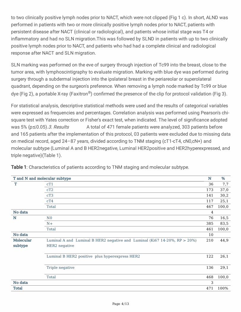
For statistical analysis, descriptive statistical methods were used and the results of categorical variableswere expressed as frequencies and percentages. Correlation analysis was performed using Pearson's chi-square test with Yates correction or Fisher's exact test, when indicated. The level of signi�cance adoptedwas 5% (p≤0.05).3. Results A total of 471 female patients were analyzed, 303 patients beforeand 165 patients after the implementation of this protocol, 03 patients were excluded due to missing dataon medical record, aged 24–87 years, divided according to TNM staging (cT1-cT4, cN0,cN+) andmolecular subtype (Luminal A and B HER2negative, Luminal HER2positive and HER2hyperexpressed, andtriple negative)(Table 1).
Table 1: Characteristics of patients according to TNM staging and molecular subtype.
T and N and molecular subtype N % T cT1 36 7,7
cT2 173 37,0cT3 141 30,2cT4 117 25,1Total 467 100,0
No data 4 N N0 76 16,5
N+ 385 83,5Total 461 100,0
No data 10 Molecularsubtype
Luminal A and Luminal B HER2 negative and Luminal (Ki67 14-20%, RP > 20%)HER2 negative
210 44,9
Luminal B HER2 positive plus hyperexpress HER2 122 26,1
Triple negative 136 29,1
Total 468 100,0No data 3 Total 471 100%
Page 5/13
Of these patients, 295 underwent mastectomy (simple, modi�ed radical, or skin-sparing) and 176underwent breast-conserving surgeries (quadrantectomy or lumpectomy). ALND was performed in 303patients, SLND in 156, and SLND followed by ALND in nine.
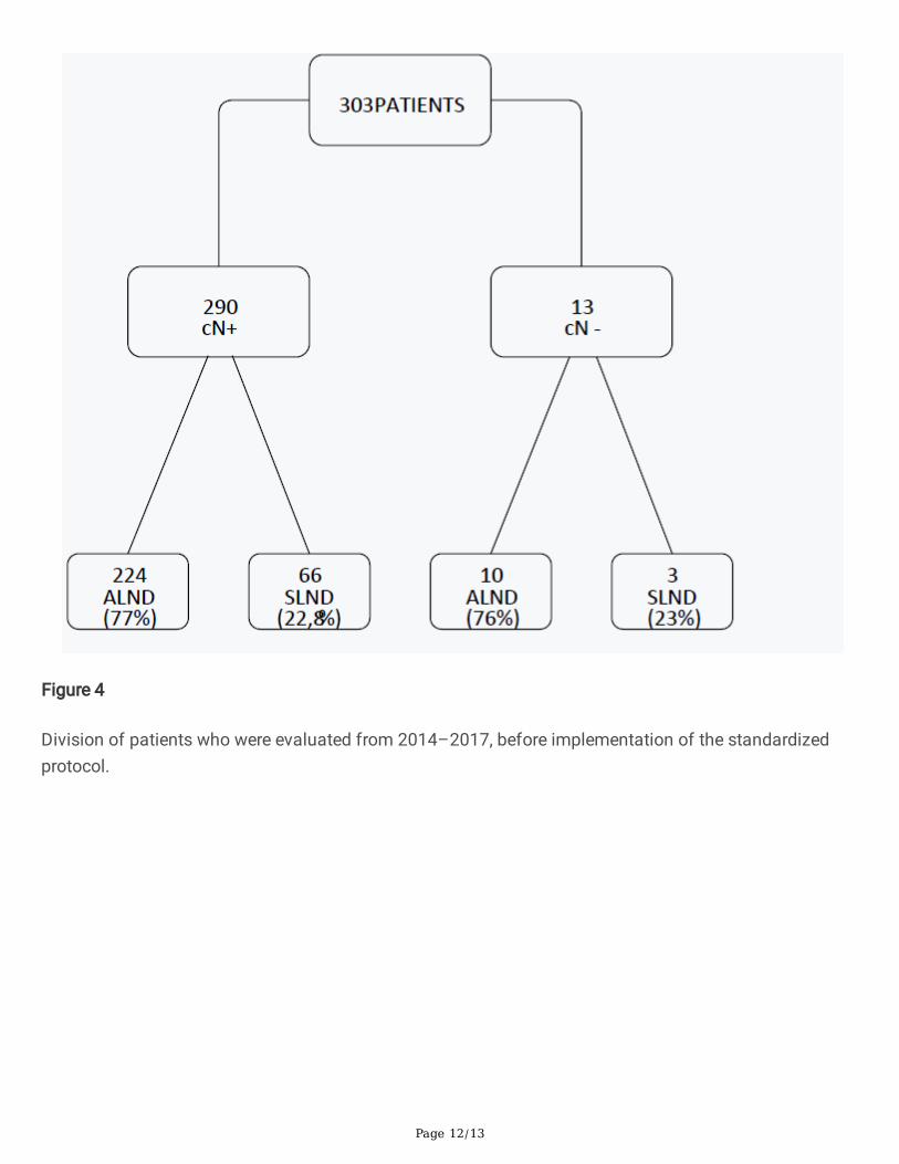
In the period from 2014 to 2017, prior to the implementation of a standardized protocol for pre-NACT axillary management, 303 patients were included (Fig 4).
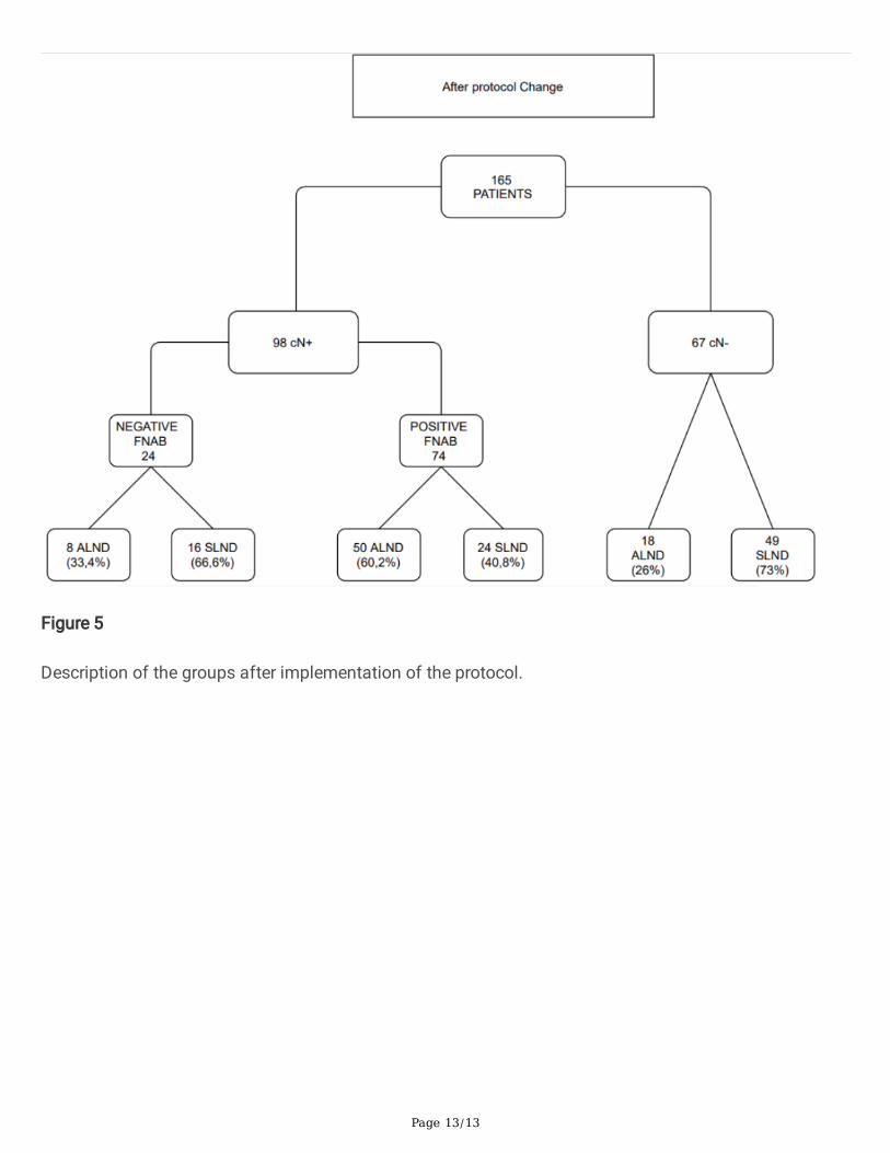
Beginning in 2017, a standardized protocol for pre-NACT axillary management was instituted, from whichtime 165 patients were included (Fig 5).
Sixty-seven of these patients were axillary clinically negative, 49 of whom underwent SLND or SLNDfollowed by ALND, and 18 of whom underwent ALND. The remaining 98 patients, who were axillaryclinically positive, were submitted to �ne needle aspiration biopsy to test for malignancy. Of the 24patients who tested negative for malignancy, sixteen underwent SLND and eight underwent ALND. Of the74 who tested positive for malignancy, 43 underwent ALND, 24 underwent SLND, and 7 underwent SLNDfollowed by ALND Prior to implementation of the standard protocol, 77.2% of patients underwentALND and 22.8% underwent SLND; after implementation, 59.2% underwent ALND and 40.8% SLND. Theincrease in SLND after protocol implementation was statistically signi�cant (p < 0.05). Rates ofSLND differed across molecular subtypes. Patients with tumors that were triple negative underwent SLNDmost frequently (44%), followed by Luminal B HER2 positive with HER2 hyperexpression (32%), andLuminal HER2 negative (26%). The difference between triple negative and Luminal patients wassigni�cant (p = 0.03).4.Discussion In the present study we describe a standardized protocol forpost-NACT axillary management which increases the e�cacy of target axillary dissection and reduces thefalse negative rate of SLND. NACT is an important tool in the treatment of certain breast cancersbecause it not only reduces tumor burden by initially treating the systemic micrometastatic disease, buthas also been shown to increase the rate of conservative surgeries in patients who would not otherwisebe candidates [12-14] NACT also plays a role in axillary downstaging, improving outcomes of clinicallypositive patients who underwent chemotherapy prior to SLND. The present study similarly found that, inthe ninety-eight patients who presented clinically positive axilla, NACT prevented lymphadenectomy in46% of cases. This result is nearly identical to that of Mamtani et al., whoreported in a prospective studythat 70% of clinically positive patients were eligible for SLND after NACT and 48% were able to avoidALND [15]. Together these studies demonstrate the role of NACT in reducing the need for ALND amongpatients with lymph node metastases. Considering the performance of lumpectomy after NACT,Bonadonna and Veronesi reported that NACT reduced large tumors to less than 3 cm in 81% of patients,allowing lumpectomy instead of radical mastectomy in 50–75% of patients for whom mastectomy wasinitially indicated [13-14]. The results of our study were in agreement, allowing lumpectomy in 52.6% ofpatients with T2 staging. Regarding molecular subtypes, we observed complete radiologicalresponse in 56% of triple negative tumors, and 54% of HER2 positive. A similar study also observedcomplete radiological response in 59% of cases [16]. Moreover, patients who were triple negativeunderwent signi�cantly more SLND than those in the Luminal (HER2 negative) subgroup. Thisstudy has limitations inherent to its retrospective design. First, the medical records were not standardized,

Page 6/13
especially regarding axillary status prior to the implementation of institutional protocol in 2017. Inaddition, the relatively small number of patients in the sample may have limited statistical analyses.Despite this, the present study demonstrates effectiveness in implementing a standardized protocol foraxillary management before and after NACT. The institutional protocol used was created with theobjective of reducing the number of false-negative results of SLND and minimizing technical limitationsof the target axillary dissection that make its routine implementation unfeasible, such as di�culty inlocating the clipped lymph node, especially when it has a complete response to treatment [17].
In conclusion, the results of this study showed an increase in the frequency of SLND afterimplementation of a standardized protocol for management of axilla after NACT in breast cancerpatients. Prospective studies, with a larger sample and in other centers, are necessary to validate theproposed technique.
DeclarationsFunding - Not applicable
Con�icts of interest/Competing interests - None
Availability of data and material – The datasets generated during and/or analysed during the currentstudy are not publicly available due to restrictions by our ethics committee but are available from thecorresponding author on reasonable request.
Code availability – Not applicable
Authors' contributions (all of them):
Substantial contributions to the conception or design of the work; or the acquisition, analysis, orinterpretation of data for the work; AND
Drafting the work or revising it critically for important intellectual content; AND
Final approval of the version to be published; AND
Agreement to be accountable for all aspects of the work in ensuring that questions related to theaccuracy or integrity of any part of the work are appropriately investigated and resolved.
Ethics approval :Trial registration number 2786/19, approved in October, 19, 2019.
References1. Nguyen TT, Hoskin TL, Day CN, Degnim AC, Jakub JW, Hieken TJ, Boughey JC, et al. Decreasing Use
of Axillary Dissection in Node-Positive Breast Cancer Patients Treated with NeoadjuvantChemotherapy. Ann SurgOncol. 2018 Sep;25(9):2596-2602

Page 7/13
2. Rubovszky G, Horváth Z, et al. Recent Advances in the Neoadjuvant Treatment of Breast Cancer. JBreastCancer. 2017;20(2):119-31
3. Haffty BG, McCall LM, BallmanKV, McLaughlin S, Jagsi R, Ollila DW, Hunt KK, Buchholz TA,BougheyJC, et al.Patterns of local-regional management following neoadjuvant chemotherapy inbreast cancer: Results from ACOSOG Z1071 (Alliance). Int J RadiatOncolBiolPhys. 2016;1;94(3):493-502.
4. Fleissig A, Fallow�eld LJ, Langridge CI, et. al. Post-operative arm morbidity and quality of life: resultsof the ALMANAC randomized trial comparing sentinel node biopsy with standard axillary treatmentin the management of patients with early breast cancer. Breast Cancer ResTreat. 2006;95(3):279-93
5. Boughey JC, Suman VJ, Mittendorf EA, et. al. Sentinel lymph node surgery after neoadjuvantchemotherapy in patients with node-positive breast cancer: the ACOSOG Z1071 (Alliance) clinicaltrial. JAMA. 2013;310(14):1455-61
�. Kuehn T, Bauerfeind I, Fehm T, et. al. Sentinel-lymph-node biopsy in patients with breast cancerbefore and after neoadjuvant chemotherapy (SENTINA): a prospective, multicentre cohort study.Lancet Oncol. 2013;14(7):609-18
7. Boileau JF, Poirier B, Basik M, et. al. Sentinel node biopsy after neoadjuvant chemotherapy in biopsy-proven node positive breast cancer: the SN FNAC Study. J Clin Oncol. 2015:33(3):258-64
�. Pilewskie M, Morrow M, et al. Axillary Nodal Management Following Neoadjuvant Chemotherapy: AReview. JAMA Oncol. 2017;1;3(4):549-555
9. Caudle AS, Yang WT, Krishnamurthy S, et. al. Improved Axillary Evaluation Following NeoadjuvantChemotherapy for Patients With Node-Positive Breast Cancer Using Selective Evaluation of ClippedNodes: Implementation of Target Axillary Dissection. J Clin Oncol. 2016;34:1072-8
10. Symmans WF, Peintinger F, Hatzis C, Rajan R, Kuerer H, Valero V, Assad L, et at. Measurement ofresidual breast cancer burden to predict survival after neoadjuvant chemotherapy. J Clin Oncol. 2007Oct 1;25(28):4414-22.
11. Osório C T; Soares F A, et al. Assessment of pathological response in breast cancer afterneoadjuvant chemotherapy: standardization of adapted protocol. Bras. Patol. Med.Lab. vol.48 no.6 Rio de Janeiro Dec. 2012
12. Aghian A, El-Ghamry MN, Merajver SD, et al. Clinical features and management of locally advancedand in�ammatory breast cancer. UpToDate Online 17.3 2009 Sep-Dec [last updated May 12].
13. Mauriac L, Durand M, Avril A, Dilhuydy JM, et al. Effects of primary chemotherapy in conservativetreatment of breast cancer patients with operable tumors larger than 3 cm. Results of a randomizedtrial in a single centre. Ann Oncol. 1991;2(5):347-54.
14. Bonadonna G, Veronesi U, Brambilla C, Ferrari L, Luini A, Greco M, et al.Primary chemotherapy toavoid mastectomy in tumors with diameters of three centimeters or more. J Nat Cancer Inst.1990;82(19):1539-45.
Page 8/13
15. Mantani A, Barrio A, King T, Van Zee K J, Pilewskie M et al. How Often Does NeoadjuvantChemotherapy Avoid Axillary Dissection in Patients With Histologically Con�rmed NodalMetastases? Resultsof a ProspectiveStudy .AnnSurgOncol 2016 Oct;23(11):3467-3474.
1�. Batt, J, Chambers, A, Al‐Allak, A, Vestey, S, Hunt, R, Massey, et al. Neo‐Adjuvant chemotherapy and itsaffects to the axilla—Can we safely downgrade axillary surgery to mirror the approach in the breast.The Breast Journal 2020, Aug; 26(09): 1667-1672.
17. Ecanow J, Abe H, Newstead G, Ecanow D, Jeske J, et al. Axillary staging of breast cancer: what theradiologist should know. Radiographics. 2013;33(6):1589-1612.
Figures

Page 9/13
Figure 1
Top panel: A: Protocol for patients with axilla clinically negative prior to NACT. Middle panel: 1B: Protocolfor patients with up to two clinically positive lymph nodes prior to NACT, which were clipped. Bottompanel: Protocol for patients with up to two clinically positive lymph nodes prior to NACT, which were notclipped.

Page 10/13
Figure 2
Photo of SLN stained with blue dye.

Page 11/13
Figure 3
X-ray photo of clipped lymph node. Pathological analysis (frozen section) of lymph nodes after NACT[10} was performed in three parts: 1- Macroscopic examination. In the perioperative examination, SLNswere sliced transversely to a thickness of 2 mm and examined by a pathologist for the presence of anywhitish areas of hardened consistency, suggestive of residual lymph node metastasis. All slices of lymphnode tissue were �xed in 10% buffered formalin and included in one or more para�n blocks for thede�nitive histological evaluation. 2- Microscopic evaluation. In the microscopic evaluation, a pathologistmeasured the linear dimension of the largest metastatic focus and described the presence of a possiblearea of pathological response, characterized by �brosis, hemorrhage, accumulations of macrophagesand a decrease in the lymph node parenchyma. Additional sections of 4-µm thickness were stained withhematoxylin-eosin and analyzed by a pathologist for the presence of isolated cells, or a group of atypicalepithelial cells compatible with residual neoplasia, which would determine ALND. The metal clip area wasalso described, characterized by foreign body-type gigantocellular reaction and lymphocytic in�ltratearound amorphous acidophilic material, compatible with the gel shell present in the clips used. 3- Tumorpresence. The presence of axillary nodal tumor deposits of any size, including isolated tumor cells,excluded a complete pathological response. Finally, the number of compromised lymph nodes wascounted and classi�ed to obtain the rCR index and classi�cation[11].
Page 12/13
Figure 4
Division of patients who were evaluated from 2014–2017, before implementation of the standardizedprotocol.
Page 13/13
Figure 5
Description of the groups after implementation of the protocol.