left-handed deoxyribonucleic acid double helix in solution
TRANSCRIPT

2036 Biochemistry 1981, 20, 2036-2041
Left-Handed Deoxyribonucleic Acid Double Helix in Solution+
C . K. Mitra, M. H. Sarma, and Ramaswamy H. Sarma*
ABSTRACT: Magnetic shielding constants were calculated for the synthetic deoxyribonucleic acid (DNA) double helix poly(dG-dC)*poly(dG-dC) from the x, y, and z coordinates of Z-DNA of Rich and co-workers [Wang, A. H-J., Quigley, G. J., Kolpak, F. J., Crawford, J. L., van Boom, J. H., van der Marel, G., & Rich, A. (1979) Nature (London) 282, 680-6861 and B-DNA of Arnott & Hukins [Arnott, S. , & Hukins, D. W. L. (1972) Biochem. Biophys. Res. Commun. 47, 1504-15091, taking into account the contribution to shielding from ring current effects and effects from the dia-
F r o m single-crystal crystallographic studies of the double- stranded deoxyhexanucleoside pentaphosphate d- CpGpCpGpCpG, Wang et al. (1979) have concluded that ... dCdG ... tracks of deoxyribonucleic acid (DNA) have the potentiality to achieve the left-handed double-helical spatial configurations in which the repeating unit is a dinucleotide with the structural features illustrated in Figure 1. Z-DNA is the name given by the authors to this structure; a hexamer duplex segment of Z-DNA is stereo is illustrated in Figure 2, and this structure is not identical with the one in Wang et al. (1979), but idealized (see legend of Figure 2). As early as 1972, Pohl & Jovin (1972) have demonstrated that addition of salt to poly(dG-dC).poly(dG-dC) results in a definite transformation of conformation. In a series of reports, Pohl and co-workers (Pohl & Jovin, 1972; Pohl, 1974, 1976; Pohl et al., 1972) presented evidence that the transition occurred for poly(dG-dC).poly(dG-dC) and estimated the thermody- namic and kinetic parameters associated with the transition. These pioneering investigations were recently confirmed by Pate1 et al. (1 979) by nuclear magnetic resonance (NMR) studies, and they have provided the first qualitative insight about the structure of poly(dG-dC)-poly(dG-dC) in high salt concentration. They have suggested that in these structures the symmetry unit repeats every two base pairs, the base pairing to be likely of the Watson-Crick type, and that every other glycosidic torsion angle and phosphodiester linkage adopts values other than those in B-DNA.
It is the thesis of Wang et al. (1979) that poly(dG-dC). poly(dG-dC) in high salt solutions may take up the Z con- figuration. Here, we present NMR results which clearly show that this prediction of Wang et al. (1979) about the confor- mation of the polymer in solution arrived at from single-crystal crystallographic investigations of a double-helical hexamer is essentially correct.
Methodology, Computations, and Experiments In the NMR approach, it is assumed that the chemical shifts
of the protons of the central base-paired nucleotides in a
+From the Institute of Biomolecular Sterecdynamics, State University of New York at Albany, Albany, New York 12222. Received July 25, 1980. This research was supported by grants from the National Cancer Institute of the National Institutes of Health (CA12462) and the Na- tional Science Foundation (PCM7822531). The NMR spectra were obtained at the North Eastern NSF Facility located at Yale.
magnetic and paramagnetic components of the atomic mag- netic anisotropy. Comparison of the calculated shielding values with the experimentally observed nuclear magnetic resonance shift data for poly(dG-dC)*poly(dG-dC) in high salt solution shows striking agreement for Z-DNA and considerable de- viation for EDNA, indicating that this synthetic DNA double helix in high salt solution can assume the spatial configuration of the left-handed Z-DNA double helix known to occur in crystals.
heptamer duplex are the shifts of protons in any nucleotide unit in the polymer duplex and that there are no end effects. This is a reasonable assumption because the polymer contains about 100 base pairs and the chemical shifts are not signifi- cantly affected by units beyond the third neighbor. First of all, we will assume that the heptamer duplex
d-CGCGCGC dGCGCGCG
exists in the Z conformation (Figure 2), and this is drawn schematically for ease of discussion in Figure 3. The chemical shifts of the central cytidine Co (Figure 3) will be affected by (a) the complementary unit Go, (b) the nearest-neighbor units on the same strand, GI and G+ (c) the nearest-neighbor units C-l and C1 on the complementary strand, (d) the next near- est-neighbors C2 and C2 on the same strand, (e) the next nearest-neighbors G-* and G2 on the complementary strand, (f) the next next nearest-neighbors G3 and G-3 on the same strand, and (g) the next next nearest-neighbors C3 and C-3 on the complementary strand. In the same way, the chemical shifts of the guanosine unit Go will be affected by the re- maining 13 nucleotide units.
The contribution to the chemical shifts originates from (a) the ring current effect of the bases, (b) the diamagnetic component of the atomic magnetic anisotropy of the bases, (c) the paramagnetic component of the atomic magnetic an- isotropy of the bases, and (d) the diamagnetic and paramag- netic components of the atomic magnetic anisotropy of the sugar-phosphate backbone. Shielding constants for a given proton of a nucleotide unit in a structure like the one in Figure 2 can be computed from x, y , and z coordinates, taking all the previously mentioned contributions into account (Mitra et al., 1980a,b; Cheng et al., 1980). The calculated shielding con- stant essentially provides the magnitude and direction of shielding a proton in a nucleotide unit such as Co (Figure 3) will experience as the unit is moved from an isolated envi- ronment to that in an organized structure like Z-DNA (Figure 2 ) . Obviously, in such a calculation, one cannot include the contribution to shielding from the parent nucleotide unit to which the proton belongs; Le., Co should be excluded when computing the effect of the remaining 13 units on Co; Go should be excluded when computing the effect of the remaining 13 units on Go. This creates some complications because the conformation of an isolated nucleotide may not be the same
0006-2960/8 1 /0420-2036$01.25/0 0 198 1 American Chemical Society
LEFT-HANDED DNA VOL. 2 0 , N O . 7 , 1 9 8 1 2037
Table I: Various Contributions (in ppm) to the Chemical Shifts of H6 of C, from the Remaining Bases in B-DNA and Z-DNA effect of
ring current diamagnetic anisotropy paramagnetic anisotropy total
effect froma B-DNA Z-DNA EDNA Z-DNA B-DNA Z-DNA B-DNA Z-DNA
Go -0.0 262 -0.0266 0.001 1 0.0011 -0.0444 -0.0450 -0.0696 -0.0705 GI 0.0025 0.1430 -0.0001 0.0033 -0.0103 0.2089 -0.0079 0.3552 G-I 0.0595 0.1067 -0.0056 -0.0068 0.0581 0.0956 0.1119 0.1955 c* -0.0048 -0.0055 0.0001 0 -0.0189 -0.0171 -0.0236 -0.0226 c-1 -0.0040 0.0158 0.0005 -0.0032 -0.01 13 -0.0174 -0.0147 -0.0049 C2 0.0053 0.0201 -0.0010 -0.0020 0.0133 0.0249 0.0 176 0.0429 C-2 0.0049 0.0060 0.0001 -0.0003 0.0168 0.0075 0.0218 0.0132 G2 0.0122 0.0009 -0.0015 -0.0001 0.0 16 1 0.0008 0.0268 0.0017 G-2 0.0050 0.0373 -0.0001 -0.0009 0.0072 0.0340 0.0121 0.0704 G3 0.0079 0.0072 -0.0002 -0.0003 0.0069 0.0060 0.0146 0.0129 G-3 0.0078 0.0074 -0.0006 -0.0004 0.0077 0.0065 0.0150 0.0134 c3 0.0063 0.0018 -0.0002 -0.0001 0.0112 0.0036 0.0173 0.0054 c-3 0.0046 0.01 11 -0.0005 -0.0008 0.0076 0.0165 0.0117 0.0269 total: 0.0810 0.3252 -0.0081 -0.0105 0.0600 0.3248 0.1330 0.6395
The relative dispositions of the various units are displayed in Figure 3.
Table 11: Contribution to Shielding (in ppm) of CH5, CH6, CHl', GH8, and GH1' in Z-DNA and B-DNA from the Various Parts of the Molecule
columna proton type of DNA 1 2 3 4 5
CHS EDNA Z-DNA
CH6 B-DNA Z-DNA
Z-DNA GH8 B-DNA
Z-DNA GH1' B-DNA
Z-DNA
CH1' EDNA
-0.0981 -0.0932 -0.0696 -0.0705 -0.0755 -0.0731 - 0.03 5 5 -0.0371 - 0.0397 -0.0294
0.7249 0.7533 0.1040 0.5507 0.2573 0.3016 0.1617
-0.0161 0.3225
-0.0131
-0.0405 0.0727
-0.0383 -0.0275 -0.0343
0.0374 -0.0536 -0.0284 -0.0402 -0.0279
0.0694 0.0228 0.0394 0.0561 0.0491 0.1171 0.0730 0.0159 0.0680 0.026 1
0.0609 0.1361 0.03 89 0.0721 0.0542 0.0451 0.0124 0.0175 0.0236 0.0150
6
0.046 1 0.0239 0.0296 0.0263 0.0292 0.0460 0.0135 0.0130 0.0146 0.023 1
7
0.0329 0.0229 0.0290 0.0323 0.0270 0.0413 0.0576 0.0135 0.0566 0.0300
total
0.7956 0.9385 0.1330 0.6395 0.3070 0.5154 0.2291
0.4054 0.0238
-0.0217
a Column 1, contributions from the complementary unit; column 2, contributions from the nearest neighbors in the same strand; column 3, contributions from the nearest neighbors in the complementary strand; column 4, contributions from the next nearest neighbors in the same strand; column 5, contributions from the next nearest neighbors in the complementary strand; column 6, contributions from the next next nearest neighbors in the same strand; column 7, contributions from the next next nearest neighbors in the complementary strand.
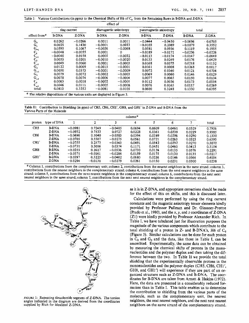
FIGURE 1: Repeating dinucleotide segment of Z-DNA. The torsion angles indicated in the diagram are derived from the coordinates supplied by Rich for idealized Z-DNA.
as it is in Z-DNA, and appropriate corrections should be made for the effect of this on shifts, and this is discussed later.
Calculations were performed by using the ring current constants and the magnetic anisotropy tensor elements kindly provided by Professor Pullman and Dr. Giessner-Prettre (Prado et al., 1980), and the x, y , and z coordinates of Z-DNA (Zl ) were kindly provided by Professor Alexander Rich. In Table I, we have tabulated just for illustration purposes the magnitude of the various components which contribute to the total shielding of a proton in Z- and B-DNA's, H6 of Co (Figure 3). Similar calculations can be done for each proton in Co and Go and the data, like those in Table I, can be assembled. Experimentally, the same data can be obtained by measuring the chemical shifts of protons in the mono- nucleotides and the polymer duplex and then taking the dif- ference between the two. In Table I1 we provide the total shielding that the experimentally observable protons in the mononucleotides and the polymer duplex (CH5, CH6, CHl', GH8, and GH1') will experience if they are part of an or- ganized structure such as Z-DNA and B-DNA. The coor- dinates for B-DNA are taken from Arnott & Hukins (1972). Here, the data are presented in a considerably reduced for- mation than in Table I. This table enables us to determine the contribution to shielding from the various parts of the molecule, such as the complementary unit, the nearest neighbors, the next nearest neighbors, and the next next nearest neighbors on the same strand of the complementary strand.

2038 B I O C H E M I S T R Y M I T R A , S A R M A , A N D S A R M A
P
B r/
9
FIGURE 2: Stereographic projection of a double-helical hexamer segment of B-DNA (top) and idealized Z-DNA (bottom) drawn from the coordinates of Arnott and from those supplied by Rich. Note that the Z-DNA segment shown is not identical with the one in Wang et al. (1979), where d-CpGpCpGpCG is shown as it appears in the crystal lattice as a continuous double helix and the terminal dG has a *E sugar conformation. The present drawing is for an idealized Z-DNA, called Z1, with the torsion angles as in Figure 1.
s3, 5'
FIGURE 3: Schematic drawing of a heptamer segment of poly(dG-
The total shielding for each of the above protons in Z- and B-DNA's is also shown in columns 6 and 8 in Table 111.
We have mentioned before that in these computations of magnetic shielding constants as a first approximation it is assumed that the conformation of the mononucleotide is the
dC).poly(dG-dC).
same when it is in an isolated state and when it is part of an organized structure of Z- and B-DNA's. Obviously, this is not a correct assumption, and, hence, appropriate amendments to the computed shielding values should be made. The dom- inant factor which affects the shielding values in these instances is a large change in the sugar-base torsion angle ( x ) as the monomer becomes part of the double helix. For example, if a purine mononucleotide in an isolated state has a x of 20' and if x changes to 250' when it becomes part of an organized structure, this change in x itself will cause the shielding of H8 to change as much as 1.3 ppm (Prado et al., 1978; Giess- ner-Prettre & Pullman, 1978). It is known that pyrimidine nucleotides exist in solution overwhelmingly in the anti con- formation (Davies, 1976; Lee et al. 1976; Ezra et al., 1977; Cheng & Sarma, 1977), as they are in Z- and B-DNA's, and, hence, no corrections are absolutely essential for the computed shielding constants for the cytidine protons. However, this is not true for the purine systems. It has been reported (Son et al., 1972) that 3'-guanine nucleotides in solution exist as a syn e anti equilibrium; the authors report that the observed x in the syn conformation is 240' and that in anti is 95' and that the percentage of the syn conformation is 72 and that of

LEFT-HANDED DNA V O L . 2 0 , N O . 7 , 1 9 8 1 2039
Table 111: Experimentally Observed Chemical Shifts for 3’-dCMP, 5-dCMP, 3‘-dGMP, and 5’-dGMP (10 mM in 4 M NaCl, pH 5.3-5.6, 90 “C) and That of Double-Helical Poly(dGdC).Poly(dG-dC) (37.5 mM in Nucleotides, pH 6 .3 ,4 M NaCI, 90 “C)“
column: 1 2 3 4 5 6 7 8 9 experimental theoretical
av of 3’- 3‘- 5 ‘- and 5‘- poly(dG-dC). Aexptl (high
proton nucleotide nucleotide nucleotides poly(dG-dC) salt concn)
CH5 2.91 2.95 2.96 1.91 1.05 CH6 4.64 4.66 4.65 4.03 0.62 CH1‘ 3.08 3.08 3.08 2.39 0.69 GH8 4.81 4.88 4.85 4.55 0.30 GH1’ 3.10 3.14 3.12 2.97 0.15
1.
AZ-DNA, AZ-DNA, AB-DNA, uncor for cor for uncor for AB-DNA, COI x change x change x change for x change
0.94 0.94 0.79 0.79 0.64 0.64 0.13 0.13 0.52 0.52 0.31 0.3 1
0.02 0.19 0.4 1 0.25 -0.02 0.25 0.23 -0.16
4 - compare- compare ’
Also given are the experimentally observed and theoretically computed differences in chemical shifts (A) between monomers and Z-DNA as well as B-DNA. All shifts are in ppm with respect to internal tetramethylammonium chloride.
anti 28. The 5’-guanine nucleotides also exist as a syn-anti equilibrium (Son et al., 1972), the percentages being 53 and 47, respectively. In addition, the sugar ring in the monomers is an equilibrium blend of 3E and 2E conformers (Lee et al., 1976; Ezra et al., 1977; Cheng & Sarma, 1977). The com- puted shielding values for GH8 and GH1’ were amended, taking the above synanti equilibrium into account and giving equal weights to the two modes of sugar pucker. The dominant factor which affects the shifts of GH8 and GH1’ in the mo- nomer is x and not sugar pucker. We used equal weights to the two modes of sugar pucker even though our own data (Cheng & Sarma, 1977) show that 2E/3E 70:30 to simplify calculations. Whether we use 50% 3E or 30% 3E, the results are not significantly influenced. The corrections for x were made as rigorously as possible within the framework of available theoretical (Prado et al., 1978; Giessner-Prettre & Pullman, 1977,1978) and experimental data (Son et al., 1972). The corrected shielding values for Z-DNA and Arnott & Hukins B-DNA (hereinafter called AH-BDNA) are displayed in columns 7 and 9, Table 111.
Experimentally, the chemical shifts of an isolated mono- nucleotide in the same environmental conditions as poly(dG- dC).poly(dG-dC) in 4 M salt are obtained by recording the ‘H NMR spectra of 3’-dCMP, S’dCMP, 3’-dGMP, and 5’- dGMP, 8 mM in 4 M NaCl, 90 OC, by using the supercon- ducting 270-MHz Fourier-transform NMR spectrometer. In order to make sure that we are dealing with isolated mono- nucleotides, data for 3’-dGMP were obtained at 2 mM con- centration as well. The shifts were identical at 2 and 8 mM concentrations, 90 OC. Both DDS and TMA were used as an internal standards. The assignments of the signals were made according to Cheng & Sarma (1977). The data are sum- marized in Table 111. The data for poly(dG-dC).poly(dG-dC) in 4 M NaCl at 90 OC were taken from the literature (Patel, 1979) are are reproduced in Table I11 after conversation to TMA scale. The assignments for CH5, CH6, and GH8 are according to Patel (1979). For Hl’, we have assigned the high-field resonance to CH1’ and the low-field one to GHl’, on the basis of simple, generally valid qualitative arguments such as the following: (a) intrinsically, GH1’ will be shifted to lower fields compared to CH1’ because of the larger in-plane deshielding ability of guanine, and (b) the shielding ability (perpendicular to the plane) of G is greater than C, so much so that CH1’ may appear at a higher field than GH1’. We realize that these rules are primitive and other geometric factors can affect the position of the signal, and that un- equivocal assignments are possible only by the synthesis of polymers containing selectively deuterated residues as has been reported from this laboratory before (Lee et al., 1976; Ezra
et al., 1977). However, as a first approximation, these guidelines and the assignments outlined above are satisfactory.
Evidence in Support of the Left-Handed Z-DNA Double Helix in Solution. The experimentally observed difference (A) in chemical shifts between the mononucleotides and the poly(dG-dC).poly(dG-dC) double helix is shown in column 5 , Table 111. Comparison of these data with those computed for Z-DNA (column 7, Table 111) and AH-BDNA (column 9, Table 111) makes it unmistakably clear that poly(dG- dC).poly(dG-dC) in high salt concentration assumes the spatial configuration of Z-DNA and never that of B-DNA. Partic- ularly striking is that in our experiments and calculations we have used five separate sites, Le., CH5, CH6, CHl’, GH8, and GHl’, in the double helix, and there is overall agreement between the computations and experimental observations in each and every case for Z-DNA; in four sites (CH5, CH6, GH8, and GHl’), the agreement is within 0.1 ppm, and in one case, CHl’, it is within 0.17 ppm. In the type of approach we have undertaken in the present study, an agreement be- tween theoretical computation and experimental data should be considered excellent if it is within 0.1 ppm and very good if it is between 0.1 and 0.2 ppm, provided one is comparing shielding for a collection of different protons. This small discrepancy is due to some of the intrinsic assumptions involved in the computation of magnetic shielding constants (Giess- ner-Prettre & Pullman, 1970a,b) and due to our treatment of the organized structure of the helix as having no internal motions; Le., we treat Z-DNA in solution as a static structure identical with the crystal structure devoid of any internal torsional and flexural motions. Even though some informaiton is available about the motions of the DNA double helix (Hogan & Jardetzky, 1979), little is known about Z-DNA dynamics. Until quantitative information is available on this aspect of Z-DNA, we are unable to incorporate this in our calculations.
The NMR methodology based upon derivation of magnetic shielding constants from x , y, and z coordinates described in this paper will tell if the calculated structure is widely different form the actual one as manifested in the experimental shielding constants. A comparison of the calculated magnetic shielding constants for AH-BDNA (column 9, Table 111) with the ex- perimental values (column 5 , Table 111) illustrates this aspect of our approach. For example, one notices that GH8 of the mononucleotide will undergo a deshielding of 0.16 ppm when it becomes part of the B form of poly(dG-dC).poly(dG-dC) in high salt concentration. Also notice the enormous dis- crepancy (about 0.5 ppm) in the value of CH6 for the ex- perimental shielding constant and that computed for the B forms, 0.62 vs. 0.13 ppm; on the other hand, in the Z form,

2040 B I O C H E M I S T R Y M I T R A , S A R M A , A N D S A R M A
BDNA
b b
ZDNA FIGURE 4: Top view in stereo of the sequence GCG in B-DNA (top) and Z-DNA (bottom), drawn from the coordinates of Arnott and from those supplied by Rich. The central base pair is drawn in dark, and hydrogens were removed for clarity. The position of CH6 is indicated. Note that in the GCG system the CH6 occupies a strong shielding zone in Z-DNA and considerably less shielded zone in B-DNA. Also see Table I.
there is very close agreement for the value of CH6, 0.62 vs. 0.64 ppm. Stereoscopic examination of the top view of the segment GCG in one strand of Z-DNA and that of AH- BDNA (Figure 4) clearly reveals that CH6 in the Z form lies in a strongly shielded zone unlike that in the AH-B form, and the experimental data in high salt concentration for poly(dG- dC)-poly(dG-dC) clearly show that CH6 is very strongly shielded, supporting the Z form. The data in Table I dem- onstrate that CH6 of Co is strongly shielded by the para- magnetic component of the magnetic anisotropy and ring current effects of GI and G-l in Z-DNA, and these effects are small in B-DNA. In addition to the disagreement between the experimentally observed ACH6 and AGH8 and that computed for the B form, the experimental A values for CH5 and CH 1’ display considerable difference from those computed for the B form (Table 111).
It is important to point out that the detailed computations of the magnetic shielding constants presented in this paper are based on the NMR theory for nucleic acids developed by Pullman and Giessner-Prettre over the period of the last decade; the several references cited in this paper from their laboratory are only a small part of this monumental work. In the past, this NMR theory has been successfully employed to derive the solution conformations of single-stranded oligo-
nucleotides and miniature double helices (Cheng et al., 1980; Mitra et al., 1980a; Sarma et al., 1979). The present paper represents their successful application to larger systems. Striking agreement is observed between the computed mag- netic shielding constants and those observed experimentally because the structure of Z-DNA is known to a resolution of 0.9 A and because this organized structure is maintained intact to a large extent under solution conditions.
Acknowledgments We are deeply indebted to Professor Pullman and Dr.
Giessner-Prettre for providing us with the tensor elements to compute the paramagnetic and diamagnetic components of the atomic magnetic anisotropy and ring current constants. We are indebted to Professor Alexander Rich for providing us with the coordinates of Z-DNA. We thank Dr. D. Patel, Professor N. C. Seeman, and L. Lerman for providing ex- tensive review of the manuscript.
References Arnott, S . , & Hukins, D. W. L. (1972) Biochem. Biophys.
Cheng, D. M., & Sarma, R. H. (1977) J . Am. Chem. SOC. Res. Commun. 47, 1504-1 509.
99, 7333-7348.

Biochemistry 1981,
Cheng, D. M., Danyluk, S. S., Dhingra, M. M., Ezra, F. S., MacCoss, M., Mitra, C. K., & Sarma, R. H. (1980) Bio- chemistry 19, 2591-2597.
Davies, D. B. (1976) Stud. Biophys. 55, 29-38. Ezra, F. S., Lee, C. H., Kondo, N. S., Danyluk, S. S., &
Giessner-Prettre, C., & Pullman, B. (1970a) J . Theor. Biol.
Giessner-Prettre, C., & Pullman, B. (1970b) J. Theor. Biol.
Giessner-Prettre, C., & Pullman, B. (1977) J . Theor. Biol.
Giessner-Prettre, C., & Pullman, B. (1978) in Nuclear Magnetic Resonance Spectroscopy in Molecular Biology (Pullman, B., Ed.) pp 147-161, Reidel, Dordrecht, The Netherlands.
Hogan, M. E., & Jardetzky, 0. (1979) Proc. Natl. Acad. Sci.
Lee, C. H., Ezra, F. S., Kondon, N. S., Danyluk, S. S., &
Mitra, C. K., Dhingra, M. M., & Sarma, R. H. (1980a)
Mitra, C. K., Sarma, R. H., Giessner-Prettre, C., & Pullman,
Sarma, R. H. (1977) Biochemistry 17, 1977-1987.
27, 87-93.
27, 341-342.
65, 189-202.
U.S.A. 76, 6341-6344.
Sarma, R. H. (1976) Biochemistry 16, 3627-3639.
Biopolymers 19, 1435-1450.
20, 204 1-2047 204 1
B. (1980b) Znt. J . Quantum Chem. (in press). Patel, D. J. (1979) in Stereodynamics of Molecular Systems
(Sarma, R. H., Ed.) pp 397-435, Pergamon Press, Oxford and New York.
Patel, D. J., Canuel, L. L., t Pohl, F. M. (1979) Proc. Natl. Acad. Sci. U.S.A. 76, 2508-2511.
Pohl, F. M. (1974) Eur. J . Biochem. 42, 495-504. Pohl, F. M. (1976) Nature (London) 260, 365-366. Pohl, F, M., & Jovin, T. M. (1 972) J. Mol. Biol. 67,375-396. Pohl, F. M., Jovin, T. M., Baehr, W., & Holbrook, J. J. (1972)
Proc. Natl. Acad. Sci. U.S.A. 69, 3805-3809. Prado, F. R., Giessner-Prettre, C., & Pullman, B. (1 978) J.
Theor. Biol. 74, 259-277. Prado, F. R., Giessner-Prettre, C., & Pullman, B. (1980) J .
Mol. Struct. (in press). Sarma, R. H., Dingra, M. M., & Feldmann, R. J. (1979) in
Stereodynamics of Molecular Systems (Sarma, R. H., Ed.) pp 251-264, Pergamon Press, New York.
Son, T-D., Guschlbauer, W., & Gueron, M. (1972) J . Am. Chem. SOC. 94, 7903-791 1,
Wang, A. H-J., Quigley, G. J., Kolpak, F. J., Crawford, J. L., van Boom, J. H., van der Marel, G., & Rich, A. (1 979) Nature (London) 282, 680-686.
Genetic Identification and Purification of the Respiratory NADH Dehydrogenase of Escherichia coli+ Anthony Jaworowski, Hugh D. Campbell, Maria I. Poulis, and Ian G. Young*
ABSTRACT: Escherichia coli membrane particles were solu- bilized with potassium cholate. An NADH:ubiquinone oxi- doreductase was resolved by hydroxylapatite chromatography of the solubilized material. This enzyme has been identified as the respiratory NADH dehydrogenase since it is absent in chromatograms of solubilized material from an ndh mutant strain. Such mutants lack membrane-bound NADH oxidase activity and have previously been shown to have an inactive NADH dehydrogenase complex [Young, I. G., & Wallace, B. J. (1976) Biochim. Biophys. Acta 449, 376-3851. The respiratory NADH dehydrogenase was amplified 50- to 100-
x e respiratory NADH dehydrogenase catalyzes the transfer of electrons from NADH, generated mainly by glycolysis and the tricarboxylic acid cycle, to the respiratory chain. The immediate electron acceptor for the enzyme is believed to be ubiquinone. The electrons are passed ultimately to molecular oxygen, and ATP is synthesized via oxidative phosphorylation. The enzyme therefore links the major catabolic and energy- producing pathways in the cell. The mitochondrial enzyme has received considerable attention [e.g., see Ragan (1976) for a review], but, despite intensive investigation, very little is known about the mechanism of electron transfer, the energy conservation associated with it, OT even the structure of the enzyme complex catalyzing these reactions.
Comparatively little work has been done with the corre- sponding enzyme from E. coli despite the advantages that
'From the Department of Biochemistry, John Curtin School of Medical Research, Australian National University, Canberra City, A. C.T., 2601 Australia. Received August 20, 1980.
fold in vivo by using multicopy plasmid vectors carrying the ndh gene and then purified to homogeneity on hydroxylapatite. Hydroxylapatite chromatography of cholate-solubilized ma- terial from genetically amplified strains purified the enzyme -800- to 1000-fold relative to the activity in wild-type mem- branes. By use of a large-scale purification procedure, 50-100 mg of protein with a specific activity of 500-600 pmol of reduced nicotinamide adenine dinucleotide oxidized min-' mg-l at pH 7.5, 30 OC, was obtained. Sodium dodecyl sulfate gel electrophoresis of the purified enzyme showed that the enzyme consists of a single polypeptide with an apparent M, of 45 OOO.
genetic manipulation of this system can offer. In early at- tempts to solubilize and purify the enzyme from crude extracts (Wosilait & Nason, 1954; Bragg, 1965) or small particles (Kashket & Brodie, 1963; Bragg & Hou, 1967a,b; Gutman et al., 1968), no indications were given of the purity of prep arations, nor were there adequate criteria to assign the ac- tivities purified to the respiratory dehydrogenase. Recently, Dancey et al. (1976) reported the purification of an NADH dehydrogenase from osmotically lysed membrane vesicles. The purified enzyme, which was composed of a single subunit with a molecular weight of 38 000, could catalyze reduction of artificial electron acceptors such as potassium ferricyanide or dichlorophenolindophenol. Its activity toward ubiquinone was not reported.
We have previously isolated E. coli mutants (designated ndh) which have <2% of the wild-type level of membrane- bound NADH oxidase activity (Young & Wallace, 1976). The mutation was shown to affect the NADH dehydrogenase complex. By use of these mutants, the gene coding for the
0006-2960/81/0420-2041%01.25/0 , , ~ ~. ~ - . ~ _. , . 0 1981 American Chemical Society