review dna structure. deoxyribonucleic acid dna deoxyribose sugar double helix a -2-t, c-3-g strands...
TRANSCRIPT
REVIEW
DNA Structure

Deoxyribonucleic Acid
DNADeoxyribose sugarDouble helixA -2-T, C-3-GStrands are complementaryPurines: A and GPyrimidines: T and C
Ribonucleic Acid
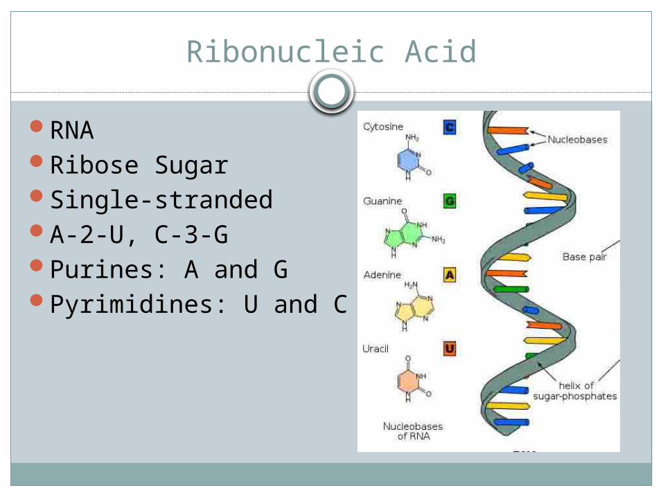
RNARibose SugarSingle-strandedA-2-U, C-3-GPurines: A and GPyrimidines: U and C
DNA Replication
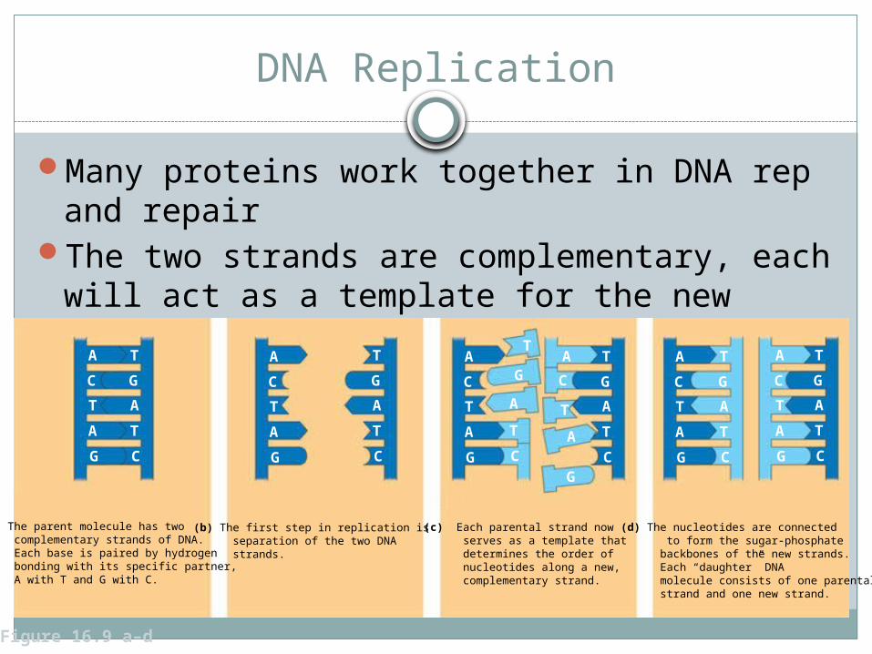
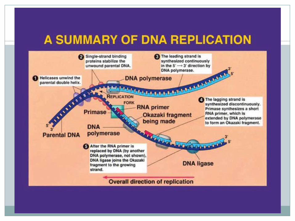
Many proteins work together in DNA rep and repair
The two strands are complementary, each will act as a template for the new strand in rep
(a) The parent molecule has two complementary strands of DNA. Each base is paired by hydrogen bonding with its specific partner, A with T and G with C.
(b) The first step in replication is separation of the two DNA strands.
(c) Each parental strand now serves as a template that determines the order of nucleotides along a new, complementary strand.
(d) The nucleotides are connected to form the sugar-phosphate backbones of the new strands. Each “daughter” DNA molecule consists of one parental strand and one new strand.
A
C
T
A
G
A
C
T
A
G
A
C
T
A
G
A
C
T
A
G
T
G
A
T
C
T
G
A
T
C
A
C
T
A
G
A
C
T
A
G
T
G
A
T
C
T
G
A
T
C
T
G
A
T
C
T
G
A
T
C
Figure 16.9 a–d

DNA Replication is semiconservative
Each of the two new daughter molecules will have one old strand (parent strand) and one newly made strand
Figure 16.10 a–c
Conservativemodel. The twoparental strandsreassociate after acting astemplates fornew strands,thus restoringthe parentaldouble helix.
Semiconservativemodel. The two strands of the parental moleculeseparate, and each functionsas a templatefor synthesis ofa new, comple-mentary strand.
Dispersivemodel. Eachstrand of bothdaughter mol-ecules containsa mixture ofold and newlysynthesizedDNA.
Parent cellFirstreplication
Secondreplication
Origins of Replication
Sites where the two strands are separated Unwound by Helicase
Euks: may have hundreds or even thousandsStabilized by SSBPs until replication is
complete
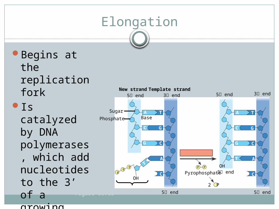
Elongation
Begins at the replication fork
Is catalyzed by DNA polymerases, which add nucleotides to the 3’ of a growing strand
Figure 16.13
New strandTemplate strand5 end 3 end
Sugar A TBase
C
G
G
C
A
C
T
PP
P
OH
P P
5 end 3 end
5 end 5 end
A T
C
G
G
C
A
C
T
3 endPyrophosphate
2 P
OH
Phosphate
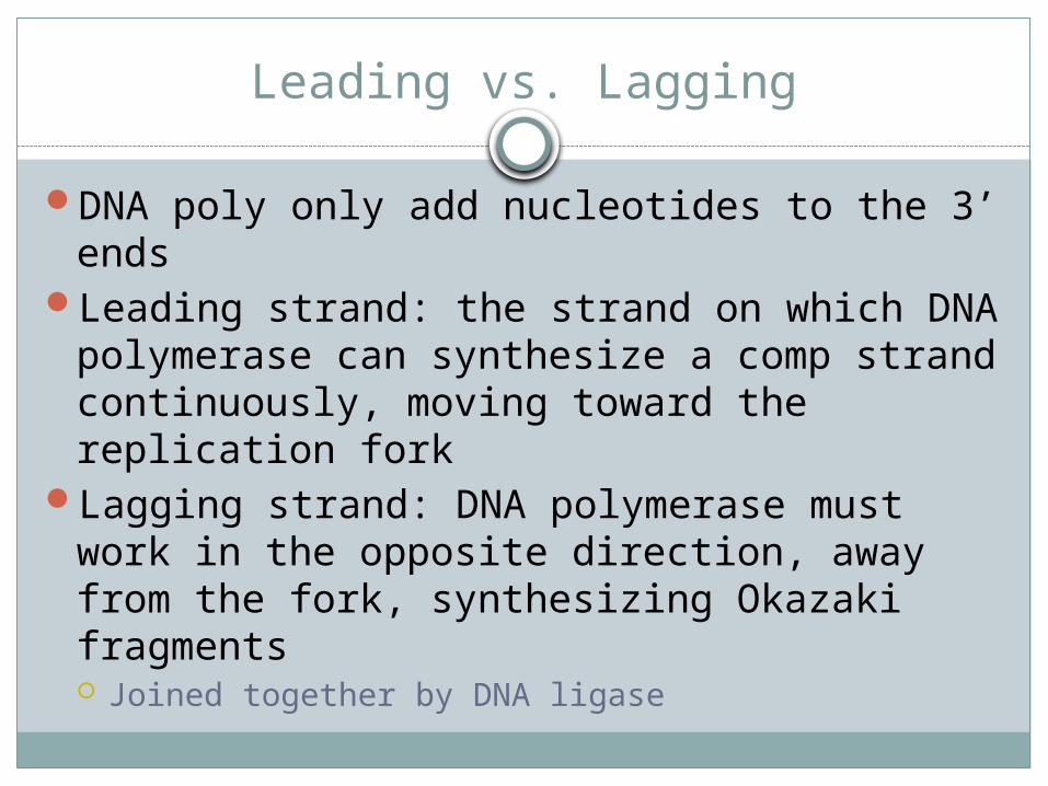
Leading vs. Lagging
DNA poly only add nucleotides to the 3’ endsLeading strand: the strand on which DNA
polymerase can synthesize a comp strand continuously, moving toward the replication fork
Lagging strand: DNA polymerase must work in the opposite direction, away from the fork, synthesizing Okazaki fragments Joined together by DNA ligase


Initiation
DNA poly cannot initiate rep, since they can only add nucleotides to the 3’ end
And RNA primer is required, synthesized by Primase One for the leading strand Multiple for lagging strand, each Okazaki fragment
must be primed separately
DNA Replication “Machine”
Various proteins participate in rep DNA poly, helicase, ligase, etc
They form a single, large complex or machine

Proofreading and Repairing DNA
DNA poly proofread newly synthesized DNA an replace any incorrect nucleotides
Excision repair: enzymes cut out and replace damaged stretches of DNA
Replicating the Ends of DNA Molecules
The ends of euk chromosomal DNA get shorter with each round of replication
So, the ends are made of repeating nucleotide sequences called telomeres Postpone the erosion of genes near the ends of DNA
moleculesIn germ, cancer, stem cells:
Telomerase catalyzed the lengthening of telomeres to prevent the loss of essential genes
Teach it to a 7th Grader
Draw a flow chart that uses analogies to explain the roles of each protein/enzyme involved
Must have drawings and explanationsAnalogy example:
Helicase : Pair of scissors