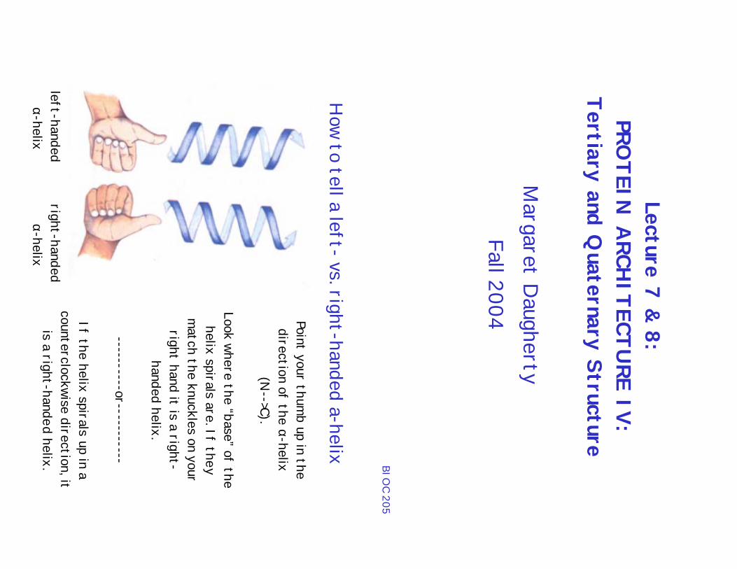
how to tell a left- vs. right-handed a-helix tertiary and...
TRANSCRIPT
Lecture 7 & 8:PRO
TEIN A
RCHITECTU
RE IV:Tertiary and Q
uaternary Structure
Margaret D
aughertyFall 2004
BIOC 205
right-handed α-helix
left-handed α-helix
Point your thumb up in the
direction of the α-helix(N
-->C).
Look where the “base” of thehelix spirals are. If they
match the knuckles on yourright hand it is a right-
handed helix.
----------or-----------
If the helix spirals up in acounterclockwise direction, it
is a right-handed helix.
How to tell a left- vs. right-handed a-helix

Tertiary Structure
•Tertiary structuredescribes how the
secondary structureunits associatewithin a single
polypeptide chain togive a three-dim
ensionalstructure
flavodoxin
BIOC 205
formation of large num
ber of intramolecular hydrogen bonds
reduction in hydrophobic surface area from solvent
Tertiary Structure: Basic Tenets - the “truths”1). A
ll information for folding is contained in the prim
ary sequence.2). Secondary structure form
ation is spontaneous - a consequence of theform
ation of hydrogen bonds.3). N
o protein is stable as a single layer - hence secondary structuralelem
ents pack together in sheets.4). Connections between structural elem
ents are short - minim
ization ofdegrees of freedom
- keeps structures compact.
Consequences1). Secondary structures are arranged in a few com
mon patterns - i.e,
resulting in protein “families”.
2). Proteins fold to form the m
ost stable structure. Stability arises from:
BIOC 205

All alpha
(human growth horm
one)
Note that som
eparts of a proteinstructure are not
regular (i.e., helical-like or sheet-like) .
These are oftenreferred to asdisordered or
random coil regions.
However a betternom
enclature is“natively random
”.A
ll beta(retinol binding protein)
Tertiary Structures
Alpha-beta barrel(triose isom
erase)
BIOC 205
Tertiary Structures
Globularproteins:
compact structures;
different folds fordifferent functions
Mem
braneProteins:
found associatedwith various
mem
brane systems
BIOC 205
Fibrousproteins:
Filamentous; playa m
ajorstructural role in
cells & tissues

Fibrous Proteins•
Share properties that give strength &/or flexibilityto the structures in which they occur;
• Fundam
ental unit is a simple repeating elem
ent ofsecondary structure;
• Insoluble in water; large percentage of hydrophobicam
ino acids;•
Usually the hydrophobic surfaces are hidden in the
elaborate supramolecular com
plexes;•
mechanically strong ; perform
important structural
functions•
Strength is enhanced by cross-links (disulfidebonds).
BIOC 205
BIOC 205
Secondary Structures andProperties of Fibrous Proteins
Structu
reC
haracteristicsExam
ples ofoccu
rrence
α-H
elix, Cross-linked
by disulfide bondsT
ough, insolubleprotective structures ofvarying hardness andflexibility
α – K
eratin of hair,feathers and nails
β-Conform
ationSoft, flexible filam
entsSilk fibroin
Collagen triple helix
High tensile strength,
without stretch
Collagen of tendons,
bone matrix

FIBROUS PRO
TEINS: α-Keratin
What: Part of the “interm
ediate filament proteins” which have m
ajorstructural roles in nuclei, cytoplasm
and cell surfacesW
here: Found in hair, fingernails, claws, horns, animal skin
Composition: Long stretches of α-helices (> 300 residues)
BIOC 205
Coiled-Coils
•Interactions are stabilized byhydrophobic interactions between the α-helices;
•H
eptad repeat (a-b-c-d-e-f-g)n where a &d are nonpolar & lie in the center of thecoiled coil;
BIOC 205
Evolved for strength; helicalnature confers flexibility
Coiled-coil is a “super twist”left-handed helix
Distortion of helix to 3.5
residues/turn
Hydrophobic faces interacting
in a close interlocking pattern

α-keratin:
Contact side chains (red balls) interlock
FIBROUS PRO
TEINS: β-Keratin
What: Part of the “fibroin proteins”
Where: silk, bird feathers
Composition: stacked anti-parallel β-sheets; strength
Sequence: Alternating G
ly-Ala/S
er
BIOC 205
Ala/Ser faces
interact withone another
Gly faceinteracts withanother glyface
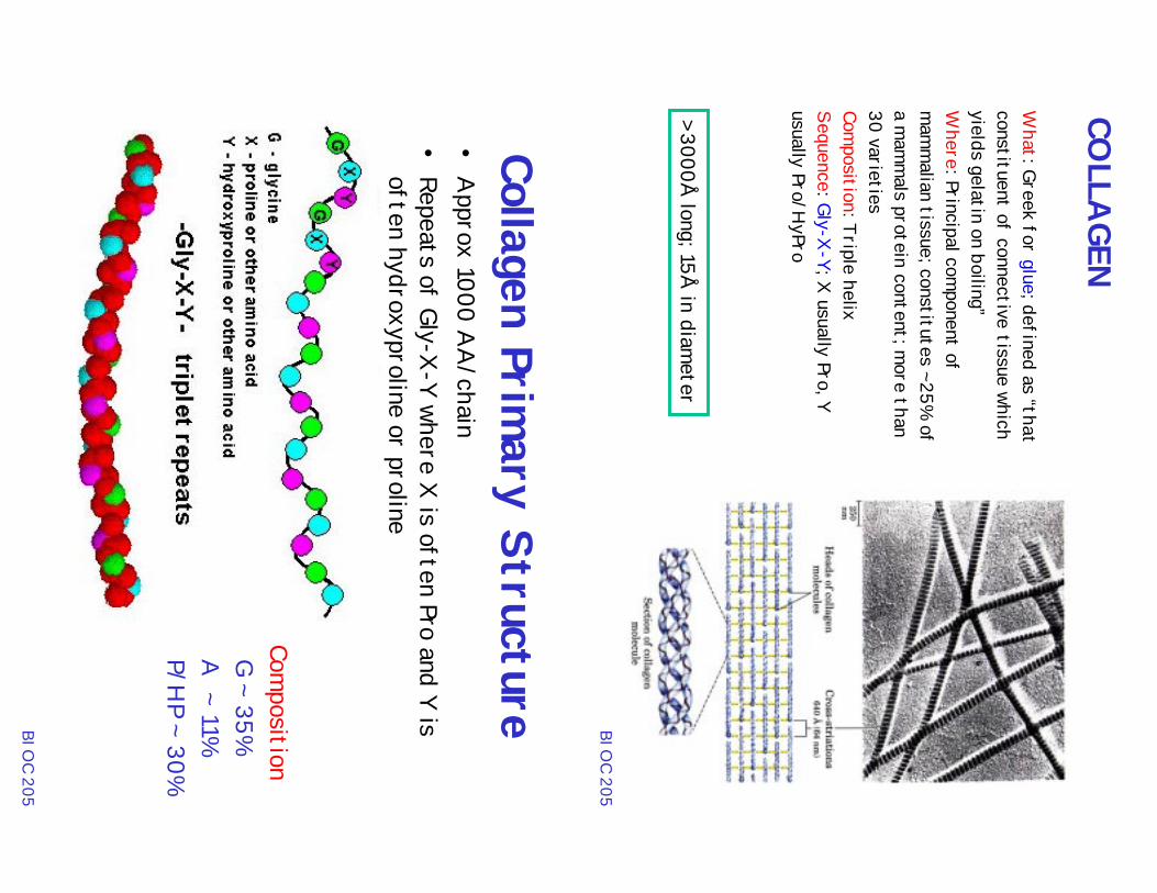
COLLA
GEN
What: Greek for glue; defined as “that
constituent of connective tissue whichyields gelatin on boiling”W
here: Principal component of
mam
malian tissue; constitutes ~25%
ofa m
amm
als protein content; more than
30 varietiesCom
position: Triple helixSequence: Gly-X
-Y; X usually Pro, Y
usually Pro/HyPro
> 3000Å long; 15Å
in diameter
BIOC 205
Collagen Primary Structure
•A
pprox 1000 AA
/chain•
Repeats of Gly-X-Y where X
is often Pro and Y isoften hydroxyproline or proline
Composition
G ~ 35% A
~ 11% P/H
P ~ 30%
BIOC 205
MO
DIFIED
AM
INO
ACID
Spost-translational m
odifications add functionality to amino acid
Hyp: stabilizes
tropocollagen viaintrachain H
-bonds
Hyl: stabilizes fibrils
via its ability to cross-link; attachm
ent ofCH
O groups
BIOC 205
Consequences of Collagen Primary Structure
Distortion of backbone
due to high content ofglycines and prolines
Can’t form “norm
al”secondary structures
Forms triple helix
Every third residuefaces inside
Interior is compact;
hence interior residueis glycine
BIOC 205
Consequences of Collagen Primary Structure
Fit occurs because Glystrand1 lies adjacent to X
strand2 and Ystrand3
Stabilization from hydrogen bonds
Glystrand1 N
-H to X
strand2 C=O hydrogen bond
Hydroxyproline form
s hydrogen bonds BIO
C 205
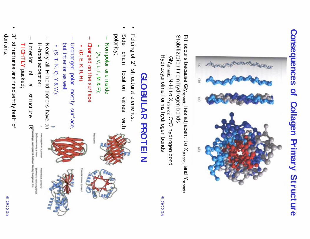
•Folding of 2˚ structural elem
ents;•
Side chain
location varies
withpolarity;–
Non-polar are inside
• (A
, V, L, I, M & F);
–Charged on the surface•
(D, E, K, R, H
);
–U
ncharged polar mostly surface,
but interior as well•
(S, T, N, Q
, Y & W);
–N
early all H-bond donors have an
H-bond acceptor;
–Interior
of a
structure is
TIGHTLY packed;
•3˚ structures are frequently built ofdom
ains.
GLOBU
LAR PRO
TEIN
BIOC 205

Globular Proteins: 4 major classes
I: antiparallel α-helixPacks in bundles; Left-handed twistU
sually regular, uniform; U
sually 4-helix bundles
Globin proteinsII: Parallel or m
ixed β-sheet proteins parallel β-sheetshave hydrophobes on both sides of sheet ---> thesem
ust be core structuresIII. A
ntiparallel β-sheet structures antiparallel β-sheetshave hydrophobes on one side of sheet and polarresidues on the other side. These β-sheet structurescan be surface exposed.
IV: Metal & D
isulfide rich proteins Small, < 100 a.a.
Structure dependent upon either the metal or disulfide
CORES O
F PROTEIN
S: α-HELICES A
ND β-SH
EETS
Ribonuclease ACitrate synthase
BIOC 205
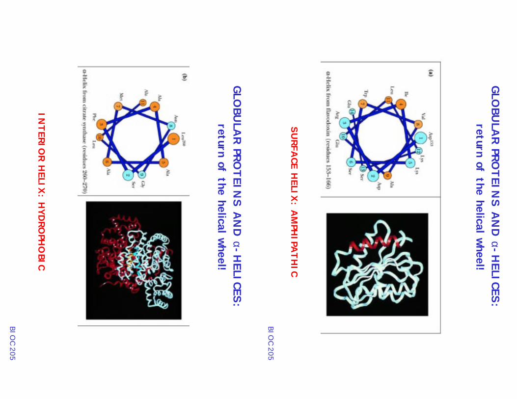
GLOBU
LAR PRO
TEINS A
ND α-H
ELICES:return of the helical wheel!
SURFA
CE HELIX
: AM
PHIPA
THIC
BIOC 205
GLOBU
LAR PRO
TEINS A
ND α-H
ELICES:return of the helical wheel!
INTERIO
R HELIX
: HYD
ROPH
OBIC
BIOC 205

GLOBU
LAR PRO
TEINS A
ND α-H
ELICES:return of the helical wheel!
SOLVEN
T-EXPO
SED H
ELIX: PO
LAR/CH
ARGED
BIOC 205
β-sheets: an example of IFA
BP
Hydrophobic = green
Charged = red
Looking in cavityfrom
bottomleft of the 3front sheets
pdb file: 1AEL
BIOC 205
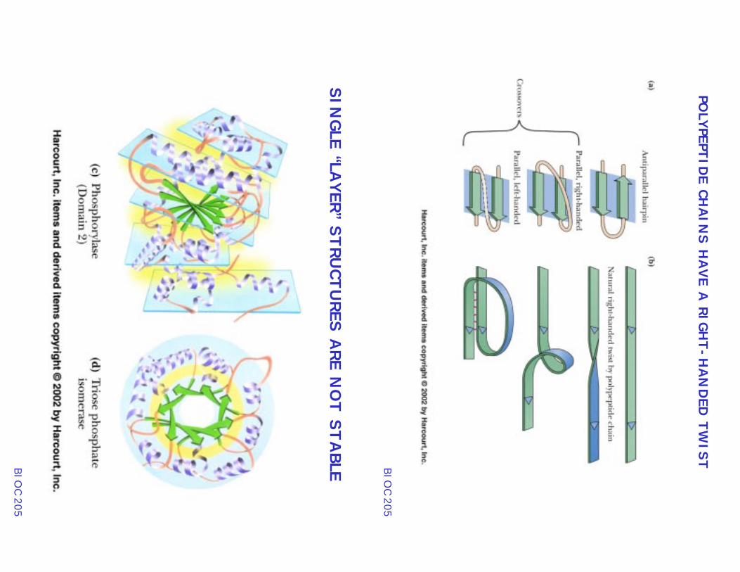
POLYPEPTID
E CHAIN
S HAVE A
RIGHT-H
AN
DED
TWIST
BIOC 205
SINGLE “LA
YER” STRUCTU
RES ARE N
OT STA
BLEBIOC 205
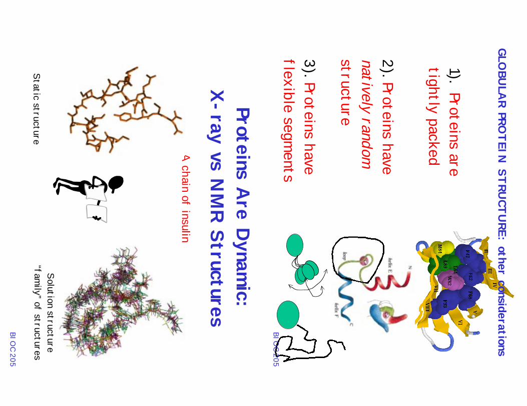
1). Proteins aretightly packed
GLOBU
LAR PRO
TEIN STRU
CTURE: other considerations
2). Proteins havenatively randomstructure
3). Proteins haveflexible segm
entsBIO
C 205
Proteins Are D
ynamic:
X-ray vs N
MR Structures
A chain of insulin
Solution structure“fam
ily” of structuresStatic structure
BIOC 205
MOTIO
NS CO
VER A RA
NGE O
F TIME: seconds to fem
toseconds
BIOC 205
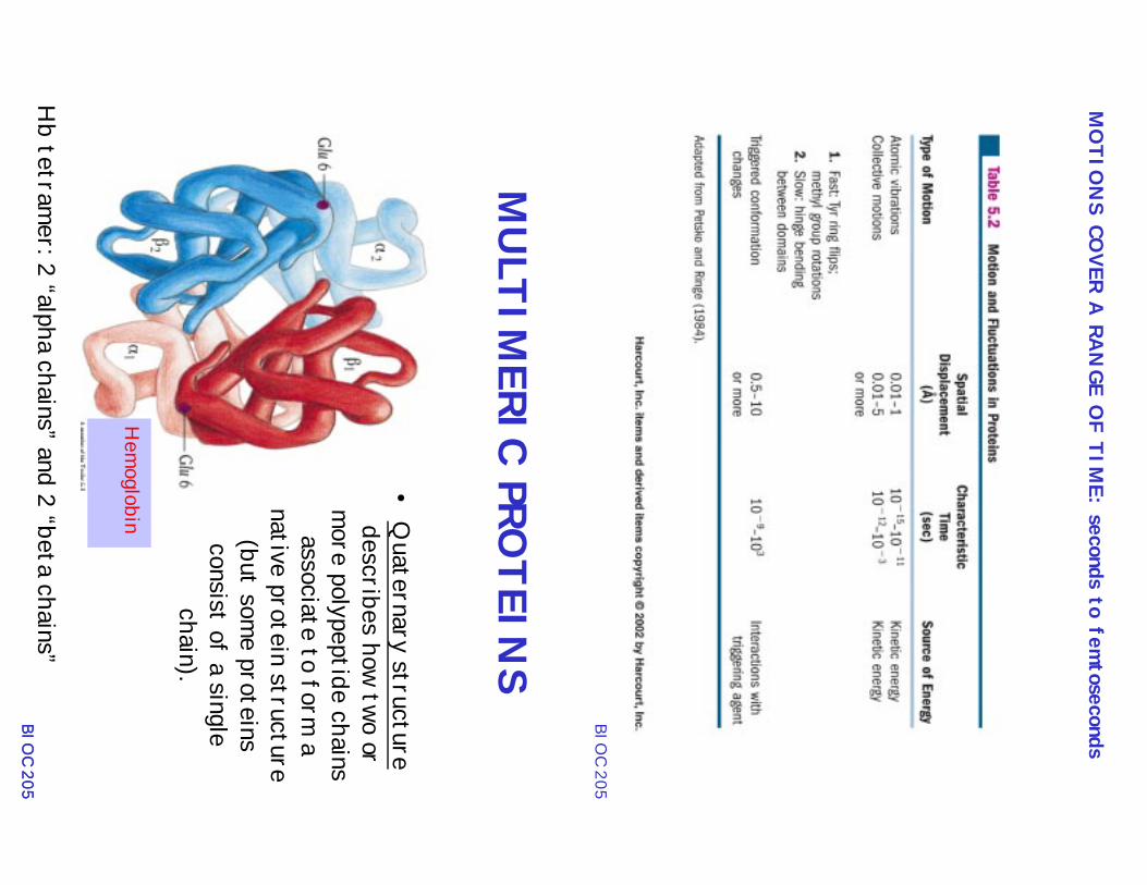
MULTIM
ERIC PROTEIN
S
•Q
uaternary structuredescribes how two or
more polypeptide chainsassociate to form
anative protein structure
(but some proteins
consist of a singlechain).
Hem
oglobin
Hb tetram
er: 2 “alpha chains” and 2 “beta chains”BIO
C 205BIO
C 205

CLOSED
QU
ATERN
ARY STRU
CTURES
The association stiochiometry is finite
BIOC 205
Isologous vs. Heterologous A
ssociations
How the packing interfaces of the m
onomers com
e together
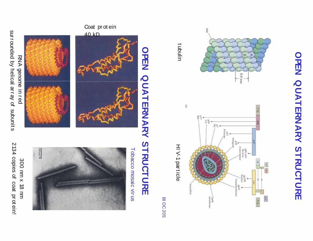
OPEN
QUATERN
ARY STRU
CTUREBIO
C 205
tubulinH
IV-1 particle
Tobacco mosaic virus
OPEN
QUATERN
ARY STRU
CTURE
Coat protein40 kD
RNA
genome in red
surrounded by helical array of subun its300 nm
x 18 nm2134 copies of coat protein!
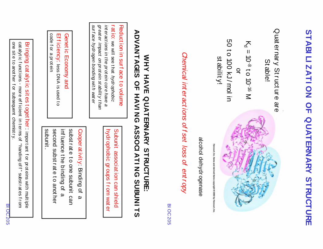
STABILIZA
TION O
F QUATERN
ARY STRU
CTURE
Quaternary Structure are
Stable!
Kd = 10
-8 to 10-16 M
or50 to 100 kJ/m
ol instability!
alcohol dehydrogenase
Chemical interactions offset loss of entropy
BIOC 205
WHY H
AVE Q
UATERN
ARY STRU
CTURE:
ADVA
NTA
GES OF H
AVIN
G ASSO
CIATIN
G SUBU
NITS
Subunit association can shieldhydrophobic groups from
water
Reduction in surface to volume
ratio: we will see that hydrophobicinteractions in the protein core have agreater im
pact on protein stability thansurface hydrogen bonding with water
Bringing catalytic sites together: important for proteins with m
ultiplecatalytic functions - m
ore efficient in terms of “handing off” substrates from
one site to another for subsequent chemistry.
Genetic Economy and
Efficiency: less DN
A is used to
code for a protein
BIOC 205
Cooperativity: Binding of asubstrate to one subunit caninfluence the binding of asecond substrate to anothersubunit.
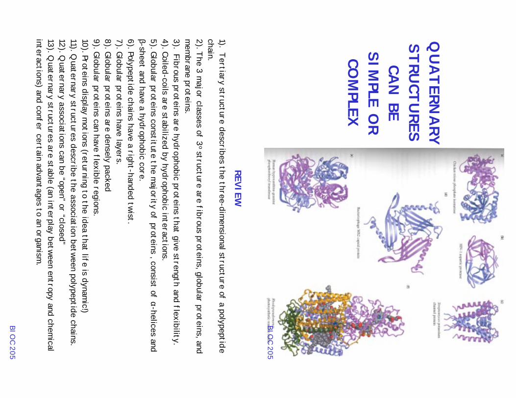
QUATERN
ARY
STRUCTU
RESCA
N BE
SIMPLE O
RCO
MPLEX
BIOC 205
REVIEW1). Tertiary structure describes the three-dim
ensional structure of a polypeptidechain.2). The 3 m
ajor classes of 3o structure are fibrous proteins, globular proteins, and
mem
brane proteins.3). Fibrous proteins are hydrophobic proteins that give strength and flexibility.4). Coiled-coils are stabilized by hydrophobic interactions.5). Globular proteins constitute the m
ajority of proteins , consist of α-helices andβ-sheet and have a hydrophobic core.6). Polypeptide chains have a right-handed twist.7). Globular proteins have layers.8). Globular proteins are densely packed9). Globular proteins can have flexible regions.10). Proteins display m
otions (returning to the idea that life is dynamic!)
11). Quaternary structures describe the association between polypeptide chains.
12). Quaternary associations can be “open” or “closed”
13). Quaternary structures are stable (an interplay between entropy and chem
icalinteractions) and confer certain advantages to an organism
.
BIOC 205