lab manual for biochemistry for ptu students
DESCRIPTION
the document is regarding the practical laboratory manual of BIOCHEMISTRY for PTU studentsTRANSCRIPT

1
LAB MANUAL FOR BIOCHEMSRTY FOR PTU
STUDENTS
1. Preparation of standard buffers (citrate, phosphate and carbonate) and measurement of
pH.
2. Titration curve for amino acids.
3. Separation of amino acids by two dimensional paper chromatography and gel
electrophoresis.
4. Separation of lipids by TLC.
5. Separation of serum proteins by electrophoresis on cellulose acetate.
6. Quantitative estimation of amino acids.
7. Quantitative estimation of proteins.
8. Determination of glucose by means of the enzyme glucose oxidase.
9. Enzymatic hydrolysis of glycogen by alpha- and beta- amylases.
10. Isolation and determination of RNA and DNA.
11. Effect of temperature on the activity of alpha-amylase.
12. Estimation of SGOT, SGPT, Alkaline phosphotase and Bilirubinu in the serum.

2
Experiment – 1
Aim : To prepare standard buffer and measure its pH
Reference : Sharma P.K., Dandiya P.C., “Pharmaceutical Biochemistry”, Vallabh
Prakashan, 1st edition, 2006, 263-266.
Requirement
Apparatus : pH meter
Chemical : Standard buffer tablet, potassium dihydrogen phosphate, disodium hydrogen
phosphate
Theory : Buffers are solutions of weak acids (or bases) together with their conjugate
bases (or acids) which diminish the change in pH which would otherwise occur from
addition of acid or base.
pH = pKa + log [salt] / [acid]
Procedure :
Preparation of standard buffer solution : Stock solutions: (i) Dissolve 14.2g Na2HPO4 in
one litre water (0.1mol/L). (ii) Dissolve 13.6g KH2PO4 in one litre water (0.1mol/L).
Mix the volumes of two solutions given below for 10 ml phosphate buffers of different
pH values.
Na2HPO4 (ml) (0.1mol/L) KH2PO4(ml) (0.1mol/L) pH at 20º C
0.25 9.75 5.29
0.50 9.50 5.59
1.00 9.00 5.91
2.00 8.00 6.24
3.00 7.00 6.47
4.00 6.00 6.64
5.00 5.00 6.81
6.00 4.00 6.98
7.00 3.00 7.17
8.00 2.00 7.38
9.00 1.00 7.73
9.50 0.50 8.04
3
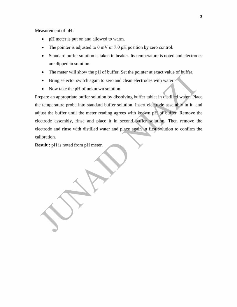
Measurement of pH :
pH meter is put on and allowed to warm.
The pointer is adjusted to 0 mV or 7.0 pH position by zero control.
Standard buffer solution is taken in beaker. Its temperature is noted and electrodes
are dipped in solution.
The meter will show the pH of buffer. Set the pointer at exact value of buffer.
Bring selector switch again to zero and clean electrodes with water.
Now take the pH of unknown solution.
Prepare an appropriate buffer solution by dissolving buffer tablet in distilled water. Place
the temperature probe into standard buffer solution. Insert electrode assembly in it and
adjust the buffer until the meter reading agrees with known pH of buffer. Remove the
electrode assembly, rinse and place it in second buffer solution. Then remove the
electrode and rinse with distilled water and place again in first solution to confirm the
calibration.
Result : pH is noted from pH meter.

4
Experiment – 2
Aim : To perform identification test for a given sample of carbohydrate.
Reference : Satyanarayan U., Chakrapani U., “Biochemistry”, Uppola , 3rd
edition, 16-
17.
Requirement
Apparatus : Test tube, water bath, holder.
Chemical : Molisch’ reagent, Fehling’s solution, Benedict’s solution, conc. sulphuric
acid, picric acid, sodium carbonate, phenyl hydrazone, sodium acetate, Barford’s
solution, sample solution.
Theory :
Molisch's Test (named after Austrian botanist Hans Molisch) is a sensitive chemical test
for the presence of carbohydrates, based on the dehydration of the carbohydrate by
sulfuric acid to produce an aldehyde, which condenses with two molecules of phenol
(usually α-naphthol, though other phenols (e.g. resorcinol, thymol) also give colored
products) resulting in a red- or purple-colored compound.
The Iodine test is used to test for the presence of starch. Iodine solution — iodine
dissolved in an aqueous solution of potassium iodide — reacts with starch producing a
purple black color.
Benedict's reagent is used as a test for the presence of reducing sugars. This includes all
monosaccharides and the disaccharides, lactose and maltose. Even more generally,
Benedict's test will detect the presence of aldehydes (except aromatic ones), and alpha-
hydroxy-ketones, including those that occur in certain ketoses. Thus, although the ketose
fructose is not.
Seliwanoff’s test is a chemical test which distinguishes between aldose and ketose
sugars. Ketoses are distinguished from aldoses via their ketone/aldehyde functionality. If
the sugar contains a ketone group, it is a ketose and if it contains an aldehyde group, it is
an aldose. This test is based on the fact that, when heated, ketoses are more rapidly
dehydrated than aldoses. In concentrated HCl, ketoses undergo dehydration to yield
furfural derivatives more rapidly than do aldoses. These derivatives form complexes with
resorcinol to yield deep red colour.

5
Procedure :
Iodine solution: Add a few crystals of iodine to 2% potassium iodide solution till the
colour becomes deep yellow.
Fehling’s reagent A: Dissolve 34.65 g copper sulphate in distilled water and make up to
500 mL.
Fehling’s reagent B: Dissolve 125 g potassium hydroxide and 173 g Rochelle salt
(potassium sodium tartrate) in distilled water and make up to 500 mL.
Benedict’s qualitative reagent: Dissolve 173 g sodium citrate and 100 g sodium
carbonate in about 500 mL water. Heat to dissolve the salts and filter, if necessary.
Dissolve 17.3 g copper sulphate in about 100 mL water and add it to the above solution
with stirring and make up the volume to 1 L with water.
Barfoed’s reagent: Dissolve 24 g copper acetate in 450 mL boiling water. Immediately
add 25 mL of 8.5% lactic acid to the hot solution. Mix well, Cool and dilute to 500 mL.
Seliwanoff’s reagent: Dissolve 0.05 g resorcinol in 100 mL dilute (1:2) hydrochloric
acid.
Bial’s Test: It is specific for pentoses. They get converted to furfural. In the presence of ferric
ion orcinol and furfural condense to yield a coloured product.
The reactions of carbohydrates are given in Table:
Experiment Observation Remarks
Molisch’s Test
Add two drops of Molisch’s
reagent (5% 1-naphthol in
alcohol) to about 2 mL of test
solution and mix well.
Incline the tube and add about
1 ml of concentrated sulphuric
acid along the sides of the
tube.
A red-cum-violet ring appears
at the junction of the two
liquids.
The colour formed is due to
the reaction of alpha-naphthol
with furfural and/or its
derivatives formed by the
dehydration of sugars by
concentrated sulphuric acid.
All carbohydrates react
positively with this reagent.
Iodine Test
Add a few drops of iodine
solution to about 1 mL of the
test solution.
Appearance of deep blue
colour
This indicates the presence of
starch in the solution. The blue
colour is due to the formation
of starch-iodine complex.

6
Fehling’s Test
To 1 mL of Fehling’s solution
‘A’, add 1 mL of Fehling’s
solution ‘B’ and a few drops
of the test solution. Boil for a
few minutes.
Formation of yellow or
brownish-red precipitate.
The blue alkaline cupric
hydroxide present in Fehling’s
solution, when heated in the
presence of reducing sugars,
gets reduced to yellow or red
cuprous oxide and it gets
precipitated. Hence, formation
of the coloured precipitate
indicates the presence of
reducing sugars in the test
solution.
Benedict’s Test
To 2 mL of Benedict’s reagent
add five drops of the test
solution. Boil for five minutes
in a water bath. Cool the
solution.
Formation of red, yellow or
green colour/precipitate.
Depending on the sugar
concentration yellow to green
colour is developed.
Barfoed’s Test
To 1 mL of the test solution
add about 2 mL of Barfoed’s
reagent.
Boil it for one minute and
allow to stand for a few
minutes.
Formation of brick-red
precipitate.
Only monosaccharides answer
this test. Since Barfoed’s
reagent is weakly acidic, it is
reduced only by
monosaccharides.
Seliwanoff’s Test
To 2 mL of Seliwanoff’s
reagent add two drops of test
solution and heat the mixture
to just boiling.
Appearance of deep red colour It is a timed colour reaction
specific for ketoses.
Bial’s Test
To 5 mL of Bial’s reagent add
2-3 mL of solution and warm
gently. When bubbles rise to
Appearance of green colour or
precipitate.
It is specific for pentoses.
They get converted to furfural.
In the presence of ferric ion
orcinol and furfural condense

7
the surface cool under the tap. to yield a coloured
product.
Osazone Test
To 0.5 g of phenylhydrazine
hydrochloride add 0.1 g of
sodium acetate and 10 drops
of glacial acetic acid. To this
mixture add 5 mL of test
solution and heat on a boiling
water bath for about half an
hour. Allow the tube to cool
slowly and examine the
crystals under a microscope.
Glucose, fructose and
mannose produce needle-
shaped yellow osazone
crystals, whereas lactosazone
is mushroom shaped. Maltose
produces flower-shaped
crystals.
The ketoses and aldoses react
with phenylhydrazine to
produce a phenylhydrazone
which in turn reacts with
another two molecules of
phenylhydrazine to form the
osazone.
Observation : Note the various changes occurring after completion of reaction.
Result : According to observation write down the result whether test is positive or
negative.

8
Experiment –3
Aim : To perform identification test for a given sample of protein.
Reference : Sharma P.K., Dandiya P.C., “Pharmaceutical Biochemistry”, Vallabh
Prakashan, 1st edition, 2006, 316.
Requirements : Test tube, dropper, pipette, beaker
Chemicals : Conc. nitric acid, chlorophenol, acetic acid.
Theory :
Heller's test: Proteins get denatured when acid is added and this forms a white coagulum
which is slightly yellow in colour because of nitro- derivatives of proteins given by
aromatic amino acids.
Heat coagulation test: Coagulation is a result of irreversible denaturation of proteins like
albumin and globulins.
Procedure :
Heller's test:
In 3ml conc. Nitric acid add 3 ml of protein solution.
White ppt. appears at junction of two fluids indicating that protein is present.
Heat coagulation test:
Fill 2/3 of test tube with protein solution. Add 4-5 drops of chlorophenol red and
mix.
A purple colour develops. Add 1% acetic acid until colour changes to faint pink.
Incline the test tube and heat the upper part and a dense coagulum is formed.
Observation: Note the various changes occurring after completion of reaction.
Result : According to observation write down the result whether test is positive or
negative.

9
Experiment –4
Aim : To separate amino acids by thin layer chromatography.
Reference : Sharma P.K., Dandiya P.C., “Pharmaceutical Biochemistry”, Vallabh
Prakashan, 1st edition, 2006, 270-273, 275.
Requirements : Glass slide , beaker
Chemicals : Amino acids, n-propanol, Butanol, glacial acetic acid, ninhydrin reagent,
silica gel.
Solvent system : n-butanol : glacial acetic acid : water :: 12 : 5 : 3
Ninhydrin Solution : 250 mg ninhydrin in 100 ml acetone.
Theory : Chromatography is the collective term for a set of laboratory techniques for the
separation of mixtures. It involves passing a mixture dissolved in a "mobile phase"
through a stationary phase, which separates the analyte to be measured from other
molecules in the mixture based on differential partitioning between the mobile and
stationary phases.
Thin layer chromatography is performed on a sheet of glass, plastic, or aluminum foil,
which is coated with a thin layer of adsorbent material, usually silica gel, aluminium
oxide, or cellulose (blotter paper). This layer of adsorbent is known as the stationary
phase. After the sample has been applied on the plate, a solvent or solvent mixture
(known as the mobile phase) is drawn up the plate via capillary action. Because different
analytes ascend the TLC plate at different rates, separation is achieved.
10
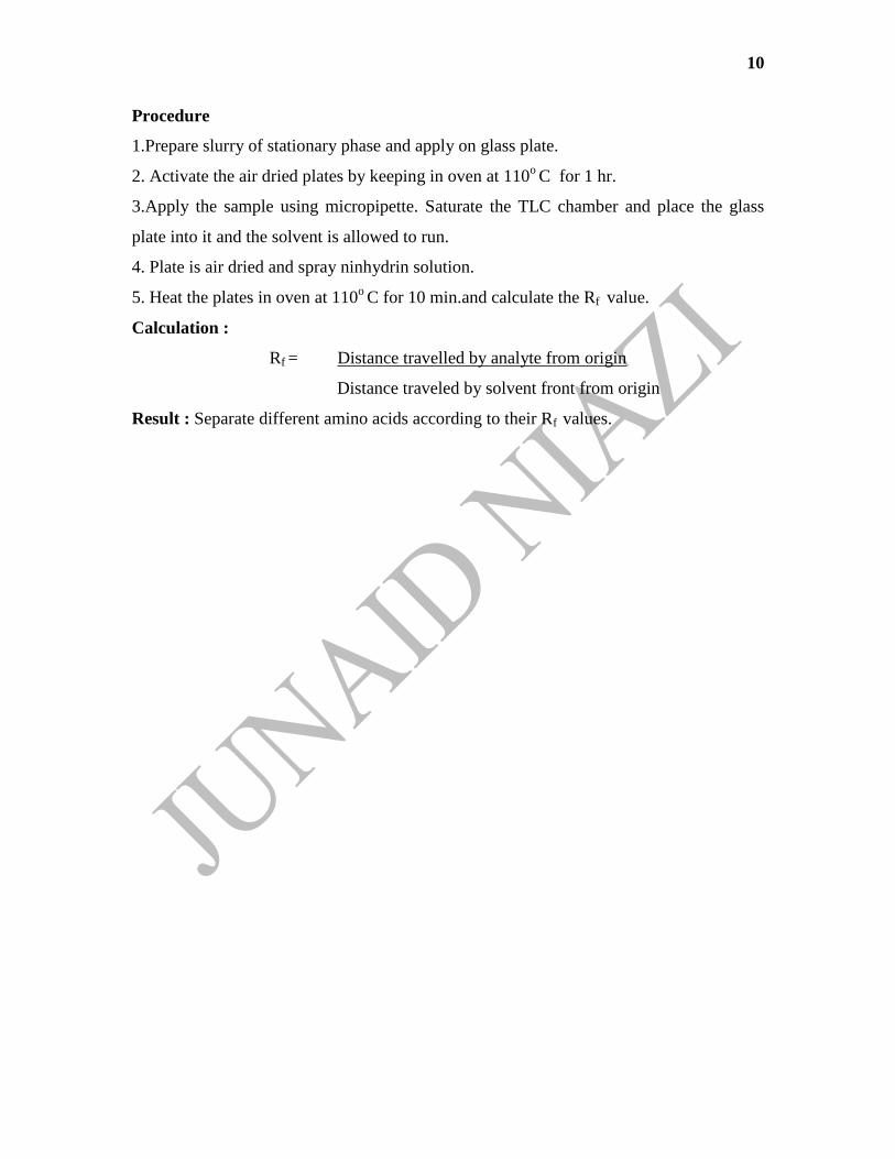
Procedure
1.Prepare slurry of stationary phase and apply on glass plate.
2. Activate the air dried plates by keeping in oven at 110o C for 1 hr.
3.Apply the sample using micropipette. Saturate the TLC chamber and place the glass
plate into it and the solvent is allowed to run.
4. Plate is air dried and spray ninhydrin solution.
5. Heat the plates in oven at 110o C for 10 min.and calculate the Rf value.
Calculation :
Rf = Distance travelled by analyte from origin
Distance traveled by solvent front from origin
Result : Separate different amino acids according to their Rf values.

11
Experiment – 5
Aim : To separate amino acids by paper chromatography.
Reference : Sharma P.K., Dandiya P.C., “Pharmaceutical Biochemistry”, Vallabh
Prakashan, 1st edition, 2006, 270-273, 275.
Requirements : Whatman filter paper no.1, beaker
Chemicals : Amino acids, n-propanol, Butanol, glacial acetic acid, ninhydrin reagent,
silica gel.
Solvent system : n-butanol : glacial acetic acid : water :: 12 : 5 : 3
Ninhydrin Solution : 250 mg ninhydrin in 100 ml acetone.
Theory : Paper chromatography is an analytical chemistry technique for separating and
identifying mixtures that are or can be colored, especially pigments. This can also be used
in secondary or primary colors in ink experiments. This method has been largely replaced
by thin layer chromatography, however it is still a powerful teaching tool. Two-way
paper chromatography, also called two-dimensional chromatography, involves using two
solvents and rotating the paper 90° in between. This is useful for separating complex
mixtures of similar compounds, for example, amino acids.
Procedure :
1.Apply the sample using micropipette. Saturate the chamber and place the paper into it
and the solvent is allowed to run.
2. Paper is air dried and spray ninhydrin solution and calculate the Rf value.

12
Calculation :
Rf = Distance travelled by analyte from origin
Distance traveled by solvent front from origin
Result : Separate different amino acids according to their Rf values.

13
Experiment – 6
Aim : To separate lipids by thin layer chromatography.
Reference : Sharma P.K., Dandiya P.C., “Pharmaceutical Biochemistry”, Vallabh
Prakashan, 1st edition, 2006, 278-279.
Requirements : Glass slide , beaker
Chemicals : Petroleum ether, diethyl ether, glacial acetic acid, silica gel, sulphuric acid
(50%v/v)
Solvent system : Petroleum ether : diethyl ether : glacial acetic acid :: 80 : 20 : 1
Theory : Chromatography is the collective term for a set of laboratory techniques for the
separation of mixtures. It involves passing a mixture dissolved in a "mobile phase"
through a stationary phase, which separates the analyte to be measured from other
molecules in the mixture based on differential partitioning between the mobile and
stationary phases.
Thin layer chromatography is performed on a sheet of glass, plastic, or aluminum foil,
which is coated with a thin layer of adsorbent material, usually silica gel, aluminium
oxide, or cellulose (blotter paper). This layer of adsorbent is known as the stationary
phase. After the sample has been applied on the plate, a solvent or solvent mixture
(known as the mobile phase) is drawn up the plate via capillary action. Because different
analytes ascend the TLC plate at different rates, separation is achieved.

14
Procedure:
1. Prepare slurry of stationary phase and apply on glass plate.
2. Activate the air dried plates by keeping in oven at 110o C for 1 hr.
3. Apply the sample using micropipette. Saturate TLC chamber and place the glass
plate into it and the solvent is allowed to run.
4. Plate is air dried and spray sulphuric acid solution.
5. Heat the plates in oven at 110o C for 10 min.and calculate the Rf value.
Calculation :
Rf = Distance travelled by analyte from origin
Distance traveled by solvent front from origin
Result : Separate different amino acids according to their Rf values.

15
Experiment -7
Aim : To determine casein in the milk.
Reference : Sharma P.K., Dandiya P.C., “Pharmaceutical Biochemistry”, Vallabh
Prakashan, 1st edition, 309,311.
Requirements : Flask, beaker
Chemicals : Acetic acid 10%, KOH 0.1N, HCl 0.1N, Phenolphthalein indicator
Theory : Casein (from Latin caseus, "cheese") is the name for a family of related
phosphoprotein proteins (αS1, αS2, β, κ). These proteins are commonly found in
mammalian milk, making up 80% of the proteins in cow milk and between 60% and 65%
of the proteins in human milk. Casein has a wide variety of uses, from being a major
component of cheese, to use as a food additive, to a binder for safety matches. As a food
source, casein supplies essential amino acids, carbohydrates and two inorganic elements,
calcium and phosphorus.
Procedure:
1. Take 10 ml milk in 200 ml flask and add 75 ml of distilled water and 1.0-1.5 ml of
10% acetic acid.
2. Mix content vigorously. Filter the ppt. and wash with cold water to remove acetic acid.
3. Transfer ppt. and paper in flask, add 75-80 ml of neutral water, 10 ml of 0.1N KOH
and a few drops of phenolphthalein.
4. Stopper the flask and shake vigorously and titrate alkaline casein solution with 0.1N
HCl until all red colour disappears.
Calculation : Substract the corrected acid value from 10ml of alkali used to give % of
casein in milk.
% Casein = 10 – (actual value + check value)
Check value = 0.2 - 0.3
Result : Write down the % casein present in milk.

16
Experiment – 8
Aim : To estimate level of glucose in blood serum.
Reference : Sharma P.K., Dandiya P.C., “Pharmaceutical Biochemistry”, Vallabh
Prakashan, 1st edition, 2006, 294-295.
Requirements : Beaker, test tube, syringe, Centrifuge, Colorimeter.
Chemicals : Blood sample, glucose estimation kit.
Theory : Enzymatic method yields maximum specificity for glucose estimation. Glucose
can be measured by its reaction with glucose oxidase, in which gluconic acid and
hydrogen peroxide are formed. Hydrogen peroxide than reacts with an oxygen acceptor,
such as ortho-dianisidine, phenylamine-phenazone or any other chromogenic oxygen
acceptors, in a reaction catalysed by peroxidase to form a colour.
Procedure
Mix them and incubate at 370
C for 20 min. Read the absorbance of standard and test
against blank at 540 nm.
Calculation :
Plasma glucose (mg/dl) = reading of test x 100
reading of standard
Result : Write down the concentration of glucose in blood.
Sr.No. Reagents Blank Standard Test tube
1. Glucose reagent 1 ml 1 ml I ml
2. Glucose standard - 0.01 ml -
3. Serum sample - - 0.01 ml
4. Distilled water 0.01 ml - -

17
Experiment – 9
Aim : To perform the quantitative estimation of proteins in serum using Biuret method.
Reference : Sharma P.K., Dandiya P.C., “Pharmaceutical Biochemistry”, Vallabh
Prakashan, 1st edition, 2006, 296.
Requirements : Beaker, test tube, syringe, Centrifuge, Colorimeter.
Chemicals : Blood sample, protein estimation kit.
Theory : Copper in alkaline solution reacts with peptide linkages of amino acids in
protein producing a violet colour which is measured colorimetrically.
Procedure :
Mix them and incubate at 370
C for 10 min. Read the absorbance of standard and test
against blank at 540 nm.
Calculation :
Serum Total Protein (g/dl) = reading of test x 6
reading of standard
Result : Write down the concentration of proteins in serum.
Sr. No. Reagents Blank Standard Test tube
1. Biuret reagent 1 ml 1 ml I ml
2. Protein standard - 0.01 ml -
3. Serum sample - - 0.01 ml
4. Distilled water 0.01 ml - -
18
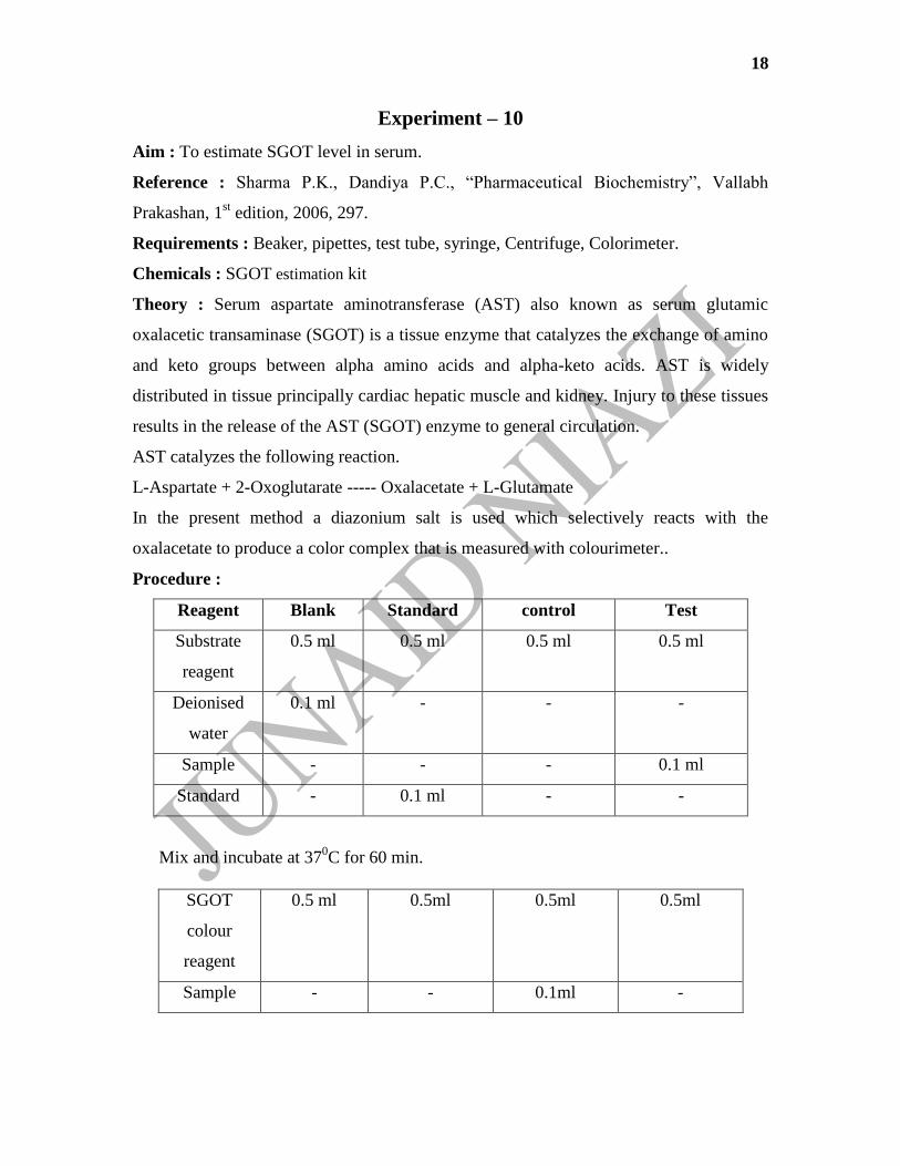
Experiment – 10
Aim : To estimate SGOT level in serum.
Reference : Sharma P.K., Dandiya P.C., “Pharmaceutical Biochemistry”, Vallabh
Prakashan, 1st edition, 2006, 297.
Requirements : Beaker, pipettes, test tube, syringe, Centrifuge, Colorimeter.
Chemicals : SGOT estimation kit
Theory : Serum aspartate aminotransferase (AST) also known as serum glutamic
oxalacetic transaminase (SGOT) is a tissue enzyme that catalyzes the exchange of amino
and keto groups between alpha amino acids and alpha-keto acids. AST is widely
distributed in tissue principally cardiac hepatic muscle and kidney. Injury to these tissues
results in the release of the AST (SGOT) enzyme to general circulation.
AST catalyzes the following reaction.
L-Aspartate + 2-Oxoglutarate ----- Oxalacetate + L-Glutamate
In the present method a diazonium salt is used which selectively reacts with the
oxalacetate to produce a color complex that is measured with colourimeter..
Procedure :
Reagent Blank Standard control Test
Substrate
reagent
0.5 ml 0.5 ml 0.5 ml 0.5 ml
Deionised
water
0.1 ml - - -
Sample - - - 0.1 ml
Standard - 0.1 ml - -
Mix and incubate at 370C for 60 min.
SGOT
colour
reagent
0.5 ml 0.5ml 0.5ml 0.5ml
Sample - - 0.1ml -
19
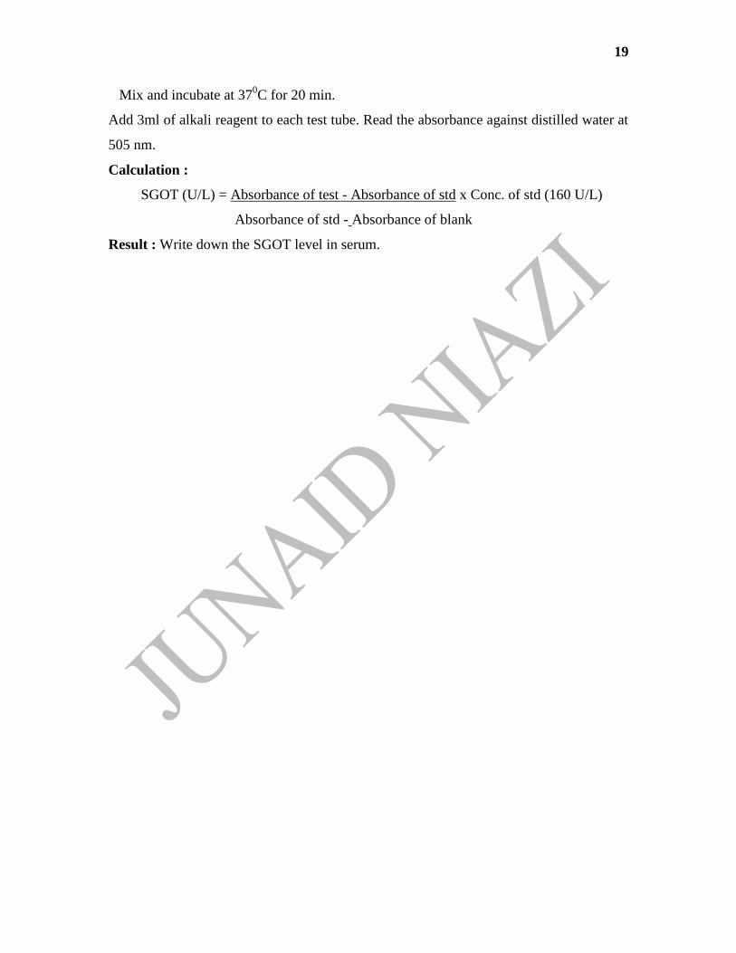
Mix and incubate at 370C for 20 min.
Add 3ml of alkali reagent to each test tube. Read the absorbance against distilled water at
505 nm.
Calculation :
SGOT (U/L) = Absorbance of test - Absorbance of std x Conc. of std (160 U/L)
Absorbance of std - Absorbance of blank
Result : Write down the SGOT level in serum.

20
Experiment -11
Aim : To estimate SGPT level in serum.
Reference : Sharma P.K., Dandiya P.C., “Pharmaceutical Biochemistry”, Vallabh
Prakashan, 1st edition, 2006, 297.
Requirements : Beaker, pipettes, test tube, syringe, Centrifuge, Colorimeter.
Chemicals : SGPT estimation kit
Theory : Alanine transaminase or ALT is a transaminase enzyme. It is also called serum
glutamic pyruvic transaminase (SGPT) or alanine aminotransferase (ALAT). ALT is
found in serum and in various bodily tissues, but is most commonly associated with the
liver. It catalyzes the two parts of the alanine cycle.
It catalyzes the transfer of an amino group from alanine to a-ketoglutarate, the products
of this reversible transamination reaction being pyruvate and glutamate.
glutamate + pyruvate ⇌ α-ketoglutarate + alanine
Procedure :
Reagent Blank Standard control Test
Substrate
reagent
0.5 ml 0.5 ml 0.5 ml 0.5 ml
Deionised
water
0.1 ml - - -
Sample - - - 0.1 ml
Standard - 0.1 ml - -
Mix and incubate at 370C for 30 min.
Mix and incubate at 370C for 20 min.
SGPT
colour
reagent
0.5 ml 0.5ml 0.5ml 0.5ml
Sample - - 0.1ml -

21
Add 3ml of alkali reagent to each test tube. Read the absorbance against distilled water at
505 nm.
Calculation :
SGPT (U/L) = Absorbance of test - Absorbance of std x Conc. of std (160 U/L)
Absorbance of std - Absorbance of blank
Result : Write down the SGPT level in serum.

22
Experiment – 12
Aim : To estimate Alkaline Phosphatase level in serum.
Reference : Sharma P.K., Dandiya P.C., “Pharmaceutical Biochemistry”, Vallabh
Prakashan, 1st edition, 2006, 299-301.
Requirements : Beaker, pipettes, test tube, syringe, Centrifuge, Colorimeter.
Chemicals : Alkaline Phosphatase estimation kit
Theory : Alkaline phosphatase (ALP, ALKP) is a hydrolase enzyme responsible for
removing phosphate groups from many types of molecules, including nucleotides,
proteins, and alkaloids.
Procedure :
Reagent Blank Standard Control Test
Working
buffered
substrate
0.5 ml 0.5ml 0.5ml 0.5ml
Distilled water 1.5 ml 1.5ml 1.5ml 1.5ml
Mix and incubate at 37ºC for 3 min.
Serum - - -
0.05ml
Reagent 3 - 0.05 ml -
-
Mix well and incubate at 37ºC for 15 min.
Reagent 2 1ml 1ml 1ml 1ml
Serum - -
0.05ml -
Mix well and measure optical density of all the test tubes against distilled water at
510nm.
Calculation :
Serum Alkaline Phosphatase (KA units/ 100 ml) = Abs of test - Abs of std x 10
Abs of std
Result : Write down the Alkaline Phosphatase level in serum.

23
Experiment -13
Aim : To estimate Billirubin level in serum.
Reference : Sharma P.K., Dandiya P.C., “Pharmaceutical Biochemistry”, Vallabh
Prakashan, 1st edition, 2006, 298-299.
Requirements : Beaker, pipettes, test tube, syringe, Centrifuge, Colorimeter.
Chemicals : Billirubin estimation kit
Theory : Bilirubin is the yellow breakdown product of normal heme catabolism. Heme is
found in hemoglobin, a principal component of red blood cells. Bilirubin is excreted in
bile and urine, and elevated levels may indicate certain diseases. It is responsible for the
yellow color of bruises, the yellow color of urine (via its reduced breakdown product,
urobilin), the brown color of faeces (via its conversion to stercobilin), and the yellow
discoloration in jaundice.
Procedure :
Reagent Blank Test
Total bilirubin
reagent
1 ml 1 ml
Sodium nitrite
reagent
- 0.5 ml
Distilled Water 0.5 ml -
Sample 0.5 ml 0.5 ml
Mix well reagents and wait for 30 sec before next addition.
Then add 0.15 ml of serum sample to each test tube.mix well and incubate at 370C for 5
min and note the absorbance at 540nm against distilled water.
Calculation :
Total Billirubin (mg/dl) = Abs of test - Abs blank x 10
Abs of std
Result : Write down the Total Billirubin level in serum.

24
Experiment -14
Aim : To estimate Total Serum Cholesterol.
Reference : Sharma P.K., Dandiya P.C., “Pharmaceutical Biochemistry”, Vallabh
Prakashan, 1st edition, 2006, 301-302.
Requirements : Beaker, pipettes, test tube, syringe, Centrifuge, Colorimeter.
Chemicals : Cholesterol estimation kit
Theory : Serum is treated with ferric chloride - acetic acid reagent to precipitate proteins.
The protein free supernatant is treated with conc. sulphuric acid. A reddish purple colour
is developed which is measured colorimetrically at 560nm0
Procedure :
Reagent Blank Standard Test
Cholesterol
reagent
1 ml 1 ml 1 ml
Cholesterol Std - 0.1 ml -
Sample - - 0.1s ml
Mix well and incubate at 370C for 5 min and note the absorbance at 560nm.
Calculation :
Total Serum Cholesterol (mg/ 100ml) = Abs of test x 200
Abs of std
Result : Write down the Total Serum Cholesterol.