j. biol. chem.-1996-gregoire-32951-9
TRANSCRIPT
8/3/2019 J. Biol. Chem.-1996-Gregoire-32951-9
http://slidepdf.com/reader/full/j-biol-chem-1996-gregoire-32951-9 1/9
cDNA Cloning and Sequencing Reveal the Major Horse AllergenEqu c1 to Be a Glycoprotein Member of the Lipocalin Superfamily*
(Received for publication, August 2, 1996, and in revised form, October 4, 1996)
Christophe Gregoire‡§, Isabelle Rosinski-Chupin¶, Jacques Rabillon‡, Pedro M. Alzari,
Bernard David‡, and Jean-Pierre Dandeu‡ From ‡Unite d’Immuno-Allergie, Departement de Physiopathologie, ¶Unite de Genetique et de Biochimie du Developpement, Departement d’Immunologie, and Unite d’Immunologie Structurale, Departement d’Immunologie, Institut Pasteur, 28 rue du Dr Roux, 75024 Paris Cedex 15, France
The gene encoding the major horse allergen, desig-nated Equus caballus allergen 1 (Equ c1), was clonedfrom total cDNA of sublingual salivary glands by reversetranscription-polymerase chain reaction using syn-thetic degenerate oligonucleotides deduced from N-ter-minal and internal peptide sequences of the glycosy-lated hair dandruff protein. A recombinant form of theprotein, with a polyhistidine tail, was expressed in Esch-erichia coli and purified by immobilized metal affinity
chromatography. The recombinant protein is able to in-duce a passive cutaneous anaphylaxis reaction in rat,and it behaves similarly to the native Equ c1 in severalimmunological tests with allergic patients’ IgE antibod-ies, mouse monoclonal antibodies, or rabbit polyclonalIgG antibodies. Amino acid sequence identity of 49–51%with rodent urinary proteins from mice and rats sug-gests that Equ c1 is a new member of the lipocalin su-perfamily of hydrophobic ligand-binding proteins thatincludes several other major allergens. An RNA blotanalysis demonstrates the expression of mRNA Equ c1in liver and in sublingual and submaxillary salivaryglands.
Exposure to animal danders, commonly present in the envi-ronment, is known to be a frequent cause of allergy. The inha-
lation of these potent animal dandruff allergens induces im-
munoglobulin E antibody (IgE) and subsequent development of
asthma in atopic individuals. Among these allergens, a major
allergen is defined to be the one that elicits an anaphylactic
reaction in a majority of patients, presenting an immediate
hypersensitivity response mediated by IgE against the basic
raw material (1).
The reasons why a protein is allergenic are not clearly un-
derstood to date, although several authors favor the hypothesis
of a possible relationship between the structure and the func-
tion of proteins and their allergenicity (2). The enzymatic ac-
tivity of certain proteins has been assumed to have a capacity
to enhance the IgE response (2). A family of proteins, the
lipocalin superfamily, is known to include several allergens,
such as the mouse major urinary protein mMUP1 (3), the rat
-2-microglobulin (rA2U) (4), the bovine -lactoglobulin (lg)
(5), the cockroach allergen Bla g4 (6), and the recently de-
scribed bovine dander allergen Bos d2 (7). Based on this obser-
vation, Arruda et al. suggested that lipocalins may contain a
common structure that is able to induce the IgE response.
Members of this superfamily, which bind or transport small
hydrophobic molecules, are generally expressed in the liver
and/or secretory glands. This is particularly true for the mMUP
and rA2U proteins, which are multigenic families at about
35–40 members in the case of the mMUP family (8) and about
25 for the rA2U (9, 10). These members are differentially ex-
pressed in the liver as well as salivary, lachrimal, and other
secretory glands (11).
The major horse allergen, Equ c1, is a potent allergen re-
sponsible for about 80% of anti-horse IgE antibody response in
patients who are chronically exposed to horse allergens. Al-
though much work has been carried out on the isolation and
identification of the horse allergenic agents responsible for
human hypersensitivity response (12–16), the major horse al-
lergen was only recently purified from hair and dandruff (17).
A previous study by SDS-polyacrylamide gel electrophoresis
(SDS-PAGE) and isoelectric focusing-PAGE showed that Equ
c1 appears as a single polypeptide with a relative molecular
mass of 21,500 daltons and a pI of 3.9. The purification of Equc1 allowed the sequencing of the 27 N-terminal amino acids
and of internal peptides (18).
To obtain more information on the structural and functional
features of Equ c1, we have cloned the corresponding cDNA
from the sublingual salivary gland (SLG). Here we report the
molecular cloning and sequencing of this cDNA and expression
of a recombinant allergen rSLG Equ c1 in a bacterial system.
The recombinant protein was compared with natural Equ c1 for
its recognition by antibodies raised against the natural Equ c1
in immunoblots and in inhibition/competition enzyme-linked
immunosorbent assay (ELISA). We also show that the recom-
binant protein is able to elicit a rat mast cell degranulation by
passive cutaneous anaphylaxis reaction.
Sequence comparisons reveal that Equ c1 is a new member of
the lipocalin superfamily.
EXPERIMENTAL PROCEDURES
Materials—The horse salivary glands were obtained from a slaugh-
terhouse and rapidly frozen in liquid nitrogen after dissection. Theywere stored at 80 °C until protein and nucleotidic extractions wereperformed.
Protein Purification and N-terminal Sequencing—Equ c1 was puri-
* The costs of publication of this article were defrayed in part by thepayment of page charges. This article must therefore be hereby marked“advertisement” in accordance with 18 U.S.C. Section 1734 solely toindicate this fact.
The nucleotide sequence(s) reported in this paper has been submittedto the GenBankTM/EBI Data Bank with accession number(s) U70823.
§ To whom correspondence should be addressed. Tel.: 33-1-45-68-84-48; Fax: 33-1-40-61-31-60: E-mail: [email protected].
1 The abbreviations used are: mMUP, mouse major urinary protein;SLG, sublingual gland; HD, hair dandruff; SMG, submaxillary gland;rSLG Equ c1, recombinant SLG Equ c1; rA2U, rat -2-microglobulin;
mAb, monoclonal antibody; FPLC, fast protein liquid chromatography;PAGE, polyacrylamide gel electrophoresis; PBS, phosphate-bufferedsaline; PCR, polymerase chain reaction; RACE, rapid amplification of cDNA ends; ELISA, enzyme-linked immunosorbent assay; BSA, bovineserum albumin.
THE JOURNAL OF BIOLOGICAL CHEMISTRY Vol. 271, No. 51, Issue of December 20, pp. 32951–32959, 1996 © 1996 by The American Society for Biochemistry and Molecular Biology, Inc. Printed in U.S.A.
This paper is available on line at http://www-jbc.stanford.edu/jbc/ 32951
8/3/2019 J. Biol. Chem.-1996-Gregoire-32951-9
http://slidepdf.com/reader/full/j-biol-chem-1996-gregoire-32951-9 2/9
fied from salivary glands and dander extracts by a combination of size
exclusion chromatography in fast protein liquid chromatography(FPLC) and hydrophobic interaction chromatography as described pre-
viously (17).
An Equ c1 tryptic proteolysis was performed for 15 min at 37 °C in abuffer containing 50 mM Tris-HCl, 1 mM CaCl2, pH 7.0, with an enzymeratio of 1:1000 (w/w). The sequencing was processed, using the method
described by Baw et al. (19), in the microsequencing laboratory of thePasteur Institute. Protein assays were performed with the colorimetricmethod using Micro BCA protein assay reagent from Pierce, accordingto Smith et al. (20).
Preparation of RNA—Total RNA was isolated from sublingual (SLG)and submaxillary (SMG) salivary glands and from liver according toChirgwin’s protocol (21), modified as described previously (22, 23).
Equ c1 cDNA Cloning—cDNA first strand synthesis was performedon 5 g of horse SLG total RNA for 1 h at 37 °C in a total volume of 50
l with 20 pmol of the primer adapter oligo(dT): 5-AAC CCG GCT CGA
GCG GCC GCT TTT TTT TTT TTT TT-3, 800 units of Moloney murineleukemia virus reverse transcriptase (Life Technologies, Inc.) in themanufacturer’s buffer. The cDNAs so obtained were amplified by po-
lymerase chain reaction (PCR) with the Opti Prime PCR optimizationkit (Stratagene), with the oligomer 5-GGY GAG TGG TAY TCY ATYTT-3 as primer 1 and the oligomer 5-GGY GAG TGG TAY AGY ATY
TT-3 as primer 2 derived from the Gly35–Ser39 sequence and the5-GTS AGP TCR ATR ATR TTY TC-3 as primer 3 derived from theGlu165–Leu170 sequence. The letter Y represents a 50% mixture (w/w) of
nucleotides T and C, S a mixture of G and C, and R a mixture of A andG. After a first denaturation cycle at 98 °C for 2 min, 30 cycles of PCRconsisting of a 30-s denaturation step at 94 °C followed by annealing at
50 °C for 35 s and elongation at 72 °C for 30 s were carried out in athermocycler Hybaid (Ceralabo, Aubervilliers, France). Each reactioncontained 1 l of cDNA reaction product, 0.2 mM dNTP, 2.4 units of TaqDNA polymerase, 68.8 pmol of the primer 3, and 34.4 pmol of each otherprimer. The variable parameters of buffers are pH, MgCl2, and KClconcentrations. The best amplification was obtained with buffer 6 (10
mM Tris-HCl, pH 8.8, 1.5 mM MgCl2, and 75 mM KCl) and buffer 12 (10mM Tris-HCl, pH 9.2, 3.5 mM MgCl2, and 75 mM KCl). After separationby electrophoresis in a 1.2% agarose gel and purification, the products
from the PCR reactions were inserted in pMOS Blue T vector (Amer-sham Life Sciences). Sequencing was performed after alkaline denatur-ation by the dideoxy chain termination method (24) using Sequenase
version 2.0 (U.S. Biochemical Corp.) and -35S-labeled dATP.
Amplification of the cDNA Ends—The rapid amplification of cDNAends (RACE) strategy was applied to clone 3 and 5 cDNA extremities.For 5 RACE, 12.5 l of the first single strand cDNA (as described
above) were directly used for dC tailing, for 5 min at 37 °C, in 10 mM
Tris-HCl, pH 8.4, 25 mM KCl, 1.25 mM MgCl2, 50 g/ml BSA, and 10units of terminal transferase. Reactions were stopped by increasing the
temperature to 65 °C for 10 min. The cDNA amplification was per-formed in the presence of 5 pmol of the oligomer 5-GCG CCC AGT GTGCTG GCT GCA GGG GGG GGG GG-3, complementary to the dC tail,
and the oligomer 5-CTT TTC CTT GAC GTC TGA AGC C-3 corre-sponding to the nucleotide sequence G189–G210, as a specific primer(antisense). A 5-l aliquot of dC-tailed cDNA was amplified by PCR in
a 50-l volume in 20 mM Tris-HCl, pH 8.4, 50 mM KCl, 2.5 mM MgCl2,100 g/ml BSA) and 0.2 mM each dNTP. The conditions of 35 cycles of PCR consisted of a 30-s denaturation step at 95 °C followed by a 35-s
annealing step at 60 °C and a 30-s extension step at 72 °C.For cloning of the 3 region, the same experimental conditions were
applied to the PCR amplification using the specific primer 5-GCC CGA
GAA CCA GAT GTG AGT-3 corresponding to the nucleotide sequenceG481–T501 and the primer adapter oligo(dT). All amplified products werecloned in pMOS Blue vector and sequenced as described above.
Bacterial Expression of Recombinant Equ c1— A cDNA correspondingto the nearly complete Equ c1 sequence was amplified by PCR andcloned in a pET vector. Primers for PCR were designed to specifically
hybridize with Equ c1 cDNA and contained EcoRI and XhoI sites. Theprimers used were 5-CTT GAA TTC ATC GAG GGG AGA GAA AAC
AGT GAT GTT GCG-3 (5 end primer) and 5-CCA CTC GAG GAAGTA TTC ACT GTC-3 (3 end primer). In addition, the 5 primer
provides the recombinant protein with a new proteolytic cleavage sitefor the factor Xa. PCR products were cloned into the EcoRI/ XhoI sites of the plasmid pET 28 (a) under control of the T7 lac promoter (Fig. 1).
This expression vector contains the kanamycin resistance gene and aHis6 tag at the N terminus of the recombinant protein. Competent
Escherichia coli XL1 cells were transformed, and supercoiled plasmid
was sequenced and transfected in E. coli BL 21 (DE3). Induction wasperformed by adding isopropyl -D-thiogalactopyranoside to the me-dium at a final concentration of 1 mM for 180 min at 37 °C. Induction
was controlled by taking aliquots every 30 min. Cells were then har- vested by centrifugation and resuspended in 50 mM Tris-HCl, pH 7.0,containing 1% (v/v) Triton X-100 and 100 g/ml lysozyme. The cells
were incubated for 15 min at 30 °C, and the DNA was disrupted bysonication. The supernatant obtained after centrifugation was filteredon a 0.2-m membrane and dialyzed against phosphate-buffered saline
(PBS) with 0.5 mM NaCl. The resulting product was used for chromato-graphic purification.
Purification of the Recombinant Equ c1— An HR 5/5 column was
packed with chelating Sepharose fast flow (Pharmacia Biotech, Inc.),washed according to the manufacturer’s suggestions, and charged untilsaturation with metal ions from a 0.5% (w/v) copper(II) chloride solu-
tion. After thorough rinsing with water, the column was presaturatedwith buffer (PBS/0.5 mM NaCl) containing 10 mM imidazole (25). Afterequilibration of the column with the starting buffer (PBS/0.5 mM NaCl),
6 column volumes of supernatant was loaded, and the unbound mate-rial was collected. Competitive elution was carried out using imidazoleat 40 and 120 mM (PBS/0.5 mM NaCl), pH 7.0, collecting 6 column
volumes at each step (26). The whole process was controlled by an FPLCapparatus (Pharmacia). The fractions were concentrated using stirredcell ultrafiltration with a PM 10 membrane (Amicon) and dialyzed
against the proteolysis buffer (50 mM Tris-HCl, pH 8.0, 100 mM NaCl,and 1 mM CaCl2). Digestion with the factor Xa was performed overnightat 30 °C. After proteolysis, the digest was dialyzed to remove the smalldigest peptides and lyophilized.
SDS-PAGE and Western Blots— All analysis of the different fractionswas performed with the Adjustable Stab Gel kit ASG 400 (Prolabo)using 18% acrylamide/bisacrylamide (29:1) gels (27). Proteins were
visualized with Coomassie Blue and/or silver nitrate staining. Electro-blotting experiments were performed using nitrocellulose membrane(Schleicher & Schull). For immunological detection, polyclonal antibod-
ies from human and rabbit sera and mouse monoclonal antibody di-rected against Equ c1 were used.
The rabbit immunization was performed by intradermal injection of
100 g of pure allergen. Sixteen patients with established allergy tonatural Equ c1 were selected, and a pool from three nonallergic healthydonors was used as negative control. Bound IgE were detected using
peroxidase conjugated to rabbit anti-human IgE. When mouse mAbanti-Equ c1 or the polyclonal rabbit IgG was used, the detection was
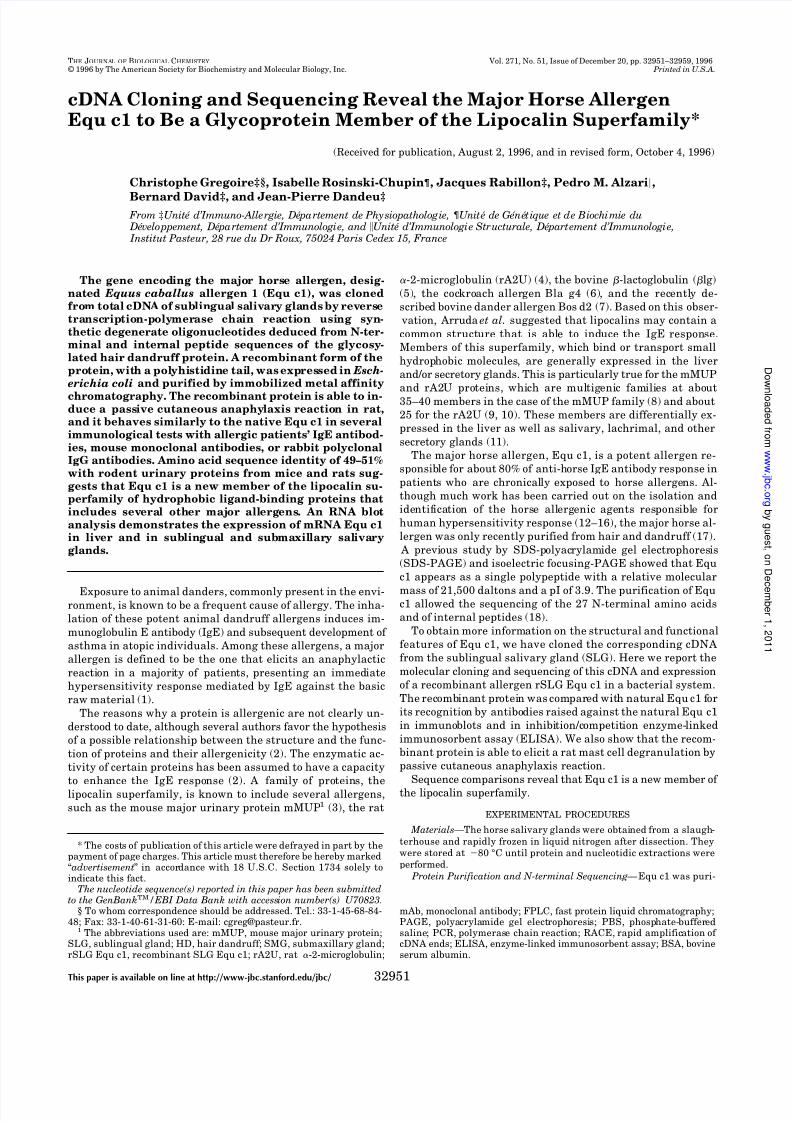
FIG. 1. Plasmid construct for thebacterial expression of rSLG Equ c1in E. coli. cDNA Equ c1 was inserted inpET 28 (a) after digestion with EcoRIand XhoI. The plasmid contains the lacoperator used to induce, with 1 m M of isopropyl -D-thiogalactopyranoside, the
recombinant protein tailed at its N-ter-minal end. Factor Xa proteolytic site(LEFIEGR2ENSDVA) was introducedbetween rSLG Equ c1 ant the tail contain-ing the polyhistidine tag.
cDNA Cloning and Sequencing of the Major Horse Allergen Equ c132952
8/3/2019 J. Biol. Chem.-1996-Gregoire-32951-9
http://slidepdf.com/reader/full/j-biol-chem-1996-gregoire-32951-9 3/9
performed with peroxidase conjugated to rabbit anti-mouse IgG or
peroxidase conjugated to goat anti-rabbit IgG, respectively, using thediamino-3,3-benzidine tetrachlorydrate as specific reagent.
The mouse anti-HD Equ c1 mAb were prepared in the Hybridolab of
the Pasteur Institute according to the methods described by Kohler andMilstein (28).
Passive Cutaneous Anaphylaxis—Each mouse was immunized sub-
cutaneously at day 0 and boosted at days 21 and 35 with 5 g of antigen(purified HD Equ c1, protein extract from horse hair dandruff, horseserum albumin, or ovalbumin) in the presence of 4% (w/w) Al(OH)3 in a
physiological solution. Each mouse was bled after being anesthetized,at day 42 by retro-orbital puncture in order to study IgE immuneresponse. The IgE antibody titers were determined by the passive
cutaneous anaphylaxis reaction in rats (29).Serum samples were diluted in a physiological solution and 100-l
aliquots inoculated intradermally on the shaved back of Lewis rats.
Twenty-four hours later, each rat was challenged by intravenous inoc-ulation in the tail of 1 ml of a physiological solution containing 50g of antigen and 0.5% Evans blue. Thirty minutes later, rats were killed,
and skin was excised for examination. The reciprocal of the highestdilution giving a blueing reaction of 10-mm diameter was taken as thepassive cutaneous anaphylaxis titer.
Inhibition/Competition Experiments—These experiments were per-formed using ELISA as follows. Each well of the assay plate (Maxisorb,Nunc, Roskild, Denmark) was coated with 100 l of a highly purified
HD Equ c1 or rSLG Equ c1, 10 g/ml in 0.1 M carbonate/bicarbonatebuffer, pH 9.6. After saturation of the unoccupied sites with 0.5% BSA
in PBS and appropriate washing, mAbs, after being previously prein-cubated 1 h at 37 °C with different dilutions of competitor, were added
in duplicate to the sample-coated wells and incubated for 1 h at 37 °C.Bound mAb and rabbit antibodies were detected with peroxidase-con-
jugated rabbit anti-mouse IgG (Sigma) and peroxidase-conjugated goat
anti-rabbit IgG, respectively, and revealed with o-phenylenediamineaccording to the manufacturer’s recommendations.
Determination of Sugar Content— A study was done to perform de-
glycosylation on Equ c1, using anhydrous trifluoromethane sulfonicacid, as described by Sojar and Bahl (30). Each dry sample was acid-treated with a mixture of trifluoromethane sulfonic acid and toluene for
4 h at 20 °C. Then trifluoromethane sulfonic acid was neutralized byadding to the reaction mixture pyridine and ammonium bicarbonateand dialyzed against 50 mM Tris/HCl, pH 7.5, 100 mM NaCl. Each
sample was submitted to electrophoresis in SDS-PAGE. Gels werestained with silvernitrate. Analysis of thesaccharide composition of theHD Equ c1 and Saliva Equ c1 was done using gas phase chromatogra-
phy after acidic treatment, as described by Kamerling et al. (31).
RNA Analysis—Total mRNA was electrophoresed in an agarose/ formaldehyde gel (32) transferred to a nylon membrane, and hybridized
with the Equ c1 cDNA probe. The probe was the full-length cDNA insertlabeled by the random priming method (33).
The search for homologies between the deduced amino acid sequence
of Equ c1 and the proteins of the Swiss-Prot data base or the Equc1 cDNA and the GenBankTM nucleotide sequence data base weredone, respectively, with the FASTP and FASTN program according to
Altschul et al. (34).
RESULTS
Molecular Cloning of the Equ c1 cDNA—Tryptic fragments
were generated from HD Equ c1 isolated and purified from
horse hair dandruff extract by a combination of size exclusion
chromatography and hydrophobic interaction chromatography.
These fragments were microsequenced, and two of them(shown in boldface type on Fig. 2) were used to design three
degenerate primers. The design of the primers took into con-
sideration the codon usage in horse.
It was previously demonstrated by Dandeu et al. (17) that
Equ c1 from different sources, i.e. saliva, urine, and hair dan-
druff extracts, are similarly recognized by antibodies. Salivary
secretions contain the highest amount of Equ c1 protein; there-
fore, the salivary glands were chosen to clone Equ c1 cDNA.
Among the tested salivary glands, the sublingual glands had
the highest level of Equ c1 immunoreactivity and were selected
to prepare mRNA.
The mRNAs so obtained were reverse-transcribed, and the
Equ c1 cDNA was amplified by PCR using a mixture of the
three primers. This reverse transcription-PCR resulted in a
DNA fragment of about 400 base pairs in length that was
cloned in pMOS Blue; several positive clones were sequenced.
In a second step, 5 and 3 ends of the SLG Equ c1 cDNA were
obtained using a 5 and 3 RACE strategy. The two amplifica-
tion products of 250 and 450 base pairs for the 5 and 3 RACE,
respectively, were cloned and sequenced.
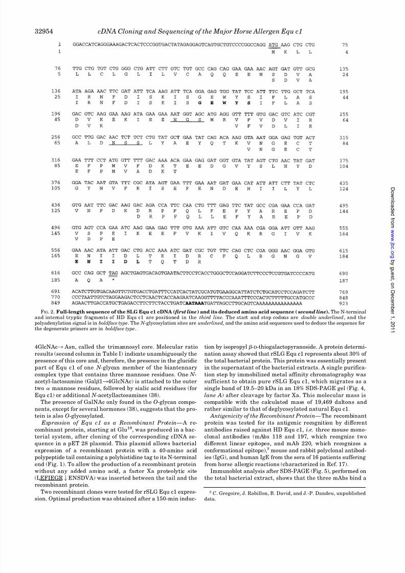
Sequence of the Equ c1 cDNA—The full-length sequence of
Equ c1 cDNA and the deduced amino acid sequence are shown
in Fig. 2. The SLG Equ c1 cDNA is 923 nucleotides long with anopen reading frame of 560 nucleotides (excluding the stop
codon), coding for a 187-amino acid protein. All peptides from
HD Equ c1 can be localized in the SLG Equ c1 sequence and
start after an arginine or a lysine residue, according to the
tryptic proteolysis consensus sites. However, some differences
in the amino acid sequence can be observed between rSLG Equ
c1 from sublingual salivary gland and the tryptic peptides
obtained from HD Equ c1. These differences are not PCR arti-
facts, because our nucleotide sequence results from the analy-
sis of 12 clones from four independent PCR experiments. These
differences are in the internal peptides, at positions 62 (Ala/
Leu), 90 (Phe/Ala), 136 (Phe/Leu), 146 (Ser/Asp), 172 (Lys/Gln),
and 173 (Ile/Thr). All analyzed clones contained a 3 noncoding
region of 298 nucleotides and a poly(A) tail 23 base pairs,
downstream from a consensus polyadenylation signal AATAAA
at position A886 /A891. All clones sequenced have identical 5
ends with a noncoding region of 63 nucleotides and with an
open reading frame beginning at A64.
Analysis of the deduced amino acid sequence revealed that
the 5 end of the coding region contains a typical signal se-
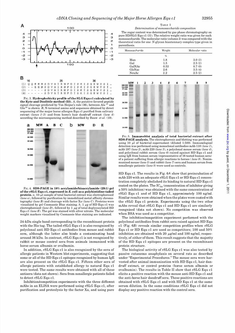
quence (35) (Fig. 3 A). According to the Von Heijne weight
matrix method (36), a favored putative signal peptidase cleav-
age site can be assigned between the Ala15 and Gln16 residues,
generating a protein beginning with QQEENSDVAI. In con-
trast, the N-terminal end of the protein initially purified from
hair dandruff (SDVAI) would result from a cleavage between
Asn20 and Ser21, which is not predicted by Von Heijne’s rules.
Equ c1 was purified from saliva, and the microsequencing of its
N-terminal peptide revealed a mixture of three sequences, oneof them beginning at the predicted Gln16, but the others at
Glu18 and Ser21, respectively (Fig. 3 B). Whether these N-ter-
minal ends are due to cleavage by signal peptidase at different
sites or are generated by proteolytic processing of the secreted
protein is not known. Such heterogeneous N-terminal ends
were also reported for human tear albumin (37), another mem-
ber of the lipocalin superfamily
Excluding the putative signal peptide, the protein contains
two cysteine residues at positions 83 and 176. In a previous
study, we observed an increase in the apparent molecular mass
of Equ c1 from 21,500 to 25,000 daltons in SDS-PAGE gels
under reducing conditions, indicating that these two cysteines
could form a disulfide bridge. Equ c1 is highly rich in charged
residues and aromatic residues. The calculated pI is 4.57, a value close to that determined by Dandeu et al.
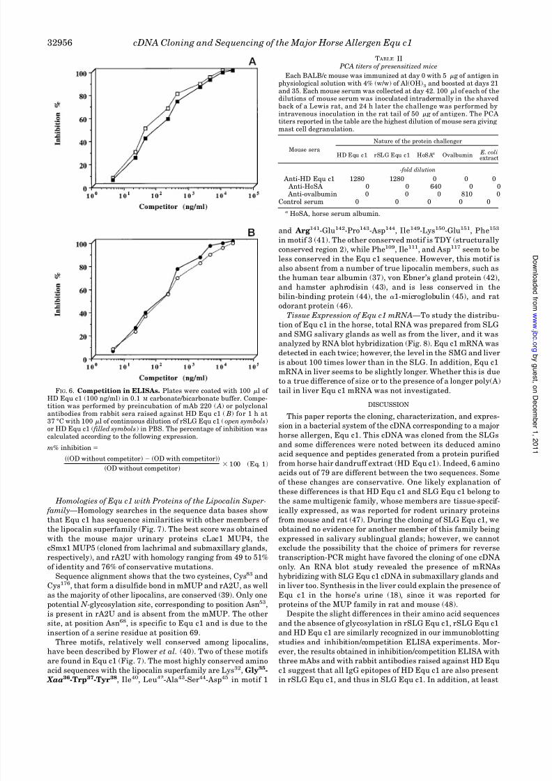
Glycosylation of Equ c1—Two putative N -glycosylation sites
are present at positions Asn53 and Asn68. Glycosylation of HD
and SLG Equ c1 was confirmed by gas phase chromatography,
which revealed the presence of approximately 8.6% (w/w) of
carbohydrates, representing 1,850 daltons. These results could
explain the decrease in apparent molecular weight of Equ c1 in
SDS-PAGE (Fig. 4) and the modification of the pI after
deglycosylation.
Analysis of the sugar residue composition in Table I shows
the presence of GalNAc, Gal, NeuAc, GlcNAc, and Man. Car-
bohydrates attached to proteins can be classified into two
groups, N -glycans and O-glycans. All N -glycans contain a com-
mon structure, Man13 6(Man13 3)Man13 4GlcNAc13
cDNA Cloning and Sequencing of the Major Horse Allergen Equ c1 32953
8/3/2019 J. Biol. Chem.-1996-Gregoire-32951-9
http://slidepdf.com/reader/full/j-biol-chem-1996-gregoire-32951-9 4/9
4GlcNAc3 Asn, called the trimannosyl core. Molecular ratio
results (second column in Table I) indicate unambiguously the
presence of this core and, therefore, the presence in the glucidic
part of Equ c1 of one N -glycan member of the biantennary
complex type that contains three mannose residues. One N -
acetyl-lactosamine (Gal13 4GlcNAc) is attached to the outer
two mannose residues, followed by sialic acid residues (for
Equ c1) or additional N -acetyllactosamines (38).
The presence of GalNAc only found in the O-glycan compo-nents, except for several hormones (38), suggests that the pro-
tein is also O-glycosylated.
Expression of Equ c1 as a Recombinant Protein— A re-
combinant protein, starting at Glu19, was produced in a bac-
terial system, after cloning of the corresponding cDNA se-
quence in a pET 28 plasmid. This plasmid allows bacterial
expression of a recombinant protein with a 40-amino acid
polypeptide tail containing a polyhistidine tag to its N-terminal
end (Fig. 1). To allow the production of a recombinant protein
without any added amino acid, a factor Xa proteolytic site
(LEFIEGR2ENSDVA) was inserted between the tail and the
recombinant protein.
Two recombinant clones were tested for rSLG Equ c1 expres-
sion. Optimal production was obtained after a 150-min induc-
tion by isopropyl -D-thiogalactopyranoside. A protein determi-
nation assay showed that rSLG Equ c1 represents about 30% of
the total bacterial protein. This protein was essentially present
in the supernatant of the bacterial extracts. A single purifica-
tion step by immobilized metal affinity chromatography was
sufficient to obtain pure rSLG Equ c1, which migrates as a
single band of 19.5–20 kDa in an 18% SDS-PAGE gel (Fig. 4,
lane A) after cleavage by factor Xa. This molecular mass is
compatible with the calculated mass of 19,469 daltons andrather similar to that of deglycosylated natural Equ c1.
Antigenicity of the Recombinant Protein—The recombinant
protein was tested for its antigenic recognition by different
antibodies raised against HD Equ c1, i.e. three mouse mono-
clonal antibodies (mAbs 118 and 197, which recognize two
different linear epitopes, and mAb 220, which recognizes a
conformational epitope),2 mouse and rabbit polyclonal antibod-
ies (IgG), and human IgE from the sera of 16 patients suffering
from horse allergic reactions (characterized in Ref. 17).
Immunoblot analysis after SDS-PAGE (Fig. 5), performed on
the total bacterial extract, shows that the three mAbs bind a
2 C. Gregoire, J. Rabillon, B. David, and J.-P. Dandeu, unpublisheddata.
FIG. 2. Full-length sequence of the SLG Equ c1 cDNA ( first line) and its deduced amino acid sequence ( second line). The N-terminaland internal tryptic fragments of HD Equ c1 are positioned in the third line. The start and stop codons are double underlined, and thepolyadenylation signal is in boldface type. The N -glycosylation sites are underlined, and the amino acid sequences used to deduce the sequence forthe degenerate primers are in boldface type.
cDNA Cloning and Sequencing of the Major Horse Allergen Equ c132954
8/3/2019 J. Biol. Chem.-1996-Gregoire-32951-9
http://slidepdf.com/reader/full/j-biol-chem-1996-gregoire-32951-9 5/9
24-kDa single band corresponding to the recombinant protein
with the His tag. The tailed rSLG Equ c1 is also recognized by
polyclonal anti HD Equ c1 antibodies from mouse and rabbit
sera, although the latter also binds a contaminating bandaround 36 kDa. In contrast, rSLG Equ c1 is not recognized by
rabbit or mouse control sera from animals immunized with
horse serum albumin or ovalbumin.
In addition, rSLG Equ c1 is also recognized by the sera of
allergic patients in Western blot experiments, suggesting that
some or all of the HD Equ c1 epitopes recognized by human IgE
are also present on the rSLG Equ c1. Fifteen other sera of
allergic patients with established allergy to natural Equ c1
were tested. The same results were obtained with all of these
antisera (data not shown). Sera from nonallergic patients failed
to detect rSLG Equ c1.
Inhibition/competition experiments with the three different
mAbs in an ELISA were performed using rSLG Equ c1, after
purification and proteolysis by the factor Xa, and using pure
HD Equ c1. The results in Fig. 6 A show that preincubation of mAb 220 with an adequate rSLG Equ c1 or HD Equ c1 concen-
tration completely abolished its binding to natural HD Equ c1
coated on the plates. The IC50 (concentration of inhibitor giving
a 50% inhibition) was obtained with the same concentration of
rSLG Equ c1 and of HD Equ c1, approximately 100 ng/ml.
Similar results were obtained when the plates were coated with
the rSLG Equ c1 protein. Experiments using the two other
mAbs reveal that rSLG Equ c1 and HD Equ c1 are similarly
recognized (data not shown). No competition was observed
when BSA was used as a competitor.
The inhibition/competition experiment performed with the
polyclonal antibodies from rabbit sera raised against HD Equ
c1 (Fig. 6 B) reveals similar competition profiles when rSLG
Equ c1 or HD Equ c1 are used as competitors; 100 and 50%
inhibition are obtained with 20 g/ml and 100 ng/ml, respec-tively, of either of them. This result suggests that the majority
of the HD Equ c1 epitopes are present on the recombinant
protein structure.
The biological activity of rSLG Equ c1 was also tested by
passive cutaneous anaphylaxis on several rats as described
under “Experimental Procedures.” The mouse sera were har-
vested after animal immunization with HD Equ c1, hair dan-
druff extract, or control proteins (horse serum albumin or
ovalbumin). The results in Table II show that rSLG Equ c1
elicits a positive reaction with the mouse anti-HD Equ c1 and
the anti-horse hair dandruff sera. These positive reactions are
obtained with rSLG Equ c1 and with HD Equ c1 at the same
serum dilution. In the same conditions rSLG Equ c1 did not
display any positive reaction with the control sera.
T ABLE I Determination of monosaccharide composition
The sugar content was determined by gas phase chromatography onpure HD/SLG Equ c1 (31). The relative weight ratio was given for eachmonosaccharide. The molecular ratio (column 2) was compared with thetheoretical ratio for one N -glycan biantennary complex type given inparenthesis.
Monosaccharide Weight Molecular ratio
%
Man 1.8 3.0 (3)Gal 1.5 2.5 (2)GalNAc 0.53 0.7 (0)GlcNac 2.6 3.5 (4)NeuAc 2.2 2.5 (2)
FIG. 5. Immunoblot analysis of total bacterial extract afterSDS-PAGE analysis. The electrophoresis and blotting was performedusing 10 l of bacterial supernatant (diluted 1:500). Immunologicaldetection was performed using monoclonal antibodies mAb 118 (lane 1),mAb 197 (lane 2), mAb 220 (lane 3), a polyclonal mouse serum (lane 4),and polyclonal rabbit serum (lane 6) raised against HD Equ c1 andusing IgE from human serum (representative of 16 tested human sera)of a patient suffering from allergic reactions to horses (lane 8). Nonim-munized mouse (lane 5) and rabbit (lane 7 ) sera and human serum fromnonallergic patients (lane 9) were used as controls.
FIG. 3. Hydrophobicity profile of the SLG Equ c1 calculated bythe Kyte and Doolittle method (53). A, the putative favored peptidesignal cleavage predicted by Von Heijne’s rule (36), between Ala15 andGln16 is shown. B, N-terminal amino acid sequences obtained by directsequencing of the major horse allergen Equ c1 purified from salivaryextract (lanes 1–3) and from horse’s hair dandruff extract (lane 4)according the microsequencing method described by Bauw et al. (19).
FIG. 4. SDS-PAGE in 18% acrylamide/bisacrylamide (29:1) gelof the rSLG Equ c1, expressed in E. coli as a polyhistidine-tailedprotein. a, 10-l sample of total bacterial extract was electrophoresed(lane a), followed by purification by immobilized metal affinity chroma-tography (lane B) and cleavage with factor Xa (lane C). Proteins were
visualized by gel Coomassie Blue staining. b, 1 g of HD Equ c1 waselectrophoresed (lane D), followed by 1 g of total deglycosylated HDEqu c1 (lane E). The gel was stained with silver nitrate. The molecularweight markers visualized by Coomassie blue staining are indicated.
cDNA Cloning and Sequencing of the Major Horse Allergen Equ c1 32955
8/3/2019 J. Biol. Chem.-1996-Gregoire-32951-9
http://slidepdf.com/reader/full/j-biol-chem-1996-gregoire-32951-9 6/9
Homologies of Equ c1 with Proteins of the Lipocalin Super-
family—Homology searches in the sequence data bases show
that Equ c1 has sequence similarities with other members of
the lipocalin superfamily (Fig. 7). The best score was obtained
with the mouse major urinary proteins cLac1 MUP4, the
cSmx1 MUP5 (cloned from lachrimal and submaxillary glands,respectively), and rA2U with homology ranging from 49 to 51%
of identity and 76% of conservative mutations.
Sequence alignment shows that the two cysteines, Cys83 and
Cys176, that form a disulfide bond in mMUP and rA2U, as well
as the majority of other lipocalins, are conserved (39). Only one
potential N -glycosylation site, corresponding to position Asn53,
is present in rA2U and is absent from the mMUP. The other
site, at position Asn68, is specific to Equ c1 and is due to the
insertion of a serine residue at position 69.
Three motifs, relatively well conserved among lipocalins,
have been described by Flower et al. (40). Two of these motifs
are found in Equ c1 (Fig. 7). The most highly conserved amino
acid sequences with the lipocalin superfamily are Lys32, Gly35-
Xaa36-Trp37-Tyr38, Ile40, Leu42-Ala43-Ser44-Asp45 in motif 1
and Arg141-Glu142-Pro143-Asp144, Ile149-Lys150-Glu151, Phe153
in motif 3 (41). The other conserved motif is TDY (structurally
conserved region 2), while Phe109, Ile111, and Asp117 seem to be
less conserved in the Equ c1 sequence. However, this motif is
also absent from a number of true lipocalin members, such as
the human tear albumin (37), von Ebner’s gland protein (42),
and hamster aphrodisin (43), and is less conserved in the
bilin-binding protein (44), the 1-microglobulin (45), and rat
odorant protein (46).
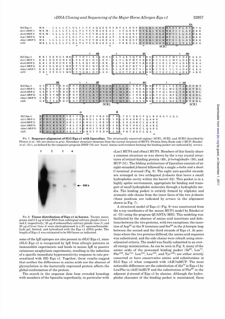
Tissue Expression of Equ c1 mRNA—To study the distribu-
tion of Equ c1 in the horse, total RNA was prepared from SLG
and SMG salivary glands as well as from the liver, and it was
analyzed by RNA blot hybridization (Fig. 8). Equ c1 mRNA was
detected in each twice; however, the level in the SMG and liver
is about 100 times lower than in the SLG. In addition, Equ c1
mRNA in liver seems to be slightly longer. Whether this is due
to a true difference of size or to the presence of a longer poly(A)
tail in liver Equ c1 mRNA was not investigated.
DISCUSSION
This paper reports the cloning, characterization, and expres-
sion in a bacterial system of the cDNA corresponding to a major
horse allergen, Equ c1. This cDNA was cloned from the SLGs
and some differences were noted between its deduced amino
acid sequence and peptides generated from a protein purified
from horse hair dandruff extract (HD Equ c1). Indeed, 6 amino
acids out of 79 are different between the two sequences. Some
of these changes are conservative. One likely explanation of
these differences is that HD Equ c1 and SLG Equ c1 belong to
the same multigenic family, whose members are tissue-specif-
ically expressed, as was reported for rodent urinary proteins
from mouse and rat (47). During the cloning of SLG Equ c1, we
obtained no evidence for another member of this family being
expressed in salivary sublingual glands; however, we cannot
exclude the possibility that the choice of primers for reversetranscription-PCR might have favored the cloning of one cDNA
only. An RNA blot study revealed the presence of mRNAs
hybridizing with SLG Equ c1 cDNA in submaxillary glands and
in liver too. Synthesis in the liver could explain the presence of
Equ c1 in the horse’s urine (18), since it was reported for
proteins of the MUP family in rat and mouse (48).
Despite the slight differences in their amino acid sequences
and the absence of glycosylation in rSLG Equ c1, rSLG Equ c1
and HD Equ c1 are similarly recognized in our immunoblotting
studies and inhibition/competition ELISA experiments. Mor-
ever, the results obtained in inhibition/competition ELISA with
three mAbs and with rabbit antibodies raised against HD Equ
c1 suggest that all IgG epitopes of HD Equ c1 are also present
in rSLG Equ c1, and thus in SLG Equ c1. In addition, at least
T ABLE II PCA titers of presensitized mice
Each BALB/c mouse was immunized at day 0 with 5 g of antigen inphysiological solution with 4% (w/w) of Al(OH)3 and boosted at days 21and 35. Each mouse serum was collected at day 42. 100l of each of thedilutions of mouse serum was inoculated intradermally in the shavedback of a Lewis rat, and 24 h later the challenge was performed byintravenous inoculation in the rat tail of 50 g of antigen. The PCAtiters reported in the table are the highest dilution of mouse sera givingmast cell degranulation.
Mouse sera
Nature of the protein challenger
HD Equ c1 rSLG Equ c1 HoSAa OvalbuminE. coliextract
-fold dilution
Anti-HD Equ c1 1280 1280 0 0 0 Anti-HoSA 0 0 640 0 0 Anti-ovalbumin 0 0 0 810 0Control serum 0 0 0 0 0
a HoSA, horse serum albumin.
FIG. 6. Competition in ELISAs. Plates were coated with 100 l of HD Equ c1 (100 ng/ml) in 0.1 M carbonate/bicarbonate buffer. Compe-tition was performed by preincubation of mAb 220 ( A) or polyclonalantibodies from rabbit sera raised against HD Equ c1 ( B) for 1 h at37 °C with 100 l of continuous dilution of rSLG Equ c1 (open symbols)or HD Equ c1 ( filled symbols) in PBS. The percentage of inhibition wascalculated according to the following expression.
m% inhibition
OD without competitor OD with competitor
OD without competitor 100 (Eq. 1)
cDNA Cloning and Sequencing of the Major Horse Allergen Equ c132956
8/3/2019 J. Biol. Chem.-1996-Gregoire-32951-9
http://slidepdf.com/reader/full/j-biol-chem-1996-gregoire-32951-9 7/9
some of the IgE epitopes are also present in rSLG Equ c1, since
rSLG Equ c1 is recognized by IgE from allergic patients in
immunoblot experiments and binds to mouse IgE in passive
cutaneous anaphylaxis experiments, resulting in the induction
of a specific immediate hypersensitivity response in rats pre-
sensitized with HD Equ c1. Together, these results suggest
that neither the differences in amino acids nor the absence of
glycosylation in the bacterially expressed protein affects the
global conformation of the protein.
The search in the sequence data base revealed homology
with members of the lipocalin superfamily, in particular with
cLac1 MUP4 and cSmx1 MUP5. Members of this family share
a common structure as was shown by the x-ray crystal struc-
tures of retinol-binding protein (49), -lactoglobulin (50), and
MUP (51). The folding architecture of lipocalins consists of an
eight-stranded -barrel followed by a single -helix and a shortC-terminal -strand (Fig. 9). The eight anti-parallel strands
are arranged in two orthogonal -sheets that leave a small
hydrophobic cavity within the barrel (52). This pocket is in a
highly apolar environment, appropriate for binding and trans-
port of small hydrophobic molecules through a hydrophilic me-
dia. The binding pocket is entirely formed by aliphatic and
aromatic side chains from the inner faces of the two -sheets
(these positions are indicated by arrows in the alignment
shown in Fig. 7).
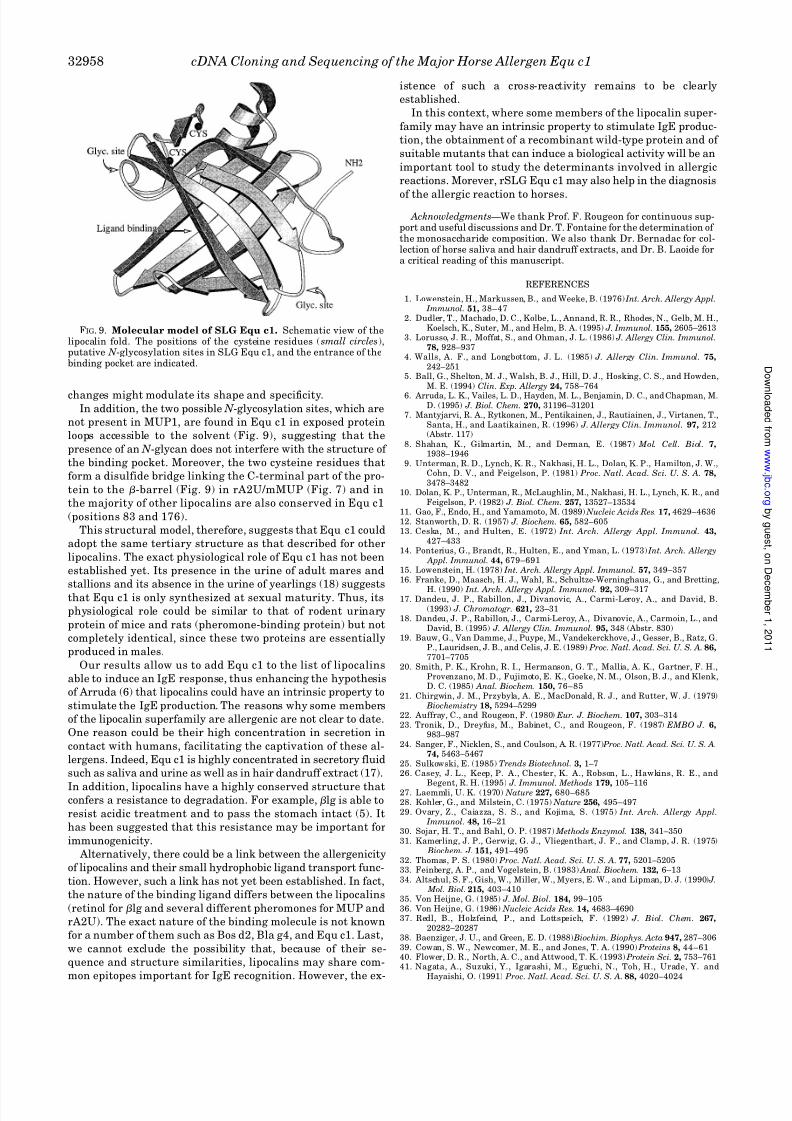
A structural model of Equ c1 (Fig. 9) was constructed from
the x-ray coordinates of the mouse MUP1 model by Bocskei et
al. (51) using the program QUANTA (MSI). This modeling was
facilitated by the absence of amino acid insertions and dele-
tions between the two proteins, with two exceptions: the inser-tion of Asp22 at the N terminus and Ser69 in the -hairpin loop
between the second and the third strands of Equ c1. At posi-
tions where the two proteins differed, the amino acid sequence
was substituted, and the side chains were rebuilt using stere-
ochemical criteria. The model was finally submitted to an over-
all energy minimization. As can be seen in Fig. 9, many of the
amino acids of the presumed binding pocket (Ile63, Leu71,
Phe109, Ile111, Leu124, Leu135, and Tyr139) are either strictly
conserved or have conservative amino acid substitutions in
SLG Equ c1 when compared with rA2U/mMUP. The most
noticeable differences are the substitution of Ala73 in Equ c1 by
Leu/Phe in rA2U/mMUP and the substitution of Phe90 in the
adjacent -strand of Equ c1 by alanine. Although the hydro-
phobic character of the binding pocket is maintained, these
FIG. 7. Sequence alignment of SLG Equ c1 with lipocalins. The structurally conserved regions ( SCR1, SCR2, and SCR3) described byFlower et al. (40) are shown in gray. Secondary structure elements from the crystal structure of MUP1 (Protein Data Bank code 1 MUP; Bocskei
et al. (51)), as defined by the computer program DSSP (54) are boxed. Amino acid residues forming the binding pocket are indicated by arrows.
FIG. 8. Tissue distribution of Equ c1 in horses. Twenty micro-grams and 0.2 g of total RNA from sublingual salivary glands (lanes 1
and 2, respectively), 20 g of submaxillary salivary glands (lane 3), and20 g of liver (lane 4) were electrophoresed in a 2% agarose/formalde-hyde gel, blotted, and hybridized with the Equ c1 cDNA probe. Thelength of Equ c1 was estimated to be 950 bases as indicated.
cDNA Cloning and Sequencing of the Major Horse Allergen Equ c1 32957
8/3/2019 J. Biol. Chem.-1996-Gregoire-32951-9
http://slidepdf.com/reader/full/j-biol-chem-1996-gregoire-32951-9 8/9
changes might modulate its shape and specificity.In addition, the two possible N -glycosylation sites, which are
not present in MUP1, are found in Equ c1 in exposed protein
loops accessible to the solvent (Fig. 9), suggesting that the
presence of an N -glycan does not interfere with the structure of
the binding pocket. Moreover, the two cysteine residues that
form a disulfide bridge linking the C-terminal part of the pro-
tein to the -barrel (Fig. 9) in rA2U/mMUP (Fig. 7) and in
the majority of other lipocalins are also conserved in Equ c1
(positions 83 and 176).
This structural model, therefore, suggests that Equ c1 could
adopt the same tertiary structure as that described for other
lipocalins. The exact physiological role of Equ c1 has not been
established yet. Its presence in the urine of adult mares and
stallions and its absence in the urine of yearlings (18) suggeststhat Equ c1 is only synthesized at sexual maturity. Thus, its
physiological role could be similar to that of rodent urinary
protein of mice and rats (pheromone-binding protein) but not
completely identical, since these two proteins are essentially
produced in males.
Our results allow us to add Equ c1 to the list of lipocalins
able to induce an IgE response, thus enhancing the hypothesis
of Arruda (6) that lipocalins could have an intrinsic property to
stimulate the IgE production. The reasons why some members
of the lipocalin superfamily are allergenic are not clear to date.
One reason could be their high concentration in secretion in
contact with humans, facilitating the captivation of these al-
lergens. Indeed, Equ c1 is highly concentrated in secretory fluid
such as saliva and urine as well as in hair dandruff extract (17).
In addition, lipocalins have a highly conserved structure thatconfers a resistance to degradation. For example, lg is able to
resist acidic treatment and to pass the stomach intact (5). It
has been suggested that this resistance may be important for
immunogenicity.
Alternatively, there could be a link between the allergenicity
of lipocalins and their small hydrophobic ligand transport func-
tion. However, such a link has not yet been established. In fact,
the nature of the binding ligand differs between the lipocalins
(retinol for lg and several different pheromones for MUP and
rA2U). The exact nature of the binding molecule is not known
for a number of them such as Bos d2, Bla g4, and Equ c1. Last,
we cannot exclude the possibility that, because of their se-
quence and structure similarities, lipocalins may share com-
mon epitopes important for IgE recognition. However, the ex-
istence of such a cross-reactivity remains to be clearly
established.
In this context, where some members of the lipocalin super-
family may have an intrinsic property to stimulate IgE produc-
tion, the obtainment of a recombinant wild-type protein and of
suitable mutants that can induce a biological activity will be an
important tool to study the determinants involved in allergic
reactions. Morever, rSLG Equ c1 may also help in the diagnosis
of the allergic reaction to horses.
Acknowledgments—We thank Prof. F. Rougeon for continuous sup-port and useful discussions and Dr. T. Fontaine for the determination of the monosaccharide composition. We also thank Dr. Bernadac for col-lection of horse saliva and hair dandruff extracts, and Dr. B. Laoide fora critical reading of this manuscript.
REFERENCES
1. Lowenstein, H., Markussen, B., and Weeke, B. (1976) Int. Arch. Allergy Appl. Immunol. 51, 38–47
2. Dudler, T., Machado, D. C., Kolbe, L., Annand, R. R., Rhodes, N., Gelb, M. H.,Koelsch, K., Suter, M., and Helm, B. A. (1995) J. Immunol. 155, 2605–2613
3. Lorusso, J. R., Moffat, S., and Ohman, J. L. (1986) J. Allergy Clin. Immunol.78, 928–937
4. Walls, A. F., and Longbottom, J. L. (1985) J. Allergy Clin. Immunol. 75,242–251
5. Ball, G., Shelton, M. J., Walsh, B. J., Hill, D. J., Hosking, C. S., and Howden,M. E. (1994) Clin. Exp. Allergy 24, 758–764
6. Arruda, L. K., Vailes, L. D., Hayden, M. L., Benjamin, D. C., and Chapman, M.D. (1995) J. Biol. Chem. 270, 31196–31201
7. Mantyjarvi, R. A., Rytkonen, M., Pentikainen, J., Rautiainen, J., Virtanen, T.,Santa, H., and Laatikainen, R. (1996) J. Allergy Clin. Immunol. 97, 212(Abstr. 117)
8. Shahan, K., Gilmartin, M., and Derman, E. (1987) Mol. Cell. Biol. 7,1938–1946
9. Unterman, R. D., Lynch, K. R., Nakhasi, H. L., Dolan, K. P., Hamilton, J. W.,Cohn, D. V., and Feigelson, P. (1981) Proc. Natl. Acad. Sci. U. S. A. 78,3478–3482
10. Dolan, K. P., Unterman, R., McLaughlin, M., Nakhasi, H. L., Lynch, K. R., andFeigelson, P. (1982) J. Biol. Chem. 257, 13527–13534
11. Gao, F., Endo, H., and Yamamoto, M. (1989) Nucleic Acids Res. 17, 4629–463612. Stanworth, D. R. (1957) J. Biochem. 65, 582–60513. Ceska, M., and Hulten, E. (1972) Int. Arch. Allergy Appl. Immunol. 43,
427–43314. Ponterius, G., Brandt, R., Hulten, E., and Yman, L. (1973) Int. Arch. Allergy
Appl. Immunol. 44, 679–69115. Lowenstein, H. (1978) Int. Arch. Allergy Appl. Immunol. 57, 349–35716. Franke, D., Maasch, H. J., Wahl, R., Schultze-Werninghaus, G., and Bretting,
H. (1990) Int. Arch. Allergy Appl. Immunol. 92, 309–31717. Dandeu, J. P., Rabillon, J., Divanovic, A., Carmi-Leroy, A., and David, B.
(1993) J. Chromatogr. 621, 23–3118. Dandeu, J. P., Rabillon, J., Carmi-Leroy, A., Divanovic, A., Carmoin, L., and
David, B. (1995) J. Allergy Clin. Immunol. 95, 348 (Abstr. 830)19. Bauw, G., Van Damme, J., Puype, M., Vandekerckhove, J., Gesser, B., Ratz, G.
P., Lauridsen, J. B., and Celis, J. E. (1989) Proc. Natl. Acad. Sci. U. S. A. 86,7701–7705
20. Smith, P. K., Krohn, R. I., Hermanson, G. T., Mallia, A. K., Gartner, F. H.,Provenzano, M. D., Fujimoto, E. K., Goeke, N. M., Olson, B. J., and Klenk,D. C. (1985) Anal. Biochem. 150, 76–85
21. Chirgwin, J. M., Przybyla, A. E., MacDonald, R. J., and Rutter, W. J. (1979) Biochemistry 18, 5294–5299
22. Auffray, C., and Rougeon, F. (1980) Eur. J. Biochem. 107, 303–31423. Tronik, D., Dreyfus, M., Babinet, C., and Rougeon, F. (1987) EMBO J. 6,
983–98724. Sanger, F., Nicklen, S., and Coulson, A. R. (1977) Proc. Natl. Acad. Sci. U. S. A.
74, 5463–546725. Sulkowski, E. (1985) Trends Biotechnol. 3, 1–726. Casey, J. L., Keep, P. A., Chester, K. A., Robson, L., Hawkins, R. E., and
Begent, R. H. (1995) J. Immunol. Methods 179, 105–11627. Laemmli, U. K. (1970) Nature 227, 680–68528. Kohler, G., and Milstein, C. (1975) Nature 256, 495–49729. Ovary, Z., Caiazza, S. S., and Kojima, S. (1975) Int. Arch. Allergy Appl.
Immunol. 48, 16–2130. Sojar, H. T., and Bahl, O. P. (1987) Methods Enzymol. 138, 341–35031. Kamerling, J. P., Gerwig, G. J., Vliegenthart, J. F., and Clamp, J. R. (1975)
Biochem. J. 151, 491–49532. Thomas, P. S. (1980) Proc. Natl. Acad. Sci. U. S. A. 77, 5201–520533. Feinberg, A. P., and Vogelstein, B. (1983) Anal. Biochem. 132, 6–1334. Altschul, S. F., Gish, W., Miller, W., Myers, E. W., and Lipman, D. J. (1990) J.
Mol. Biol. 215, 403–41035. Von Heijne, G. (1985) J. Mol. Biol. 184, 99–10536. Von Heijne, G. (1986) Nucleic Acids Res. 14, 4683–469037. Redl, B., Holzfeind, P., and Lottspeich, F. (1992) J. Biol. Chem. 267,
20282–2028738. Baenziger, J. U., and Green, E. D. (1988) Biochim. Biophys. Acta 947, 287–30639. Cowan, S. W., Newcomer, M. E., and Jones, T. A. (1990) Proteins 8, 44–6140. Flower, D. R., North, A. C., and Attwood, T. K. (1993) Protein Sci. 2, 753–76141. Nagata, A., Suzuki, Y., Igarashi, M., Eguchi, N., Toh, H., Urade, Y. and
Hayaishi, O. (1991) Proc. Natl. Acad. Sci. U. S. A. 88, 4020–4024
FIG. 9. Molecular model of SLG Equ c1. Schematic view of thelipocalin fold. The positions of the cysteine residues (small circles),putative N -glycosylation sites in SLG Equ c1, and the entrance of thebinding pocket are indicated.
cDNA Cloning and Sequencing of the Major Horse Allergen Equ c132958
8/3/2019 J. Biol. Chem.-1996-Gregoire-32951-9
http://slidepdf.com/reader/full/j-biol-chem-1996-gregoire-32951-9 9/9
42. Schmale, H., Holtgreve-Grez, H., and Christiansen, H. (1990) Nature 343,366–369
43. Henzel, W. J., Rodriguez, H., Singer, A. G., Stults, J. T., Macrides, F., Agosta,W. C., and Niall, H. (1988) J. Biol. Chem. 263, 16682–16687
44. Suter, F., Kayser, H., and Zuber, H. (1988) Biol. Chem. Hoppe-Seyler 369,497–505
45. Kaumeyer, J. F., Polazzi, J. O., and Kotick, M. P. (1986) Nucleic Acids Res. 14,7839–7850
46. Pevsner, J., Reed, R. R., Feinstein, P. G., and Snyder, S. H. (1988) Science 241,336–339
47. MacInnes, J. I., Nozik, E. S., and Kurtz, D. T. (1986) Mol. Cell. Biol. 6,3563–3567
48. Shahan, K., Denaro, M., Gilmartin, M., Shi, Y., and Derman, E. (1987) Mol.Cell. Biol. 7, 1947–1954
49. Newcomer, M. E., Jones, T. A., Aqvist, J., Sundelin, J., Eriksson, U., Rask, L.,and Peterson, P. A. (1984) EMBO J. 3, 1451–1454
50. Papiz, M. Z., Sawyer, L., Eliopoulos, E. E., North, A. C., Findlay, J. B.,Sivaprasadarao, R., Jones, T. A., Newcomer, M. E., and Kraulis, P. J. (1986) Nature 324, 383–385
51. Bocskei, Z., Groom, C. R., Flower, D. R., Wright, C. E., Phillips, S. E.,Cavaggioni, A., Findlay, J. B., and North, A. C. (1992) Nature 360, 186–188
52. North, A. C. (1989) J. Mol. Graph. 7, 67–7053. Kyte, J., and Doolittle, R. F. (1982) J. Mol. Biol. 157, 105–13254. Kabsch, W., and Sander, C. (1983) Biopolymers 22, 2577–2637
cDNA Cloning and Sequencing of the Major Horse Allergen Equ c1 32959