investigations for iron- deficiency anemia in … · 2. peripheral smear : thin smear - rbc...
TRANSCRIPT
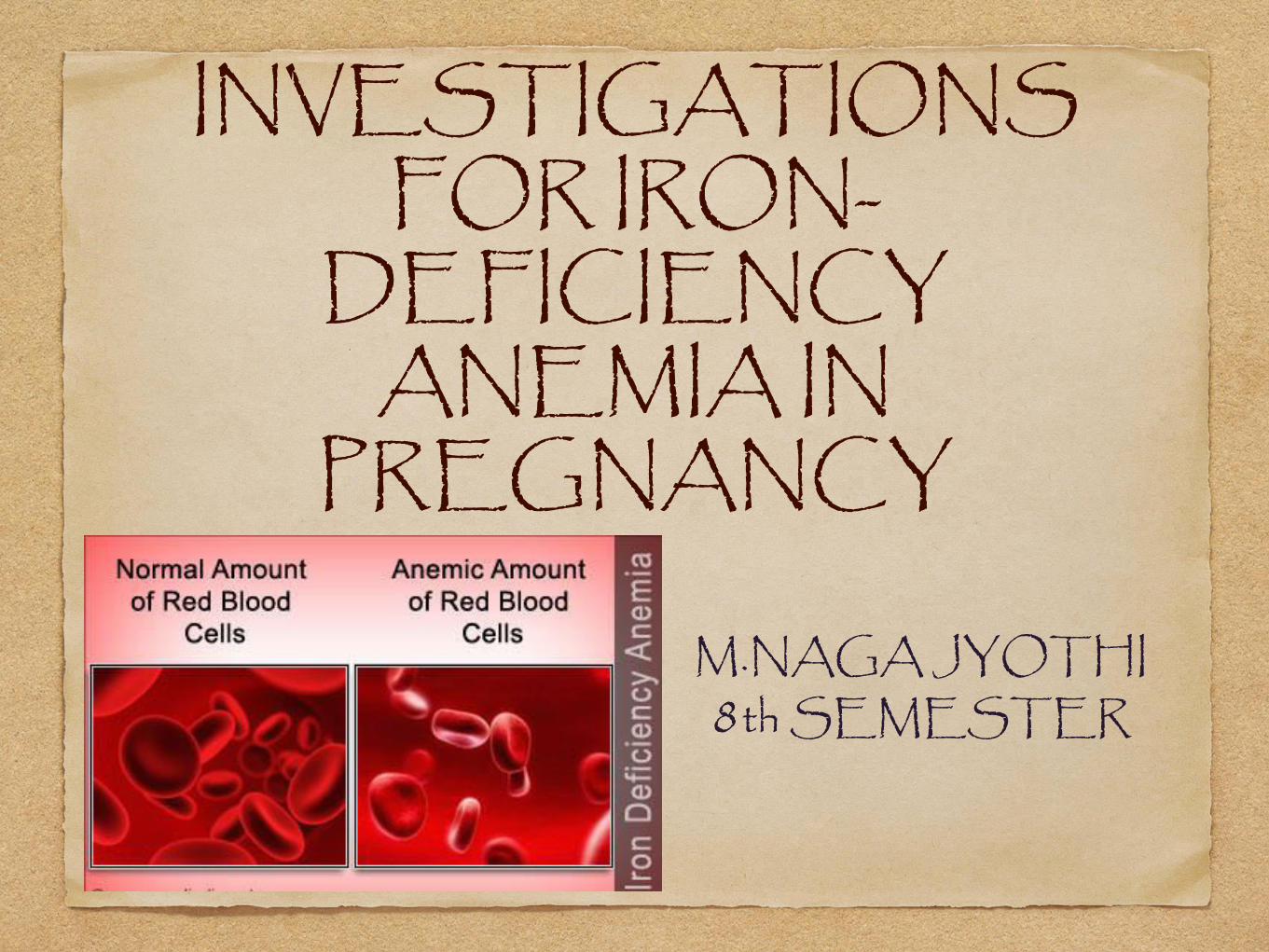
INVESTIGATIONS FOR IRON-
DEFICIENCY ANEMIA IN
PREGNANCY
M.NAGA JYOTHI 8th SEMESTER

1. Hemoglobin and hematocrit :
• Hemoglobin - <10g% (NORMAL : 11-14g%) WHO Grading:
MILD 8-10g%
MODERATE 7-8g%
SEVERE 4-7g%
VERY SEVERE <4g%
• PCV - <32% (NORMAL : 32%-36%)
• RBC Count - <3.2million (NORMAL : 4-4.5million/cubic millimetre)


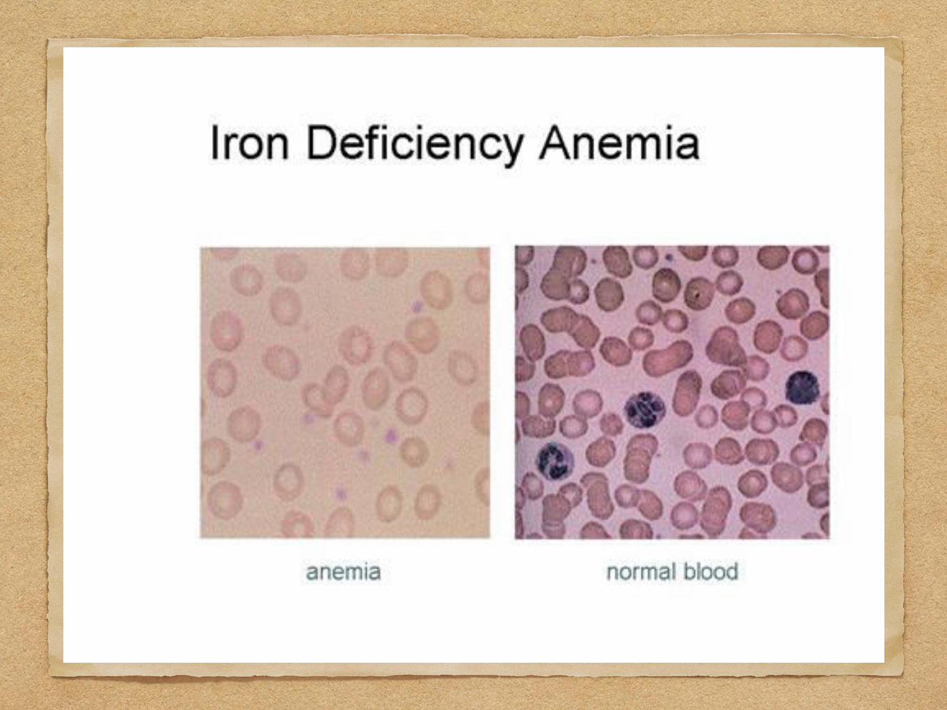
2. Peripheral Smear :
• Thin smear -
RBC Morphology - Microcytic hypochromic RBC’s , anisocytosis , poikilocytosis and target cells.
• Thick smear -
Useful in identifying parasites - malaria , leishmania

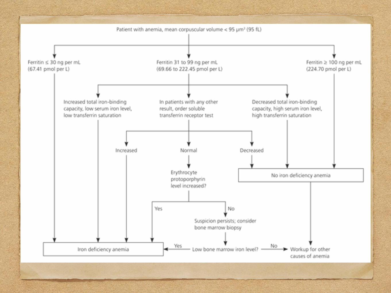
3. Red cell indices :
• Mean corpuscular volume (MCV =Hct/RBC*10) - decreased(<80fl ) (NORMAL : 80-100fl)
• Mean corpuscular haemoglobin (MCH =Hb/RBC*10) - decreased(<25pg) (NORMAL : 27-31pg)
• Mean cell hemoglobin concentration(MCHC = Hb/Hct*100)- reduced(<30% is sensitive indicator) (NORMAL : 32-36g/dl)
• Red cell distribution width (RDW) - increased(>14%)[helps to differentiate from thalassemia.] (NORMAL : 11.5-14.5%)

4 . Special tests : A.ferrokinetic studies •Serum iron and Total iron binding capacity: <30mg/dl and
>400mg/dl (NORMAL: 65-165mg/dl and 300-400mg/dl) respectively
•Transferin % saturation : <16% (NORMAL : 20-50%) •Serum ferritin : <12ng/ml (NORMAL : 15-300ng/ml) •Serum transferrin receptor(TfR) : increased (>2.8mg/L)
(NORMAL: 1-2mg/dl) •Zinc protoporphyrin : increased (NORMAL : 0-
35microgram/dl) B.Bone marrow(prussian blue stain)studies: <10% hemosideroblasts - not done routinely.
5. Investigations to determine the aetiology :
• Urine for hematuria and pyuria(culture & sensitivity)
• Stool examination for occult blood , ova and cysts.
• Renal function tests for chronic renal disease.
• Tests for tuberculosis(x-ray chest)
• Fractional test meal analysis of gastric juice.
• Serum protien.
• Osmotic fragility.

PHASES OF IRON- DEFICIENCY ANEMIA
1. Decreased iron stores(tissue iron only): decreased ferritin levels
2. Decrease in iron for erythropoiesis:( no clinical anemia)- serum transferrin receptors increases , decreased ferritin & %saturation of iron, increased FEP, decreased hemoglobin & hematocrit
3. Decrease in peripheral blood haemoglobin : decreased ferritin, %saturation of iron, haemoglobin , hematocrit , increased FEP and microcytic hypo chromic anemia.
4. Decrease in tissue oxygen delivery : clinical signs and symptoms.


DIFFERENTIAL DIAGNOSIS
Anemia due to chronic disease or an inflammatory process
Thalassemia trait
Sideroblastic anemia
Anemia due to lead poisoning
Infection
Nephritis & pre-eclampsia
Hemoglobinopathies


COMPLICATIONS 1. Maternal :
•Spontaneous abortion
•Susceptibility to infections
•Preterm labour
•Pre-eclampsia
•Inability to withstand postpartum hemorrhage
•Puerperal sepsis
•Congestive cardiac failure
•Sideropenic dysphagia(paterson-kelly syndrome,plummer-vinson syndrome[rare])


2. Fetal :
• Intrauterine growth restriction
• Prematurity
• Intrauterine fetal death (severe cases)
• Non-immune hydrops
• Increased morbidity and mortality
• Neonatal anemia
• Behavioural abnormalities in children

3. Puerperium :
• Subinvolution
• Poor lactation
• Puerperal venous thrombosis
• Pulmonary embolism


PROGNOSIS MATERNAL -
• If detected early and proper treatment is instituted,anemia improves promptly.
• At times, recurrence in subsequent pregnancy is seen.
• Anemia directly or indirectly contributes to about 20% of the maternal deaths.

FETAL :
• In severe cases fetal prognosis is adversely affected by prematurity with its hazards.
•Baby born at term, to severely anaemic mother will not be anaemic at birth, but as there is little or no reserve iron, anaemia develops in neonatal periods.
•Mean cord blood levels of serum iron, ferritin, B12 and folate are higher than that of mother.
•However, total iron binding capacity and serum levels of vitamin E are lower than that of mother.
