international journal of scientific research and …deferoxamine (dfo) is isolated from the...
TRANSCRIPT
Lata Shahani et al., IJSRR 2019, 8(2), 2122-2140
IJSRR, 8(2) April. – June., 2019 Page 2122
Research article Available online www.ijsrr.org ISSN: 2279–0543
International Journal of Scientific Research and Reviews
Ameliorative effect of Nigella sativa and Deferrioxamine against neurotoxicity induced by combination of fluoride and aluminium
1Nisha Rathore, *2Lata Shahani and Trishna Patel3
1Department of Zoology, The IIS University, Jaipur, Rajasthan,India.
email: nisha89rathore@gmailcom, Mob no:7665820978 2*Department of Zoology, The IIS University, Jaipur, Rajasthan,India.
email: [email protected], Mob no:8949293634 3Department of Zoology, The IIS University, Jaipur, Rajasthan,India.
email: trishnapatel92@gmailcom, Mob no:8949678599 ABSTRACT
The most common source of human exposure to fluoride and aluminium include drinking
water and various food items. Both fluoride and aluminium have been reported to induce oxidative
stress mediated neurodegeneration. The aim of the present study is to evaluate the protective effect of
Nigella sativa and deferrioxamine against combination of fluoride aluminium induced toxicity in
brain of male Swiss albino mice. Mice were treated intraperitoneally with fluoride and aluminium for
one month (thrice a week on alternate days) which resulted in decrease in various antioxidant
enzymes like SOD, catalase, GPx, GSH and increase in LPO levels. Neurodegenerataive changes
were also observed in histology of brain. DNA fragmentation and increased GFAP and Caspase 3
expression was also observed indicating that combination of fluoride and aluminium leads to
oxidative stress mediated apoptosis in brain cells. Treatment with Nigella sativa and deferrioxamine
resulted in amelioration of neurodegeneration induced by fluoride and aluminium indicating their
therapeutic potential.
KEY WORDS: Brain, Oxidative stress, Fluoride, Aluminium, Apoptosis, Nigella sativa
*Corresponding author:
Lata Shahani Department of Zoology,
The IIS University, Jaipur,
Rajasthan,India.
email: [email protected], Mob no:8949293634

Lata Shahani et al., IJSRR 2019, 8(2), 2122-2140
IJSRR, 8(2) April. – June., 2019 Page 2123
INTRODUCTION: Metals form an important part of earth’s crust. Most of the metals are present in rocks from
which they need to be extracted in the form. Metals like iron , copper , zinc, manganese, cobalt and
calcium are required for important functions of our body like respiration, circulation and
reproduction where they perform catalytic roles. Certain metals are not needed by the body and they
form poisonous soluble compounds inside the body. These toxic metals produce adverse effects in
the body by interfering with the metabolic processes as they imitate the action of an essential element
required in the metabolic process. Fluorine is distributed widely in nature and is one of the most abundant elements in nature.
Fluoride can enter into the body of the animals commonly through drinking water, but there can also
be various other sources of fluoride exposure like household and agricultural compounds having
fluoride in them, vegetation contaminated with fluoride, emissions from industries or volcanic ash.
Aluminium is another widely distributed metal found on earth 1. It is used in the production of many
products which are used in day to-day life like cookware , soda cans, aluminium foil , antacids,
aspirin, vaccines etc. The body does not need aluminium due to which it accumulates in the kidneys,
brain, lungs, liver and even thyroid where it act as a competitor of calcium for absorption and can
cause decrease in bone density2. Aluminium exposure occurs frequently in some occupations like
mining, factory work, and welding.
In today’s world looking at the side effects of synthetic drugs, there is need to find natural
products which have theurapeutic potential and can serve as an alternative to treat various diseases.
One of these products is Nigella sativa (NS) which has powerful healing properties for many
diseases3. It is a store house of active ingredients like many volatile and non-volatile oils, proteins,
carbohydrates, alkaloids, saponins, minerals, phenolic compounds, steroidal compounds etc4. It also
has a high content of unsaturated fatty acids5 and various essential oil components6. Other main
compounds include thymoquinone, P-cymene, Carvacrol , 4-terpineol , T-anethole and
Sesquiterpene7. But the most active component of NS is thymoquinone and its derivatives8. Due to its
antioxidant activity, it has the ability to remove free radicals and inhibit lipid peroxidation which
becomes the main reason behind most of its ameliorative abilities9.
Deferoxamine (DFO) is isolated from the bacterium Streptomyces pilosus and has a binding
affinity towards iron and aluminium. It is used in case of iron and aluminum toxicity because it only
binds with ferric iron whereas the iron in hemoglobin or cytochromes remains unaffected.
Deferoxamine was approved for medical use in the United States in 1968 and it is also approved by
WHO as a safe medicine which is required in the health system10.
Lata Shahani et al., IJSRR 2019, 8(2), 2122-2140
IJSRR, 8(2) April. – June., 2019 Page 2124
Metals co-exist in the environment and humans are simultaneously exposed to multiple metals which
after entering the body interact with each other and form toxic complexes11,12. But only few reports
are available discussing the toxic nature of the metal combinations . Lot of research has been done on
toxic effects of individual metals however, multimetal exposure is a new area in which lot of work
needs to be done and thus this has become an area of interest for researchers around the world.
The main objective of our study is the analysis of toxicity induced by intraperitoneal
treatment of combination of Flouride and aluminium in brain in terms of level of oxidative stress,
DNA damage and apoptosis and possible reversal of toxicity by treatment with NS , DFO (chelating
agent) alone and in combination.
MATERIALS AND METHOD:
Experimental animal: Male Swiss albino mice weighing around 25-30 g were used. The animals
were kept in IIS (deemed to be university) animal house approved by CPCSEA ( Registration No:
1689/PO/a/13/CPCSEA) .They were maintained on natural light and dark cycle and were given free
access to food and water. The animals were kept in the animal house for 15 days before the start of
the experiment so that they can adjust to the surrounding conditions. Each group had minimum six
animals at the time of autopsy.
Treatment agents and their dose: 20% of LD50 dose was used13.
Sodium fluoride+ Aluminium chloride( F-AL): 5mg/kg b.w.+ 50 mg/kg b.w.
Nigella sativa extract( NS) : 200 mg/ kg b.w.14. The ethanolic extract was prepared according
to WHO protocol CG-0415.
Deferrioxamine (DFO): 100 mg/kg b.w.16
Mode of administration of dose: The dose was administered intraperitoneally using syringe.
Treatment groups: Four groups were formed which were treated intraperitoneally for one month (thrice a week on
alternate days).
GROUP I: Control receiving distilled water
GROUP II: NaF+AlCl3
GROUP III: NaF+AlCl3+NS
GROUP IV: NaF+AlCl3+ DFO
GROUP V: NaF+AlCl3 + NS+ DFO
Lata Shahani et al., IJSRR 2019, 8(2), 2122-2140
IJSRR, 8(2) April. – June., 2019 Page 2125
Sample preparation: At the end of the every dosing period, animals were killed by cervical dislocation and the
brain was then gently removed on an ice-chilled glass plate and was homogenised saperately in
phosphate buffer saline (PBS). The homogenate was centrifuged at 10,000 rpm in cold centrifuge
and the supernatant was used for biochemical analysis.
Biochemical parameters:
Catalase: Estimation of Catalase activity was done by method of Luck, 197417.The activity of
enzyme is expressed as micromole of H2O2 decomposed /min/ mg of protein using molar extinction
coefficientof H2O2 as 0.036mM-1cm-1.
Superoxide dismutase (SOD): Estimation of SOD was done by method Kono et al,197818.The
Enzyme activity was expressed as units /mg protein where one unit of enzyme is defined as the
amount of enzyme inhibiting rate of reaction by 50%.
Glutathione peroxidise (GPx) : GPx activity was measured using a modification of the method
of Paglia and Valentine 196719. The activity of enzyme is expressed as nmole NADPH oxidised
/minute/mg of protein.
Lipid peroxidation (LPO) : LPO levels were measured using the conventional method of Beuge
and Aust 197820.The concentration of MDA is calculated using extinction coefficient of MDA-TBA
complex which is 1.56 × 10⁵ M-1 cm-1 and the results are expressed as nanomoles MDA/mg
protein.
Reduced glutathione(GSH): GSH levels were estimated by method of Moron et al, 197921 and
calculations were performed using standard graph. Results are expressed as nanomole / mg protein.
Protein: Total Protein was estimated by method of Lowry et al, 195122.The result were expressed
as mg of protein/g of tissue.
Histology of brain: The control and experimental animals were sacrificed by cervical dislocation for each
experimental group. Brain was dissected out and washed in normal saline and was fixed in 10%
formalin. Then the brain was dehydrated through various alcohol gradients and was embedded in
paraffin wax. Slides were prepared using haemotoxylin and eosin staining.
DNA extraction : Small amount of brain tissue was finely chopped with a sterile scalpel blade and was
transferred to micro centrifuge tube. TNES buffer (600 µl) and 35 µl Proteinase-K (20 mg/ml) was
Lata Shahani et al., IJSRR 2019, 8(2), 2122-2140
IJSRR, 8(2) April. – June., 2019 Page 2126
added to the tube. The samples were incubated overnight (or 5-24 hours) at 50ᴼC. NaCl (6 M ) was
added and then the tubes were centrifuged at 12-14,000 rpm for 5-10 minutes at room temperature.
Supernatant was transferred to a new microfuge tube to which an equal volume of cold 100 %
ethanol was added. The contents of the tube were gently mixed by inverting the tube until white
DNA precipitates out in the solution. The sample was then centrifuged at 12-14,000 rpm for 10-20
minutes at 4ᴼC. DNA forms a pellet and the supernatant was removed without disturbing the DNA
pellet. DNA pellet was washed with 70 % ethanol. After washing, 70 % ethanol was completely
removed by pipeting. The DNA sample was left in air for 10-30 min to dry. After drying, the DNA
was re-suspended in 100 µl of Tris-EDTA. Nanodrop spectrophotometer was used to quantify DNA
and also to check its purity. The absorbance ratio was used to estimate sample purity. Only samples
having ratio of 260 nm/280 nm= ~1.8 were used for electrophoresis to visualize DNA fragmentation
(laddering), characteristic of apoptosis and then photographs were also taken.
Immunohistochemistry: Formalin fixed , paraffin embedded tissue section were taken on the polylysine coated slides,
and were kept in 0.3% Peroxidase solution for 5 minutes. Slides were then transferred to Tris buffer
(2-3 minutes). For antigen retrieval the slides were a kept in citrate buffer (retrieval box) and then
were washed with tris buffer. Background snipper was applied for 5 minutes. Primary antibody (
GFAP and Caspase 3) was applied followed by washing with Tris Buffer twice. Secondary antibody
was applied and were again washed with tris buffer. Incubation with DAB was done for visualization
followed by counter staining with heamotoxylin.
Lata Shahani et al., IJSRR 2019, 8(2), 2122-2140
IJSRR, 8(2) April. – June., 2019 Page 2127
RESULTS:
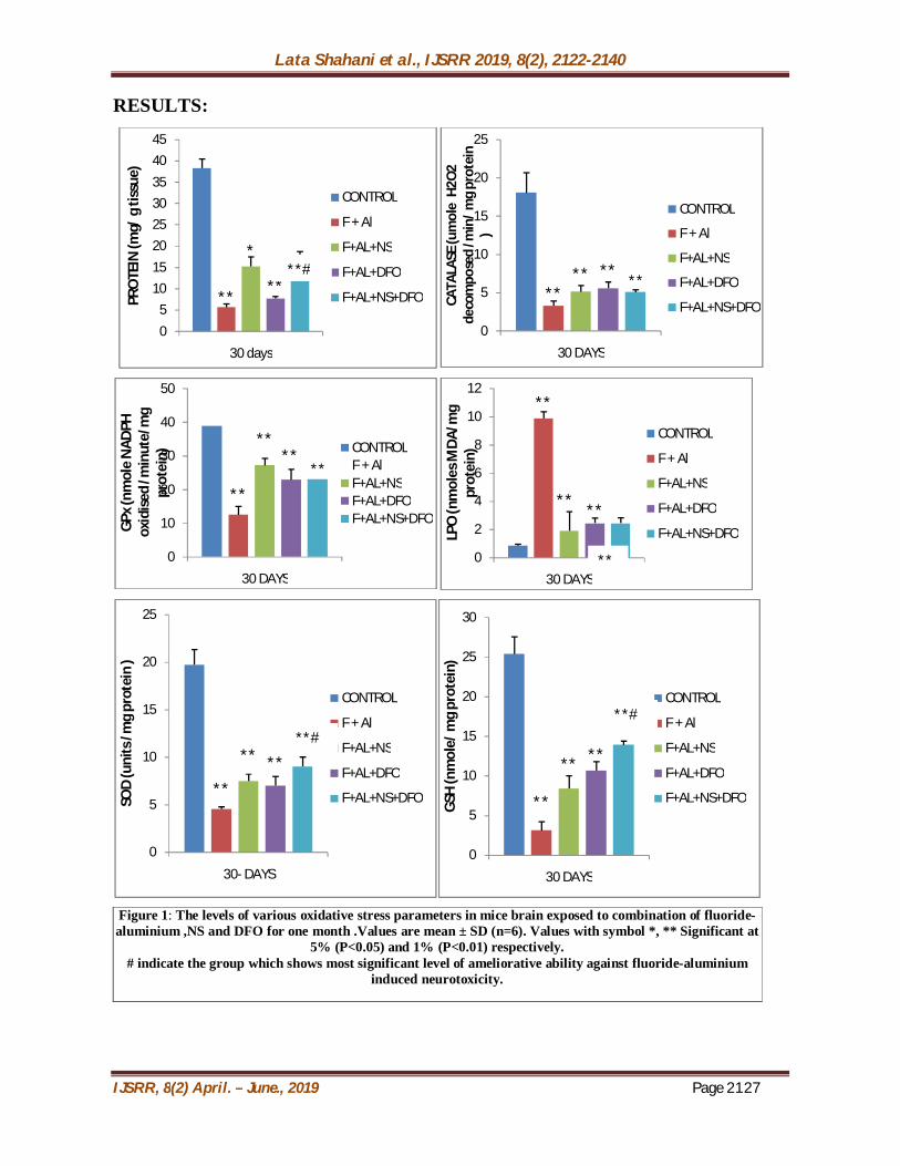
Figure 1: The levels of various oxidative stress parameters in mice brain exposed to combination of fluoride-aluminium ,NS and DFO for one month .Values are mean ± SD (n=6). Values with symbol *, ** Significant at
5% (P<0.05) and 1% (P<0.01) respectively. # indicate the group which shows most significant level of ameliorative ability against fluoride-aluminium
induced neurotoxicity.
**
*
**
05
1015202530354045
30 days
PRO
TEIN
(mg/
g ti
ssue
)
CONTROL
F + Al
F+AL+NS
F+AL+DFO
F+AL+NS+DFO **** **
0
5
10
15
20
25
30 DAYS
CATA
LASE
(um
ole
H2O
2 de
com
pose
d /m
in/ m
g pr
otei
n )
CONTROL
F + Al
F+AL+NS
F+AL+DFO
F+AL+NS+DFO
**
**
****
0
10
20
30
40
50
30 DAYS
GPx
(nm
ole
NAD
PH
oxid
ised
/min
ute/
mg
pr
otei
n) CONTROLF + AlF+AL+NSF+AL+DFOF+AL+NS+DFO
**
** **
0
2
4
6
8
10
12
30 DAYS
LPO
(nm
oles
MDA
/mg
prot
ein)
CONTROL
F + Al
F+AL+NS
F+AL+DFO
F+AL+NS+DFO
**
** **
0
5
10
15
20
25
30- DAYS
SOD
(uni
ts /m
g pr
otei
n )
CONTROL
F + Al
F+AL+NS
F+AL+DFO
F+AL+NS+DFO **
** **
0
5
10
15
20
25
30
30 DAYS
GSH
(nm
ole/
mg
prot
ein)
CONTROL
F + Al
F+AL+NS
F+AL+DFO
F+AL+NS+DFO
**#
**
**
**# **#
Lata Shahani et al., IJSRR 2019, 8(2), 2122-2140
IJSRR, 8(2) April. – June., 2019 Page 2128
Effect of NS and DFO treatment on fluoride- aluminium combination induced
changes in various oxidative stress parameters (figure 1): When mice were treated with combination of fluoride and aluminium intraperitoneally for
one month, it resulted in a significant decrease (P<0.01) in levels of protein, catalase, GPx, SOD,
GSH and and a significant increase in LPO levels as compared to the control group. Simultaneous
administration of NS along with fluoride and aluminium combination resulted in a significant
increase (P<0.01) in the levels of protein, catalase, GPx, SOD, GSH and and a significant decrease in
LPO levels as compared to the fluoride and aluminium combination group. Simultaneous
administration of DFO along with fluoride and aluminium combination resulted in a significant
increase (P<0.01) in the levels of catalase, GPx, SOD, GSH and and protein (P<0.05).A significant
decrease (P<0.01) in LPO levels was also observed as compared to the fluoride and aluminium
combination group. The amelioration produced by NSwas more significant than DFO. But when
combination of NS and DFO was given along with fluoride-aluminium treatment the increase in
levels of protein, catalase, GPx, SOD, GSH and decrease in LPO levels was most significant when
compared to the groups in which NSand DFO were given individually along with fluoride and
aluminium combination. Our results indicate that combination of NS and DFO has maximum
ameliorative ability against fluoride-aluminium induced oxidative stress in brain.
HISTOLOGY:
Cerebellum (figure 2): When mice was intraperitoneally treated with fluoride and aluminium (thrice a week on
alternate days) for 30 days resulted in vacoulation and constriction of Purkinje cells ( shown by the
arrow) as compared to the control mice which showed normal appearance of purkinje cells (( figure
N1). Pyknosis is also seen in purkinje cells with loss of Nissl substance from neuroplasm. There is a
decrease in the density in the Purkinje cell layer, degeneration of nuclear contents (indicated by
arrow) and chromatolysis is also observed in the granule cells (figure A). But when NS was
simultaneously given along with fluoride and aluminium resulted in an increase in the density in the
Purkinje cell layer, normal appearance of nucleus and dendrites of purkinje neurons ( shown by
arrow). The granule cells also appear to be normal (figure B).Combination of fluoride, aluminium
and DFO lead to slight vacoulation around purkinje cells (indicated by arrow). Degeneration of
dendrites of purkinje cells is also observed but the granule cells appear normal (figure C). When
mice were treated with combination of fluoride, aluminium , NS and DFO , the purkinje cells
appeared normal with a central nucleus and long dendrites ( shown by arrow). The granule cells also
appear to be normal with a round heterochromatic nucleus (figure D).

Lata Shahani et al., IJSRR 2019, 8(2), 2122-2140
IJSRR, 8(2) April. – June., 2019 Page 2129
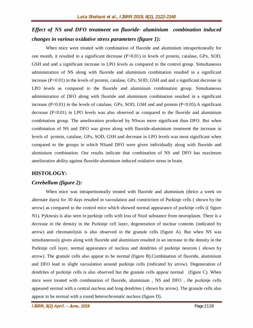
Cerebrum (figure 3): Intraperitoneal treatment of mice for 30 days with fluoride and aluminium resulted in
constriction of pyramidal cells and shrinkage of nucleus ( indicated by arrow). Disorganisation of
dendrites of pyramidal cells occurs is also observed. Astrocytes increase in size and number
indicating gliosis. Pericellular edema is visible and capillaries appear dilated (figure A) as compared
to the control group which normal appearance of pyramidal cells (figure N2). Fluoride, aluminium
and NS administered simultaneously resulted in an increase in the density of the pyramidal cells.
Nucleus and dendrites of pyramidal neurons also appear normal (indicated by arrow). Decrease in
celluar edema in neuropil is seen. Capillaries also appear normal (figure B). When DFO was given
along with fluoride and aluminium , it leads to slight vacoulation and edema in neuropil. But the
pyramidal cells appear normal (shown by arrow) (figure C).Combination of fluoride, aluminium ,NS
and DFO resulted in normal histology of Pyramidal cells and astrocytes. Nucleus and dendrites of
pyramidal neurons also appear normal (shown by arrow). Decrease in celluar edema in neuropil is
also seen. The capillaries and neurofibrillar network also appear normal(figure D).
FIGURE 2: Showing T.S. Cerebellum ( I.P. treament) : (N1) Control (A) F +AL (B) F+AL+NS (C) F+AL+DFO (D) F+AL+NS+DFO
N1 N2
Lata Shahani et al., IJSRR 2019, 8(2), 2122-2140
IJSRR, 8(2) April. – June., 2019 Page 2130
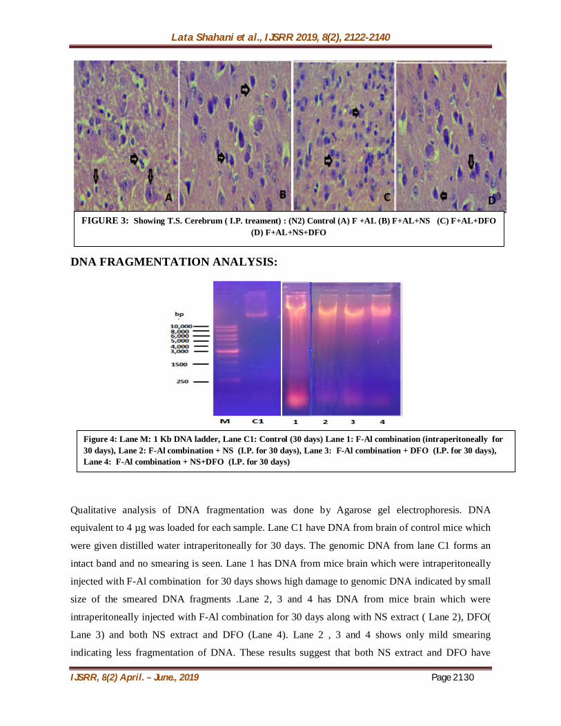
DNA FRAGMENTATION ANALYSIS:
Qualitative analysis of DNA fragmentation was done by Agarose gel electrophoresis. DNA
equivalent to 4 µg was loaded for each sample. Lane C1 have DNA from brain of control mice which
were given distilled water intraperitoneally for 30 days. The genomic DNA from lane C1 forms an
intact band and no smearing is seen. Lane 1 has DNA from mice brain which were intraperitoneally
injected with F-Al combination for 30 days shows high damage to genomic DNA indicated by small
size of the smeared DNA fragments .Lane 2, 3 and 4 has DNA from mice brain which were
intraperitoneally injected with F-Al combination for 30 days along with NS extract ( Lane 2), DFO(
Lane 3) and both NS extract and DFO (Lane 4). Lane 2 , 3 and 4 shows only mild smearing
indicating less fragmentation of DNA. These results suggest that both NS extract and DFO have
FIGURE 3: Showing T.S. Cerebrum ( I.P. treament) : (N2) Control (A) F +AL (B) F+AL+NS (C) F+AL+DFO (D) F+AL+NS+DFO
Figure 4: Lane M: 1 Kb DNA ladder, Lane C1: Control (30 days) Lane 1: F-Al combination (intraperitoneally for 30 days), Lane 2: F-Al combination + NS (I.P. for 30 days), Lane 3: F-Al combination + DFO (I.P. for 30 days), Lane 4: F-Al combination + NS+DFO (I.P. for 30 days)
Lata Shahani et al., IJSRR 2019, 8(2), 2122-2140
IJSRR, 8(2) April. – June., 2019 Page 2131
ameliorative abilities against F-Al induced DNA damage but their combination works best against F-
Al induced DNA fragmentation as lane 4 DNA showed least smearing of DNA and an intact band of
genomic DNA.
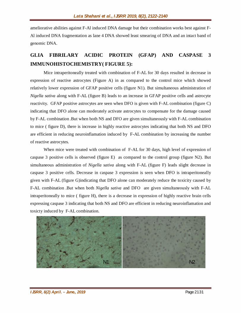
GLIA FIBRILARY ACIDIC PROTEIN (GFAP) AND CASPASE 3
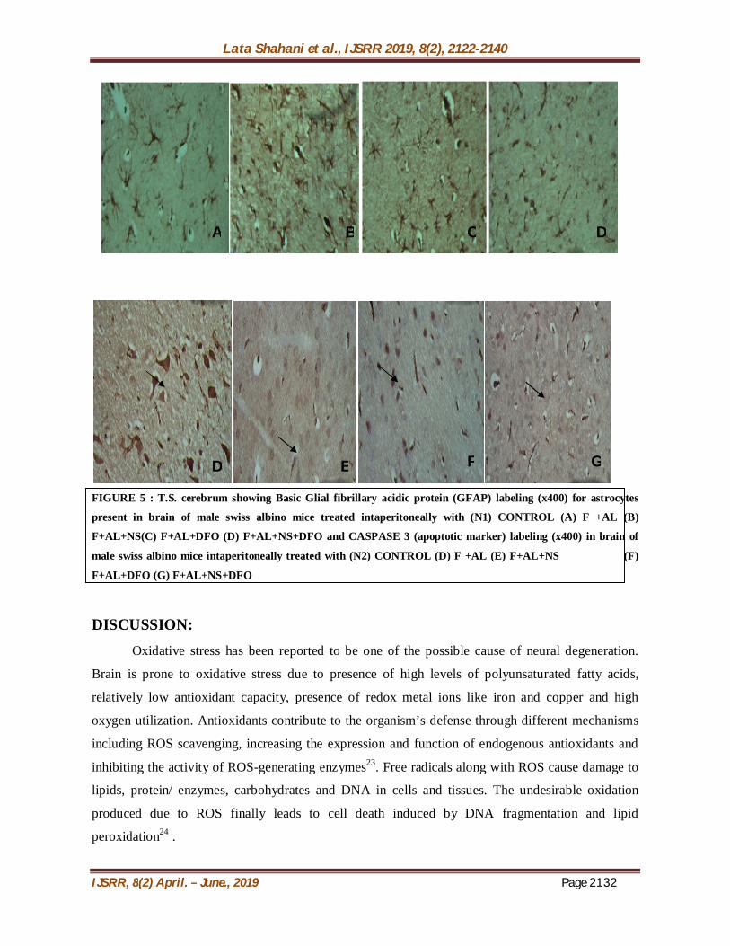
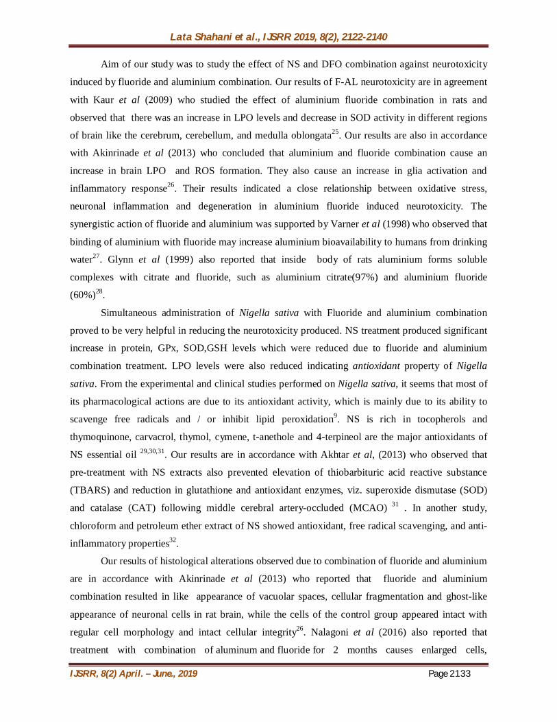
IMMUNOHISTOCHEMISTRY( FIGURE 5): Mice intraperitoneally treated with combination of F-AL for 30 days resulted in decrease in
expression of reactive astrocytes (Figure A) in as compared to the control mice which showed
relatively lower expression of GFAP positive cells (figure N1). But simultaneous administration of
Nigella sativa along with F-AL (figure B) leads to an increase in GFAP positive cells and astrocyte
reactivity. GFAP positive astrocytes are seen when DFO is given with F-AL combination (figure C)
indicating that DFO alone can moderately activate astrocytes to compensate for the damage caused
by F-AL combination .But when both NS and DFO are given simultaneously with F-AL combination
to mice ( figure D), there is increase in highly reactive astrocytes indicating that both NS and DFO
are efficient in reducing neuroinflamation induced by F-AL combination by increasing the number
of reactive astrocytes.
When mice were treated with combination of F-AL for 30 days, high level of expression of
caspase 3 positive cells is observed (figure E) as compared to the control group (figure N2). But
simultaneous administration of Nigella sativa along with F-AL (figure F) leads slight decrease in
caspase 3 positive cells. Decrease in caspase 3 expression is seen when DFO is intraperitoneally
given with F-AL (figure G)indicating that DFO alone can moderately reduce the toxicity caused by
F-AL combination .But when both Nigella sativa and DFO are given simultaneously with F-AL
intraperitoneally to mice ( figure H), there is a decrease in expression of highly reactive brain cells
expressing caspase 3 indicating that both NS and DFO are efficient in reducing neuroinflamation and
toxicty induced by F-AL combination.
N1 N2
Lata Shahani et al., IJSRR 2019, 8(2), 2122-2140
IJSRR, 8(2) April. – June., 2019 Page 2132
FIGURE 5 : T.S. cerebrum showing Basic Glial fibrillary acidic protein (GFAP) labeling (x400) for astrocytes
present in brain of male swiss albino mice treated intaperitoneally with (N1) CONTROL (A) F +AL (B)
F+AL+NS(C) F+AL+DFO (D) F+AL+NS+DFO and CASPASE 3 (apoptotic marker) labeling (x400) in brain of
male swiss albino mice intaperitoneally treated with (N2) CONTROL (D) F +AL (E) F+AL+NS (F)
F+AL+DFO (G) F+AL+NS+DFO
DISCUSSION: Oxidative stress has been reported to be one of the possible cause of neural degeneration.
Brain is prone to oxidative stress due to presence of high levels of polyunsaturated fatty acids,
relatively low antioxidant capacity, presence of redox metal ions like iron and copper and high
oxygen utilization. Antioxidants contribute to the organism’s defense through different mechanisms
including ROS scavenging, increasing the expression and function of endogenous antioxidants and
inhibiting the activity of ROS-generating enzymes23. Free radicals along with ROS cause damage to
lipids, protein/ enzymes, carbohydrates and DNA in cells and tissues. The undesirable oxidation
produced due to ROS finally leads to cell death induced by DNA fragmentation and lipid
peroxidation24 .
D E F G
A B C D
Lata Shahani et al., IJSRR 2019, 8(2), 2122-2140
IJSRR, 8(2) April. – June., 2019 Page 2133
Aim of our study was to study the effect of NS and DFO combination against neurotoxicity
induced by fluoride and aluminium combination. Our results of F-AL neurotoxicity are in agreement
with Kaur et al (2009) who studied the effect of aluminium fluoride combination in rats and
observed that there was an increase in LPO levels and decrease in SOD activity in different regions
of brain like the cerebrum, cerebellum, and medulla oblongata25. Our results are also in accordance
with Akinrinade et al (2013) who concluded that aluminium and fluoride combination cause an
increase in brain LPO and ROS formation. They also cause an increase in glia activation and
inflammatory response26. Their results indicated a close relationship between oxidative stress,
neuronal inflammation and degeneration in aluminium fluoride induced neurotoxicity. The
synergistic action of fluoride and aluminium was supported by Varner et al (1998) who observed that
binding of aluminium with fluoride may increase aluminium bioavailability to humans from drinking
water27. Glynn et al (1999) also reported that inside body of rats aluminium forms soluble
complexes with citrate and fluoride, such as aluminium citrate(97%) and aluminium fluoride
(60%)28.
Simultaneous administration of Nigella sativa with Fluoride and aluminium combination
proved to be very helpful in reducing the neurotoxicity produced. NS treatment produced significant
increase in protein, GPx, SOD,GSH levels which were reduced due to fluoride and aluminium
combination treatment. LPO levels were also reduced indicating antioxidant property of Nigella
sativa. From the experimental and clinical studies performed on Nigella sativa, it seems that most of
its pharmacological actions are due to its antioxidant activity, which is mainly due to its ability to
scavenge free radicals and / or inhibit lipid peroxidation9. NS is rich in tocopherols and
thymoquinone, carvacrol, thymol, cymene, t-anethole and 4-terpineol are the major antioxidants of
NS essential oil 29,30,31. Our results are in accordance with Akhtar et al, (2013) who observed that
pre-treatment with NS extracts also prevented elevation of thiobarbituric acid reactive substance
(TBARS) and reduction in glutathione and antioxidant enzymes, viz. superoxide dismutase (SOD)
and catalase (CAT) following middle cerebral artery-occluded (MCAO) 31 . In another study,
chloroform and petroleum ether extract of NS showed antioxidant, free radical scavenging, and anti-
inflammatory properties32.
Our results of histological alterations observed due to combination of fluoride and aluminium
are in accordance with Akinrinade et al (2013) who reported that fluoride and aluminium
combination resulted in like appearance of vacuolar spaces, cellular fragmentation and ghost-like
appearance of neuronal cells in rat brain, while the cells of the control group appeared intact with
regular cell morphology and intact cellular integrity26. Nalagoni et al (2016) also reported that
treatment with combination of aluminum and fluoride for 2 months causes enlarged cells,
Lata Shahani et al., IJSRR 2019, 8(2), 2122-2140
IJSRR, 8(2) April. – June., 2019 Page 2134
neurofibrillary tangles, and vacuolar spaces in the cerebral cortex and suggested that the histological
alteration observed may be a result of Oxidative stress caused by aluminium and fluoride
combination32. The use of NS extract to compensate for the decreased antioxidant status of brain
cells due to fluoride –aluminium toxicity only has little ameliorative action as shown by our
histology results. The seed extract was not able to completely avoid the toxic changes induced by the
metal combination but was surely able to reduce the degeneration of brain cells. Ameliorative abililty
shown by DFO against fluoride and aluminium combination induced changes in histology of brain
cells is comparatively less than NS but the combination of both agents (NS and DFO) produces most
protective effect. These results of histology correlate with results of biochemical parameters obtained
in our study. NS comparatively showed more potential than DFO to increase the activity of various
antioxidant enzymes. Since NS is rich in various antioxidants, it decreases the oxidative stress
induced by fluoride and aluminium combination. DFO being a chelator of aluminium helps in
reducing the toxicity produced by removing aluminium in either in feaces or urine. Both these agents
decrease oxidative stress which indirectly reduces the histopathological alterations produced due to
fluoride and aluminium combination.
Our results of DNA fragmentation are in accordance with Ohyashiki et al (2002) who
reported that AlCl3 treatment induce chromatin condensation and DNA ladder formation in PC12
cells33 . Aluminium is also reported to cause oxidation of nucleic acids, including the oxidation of
DNA and the breaking of α-helix. The accumulation of many such changes in the DNA, detected by
repair mechanisms, leads to the activation of the apoptosis pathway34. The DNA damaging ability of
aluminium is also observed by Walton et al( 2006)35 who reported that Al is centrally localized in the
nuclear region compared to other intracellular organelles which indicate its DNA damaging
potential. Further Lima et al (2007) and Mohan et al (2007) observed that aluminium treatment
induces gaps and breaks in the chromosomes with higher frequency36, 37. Tsubouchi et al (2001) also
found Al induces DNA strand breaks in PCD12 cells by the generation of reactive oxygen species
(ROS), thus leading to apoptosis38. Li et al (1987) reported that Fluorine being a negatively charged
ion has strong affinity for uracil and amide bonds by the interaction with –NH. Fl also induce lipid
peroxidation which can further lead to generation of free radicals39. Theses free radicals can damage
DNA by decreasing the activity of DNA polymerase thus affecting DNA replication or repair
mechanism40.
In our study combination of fluoride and aluminium initially increases the GFAP
immunoreactivity which could be explained by the fact that astrocytes play an important role in the
formation of the blood-brain barrier and fighting against oxidative stress41. Yu et al (2015) found that
astrocytes are more resistant to oxidative stress mediated apoptosis as they have low production of
Lata Shahani et al., IJSRR 2019, 8(2), 2122-2140
IJSRR, 8(2) April. – June., 2019 Page 2135
reactive oxygen species (ROS) due to high buffering ability against ROS induced toxicity42.
However inspite of high GFAP expression, brain cells also showed moderate level of caspase 3
expression which can be explained by the finding that although caspase-3 is involved in the cleavage
of cytoskeletal proteins including vimentin43 and GFAP in astrocytes indicating cell death44. But
cleaved GFAP and vimentin have also been found to co-localize with activated caspase-3 in non-
apoptotic astrocytes45suggesting that caspase activation alone is not sufficient for cytoskeletal
remodeling but may contribute to astrogliosis46. The decrease in GFAP reactivity and increase in
caspase 3 expression after intraperitoneal treatment of mice with combination of fluoride and
aluminium for one month can be explained by the ability of F-AL combination to activate caspases
and induce apoptosis of astrocytes47. Our results are similar to the findings of Varner et al (1998)
who observed that chronic administration of aluminium fluoride and sodium fluoride in the drinking
water of rats resulted in distinct morphological alterations in the brain27 and leads to the formation of
abnormal connections between nerve fibres by interfering with the metabolism of the cytoskeleton
in the nerve cells48.
The exact mechanisms of neurotoxic actions induced by the interaction of fluoride and
aluminium have still not been clearly established. But study conducted by Miles et al, 200249 and
Blaylock et al , 2004 reported that fluoro-aluminium complexes formed due to the interaction of F
and AL mimic phosphate groups in biological systems50. One of the possible mechanism of fluoride
induced toxicity is excitotoxicity which involves the accumulation of acidic amino acids like
cysteine, cysteine sulfinic acid and neurotransmitters like glutamate and aspartate in synaptic cleft. If
theses excitatory amino acids are not removed from synaptic cleft they cause prolonged stimulation
and neuronal destruction by both apoptosis and necrosis51. Due to this excitotoxicity free radicals and
lipid peroxidation products are generated to damage dendrites and synaptic connections leading to
neuronal destruction52.Thus even if fluoride does not directly trigger excitotoxicity , it can cause it
indirectly by production of free radicals and lipid peroxidation products. Ghribi et al,2001 reported
about the important role played by endoplasmic reticulum (ER) in regulating aluminium induced
neurotoxicity. The ER is the major storage location for calcium and contains members of the Bcl-2
family of proteins, Bcl-2 and Bcl-XL. The stress induced by aluminium in the ER has also been
shown to result in a specific type of apoptosis mediated by caspase-12 and is independent of
mitochondrial-targeted apoptotic signals53. Blaylock,2012 also highlighted the aluminium activates
glial cells leading to immunoexcitotoxicity, impairs a number of energy related enzymes, promotes
brain inflammation, oxidative damage, reduces the levels of brain antioxidants (i.e., glutathione) and
disturbs calcium homeostasis, thus confirming its role in neuurodegenration54. The exact mechanism
of aluminium toxicity to cells still remains unclear.
Lata Shahani et al., IJSRR 2019, 8(2), 2122-2140
IJSRR, 8(2) April. – June., 2019 Page 2136
Simultaneous administration of NS with combination of fluoride and aluminium results in
gradual increase in GFAP expression and decrease in caspase 3 immunoreactivity which might be
due the ameliorative activity of NS to reduce the oxidative stress and induce astrogliosis to repair the
damaged CNS and maintain the integrity of blood brain barrier (BBB). Administration of NS and
DFO individually and simultaneously with F-AL combination causes an increase in astrocyte
reactivity which indicates that NS and DFO individually have moderate ability to activate astrocytes
to repair the damaged CNS but together they can more efficiently decrease the toxic effects of F-Al
combination by chelating aluminium from body and acting against the induced oxidative stress and
neurodegenerative changes by activating cells like astrocytes.
CONCLUSION: Fluoride and aluminium combination was administered intraperitoneally for one month and
NSand DFO, both alone and in combination were also given simultaneously through the same route.
Our findings revealed that both NS and DFO resulted in a significant increase in various
antioxidant enzymes and decrease in LPO levels showing its ameliorative ability against fluoride and
aluminium induced toxicity. But NS caused more significant increase in antioxidant enzymes as
compared to DFO. However combination of NS and DFO had the maximum ameliorative ability
against fluoride and aluminium induced neurotoxicity.
BIBLIOGRAPHY: 1. Blaylock RL. Aluminum Induced Immunoexcitotoxicity in Neurodevelopmental and
Neurodegenerative Disorders, Curr. Inorg. Chem.. 2012; 2(1):1877-9441.
2. Dr. Edward Group. Why I’m Concerned About the Dangers of Aluminum.Global healing
center;2012.
3. Al-Hijazi AA. Evaluation of the Effect of NS Oil and Powder on Socket Healing Process.
Jour. of Nat. Sci. Res,2013; 3(11): 135-140.
4. El-tahir KH and Bakeet D. The black seed Nigella sativa: A plea for urgent clinical
evaluation of its volatile oil. JTU Med Sc. 2006; 1(1): 1-19.
5. Nickavar B, Mojab F, Javidnia, K, et al. Chemical composition of the fixed and volatile oils
of NS. from iran, Z.naturforsch.2003; 58: 629-631.
6. Hajhashemi V, Ghannadi A, Jafarabadi, H.Black cumin seed essential oil, as a potent
analgesic and anti-inflammatory drug. Phytother. Res.2004; 18:195-199.
7. Burits M, Bucar F.Antioxidant activity of NS essential oil. Phytother Res.2000;14:323-328.
Lata Shahani et al., IJSRR 2019, 8(2), 2122-2140
IJSRR, 8(2) April. – June., 2019 Page 2137
8. Padhye S, Banerjee S, Ahmad A et al. From here to eternity- the secret of pharaohs:
therapeutic potential of black cumin seeds and beyond.Cancer Ther.2008; 6: 495-510.
9. Gupta M, Mazumder, UK, Kumar TS, et al. Antioxidant and hepatoprotective effects of
bauhinia racemosa against paracetamol and carbon tetrachloride induced liver damage in
rats. Iranian Jour. of Pharma. and Therap.2004; 3(1): 12-20.
10. ASHSP (the american society of health-system pharmacists), deferoxamine mesylate"2016.
11. Carpenter DO, Arcaro K, Spink DC. Understanding the human health effects of chemical
mixtures. Environ Health Perspect. 2002; 110(1): 25–42.
12. Hertzberg RC, Teuschler LK. Evaluating quantitative formulas for dose-response
assessment of chemical mixtures. Environ Health Perspect.2002;110(6):965–70.
13. Chinoy NJ, Patel TN. The influence of fluoride and aluminium on free radical toxicity in the
brain of female mice and beneficial effects of some antidotes. Fluoride. 2000 ;33(1):S8.
14. Abbasnezhad A, Hayatdavoudi P, Niazmand S, Mahmoudabady M. The effects of
hydroalcoholic extract of NS seed on oxidative stress in hippocampus of STZ-induced
diabetic rats. Avicenna J Phytomed. 2015; 5(4):333-40.
15. WHO protocol CG-04. Preparation of alcoholic extract for bioassay and phytochemical
studies (APJF/IP, 1001 A) Geneva: World Health Organization,1983.
16. Cappellini MD. Iron-chelating therapy with the new oral agent ICL670 (Exjade). Best Pract
Res Clin Haematol.2005;18(2): 289-98.
17. Luck H. Methods in Enzymatic Analysis . 2nd edition. Bergmeyer (Publ.) Academic Press:
New York; 1974; 885.
18. Kono Y. Generation of superoxide radical during auto-oxidation of hydroxylamine and an
assay for Superoxide Dismutase. Arch.Biochem. Biophys.1978; 186:189-195.
19. Paglia DE, and Valentine WN. Studies on the quantitative and qualitative characterization
of erythrocyte glutathione peroxidase. J. Lab Clin. Med.1967; 158-169.
20. Beuge JA, Aust SD. Methods Enzymol. 1978; 52 : 302-310.
21. Moron MS, Depierre JW, Mannervik B. Levels of glutathione, glutathione reductase and
glutathione- S-transferase activities in rat lung and liver. Biochem. Biophys Acta.
1979 ; 582: 67-78.
22. Lowry OH, Rosebrough NJ, Farr AL et al. Protein measurement with the Folin phenol
reagent. J. Biol. Chem. 1951; 193:265-75.
23. Al-Olayan E, El-Khadragy M, Metwally D et al. Protective effects of pomegranate
(Punicagranatum) juice on testes against carbon tetrachloride intoxication in rats. BMC
Comple. and Alter. Medi. 2014; 14:164.
Lata Shahani et al., IJSRR 2019, 8(2), 2122-2140
IJSRR, 8(2) April. – June., 2019 Page 2138
24. Singh, R.P, Sharad S, Kapur S. Free radicals and oxidative stress in neurodegenerative
diseases:relevance of dietary antioxidants. Jour. Ind. Acad. Of Gen. Med. 2004; 5(3):218-25.
25. Kaur T, Bijarnia RK, Nehru B. Effect of concurrent chronic exposure of fluoride and
aluminum on rat brain. Drug Chem Toxicol.2009; 32(3):215-21.
26. Akinrinade ID, Ogundele OM., Memudu AE et al. Degeneration of neuronal cells: A
product of fluoride and aluminium assault to the prefrontal cortex. Jour. Cell and Anim. Bio.
2013; 7(6):63-66.
27. Varner JA , Jensen KF , Horvath W. et al. Chronic administration of aluminum–fluoride or
sodium–fluoride to rats in drinkingwater: alterations in neuronal and cerebrovascular
integrity. Brain Res. 1998; 784 :284–298.
28. Glynn AW, Sparen A, Danielsson LG et al. Concentration dependent absorption of
aluminum in rats exposed to labile aluminum in drinking water. J Toxicol Environ Health.
1999; 56:501 –512.
29. Wajs A, Bonikowski R, Kalemba D. Composition of essential oil from seeds of 22 NSL
cultivated in Poland. Flavour Fragr. J. 2008; 23: 126-132.
30. Sultan MT, Butt MS, Anjum FM et al. Nutritional profile of indigenous cultivar of NS
seeds and antioxidant potential of its fixed and essential oil. Pak. J. Bot. 2009; 41(3): 1321-
1330.
31. Akhtar M, Maikiyo AM, Najmi AK. Neuroprotective effects of chloroform and petroleum
ether extracts of NS seeds in cerebral ischemia. J. Pharm. Bio. Allied. Sci. 2013; 5:119–25
32. Nalagoni CSR., Karnati PR. Protective effect of resveratrol against neuronal damage through
oxidative stress in cerebral hemisphere of aluminum and fluoride treated rats. 2002; 9(2):78-
82.
33. Ohyashiki T, Satoh E, Okada M et al. Nerve growth factor protects against aluminum-
mediated cell death. Toxicology,2002; 176: 195-207.
34. Temple MD, Perrone GG, Dawes IW. Complex cellular responses to reactive oxygen
species. Trends Cell. Biol. 2005; 15(6):319–326
35. Walton JR. Aluminum in hippocampal neurons from humans with Alzheimer's disease.
Neurotoxicology. 2006; 27(3):385-94.
36. Lima PD, Leite DS, Vasconcellos MC et al. Genotoxic effects of aluminum chloride in
cultured human lymphocytes treated in different phases of cell cycle. Food Chem. Toxicol.
2007; 45: 1154-9.
37. Mohan Murali Achary V, Jena S, Panda KKet al. Aluminium induced oxidative stress and
DNA damage in root cells of Allium cepa L. Ecotoxicol. Environ. Saf. 2007; 70 : 300-10.
Lata Shahani et al., IJSRR 2019, 8(2), 2122-2140
IJSRR, 8(2) April. – June., 2019 Page 2139
38. Tsubouchi R, Htay HH, Murakami K. Aluminum-induced apoptosis in PC12D cells.
Biometals. 2001; 14 :181-5.
39. Li YM, Heerema NA, Dunipace AJet al..Genotoxic effects of fluoride evaluated by sister-
chromatid exchange. Mutat. Res.1987; 192:191-201.
40. Aardema MJ, Tsutsui T. Sodium fluoride-induced chromosome aberrations in different cell
cycle stages.1995; 331(1): 171-2 .
41. Nagele RG, Wegiel J, Venkataraman V et al. Contribution of glial cells to the development
of amyloid plaques in Alzheimer’s disease. Neurobiol. Aging. 2004; 25: 663-674.
42. Yu HT, Zhen J, Pang B. Ginsenoside Rg1 ameliorates oxidative stress and myocardial
apoptosis in streptozotocin-induced diabetic rats .J. Zhejiang Univ-Sci B (Biomed &
Biotechnol). 2015. 16(5): 344–350.
43. Byun Y, Chen F, Chang R. 2001,Caspase cleavage of vimentin disrupts intermediate
filaments and promotes apoptosis. Cell Death Differ. 2001; 8(5):443-50.
44. Mouser PE, Head E, Ha KH. Caspase-mediated cleavage of glial fibrillary acidic protein
within degenerating astrocytes of theAlzheimer’s disease brain. Am. J. Pathol, 2000; 168:
936-946.
45. Acarin L, Villapol S, Faiz M. Caspase-3 activation in astrocytes following postnatal
excitotoxic damage correlates with cytoskeletal remodeling but not with cell death or
proliferation. Glia .2007; 55: 954-965.
46. Abe K, Saito H. Na+ and K+ dependence of morphological changes of cultured rat cortical
astrocytes. Pharmacol. Toxicol. 2001; 88:319–24.
47. Troy T R, Lindsey WC, Wayne WP.Caspase-cleaved glial fibrillary acidic protein within
cerebellar white matter of the Alzheimer’s disease brain,. Int. J. Clin. Exp. Pathol. 2013;
6(1):41-48.
48. Allain P, Gauchard F, Krari N. Enhancement of aluminum digestiveabsorption by fluoride in
rats. Res. Commun. Mol. Pathol. Pharmacol.1996; 91:225–231.
49. Miles RD , Gorrell A , Ferry JG. Evidence for a transition stateanalog, MgADP-aluminum
fluoride-acetate, in acetate kinase from Methanosarcina thermophila. J. Biol. Chem. 2002;
277: 22547–22552
50. Blaylock RL. Excitotoxicity a possible central mechanism in fluoride neurotoxicity. Fluoride
.2004; 37:301–314.
51. Szatkowski M, Attwell D. Triggering and execution of neuronal death in brain ischemia:
two phases of glutamate release by different mechanisms. Trends Neurosci. 1994; 17:359-
65.,
Lata Shahani et al., IJSRR 2019, 8(2), 2122-2140
IJSRR, 8(2) April. – June., 2019 Page 2140
52. Isokawa M, Levesque MF. Increased NMDA responses and dendritic degeneration in human
epileptic hippocampal neurons in slices. Neurosci Lett. 1991; 132:212-6.
53. Ghribi O, DeWitt DA, Forbes MS et al. Cyclosporin A inhibits A1-induced cytochrome c
release from mitochondria in aged rabbits. J Alz. Dis. 2001;3:387-91.
54. Dewitt DA, Hurd JA, Fox N, Townsend BE, Griffioen KJ, Ghribi O et al. Peri-nuclear
clustering of mitochondria is tr iggered during aluminum maltolate induced
apoptosis. J Alzheimers Dis 2006; 9 : 195-205
55. Ghribi O, Dewitt DA, Forbes MS, Herman MM, Savory J. Involvement of mitochondria
and endoplasmic reticulum in regulation of apoptosis: changes in cytochrome-c, Bcl-2 and
Bax in the hippocampus of Aluminium treated rabbits. Brain Res 2001; 8 : 764-73
56. Ghribi O, Dewitt DA, Forbes MS, Herman MM, Savory J. Involvement of mitochondria
and endoplasmic reticulum in regulation of apoptosis: changes in cytochrome-c, Bcl-2 and
57. Bax in the hippocampus of Aluminium treated rabbits. Brain Res 2001; 8 : 764-73
58. Blaylock L. Aluminum Induced Immunoexcitotoxicity in Neurodevelopmental and
Neurodegenerative Disorders. Curr. Inorg. Chem. 2012; 2:1.