high-fat diet induces apoptosis of hypothalamic neurons · high-fat diet induces apoptosis of...
TRANSCRIPT
High-Fat Diet Induces Apoptosis of HypothalamicNeuronsJuliana C. Moraes1, Andressa Coope1, Joseane Morari1, Dennys E. Cintra1, Erika A. Roman1, Jose R.
Pauli1, Talita Romanatto1, Jose B. Carvalheira1, Alexandre L. R. Oliveira2, Mario J. Saad1, Licio A.
Velloso1*
1 Department of Internal Medicine, University of Campinas, Campinas, Brazil, 2 Department of Anatomy, University of Campinas, Campinas, Brazil
Abstract
Consumption of dietary fats is amongst the most important environmental factors leading to obesity. In rodents, theconsumption of fat-rich diets blunts leptin and insulin anorexigenic signaling in the hypothalamus by a mechanismdependent on the in situ activation of inflammation. Since inflammatory signal transduction can lead to the activation ofapoptotic signaling pathways, we evaluated the effect of high-fat feeding on the induction of apoptosis of hypothalamiccells. Here, we show that consumption of dietary fats induce apoptosis of neurons and a reduction of synaptic inputs in thearcuate nucleus and lateral hypothalamus. This effect is dependent upon diet composition, and not on caloric intake, sincepair-feeding is not sufficient to reduce the expression of apoptotic markers. The presence of an intact TLR4 receptor,protects cells from further apoptotic signals. In diet-induced inflammation of the hypothalamus, TLR4 exerts a dual function,on one side activating pro-inflammatory pathways that play a central role in the development of resistance to leptin andinsulin, and on the other side restraining further damage by controlling the apoptotic activity.
Citation: Moraes JC, Coope A, Morari J, Cintra DE, Roman EA, et al. (2009) High-Fat Diet Induces Apoptosis of Hypothalamic Neurons. PLoS ONE 4(4): e5045.doi:10.1371/journal.pone.0005045
Editor: Xin-Yun Lu, University of Texas Health Science Center, United States of America
Received October 28, 2008; Accepted March 2, 2009; Published April 2, 2009
Copyright: � 2009 Moraes et al. This is an open-access article distributed under the terms of the Creative Commons Attribution License, which permitsunrestricted use, distribution, and reproduction in any medium, provided the original author and source are credited.
Funding: This work was supported by grants from Fundacao de 395 Amparo a Pesquisa do Estado de Sao Paulo and Conselho Nacional de 396 DesenvolvimentoCientıfico e Tecnologico. The funders had no role in study design, data collection and analysis, decision to publish, or preparation of the manuscript.
Competing Interests: The authors have declared that no competing interests exist.
* E-mail: [email protected]
Introduction
Obesity results from an imbalance between caloric intake and
energy expenditure. Changes in lifestyle, resulting in increased
consumption of dietary fats and reduced physical activity have
contributed to the worldwide obesity epidemic [1]. Recent studies
have shown that consumption of dietary fats promotes hypotha-
lamic resistance to the main anorexigenic hormones, leptin and
insulin, leading to the progressive loss of the balance between food
intake and thermogenesis and, therefore, resulting in body mass
gain [2–4]. The functional resistance to leptin and insulin in the
hypothalamus is a consequence of diet-induced activation of
inflammatory signaling, specifically in this site of the brain, which
leads to the molecular impairment of leptin and insulin signal
transduction by at least four distinct mechanisms; induction of
suppressor of cytokine signaling-3 (SOCS3) expression [5],
activation of c-Jun N-terminal kinase (JNK) and I kappa kinase
(IKK) [3], and induction of protein tyrosine phosphatase 1B
(PTP1B) [6].
Inflammatory and apoptotic pathways are tightly connected and
subtle changes in some of the respective determining factors can
swing the balance towards one or another outcome [7]. For
example, the activation of signal transduction pathways by
cytokines such as TNF-a and IL-1b can lead either to pro-, or
anti-apoptotic effects in addition to their classical inflammatory
activity [8,9].
Since the balance between survival and loss of hypothalamic
neurons may have an impact on the coordinated control of feeding
and thermogenesis [10,11], we decided to evaluate whether the
consumption of high amounts of dietary fat can induce apoptosis
of cells in this anatomical region. Our results show that neuronal
apoptosis is induced by the fat-rich diet and that the presence of a
functional TLR4 receptor protects hypothalamic cells from
apoptotic damage.
Materials and Methods
Antibodies, chemicals and buffersThe reagents for SDS–polyacrylamide gel electrophoresis and
immunoblotting were from Bio-Rad (Richmond, CA, USA).
HEPES, phenylmethylsulfonyl fluoride (PMSF), aprotinin, dithio-
threitol (DTT), Triton X-100, Tween 20, glycerol and bovine
serum albumin (fraction V) were from Sigma (St. Louis, MO,
USA). The reagents for chemiluminescence labeling of proteins in
blots were from Amersham (Aylesbury, UK). Antibodies against
SOCS3 (rabbit polyclonal, sc-9023), PARP (rabbit polyclonal, sc-
7150), Bcl2 (rabbit polyclonal, sc-492), phospho-Bad (pBad) (goat
polyclonal, sc-7999), Bax (rabbit polyclonal, sc-493), Apaf1 (goat
polyclonal, sc-26685), caspase-9 (rabbit polyclonal, sc-7885),
FADD (rabbit polyclonal, sc-5559), caspase-8/p20 (rabbit poly-
clonal, sc-7890), neuromedin-N (NeuN) (goat polyclonal, sc-7593),
phospho-PERK (pPERK) (rabbit polyclonal, sc-32577), PERK
(rabbit polyclonal, sc-13073), phospho-eIF2a (peIF2a) (rabbit
polyclonal, sc-12412), eIF2a (rabbit polyclonal, sc-11386), IkB
(rabbit polyclonal, sc-1643), NFkB/p50 (rabbit polyclonal, sc-
7178), Myd88 (rabbit polyclonal, sc-11356), phospho-JNK (pJNK)
PLoS ONE | www.plosone.org 1 April 2009 | Volume 4 | Issue 4 | e5045
(rabbit polyclonal, sc-12882), phospho-IKK (pIKK) (rabbit
polyclonal, sc-23470), POMC (rabbit polyclonal, sc-20148), AgRP
(rabbit polyclonal, sc-18634), TLR4 (rabbit polyclonal, sc-13591),
F4/80 (rabbit polyclonal, sc-25830), and FITC or rodhamin
conjugated goat and rabbit antibodies were from Santa Cruz
Biotechnology (Santa Cruz, CA, USA). The annexin V FITC was
produced by the Laboratory of Molecular and Cellular Biology at
the Institute of Biomedical Sciences, State University of Sao Paulo,
Brazil. The synaptophysin (rabbit polyclonal, A0010) antibody was
from DAKO (Glostrup, Denmark). The caspase-3 antibody was
form Cell Signaling (Danvers, MA, USA). The kit for detecting
apoptosis by the TUNEL assay was from Upstate Cell Signaling
Solutions (Temecula, CA, USA). Chemicals for real-time PCR
were from Invitrogen (Carlsbad, CA, USA) and Applied
Biosystems (Foster City, CA, USA).
Experimental model and feeding protocolsFor most of the experiments, eight-week-old (280–300 g) male
Wistar rats were employed. In addition, some experiments were
performed with recombinant mice with a loss-of-function mutation
for the TLR4 gene (C3H/HeJ) and its respective control (C3H/
HeN). In addition, some experiments were performed with male,
8-week old Swiss mice. Rats and Swiss mice were obtained from
the University of Campinas Animal Breeding Center, while
mutant mice were purchased from the Jackson Laboratory (Bar
Harbor, Maine, USA). All the animals were handled according to
the University guidelines for the use of animals in experimental
studies and conform to the Guide for the Care and Use of
Laboratory Animals, published by the US National Institutes of
Health (NIH publication No. 85-23 revised 1996). All experimen-
tal protocols were approved by the University of Campinas Ethics
Committee. The rats were always housed in individual cages and
maintained on a 12 h-light/dark cycle. After random selection,
rats and Swiss mice were submitted for eight weeks to a control or
high-fat (HF) diet, as presented in Table 1. The access to diet and
water was ad libitum. Some rats were submitted to caloric pair
feeding for eight weeks. For that, 18.43 g of high-fat chow was
offered every day, which provided a mean caloric intake of
4.3 kCal/g body weight, similar to the mean caloric intake of rats
fed on the control diet. Eight-week old C3H/HeJ and C3H/HeN
mice were randomly selected to either control or HF diet for 8
weeks. At the end of the experimental period, hypothalami were
obtained for determination of protein expression and immunohis-
tochemistry.
Intracerebroventricular (icv) cannulation and analysis ofleptin and insulin action in the hypothalamus
For the evaluation of leptin- and insulin-induced inhibition of
food intake, rats were stereotaxically instrumented using a
Stoelting stereotaxic apparatus, according to a previously
described method [12,13]. Coordinates were: anteroposterior,
0.2 mm/lateral, 1.5 mm/depth, 4.0 mm. Procedure efficiency
was tested one week after cannulation by the evaluation of the
drinking response elicited by icv angiotensin II [12]. After the
experiments, cannula placement was also evaluated by histology.
For determination of food intake, rats were food deprived for 6 h
(from 12 to 18 h) and at 18 h were icv treated with insulin
(2.0 ml, 1026 M), leptin (2.0 ml, 1026 M), or saline (2.0 ml). Food
ingestion was determined over the next 12 h, during the dark
cycle.
Real-time PCR and PCR arrayHypothalamic total RNA was extracted using Trizol reagent
(Life Technologies, Gaithersburg, MD, USA), according to the
manufacturer’s recommendations. Total RNA was rendered
genomic DNA free by digestion with Rnase-free Dnase (RQ1,
Promega, Madison, WI, USA). Samples obtained from three
hypothalami from control and HF diet fed rats and Swiss mice
were analyzed using a real-time PCR array (RT2 profiler PCR
array rat apoptosis - SuperArray Bioscience Corp., Frederick,
MD, USA) containing 84 apoptosis related genes, as shown in
www.superarray.com/rt_pcr_product/HTML/PARN-012A.html.
Real-time PCR analysis of gene expression was carried out in
an ABI Prism 7500 sequence detection system (Applied
Biosystems). The optimal concentration of cDNA and primers,
as well as the maximum efficiency of amplification, were
obtained through seven-point, 3-fold dilution curve analysis
for each gene. Each PCR reaction contained 25–30 ng of
reverse-transcribed cDNA (depending on the gene). Primers
were purchased from Applied Biosystems and were: NPY,
Rn00561681_m1; POMC, Rn00595020_m1; TLR4,
Rn00569848_m1; F4/80, Rn01527631_m1 Emr1; GAPD,
#4352338E, for rat; and, NPY, Mm00445771_m1; POMC,
Mm00435874_m1; GAPD #4352339E for mouse. The PCR
conditions were 2 min at 50uC; 10 min at 95uC, followed by 40
cycles at 95uC for 15 sec and 60uC for 60 sec. Real-time data
were analyzed using the engine provided by Applied Biosystems.
Tissue extraction, immunoprecipitation andimmunoblotting
Rats were anesthetized and the hypothalami were dissected and
immediately homogenized in solubilization buffer at 4uC [1%
Triton X-100, 100 mM Tris–HCl (pH 7.4), 100 mM sodium
pyrophosphate, 100 mM sodium fluoride, 10 mM EDTA, 10 mM
sodium orthovanadate, 2.0 mM PMSF and 0.1 mg aprotinin/ml]
with a Polytron PTA 20 S generator (model PT 10/35; Brinkmann
Instruments, Westbury, NY, USA). Insoluble material was
removed by centrifugation for 20 min at 90006g in a 70.Ti rotor
(Beckman, Fullerton, CA, USA) at 4uC. The protein concentration
of the supernatants was determined by the Bradford dye binding
method. Aliquots of the resulting supernatants containing 2.0 mg
of total protein were used for immunoprecipitation with antibodies
against TLR4, Myd88, FADD, Apaf1 and IkB at 4uC overnight,
followed by SDS–PAGE, transfer to nitrocellulose membranes and
blotting with anti-Myd88, TLR4, caspase-8, caspase-9 and NFkB,
respectively. In direct immunoblot experiments, 0.2 mg of protein
extracts were separated by SDS–PAGE, transferred to nitrocellu-
lose membranes and blotted with anti-pJNK, PERK, pPERK,
eIF2a, peIF2a, PARP, Bax, Bcl2 antibodies.
ImmunohistochemistryParaformaldehyde-fixed hypothalami were sectioned
(5.0 mm) and used in regular single- or double-immunofluo-
Table 1. Macronutrient composition of the diets.
Standard Chow High-Fat Chow
g% kJ% g% kJ%
Protein 20 19 20 14
Carbohydrate 76 72 45 31
Saturated fat 4 9 35 55
kJ/g 17.5 24.1
doi:10.1371/journal.pone.0005045.t001
Hypothalamic Apoptosis
PLoS ONE | www.plosone.org 2 April 2009 | Volume 4 | Issue 4 | e5045
rescence staining using Bax, Bcl2, pBad, pJNK, pPERK,
PERK, eIF2a, peIF2a, TLR4, F4/80, AgRP, POMC, NeuN,
caspase-3 and synaptophysin antibodies, as previously de-
scribed [14,15]. Analysis and documentation of results were
performed using a Leica FW 4500 B microscope. The
hypothalami were sectioned from Bregma 21,6 to 24,2 mm.
Every second of all consecutive section was analyzed. The
anatomical correlations were made according to the landmarks
given in a stereotaxic atlas [13]. The topographical views of
the regions to be studied were obtained by hematoxylin-eosin
staining of consecutive sections.
Transmission electronic microscopy (TEM)Hypothalami from rats fed on control and HF diets were
dissected and maintained overnight at 4uC in fixative containing
2.5% glutaraldehyde and 0.5% paraformaldehyde in phosphate
buffer (pH 7.4). The specimens were then trimmed, dehydrated
and embedded in Durcupan (Fluka-Sigma-Aldrich, Seelze,
Germany). Ultrathin sections from the arcuate nucleus were
collected on formvar-coated copper grids, counterstained with
uranyl acetate and lead citrate, and examined under a
transmission electron microscope (Leo906, Zeiss) operated at
60 KV. Hypothalamus microenvironment was analyzed and
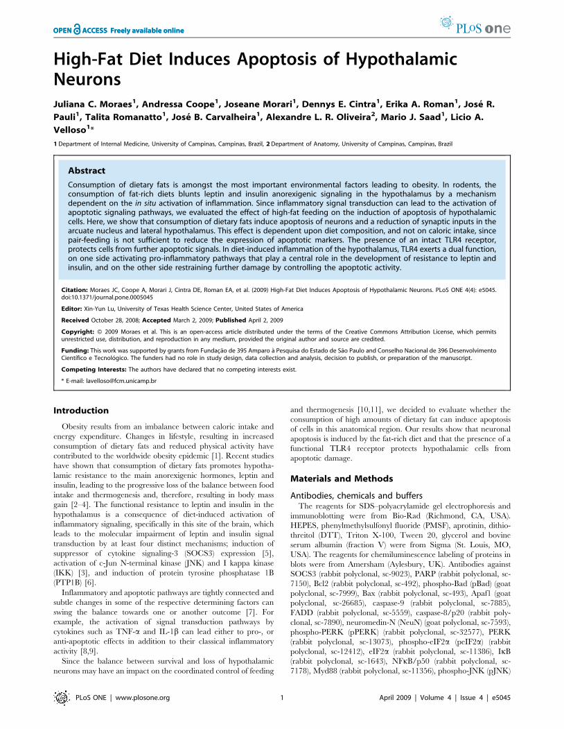
Figure 1. Leptin/insulin resistance and inflammatory markers in the hypothalamus of rats fed on high-fat diet. (A) Body mass variation(g) of Wistar rats fed on control (CT) or high-fat (HF) diets for 8 w. (B–C) Twelve hours spontaneous food intake (g) of Wistar rats fed on CT or HF dietsfor 8 w and treated icv with a single dose (2.0 ml) of saline (2), leptin (+, in B) or insulin (+, in C). (D) Immunoblots (IB) of hypothalamic proteinextracts obtained from Wistar rats fed on CT or HF diets. (E) Real-time PCR analysis of F4/80 transcript amount in samples obtained from thehypothalami of Wistar rats fed on CT or HF diets. In all experiments n = 5. In A, D and E, *p,0.05 vs. CT, values are means6SEM; in B and C, *p,0.05vs. CT+, values are means6SEM.doi:10.1371/journal.pone.0005045.g001
Hypothalamic Apoptosis
PLoS ONE | www.plosone.org 3 April 2009 | Volume 4 | Issue 4 | e5045
neurons with normal and apoptotic morphology were identified
and photographed using a digital image acquisition system
(Morada, Zeiss).
TUNELA terminal deoxynucleotidyl-transferase-mediated dUTP nick
end-labeling (TUNEL) assay was used to identify double-
stranded DNA fragmentation. Briefly, tissue slides were depar-
affinized, treated with proteinase K (20 mg/ml) for 15 min at
room temperature, and then quenched in 2.0% hydrogen
peroxide. After rinsing in phosphate-buffered saline (PBS),
pH 7.4, specimens were incubated in 16 equilibration buffer
for 10–15 s. The slides were then incubated with terminal
deoxynucleotidyl transferase (TdT) for 1.0 h at 37uC, blocked
with stop/wash buffer, and incubated with peroxidase antibody
for 30 min at room temperature. Negative control for the
TUNEL assay was confirmed by staining the tissues in the same
manner without primary antibody. Percentages of TUNEL-
positive neurons were determined in at least 10 optical fields.
Analysis was performed in five 5.0 mm non-consecutive sections
from each hypothalamus.
Statistical analysisData from the real-time PCR array were analyzed using the
engine supplied by the manufacturer. Only mRNAs undergoing
at least 2.0-fold variation from control were considered
significantly modulated by the diet. Specific bands in immuno-
blots were scanned and submitted to a quantitative analysis
using the Scion Image software (Scion Corp., Frederick, MD,
USA). TUNEL positive cells, the apoptotic cells detected in
low-magnification TEM and synaptophysin positive nerve
terminals were field counted. All these parameters and the
metabolic data obtained from the animals were analyzed by the
Student’s t-test.
Results
Initially, we evaluated the effect of the HF diet on the induction
of apoptosis in hypothalamic cells. Male Wistar rats were fed
control (4% saturated fat, 15.8 kJ/g) or HF (36% saturated fat,
24.5 kJ/g) diets from the 8th to 16th week of life. The HF diet
produced a 3667% (6963 vs. 9464 g) increase in body mass, as
compared to control (p,0.05) (Fig. 1A), which was accompanied
by functional resistance to icv-injected leptin and insulin, as
determined by the capacity of the hormones to inhibit 12-h
spontaneous food intake [leptin, 5568% and 2165% inhibition in
control and HF, respectively (p,0.05); insulin, 4166% and
1863% in control and HF, respectively (p,0.05)] (Fig. 1B–C). In
addition, the HF diet led to an increased hypothalamic expression
of the inflammatory cytokines TNF-a, IL-1b and IL-6, of proteins
involved in inflammatory signal transduction, such as SOCS3,
pJNK and pIKK (Fig. 1D), and also of a marker of glial cell
activation, F4/80 (Fig. 1E). Using a real-time PCR array, we
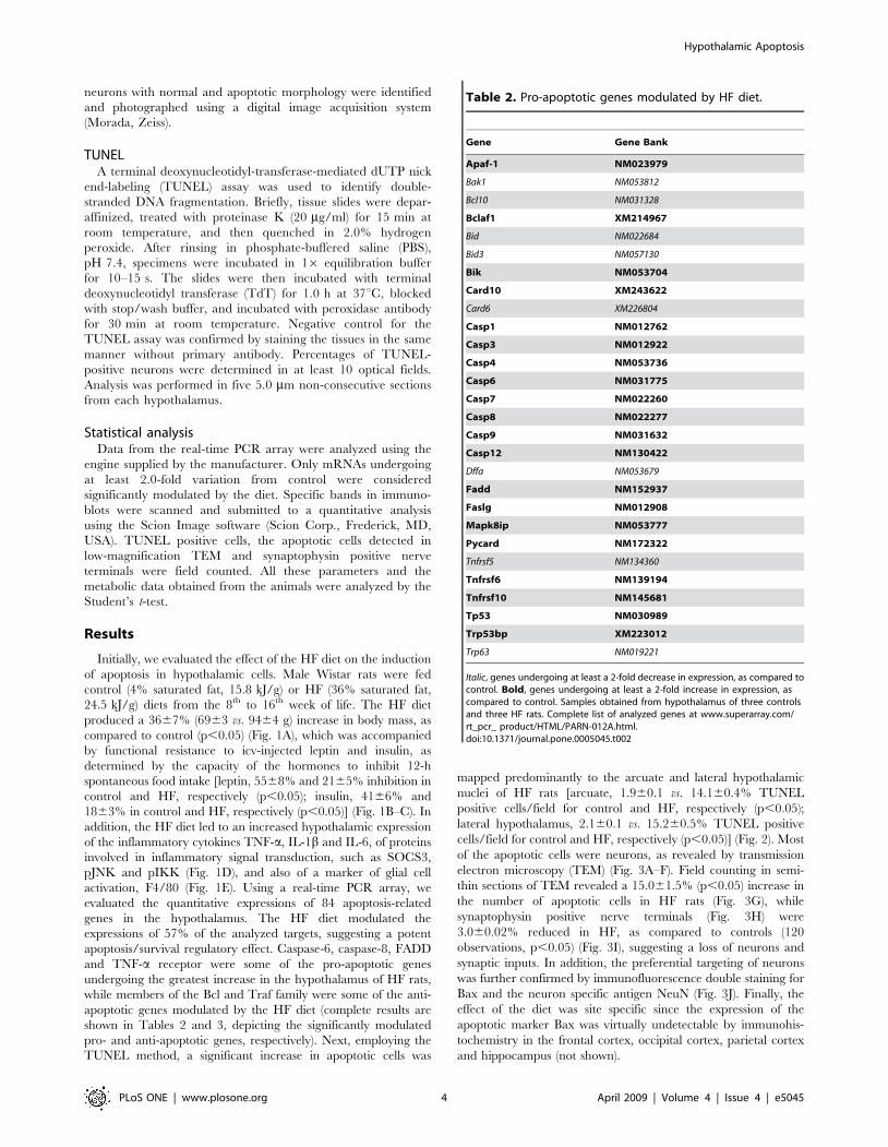
evaluated the quantitative expressions of 84 apoptosis-related
genes in the hypothalamus. The HF diet modulated the
expressions of 57% of the analyzed targets, suggesting a potent
apoptosis/survival regulatory effect. Caspase-6, caspase-8, FADD
and TNF-a receptor were some of the pro-apoptotic genes
undergoing the greatest increase in the hypothalamus of HF rats,
while members of the Bcl and Traf family were some of the anti-
apoptotic genes modulated by the HF diet (complete results are
shown in Tables 2 and 3, depicting the significantly modulated
pro- and anti-apoptotic genes, respectively). Next, employing the
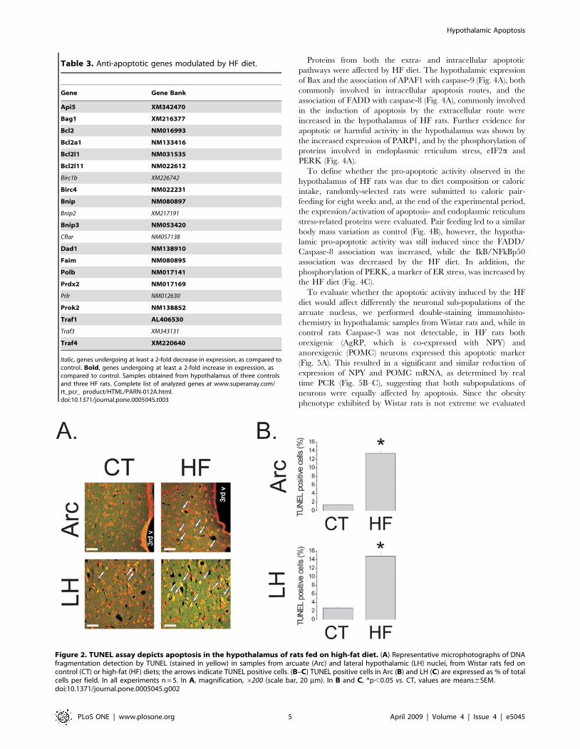
TUNEL method, a significant increase in apoptotic cells was
mapped predominantly to the arcuate and lateral hypothalamic
nuclei of HF rats [arcuate, 1.960.1 vs. 14.160.4% TUNEL
positive cells/field for control and HF, respectively (p,0.05);
lateral hypothalamus, 2.160.1 vs. 15.260.5% TUNEL positive
cells/field for control and HF, respectively (p,0.05)] (Fig. 2). Most
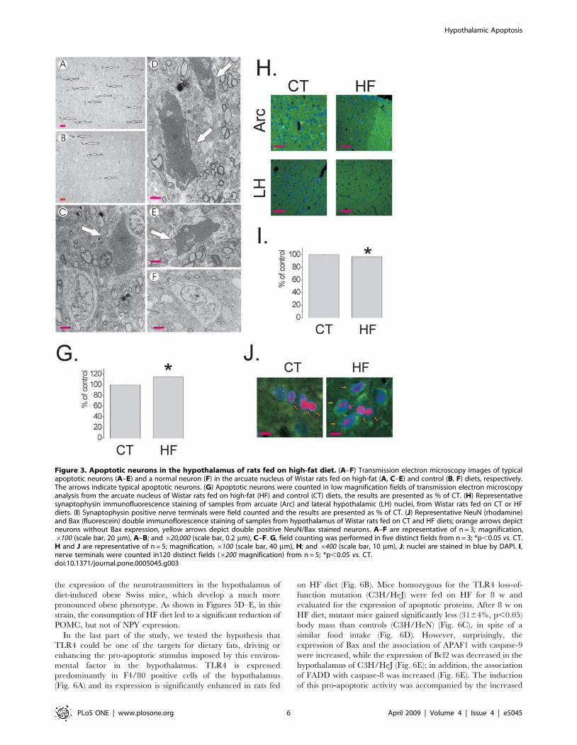
of the apoptotic cells were neurons, as revealed by transmission
electron microscopy (TEM) (Fig. 3A–F). Field counting in semi-
thin sections of TEM revealed a 15.061.5% (p,0.05) increase in
the number of apoptotic cells in HF rats (Fig. 3G), while
synaptophysin positive nerve terminals (Fig. 3H) were
3.060.02% reduced in HF, as compared to controls (120
observations, p,0.05) (Fig. 3I), suggesting a loss of neurons and
synaptic inputs. In addition, the preferential targeting of neurons
was further confirmed by immunofluorescence double staining for
Bax and the neuron specific antigen NeuN (Fig. 3J). Finally, the
effect of the diet was site specific since the expression of the
apoptotic marker Bax was virtually undetectable by immunohis-
tochemistry in the frontal cortex, occipital cortex, parietal cortex
and hippocampus (not shown).
Table 2. Pro-apoptotic genes modulated by HF diet.
Gene Gene Bank
Apaf-1 NM023979
Bak1 NM053812
Bcl10 NM031328
Bclaf1 XM214967
Bid NM022684
Bid3 NM057130
Bik NM053704
Card10 XM243622
Card6 XM226804
Casp1 NM012762
Casp3 NM012922
Casp4 NM053736
Casp6 NM031775
Casp7 NM022260
Casp8 NM022277
Casp9 NM031632
Casp12 NM130422
Dffa NM053679
Fadd NM152937
Faslg NM012908
Mapk8ip NM053777
Pycard NM172322
Tnfrsf5 NM134360
Tnfrsf6 NM139194
Tnfrsf10 NM145681
Tp53 NM030989
Trp53bp XM223012
Trp63 NM019221
Italic, genes undergoing at least a 2-fold decrease in expression, as compared tocontrol. Bold, genes undergoing at least a 2-fold increase in expression, ascompared to control. Samples obtained from hypothalamus of three controlsand three HF rats. Complete list of analyzed genes at www.superarray.com/rt_pcr_ product/HTML/PARN-012A.html.doi:10.1371/journal.pone.0005045.t002
Hypothalamic Apoptosis
PLoS ONE | www.plosone.org 4 April 2009 | Volume 4 | Issue 4 | e5045
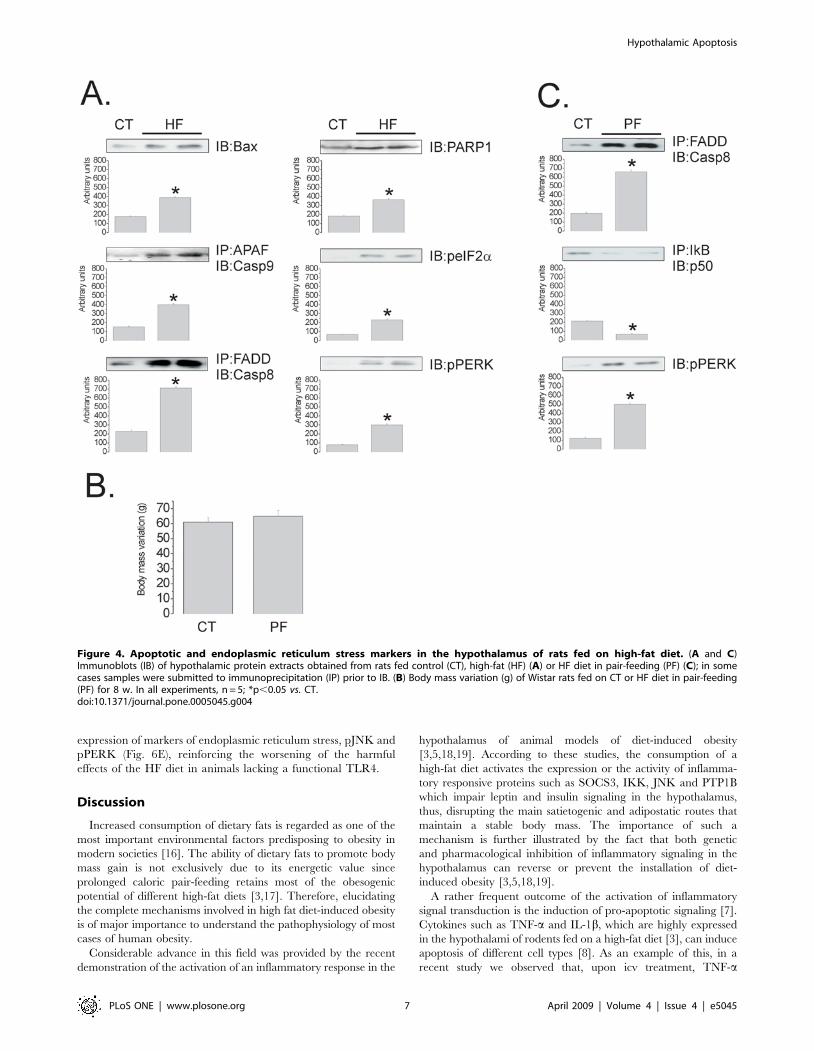
Proteins from both the extra- and intracellular apoptotic
pathways were affected by HF diet. The hypothalamic expression
of Bax and the association of APAF1 with caspase-9 (Fig. 4A), both
commonly involved in intracellular apoptosis routes, and the
association of FADD with caspase-8 (Fig. 4A), commonly involved
in the induction of apoptosis by the extracellular route were
increased in the hypothalamus of HF rats. Further evidence for
apoptotic or harmful activity in the hypothalamus was shown by
the increased expression of PARP1, and by the phosphorylation of
proteins involved in endoplasmic reticulum stress, eIF2a and
PERK (Fig. 4A).
To define whether the pro-apoptotic activity observed in the
hypothalamus of HF rats was due to diet composition or caloric
intake, randomly-selected rats were submitted to caloric pair-
feeding for eight weeks and, at the end of the experimental period,
the expression/activation of apoptosis- and endoplasmic reticulum
stress-related proteins were evaluated. Pair feeding led to a similar
body mass variation as control (Fig. 4B), however, the hypotha-
lamic pro-apoptotic activity was still induced since the FADD/
Caspase-8 association was increased, while the IkB/NFkBp50
association was decreased by the HF diet. In addition, the
phosphorylation of PERK, a marker of ER stress, was increased by
the HF diet (Fig. 4C).
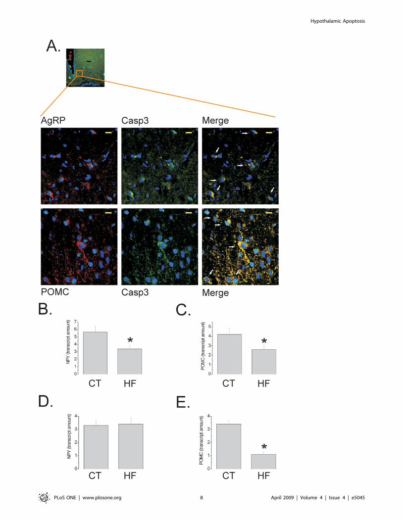
To evaluate whether the apoptotic activity induced by the HF
diet would affect differently the neuronal sub-populations of the
arcuate nucleus, we performed double-staining immunohisto-
chemistry in hypothalamic samples from Wistar rats and, while in
control rats Caspase-3 was not detectable, in HF rats both
orexigenic (AgRP, which is co-expressed with NPY) and
anorexigenic (POMC) neurons expressed this apoptotic marker
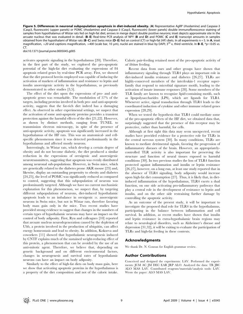
(Fig. 5A). This resulted in a significant and similar reduction of
expression of NPY and POMC mRNA, as determined by real
time PCR (Fig. 5B–C), suggesting that both subpopulations of
neurons were equally affected by apoptosis. Since the obesity
phenotype exhibited by Wistar rats is not extreme we evaluated
Table 3. Anti-apoptotic genes modulated by HF diet.
Gene Gene Bank
Api5 XM342470
Bag1 XM216377
Bcl2 NM016993
Bcl2a1 NM133416
Bcl2l1 NM031535
Bcl2l11 NM022612
Birc1b XM226742
Birc4 NM022231
Bnip NM080897
Bnip2 XM217191
Bnip3 NM053420
Cflar NM057138
Dad1 NM138910
Faim NM080895
Polb NM017141
Prdx2 NM017169
Prlr NM012630
Prok2 NM138852
Traf1 AL406530
Traf3 XM343131
Traf4 XM220640
Italic, genes undergoing at least a 2-fold decrease in expression, as compared tocontrol. Bold, genes undergoing at least a 2-fold increase in expression, ascompared to control. Samples obtained from hypothalamus of three controlsand three HF rats. Complete list of analyzed genes at www.superarray.com/rt_pcr_ product/HTML/PARN-012A.html.doi:10.1371/journal.pone.0005045.t003
Figure 2. TUNEL assay depicts apoptosis in the hypothalamus of rats fed on high-fat diet. (A) Representative microphotographs of DNAfragmentation detection by TUNEL (stained in yellow) in samples from arcuate (Arc) and lateral hypothalamic (LH) nuclei, from Wistar rats fed oncontrol (CT) or high-fat (HF) diets; the arrows indicate TUNEL positive cells. (B–C) TUNEL positive cells in Arc (B) and LH (C) are expressed as % of totalcells per field. In all experiments n = 5. In A, magnification, 6200 (scale bar, 20 mm). In B and C, *p,0.05 vs. CT, values are means6SEM.doi:10.1371/journal.pone.0005045.g002
Hypothalamic Apoptosis
PLoS ONE | www.plosone.org 5 April 2009 | Volume 4 | Issue 4 | e5045
the expression of the neurotransmitters in the hypothalamus of
diet-induced obese Swiss mice, which develop a much more
pronounced obese phenotype. As shown in Figures 5D–E, in this
strain, the consumption of HF diet led to a significant reduction of
POMC, but not of NPY expression.
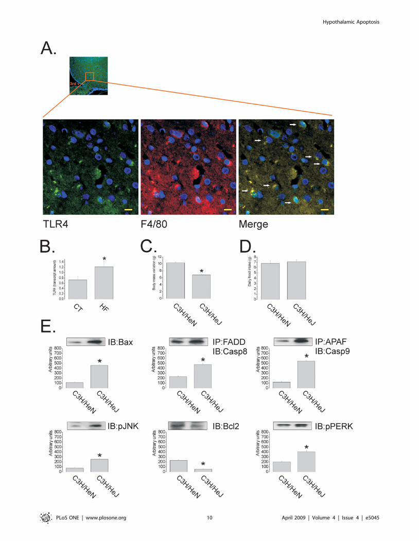
In the last part of the study, we tested the hypothesis that
TLR4 could be one of the targets for dietary fats, driving or
enhancing the pro-apoptotic stimulus imposed by this environ-
mental factor in the hypothalamus. TLR4 is expressed
predominantly in F4/80 positive cells of the hypothalamus
(Fig. 6A) and its expression is significantly enhanced in rats fed
on HF diet (Fig. 6B). Mice homozygous for the TLR4 loss-of-
function mutation (C3H/HeJ) were fed on HF for 8 w and
evaluated for the expression of apoptotic proteins. After 8 w on
HF diet, mutant mice gained significantly less (3164%, p,0.05)
body mass than controls (C3H/HeN) (Fig. 6C), in spite of a
similar food intake (Fig. 6D). However, surprisingly, the
expression of Bax and the association of APAF1 with caspase-9
were increased, while the expression of Bcl2 was decreased in the
hypothalamus of C3H/HeJ (Fig. 6E); in addition, the association
of FADD with caspase-8 was increased (Fig. 6E). The induction
of this pro-apoptotic activity was accompanied by the increased
Figure 3. Apoptotic neurons in the hypothalamus of rats fed on high-fat diet. (A–F) Transmission electron microscopy images of typicalapoptotic neurons (A–E) and a normal neuron (F) in the arcuate nucleus of Wistar rats fed on high-fat (A, C–E) and control (B, F) diets, respectively.The arrows indicate typical apoptotic neurons. (G) Apoptotic neurons were counted in low magnification fields of transmission electron microscopyanalysis from the arcuate nucleus of Wistar rats fed on high-fat (HF) and control (CT) diets, the results are presented as % of CT. (H) Representativesynaptophysin immunofluorescence staining of samples from arcuate (Arc) and lateral hypothalamic (LH) nuclei, from Wistar rats fed on CT or HFdiets. (I) Synaptophysin positive nerve terminals were field counted and the results are presented as % of CT. (J) Representative NeuN (rhodamine)and Bax (fluorescein) double immunoflorescence staining of samples from hypothalamus of Wistar rats fed on CT and HF diets; orange arrows depictneurons without Bax expression, yellow arrows depict double positive NeuN/Bax stained neurons. A–F are representative of n = 3; magnification,6100 (scale bar, 20 mm), A–B; and 620,000 (scale bar, 0.2 mm), C–F. G, field counting was performed in five distinct fields from n = 3; *p,0.05 vs. CT.H and J are representative of n = 5; magnification, 6100 (scale bar, 40 mm), H; and 6400 (scale bar, 10 mm), J; nuclei are stained in blue by DAPI. I,nerve terminals were counted in120 distinct fields (6200 magnification) from n = 5; *p,0.05 vs. CT.doi:10.1371/journal.pone.0005045.g003
Hypothalamic Apoptosis
PLoS ONE | www.plosone.org 6 April 2009 | Volume 4 | Issue 4 | e5045
expression of markers of endoplasmic reticulum stress, pJNK and
pPERK (Fig. 6E), reinforcing the worsening of the harmful
effects of the HF diet in animals lacking a functional TLR4.
Discussion
Increased consumption of dietary fats is regarded as one of the
most important environmental factors predisposing to obesity in
modern societies [16]. The ability of dietary fats to promote body
mass gain is not exclusively due to its energetic value since
prolonged caloric pair-feeding retains most of the obesogenic
potential of different high-fat diets [3,17]. Therefore, elucidating
the complete mechanisms involved in high fat diet-induced obesity
is of major importance to understand the pathophysiology of most
cases of human obesity.
Considerable advance in this field was provided by the recent
demonstration of the activation of an inflammatory response in the
hypothalamus of animal models of diet-induced obesity
[3,5,18,19]. According to these studies, the consumption of a
high-fat diet activates the expression or the activity of inflamma-
tory responsive proteins such as SOCS3, IKK, JNK and PTP1B
which impair leptin and insulin signaling in the hypothalamus,
thus, disrupting the main satietogenic and adipostatic routes that
maintain a stable body mass. The importance of such a
mechanism is further illustrated by the fact that both genetic
and pharmacological inhibition of inflammatory signaling in the
hypothalamus can reverse or prevent the installation of diet-
induced obesity [3,5,18,19].
A rather frequent outcome of the activation of inflammatory
signal transduction is the induction of pro-apoptotic signaling [7].
Cytokines such as TNF-a and IL-1b, which are highly expressed
in the hypothalami of rodents fed on a high-fat diet [3], can induce
apoptosis of different cell types [8]. As an example of this, in a
recent study we observed that, upon icv treatment, TNF-a
Figure 4. Apoptotic and endoplasmic reticulum stress markers in the hypothalamus of rats fed on high-fat diet. (A and C)Immunoblots (IB) of hypothalamic protein extracts obtained from rats fed control (CT), high-fat (HF) (A) or HF diet in pair-feeding (PF) (C); in somecases samples were submitted to immunoprecipitation (IP) prior to IB. (B) Body mass variation (g) of Wistar rats fed on CT or HF diet in pair-feeding(PF) for 8 w. In all experiments, n = 5; *p,0.05 vs. CT.doi:10.1371/journal.pone.0005045.g004
Hypothalamic Apoptosis
PLoS ONE | www.plosone.org 7 April 2009 | Volume 4 | Issue 4 | e5045
Hypothalamic Apoptosis
PLoS ONE | www.plosone.org 8 April 2009 | Volume 4 | Issue 4 | e5045
activates apoptotic signaling in the hypothalamus [20]. Therefore,
in the first part of the study, we explored the pro-apoptotic
potential of the high-fat diet evaluating the expressions of 84
apoptosis related genes by real-time PCR array. First, we showed
that the diet protocol herein employed was capable of inducing the
activation of markers of inflammation and resistance to leptin and
insulin anorexigenic activity in the hypothalamus, as previously
demonstrated in other studies [3,5].
The effect of the diet upon the expressions of pro- and anti-
apoptotic genes was remarkable. The modulation of 57% of the
targets, including proteins involved in both pro- and anti-apoptotic
activity, suggests that the fat-rich diet indeed has a damaging
effect. As observed in other experimental settings, we suspect that
the activation of some anti-apoptotic proteins provides a transient
protection against the harmful effects of the diet [21,22]. However,
as shown by distinct methods, ranging from TUNEL to
transmission electron microscopy, in spite of the presence of
anti-apoptotic activity, apoptosis was significantly increased in the
hypothalamus of the HF rats. This was an anatomical- and cell-
specific phenomenon since it was detected predominantly in the
hypothalamus and affected mostly neurons.
Interestingly, in Wistar rats, which develop a certain degree of
obesity and do not become diabetic, the diet produced a similar
reduction in the expressions of orexigenic and anorexigenic
neurotransmitters, suggesting that apoptosis was evenly distributed
among neuronal sub-populations. However, in Swiss mice, which
are genetically related to the diabetes prone AKR mouse [23] and,
likewise, display an outstanding propensity to obesity and diabetes
[24,25], the level of POMC was significantly reduced as compared
to control, suggesting that this subpopulation of neurons was
predominantly targeted. Although we have no current mechanistic
explanation for this phenomenon, we suspect that, by targeting
different subpopulations of neurons, diet-induced hypothalamic
apoptosis leads to an imbalance in orexigenic vs. anorexigenic
neurons in Swiss mice, but not in Wistar rats, therefore favoring
body mass gain only in the mice. Two recent studies have
provided strong evidence to suggest that changes in the numbers of
certain types of hypothalamic neurons may have an impact on the
control of body adiposity. First, Ryu and colleagues [10] reported
that arcuate nucleus neurodegeneration caused by the depletion of
Ubb, a protein involved in the production of ubiquitin, can affect
energy homeostasis and lead to obesity. In addition, Kokoeva and
coworkers [11] showed that hypothalamic neurogenesis induced
by CNTF explains much of the sustained weight-reducing effect of
this protein, a phenomenon that can be avoided by the use of an
anti-mitotic agent. Therefore, we believe that, depending on
genetic background and on different environmental factors,
changes in neurogenesis and survival rates of hypothalamic
neurons can have an impact on body adiposity.
Similarly to the effect of high-fat diets on body mass gain, here
we show that activating apoptotic proteins in the hypothalamus is
a property of the diet composition and not of the caloric intake.
Caloric pair-feeding retained most of the pro-apoptotic activity of
ad libitum feeding.
Recent data from ours and other groups have shown that
inflammatory signaling through TLR4 plays an important role in
diet-induced insulin resistance and diabetes [26,27]. TLRs are
highly-conserved members of the interleukin-1 receptor super-
family that respond to microbial signature motifs, leading to the
activation of innate immune responses [28]. Some members of the
TLR family are known to recognize lipid-containing motifs, such
as lipopolysaccharides (LPS), which are ligands for TLR4.
Whenever active, signal transduction through TLR4 leads to the
coordinated induction of cytokine and other immune related genes
expression [28,29].
When we tested the hypothesis that TLR4 could mediate some
of the pro-apoptotic effects of the HF diet, we obtained data that,
surprisingly suggested that the presence of this receptor may be
protective, rather than harmful, for cells.
Although at first sight this data may seem unexpected, recent
studies have provided evidence for a protective role for TLRs in
the central nervous system [30]. In many conditions, TLRs are
known to mediate detrimental signals, favoring the progression of
inflammatory diseases of the brain. However, an appropriately-
controlled TLR activity is also important for preserving the
structure and function of neural tissues exposed to harmful
conditions [30]. In two previous studies the loss of TLR4 function
protected against inflammation and insulin or leptin resistance
[26,27], however, on a long run, at least one study reported that in
the absence of TLR4 signaling, body adiposity would increase
upon high-fat diet consumption [27]. Thus, it is likely that, in diet-
induced inflammation of the hypothalamus, TLR4 exerts a dual
function, on one side activating pro-inflammatory pathways that
play a central role in the development of resistance to leptin and
insulin, and on the other side restraining further damage by
controlling the apoptotic activity.
As an outcome of the present study, it will be important to
investigate the proposed dual role for TLR4 in the hypothalamus,
participating in the balance between inflammation and cell
survival. In addition, as recent studies have shown that insulin
and leptin resistance in extra-hypothalamic brain regions may
relate to neurological disorders, such as Alzheimer’s disease and
depression [31,32], it will be exiting to evaluate the participation of
TLRs and high-fat feeding in these contexts.
Acknowledgments
We thank Dr. N. Conran for English grammar review.
Author Contributions
Conceived and designed the experiments: LAV. Performed the experi-
ments: JCM AC JM DEC EAR JRP ALO. Analyzed the data: TR JBC
ALO MAS LAV. Contributed reagents/materials/analysis tools: LAV.
Wrote the paper: ALO MAS LAV.
Figure 5. Differences in neuronal subpopulation apoptosis in diet-induced obesity. (A) Representative AgRP (rhodamine) and Caspase-3(Casp3, fluorescein) (upper panels) or POMC (rhodamine) and Caspase-3 (Casp3, fluorescein) (lower panels) double immunoflorescence staining ofsamples from hypothalamus of Wistar rats fed on high-fat diet; arrows in merge depict double positive neurons; inset depicts approximate site in thearcuate nucleus that was evaluated in detail. (B–E) Real-time PCR analysis of NPY (B and D) and POMC (C and E) transcript amounts in samplesobtained from the hypothalami of Wistar rats (B–C) and Swiss mice (D–E) fed on control (CT) or high-fat (HF) diets. In all experiments n = 5. In A, insetmagnification, 620 and captions magnification, 6400 (scale bar, 10 mm), nuclei are stained in blue by DAPI; 3rd v, third ventricle. In B–E, *p,0.05 vs.CT.doi:10.1371/journal.pone.0005045.g005
Hypothalamic Apoptosis
PLoS ONE | www.plosone.org 9 April 2009 | Volume 4 | Issue 4 | e5045
Hypothalamic Apoptosis
PLoS ONE | www.plosone.org 10 April 2009 | Volume 4 | Issue 4 | e5045
References
1. Flier JS (2004) Obesity wars: molecular progress confronts an expanding
epidemic. Cell 116: 337–350.2. Milanski M, Degasperi G, Coope A, Morari J, Denis R, et al. (2009) Saturated
fatty acids produce an inflammatory response predominantly through the
activation of TLR4 signaling in hypothalamus: implications for the pathogenesisof obesity. J Neurosci 29: 359–370.
3. De Souza CT, Araujo EP, Bordin S, Ashimine R, Zollner RL, et al. (2005)Consumption of a fat-rich diet activates a proinflammatory response and induces
insulin resistance in the hypothalamus. Endocrinology 146: 4192–4199.
4. Munzberg H, Flier JS, Bjorbaek C (2004) Region-specific leptin resistance withinthe hypothalamus of diet-induced obese mice. Endocrinology 145: 4880–4889.
5. Howard JK, Cave BJ, Oksanen LJ, Tzameli I, Bjorbaek C, et al. (2004)Enhanced leptin sensitivity and attenuation of diet-induced obesity in mice with
haploinsufficiency of Socs3. Nat Med 10: 734–738.
6. Bence KK, Delibegovic M, Xue B, Gorgun CZ, Hotamisligil GS, et al. (2006)Neuronal PTP1B regulates body weight, adiposity and leptin action. Nat Med
12: 917–924.7. Siegel RM, Muppidi J, Roberts M, Porter M, Wu Z (2003) Death receptor
signaling and autoimmunity. Immunol Res 27: 499–512.8. Muppidi JR, Tschopp J, Siegel RM (2004) Life and death decisions: secondary
complexes and lipid rafts in TNF receptor family signal transduction. Immunity
21: 461–465.9. Cnop M, Welsh N, Jonas JC, Jorns A, Lenzen S, et al. (2005) Mechanisms of
pancreatic beta-cell death in type 1 and type 2 diabetes: many differences, fewsimilarities. Diabetes 54 Suppl 2: S97–107.
10. Ryu KY, Garza JC, Lu XY, Barsh GS, Kopito RR (2008) Hypothalamic
neurodegeneration and adult-onset obesity in mice lacking the Ubb poly-ubiquitin gene. Proc Natl Acad Sci U S A 105: 4016–4021.
11. Kokoeva MV, Yin H, Flier JS (2005) Neurogenesis in the hypothalamus of adultmice: potential role in energy balance. Science 310: 679–683.
12. Carvalheira JB, Siloto RM, Ignacchitti I, Brenelli SL, Carvalho CR, et al. (2001)Insulin modulates leptin-induced STAT3 activation in rat hypothalamus. FEBS
Lett 500: 119–124.
13. Paxinos G, Watson CR, Emson PC (1980) AChE-stained horizontal sections ofthe rat brain in stereotaxic coordinates. J Neurosci Methods 3: 129–149.
14. Bertelli DF, Araujo EP, Cesquini M, Stoppa GR, Gasparotto-Contessotto M, etal. (2006) Phosphoinositide-specific inositol polyphosphate 5-phosphatase IV
inhibits inositide trisphosphate accumulation in hypothalamus and regulates
food intake and body weight. Endocrinology 147: 5385–5399.15. Araujo EP, Amaral ME, Filiputti E, De Souza CT, Laurito TL, et al. (2004)
Restoration of insulin secretion in pancreatic islets of protein-deficient rats byreduced expression of insulin receptor substrate (IRS)-1 and IRS-2. J Endocrinol
181: 25–38.16. Astrup A, Dyerberg J, Selleck M, Stender S (2008) Nutrition transition and its
relationship to the development of obesity and related chronic diseases. Obes
Rev 9 Suppl 1: 48–52.
17. Wade GN (1982) Obesity without overeating in golden hamsters. Physiol Behav
29: 701–707.
18. Picardi PK, Calegari VC, Prada Pde O, Moraes JC, Araujo E, et al. (2008)
Reduction of hypothalamic protein tyrosine phosphatase improves insulin and
leptin resistance in diet-induced obese rats. Endocrinology 149: 3870–3880.
19. Xue B, Kim YB, Lee A, Toschi E, Bonner-Weir S, et al. (2007) Protein-tyrosine
phosphatase 1B deficiency reduces insulin resistance and the diabetic phenotype
in mice with polygenic insulin resistance. J Biol Chem 282: 23829–23840.
20. Degasperi GR, Romanatto T, Denis RG, Araujo EP, Moraes JC, et al. (2008)
UCP2 protects hypothalamic cells from TNF-alpha-induced damage. FEBS Lett
582: 3103–3110.
21. Lu Y, Fukuyama S, Yoshida R, Kobayashi T, Saeki K, et al. (2006) Loss of
SOCS3 gene expression converts STAT3 function from anti-apoptotic to pro-
apoptotic. J Biol Chem 281: 36683–36690.
22. Leber B, Lin J, Andrews DW (2007) Embedded together: the life and death
consequences of interaction of the Bcl-2 family with membranes. Apoptosis 12:
897–911.
23. West DB, Goudey-Lefevre J, York B, Truett GE (1994) Dietary obesity linked to
genetic loci on chromosomes 9 and 15 in a polygenic mouse model. J Clin Invest
94: 1410–1416.
24. De Souza CT, Araujo EP, Stoppiglia LF, Pauli JR, Ropelle E, et al. (2007)
Inhibition of UCP2 expression reverses diet-induced diabetes mellitus by effects
on both insulin secretion and action. Faseb J 21: 1153–1163.
25. De Souza CT, Araujo EP, Prada PO, Saad MJ, Boschero AC, et al. (2005)
Short-term inhibition of peroxisome proliferator-activated receptor-gamma
coactivator-1alpha expression reverses diet-induced diabetes mellitus and
hepatic steatosis in mice. Diabetologia 48: 1860–1871.
26. Tsukumo DM, Carvalho-Filho MA, Carvalheira JB, Prada PO, Hirabara SM, et
al. (2007) Loss-of-function mutation in Toll-like receptor 4 prevents diet-induced
obesity and insulin resistance. Diabetes 56: 1986–1998.
27. Shi H, Kokoeva MV, Inouye K, Tzameli I, Yin H, et al. (2006) TLR4 links
innate immunity and fatty acid-induced insulin resistance. J Clin Invest 116:
3015–3025.
28. Akira S, Uematsu S, Takeuchi O (2006) Pathogen recognition and innate
immunity. Cell 124: 783–801.
29. Shimazu R, Akashi S, Ogata H, Nagai Y, Fukudome K, et al. (1999) MD-2, a
molecule that confers lipopolysaccharide responsiveness on Toll-like receptor 4.
J Exp Med 189: 1777–1782.
30. Hanisch UK, Johnson TV, Kipnis J (2008) Toll-like receptors: roles in
neuroprotection? Trends Neurosci 31: 176–182.
31. Pasinetti GM, Eberstein JA (2008) Metabolic syndrome and the role of dietary
lifestyles in Alzheimer’s disease. J Neurochem 106: 1503–1514.
32. Rintamaki R, Grimaldi S, Englund A, Haukka J, Partonen T, et al. (2008)
Seasonal changes in mood and behavior are linked to metabolic syndrome.
PLoS ONE 3: e1482.
Figure 6. TLR4 protects against diet-induced apoptosis of hypothalamic neurons. (A) Representative TLR4 (rhodamine) and F4/80(fluorescein) double immunoflorescence staining of samples from hypothalamus of Wistar rats; arrows in merge depict double positive cells; insetdepicts approximate site in the arcuate nucleus that was evaluated in detail. (B) Real-time PCR analysis of TLR4 transcript amount in samples obtainedfrom the hypothalami of Wistar rats fed on control (CT) or high-fat (HF) diets. (C) Body mass variation (g) of C3H/HeN and C3H/HeJ mice fed on HFdiet for 8 w. (D) Mean daily food intake (g) of C3H/HeN and C3H/HeJ mice fed on HF diet. (E) Immunoblots (IB) of hypothalamic protein extractsobtained from C3H/HeN and C3H/HeJ mice fed on HF diet; in some cases samples were submitted to immunoprecipitation (IP) prior to IB. In allexperiments n = 5. In A, inset magnification, 620 and captions magnification, 6400 (scale bar, 10 mm), nuclei are stained in blue by DAPI; 3rd v, thirdventricle. B, *p,0.05 vs. CT; in C and E, *p,0.05 vs. C3H/HeN.doi:10.1371/journal.pone.0005045.g006
Hypothalamic Apoptosis
PLoS ONE | www.plosone.org 11 April 2009 | Volume 4 | Issue 4 | e5045

Copyright of PLoS ONE is the property of Public Library of Science and its content may not be copied or
emailed to multiple sites or posted to a listserv without the copyright holder's express written permission.
However, users may print, download, or email articles for individual use.