chelerythrine induces apoptosis through a bax/bak-independent
TRANSCRIPT

Chelerythrine Induces Apoptosis through aBax/Bak-independent Mitochondrial Mechanism*□S
Received for publication, September 13, 2007, and in revised form, December 17, 2007 Published, JBC Papers in Press, January 29, 2008, DOI 10.1074/jbc.M707687200
Kah Fei Wan‡, Shing-Leng Chan§, Sunil Kumar Sukumaran‡, Mei-Chin Lee‡, and Victor C. Yu‡1
From the ‡Institute of Molecular and Cell Biology, A*STAR (Agency for Science, Technology, and Research), 61 Biopolis Dr. (Proteos),Singapore 138673 and §Oncology Research Institute, National University of Singapore, 28 Medical Dr., Centre For Life Sciences,Cancer Program, Singapore 117456
Although murine embryonic fibroblasts (MEFs) with Bax orBak deleted displayed no defect in apoptosis signaling, MEFswith Bax and Bak double knock-out (DKO) showed dramaticresistance to diverse apoptotic stimuli, suggesting that Bax andBak are redundant but essential regulators for apoptosis signal-ing.Chelerythrinehas recently been identified as aBcl-xL inhib-itor that is capable of triggering apoptosis via direct action onmitochondria. Here we report that in contrast to classic apopto-tic stimuli, chelerythrine is fully competent in inducing apopto-sis in the DKO MEFs. Wild-type and DKO MEFs are equallysensitive to chelerythrine-inducedmorphological andbiochem-ical changes associatedwith apoptosis phenotype. Interestingly,chelerythrine-mediated release of cytochrome c is rapid andprecedes Bax translocation and integration. Although the BH3peptide of Bim is totally inactive in releasing cytochrome c fromisolated mitochondria of DKO MEFs, chelerythrine maintainsits potency and efficacy in inducing direct release of cytochromec from these mitochondria. Furthermore, chelerythrine-medi-ated mitochondrial swelling and loss in mitochondrial mem-brane potential (��m) are inhibited by cyclosporine A, suggest-ing that mitochondrial permeability transition pore is involvedin chelerythrine-induced apoptosis. Although certain apoptoticstimuli have been shown to elicit cytotoxic effect in the DKOMEFs through alternate death mechanisms, chelerythrine doesnot appear to engage necrotic or autophagic death mechanismto trigger cell death in theDKOMEFs. These results, thus, arguefor the existence of an alternative Bax/Bak-independent apo-ptotic mechanism that involves cyclosporine A-sensitive mito-chondrial membrane permeability.
Mitochondria are the major organelles involved in the signaltransduction and biochemical execution of apoptosis (1). Pro-teins of the Bcl-2 family are the central transducers of survivaland apoptotic signals (2). They act at mitochondria by regulat-ing the permeability and integrity of the mitochondrial outermembranes, thereby controlling the release of apoptogenic fac-
tors. The Bcl-2 family consists of three major subfamilies ofpro-survival and pro-apoptotic molecules. Members of theBH3-only subfamily (Bim, Bad, Bid, Bik, Noxa, Puma, and Hrk)serve as sentinels for the initiation of apoptosis by modulatingthe function of members of the other two multidomain pro-survival (Bcl-2, Bcl-w,Mcl-1, Bcl-xL, andA1/Bfl-1) or pro-apo-ptotic (Bax and Bak) subfamilies (3–5).A widely acknowledged paradigm of apoptotic signaling cas-
cades suggests that apoptotic insults unleash one ormore of thedistinct BH3-only molecules and induce their translocation tothe outer mitochondrial membrane (2, 6, 7). In mitochondriathese proteins are thought to bind preferentially to the anti-apoptoticmembers of the Bcl-2 family (8–10) and, hence, serveto displace the multidomain pro-apoptotic members frombinding to the pro-survival molecules. The ratio between thelevels of multidomain pro-and anti-apoptotic members may,thus, play an important role in setting the rheostat for deathresponses to apoptotic insults (4, 11). Some of the BH3-onlymolecules such as Bid and certain splicing isoforms of Bim arethought to have an additional role in promoting activation ofthe multidomain pro-apoptotic molecules such as Bax and Bak(10, 12).Bax and Bak are essential but redundant regulators of the
mitochondrial apoptotic signaling pathway (8, 9, 13). Murineembryonic fibroblasts (MEFs)2 deficient in only one of the twomolecules remain relatively normal in the execution of apopto-sis signaling in response to a variety of apoptotic insults. Ani-mals deficient in both molecules, however, display majorabnormalities (14). Furthermore, MEFs derived from bax andbak double-knock-out mice (DKO) were found to be highlyresistant to a wide variety of apoptotic stimuli, including over-expression of BH3-only molecules (8, 9, 13).Despite the profound apoptotic defects, the ability of some
bax and bak double-knock-out mice to survive into adulthood
* This work was supported by the Biomedical Research Council of A*STAR(Agency for Science, Technology, and Research), Singapore. The costs ofpublication of this article were defrayed in part by the payment of pagecharges. This article must therefore be hereby marked “advertisement” inaccordance with 18 U.S.C. Section 1734 solely to indicate this fact.
□S The on-line version of this article (available at http://www.jbc.org) containssupplemental Figs. S1–S6.
1 An adjunct staff of the Dept. of Pharmacology, National University of Singa-pore. To whom correspondence should be addressed. Tel.: 65-65869672;Fax: 65-67791117; E-mail: [email protected].
2 The abbreviations used are: MEF, murine embryonic fibroblast; WT, wildtype; DKO, Bax and Bak double knockout; cyt c, cytochrome c; ��m, mito-chondrial membrane potential; CsA, cyclosporine A; mPTP, mitochondrialpermeability transition pore; hXIAP, human X-linked inhibitor of apoptosisprotein; CypD, cyclophilin D; PBS, phosphate-buffered saline; ROS, reactiveoxygen species; PARP, poly(ADP-ribose) polymerase; Q-VD-OPH, N-(2-quinolyl)valyl-aspartyl-(2,6-difluorophenoxy) methyl ketone; zVAD-fmk,N-benzyloxycarbonyl-Val-Ala-Asp(O-Me) fluoromethyl ketone; 3MA,3-methyl adenine; JC-1, 5,5�,6,6�-tetrachloro-1,1�,3,3� tetraethyl-benz-imidazolylcarbocyanine iodide; 7AAD, 7-amino-actinomycin D;CM-H2DCFDA, 5-(and 6-)chloromethyl-2�,7�-dichlorodihydrofluoresceindiacetate, acetyl ester dye; HMG-B1, high mobility group B1 protein; LC3,light chain-3; WST, water-soluable tetrazolium salts.
THE JOURNAL OF BIOLOGICAL CHEMISTRY VOL. 283, NO. 13, pp. 8423–8433, March 28, 2008© 2008 by The American Society for Biochemistry and Molecular Biology, Inc. Printed in the U.S.A.
MARCH 28, 2008 • VOLUME 283 • NUMBER 13 JOURNAL OF BIOLOGICAL CHEMISTRY 8423
by guest on April 9, 2018
http://ww
w.jbc.org/
Dow
nloaded from
suggest the existence of other forms of cell death mechanismfor directing proper development and maintaining tissuehomeostasis by eliminating excess or damaged cells (14). Inter-estingly, several recent reports have shown that certain chemi-cal apoptotic stimuli are capable of engaging in alternativeforms of cell death in the absence of Bax and Bak. For example,DNA alkylating agents appeared to induce a mix of both apo-ptotic and necrotic cell death in MEFs, whereas they inducedmainly necrotic death in MEFs lacking both Bax and Bak (15).Furthermore, although etoposide and staurosporine predomi-nantly induce apoptosis in MEFs, they activate a caspase-inde-pendent cell deathmechanism largely dependent on autophagyin the DKOMEFs (16). Therefore, the DKOMEFs could serveas a valuable tool in delineatingmultiple cell deathmechanismsthat can be engaged by cytotoxic agents which may otherwiseescape detection in cells with intact apoptotic machinery.Chelerythrine, a benzophenanthridine alkaloid, is known to
trigger apoptosis in a variety of tumor cells (17). Recently, chel-erythrinewas identified by us as an inhibitorymolecule that canblock the heterodimerization of the Bcl-xL and Bak BH3 pep-tide in a high-throughput screen of 107,423 extracts derivedfromnatural products (18). Although etoposide, staurosporine,and chelerythrine effectively triggered cytochrome c (cyt c)release from mitochondria in intact cells, only chelerythrinewas able to induce cyt c release from isolatedmitochondria (18),suggesting that chelerythrine acts directly on mitochondria. Inthis study we compared the cytotoxic activity of chelerythrinein the wild-type (WT) and DKOMEFs in an attempt to furtherdelineate the mechanism by which chelerythrine mediates itscytotoxic effect in mammalian cells. Interestingly, cheleryth-rine is cytotoxic to both cell types. Surprisingly, themorpholog-ical and biochemical features associated with chelerythrine-in-duced cytotoxicity in DKO MEFs are indistinguishable fromthose observed in the WT MEFs. These are consistent withapoptotic rather than the autophagic or necrotic phenotypesassociated with certain classic cytotoxic stimuli in the DKOMEFs. Furthermore, although chelerythrine and the Bim BH3peptide are effective in triggering cyt c release from mitochon-dria isolated fromWTMEFs, only chelerythrine remains activeinmediating this effect onmitochondria isolated from theDKOMEFs. Chelerythrine induces mitochondrial swelling and lossof mitochondrial membrane potential (��m), and these effectsare inhibited by cyclosporine A (CsA), suggesting the involve-ment of CsA-sensitive mitochondrial permeability transitionpore (mPTP) components in both cell types. Taken together,these data strongly suggest that chelerythrine induces apopto-sis in MEFs through a Bax- and Bak-independent mitochon-drial mechanism that is distinct from the previously definedapoptosis gateway.
EXPERIMENTAL PROCEDURES
Reagents and Cell Lines—SV40 T antigen-transformed WTand DKO MEFs were grown in Dulbecco’s modified Eagle’smedium supplemented with 10% fetal bovine serum andantibiotics (100 �g of streptomycin/ml and 100 IU of peni-cillin/ml, Invitrogen). Lipofectamine (Invitrogen) was usedfor transfections according to the user’s manual. The caspaseinhibitors N-benzyloxycarbonyl-Val-Ala-Asp(O-Me) flu-
oromethyl ketone (zVAD-fmk) and N-(2-quinolyl)valyl-as-partyl-(2,6-difluorophenoxy)-methyl Ketone (Q-VD-OPH)were from Calbiochem. Staurosporine, etoposide, cheleryth-rine, camptothecin, 3-methyladenine (3MA), and CsA werefrom Sigma. Anti-cyt c antibody was purchased from BD Bio-sciences Pharmingen. Anti-Myc antibody was from Santa CruzBiotechnology. Antibody against high mobility group B1 pro-tein (HMG-B1) was from Abcam. Antibodies against actin andHSP60 were from Sigma.Cyt c Release and Bax Translocation in Intact Cells—Cells
were harvested andwashed once with ice-cold phosphate-buff-ered saline (PBS). To separate the cytosolic fraction from othercellular components, cells were lysed for 5 min on ice in a solu-tion consisting of 20 mM HEPES, pH 7.2, 50 mM KCl, 5 mMMgCl2, 1 mM EDTA, 1 mM EGTA, 250 mM sucrose, 200 �g/mldigitonin and protease inhibitors mixture as described (19).The organelles, cytoskeleton, and membranes were pelleted bycentrifugation at 12,000 rpm for 10 min at 4 °C. The superna-tant (cytosol) was carefully removed, and the pellet containingthe mitochondria was solubilized in radioimmune precipita-tion assay buffer (150 mM NaCl, 0.1% SDS, 10% sodiumdeoxycholate, 1% Nonidet P-40, 2 mM EDTA, and 10 mMHEPES (pH 7.3)) containing the Complete protease inhibi-tors mixture. Protein content was determined by the Brad-ford reaction (Bio-Rad), and equivalent amount from eachfraction were used for detection of cyt c release and Baxtranslocation using immunoblotting.Analysis of Isolated Mitochondria—Mitochondria were iso-
lated fromMEFs as previously described (20). For in vitro cyt crelease assays, cells were suspended in isolation buffer (320mMsucrose, 1mM EDTA, 50mMHEPES (pH 7.5)) and disrupted by20 expulsions through a 27-gauge needle. Disrupted cells werespun at 1000 � g for 10 min to remove cell debris and nucleus.The supernatants were centrifuged at 7000 � g for 10 min topellet the heavy membrane fraction containing the mitochon-dria. Themitochondria-containing pellets were resuspended inassay buffer (250 mM sucrose, 2 mM KH2PO4, 5 mM sodiumsuccinate, 25 mM EGTA, and 10 mM HEPES (pH 7.5)) at 0.5mg/ml. Equal amounts of mitochondria were treated with theindicated compounds for 15min at room temperature followedby centrifugation (20). Cyt c released into the supernatant wassubjected to fractionation on 13.5% SDS-PAGE followed byWestern blotting analysis. For light-scattering studies, mito-chondrial isolated as described above were suspended in assaybuffer (215 mM mannitol, 71 nM sucrose, 10 mM succinate, and10 mM HEPES, pH 7.4) (21). Changes in absorbance at 540 nM(A540), indicating mitochondrial swelling as a consequence ofmPTP opening, were measured after the addition of the indi-cated compounds using a microplate reader (Tecan). For dyeretention assays, the mitochondrial suspension was incubatedwith 5,5�,6,6�-tetrachloro-1,1�,3,3� tetraethylbenzimidazolyl-carbocyanine iodide (JC-1 100 ng/ml) for 10 min, then washedand resuspended in PBS; 5 min after the addition of indicatedcompounds, the level of JC-1 red fluorescence (FL2) retained bymitochondria was determined by flow cytometry (22).Assessment of Cell Viability, Cell Proliferation, and Caspase
3/7 Activity—Cells were seeded onto 96-well plates. After 48 hthey were treated with the indicated concentrations of com-
Chelerythrine Induces Bax/Bak-independent Apoptosis
8424 JOURNAL OF BIOLOGICAL CHEMISTRY VOLUME 283 • NUMBER 13 • MARCH 28, 2008
by guest on April 9, 2018
http://ww
w.jbc.org/
Dow
nloaded from

pounds. Cell death was assessed usingWST assay according tothe supplier’s protocols (Roche Applied Science). For the cellproliferation assay, cells were seeded onto 6-well dishes. After2 h they were treated with the indicated compounds in thepresence and absence of 3MA (10 mM). The cells were thenrecovered and re-cultured in standard medium onto 96-wellplates. Viable cell numbers were measured on the indicateddays by WST assay. Caspase 3/7 activity was measured using aCaspase-Glo 3/7 kit from Promega.Reactive Oxygen Species (ROS) Measurement—Cells were
treated with the indicated concentrations of compounds for16 h. After such treatment, cells werewashed in PBS and loadedwith 1 �M 5-(and 6-)chloromethyl-2�,7�-dichlorodihydrofluo-rescein diacetate, acetyl ester dye (CM-H2DCFDA; MolecularProbes) in PBS for 30 min at 37 °C as described (23). TheCM-H2DCFDA buffer was then removed, and the cells werewashed twice with PBS before being analyzed by microplatereader (Tecan). Measurement was taken at an excitation wave-length of 500 nm and an emission wavelength of 520 nm. Emis-sion from cells samples that were not loaded withCM-H2DCFDA was the same as for those with PBS.Fluorescence Microscope Analysis—Nuclear DNA fragmen-
tationwas assessed by nuclearmorphology afterHoechst 33342staining. Cells were stained with 1 �MHoechst 33342 for 5 minat room temperature. In another set of experiment designed fordetecting early phase of apoptosis, cells were stained withannexin V-fluorescein isothiocyanate and propidium iodide byfollowing themanufacturer’s instructions (ApoAlertTM AnnexinVApoptosis kit, Clontech). Apoptotic cells were photographedusingZeiss epifluorescencemicroscopewith an attachedNikonCoolpix digital camera.Flow Cytometry—For the detection of sub-G1 DNA, cells
were washed once, resuspended in 200 �l of PBS, and fixed in a50-fold excess of ice-cold 70% ethanol. Cells were recovered bycentrifugation at 1000 � g for 5 min at 4 °C, washed, stainedwith 50mg/ml of propidium iodide for 30min at room temper-ature, and analyzed in a FACScan flow cytometer (BD Bio-sciences).Mitochondrialmembrane potential change (��m) asmeasured by JC-1 and tetramethylrhodamine ethyl ester stain-ing was performed in accordance with the manufacturer’sinstructions (Molecular Probes). The exclusion assay for non-viable cells was performed by staining with 7-amino-actinomy-cin D (7AAD) followed by flow cytometry (BD Pharmingen). Aminimum of 10,000 cells per sample were analyzed. The resultspresented are representative of at least three experiments.
RESULTS
Chelerythrine Triggers Caspase-dependent Cytotoxicity in theAbsence of Bax and Bak—In agreement with the published lit-erature, DKO MEFs were found to be resistant to a variety ofapoptotic stimuli including etoposide, staurosporine, campto-thecin, cisplatin, methotrexate, and UV irradiation (Fig. 1, A,panel c and d, and B; data not shown). Surprisingly, in responseto treatment with chelerythrine, both WT and DKO MEFsunderwent similar morphological and biochemical changesthat are hallmarks of apoptosis. The chelerythrine-treatedMEFs displayed nuclear condensation (Fig. 1A, panels e and f)and the appearance of sub-G1 DNA (Fig. 1B). The appearance
of sub-G1 DNAwas seen in DKOMEFs treated with cheleryth-rine, but not etoposide, staurosporine, or camptothecin eventhough these compounds were fully effective in inducingsub-G1 DNA appearance in the WT MEFs (Fig. 1B; data notshown). Progressive stages of apoptosis could easily be de-tected in the chelerythrine-treated MEFs. At 5 �M, cheleryth-rine induced the appearance of annexin V-positive cells with-out an increase in propidium iodine staining at 16 h (Fig. 1C,panel b), indicative of early stage apoptosis. Cells exposed to 5�M chelerythrine for prolonged period (Fig. 1C, panels g and h)or treated with higher concentrations of chelerythrine (Fig. 1C,panel c and d) became both annexin V- and propidium iodide-positive, presumably because of an increase in permeability ofplasmamembrane during the late stage of apoptosis.Moreover,chelerythrine was able to induce comparable caspase 3/7 acti-vation in both WT and DKO MEFs (Fig. 1D). In contrast, eto-poside, staurosporine, and UV irradiation were able to activatecaspases 3/7 only in theWTMEFs (Fig. 1D). The appearance ofsub-G1 DNA in DKOMEFs upon chelerythrine treatment wasclearly caspase-dependent, as the effect was abolished by pre-treatment with caspase inhibitor (zVAD-fmk) (Fig. 1E, upperpanel) or overexpression of hXIAP (human X-linked inhibitorof apoptosis protein) (Fig. 1E, lower panel), a protein that isknown to sequester and inhibit activated caspases (24). Chel-erythrine failed to induce caspase-8 activation in bothWT andDKO MEFs (data not shown), suggesting that the cytotoxiceffect of chelerythrine inMEFs is not attributable to the extrin-sic apoptosis signaling pathway.Chelerythrine Remains Fully Active in Inducing Loss of Mito-
chondrialMembrane Potential andCyt c Release in the Absenceof Bax and Bak—Bax and Bak are thought to be the gateway toregulate the release of apoptogenic factors frommitochondria.Because chelerythrine appears to induce apoptosis in cells thatare deficient in these two proteins, we next evaluated mito-chondrial apoptosis signaling in response to chelerythrine withthe fluorescent dye JC-1 that allows the detection of loss of��m(mitochondrial depolarization). Treatment of WT and DKOMEFs with chelerythrine induced a substantial decrease in��m (Fig. 2A; supplemental Fig S1A, left panel). In contrast,etoposide (Fig. 2B; supplemental Fig S1A, right panel), stauro-sporine (Fig. 2C), camptothecin (Fig. 2D), and UV irradiation(data not shown)were ineffective in inducing loss of��m in theabsence of Bax and Bak. The loss of��m induced by cheleryth-rine (Fig. 2E) but not by etoposide (supplemental Fig. S1B) inDKO MEFs was further confirmed using another fluorescentcationic dye, tetramethylrhodamine ethyl ester.Consistent with the notion that the efficacy of chelerythrine
in engagingmitochondrial apoptosis signaling is not affected bythe absence of Bax and Bak, the kinetics and efficacy of cyt crelease frommitochondria to cytosol triggered by chelerythrinein both WT and DKO MEFs were similar (Fig. 3A). Interest-ingly, chelerythrine appeared to cause rapid release of cyt c (Fig.3A, right panel). Because translocation of Bax from cytosol tomitochondria and subsequent integration into outermitochon-drial membrane are thought to be the key signaling events pre-ceding cyt c release upon apoptosis induction (20), we nextcompared the kinetics of these two events upon treatment withchelerythrine in WT and DKO MEFs. Surprisingly, cheleryth-
Chelerythrine Induces Bax/Bak-independent Apoptosis
MARCH 28, 2008 • VOLUME 283 • NUMBER 13 JOURNAL OF BIOLOGICAL CHEMISTRY 8425
by guest on April 9, 2018
http://ww
w.jbc.org/
Dow
nloaded from

rine-mediated Bax translocation and integration in the WTMEFs occurred at much slower kinetics than the cyt c release(Fig. 3B, left panel). In the DKO MEFs, despite the absence ofBax and Bak, the rapid kinetics of cyt c release was unaltered inresponse to chelerythrine (Fig. 3B, right panel). In contrast,staurosporine-induced Bax activation in WT MEFs coincidedwith cyt c release (supplemental Fig. S2A). DKO MEFs weresignificantly resistant to staurosporine-induced cyt c release
(supplemental Fig. S2B), confirming the critical role of the Bax/Bak gateway in mediating staurosporine-induced apoptosis.BH3-only molecules are thought to integrate and relay apo-
ptosis signals to mitochondria. Polypeptides consisting ofamino acids representing the sequence of the BH3 domains aresufficient to trigger the direct release of cyt c from isolatedmitochondria (10). Certain chemical inhibitors of pro-survivalmembers of Bcl-2 family including chelerythrine are thought to
Chelerythrine Induces Bax/Bak-independent Apoptosis
8426 JOURNAL OF BIOLOGICAL CHEMISTRY VOLUME 283 • NUMBER 13 • MARCH 28, 2008
by guest on April 9, 2018
http://ww
w.jbc.org/
Dow
nloaded from

act asmimetic of the BH3-onlymolecules (18, 22, 25–30). Chel-erythrine was indeed shown to be competent in releasing cyt cfrom isolated mitochondria (18). Consistent with the observa-
tion that BH3-only molecules areinactive in triggering apoptosis inDKO MEFs, the BH3 peptideencompassing the BH3 domain ofBim was effective only in releasingcyt c from mitochondria isolatedfrom WT but not DKO cells (Fig.3C, left panel). On the contrary,chelerythrine triggered the releaseof cyt c from mitochondria isolatedfrom bothWT andDKOMEFswithsimilar efficacy (Fig. 3C, rightpanel), suggesting that cheleryth-rine may act through a mechanismdistinct from that of the BH3-onlymolecules.Chelerythrine Induces Cyclospo-
rine A-sensitive Mitochondrial Per-meability Transition Independent ofBax and Bak—Two major mecha-nisms have been proposed toaccount for the release of cyt c frommitochondria. One involves activa-tion and oligomerization of Bax andBak followed by direct pore forma-tion by the oligomers (11, 31).Another proposed mechanism isthe interaction of multidomain pro-apoptotic Bcl-2 family memberswith components of themPTP, suchas the voltage-dependent anionchannel, the adenine nucleotidetranslocator, and cyclophilin D(CypD), which results in mitochon-drial depolarization and swelling,followed by mitochondrial outermembrane rupture and release ofthe inter-membrane content (32).Because chelerythrine-mediatedrelease of cyt c does not appear to be
dependent on Bax and Bak, its effect on cyt c releasemay, there-fore, involve certain components described in the latter mech-anism.We next checked whether the ability of chelerythrine to
FIGURE 1. Chelerythrine-induced apoptosis is unaffected by the absence of Bax and Bak. A, chelerythrine induced nuclear condensation in DKO MEFs.MEFs were treated with 20 �M etoposide and 10 �M chelerythrine for 16 h. Nuclei were stained with Hoechst 33342 and then visualized using fluorescencemicroscopy. Arrows indicate condensed apoptotic nuclei. B, chelerythrine, but not staurosporine or etoposide, was effective in inducing accumulation of cellswith sub-G1 DNA in DKO MEFs. MEFs were treated with etoposide (20 �M), staurosporine (1 �M), or chelerythrine (10 �M) for 48 h before they were ethanol-fixedand stained with propidium iodide for DNA and analyzed by flow cytometry. Percentages of cells with sub-G1 DNA are shown. Data are representative of at leastthree experiments. DNA Con., DNA content. C, chelerythrine was capable of inducing apoptotic phenotype on DKO MEFs. DKO MEFs were treated with theindicated concentrations of chelerythrine for 16 h or with 5 �M chelerythrine for the indicated durations. Cells were then incubated in binding buffer containing0.2 �g/ml annexin V-fluorescein isothiocyanate and 10 �g/ml propidium iodide for 10 min and examined using fluorescence microscopy. Panel b and f showtypical early apoptotic staining with green annexin V-fluorescein isothiocyanate, whereas the yellow signal in panels d and h show staining of the nuclei at latestages of apoptosis by which time the plasma membrane becomes permeable to propidium iodide. D, chelerythrine remains effective in inducing caspase 3/7activation in the absence of Bax and Bak. MEFs were incubated with the indicated concentrations of chelerythrine, etoposide, staurosporine, or treated withindicated doses of UV irradiation. After 16 h, caspase 3/7 activities were measured according to caspase-Glo 3/7 kit (Promega). RLU, relative luminescence units.E, chelerythrine-induced accumulation of cells with sub-G1 DNA in DKO MEFs was efficiently blocked by zVAD-fmk (upper panel) and hXIAP overexpression(lower panel). DKO MEFs were treated with the indicated concentrations of chelerythrine in the presence (closed squares) or absence (open squares) of the broadspectrum caspase inhibitor zVAD-fmk (10 �M) or in the presence (closed circles) or absence (open circles) of hXIAP overexpression for 48 h. Cell lysates werefractionated by SDS-PAGE followed by Western blotting analyses using anti-Myc antibody to monitor the overexpression of Myc-tagged hXIAP (hXIAP). VC,vector control. DNA fragmentation was assessed using flow cytometry after ethanol-fixing and propidium iodide-staining. Percentages of cells with sub-G1DNA (mean � S.D., n � 3) are shown.
FIGURE 2. Chelerythrine remains effective in inducing loss of ��m in the absence of Bax and Bak.A–D, MEFs were treated with indicated concentrations of chelerythrine (A), etoposide (B), staurosporine (C),and camptothecin (D). After 16 h, cells were harvested, stained with JC-1, and analyzed by flow cytometry. Theincreases in JC-1 green fluorescence indicate the depolarized mitochondria. Percentages of cells with greenfluorescence (Cells with depolarized mit.) upon treatment of WT or DKO MEFs with chelerythrine, etoposide,staurosporine, or camptothecin are plotted (mean � S.D., n � 3). E, WT (left panel) and DKO (right panel) MEFswere treated with the indicated concentrations of chelerythrine. After 16 h cells were harvested, stained with5 �M tetramethylrhodamine ethyl ester, and analyzed by flow cytometry. Fluorescence emissions (FL2-Height)of control (thick line), 10 �M (dotted line), and 20 �M (thin line) chelerythrine are shown. Chelerythrine-inducedmitochondrial depolarization was indicated by reduced fluorescence intensity in the FL2 channel. Data arerepresentative of at least three experiments.
Chelerythrine Induces Bax/Bak-independent Apoptosis
MARCH 28, 2008 • VOLUME 283 • NUMBER 13 JOURNAL OF BIOLOGICAL CHEMISTRY 8427
by guest on April 9, 2018
http://ww
w.jbc.org/
Dow
nloaded from

induce loss in��m inMEFs is affected by the mPTP inhibitorCsA, which can act as a pseudo-substrate of CypD that pre-vents it from interacting with the mPTP (32). Pretreatmentof WT (Fig. 4A, left panel) and DKO MEFs (Fig. 4A, rightpanel) with CsA was able to inhibit the increase in loss of��m induced by chelerythrine, suggesting chelerythrinemay affect the mitochondrial integrity at least in part via thedisruption of mPTP components.To investigate further the role of mPTP in mediating the
effect of chelerythrine, we used isolated mitochondria fromWTandDKOMEFs to study its effect in causingmitochondrialswelling, which is indicative of the opening of mPTP. Direct
addition of chelerythrine to mito-chondria isolated fromWT (Fig. 4B,left panel) and DKO (Fig. 4B, rightpanel) MEFs caused a fall in lightattenuation in a dose-dependentmanner, characteristic of large-am-plitude swelling. In contrast, classicapoptotic stimuli (etoposide andstaurosporine) failed to induceswelling of mitochondria isolatedfrom both WT and DKO MEFs(data not shown). Bim BH3 peptide,on the other hand, was effective ininducing swelling of mitochondriaisolated from WT MEFs (Fig. 4C,left panel) but not DKO MEFs (Fig.4C, right panel), which supports therecent observation that Bim BH3peptides can directly regulate Bax-mediated mitochondrial membranepermeabilization (33). Becausechelerythrine-induced loss of ��mwas inhibited by CsA in intact cells,we next examined whether mito-chondrial swelling can also beblocked by CsA. CsA completelyattenuated chelerythrine-inducedswelling of mitochondria isolatedfrom WT (Fig. 4D, left panel) andDKO MEFs (Fig. 4D, right panel),providing further evidence toargue that chelerythrine inducesmPTP opening by affecting mPTPcomponents.We also tested the effect of chel-
erythrine on mitochondrial mem-brane potential in isolated mito-chondria. Isolated mitochondriawere loadedwith the��m-sensitiveJC-1 probe before treatment, andmitochondrial labeling was deter-mined by flow cytometry. Similar tothe data in intact cells, chelerythrineinduced a loss of ��m in both iso-lated mitochondria from WT (Fig.4E, left panel) and DKO MEFs (Fig.
4E, right panel). Consistent with the mitochondrial swellingobservations, pretreatment of the mitochondria with CsAresulted in inhibition of loss of ��m induced by chelerythrine(Fig. 4E). Collectively, our data indicate that chelerythrine has adirect effect on the CsA-sensitive, but Bax- and Bak-independ-ent mPTP.Chelerythrine Does Not Cause Necrotic Death—Although it
has beenwell documented that the CsA-sensitivemPTP plays arole in mitochondrial apoptotic signaling, recent data fromCypD-deficient mice showed that CypD-deficient mitochon-dria did not undergo CsA-sensitive mPTP and loss of ��m.These CypD-deficient MEFs were resistant only to necrotic
10 μM Chelerythrine (h)0 0.5 1 2 3 0 0.5 1 2 3
Cyt. C
Cyt. C
HSP60
Actin
WT MEF DKO MEF
B
A
C
0 0.5 1 4 8 12 0 0.5 1 4 8 12
WT MEF DKO MEF
Cyt. C
Cyt. C
Bax Translocation
Bax Integration
VDAC
VDAC Integration
Cytosol
Pellet
Chelerythrine 10 μM (h)
S/N
WT MEF DKO MEF WT MEF DKO MEF
0 12.5 50 100 0 12.5 50 100
Bim BH3 peptide (μM)
0 12.5 25 50 0 12.5 25 50
Chelerythrine (μM)
Cyt. C
Cyt. C
HSP60
Chelerythrine (µM)
0 5 7.5 10 20 0 5 7.5 10 20
WT MEF DKO MEF A
Cytosol
Pellet
Pellet
FIGURE 3. The ability of chelerythrine in triggering cyt c release from intact cells and isolated mitochon-dria is not affected by the absence of Bax and Bak. A, Bax and Bak were dispensable for chelerythrine-induced cyt c release in intact cells. MEFs were treated with chelerythrine at the indicated concentrations for16 h (left panel) or 10 �M chelerythrine for the indicated duration (right panel) and harvested for cytosolic andpellet fractions as described under “Experimental Procedures.” Proteins in the preparations were subjected toSDS-PAGE and immunoblotted with cyt c, actin (loading control for cytosolic fraction), and HSP60 antibodies(loading control for pellet fraction). B, chelerythrine induced rapid release of cyt c that preceded Bax translo-cation and integration in WT MEFs. MEFs were treated with 10 �M chelerythrine for the indicated durations andharvested for cytosolic and pellet fractions as described in A. For Bax and voltage-dependent anion channel(VDAC) integration analysis, mitochondria isolated from chelerythrine-treated (10 �M) WT and DKO MEFs forthe indicated durations were resuspended in 0.1 M NaCO3, pH 10.5, and incubated on ice for 20 min followed bysonication for 5 min. Mitochondria were repelleted by centrifugation (100,000 rpm, 20 min) and immuno-blotted for the indicated proteins. C, chelerythrine, but not Bim BH3 peptides, was efficacious in inducing cyt crelease from mitochondria isolated from DKO MEFs. Mitochondria were isolated as described under “Experi-mental Procedures” and incubated at room temperature with the indicated concentrations of either Bim BH3peptide (left panel) or chelerythrine (right panel). Supernatant (S/N), and pellet fractions containing mitochon-dria were then subjected to SDS-PAGE and immunoblotted with cyt c and HSP60 antibodies (loading controlfor pellet fractions). Data are representative of at least three experiments.
Chelerythrine Induces Bax/Bak-independent Apoptosis
8428 JOURNAL OF BIOLOGICAL CHEMISTRY VOLUME 283 • NUMBER 13 • MARCH 28, 2008
by guest on April 9, 2018
http://ww
w.jbc.org/
Dow
nloaded from

stimuli, suggesting that necrosis,but not apoptosis, is regulated bythe CsA-sensitive mPTP (34).Because chelerythrine-induced lossof ��m and mitochondrial swellingcan be inhibited by CsA (Fig. 4), itraises the question of whether chel-erythrine-mediated cytotoxicitymay at least in part associate withnecrotic mechanism. To examinenecrosis as a contributing factor ofchelerythrine-mediated cytotoxic-ity, we first compared the cytotoxic-ity of chelerythrine, and the necroticdeath resulted from exposure tohigh concentration of hydrogenperoxides (500 �M H2O2) (34, 35).As expected, high levels of H2O2failed to induce apoptosis in DKOMEFs as indicated by the absence ofcaspases activation, sub-G1 DNAappearance, and nuclear fragmenta-tion (data not shown). H2O2, how-ever, was capable of triggering ROSproduction (Fig. 5A; supplementalFig. S3A) and cell death (Fig. 5B).Although chelerythrine was alsocapable of elevating ROS level, abroad spectrum caspase inhibitor(Q-VD-OPH) was effective in ablat-ing chelerythrine-induced, but notH2O2-induced ROS elevation (Fig.5A; supplemental Fig. S3A) and celldeath (Fig. 5B), suggesting thatmitochondria-derived ROS origi-nated from caspase feedback mech-anism may play a role in cheleryth-rine, but not H2O2-induced celldeath in MEFs.Interestingly, another regulated
form of necrotic death that is inde-pendent of Bax/Bak mitochondrialapoptosis pathway but is dependenton poly(ADP-ribose) polymerase(PARP) activation has recently beendescribed in cells treated with alky-lating DNA damaging agents (15).In agreement with the reporteddata, PARP inhibitor (DPQ) waseffective in partial rescue of celldeath induced by alkylating DNAdamage agent mechlorethaminehydrochloride (nitrogen mustard)in WT (supplemental Fig. S3B, leftpanel) and DKOMEFs (Fig. 5C, leftpanel). PARP inhibitor (DPQ, how-ever, did not appear to have a signif-icant effect on blocking cheleryth-
FIGURE 4. Chelerythrine directly induces CsA-sensitive mitochondrial permeability transition poreopening in the absence of Bax and Bak. A, chelerythrine-induced mitochondrial depolarization was inhib-ited by CsA in MEFs. WT (left panel) and DKO (right panel) MEFs were treated with the indicated concentrationsof chelerythrine in the presence (closed bar) or absence (open bar) of CsA (5 �M) for 16 h. Cells were thenharvested, stained with JC-1, and analyzed by flow cytometry. Data are shown as the mean � S.D. (n � 3).Statistical analysis was performed by the Student’s t test. B, chelerythrine induced mitochondrial swelling inWT and DKO MEFs. Mitochondria from WT (left panel) and DKO MEFs (right panel) were treated with theindicated concentrations of chelerythrine (10 and 25 �M) and monitored for mitochondrial swelling (bylight scatter). C, Bim BH3 peptide induced mitochondrial swelling in WT but not DKO MEFs. Mitochondriafrom WT (left panel) and DKO MEFs (right panel) were treated with the indicated concentrations of Bim BH3peptide (25 and 50 �M) and monitored for mitochondrial swelling (by light scatter). D, chelerythrine-induced swelling of mitochondria from WT and DKO MEFs was sensitive to CsA. Mitochondria isolatedfrom WT (left panel) and DKO MEFs (right panel) were incubated for 15 min with CsA (10 �M) before theaddition of 10 �M chelerythrine. Data from control non-treated mitochondria are also shown. Lines rep-resent the values of one experiment performed at least three times. E, chelerythrine-induced CsA-sensi-tive loss of ��m in mitochondria isolated from WT and DKO MEFs. Dye retention assays were carried outas described under “Experimental Procedures.” Histograms shown are control (thin lines) and 10 �M
chelerythrine-treated mitochondria in the absence (thick line) or presence (dotted lines) of 10 �M CsA. Datashow a decrease in red fluorescence indicating loss of ��m in chelerythrine-treated in contrast to controlmitochondria from WT or DKO MEFs, and the decrease was inhibited by CsA. Data are representative of atleast three experiments.
Chelerythrine Induces Bax/Bak-independent Apoptosis
MARCH 28, 2008 • VOLUME 283 • NUMBER 13 JOURNAL OF BIOLOGICAL CHEMISTRY 8429
by guest on April 9, 2018
http://ww
w.jbc.org/
Dow
nloaded from
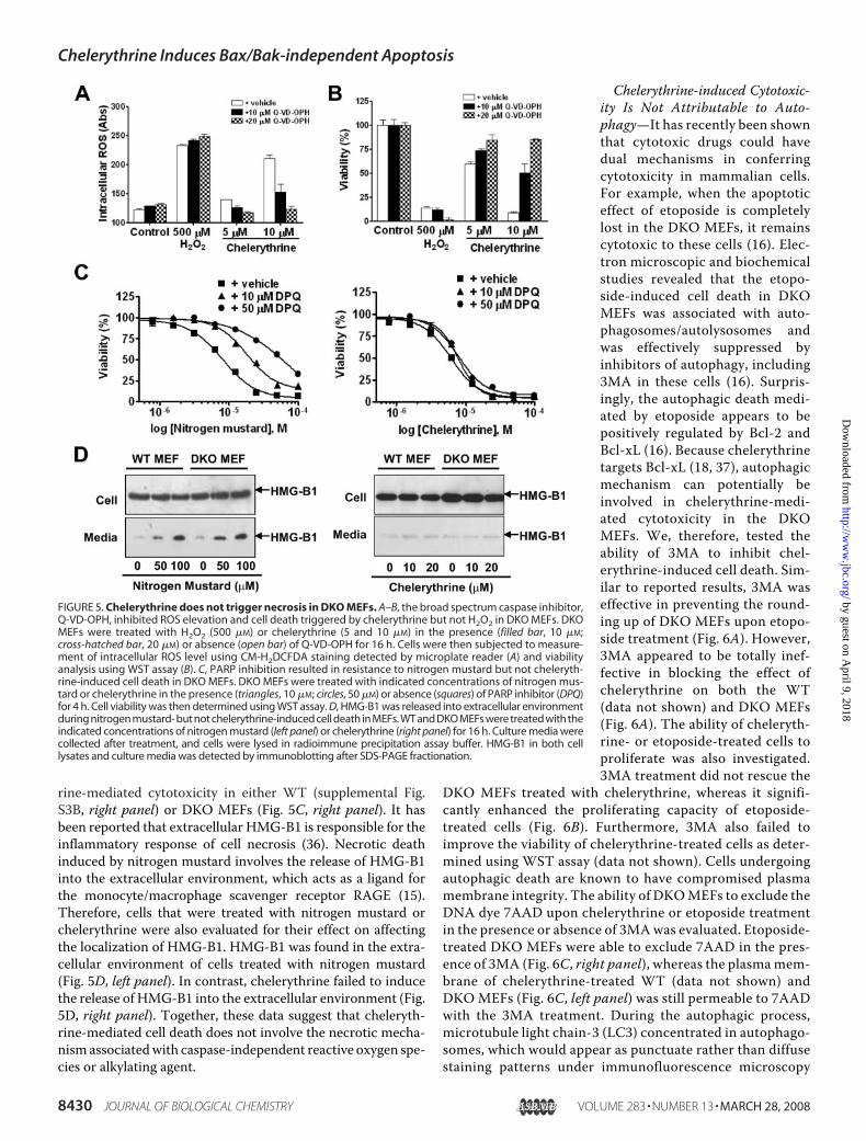
rine-mediated cytotoxicity in either WT (supplemental Fig.S3B, right panel) or DKO MEFs (Fig. 5C, right panel). It hasbeen reported that extracellular HMG-B1 is responsible for theinflammatory response of cell necrosis (36). Necrotic deathinduced by nitrogen mustard involves the release of HMG-B1into the extracellular environment, which acts as a ligand forthe monocyte/macrophage scavenger receptor RAGE (15).Therefore, cells that were treated with nitrogen mustard orchelerythrine were also evaluated for their effect on affectingthe localization of HMG-B1. HMG-B1 was found in the extra-cellular environment of cells treated with nitrogen mustard(Fig. 5D, left panel). In contrast, chelerythrine failed to inducethe release of HMG-B1 into the extracellular environment (Fig.5D, right panel). Together, these data suggest that cheleryth-rine-mediated cell death does not involve the necrotic mecha-nismassociatedwith caspase-independent reactive oxygen spe-cies or alkylating agent.
Chelerythrine-induced Cytotoxic-ity Is Not Attributable to Auto-phagy—It has recently been shownthat cytotoxic drugs could havedual mechanisms in conferringcytotoxicity in mammalian cells.For example, when the apoptoticeffect of etoposide is completelylost in the DKO MEFs, it remainscytotoxic to these cells (16). Elec-tron microscopic and biochemicalstudies revealed that the etopo-side-induced cell death in DKOMEFs was associated with auto-phagosomes/autolysosomes andwas effectively suppressed byinhibitors of autophagy, including3MA in these cells (16). Surpris-ingly, the autophagic death medi-ated by etoposide appears to bepositively regulated by Bcl-2 andBcl-xL (16). Because chelerythrinetargets Bcl-xL (18, 37), autophagicmechanism can potentially beinvolved in chelerythrine-medi-ated cytotoxicity in the DKOMEFs. We, therefore, tested theability of 3MA to inhibit chel-erythrine-induced cell death. Sim-ilar to reported results, 3MA waseffective in preventing the round-ing up of DKO MEFs upon etopo-side treatment (Fig. 6A). However,3MA appeared to be totally inef-fective in blocking the effect ofchelerythrine on both the WT(data not shown) and DKO MEFs(Fig. 6A). The ability of cheleryth-rine- or etoposide-treated cells toproliferate was also investigated.3MA treatment did not rescue the
DKO MEFs treated with chelerythrine, whereas it signifi-cantly enhanced the proliferating capacity of etoposide-treated cells (Fig. 6B). Furthermore, 3MA also failed toimprove the viability of chelerythrine-treated cells as deter-mined using WST assay (data not shown). Cells undergoingautophagic death are known to have compromised plasmamembrane integrity. The ability of DKOMEFs to exclude theDNA dye 7AAD upon chelerythrine or etoposide treatmentin the presence or absence of 3MAwas evaluated. Etoposide-treated DKO MEFs were able to exclude 7AAD in the pres-ence of 3MA (Fig. 6C, right panel), whereas the plasmamem-brane of chelerythrine-treated WT (data not shown) andDKOMEFs (Fig. 6C, left panel) was still permeable to 7AADwith the 3MA treatment. During the autophagic process,microtubule light chain-3 (LC3) concentrated in autophago-somes, which would appear as punctuate rather than diffusestaining patterns under immunofluorescence microscopy
FIGURE 5. Chelerythrine does not trigger necrosis in DKO MEFs. A–B, the broad spectrum caspase inhibitor,Q-VD-OPH, inhibited ROS elevation and cell death triggered by chelerythrine but not H2O2 in DKO MEFs. DKOMEFs were treated with H2O2 (500 �M) or chelerythrine (5 and 10 �M) in the presence (filled bar, 10 �M;cross-hatched bar, 20 �M) or absence (open bar) of Q-VD-OPH for 16 h. Cells were then subjected to measure-ment of intracellular ROS level using CM-H2DCFDA staining detected by microplate reader (A) and viabilityanalysis using WST assay (B). C, PARP inhibition resulted in resistance to nitrogen mustard but not cheleryth-rine-induced cell death in DKO MEFs. DKO MEFs were treated with indicated concentrations of nitrogen mus-tard or chelerythrine in the presence (triangles, 10 �M; circles, 50 �M) or absence (squares) of PARP inhibitor (DPQ)for 4 h. Cell viability was then determined using WST assay. D, HMG-B1 was released into extracellular environmentduring nitrogen mustard- but not chelerythrine-induced cell death in MEFs. WT and DKO MEFs were treated with theindicated concentrations of nitrogen mustard (left panel) or chelerythrine (right panel) for 16 h. Culture media werecollected after treatment, and cells were lysed in radioimmune precipitation assay buffer. HMG-B1 in both celllysates and culture media was detected by immunoblotting after SDS-PAGE fractionation.
Chelerythrine Induces Bax/Bak-independent Apoptosis
8430 JOURNAL OF BIOLOGICAL CHEMISTRY VOLUME 283 • NUMBER 13 • MARCH 28, 2008
by guest on April 9, 2018
http://ww
w.jbc.org/
Dow
nloaded from

(38). Diffuse cytoplasmic localization of transiently ex-pressed LC3 was observed in healthy and chelerythrine-treated DKO MEFs, whereas etoposide- and staurosporine-treated DKOMEFs showed punctuate fluorescence staining ofLC3 under immunofluorescence microscopy (Fig. 6D). Etopo-side- and staurosporine-induced aggregation of LC3was inhib-ited by 3MA. In contrast, 3MA had no effect on LC3 localiza-tion either in healthy or chelerythrine-treated DKOMEFs (Fig.6D). In addition, 3MA was also unable to block chelerythrine-induced mitochondrial membrane potential change (supple-mental Fig. S4). Together, these data reveal no evidence thatchelerythrine can mediate autophagic death in either the WTor DKOMEFs.
DISCUSSION
Chelerythrine was identified as Bcl-xL inhibitor from a highthroughput screen and was subsequently shown that it caninteract with the BH groove instead of the classic BH3 binding
cleft when many other chemicalinhibitors of Bcl-2/Bcl-xL areknown to bind. Chelerythrine canact on isolated mitochondria fromtumor cells for triggering the directrelease of cyt c (18). In this study wecharacterized and compared thecytotoxic mechanism of cheleryth-rine in bothWT and DKOMEFs. Incontrast to other established chem-ical apoptotic stimuli, chelerythrineappears to induce classic apoptoticphenotypes in both cell types withsimilar kinetics and efficacy.Although chelerythrine was capableof inducing Bax integration in WTMEFs, this event occurred only aftera long delay (�10 h after cyt crelease), suggesting that Bax activa-tion in theWTMEFs may occur viapositive feedback mechanism trig-gered by downstream apoptosisevents. Furthermore, the kinetics ofcyt c release frommitochondria iso-lated from both WT and DKOMEFs were almost identical, sug-gesting that Bax activation may notbe a major contributing factor inmediating the apoptotic phenotypestriggered by chelerythrine. Indeed,the rapid release of cyt c that pre-ceded Bax activation and the inhib-itory activity of CsA toward mito-chondrial swelling strongly suggestthat chelerythrine-induced apopto-sis in MEFs is predominantly due tothe CsA-sensitive mPTP openinginstead of the Bax/Bak-mediatedpermeabilization mechanism.Because chelerythrine is able to
interact with Bcl-xL and acts directly onmitochondria (18), it istempting to speculate that the effect of chelerythrine on themPTP is related to Bcl-2/Bcl-xL inhibition. Although themolecular mechanism by which Bcl-2/Bcl-xL inhibits mPTP isstill controversial, several independent evidence have emergeddemonstrating that these proteins can interact with sessilemitochondrial proteins, including adenine nucleotide translo-cator (39) and voltage-dependent anion channel (40). In vitro,overexpression of Bcl-2 in cells or the addition of Bcl-2 to iso-lated mitochondria suppressed the mPTP induced by a varietyof apoptotic insults (39, 41, 42). Therefore, it is plausible thatthe inhibition of Bcl-2/Bcl-xL by chelerythrine might result inthe sensitization of themitochondrialmembranes permeabilityvia mPTP components, release of cyt c, and functional collapseof the organelle and apoptosis. However, alternative mecha-nisms such as activation of other unidentified mitochondrialtargets that are dependent onmPTP components by cheleryth-rine are certainly possible and remain to be explored.
FIGURE 6. Autophagic mechanism does not contribute significantly to the cytotoxic effect of cheleryth-rine in DKO MEFs. A–C, 3MA blocked cell death triggered by etoposide but not chelerythrine in DKO MEFs.A, DKO MEFs were treated with 10 �M etoposide or 20 �M chelerythrine in the absence (�3MA) or presence(3MA) of 10 mM 3MA for 48 h and then examined by phase-contrast microscopy. B and C, DKO MEFs weretreated with indicated concentrations of chelerythrine (circle symbols) or etoposide (square symbols) in theabsence (open symbols) or presence (closed symbols) of 3MA. Cell viability was measured by a cell proliferationassay after re-seeding the cells for the indicated days followed by WST assay (B) and 7AAD exclusion staining(C); cell death was expressed as percentage of 7AAD positive cells. Data shown are the mean � S.D. (n � 3).D, chelerythrine did not induce punctuate distribution of LC3 in DKO MEFs. MEFs transiently transfected withpXJ-myc-LC3 were treated with chelerythrine (20 �M), etoposide (10 �M), or staurosporine (1 �M) for 16 h, andcells were fixed and permeabilized for immunofluorescence confocal microscopy using anti-Myc epitopeantibody.
Chelerythrine Induces Bax/Bak-independent Apoptosis
MARCH 28, 2008 • VOLUME 283 • NUMBER 13 JOURNAL OF BIOLOGICAL CHEMISTRY 8431
by guest on April 9, 2018
http://ww
w.jbc.org/
Dow
nloaded from

Although we were investigating the Bax- and Bak-independ-ent apoptotic mechanism triggered by chelerythrine, two stud-ieswere published to suggest that two distinct apoptotic insults,such as gossypol and A23187/arachidonic acid, are capable ofinducing apoptosis in the DKOMEFs. In contrast to cheleryth-rine, the apoptotic effects of gossypol (supplemental Fig. S5A)and the A23187/arachidonic acid (43) are significantly weakerin the DKO than the WT MEFs, suggesting that the apoptoticeffects emulated from these stimuli may in part still be depend-ent on the Bax/Bak gateway. Furthermore, our data showedthat neither gossypol (supplemental Fig. S5, B and C) norA23187/arachidonic acid (43) was effective in triggering cyt crelease and swelling in isolated mitochondrial preparations,suggesting that unlike chelerythrine, these compounds do notact directly onmitochondria tomediate their apoptotic effect inMEFs. In fact, our results are in disagreement with a previousreport showing that gossypol was able to induce cyt c releasefrommitochondria isolated fromDKOMEFs (44). In the studyby Lei et al. (44),mitochondriawere incubated for 1 h in a bufferdevoid of calcium chelator EGTA, whereas our experimentswere performed for 30 min in a buffer containing EGTA. Thismay explain why their mitochondrial preparation was moresensitive to gossypol, as it has been documented thatmitochon-dria have a tendency to release spontaneously high levels of cytc in the absence of EGTA (45). The mechanistic basis for themitochondrial effects of these drugs also appears to be differentas gossypol- andA23187/arachidonic acid-induced cyt c releaseare insensitive to CsA and independent of mPTP opening (43,44), whereas the mitochondrial event induced by chelerythrineis mediated by CsA-sensitive mPTP.Recently, the ability of several chemical inhibitors of Bcl-2/
Bcl-xL (ABT-737, BH3I-1, HA14-1, antimycin A, gossypol, andchelerythrine) to induce cell death in the DKO MEFs has alsobeen investigated (46). Except ABT-737, these inhibitorsappeared cytotoxic to DKO MEFs. However, the nature of thecytotoxic mechanisms associated with each of these inhibitorsin DKO MEFs was not known as detailed phenotypic, andmolecular characterizations were not performed. It was con-cluded from that study that onlyABT-737 qualifies as a genuinechemical BH3mimetic as it behaves just like BH3-only proteinsin that they fail to induce apoptosis in the absence of Bax andBak. Surprisingly, our in-depth characterization and compari-son of cytotoxic mechanism of chelerythrine in WT and DKOMEFs provides strong and comprehensive evidence to suggestthat chelerythrine is an apoptotic agent that acts through Bax/Bak-independent mitochondrial mechanism. As revealed byNMR and modeling analysis, chelerythrine can induce a globalchange in the Bcl-xL protein structure through direct interac-tion with the BH groove (37) instead of the classic BH3 domainbinding cleft where BH3 peptides and ABT-737 are shown todock (26). This finding would suggest that chelerythrine mayconfer its inhibitory action on Bcl-xL by directly or indirectlycompeting with unknown regulatory proteins. It would be ofinterest to investigate further on the structure activity relation-ship of chelerythrine-related compounds on their apoptoticeffect in the DKO MEFs versus their binding and inhibitoryactivities on pro-survival members of Bcl-2 family to gain fur-ther insight on whether the Bax/Bak-independent pro-apopto-
tic mechanism is dependent on direct interaction with the pro-survival molecules.Chelerythrine has recently been shown to display a biphasic
effect on mitochondrial respiration with energy uncoupling atlow concentration and respiration inhibition at high concentra-tion (47). Based on this study, chelerythrine-induced apoptosisin MEFs might just be a simple consequence of mitochondrialtoxicity. Interestingly, in contrast to chelerythrine, respiratorycomplex IV inhibitor (sodium azide) (48) and uncoupler ofmitochondrial oxidation (carbonyl cyanide p-trifluorome-thoxyphenylazone) (49) that directly interfere energy couplingwere cytotoxic to WT but not the DKO MEFs (supplementalFig. S6). These data would serve to argue that themainmode ofaction of chelerythrine in causing cytotoxicity in MEFs isunlikely due to its effect on mitochondrial respiration.It was suggested that Bax and Bak might share a redundant
function that suppresses tumorigenicity through the independ-ent ability of either of these proteins to initiate apoptosis inresponse to oncogenic transformation (9). Using transformedprimary baby kidney epithelial cells, Degenhardt et al. (50) alsoshowed that Bax and Bak function to suppress tumorigenesis,and their deficiency resulting from genemutation was found tobe prevalent in tumor cells detected in metastatic sites in amouse tumormodel. Therefore, designing specific strategies toovercome survival of the tumor cells in the absence of Bax andBak function may provide novel opportunities in anticancerdrug discovery. Indeed, it has been shown that chelerythrineinduces tumor growth delays in vivo and appears to have amin-imum adverse effect in mice (51). In this respect chelerythrinemay have the potential to serve as a lead compound to supportexploration of the Bax/Bak-independent mechanism as amolecular target for anticancer drug development.
Acknowledgments—We are grateful to the late Dr. Stanley Korsmeyerfor providing the DKO MEFs. We thank NaiYang Fu for valuablecomments on the manuscript.
REFERENCES1. Wang, X. (2001) Genes Dev. 15, 2922–29332. Chan, S. L., and Yu, V. C. (2004) Clin. Exp. Pharmacol. Physiol. 31,
119–1283. Strasser, A. (2005) Nat. Rev. Immunol. 5, 189–2004. Danial, N. N., and Korsmeyer, S. J. (2004) Cell 116, 205–2195. Willis, S. N., and Adams, J. M. (2005) Curr. Opin. Cell Biol. 17, 617–6256. Bouillet, P., and Strasser, A. (2002) J. Cell Sci. 115, 1567–15747. Willis, S. N., Fletcher, J. I., Kaufmann, T., van Delft, M. F., Chen, L., Cz-
abotar, P. E., Ierino, H., Lee, E. F., Fairlie, W. D., Bouillet, P., Strasser, A.,Kluck, R.M., Adams, J.M., andHuang, D. C. (2007) Science 315, 856–859
8. Cheng, E.H.,Wei,M.C.,Weiler, S., Flavell, R. A.,Mak, T.W., Lindsten, T.,and Korsmeyer, S. J. (2001)Mol. Cell 8, 705–711
9. Zong, W. X., Lindsten, T., Ross, A. J., MacGregor, G. R., and Thompson,C. B. (2001) Genes Dev. 15, 1481–1486
10. Letai, A., Bassik, M. C., Walensky, L. D., Sorcinelli, M. D., Weiler, S., andKorsmeyer, S. J. (2002) Cancer Cell 2, 183–192
11. Reed, J. C. (1998) Oncogene 17, 3225–323612. Kim,H., Rafiuddin-Shah,M., Tu,H. C., Jeffers, J. R., Zambetti, G. P., Hsieh,
J. J., and Cheng, E. H. (2006) Nat. Cell Biol. 8, 1348–135813. Wei,M.C., Zong,W.X., Cheng, E.H., Lindsten, T., Panoutsakopoulou, V.,
Ross, A. J., Roth, K. A., MacGregor, G. R., Thompson, C. B., and Kors-meyer, S. J. (2001) Science 292, 727–730
14. Lindsten, T., Ross, A. J., King, A., Zong,W. X., Rathmell, J. C., Shiels, H. A.,
Chelerythrine Induces Bax/Bak-independent Apoptosis
8432 JOURNAL OF BIOLOGICAL CHEMISTRY VOLUME 283 • NUMBER 13 • MARCH 28, 2008
by guest on April 9, 2018
http://ww
w.jbc.org/
Dow
nloaded from
Ulrich, E., Waymire, K. G., Mahar, P., Frauwirth, K., Chen, Y., Wei, M.,Eng, V. M., Adelman, D. M., Simon, M. C., Ma, A., Golden, J. A., Evan, G.,Korsmeyer, S. J., MacGregor, G. R., and Thompson, C. B. (2000)Mol. Cell6, 1389–1399
15. Zong, W. X., Ditsworth, D., Bauer, D. E., Wang, Z. Q., and Thompson,C. B. (2004) Genes Dev. 18, 1272–1282
16. Shimizu, S., Kanaseki, T.,Mizushima, N.,Mizuta, T., Arakawa-Kobayashi,S., Thompson, C. B., andTsujimoto, Y. (2004)Nat. Cell Biol. 6, 1221–1228
17. Malikova, J., Zdarilova, A., Hlobilkova, A., and Ulrichova, J. (2006) CellBiol. Toxicol. 22, 439–453
18. Chan, S. L., Lee, M. C., Tan, K. O., Yang, L. K., Lee, A. S., Flotow, H., Fu,N. Y., Butler, M. S., Soejarto, D. D., Buss, A. D., and Yu, V. C. (2003) J. Biol.Chem. 278, 20453–20456
19. Wilson-Annan, J., O’Reilly, L. A., Crawford, S. A., Hausmann, G., Beau-mont, J. G., Parma, L. P., Chen, L., Lackmann, M., Lithgow, T., Hinds,M. G., Day, C. L., Adams, J. M., and Huang, D. C. (2003) J. Cell Biol. 162,877–887
20. Tan, K. O., Fu, N. Y., Sukumaran, S. K., Chan, S. L., Kang, J. H., Poon, K. L.,Chen, B. S., and Yu, V. C. (2005) Proc. Natl. Acad. Sci. U. S. A. 102,14623–14628
21. Galindo, M. F., Jordan, J., Gonzalez-Garcia, C., and Cena, V. (2003) Br. J.Pharmacol. 139, 797–804
22. Tzung, S. P., Kim, K.M., Basanez, G., Giedt, C. D., Simon, J., Zimmerberg,J., Zhang, K. Y., and Hockenbery, D. M. (2001) Nat. Cell Biol. 3, 183–191
23. Kroll, S. L., and Czyzyk-Krzeska, M. F. (1998) Am. J. Physiol. 274,C167–C174
24. Srinivasula, S. M., Hegde, R., Saleh, A., Datta, P., Shiozaki, E., Chai, J., Lee,R. A., Robbins, P. D., Fernandes-Alnemri, T., Shi, Y., and Alnemri, E. S.(2001) Nature 410, 112–116
25. Oliver, C. L.,Miranda,M. B., Shangary, S., Land, S.,Wang, S., and Johnson,D. E. (2005)Mol. Cancer Ther. 4, 23–31
26. Oltersdorf, T., Elmore, S. W., Shoemaker, A. R., Armstrong, R. C., Augeri,D. J., Belli, B. A., Bruncko, M., Deckwerth, T. L., Dinges, J., Hajduk, P. J.,Joseph, M. K., Kitada, S., Korsmeyer, S. J., Kunzer, A. R., Letai, A., Li, C.,Mitten, M. J., Nettesheim, D. G., Ng, S., Nimmer, P. M., O’connor, J. M.,Oleksijew, A., Petros, A.M., Reed, J. C., Shen,W., Tahir, S. K., Thompson,C. B., Tomaselli, K. J.,Wang, B.,Wendt,M. D., Zhang, H., Fesik, S.W., andRosenberg, S. H. (2005) Nature 435, 677–681
27. Lickliter, J. D., Wood, N. J., Johnson, L., McHugh, G., Tan, J., Wood, F.,Cox, J., and Wickham, N. W. (2003) Leukemia 17, 2074–2080
28. Kitada, S., Leone, M., Sareth, S., Zhai, D., Reed, J. C., and Pellecchia, M.(2003) J. Med. Chem. 46, 4259–4264
29. Real, P. J., Cao, Y.,Wang, R., Nikolovska-Coleska, Z., Sanz-Ortiz, J.,Wang,S., and Fernandez-Luna, J. L. (2004) Cancer Res. 64, 7947–7953
30. Zhai, D., Jin, C., Satterthwait, A. C., andReed, J. C. (2006)Cell DeathDiffer.13, 1419–1421
31. Chao, D. T., andKorsmeyer, S. J. (1998)Annu. Rev. Immunol. 16, 395–41932. Crompton, M., Virji, S., Doyle, V., Johnson, N., and Ward, J. M. (1999)
Biochem. Soc. Symp. 66, 167–179
33. Kuwana, T., Bouchier-Hayes, L., Chipuk, J. E., Bonzon, C., Sullivan, B. A.,Green, D. R., and Newmeyer, D. D. (2005)Mol. Cell 17, 525–535
34. Nakagawa, T., Shimizu, S., Watanabe, T., Yamaguchi, O., Otsu, K., Yama-gata, H., Inohara, H., Kubo, T., and Tsujimoto, Y. (2005) Nature 434,652–658
35. Baines, C. P., Kaiser, R. A., Purcell, N. H., Blair, N. S., Osinska, H., Hamble-ton, M. A., Brunskill, E. W., Sayen, M. R., Gottlieb, R. A., Dorn, G. W.,Robbins, J., and Molkentin, J. D. (2005) Nature 434, 658–662
36. Rovere-Querini, P., Capobianco, A., Scaffidi, P., Valentinis, B., Catalanotti,F., Giazzon, M., Dumitriu, I. E., Muller, S., Iannacone, M., Traversari, C.,Bianchi, M. E., and Manfredi, A. A. (2004) EMBO Rep. 5, 825–830
37. Zhang, Y. H., Bhunia, A., Wan, K. F., Lee, M. C., Chan, S. L., Yu, V. C., andMok, Y. K. (2006) J. Mol. Biol. 364, 536–549
38. Kabeya, Y., Mizushima, N., Ueno, T., Yamamoto, A., Kirisako, T., Noda,T., Kominami, E., Ohsumi, Y., and Yoshimori, T. (2000) EMBO J. 19,5720–5728
39. Marzo, I., Brenner, C., Zamzami, N., Susin, S. A., Beutner, G., Brdiczka, D.,Remy, R., Xie, Z. H., Reed, J. C., and Kroemer, G. (1998) J. Exp. Med. 187,1261–1271
40. Shimizu, S., Konishi, A., Kodama, T., and Tsujimoto, Y. (2000) Proc. Natl.Acad. Sci. U. S. A. 97, 3100–3105
41. Susin, S. A., Zamzami, N., Castedo, M., Daugas, E., Wang, H. G., Geley, S.,Fassy, F., Reed, J. C., and Kroemer, G. (1997) J. Exp. Med. 186, 25–37
42. Shimizu, S., Eguchi, Y., Kamiike, W., Funahashi, Y., Mignon, A., La-cronique, V., Matsuda, H., and Tsujimoto, Y. (1998) Proc. Natl. Acad. Sci.U. S. A. 95, 1455–1459
43. Mizuta, T., Shimizu, S., Matsuoka, Y., Nakagawa, T., and Tsujimoto, Y.(2007) J. Biol. Chem. 282, 16623–16630
44. Lei, X., Chen, Y., Du, G., Yu,W.,Wang, X., Qu, H., Xia, B., He, H., Mao, J.,Zong, W., Liao, X., Mehrpour, M., Hao, X., and Chen, Q. (2006) FASEB J.20, 2147–2149
45. Eskes, R., Antonsson, B., Osen-Sand, A., Montessuit, S., Richter, C., Sa-doul, R., Mazzei, G., Nichols, A., and Martinou, J. C. (1998) J. Cell Biol.143, 217–224
46. van Delft, M. F., Wei, A. H., Mason, K. D., Vandenberg, C. J., Chen, L.,Czabotar, P. E., Willis, S. N., Scott, C. L., Day, C. L., Cory, S., Adams, J. M.,Roberts, A. W., and Huang, D. C. (2006) Cancer Cell 10, 389–399
47. Milanesi, E., Costantini, P., Gambalunga, A., Colonna, R., Petronilli, V.,Cabrelle, A., Semenzato, G., Cesura, A. M., Pinard, E., and Bernardi, P.(2006) J. Biol. Chem. 281, 10066–10072
48. Wang, J.,Wei,Q.,Wang, C. Y., Hill,W.D.,Hess, D. C., andDong, Z. (2004)J. Biol. Chem. 279, 19948–19954
49. Armstrong, J. S., Steinauer, K. K., French, J., Killoran, P. L., Walleczek, J.,Kochanski, J., and Knox, S. J. (2001) Exp. Cell Res. 262, 170–179
50. Degenhardt, K., Chen,G., Lindsten, T., andWhite, E. (2002)Cancer Cell 2,193–203
51. Chmura, S. J., Dolan, M. E., Cha, A., Mauceri, H. J., Kufe, D. W., andWeichselbaum, R. R. (2000) Clin. Cancer Res. 6, 737–742
Chelerythrine Induces Bax/Bak-independent Apoptosis
MARCH 28, 2008 • VOLUME 283 • NUMBER 13 JOURNAL OF BIOLOGICAL CHEMISTRY 8433
by guest on April 9, 2018
http://ww
w.jbc.org/
Dow
nloaded from

YuKah Fei Wan, Shing-Leng Chan, Sunil Kumar Sukumaran, Mei-Chin Lee and Victor C.
MechanismChelerythrine Induces Apoptosis through a Bax/Bak-independent Mitochondrial
doi: 10.1074/jbc.M707687200 originally published online January 29, 20082008, 283:8423-8433.J. Biol. Chem.
10.1074/jbc.M707687200Access the most updated version of this article at doi:
Alerts:
When a correction for this article is posted•
When this article is cited•
to choose from all of JBC's e-mail alertsClick here
Supplemental material:
http://www.jbc.org/content/suppl/2008/01/29/M707687200.DC1
http://www.jbc.org/content/283/13/8423.full.html#ref-list-1
This article cites 51 references, 23 of which can be accessed free at
by guest on April 9, 2018
http://ww
w.jbc.org/
Dow
nloaded from