prima-1 induces apoptosis in bladder cancer cell … induces apoptosis in bladder cancer cell ... 1...
TRANSCRIPT
Prima-1 induces apoptosis in bladder cancer cell linesby activating p53Camila B. Piantino, Sabrina T. Reis, Nayara I. Viana, Iran A. Silva, Denis R. Morais, Alberto A. Antunes,
Nelson Dip, Miguel Srougi, Katia R. Leite
Faculdade de Medicina da Universidade de Sao Paulo, Laboratory of Medical Investigation, Urology Department – LIM55, Sao Paulo/SP, Brazil.
OBJECTIVES: Bladder cancer represents 3% of all carcinomas in the Brazilian population and ranks second inincidence among urological tumors, after prostate cancer. The loss of p53 function is the main geneticalteration related to the development of high-grade muscle-invasive disease. Prima-1 is a small molecule thatrestores tumor suppressor function to mutant p53 and induces cancer cell death in various cancer types. Ouraim was to investigate the ability of Prima-1 to induce apoptosis after DNA damage in bladder cancer cell lines.
METHOD: The therapeutic effect of Prima-1 was studied in two bladder cancer cell lines: T24, which ischaracterized by a p53 mutation, and RT4, which is the wild-type for the p53 gene. Morphological features ofapoptosis induced by p53, including mitochondrial membrane potential changes and the expression of thirteengenes involved in apoptosis, were assessed by microscopic observation and quantitative real-time PCR (qRT-PCR).
RESULTS: Prima-1 was able to reactivate p53 function in the T24 (p53 mt) bladder cancer cell line and promoteapoptosis via the induction of Bax and Puma expression, activation of the caspase cascade and disruption of themitochondrial membrane in a BAK-independent manner.
CONCLUSION: Prima-1 is able to restore the transcriptional activity of p53. Experimental studies in vivo may beconducted to test this molecule as a new therapeutic agent for urothelial carcinomas of the bladder, whichcharacteristically harbor p53 mutations.
KEYWORDS: Bladder cancer; p53; Apoptosis; Prima-1.
Piantino CB, Reis ST, Viana NI, Silva IA, Morais DR, Antunes AA, et al. Prima-1 induces apoptosis in bladder cancer cell lines by activatingp53. Clinics. 2013;68(3):297-303.
Received for publication on August 20, 2012; First review completed on October 3, 2012; Accepted for publication on November 4, 2012
E-mail: [email protected]
Tel.: 55 11 3061-7183
& INTRODUCTION
Bladder cancer (BC) represents 3% of all carcinomas in theBrazilian male population and ranks second in incidenceamong urological tumors, after prostate cancer (1), with73,310 new cases and 14,880 deaths estimated for 2012 in theUSA (2).
Tobacco smoking, occupational exposure to aromaticamines, arsenic, radiation exposure from the radiationtherapy of neighboring organs and the therapeutic use ofalkylating agents are the main risk factors for the develop-ment of urothelial tumors (3).
A large number of genetic changes have been associatedwith the genesis and progression of BC, and many of themappear to abrogate the G1/S cell cycle checkpoint (4). In
particular, p53 loss of function has been related to thedevelopment of high-grade muscle-invasive disease (5).
As a key gatekeeper of the G1/S checkpoint for cell-cycleprogression, p53 is indispensable for maintaining genomicstability and keeping urothelial growth in check. Structuraland functional p53 defects are found in over one-half ofhuman urothelial carcinomas, contributing to genomicinstability and consequently, numerous chromosomal aber-rations. p53 mutations first affect one allele, followed by asecond that may be a mutation, deletion or silencing bymethylation of the additional wild-type allele, whichdisables the function of this tumor suppressor gene (6).Consistent with this action, the expression of p21 (WAF1),an important p53 downstream target, is downregulated inthe majority of urothelial carcinomas that harbor p53mutations (7). Several codons in the p53 gene appear to bepreferentially mutated, including codons 280 and 285 (8).The specific mutation of these codons is rare in otherepithelial tumors; thus, it has been suggested that theyresult from a urothelium-specific carcinogen or carcinogenicevent.
Prima-1 (p53 reactivation and induction of massiveapoptosis 1) is a low–molecular weight compound identified
Copyright � 2013 CLINICS – This is an Open Access article distributed underthe terms of the Creative Commons Attribution Non-Commercial License (http://creativecommons.org/licenses/by-nc/3.0/) which permits unrestricted non-commercial use, distribution, and reproduction in any medium, provided theoriginal work is properly cited.
No potential conflict of interest was reported.
DOI: 10.6061/clinics/2013(03)OA03
CLINICAL SCIENCE
297
in a cell-based chemical library screen at the National CancerInstitute (National Institutes of Health, Bethesda, MD). Thiscompound has been demonstrated to suppress the growth ofcells that harbor mutant p53 by restoring the sequence-specific DNA-binding, wild-type conformation and tran-scriptional transactivation of mutant p53. Prima-1 has beenshown to induce apoptosis in human tumor cells in a p53-dependent manner and suppress the growth of human tumorxenografts carrying mutant p53 (9).
In this study, we examined the effects of Prima-1 on theT24 and RT4 BC cell lines, which express a mutant and wild-type p53 gene, respectively, and analyzed whether thiscompound was able to induce p53 activity after UVCdamage. To test this hypothesis, we evaluated whetherexposing p53 wild-type (RT4) and p53 mutant (T24) cells toPrima-1 and ultraviolet radiation (UVC) alters the expres-sion of p53 downstream genes that are related to apoptosisinduction.
& MATERIALS AND METHODS
Cell Lines and TreatmentThe human urothelial carcinoma T24 (p53 mt) cell line
was kindly provided by Dr. Salvadori (Botucatu, Brazil).RT4, a primary urothelial carcinoma bladder cell line from aCaucasian (p53 wt) was purchased from EACC (Salisbury,UK). The cell lines were cultured in McCoy’s mediumsupplemented with 20% (v/v) heat-inactivated fetal bovineserum (Sigma, St.Louis, MO, USA) under an atmosphere of5% CO2 at 37 C. For the experiments, T24 and RT4 cells wereseeded in 25 cm2 tissue culture flasks and maintained at37 C/5% CO2 overnight to allow the cells to adhere. On thenext day, the cells were exposed to 253.7 nm of UVC for 2 hto induce DNA damage. UVC radiation causes specificDNA lesions, and in addition to being a powerful tool forscientific studies, it is a methodology that is easilyimplemented in the laboratory because the majority ofgermicidal lamps emit UVC light (10). After exposure, thecells were treated with 50 mM Prima-1 (Sigma, St.Louis, MO,USA) for 1 and 192 h.
RNA Isolation and cDNA SynthesisFollowing the above experiment, total RNA was isolated with
the mirVanaTM Kit (Applied Biosystems, Foster City, CA, USA)according to the manufacturer’s instructions. As a control, weused T24 and RT4 cell lines cultured in McCoy’s mediumsupplemented with 20% (v/v) heat-inactivated fetal bovine
serum (Sigma, St. Louis, MO, USA) in a 5% CO2 atmosphere at37 C. RNA concentrations were determined by measuring theabsorbance at 260/280 nm using a Nanodrop ND-1000spectrophotometer (Thermo Scientific, Wilmington, Delaware,USA). cDNA was generated using the High Capacity cDNAReverse Transcription Kit (Applied Biosystems, Foster City, CA,USA). Reactions were incubated at 25 C for 10 min, followed by37 C for 120 min and 85 C for 5 min. The cDNA was stored at220 C until use.
Quantitative Real-Time PCR (qRT-PCR) and GeneExpression
The expression level of 13 genes (Table 1) was analyzedby qRT-PCR using an ABI 7500 Fast Real-Time PCR System(Applied Biosystems). Target sequences were amplified in a10 ml reaction containing 5 ml of TaqMan Universal PCRMaster Mix, 0.5 ml of a TaqMan Gene Expression Assay(Applied Biosystems, Foster City, CA, USA) (primers andprobes, see Table I), 1 ml of cDNA and 3.5 ml of DNase-freewater. The PCR cycling conditions were as follows: 2 min at50 C, 10 min at 95 C and 40 cycles of 15 seconds at 95 C and1 min at 60 C. A TaqMan B2M (Applied Biosystems, FosterCity, CA, USA) (b2 microglobulin) Assay was used as anendogenous control.
We used the DDCT method to calculate the relativeexpression of the 13 target genes using the followingformula: DDCT = (CT of the target gene in T24 and RT4cells after 1, 18 and 192 h after exposure to 50 mM Prima-1 –the CT of the endogenous control under the same condi-tions) – (CT of the target gene in T24 and RT4 cells withoutPrima-1 treatment - CT of the endogenous control under thesame conditions). The gene expression fold change wascalculated as 2-DDCT.
Evaluation of the mitochondrial membranepotential by DePsipher
The DePsipher Kit (Trevigen, Gaithersburg, MD, USA)was used to measure the mitochondrial membrane potentialunder a fluorescence microscope (11,12). T24 and RT4 cells(26105/ml) were seeded in a 24 well plate 24 h prior to theexperiments. Then, the plate was exposed to 253.7 nm ofUVC for 2 h for the induction of DNA damage. Prima-1(50 mM) was added, and 18 h later, the cells were washedwith PBS and detached from the cell culture wells withtrypsin. As a control, we used T24 and RT4 cell linesexposed to UVC for 2 h in the absence of treatment. The
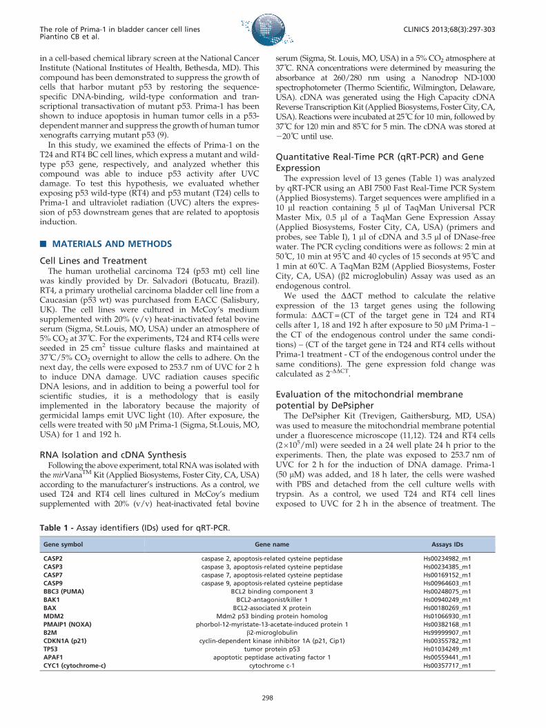
Table 1 - Assay identifiers (IDs) used for qRT-PCR.
Gene symbol Gene name Assays IDs
CASP2 caspase 2, apoptosis-related cysteine peptidase Hs00234982_m1
CASP3 caspase 3, apoptosis-related cysteine peptidase Hs00234385_m1
CASP7 caspase 7, apoptosis-related cysteine peptidase Hs00169152_m1
CASP9 caspase 9, apoptosis-related cysteine peptidase Hs00964603_m1
BBC3 (PUMA) BCL2 binding component 3 Hs00248075_m1
BAK1 BCL2-antagonist/killer 1 Hs00940249_m1
BAX BCL2-associated X protein Hs00180269_m1
MDM2 Mdm2 p53 binding protein homolog Hs01066930_m1
PMAIP1 (NOXA) phorbol-12-myristate-13-acetate-induced protein 1 Hs00382168_m1
B2M b2-microglobulin Hs99999907_m1
CDKN1A (p21) cyclin-dependent kinase inhibitor 1A (p21, Cip1) Hs00355782_m1
TP53 tumor protein p53 Hs01034249_m1
APAF1 apoptotic peptidase activating factor 1 Hs00559441_m1
CYC1 (cytochrome-c) cytochrome c-1 Hs00357717_m1
The role of Prima-1 in bladder cancer cell linesPiantino CB et al.
CLINICS 2013;68(3):297-303
298
cells were incubated in the dark with 5 mg/ml DePsipher(5.59.6.69-tetrachloro-1.19.3.39-tetraethyl-benzimidazolyl car-bocyanin iodide) solution for 30 min at 37 C, washed withreaction buffer with stabilizer, placed on a glass slide andcovered with a glass coverslip. The stained cells wereobserved under an inverted fluorescence microscope usinga fluorescein long-pass filter (fluorescein and rhodamine).DePsipher visualizes the potential-dependent accumulationin mitochondria, which is indicated by a fluorescenceemission shift from red (590 nm) to green (530 nm).
Apoptosis analysisFollowing the conditions previously reported, the mor-
phological aspects of the cells were analyzed and photo-graphed under a microscope.
EthicsThis project was submitted to the research ethics
committee of the Hospital das Clınicas da Faculdade deMedicina da Universidade de Sao Paulo and approved atthe August 5th, 2009 meeting (Protocol No. 0718/09).
& RESULTS
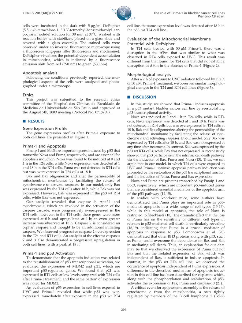
Gene Expression ProfileThe gene expression profiles after Prima-1 exposure to
both cell lines are presented in Figure 1.
Prima-1 and ApoptosisPmaip 1 and Bbc3 are important genes induced by p53 that
transcribe Noxa and Puma, respectively, and are essential forapoptosis induction. Noxa was found to be induced at 0 and1 h in the T24 cells, while Noxa expression was detected at 1and 18 h in the RT4 cells. Puma was not detected in RT4 cellsbut was overexpressed in T24 cells at 18 h.
Bak and Bax oligomerize and alter the permeability ofmitochondrial membranes by facilitating the release ofcytochrome c to activate caspases. In our model, only Baxwas expressed by the T24 cells after 18 h, while Bak was notexpressed. However, Bak was expressed in the p53 wt RT4cells, while Bax was not expressed.
Our analysis revealed that caspase 9, Apaf-1 andcytochrome-c, which are involved in the activation of thecaspase cascade, were progressively overexpressed in theRT4 cells; however, in the T24 cells, these genes were moreexpressed at 0 h and upregulated at 1 h; an even greaterincrease was observed at 18 h. Caspase 2 is considered anorphan caspase and thought to be an additional initiatingcaspase. We observed progressive caspase 2 overexpressionin the T24 and RT4 cells. An analysis of the effector caspases7 and 3 also demonstrated a progressive upregulation inboth cell lines, with a peak at 18 h.
Prima-1 and p53 ActivationTo demonstrate that the apoptosis induction was related
to the reestablishment of p53 transcriptional activation, weevaluated the expression of MDM2 and p21, which areimportant p53-regulated genes. We found that p21 wasexpressed in RT4 cells at low levels compared with T24 cellsafter Prima-1 treatment, and the same pattern of expressionwas noted for MDM2.
An evaluation of p53 expression in cell lines exposed toUVC and Prima-1 revealed that while p53 was over-expressed immediately after exposure in the p53 wt RT4
cell line, the same expression level was detected after 18 h inthe p53 mt T24 cell line.
Evaluation of the Mitochondrial MembranePotential with DePsipher
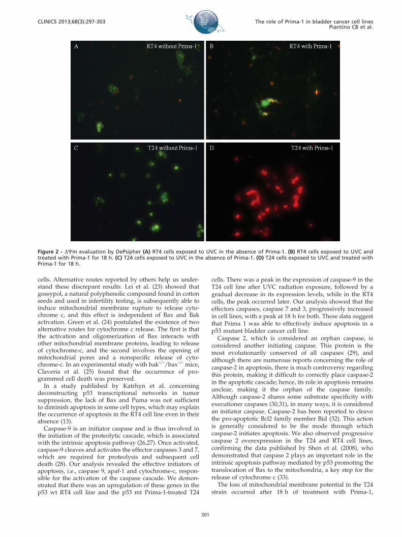
In T24 cells treated with 50 mM Prima-1, there was adisruption in the DYm that was similar to what wasobserved in RT4 cells exposed to UVC. This result wasdifferent from that found for T24 cells that did not exhibit adisruption in DYm in the absence of Prima-1 (Figure 2).
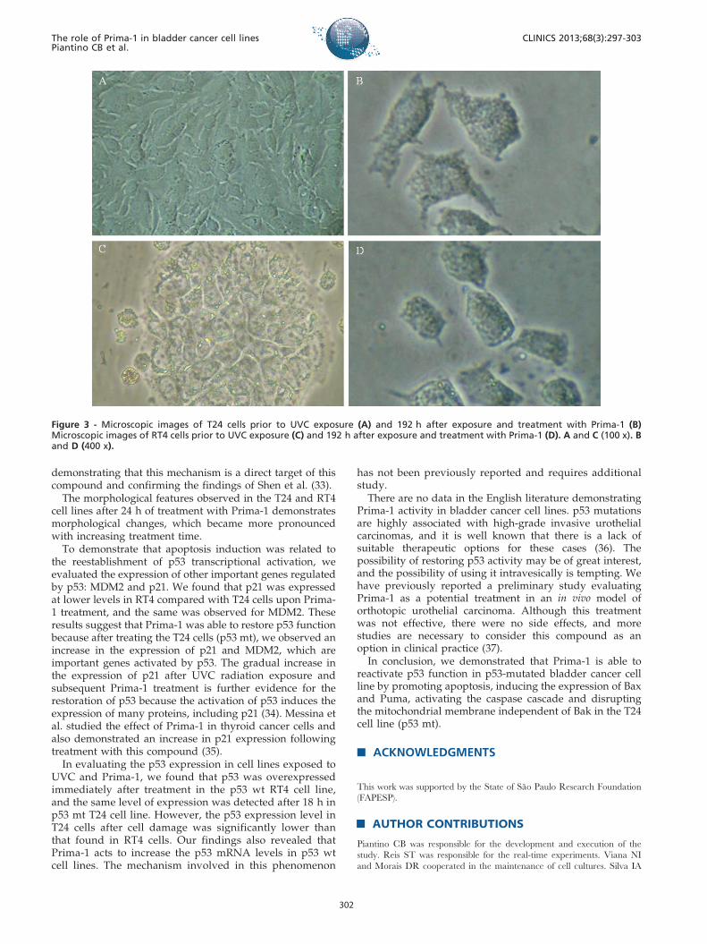
Morphological analysisAfter a 2 h of exposure to UVC radiation followed by 192 h
of 50 mM Prima-1 treatment, we observed similar morpholo-gical changes in the T24 and RT4 cell lines (Figure 3).
& DISCUSSION
In this study, we showed that Prima-1 induces apoptosisin a p53 mutant bladder cancer cell line by reestablishingp53 transcriptional activity.
Noxa was induced at 0 and 1 h in T24 cells, while in RT4cells, Noxa expression was detected at 1 and 18 h. Puma wasnot detected in RT4 cells but was overexpressed in T24 cells at18 h. Bak and Bax oligomerize, altering the permeability of themitochondrial membrane by facilitating the release of cyto-chrome c and activating caspases. In our model, only Bax wasexpressed by T24 cells after 18 h, and Bak was not expressed atany time after treatment. In contrast, Bak was expressed by thep53 wt RT4 cells, while Bax was not expressed. A recent studyshowed that p53 participates in the intrinsic cell death pathwayvia the induction of Bax, Puma and Noxa (13). Thus, we canargue that in our model, in which T24 cells were exposed toUVC and Prima-1, intrinsic apoptosis pathway induction waspromoted by the restoration of the p53 transcriptional functionand the induction of Noxa, Puma and Bax expression.
Noxa and Puma are proteins transcribed by Pmaip 1 andBbc3, respectively, which are important p53-induced genesthat are considered essential mediators of the apoptotic armof the p53 pathway (14,15).
In studies with knockout mice, some authors havedemonstrated that Puma plays an important role in p53-mediated apoptosis in a wide range of cell types (15-17),while in this model of apoptosis, the role of Noxa isrestricted to fibroblasts (18). The dramatic effect that the lossof Puma has on the sensitivity of different cell types inrelation to p53-mediated cell death is particularly revealing(16,19), indicating that Puma is a crucial mediator ofapoptosis in response to p53. Lomonosova et al. (20)demonstrated that other BH3 proteins along with p53, suchas Puma, could overcome the dependence on Bax and Bakin mediating cell death. Thus, an explanation for our datamay be that we observed the expression of Puma but notBax and that the isolated expression of Bak, which wasindependent of Bax, is sufficient to induce apoptosis. Incontrast, in the p53 wt RT4 cell line, we observed theoccurrence of apoptosis independent of Puma expression. Adifference in the described mechanism of apoptosis induc-tion in this cell line has been described for cisplatin, which,along with the phosphorylation and stabilization of p53,activates the expression of Fas, Puma and caspase-10 (21).
A critical event for apoptosome assembly is the release ofcytochrome c from the mitochondria, a step tightlyregulated by members of the B cell lymphoma 2 (Bcl-2)
CLINICS 2013;68(3):297-303 The role of Prima-1 in bladder cancer cell linesPiantino CB et al.
299
protein family (22). This family is comprised of threesubfamilies, depending on the number of Bcl-2 homolog(BH) domains that they contain. The antiapoptotic sub-family consists of members containing BH4 domains, andBcl-2 is the most well-known protein in this class. The twoother subfamilies are proapoptotic in nature, either lackingthe BH4 domain (Bax, Bak) or solely containing a BH3
domain (BH3-only). Once the BH3-only subfamily isactivated, the inhibitory effects of the antiapoptotic Bcl-2protein family are overcome, enabling the oligomerizationof Bak-Bax within the mitochondrial outer membrane(MOM). Our results demonstrated differences in theinvolvement of Bak and Bax, and the former was shownto participate in apoptosis in RT4 cells and the latter in T24
Figure 1 - Profile of MDM2, Puma, Bax, Noxa, Casp9, Casp7, Casp3, Casp2, p53, Apaf-1, p21 and cytochrome-c gene expression in T24(p53 mt) and RT4 (p53 wt) cells after UVC radiation and treatment with Prima -1 for 1 and 18 h, compared with the control.
The role of Prima-1 in bladder cancer cell linesPiantino CB et al.
CLINICS 2013;68(3):297-303
300
cells. Alternative routes reported by others help us under-stand these discrepant results. Lei et al. (23) showed thatgossypol, a natural polyphenolic compound found in cottonseeds and used in infertility testing, is subsequently able toinduce mitochondrial membrane rupture to release cyto-chrome c, and this effect is independent of Bax and Bakactivation. Green et al. (24) postulated the existence of twoalternative routes for cytochrome c release. The first is thatthe activation and oligomerization of Bax interacts withother mitochondrial membrane proteins, leading to releaseof cytochrome-c, and the second involves the opening ofmitochondrial pores and a nonspecific release of cyto-chrome-c. In an experimental study with bak-/-/bax-/- mice,Claveria et al. (25) found that the occurrence of pro-grammed cell death was preserved.
In a study published by Katrhyn et al. concerningdeconstructing p53 transcriptional networks in tumorsuppression, the lack of Bax and Puma was not sufficientto diminish apoptosis in some cell types, which may explainthe occurrence of apoptosis in the RT4 cell line even in theirabsence (13).
Caspase-9 is an initiator caspase and is thus involved inthe initiation of the proteolytic cascade, which is associatedwith the intrinsic apoptosis pathway (26,27). Once activated,caspase-9 cleaves and activates the effector caspases 3 and 7,which are required for proteolysis and subsequent celldeath (28). Our analysis revealed the effective initiators ofapoptosis, i.e., caspase 9, apaf-1 and cytochrome-c, respon-sible for the activation of the caspase cascade. We demon-strated that there was an upregulation of these genes in thep53 wt RT4 cell line and the p53 mt Prima-1-treated T24
cells. There was a peak in the expression of caspase-9 in theT24 cell line after UVC radiation exposure, followed by agradual decrease in its expression levels, while in the RT4cells, the peak occurred later. Our analysis showed that theeffectors caspases, caspase 7 and 3, progressively increasedin cell lines, with a peak at 18 h for both. These data suggestthat Prima 1 was able to effectively induce apoptosis in ap53 mutant bladder cancer cell line.
Caspase 2, which is considered an orphan caspase, isconsidered another initiating caspase. This protein is themost evolutionarily conserved of all caspases (29), andalthough there are numerous reports concerning the role ofcaspase-2 in apoptosis, there is much controversy regardingthis protein, making it difficult to correctly place caspase-2in the apoptotic cascade; hence, its role in apoptosis remainsunclear, making it the orphan of the caspase family.Although caspase-2 shares some substrate specificity withexecutioner caspases (30,31), in many ways, it is consideredan initiator caspase. Caspase-2 has been reported to cleavethe pro-apoptotic Bcl2 family member Bid (32). This actionis generally considered to be the mode through whichcaspase-2 initiates apoptosis. We also observed progressivecaspase 2 overexpression in the T24 and RT4 cell lines,confirming the data published by Shen et al. (2008), whodemonstrated that caspase 2 plays an important role in theintrinsic apoptosis pathway mediated by p53 promoting thetranslocation of Bax to the mitochondria, a key step for therelease of cytochrome c (33).
The loss of mitochondrial membrane potential in the T24strain occurred after 18 h of treatment with Prima-1,
Figure 2 - DYm evaluation by DePsipher (A) RT4 cells exposed to UVC in the absence of Prima-1. (B) RT4 cells exposed to UVC andtreated with Prima-1 for 18 h. (C) T24 cells exposed to UVC in the absence of Prima-1. (D) T24 cells exposed to UVC and treated withPrima-1 for 18 h.
CLINICS 2013;68(3):297-303 The role of Prima-1 in bladder cancer cell linesPiantino CB et al.
301
demonstrating that this mechanism is a direct target of thiscompound and confirming the findings of Shen et al. (33).
The morphological features observed in the T24 and RT4cell lines after 24 h of treatment with Prima-1 demonstratesmorphological changes, which became more pronouncedwith increasing treatment time.
To demonstrate that apoptosis induction was related tothe reestablishment of p53 transcriptional activation, weevaluated the expression of other important genes regulatedby p53: MDM2 and p21. We found that p21 was expressedat lower levels in RT4 compared with T24 cells upon Prima-1 treatment, and the same was observed for MDM2. Theseresults suggest that Prima-1 was able to restore p53 functionbecause after treating the T24 cells (p53 mt), we observed anincrease in the expression of p21 and MDM2, which areimportant genes activated by p53. The gradual increase inthe expression of p21 after UVC radiation exposure andsubsequent Prima-1 treatment is further evidence for therestoration of p53 because the activation of p53 induces theexpression of many proteins, including p21 (34). Messina etal. studied the effect of Prima-1 in thyroid cancer cells andalso demonstrated an increase in p21 expression followingtreatment with this compound (35).
In evaluating the p53 expression in cell lines exposed toUVC and Prima-1, we found that p53 was overexpressedimmediately after treatment in the p53 wt RT4 cell line,and the same level of expression was detected after 18 h inp53 mt T24 cell line. However, the p53 expression level inT24 cells after cell damage was significantly lower thanthat found in RT4 cells. Our findings also revealed thatPrima-1 acts to increase the p53 mRNA levels in p53 wtcell lines. The mechanism involved in this phenomenon
has not been previously reported and requires additionalstudy.
There are no data in the English literature demonstratingPrima-1 activity in bladder cancer cell lines. p53 mutationsare highly associated with high-grade invasive urothelialcarcinomas, and it is well known that there is a lack ofsuitable therapeutic options for these cases (36). Thepossibility of restoring p53 activity may be of great interest,and the possibility of using it intravesically is tempting. Wehave previously reported a preliminary study evaluatingPrima-1 as a potential treatment in an in vivo model oforthotopic urothelial carcinoma. Although this treatmentwas not effective, there were no side effects, and morestudies are necessary to consider this compound as anoption in clinical practice (37).
In conclusion, we demonstrated that Prima-1 is able toreactivate p53 function in p53-mutated bladder cancer cellline by promoting apoptosis, inducing the expression of Baxand Puma, activating the caspase cascade and disruptingthe mitochondrial membrane independent of Bak in the T24cell line (p53 mt).
& ACKNOWLEDGMENTS
This work was supported by the State of Sao Paulo Research Foundation
(FAPESP).
& AUTHOR CONTRIBUTIONS
Piantino CB was responsible for the development and execution of the
study. Reis ST was responsible for the real-time experiments. Viana NI
and Morais DR cooperated in the maintenance of cell cultures. Silva IA
Figure 3 - Microscopic images of T24 cells prior to UVC exposure (A) and 192 h after exposure and treatment with Prima-1 (B)Microscopic images of RT4 cells prior to UVC exposure (C) and 192 h after exposure and treatment with Prima-1 (D). A and C (100 x). Band D (400 x).
The role of Prima-1 in bladder cancer cell linesPiantino CB et al.
CLINICS 2013;68(3):297-303
302
collaborated in the preparation of solutions and dilutions. Antunes AA, Dip
Junior N and Srougi M collaborated in project development. Leite KRM
generally supervised the project.
& REFERENCES
1. INCA (2012). Accessed on October 6, 2012 , http://www.inca.gov.br/estimativa/2012/.
2. Jemal A, Bray F, Center MM, Ferlay J, Ward E, Forman D. Global cancerstatistics. CA Cancer J Clin. 2011;61(2):69-90, http://dx.doi.org/10.3322/caac.20107
3. Debra T. Silverman SSD, Lee E. Moore, Nathaniel Rothman. (2006)Bladder Cancer. In: David Schottenfeld JFF, Jr. (ed). Epidemiology andPrevention, 3 edn. Oxford University Press, New York, pp. 1101-1127.
4. Cote RJ, Datar RH. Therapeutic approaches to bladder cancer: identify-ing targets and mechanisms. Crit Rev Oncol Hematol. 2003;46 Suppl:S67-83, http://dx.doi.org/10.1016/S1040-8428(03)00066-0.
5. Cordon-Cardo C. Molecular alterations associated with bladder cancerinitiation and progression. Scand J Urol Nephrol Suppl. 2008;(218):154-65, http://dx.doi.org/10.1080/03008880802291915.
6. Cordon-Cardo C. Molecular alterations in bladder cancer. Cancer Surv.1998;32:115-31.
7. Lu ML, Wikman F, Orntoft TF, Charytonowicz E, Rabbani F, Zhang Z,et al. Impact of alterations affecting the p53 pathway in bladder canceron clinical outcome, assessed by conventional and array-based methods.Clin Cancer Res. 2002;8(1):171-9.
8. Feng Z, Hu W, Rom WN, Beland FA, Tang MS. 4-aminobiphenyl is amajor etiological agent of human bladder cancer: evidence from its DNAbinding spectrum in human p53 gene. Carcinogenesis. 2002;23(10):1721-7, http://dx.doi.org/10.1093/carcin/23.10.1721.
9. Bykov VJ, Issaeva N, Shilov A, Hultcrantz M, Pugacheva E, ChumakovP, et al. Restoration of the tumor suppressor function to mutant p53 by alow-molecular-weight compound. Nat Med. 2002;8(3):282-8, http://dx.doi.org/10.1038/nm0302-282.
10. Batista LF, Kaina B, Meneghini R, Menck CF. How DNA lesions areturned into powerful killing structures: insights from UV-inducedapoptosis. Mutat Res. 2009;681(2-3):197-208.
11. Szliszka E, Czuba ZP, Mazur B, Paradysz A, Krol W. Chalcones anddihydrochalcones augment TRAIL-mediated apoptosis in prostatecancer cells. Molecules. 2010;15(8):5336-53, http://dx.doi.org/10.3390/molecules15085336.
12. Szliszka E, Zydowicz G, Janoszka B, Dobosz C, Kowalczyk-Ziomek G,Krol W. Ethanolic extract of Brazilian green propolis sensitizes prostatecancer cells to TRAIL-induced apoptosis. Int J Oncol. 2011;38(4):941-53.
13. Bieging KT, Attardi LD. Deconstructing p53 transcriptional networks intumor suppression. Trends in cell biology. 2012;22(2):97-106, http://dx.doi.org/10.1016/j.tcb.2011.10.006.
14. Nakano K, Vousden KH. PUMA, a novel proapoptotic gene, is inducedby p53. Mol Cell. 2001;7(3):683-94, http://dx.doi.org/10.1016/S1097-2765(01)00214-3.
15. Jeffers JR, Parganas E, Lee Y, Yang C, Wang J, Brennan J, et al. Puma is anessential mediator of p53-dependent and -independent apoptotic path-ways. Cancer Cell. 2003;4(4):321-8, http://dx.doi.org/10.1016/S1535-6108(03)00244-7.
16. Villunger A, Michalak EM, Coultas L, Mullauer F, Bock G, AusserlechnerMJ, et al. p53- and drug-induced apoptotic responses mediated by BH3-only proteins puma and noxa. Science. 2003;302(5647):1036-8, http://dx.doi.org/10.1126/science.1090072.
17. Erlacher M, Michalak EM, Kelly PN, Labi V, Niederegger H, Coultas L, et al.BH3-only proteins Puma and Bim are rate-limiting for gamma-radiation-and glucocorticoid-induced apoptosis of lymphoid cells in vivo. Blood.2005;106(13):4131-8, http://dx.doi.org/10.1182/blood-2005-04-1595.
18. Shibue T, Takeda K, Oda E, Tanaka H, Murasawa H, Takaoka A, et al.Integral role of Noxa in p53-mediated apoptotic response. Genes Dev.2003;17:2233-8, http://dx.doi.org/10.1101/gad.1103603.
19. Vousden KH. Apoptosis. p53 and PUMA: a deadly duo. Science.2005;309(5741):1685-6, http://dx.doi.org/10.1126/science.1118232.
20. Lomonosova E, Ryerse J, Chinnadurai G. BAX/BAK-independentmitoptosis during cell death induced by proteasome inhibition? MolCancer Res. 2009;7(8):1268-84, http://dx.doi.org/10.1158/1541-7786.MCR-08-0183.
21. Konstantakou EG, Voutsinas GE, Karkoulis PK, Aravantinos G,Margaritis LH, Stravopodis DJ. Human bladder cancer cells undergocisplatin-induced apoptosis that is associated with p53-dependent andp53-independent responses. Int J Oncol. 2009;35(2):401-16.
22. Youle RJ, Strasser A. The BCL-2 protein family: opposing activities thatmediate cell death. Nat Rev Mol Cell Biol. 2008;9(1):47-59, http://dx.doi.org/10.1038/nrm2308.
23. Lei X, Chen Y, Du G, Yu W, Wang X, Qu H, et al. Gossypol induces Bax/Bak-independent activation of apoptosis and cytochrome c release via aconformational change in Bcl-2. FASEB J. 2006;20(12):2147-9, http://dx.doi.org/10.1096/fj.05-5665fje.
24. Green DR. Apoptotic pathways: ten minutes to dead. Cell.2006;121(5):671-4.
25. Claveria C, Martinez-A C, Torres M. A Bax/Bak-independent mitochon-drial death pathway triggered by Drosophila Grim GH3 domain inmammalian cells. J Biol Chem. 2004;279(2):1368-75.
26. Li P, Nijhawan D, Budihardjo I, Srinivasula SM, Ahmad M, Alnemri ES,et al. Cytochrome c and dATP-dependent formation of Apaf-1/caspase-9complex initiates an apoptotic protease cascade. Cell. 1997;91(4):479-89,http://dx.doi.org/10.1016/S0092-8674(00)80434-1.
27. Budihardjo I, Oliver H, Lutter M, Luo X, Wang X. Biochemical pathwaysof caspase activation during apoptosis. Annu Rev Cell Dev Biol.1999;15:269-90, http://dx.doi.org/10.1146/annurev.cellbio.15.1.269.
28. Taylor RC, Cullen SP, Martin SJ. Apoptosis: controlled demolition at thecellular level. Nat Rev Mol Cell Biol. 2008;9(3):231-41, http://dx.doi.org/10.1038/nrm2312.
29. Lamkanfi M, Declercq W, Kalai M, Saelens X, Vandenabeele P. () Alice incaspase land. A phylogenetic analysis of caspases from worm to man.Cell Death Differ. 2002;9(4):358-61, http://dx.doi.org/10.1038/sj.cdd.4400989.
30. Talanian RV, Quinlan C, Trautz S, Hackett MC, Mankovich JA, BanachD, et al. Substrate specificities of caspase family proteases. J Biol Chem.1997;272(29):9677-82.
31. Thornberry NA, Rano TA, Peterson EP, Rasper DM, Timkey T, Garcia-Calvo M, et al. A combinatorial approach defines specificities ofmembers of the caspase family and granzyme B. Functional relationshipsestablished for key mediators of apoptosis. J Biol Chem.1997;272(29):17907-11, http://dx.doi.org/10.1074/jbc.272.29.17907.
32. Guo Y, Srinivasula SM, Druilhe A, Fernandes-Alnemri T, Alnemri ES.Caspase-2 induces apoptosis by releasing proapoptotic proteins frommitochondria. J Biol Chem. 2002;277(16):13430-7, http://dx.doi.org/10.1074/jbc.M108029200.
33. Shen J, Vakifahmetoglu H, Stridh H, Zhivotovsky B, Wiman KG. PRIMA-1MET induces mitochondrial apoptosis through activation of caspase-2.Oncogene. 2008;27(51):6571-80, http://dx.doi.org/10.1038/onc.2008.249.
34. Xiong Y, Hannon GJ, Zhang H, Casso D, Kobayashi R, Beach D. (1993)p21 is a universal inhibitor of cyclin kinases. Nature. 1993;366(6456):701-4, http://dx.doi.org/10.1038/366701a0.
35. Messina RL, Sanfilippo M, Vella V, Pandini G, Vigneri P, Nicolosi ML, et al.Reactivation of p53 mutants by p53 reactivation and induction of massiveapoptosis in thyroid cancer cells. Int J Cancer. 2012;130(10):2259-70.
36. Cory S, Adams JM. The Bcl2 family: regulators of the cellular life-or-death switch. Nat Rev Cancer. 2002;2(9):647-56, http://dx.doi.org/10.1038/nrc883.
37. Watanabe FT, Chade DC, Reis ST, Piantino C, Dall’ Oglio MF, Srougi M,Leite KR. Curcumin, but not Prima-1, decreased tumor cell proliferation inthe syngeneic murine orthotopic bladder tumor model. Clinics.2011;66(12):2121-4, http://dx.doi.org/10.1590/S1807-59322011001200019.
CLINICS 2013;68(3):297-303 The role of Prima-1 in bladder cancer cell linesPiantino CB et al.
303