h istology of digestive system oesophagus, stomach - fundus & pylorus dr. makarchuk iryna
TRANSCRIPT
HISTOLOGY OF DIGESTIVE SYSTEMOESOPHAGUS, STOMACH-
FUNDUS & PYLORUS
Dr. Makarchuk Iryna
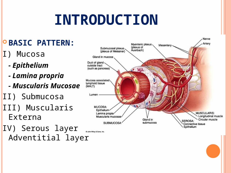
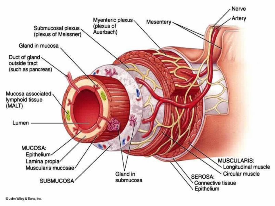
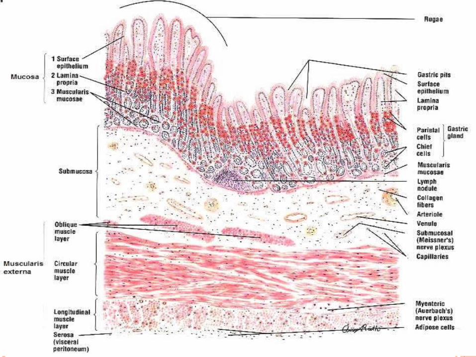
INTRODUCTION BASIC PATTERN:I) Mucosa
- Epithelium
- Lamina propria
- Muscularis Mucosae
II) SubmucosaIII) Muscularis ExternaIV) Serous layer /
Adventitial layer
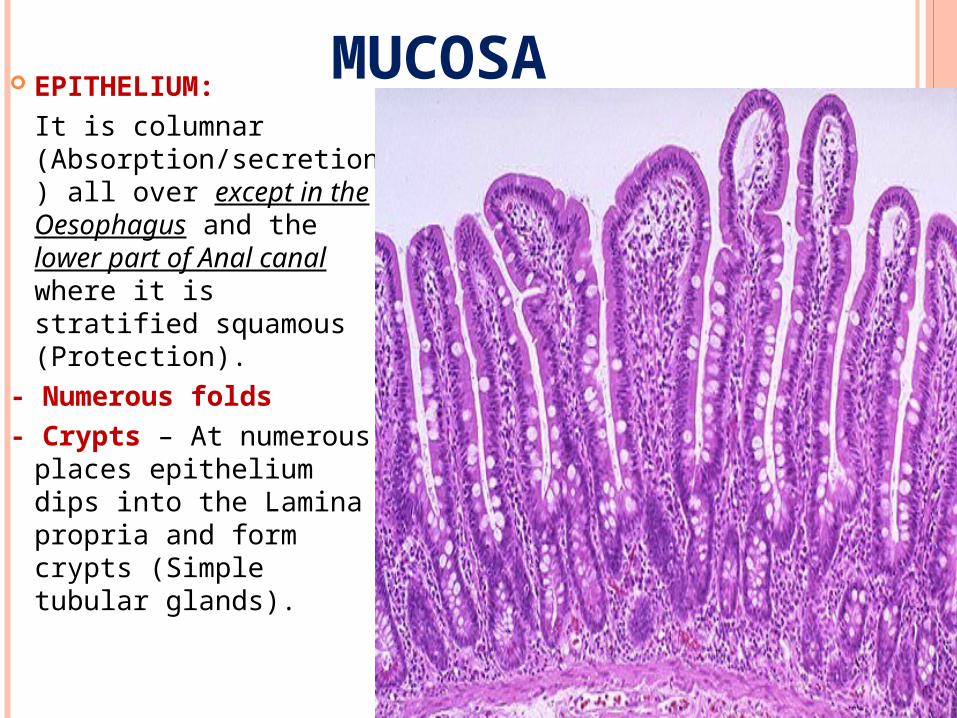
MUCOSA EPITHELIUM:
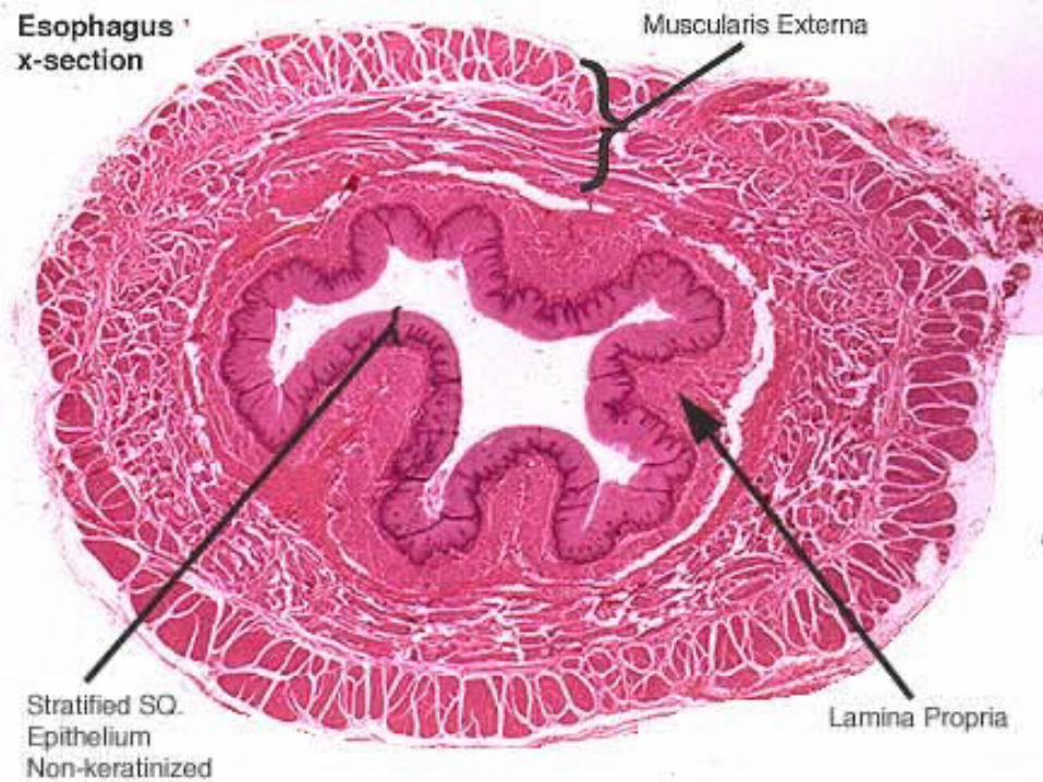
It is columnar (Absorption/secretion) all over except in the Oesophagus and the lower part of Anal canal where it is stratified squamous (Protection).
- Numerous folds- Crypts – At numerous
places epithelium dips into the Lamina propria and form crypts (Simple tubular glands).
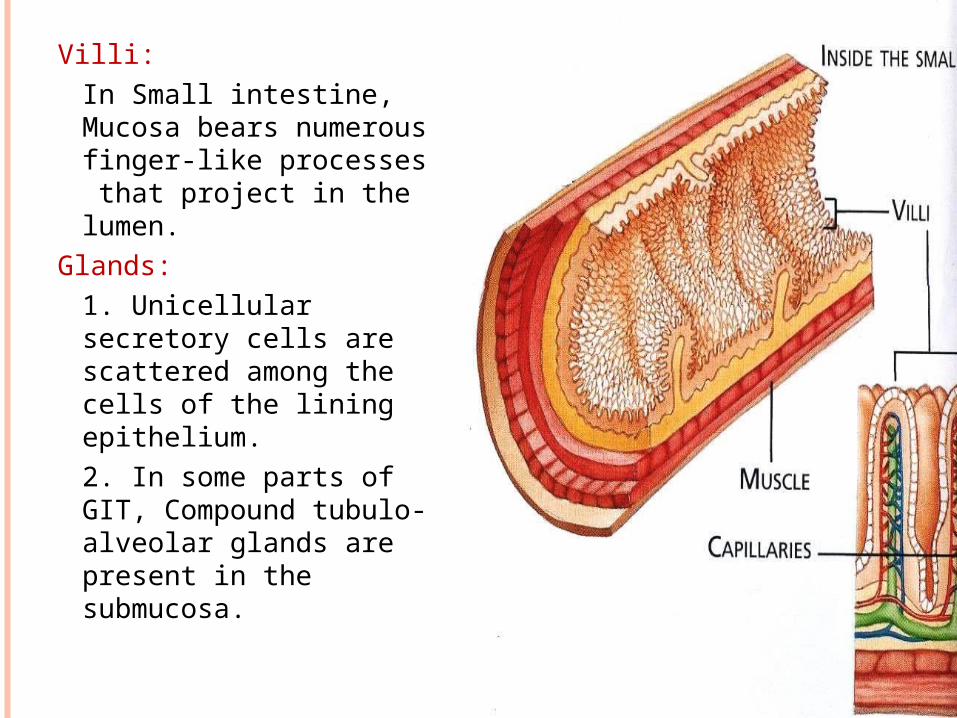
Villi:In Small intestine, Mucosa bears numerous finger-like processes that project in the lumen.
Glands:1. Unicellular secretory cells are scattered among the cells of the lining epithelium.2. In some parts of GIT, Compound tubulo-alveolar glands are present in the submucosa.
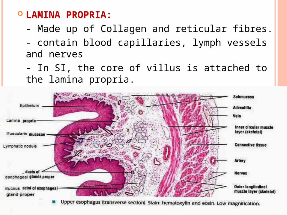
LAMINA PROPRIA:- Made up of Collagen and reticular fibres.- contain blood capillaries, lymph vessels and nerves- In SI, the core of villus is attached to the lamina propria.- Prominent aggregation of lymphatic tissue is seen in Lamina propria (MALT)
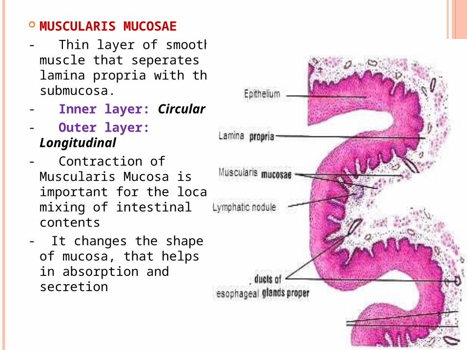
MUSCULARIS MUCOSAE- Thin layer of smooth
muscle that seperates lamina propria with the submucosa.
- Inner layer: Circular- Outer layer: Longitudinal- Contraction of Muscularis
Mucosa is important for the local mixing of intestinal contents
- It changes the shape of mucosa, that helps in absorption and secretion
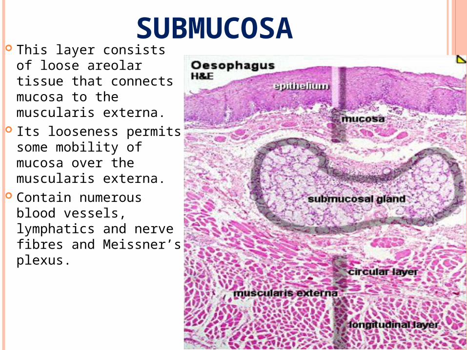
SUBMUCOSA This layer consists of
loose areolar tissue that connects mucosa to the muscularis externa.
Its looseness permits some mobility of mucosa over the muscularis externa.
Contain numerous blood vessels, lymphatics and nerve fibres and Meissner’s plexus.
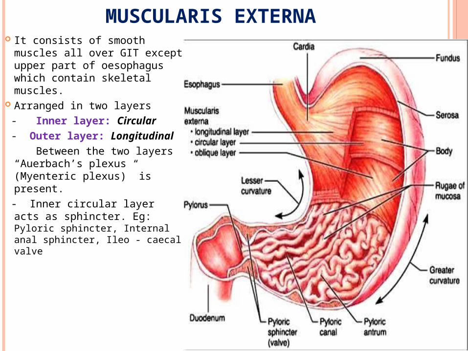
MUSCULARIS EXTERNA It consists of smooth muscles
all over GIT except upper part of oesophagus which contain skeletal muscles.
Arranged in two layers - Inner layer: Circular - Outer layer: Longitudinal Between the two layers
“Auerbach’s plexus (Myenteric plexus)” is present.
- Inner circular layer acts as sphincter. Eg: Pyloric sphincter, Internal anal sphincter, Ileo - caecal valve
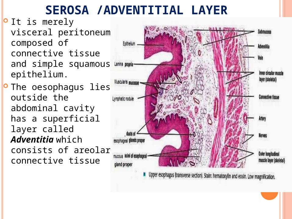
SEROSA /ADVENTITIAL LAYER It is merely visceral
peritoneum composed of connective tissue and simple squamous epithelium.
The oesophagus lies outside the abdominal cavity has a superficial layer called Adventitia which consists of areolar connective tissue
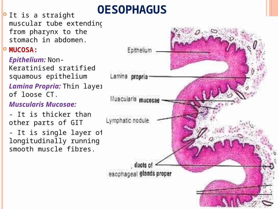
OESOPHAGUS It is a straight muscular tube extending from pharynx to the stomach in abdomen.
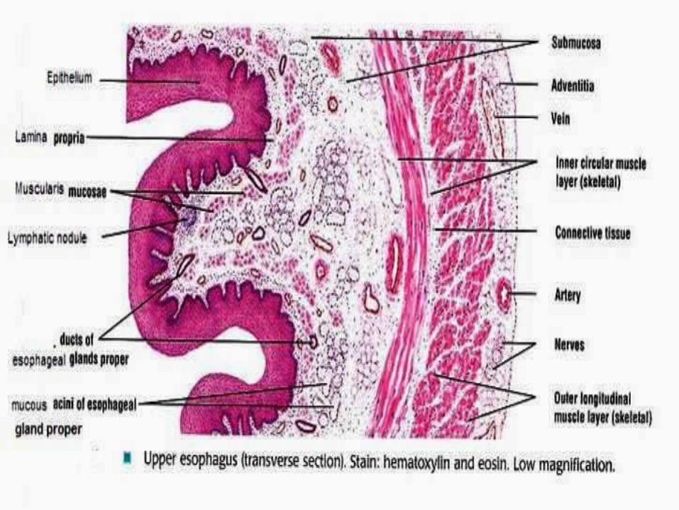
MUCOSA:Epithelium: Non-Keratinised sratified squamous epitheliumLamina Propria: Thin layer of loose CT.Muscularis Mucosae: - It is thicker than other parts of GIT- It is single layer of longitudinally running smooth muscle fibres.
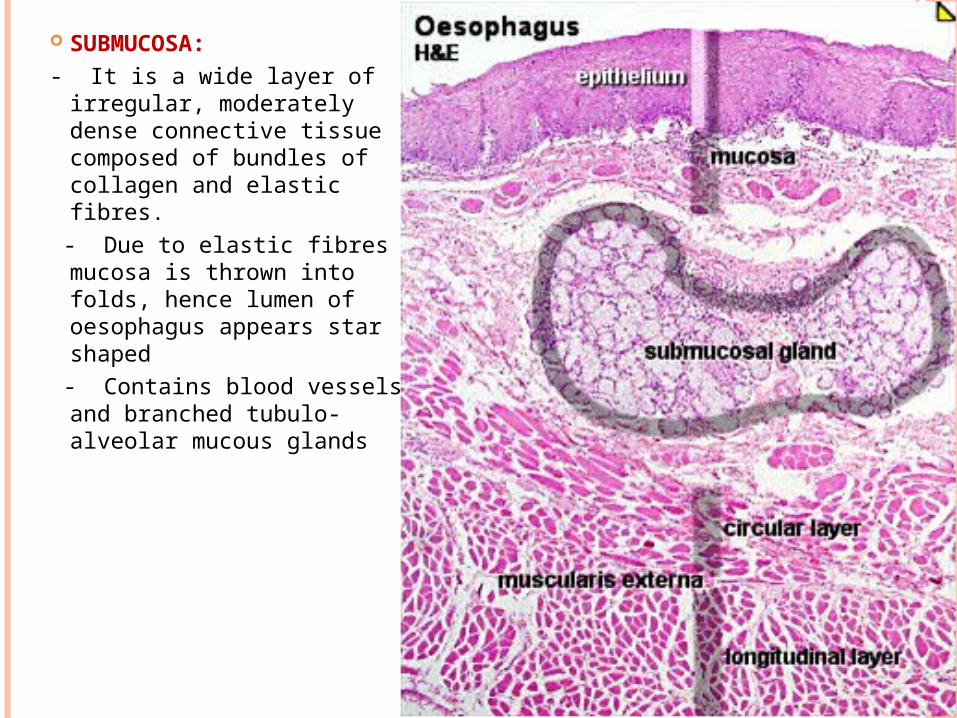
SUBMUCOSA:- It is a wide layer of irregular,
moderately dense connective tissue composed of bundles of collagen and elastic fibres.
- Due to elastic fibres mucosa is thrown into folds, hence lumen of oesophagus appears star shaped
- Contains blood vessels and branched tubulo- alveolar mucous glands
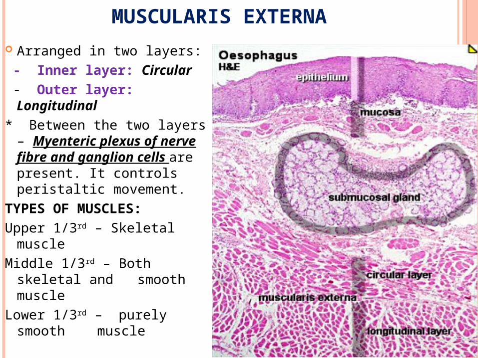
MUSCULARIS EXTERNA
Arranged in two layers: - Inner layer: Circular - Outer layer: Longitudinal* Between the two layers –
Myenteric plexus of nerve fibre and ganglion cells are present. It controls peristaltic movement.
TYPES OF MUSCLES:Upper 1/3rd – Skeletal muscleMiddle 1/3rd – Both skeletal and
smooth muscleLower 1/3rd – purely smooth
muscle
SEROSA / ADVEVTITIA
It consists of loose areolar connective tissue, which merges with the connective tissue of surrounding structures.
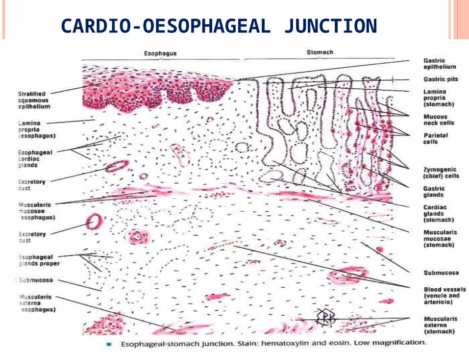
CARDIO-OESOPHAGEAL JUNCTION
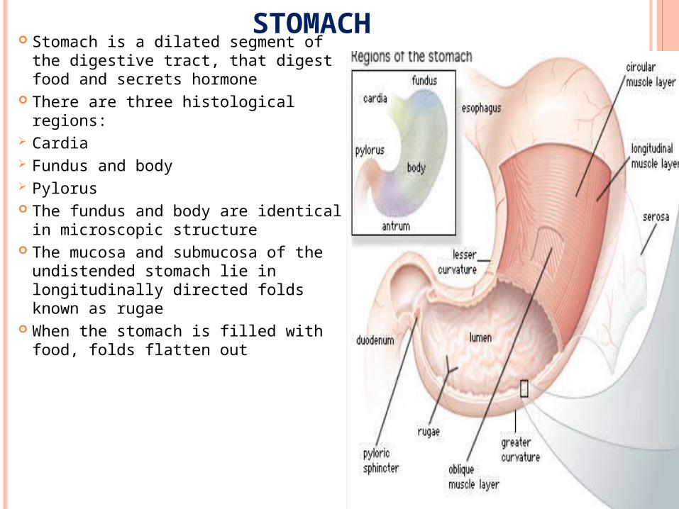
STOMACH Stomach is a dilated segment of the
digestive tract, that digest food and secrets hormone
There are three histological regions: Cardia Fundus and body Pylorus The fundus and body are identical in
microscopic structure The mucosa and submucosa of the
undistended stomach lie in longitudinally directed folds known as rugae
When the stomach is filled with food, folds flatten out
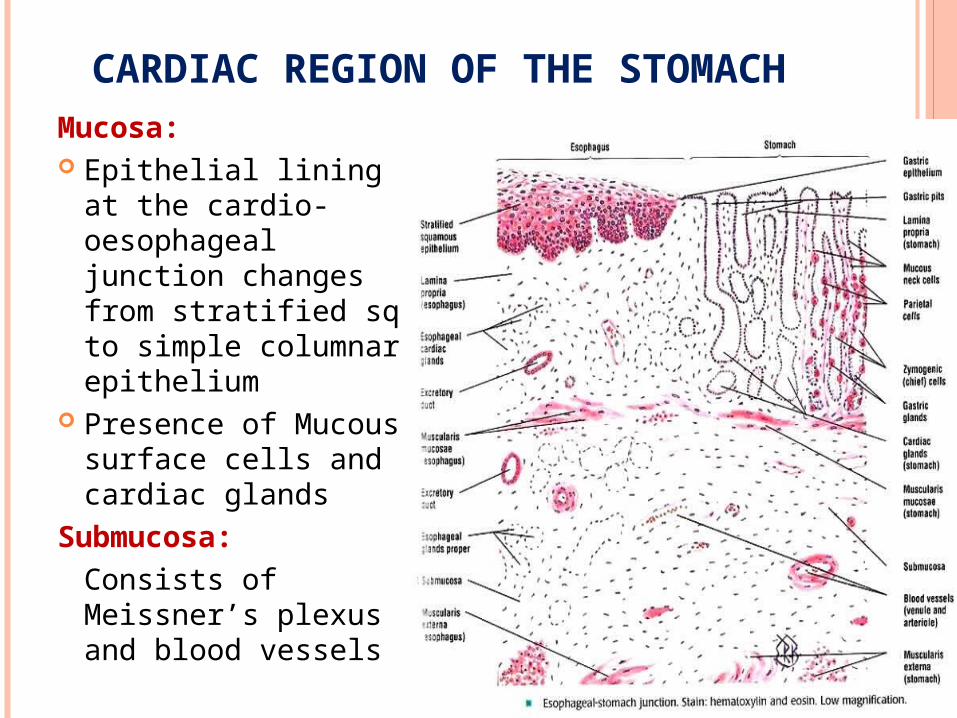
CARDIAC REGION OF THE STOMACHMucosa: Epithelial lining at the
cardio-oesophageal junction changes from stratified sq. to simple columnar epithelium
Presence of Mucous surface cells and cardiac glands
Submucosa:Consists of Meissner’s plexus and blood vessels
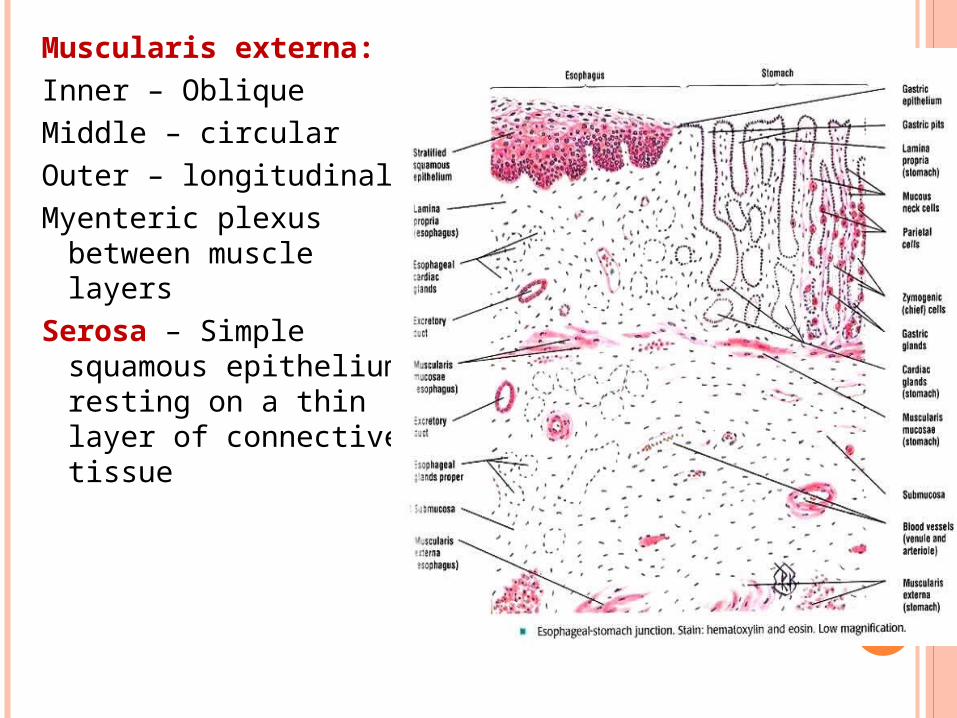
Muscularis externa:Inner – ObliqueMiddle – circularOuter – longitudinalMyenteric plexus between
muscle layersSerosa – Simple squamous
epithelium resting on a thin layer of connective tissue
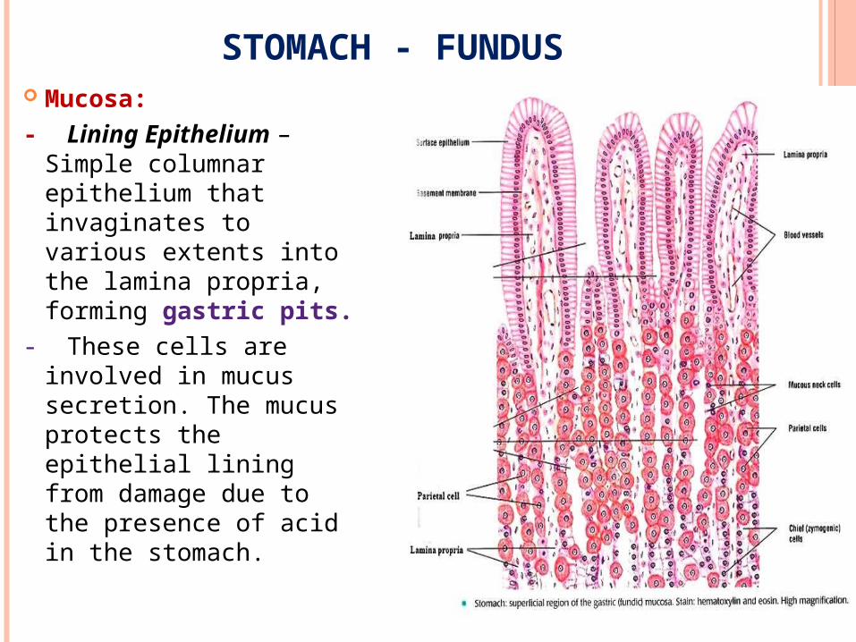
STOMACH - FUNDUS Mucosa:- Lining Epithelium – Simple
columnar epithelium that invaginates to various extents into the lamina propria, forming gastric pits.
- These cells are involved in mucus secretion. The mucus protects the epithelial lining from damage due to the presence of acid in the stomach.
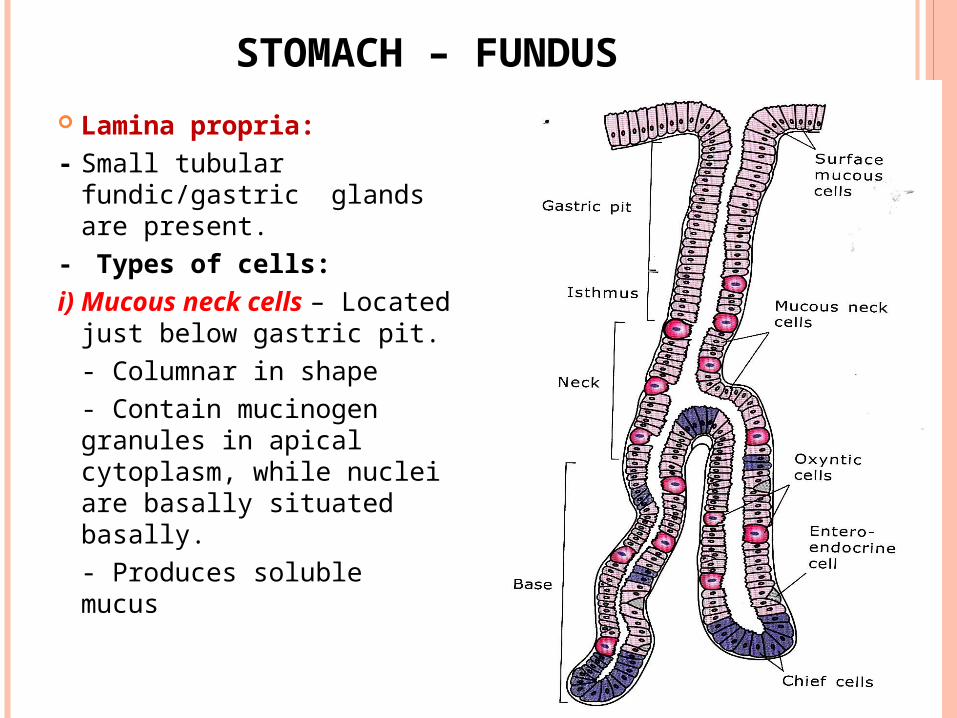
STOMACH – FUNDUS
Lamina propria:- Small tubular fundic/gastric
glands are present.- Types of cells:i) Mucous neck cells – Located
just below gastric pit. - Columnar in shape- Contain mucinogen granules in apical cytoplasm, while nuclei are basally situated basally.- Produces soluble mucus
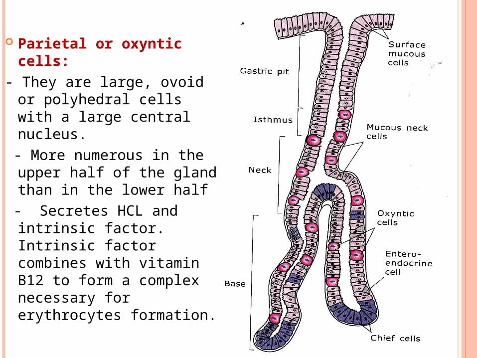
Parietal or oxyntic cells:- They are large, ovoid or
polyhedral cells with a large central nucleus.
- More numerous in the upper half of the gland than in the lower half
- Secretes HCL and intrinsic factor. Intrinsic factor combines with vitamin B12 to form a complex necessary for erythrocytes formation.
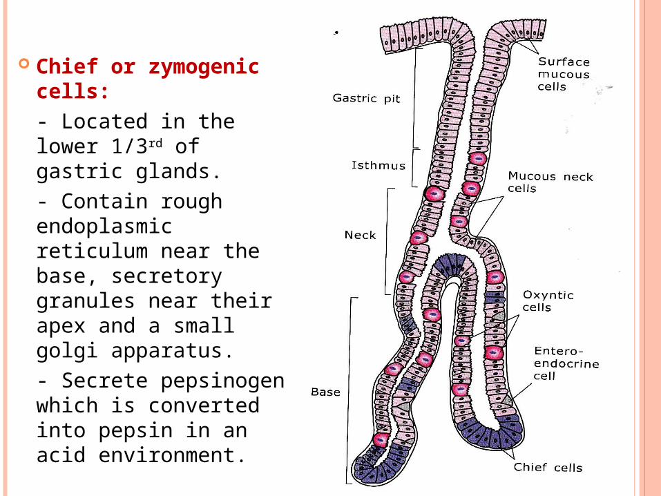
Chief or zymogenic cells:- Located in the lower 1/3rd of gastric glands.- Contain rough endoplasmic reticulum near the base, secretory granules near their apex and a small golgi apparatus.- Secrete pepsinogen which is converted into pepsin in an acid environment.
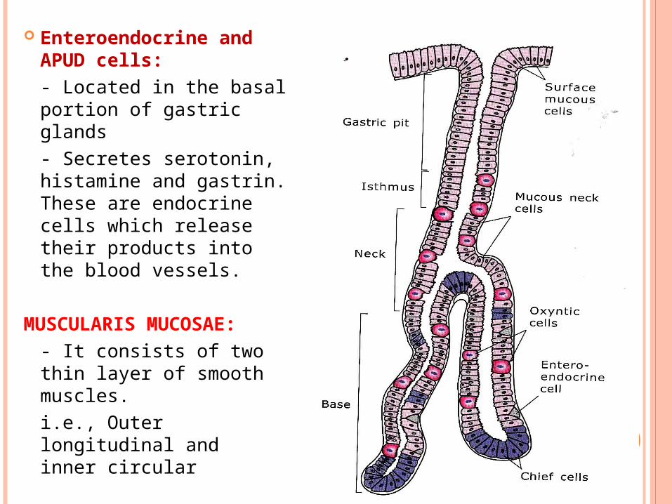
Enteroendocrine and APUD cells:- Located in the basal portion of gastric glands- Secretes serotonin, histamine and gastrin. These are endocrine cells which release their products into the blood vessels.
MUSCULARIS MUCOSAE:- It consists of two thin layer of smooth muscles.i.e., Outer longitudinal and
inner circular
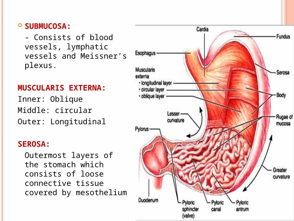
SUBMUCOSA:- Consists of blood vessels, lymphatic vessels and Meissner’s plexus.
MUSCULARIS EXTERNA:Inner: ObliqueMiddle: circularOuter: Longitudinal
SEROSA:Outermost layers of the stomach which consists of loose connective tissue covered by mesothelium
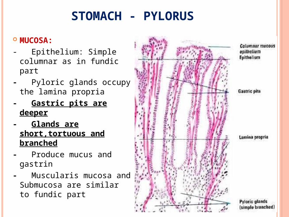
STOMACH - PYLORUS
MUCOSA:- Epithelium: Simple
columnar as in fundic part- Pyloric glands occupy the
lamina propria- Gastric pits are deeper- Glands are short,tortuous
and branched- Produce mucus and gastrin- Muscularis mucosa and
Submucosa are similar to fundic part
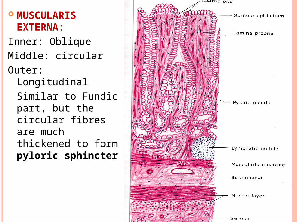
MUSCULARIS EXTERNA:Inner: ObliqueMiddle: circularOuter: Longitudinal
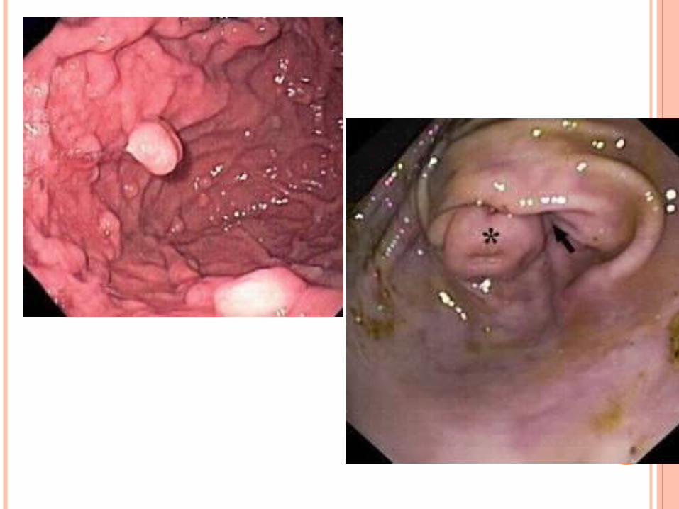
Similar to Fundic part, but the circular fibres are much thickened to form pyloric sphincter
30
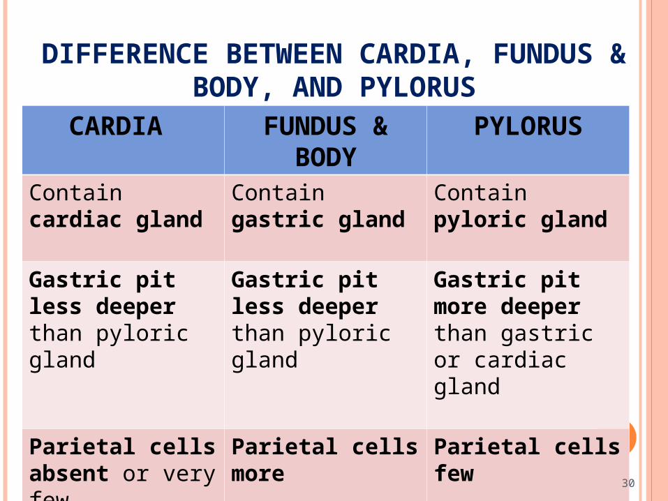
DIFFERENCE BETWEEN CARDIA, FUNDUS & BODY, AND PYLORUS
CARDIA FUNDUS & BODY PYLORUS
Contain cardiac gland
Contain gastric gland
Contain pyloric gland
Gastric pit less deeper than pyloric gland
Gastric pit less deeper than pyloric gland
Gastric pit more deeper than gastric or cardiac gland
Parietal cells absent or very few
Parietal cells more Parietal cells few
30
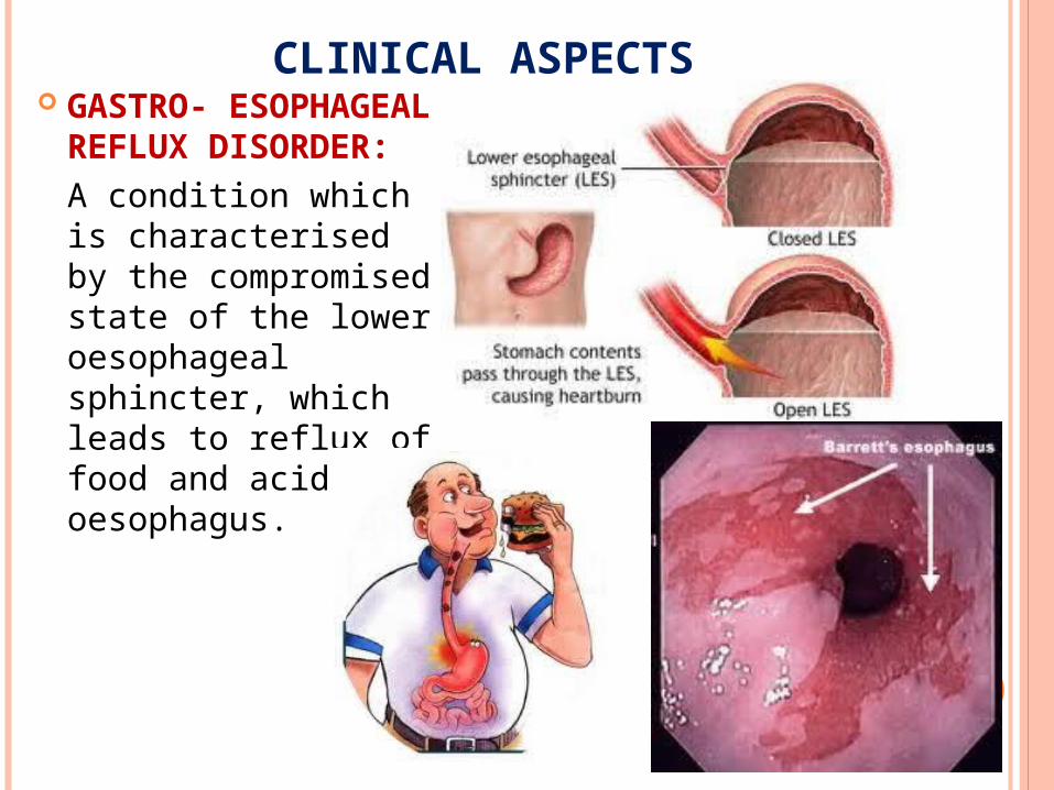
CLINICAL ASPECTS GASTRO- ESOPHAGEAL
REFLUX DISORDER:A condition which is characterised by the compromised state of the lower oesophageal sphincter, which leads to reflux of food and acid into oesophagus.
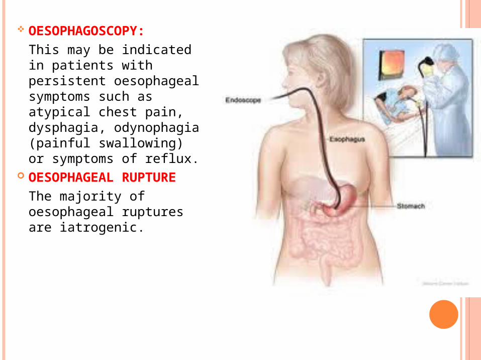
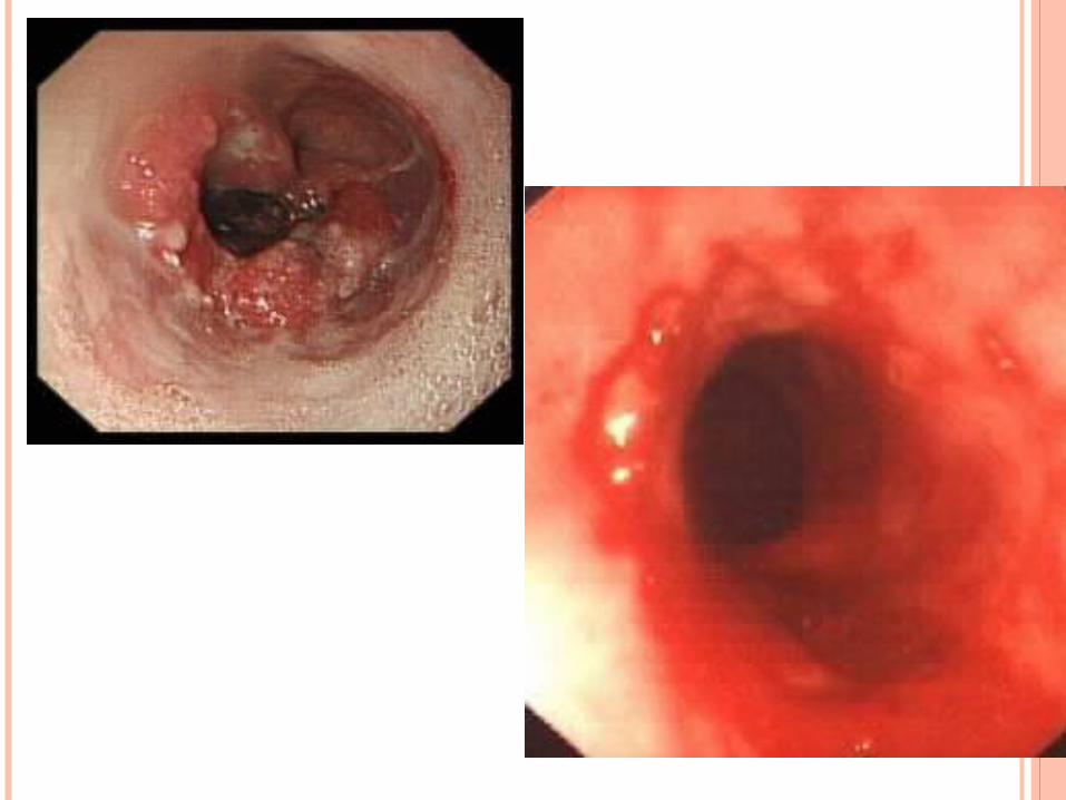
OESOPHAGOSCOPY:This may be indicated in patients with persistent oesophageal symptoms such as atypical chest pain, dysphagia, odynophagia (painful swallowing) or symptoms of reflux.
OESOPHAGEAL RUPTURE The majority of oesophageal ruptures are iatrogenic.
THANK YOU