guidelines for the diagnosis and treatment · the guidelines are designed to assist clinicians and...
TRANSCRIPT
GUIDELINES
FOR THE DIAGNOSIS AND TREATMENT
OF LOW BACK PAIN
Prepared for
The Workplace Health, Safety and Compensation Commission of New Brunswick
by
Dr. Eric R. Gozna, FRCSC Orthopedic Surgeon
Senior Medical Consultant WHSCC of New Brunswick
Copyright: E. Gozna 2001
Revised: October 2002

TABLE OF CONTENTS
GUIDELINE TOPIC PAGE NUMBER NUMBER
HOW TO USE THESE GUIDELINES 4 INTRODUCTION 5 SUMMARY TABLE OF LBP GUIDELINES 8 1. MEDICAL ASSESSMENT 9 2. MEDICAL MANANGEMENT 14 3. X-RAYS (PLAIN) 18 4. LABORATORY STUDIES 20 5. EDUCATION 21 6. ACTIVITY & WORK MODIFICATION 23 7. ACTIVE VS PASSIVE MANAGEMENT 24 8. MEDICATIONS 27 9. BED REST 29 10. PHYSIOTHERAPY MODALITIES 30 11. SPINAL MANIPULATION 32 12. TRANSCUTANEOUS ELECTRICAL NERVE STIMULATION 33 13. TRACTION 34 14. ACUPUNCTURE / DRY NEEDLING 35 15. LUMBAR SUPPORTS 36 16. SHOE INSOLES / LIFTS / ORTHOTICS 37 17. CT / MRI / MYELOGRAPHY AND CT-MYELOGRAPHY 38 18. BONE SCAN 40
Guidelines: Low Back Pain 1 ©E Gozna, 2001

TABLE OF CONTENTS (CONTINUED)
GUIDELINE TOPIC PAGE NUMBER NUMBER
19. ELECTRODIAGNOSTIC STUDIES (EMG, NCS, SEP) 41 20. PSYCHOSOCIAL ASSESSMENT 43 21. ERGONOMIC EVALUATION 44 22. WORK-CONDITIONING PROGRAMS 45 23. DISCECTOMY (STANDARD / MICRO / PERCUTANEOUS) 46 24. SPINAL STENOSIS 48 25. SPINAL FUSION 49 26. INTERDISCIPLINARY PROGRAMS 50 27. FUNCTIONAL CAPACITY TESTING 52 28. COMPUTERIZED STRENGTH AND R.O.M. TESTING 53 29. DISCOGRAPHY 54 30. THERMOGRAPHY 55 31. EPIDURAL CORTICOSTEROID INJECTIONS 56 32. FACET JOINT INJECTIONS (DIAGNOSTIC / THERAPEUTIC) 57 33. TRIGGER POINT INJECTIONS 58 34. SCLEROSANT LIGAMENTOUS INJECTIONS (PROLOTHERAPY) 59 35. CHEMONUCLEOLYSIS 60 36. BIOFEEDBACK 61 37. SPECIALIST REFERRAL 62
Guidelines: Low Back Pain 2 © E. Gozna, 2001
TABLE OF CONTENTS (CONTINUED)
GUIDELINE TOPIC PAGE NUMBER NUMBER
38. WHSCC RESOURCES 63 39. RED FLAGS 64 40. AMBER FLAGS 67 41. SIMPLE BACK PAIN AND SCIATICA 70 42. EXAMPLES OF POSTURAL CONTRIBUTORS TO LBP 76
SOURCE DOCUMENTS AND REFERENCES 80 CMA POLICY ON RETURN TO WORK 82
INDEX 87
Guidelines: Low Back Pain 3 ©E Gozna, 2001
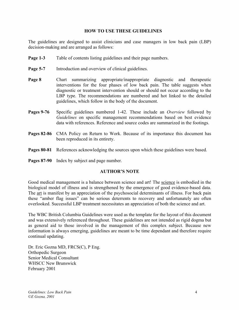
HOW TO USE THESE GUIDELINES
The guidelines are designed to assist clinicians and case managers in low back pain (LBP) decision-making and are arranged as follows: Page 1-3 Table of contents listing guidelines and their page numbers. Page 5-7 Introduction and overview of clinical guidelines. Page 8 Chart summarizing appropriate/inappropriate diagnostic and therapeutic
interventions for the four phases of low back pain. The table suggests when diagnostic or treatment intervention should or should not occur according to the LBP type. The recommendations are numbered and hot linked to the detailed guidelines, which follow in the body of the document.
Pages 9-76 Specific guidelines numbered 1-42. These include an Overview followed by
Guidelines on specific management recommendations based on best evidence data with references. Reference and source codes are summarized in the footings.
Pages 82-86 CMA Policy on Return to Work. Because of its importance this document has
been reproduced in its entirety. Pages 80-81 References acknowledging the sources upon which these guidelines were based. Pages 87-90 Index by subject and page number.
AUTHOR’S NOTE
Good medical management is a balance between science and art! The science is embodied in the biological model of illness and is strengthened by the emergence of good evidence-based data. The art is manifest by an appreciation of the psychosocial determinants of illness. For back pain these “amber flag issues” can be serious deterrents to recovery and unfortunately are often overlooked. Successful LBP treatment necessitates an appreciation of both the science and art. The WBC British Columbia Guidelines were used as the template for the layout of this document and was extensively referenced throughout. These guidelines are not intended as rigid dogma but as general aid to those involved in the management of this complex subject. Because new information is always emerging, guidelines are meant to be time dependant and therefore require continual updating. Dr. Eric Gozna MD, FRCS(C), P Eng. Orthopedic Surgeon Senior Medical Consultant WHSCC New Brunswick February 2001
Guidelines: Low Back Pain 4 ©E Gozna, 2001
INTRODUCTION AND OVERVIEW
• Objective: These guidelines were designed to assist practitioners and case managers in the
diagnosis and treatment of low back pain (LBP) based upon current best evidence recommendations. The intention is to promote improved patient outcomes through consistent, efficient, cost effective, evidence-based practice.
• Summary of Diagnosis and Management Principals: Effective medical management is based on three fundamental requirement:
1. Establishing a working diagnosis (Simple LBP, Sciatica, Something else- i.e. red flags). 2. Appreciate the importance of the phase (acute, subacute, chronic, recurrent) of the LBP. 3. Identify and address amber flag issues (workplace and psychosocial) and appreciating
their important role as a psychological enforcer of pain and deterrent of function. Towards this end the following fundamental principles are acknowledged:
1. Initial medical assessment should focus on ruling out potentially serious spinal pathology (red flags) for which specific investigations and interventions are indicated.
2. In the absence of red flags, the working diagnosis is that of non-specific (simple) low back pain or sciatica, for which management is based upon the phase (acute, subacute, chronic, recurrent) and identifying /addressing amber flag issues.
3. Amber flags are important non-physical (workplace, psychosocial) deterrents of functional recovery and return to work.
4. Amber flags can be greater deterrents to return to work than medical issues 5. Amber flag issues become increasingly important as patients progress from acute to subacute
and to chronic LBP. 6. Amber flags are an important predictor of both medical and surgical outcomes. 7. Over 80% of patients with acute simple LBP will recover uneventfully over a period of 1
month without intervention. 8. The single most important LBP treatment recommendation is to encourage patient activity. 9. Patients should be encouraged to stay at work (on a modified basis) and to return to normal
activities as soon as possible. 10. For acute simple LBP or mild sciatica, imaging studies or further investigations are not
usually necessary. Costly interventions and investigations are not warranted. 11. Generally pain relief can be effectively accomplished with non-prescription medications. 12. Bed rest beyond 24-48 hours is not helpful and may further debilitate the patient. 13. A careful baseline neurological assessment is fundamental to the management of suspected
sciatica. 14. Mild sciatica may recover more slowly than acute simple LBP, but is treated the same (i.e.
encouragement of activity) and further investigations are not required as long as the condition is clinically resolved.
15. In the first three months of low back symptoms, only patients with evidence of serious spinal
pathology, debilitating symptoms of sciatica or significant/progressive neurological impairment as corroborated on imaging studies can be expected to benefit from surgery.
Guidelines: Low Back Pain 5 © E. Gozna, 2001

• Anticipated Outcomes:
1. Reduction in morbidity due to LBP as a result of consistent best practice management, which emphasizes active versus passive treatment modalities.
2. Improved quality of life as a result of diminished pain avoidance behavior, more timely return to work and greater patient involvement in ongoing fitness and lifestyle management.
3. Shifting the emphasis of low back pain management from the biological (medical) model to the biopsychosocial model, which addresses the workplace and psychosocial reinforcers of disability.
4. Change in the process of health care as a result of increasing use of evidence based interventions and a decreasing frequency of practice not supported by currently available evidence.
• Source Documents and References: These guidelines have been adopted from the following principle sources:
1. Clinical Practice Guidelines for the Diagnosis and Treatment of Low Back Pain. WCB
British Columbia, October 4, 1995. 2. Clinical Practice Guidelines #14: Acute Low Back Problems in Adults. US Department
of Health & Human Services. AHCPR Publication #95-0642. December 1994. 3. Treatment Guidelines for Low Back Problems (Section 70) Department of Industrial
Relations, State of California 1994. 4. Neck and Back Pain: Scientific Evidence of Cause, Diagnosis and Treatment. Editors Alf
L. Nachemson, Egon Jonsson; Lippincott Williams & Wilkins, 2000. 5. Individual specific references as listed at the end of the document.
The first three source documents provided best evidence recommendations up until 1995. The Nachemson and Jonsson text published in the year 2000 updates these references to 1998. Where appropriate, more recent references have been included in the text and are listed in the References appendix. • Coding: The rating system consisted of four levels of scientific evidence based on the quality
of the studies. A fifth level (E) is based upon a consensus of an expert panel for the specific study cited:
A = Strong evidence based on multiple relevant high quality scientific studies. B = Moderate evidence based on one relevant high quality scientific study or
multiple adequate scientific studies. C = Limited evidence based on at least one adequate scientific study. D = No Evidence based upon an adequate scientific study. E = Panel interpretation of information that does not meet inclusion criteria as
research based evidence.
Recommendations borrowed from the Agency for Health Care Policy and Research (AHCPR) guidelines are identified by (*). Recommendations from the California guidelines are identified
Guidelines: Low Back Pain 6 © E. Gozna, 2001

by (#). Recommendations based upon the WCB British Columbia clinical practice guidelines are identified by ( ). Those derived from the Nachemson, Jonsson text are identified by (Θ). Recommendations made by the author and endorsed by the Workplace Health, Safety, and Compensation Commission (WHSCC) of New Brunswick are identified by (∆). Where applicable these codes are followed by the letter designation (A, B, etc.) indicating the strength of evidence. For ease of reference these codes are repeated in the footers of each page.
Guidelines: Low Back Pain 7 © E. Gozna, 2001

SUMMARY OF LOW BACK PAIN GUIDELINES
ACUTE PHASE < 1 MONTH
SUBACUTE PHASE 1-3 MONTHS
CHRONIC PHASE > 3 MONTHS
APPROPRIATE Dx Rx
APPROPRIATE Dx Rx
APPROPRIATE Dx Rx
Med. Assessment (1). IF NO RED FLAGS CONSIDER AMBER FLAG ISSUES (40)
IF RED FLAGS
Med. Assessment (1). IF NO RED FLAGS NO FURTHER DX INVESTIGATIONS NECESSARY
Med. Assessment to R/O Red Flags (1). If patient’s ability to work is in doubt, consider functional capacity testing (27).
IF RED FLAGS IDENTIFY AMBER FLAG ISSUES
Med. Management with emphasis on addressing function rather than pain/discomfort elimination (2). IF NO RED FLAGS REFER FOR INTERDISCIPLINARY PROGRAM (26)
IF AMBER FLAGS
Med. Management (2). ENCOURAGE ACTIVITY! (7) Education, Reassurance (5). Encourage stay at work with modifications (6). Exercise (7). Non-narcotic Medications (8).
IF RECURRENT
Med. Management (2). Encourage stay work or return to modified work (6). Wean off passive treatment modalities (41). PT to supervise active strengthening program (10). Work conditioning (22).
IF AMBER FLAGS
ALL PATIENTS WITH CHRONIC LBP HAVE AMBER FLAG ISSUES (40)
IF RED FLAGS
ADDRESS AMBER FLAG ISSUES (40) Amber flags must be addressed along with medical issues. Insure WHSCC aware and involved.
IF RED FLAGS
When ACUTE phase settled, arrange PT biomechanical assessment (10) and specific exercises to correct postural imbalances (42). Where appropriate address other life style, and workplace issues
IF RED FLAGES
ADDRESS AMBER FLAG ISSUES (40) Consider Interdisciplinary Program (26). Insure WHSCC aware and involved (38).
IF RED FLAGS
Specific investigations according to suspected diagnosis (39).
Treatment or referral according to specific diagnosis (39).
Specific investigations according to suspected diagnosis (39).
Treatment or referral according to specific diagnosis (39).
Specific investigations according to suspected diagnosis (39). Order only those tests that would be dictated by suspected diagnosis.
Treatment or referral according to specific diagnosis (39).
INAPPROPRIATE (NON RED FLAG) Dx Rx
INAPPROPRIATE (NON RED FLAG) Dx Rx
INAPPROPRIATE (NON RED FLAG) Dx Rx
Routine use of: • Passive treatment • X-rays (3). • Lab. studies (4). • CT, MRI,
myelography (17). • Electro-diagnostic
studies (19).
Routine use of: • Muscle relaxants,
opioids (8). • Prolonged bed
rest (9). • Hospitalization
for non-surgical treatment.
Surgery, except for acute neurological deterioration (23, 24, 25).
CT or MRI in absence of Red Flag issues.
Isolated and prolonged use of passive (6) treatments such as: • Manipulation
(11). • TENS (12). • Acupuncture (14). • Lumbar support
(15).
Continued search for “missed diagnosis” once a competent medical or interdisciplinary assessment has been made.
Continued use of opioid medications (8). Continued use of passive treatments (6) in isolation such as physical modalities (10), manipulation (11), acupuncture (14), or massage. “Recycling” through treatments and programs that have been previously tried and failed.
Guidelines: Low Back Pain 8 © E. Gozna, 2001

1. MEDICAL ASSESSMENT OF LOW BACK PAIN OVERVIEW Medical assessment should establish three things:
1. MEDICAL DIAGNOSIS • RED FLAGS • SCIATICA • SIMPLE LBP
2. PHASE 3. IDENTIFY AMBER
FLAG ISSUES
1. MEDICAL DIAGNOSIS: The initial physician assessment should assign the patient to one of three clinical categories: red flag, sciatica or non-specific simple low back pain (LBP). Subsequent physician assessments should retest and upgrade each of these parameters. The following table gives the relatively frequency of the various medical diagnoses and shows that the vast majority of back problems a simple non specific LBP (1):
CAUSES OF LBP
1. RED FLAGS < 2% • Tumor < 1% • Inflammatory < 1%
2. SCIATICA < 5% 3. SIMPLE LBP > 93%
RED FLAGS are described by the mnemonic NIFTI, which stands for:
Neurological progression Infection Fracture Tumor Inflammation
Specific investigations are required for each suspected red flag diagnosis (see Section 39). If the history and physical findings suggest serious spinal pathology, then the appropriate specialist opinion should be sought immediately. It is important to appreciate that Red flags constitute less than 2 % of back problems.
Guidelines: Low Back Pain (*, A) = AHCPR, Strength of Evidence 9 © E. Gozna 2001 (#) = Ind. Med. Council, California ( ) = LBP Practice Guidelines, WCB of BC (Θ, A) = Nachemson, Jonsson, Strength of Evidence (∆) = Author & WHSCC of NB (No.) = Specific Reference

Guidelines: Low Back Pain (*, A) = AHCPR, Strength of Evidence 10 © E. Gozna 2001 (#) = Ind. Med. Council, California ( ) = LBP Practice Guidelines, WCB of BC (Θ, A) = Nachemson, Jonsson, Strength of Evidence (∆) = Author & WHSCC of NB (No.) = Specific Reference
SCIATICA refers to a specific condition resulting from involvement of one or more of the sciatic nerve roots as they exit the spinal canal. This is often the result of disc herniation or protrusion and is characterized by combinations of the following signs and symptoms:
Nerve root irritation (3Ps):
• Pain/paresthesia below the knee and into the calf/foot • Positive bowstring sign • Paresthesia
Nerve root impairment: • Motor • Sensory • Reflex
Section 41 describes the pathomechanics and differentiating features of simple back pain and sciatica. Suspected sciatica necessitates a careful baseline neurological examination and more careful follow-up than simple LBP to document recovery or deterioration. SIMPLE LBP refers to non-specific pain that occurs primarily in the back, which suggests neither nerve root compromise (sciatica) nor a serious underlying condition (red flags).
The scientific literature does not support that further subcategorization of regional, non-specific low back pain is of any value. Such terms as facet pain, piriformis syndrome, sacroiliac dysfunction, lumbar myositis, minor intervertebral displacement, etc., are to be discouraged. (*, #, , ∆, Θ, 1) 2. PHASE: There are four phases of nonspecific LBP and each has its own unique personality:
• Acute (<1 month) is the most benign with over 80% recovering with no need for specific investigations or treatment beyond reassurance and encouragement to remain active.
• Subacute (1-3 months) is more worrisome as it is during this time that pain avoidance behavior, fear and amber flag issues start to predominate.
• Chronic (>3 months) is of greatest concern as it has the poorest prognosis. Over 80% of the health and rehabilitation resources are expended on the chronic phase of LBP! Treatment usually requires an interdisciplinary approach.
• Recurrent is in fact the most common and suggests the presence of underlying postural or biomechanical disturbances.
Knowing the phase of the LBP has prognostic significance and is especially important in planning treatment (see Section 2).
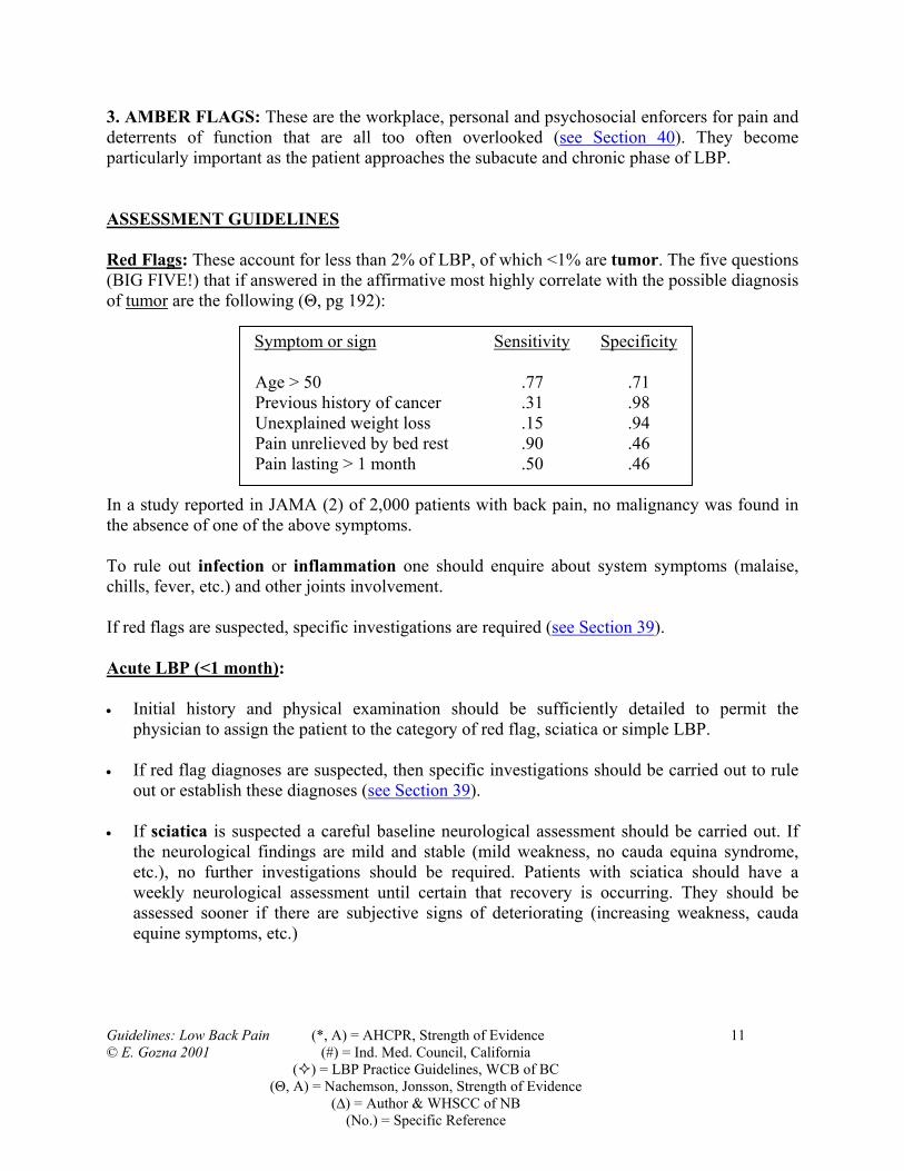
3. AMBER FLAGS: These are the workplace, personal and psychosocial enforcers for pain and deterrents of function that are all too often overlooked (see Section 40). They become particularly important as the patient approaches the subacute and chronic phase of LBP. ASSESSMENT GUIDELINES Red Flags: These account for less than 2% of LBP, of which <1% are tumor. The five questions (BIG FIVE!) that if answered in the affirmative most highly correlate with the possible diagnosis of tumor are the following (Θ, pg 192):
Symptom or sign Sensitivity Specificity
Age > 50 .77 .71 Previous history of cancer .31 .98 Unexplained weight loss .15 .94 Pain unrelieved by bed rest .90 .46 Pain lasting > 1 month .50 .46
In a study reported in JAMA (2) of 2,000 patients with back pain, no malignancy was found in the absence of one of the above symptoms. To rule out infection or inflammation one should enquire about system symptoms (malaise, chills, fever, etc.) and other joints involvement. If red flags are suspected, specific investigations are required (see Section 39). Acute LBP (<1 month): • Initial history and physical examination should be sufficiently detailed to permit the
physician to assign the patient to the category of red flag, sciatica or simple LBP. • If red flag diagnoses are suspected, then specific investigations should be carried out to rule
out or establish these diagnoses (see Section 39). • If sciatica is suspected a careful baseline neurological assessment should be carried out. If
the neurological findings are mild and stable (mild weakness, no cauda equina syndrome, etc.), no further investigations should be required. Patients with sciatica should have a weekly neurological assessment until certain that recovery is occurring. They should be assessed sooner if there are subjective signs of deteriorating (increasing weakness, cauda equine symptoms, etc.)
Guidelines: Low Back Pain (*, A) = AHCPR, Strength of Evidence 11 © E. Gozna 2001 (#) = Ind. Med. Council, California ( ) = LBP Practice Guidelines, WCB of BC (Θ, A) = Nachemson, Jonsson, Strength of Evidence (∆) = Author & WHSCC of NB (No.) = Specific Reference

Guidelines: Low Back Pain (*, A) = AHCPR, Strength of Evidence 12 © E. Gozna 2001 (#) = Ind. Med. Council, California ( ) = LBP Practice Guidelines, WCB of BC (Θ, A) = Nachemson, Jonsson, Strength of Evidence (∆) = Author & WHSCC of NB (No.) = Specific Reference
• Patient should be assessed every 1-2 weeks to test diagnoses (red flag, sciatica, LBP) and to document progress. Follow-up should continue until patient has returned to modified or full work activities.
Subacute LBP (1-3 months): • If the patient presents with steady pain of greater than one-month duration a careful focused
history and physical examination should be carried out to rule out red flags, sciatica or other serious spinal pathology (see Section 39).
• If no red flags, a careful inquiry about amber flag issues should be carried out and these
issues addressed (see Section 40). • Amber flag issues can be serious deterrents of return to work. • WHSCC case manager or medical advisor should be made aware of:
o Any identified red flags o Relevant amber flag issues, o Changes in medical diagnosis o Other medical conditions that may be impeding return to work.
Chronic LBP (> 3 months): • Less than 10% of LBP lasts more than 3 months. Therefore, it is important to carefully
reassess for red flags and investigate as appropriate. • Almost all patients with chronic LBP have amber flag issues and these can become major
obstacles to recovery. The WHSCC case manager or medical consultants are pleased to work along with the primary care physician to help solve these issues (see Section 38). Many of these patients require an interdisciplinary treatment approach (see Section 26).
For management guidelines see Section 2.

Guidelines: Low Back Pain (*, A) = AHCPR, Strength of Evidence 13 © E. Gozna 2001 (#) = Ind. Med. Council, California ( ) = LBP Practice Guidelines, WCB of BC (Θ, A) = Nachemson, Jonsson, Strength of Evidence (∆) = Author & WHSCC of NB (No.) = Specific Reference
LBP DIAGNOSTIC FLAGS CHECKLIST:
RED FLAGS (NIFTI)
NEUROLOGICAL PROGRESSION • Progressive Sciatica • Cauda Equina Syndrome • Spinal Stenosis
INFECTION FRACTURE TUMOR INFLAMATION
AMBER FLAGS WORKPLACE: • Job dissatisfaction: Does the patient enjoy the work? • Human resource issues: Was the patient about to be laid off? • Seasonal Employment: Is the job still available? • Availability of transitional return to work programs: Is this available? • Union contract issues: This can limit employer’s ability to assist. • Unrealistic Job Demands: Covered by Occupational Health and Safety Act • Is there alternative employment? Particular problem in rural areas PERSONAL: • Use of narcotics beyond four weeks of date of injury. • History of excessive alcohol and drug utilization, • History of avoidance type behavior. • Depression or somatization tendencies. • Belief that non-specific pain is harmful. • Belief that passive treatment is beneficial and active treatment harmful. • Involvement in the underground economy? Becoming a big problem! • Limited transferable skills: Can preclude finding alternative employment. • Role Reversal: Has spouse become breadwinner?
DEMOGRAPHICS: • Age > 40 • Heavy smokers • Poor language skills • Heavy utilizers of walk-in clinics • Previous compensable injuries with prolonged disability • Those with history of previous utilization of the welfare system • Those who perceive social support system as an alternative income source • Those with chronic anger and frustration over socio-economic status.

Guidelines: Low Back Pain (*, A) = AHCPR, Strength of Evidence 14 © E. Gozna 2001 (#) = Ind. Med. Council, California ( ) = LBP Practice Guidelines, WCB of BC (Θ, A) = Nachemson, Jonsson, Strength of Evidence (∆) = Author & WHSCC of NB (No.) = Specific Reference
2. MEDICAL MANAGEMENT OVERVIEW Medical management is determined by: 1) the medical diagnosis, 2) phase of LBP and 3) presence of amber flags. Diagnosis: During the course of management, the medical diagnosis can change (i.e., simple back pain can become sciatica, etc.) and new amber flag issues can arise. For this reason at each examination the medical diagnosis, red and amber flag issues have to be retested. The longer a patient continues to experience back pain and remain off work, the more amber flag issues start to predominate. Phase: Having ruled out serious spinal pathology, the phase of the back pain dictates management: • Acute (<1 month): >80% recover in 1 month with no specific Rx. • Subacute (1-3 months): The time to address all issues: medical, workplace, psychosocial. • Chronic (>3 months): <10% of patients but consume >80% of resources. Often require
interdisciplinary treatment.
Amber flags are frequently overlooked and important enforcers of pain and deterrents of function. GUIDELINES • A re-evaluation of medical diagnosis, red and amber flags, treatment effectiveness, patient
compliance, and work status should be performed every 1-2 weeks until patient has returned to modified or full work.
• WHSCC should be made aware of any suspected red or amber flag issues, changes in
diagnosis or other medical conditions that are impeding return to work. ACUTE LBP (<1 MONTH): • Having ruled out Red Flag conditions (see Section 39), there is no need for routine
investigations or specific treatment beyond education, reassurance and encouragement to remain active. Narcotic analgesics and bed rest should be avoided. Over 80 percent of patients with low back pain show signs of getting better within four weeks with absolutely no treatment!

Guidelines: Low Back Pain (*, A) = AHCPR, Strength of Evidence 15 © E. Gozna 2001 (#) = Ind. Med. Council, California ( ) = LBP Practice Guidelines, WCB of BC (Θ, A) = Nachemson, Jonsson, Strength of Evidence (∆) = Author & WHSCC of NB (No.) = Specific Reference
• Bed rest should be kept to an absolute minimum. Bed rest beyond 48 hours is of no benefit (Ref. 3) and most specialists feel rest shouldn’t exceed 24 hours. Other studies have reported on the deleterious physical effects of enforced bed rest (DVT, deconditioning, loss of muscle mass) and the adverse psychological effects of encouraging disability behavior. There is a one percent loss of muscle mass for every day of bed rest and a 15 percent reduction in aerobic capacity after only 10 days of bed rest! Further evidence is accumulating about the positive physical and psychological effects of remaining in the workplace with modified activities.
• Because the prognosis for spontaneous recovery is so strong, there is little justification for
many of the investigations or treatments that have been used in the past for treating acute LBP:
ACUTE, NON RED FLAG LBP NO PROVEN BENEFIT OF:
• X-RAYS • MRI/CT • BLOOD WORK UP • OPOIDS • BED REST • CORSETS • ORTHOTICS
• In the acute phase such interventions as bracing, corsets or orthotics have no benefits over a
program of watchful waiting and encouragement of activity. • Acetaminophen and non-steroidal anti-inflammatory drugs are as effective as opioid
analgesics and muscle relaxants, with less side effects and complications. There is no role for oral steroids in the treatment of acute simple back pain.
• Whereas in the past we treated sciatica with rest, it has now been shown (4) that sciatica
without progressive neurological impairment can be treated in the same way as acute simple back pain.
RECURRENT ACUTE: • These patients can be treated as outlined above until the acute component has settled and
they have returned to normal activities (i.e. workplace with/without modifications). • During the acute recurrent phase it is a higher priority to maintain the patient’s workplace
security than to correct the underlying disturbed biomechanics.

• To reduce the risk of recurrence, an elective physiotherapy biomechanical analysis should be carried out to identify any postural or muscle imbalance problems that should be addressed (see Section 42).
• One cannot overemphasize the importance of patient education and reassurance by treating
clinicians (see Section 5). • Patients must be taught to assume responsibility for maintaining their own fitness. • Correctable workplace issues that could contribute to LBP should be addressed. SUBACUTE LBP (1-3 MONTHS): • Less than 20 percent of people are still experiencing LBP at three months. For those who
enter the subacute phase, it is important to carry out a focused history and examination to rule out Red Flags and address Amber Flag issues. In the subacute phase, Red and Amber Flag conditions become increasingly prevalent:
• Amber flag issues should be identified and addressed. The longer a worker remains disabled
the more influential amber flags become. The WHSCC case manager or medical consultants are pleased to work along with the primary care physician to help solve these issues (see Section 38).
• Recommendations for work restrictions should be based on knowledge of the patient’s working conditions. In the absence of this knowledge, recommendations should be confined to functional limitation based on objective data related to the physician’s assessment. Taking the patient out of the work force can lead to profound long-term personal family hardship and should be avoided where possible. A modified work program is preferable and the WHSCC staff can investigate these alternatives for the physician. The role of the physician is well outlined in the CMA Policy on Return to Work.
• Rehabilitation should encourage patients to understand and gain control of their lumbar
dysfunction instead of allowing pain to control their lives. The physician’s role in providing reassurance is critical as fear and apprehension are major deterrents to recovery.
Guidelines: Low Back Pain (*, A) = AHCPR, Strength of Evidence 16 © E. Gozna 2001 (#) = Ind. Med. Council, California ( ) = LBP Practice Guidelines, WCB of BC (Θ, A) = Nachemson, Jonsson, Strength of Evidence (∆) = Author & WHSCC of NB (No.) = Specific Reference

• Passive treatment modalities (manipulation, massage, TENS, etc.) should be discouraged in preference to active exercise and client education, administered in a positive environment. During this stage, one can address underlying muscle imbalance problems and deconditioning, but the focus of rehabilitation should be on increased activity and functional improvement rather than treating discomfort. The treatment objective for subacute low back pain is increased function not pain elimination!
CHRONIC LBP (BEYOND 3 MONTHS): Though less than 10 percent of patients progress to this stage, it accounts for more than 80 percent of the cost of management (medical, lost time, replacement and retraining).
• Patients who enter the chronic phase require an aggressive workup to sort out the many (red
or amber) factors that are contributing to disability. Management of chronic pain frequently involves an interdisciplinary approach and the primary care physician should be an integral part of this team.
• Chronic pain patients can develop major workplace, personal and psychosocial (amber flag)
issues that can become the major obstacle to recovery and have to be addressed:
All amber flags should be considered (see Section 40) and every issue identified should be addressed in the same manner as if it were a medical diagnosis. The physician should take advantage of the services offered through the WHSCC for patients entering into the chronic phase of LBP. Examples of Amber flag issues that can be addressed include:
• Unrealistic working conditions fall under the Occupational Health and Safety Act. • Concerns about the patient’s ability to resume work can be addressed with work
conditioning, gradual return or modified work programs. • Physical limitations can be quantified with functional capacity testing (see Section 27). • Where physical, psychological and workplace issues start to overlap, the Interdisciplinary
Program offered through the WRC can be very helpful.
These examples illustrate how the WHSCC can be of assistance in sorting out and dealing with workplace, personal and psychosocial issues. Guidelines: Low Back Pain (*, A) = AHCPR, Strength of Evidence 17 © E. Gozna 2001 (#) = Ind. Med. Council, California ( ) = LBP Practice Guidelines, WCB of BC (Θ, A) = Nachemson, Jonsson, Strength of Evidence (∆) = Author & WHSCC of NB (No.) = Specific Reference

Guidelines: Low Back Pain (*, A) = AHCPR, Strength of Evidence 18 © E. Gozna 2001 (#) = Ind. Med. Council, California ( ) = LBP Practice Guidelines, WCB of BC (Θ, A) = Nachemson, Jonsson, Strength of Evidence (∆) = Author & WHSCC of NB (No.) = Specific Reference
3. PLAIN X-RAYS OVERVIEW Plain lumbo-sacral spine (LSS) x-rays are the most commonly ordered investigation for low back pain. Up to 50% of patients with LBP end up having LSS x-rays and it is generally accepted that routine x-rays are greatly overused in the management of routine acute LBP and for following the course of chronic back pain. No firm evidence exists for an association between x-ray findings of arthritis and simple low back pain and every guideline published in the English language since 1987 has stated that ordinary x-rays (in the absence of red flags) have no diagnostic or therapeutic value in managing simple LBP (Θ, pg 196). The following are the commonly ordered routine x-ray studies and their indications:
• Standard antero-posterior and lateral views permit assessment of lumbar alignment, comparison of vertebral body and disc space size, assessment of bony density and architecture, and gross evaluation of soft tissue structures.
• Oblique views are used to detect a spondylolysis. This is an acquired bony defect that can allow the slippage of one vertebrae on another (the slippage itself is called spondylolisthesis)
• Sacroiliac views to evaluate possible ankylosing spondylitis. • Cone views to show specific areas of the spine. • Flexion-extension or bending views to detect abnormalities or movement. GUIDELINES (*B, #, , Θ, ∆) • For acute LBP (<1 month) and in the absence of red flags, routine lumbar spine x-rays are
not required. • For chronic LBP (> 3 months) and in the absence of red flags, repeat serial x-rays are of no
benefit in management. • There is little correlation between the degree of arthritis as documented on x-ray and
the severity of LBP symptoms, clinical course or prognosis. • Plain x-rays of the lumbar spine are recommended for ruling out fractures in patients with:
o Recent significant trauma at any age. o Recent mild trauma in the older patient. o History of prolonged steroid use, osteoporosis. o History of tumor.

Guidelines: Low Back Pain (*, A) = AHCPR, Strength of Evidence 19 © E. Gozna 2001 (#) = Ind. Med. Council, California ( ) = LBP Practice Guidelines, WCB of BC (Θ, A) = Nachemson, Jonsson, Strength of Evidence (∆) = Author & WHSCC of NB (No.) = Specific Reference
• Plain x-rays in combination with Complete Blood Count (CBC), C-reactive protein (CRP) and Erythrocyte Sedimentation Rate (ESR) may be useful for ruling out tumor or infection in patients with acute low back problems when any of the following are present: prior cancer, recent infection, fever, IV drug abuse, prolonged steroid use, low back pain worse with rest, unexplained weight loss.
• Plain x-rays are not as sensitive as bone scans for detecting metastatic tumor and infection
(see Section 18). • Bone scan can be useful to help determine whether a fracture diagnosed on plain x-ray is
recent or remote (i.e., more than 1 year old). (See Section 18)

Guidelines: Low Back Pain (*, A) = AHCPR, Strength of Evidence 20 © E. Gozna 2001 (#) = Ind. Med. Council, California ( ) = LBP Practice Guidelines, WCB of BC (Θ, A) = Nachemson, Jonsson, Strength of Evidence (∆) = Author & WHSCC of NB (No.) = Specific Reference
4. LABORATORY STUDIES OVERVIEW If the patient’s history, age, or examination suggests red flag issues such as cancer, infection, inflammatory arthritis (such as ankylosing spondylitis), or metabolic-endocrinologic disorders and visceral disease, then appropriate laboratory tests may be indicated. The most common are:
• CBC (Complete Blood Count): This is a good screen for many conditions including anemia, leukemia, inflammation, infection or cancer.
• ESR (Erythrocyte Sedimentation Rate): Is very sensitive for inflammatory conditions within the body. It is not specific and therefore cannot separate between infection, cancer or systemic disease as a course of the elevation. ESR tends to be elevated naturally in the elderly and in patients with anemia.
• CRP (C Reactive Protein): The information in this test is not unlike that of the ESR but is more useful for monitoring the acute inflammatory reaction. CRP tends to respond more rapidly then ESR to changing inflammatory states.
• AP (Alkaline Phosphatase): This enzyme is elevated in active bone disease such as Paget’s disease and bone cancer. It can also be elevated with liver disease but can be distinguished by fractionation.
• Protein Immunoelectrophoresis: This is the most common screen to rule out multiple myeloma (a primary bone cancer).
• Investigations for inflammatory arthritis: There are too many to list but commonly include CBC, ESR, CRP, ANA, rheumatoid factor, uric acid, serum ferritin, etc.
GUIDELINES • Laboratory tests should not be done routinely in the initial evaluation unless an underlying
systemic illness or red flag (see Section 39) is suspected (#, , ∆). • CBC, ESR, CRP, AP and an adequate screen for metastatic bone cancer (see Section 2). • The one primary bone tumor not necessarily picked up on bone scan (most sensitive detector
of tumor) is multiple myeloma for which protein immunoelectrophoresis is usually diagnostic.

5. EDUCATION OVERVIEW
• Inexplicable pain is fear provoking as it threatens the very core of one security! • Fear is an important psychological enforcer of pain and deterrent of function. • Education is the most effective method of reducing fear.
Those entrusted with the management of LBP have a responsibility to alleviate fear through education and provide a positive, reassuring therapeutic milieu (#, , Θ, ∆). GUIDELINES • Once the clinician (physician or therapist) is comfortable that there are no underlying red
flags, the patient should be reassured about the benign nature of most back pain and the importance of increased activity and fitness. Patients should be reassured as to how common simple LBP is and how uncommon serious spinal problems are.
CAUSES OF LBP
1. SIMPLE LBP > 93% 2. RED FLAGS < 2%
1. Tumor < 1% 2. Inflammatory < 1%
3. SCIATICA < 5% • Specific rehabilitation points that the patient should be educated on include:
Rehabilitation instructions: Acute non-red flag LBP
Principles of weight control, posture, back mechanics, and core abdominal strengthening. Reasonable modifications of work, home, and recreational activities. Lifestyle modifications (e.g., diet, exercises, smoking cessation). Specific functional goals (home, recreational and work activities).
• Patient with acute simple LBP, generally require reassurance about the following issues: Guidelines: Low Back Pain (*, A) = AHCPR, Strength of Evidence 21 © E. Gozna 2001 (#) = Ind. Med. Council, California ( ) = LBP Practice Guidelines, WCB of BC (Θ, A) = Nachemson, Jonsson, Strength of Evidence (∆) = Author & WHSCC of NB (No.) = Specific Reference

Guidelines: Low Back Pain (*, A) = AHCPR, Strength of Evidence 22 © E. Gozna 2001 (#) = Ind. Med. Council, California ( ) = LBP Practice Guidelines, WCB of BC (Θ, A) = Nachemson, Jonsson, Strength of Evidence (∆) = Author & WHSCC of NB (No.) = Specific Reference
Reassurance points: Acute non-red flag LBP:
There is no indication of a serious problem and therefore no need for
further investigations. Over 80% of acute LBP disappears within a month, so the patient
can expect recovery. The only treatment highly correlated with recovery of simple LBP is
remaining as active as possible. No medications or other treatments, including home remedies, can
be expected to relieve all the discomfort immediately. The patient may need to modify some daily activities, but it is
important to stay active to prevent deconditioned during recovery. If the patient is one of the few who is slow to recover, then
investigations will be carried out to look for the reason. Even if the LBP is slow resolving, there is little chance (< 3%) of a serious condition being found.
If there is a history of recurrent LBP then after the current episode
has passed there may be some biomechanical problems that need to be addressed.
• All those involved in treatment should provide education but care should be taken to insure
that the patient is not getting conflicting explanations. All players should be singing from the same song sheet (∆).
• Handouts: In general demonstration and personal reinforcement are more successful than a
handout or picture book (#, , ∆).

Guidelines: Low Back Pain (*, A) = AHCPR, Strength of Evidence 23 © E. Gozna 2001 (#) = Ind. Med. Council, California ( ) = LBP Practice Guidelines, WCB of BC (Θ, A) = Nachemson, Jonsson, Strength of Evidence (∆) = Author & WHSCC of NB (No.) = Specific Reference
6. ACTIVITY & WORK MODIFICATIONS OVERVIEW All efforts should be expended to keep the worker in the workplace, if at all possible. This may necessitate activity and workplace modifications. The natural history of acute LBP is spontaneous recovery with minimal intervention. The intent of work modification is to achieve a tolerable comfort level while continuing adequate physical activity to avoid deconditioning and the adverse personal consequences of being taken out of the work force. To be taken out of the work force for a prolonged period of time without a strong medical indication can have serious financial, work culture, and psychological consequences. The importance of keeping the injured in the workplace cannot be overemphasized enough. The psychological and long-term effects of this can be immense, especially if there are ongoing workplace and psychosocial issues (see Section 40 Amber Flags) that can be deterrents to return to work. There is a large body of data to substantiate the positive effects of staying in the work force in a modified job description and minimizing loss of work time (*, #, , Θ, ∆). GUIDELINES • Modified duties are preferable to complete work cessation (#, , ∆). • Physician recommendations should be confined to functional limitation based on the clinical
findings ( , ∆). Recommendations for work modification require that the physician have knowledge of the workplace and the clients normal work duties (see CMA Guidelines)
• Written modified work guidelines should be as specific as possible, and it may be necessary
to contact the employer to discuss alternative work within the prescribed restrictions (#, ∆). The WHSCC case manager or staff can arrange a workplace assessment or contact the employer for the physician if requested (see section 38).
• Patients with work restrictions should be re-evaluated every 1-2 weeks for determination of
work status, response to treatment and for making appropriate decisions concerning progression to full activities (#, ∆).
• The overall goal is to aid recovery while disrupting daily activities as little as possible (#, ∆).
Guidelines: Low Back Pain (*, A) = AHCPR, Strength of Evidence 24 © E. Gozna 2001 (#) = Ind. Med. Council, California ( ) = LBP Practice Guidelines, WCB of BC (Θ, A) = Nachemson, Jonsson, Strength of Evidence (∆) = Author & WHSCC of NB (No.) = Specific Reference
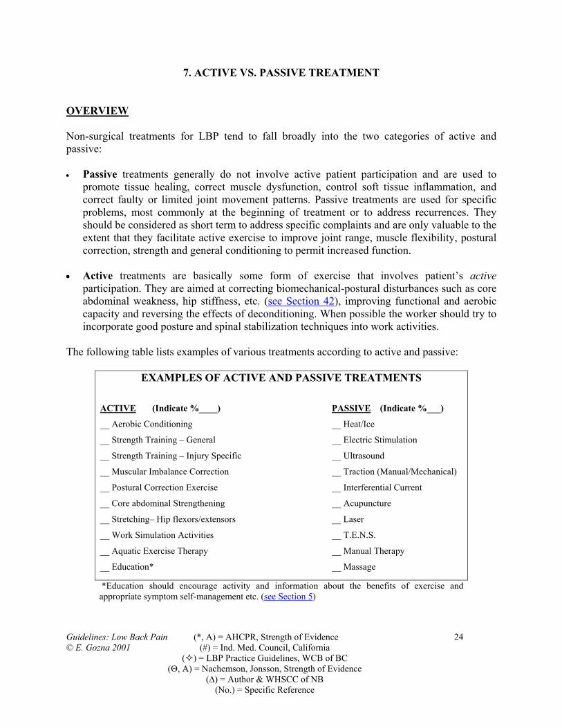
7. ACTIVE VS. PASSIVE TREATMENT OVERVIEW Non-surgical treatments for LBP tend to fall broadly into the two categories of active and passive: • Passive treatments generally do not involve active patient participation and are used to
promote tissue healing, correct muscle dysfunction, control soft tissue inflammation, and correct faulty or limited joint movement patterns. Passive treatments are used for specific problems, most commonly at the beginning of treatment or to address recurrences. They should be considered as short term to address specific complaints and are only valuable to the extent that they facilitate active exercise to improve joint range, muscle flexibility, postural correction, strength and general conditioning to permit increased function.
• Active treatments are basically some form of exercise that involves patient’s active
participation. They are aimed at correcting biomechanical-postural disturbances such as core abdominal weakness, hip stiffness, etc. (see Section 42), improving functional and aerobic capacity and reversing the effects of deconditioning. When possible the worker should try to incorporate good posture and spinal stabilization techniques into work activities.
The following table lists examples of various treatments according to active and passive:
EXAMPLES OF ACTIVE AND PASSIVE TREATMENTS ACTIVE (Indicate %____) PASSIVE (Indicate %___)
__ Aerobic Conditioning __ Heat/Ice
__ Strength Training – General __ Electric Stimulation
__ Strength Training – Injury Specific __ Ultrasound
__ Muscular Imbalance Correction __ Traction (Manual/Mechanical)
__ Postural Correction Exercise __ Interferential Current
__ Core abdominal Strengthening __ Acupuncture
__ Stretching– Hip flexors/extensors __ Laser
__ Work Simulation Activities __ T.E.N.S.
__ Aquatic Exercise Therapy __ Manual Therapy
__ Education* __ Massage
*Education should encourage activity and information about the benefits of exercise and appropriate symptom self-management etc. (see Section 5)

Guidelines: Low Back Pain (*, A) = AHCPR, Strength of Evidence 25 © E. Gozna 2001 (#) = Ind. Med. Council, California ( ) = LBP Practice Guidelines, WCB of BC (Θ, A) = Nachemson, Jonsson, Strength of Evidence (∆) = Author & WHSCC of NB (No.) = Specific Reference
Strength of Evidence: • Active: There is a strong body of data to support the importance of activity in general for
musculoskeletal rehabilitation (*, #, , Θ, ∆). There is conflicting and less quality data comparing the relative merits of the various active interventions.
• Passive: Though there is increasing evidence of the importance of certain passive modalities
for treating specific pain related conditions, currently there is little quality evidence based literature support for many of these.
In view of the paucity of strong evidence based comparative data, the WHSCC position at this time is to encourage therapists to select the treatment program based upon training and experience. However regardless of the treatment chosen, there should be evidence of improved function and progression from passive to active treatments. Passive treatment should not be used exclusively. GUIDELINES *Failure to respond to treatment is an indication for a careful repeat interview and examination to rule out red and amber flag issues (see Section 39, and Section 40). Acute LBP:
• As > 80% of acute LBP will resolve spontaneously with 1 month it should require little physiotherapy beyond: 1. Reassurance 2. Advice to stay as active as possible and to continue normal daily activities. 3. Increase physical activities progressively over a few days or weeks. 4. If patient is working, advise to stay at work or return to work as soon as possible. 5. General advice about the importance of posture, core abdominal strength, hip mobility,
fitness and weight control. Subacute LBP:
• Passive treatments can be used to treat specific conditions at the discretion of the therapist but should not be used exclusively and should have a clearly defined end point.
• When combined active and passive treatments are used, there should be documentation of
progression from passive to active methods. This can be done by utilizing a table such as shown above to record the % in effort and/or time expended in active vs. passive treatments.

Guidelines: Low Back Pain (*, A) = AHCPR, Strength of Evidence 26 © E. Gozna 2001 (#) = Ind. Med. Council, California ( ) = LBP Practice Guidelines, WCB of BC (Θ, A) = Nachemson, Jonsson, Strength of Evidence (∆) = Author & WHSCC of NB (No.) = Specific Reference
• A biomechanical analysis should be carried out identifying specific mobility problems / movement dysfunctions that should be actively addressed. Where indicated these exercises should be incorporated into the workplace and activities of daily living.
• Amber flag issues become increasingly important in subacute LBP and clinicians should be
aware and identify them. (see Section 40). The WHSCC should be made aware of these issues so that they can be addressed.
• All effort should be expended to prevent patients from progressing into the chronic phase of
LBP. Chronic LBP:
• Because of the multifactorial nature of chronic pain, these patients usually require an interdisciplinary program (see Section 26). The physiotherapy is an important part of the treatment team.
• Physiotherapy emphasis in the management of chronic pain is not pain elimination but
encouraging increased activity and function. This may include patient education on symptom self-management strategies and overcoming fear/pain avoidance behavior.
• Amber flag (workplace and psychosocial) issues are invariably present in chronic LBP and
should be enquired about (see Section 40). The WHSCC should be made aware of these issues so that they can be addressed.
Guidelines: Low Back Pain (*, A) = AHCPR, Strength of Evidence 27 © E. Gozna 2001 (#) = Ind. Med. Council, California ( ) = LBP Practice Guidelines, WCB of BC (Θ, A) = Nachemson, Jonsson, Strength of Evidence (∆) = Author & WHSCC of NB (No.) = Specific Reference
8. MEDICATIONS OVERVIEW For the management of LBP, there is a large body of data to suggest that Acetaminophen and non-steroidal anti-inflammatory medications (NSAIDs) are equally effective as opioids and muscle relaxants and have significantly less side effects and predisposition to habituation. Care should be taken in prescribing narcotics or other potentially addictive analgesics in patients with depression or a history of drug / alcohol abuse. There is considerable evidence that there is little difference between the efficacies of the various brands of NSAID or muscle relaxants. GUIDELINES A) ACETAMINOPHEN
• Acetaminophen is the safest medication for first line analgesia for acute low back pain and sciatica (#, , ∆).
B) NON-STEROIDAL ANTI-INFLAMMATORY DRUGS (NSAIDS)
• There is strong evidence (level A) that NSAIDs provide effective pain relief for acute LBP (Θ, pg 249).
• There is limited evidence (level C) that NSAIDs do not provide effective relief of sciatic
pain (Θ, pg 249).
• There is strong evidence (level A) that different types of NSAIDs are equally effective (Θ, pg 249).
C) OPIOIDS (NARCOTIC ANALGESICS)
• Oral opioids may be used for severe disabling pain in acute low back problems, but their routine use is not appropriate. It is recommended that oral opioids be used for no longer than 2 weeks and be taken on a regular schedule (time contingent) rather than as needed for pain (#, , ∆).
• The routine use of oral opioids is not recommended for chronic low back problems. Their
use should be limited to objectively document acute exacerbations with a set time frame of use (#, , ∆).
• Patients should be cautioned about the potential harm of physical dependence and
possible side effects (#, , ∆).

Guidelines: Low Back Pain (*, A) = AHCPR, Strength of Evidence 28 © E. Gozna 2001 (#) = Ind. Med. Council, California ( ) = LBP Practice Guidelines, WCB of BC (Θ, A) = Nachemson, Jonsson, Strength of Evidence (∆) = Author & WHSCC of NB (No.) = Specific Reference
• Use of injectable opioids is inappropriate (#, , ∆).
D) MUSCLE RELAXANTS • Routine use of muscle relaxants is not appropriate for low back pain (#, , ∆, Θ).
• Muscle relaxants appear to be no more effective than NSAIDs in treating acute low back
problems and are associated with a much higher incidence of side effects and carry a significant risk of habituation (#, , ∆, Θ, pg 254).
• Patients on muscle relaxants should be cautioned about possible side effects (including
drowsiness and confusion) that might interfere with job performance/safety and active therapy goals (#, , ∆, Θ).
• If muscle relaxants are prescribed, it is recommended that they be used for no longer than
1 week. Patients should also be warned that there is a significant risk of habituation and dependency even after courses as short as 1 week (Θ, pg 254).
E) ORAL CORTICOSTEROIDS
• There is no role for systemic corticosteroids in the treatment of acute, subacute or chronic back pain (#, , ∆, Θ).
F) PHENYLBUTAZONE
• Phenylbutazone is contraindicated due to risks of bone marrow suppression (* C, , ∆).
G) ANTIDEPRESSANTS • There is general acceptance that antidepressants may be useful for treatment of chronic
low back pain (#, , ∆) especially where there are features of sleep disturbance and /or depressive features ( , ∆). There is, however, some evidence that antidepressants may not be effective (ΘB, pg 254).
Guidelines: Low Back Pain (*, A) = AHCPR, Strength of Evidence 29 © E. Gozna 2001 (#) = Ind. Med. Council, California ( ) = LBP Practice Guidelines, WCB of BC (Θ, A) = Nachemson, Jonsson, Strength of Evidence (∆) = Author & WHSCC of NB (No.) = Specific Reference
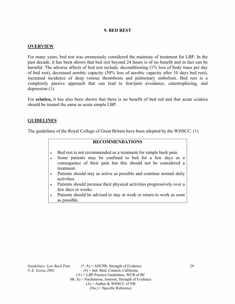
9. BED REST OVERVIEW For many years, bed rest was erroneously considered the mainstay of treatment for LBP. In the past decade, it has been shown that bed rest beyond 24 hours is of no benefit and in fact can be harmful. The adverse affects of bed rest include: deconditioning (1% loss of body mass per day of bed rest), decreased aerobic capacity (50% loss of aerobic capacity after 10 days bed rest), increased incidence of deep venous thrombosis and pulmonary embolism. Bed rest is a completely passive approach that can lead to fear/pain avoidance, catastrophizing, and depression (1). For sciatica, it has also been shown that there is no benefit of bed red and that acute sciatica should be treated the same as acute simple LBP. GUIDELINES The guidelines of the Royal College of Great Britain have been adopted by the WHSCC: (1)
RECOMMENDATIONS
• Bed rest is not recommended as a treatment for simple back pain. • Some patients may be confined to bed for a few days as a
consequence of their pain but this should not be considered a treatment.
• Patients should stay as active as possible and continue normal daily activities.
• Patients should increase their physical activities progressively over a few days or weeks.
• Patients should be advised to stay at work or return to work as soon as possible.
Guidelines: Low Back Pain (*, A) = AHCPR, Strength of Evidence 30 © E. Gozna 2001 (#) = Ind. Med. Council, California ( ) = LBP Practice Guidelines, WCB of BC (Θ, A) = Nachemson, Jonsson, Strength of Evidence (∆) = Author & WHSCC of NB (No.) = Specific Reference
10. PHYSIOTHERAPY OVERVIEW Physiotherapy can divide into four broad categories: 1. Modalities are passive treatments that often involve devices to promote tissue healing relieve
pain, inflammation and muscle tension. These include for example ice, heat, ultrasound, low power laser treatment, TENS and acupuncture.
2. Manual therapy manipulation involves high velocity, low amplitude, short lever arm thrusts and passive movements to restore accessory joint mobility to improve physiological range of motion and restore joint kinematics.
3. Exercise involves active muscle contraction and/or stretching to improve flexibility, strength, and aerobic conditioning and posture. Its purpose is to restore normal movement and muscle balance, which should result in improved function.
4. Education is an extremely important part of musculoskeletal rehabilitation. Inexplicable pain
is fear provoking! The therapist’s role in education cannot be underestimated. Education should include information about their condition and normal tissue healing, the importance of exercise and activity, self-management of their symptoms, and safe work practices and good body mechanics to prevent recurrence.
Though there is a considerable body of data evolving to support the various treatment forms for specific conditions, there is little strong evidence based comparative information. In view of this paucity of data, the WHSCC position is to encourage therapists to select the treatment program based upon training and experience. However regardless of the treatment chosen, there should be evidence of appropriate progression from passive to active treatments (see Section 7) and improved function. GUIDELINES Physiotherapist:
• Effective physiotherapy should emphasize progressive exercises with decreasing emphasis on passive treatments (*, #, , ∆, Θ).
• Passive modalities can be used for short periods of time to supplement active treatments but
they should not be used exclusively and for a limited time only (*, #, , ∆, Θ).

Guidelines: Low Back Pain (*, A) = AHCPR, Strength of Evidence 31 © E. Gozna 2001 (#) = Ind. Med. Council, California ( ) = LBP Practice Guidelines, WCB of BC (Θ, A) = Nachemson, Jonsson, Strength of Evidence (∆) = Author & WHSCC of NB (No.) = Specific Reference
• A biomechanical analysis should be part of any LBP assessment. Most patients with LBP develop biomechanical dysfunction and deconditioning that should be addressed in order to prevent recurrent symptoms. These should be addressed through exercise and education (*, #, , ∆,).
• The Physiotherapy Clinical Coordinator is pleased to discuss or assist external therapists with
any problems that may arise in managing patients (see Section 38). Referring physician:
• Though the referring physician does not need to know the minute details of PT treatment, he/she should:
o Have some appreciation of the underlying treatment principles o Know whether the treatment program is primarily passive or active o Insure that there is progression from passive to active treatment.
• If the physician has questions or concerns about their patient’s physiotherapy, it should be
discussed with the PT involved. The patient should feel that there is unanimity of opinion regarding treatment.
• The physician should enquire about the patient’s activities and exercises between
physiotherapy sessions. It is important that there be evidence that the patient is assuming responsibility for self-care and fitness and is not developing pain avoidance behavior.
• For the same reason that it is erroneous to say “the patient has seen a doctor with no success
therefore doctors are of no help”, it is fallacious to assume that because a patient has had unsuccessful PT that no other type of PT or a different therapist will not be successful.
Since the treatment “team” consists of the patient, treating physician, other medical and non-medical clinicians and the WHSCC, don’t hesitate to contact the WHSCC at any time if you have questions or concerns (see Section 38).

Guidelines: Low Back Pain (*, A) = AHCPR, Strength of Evidence 32 © E. Gozna 2001 (#) = Ind. Med. Council, California ( ) = LBP Practice Guidelines, WCB of BC (Θ, A) = Nachemson, Jonsson, Strength of Evidence (∆) = Author & WHSCC of NB (No.) = Specific Reference
11. SPINAL MANIPULATION OVERVIEW Spinal manipulation may involve many different techniques including: lumbar manipulation or adjustments, manual therapy, mobilization, therapeutic massage, craniosacral manipulation, etc. However for this guideline, manipulation is defined as manual therapy in which loads are applied to the spine using short or long lever methods. The selected joint is moved to its end range of voluntary motion, followed by application of impulse loading. The therapeutic objectives of manipulation include symptomatic relief and functional improvement. GUIDELINES • Acute LBP usually recovers spontaneously without intervention. However if it is not
resolving as anticipated manipulative therapy should be considered (ΘB, pg 264). • As manipulation is a passive form of treatment, it should be used for short periods only and
preferably in conjunction with active treatment. • There should be evidence of improved function, with an ultimate goal of discontinuation of
manipulation (see Section 7) (#, , ∆). • There is insufficient evidence to recommend manipulation for patients with radiculopathy
(*C, , ∆,). • Manipulation should not be carried out in patients with severe or progressive sciatica in view
of the risk of neurological complications (* B, #, , ∆, Θ, pg 262). • There is no evidence to support the use of manipulation for preventive treatment (#, , ∆,).

Guidelines: Low Back Pain (*, A) = AHCPR, Strength of Evidence 33 © E. Gozna 2001 (#) = Ind. Med. Council, California ( ) = LBP Practice Guidelines, WCB of BC (Θ, A) = Nachemson, Jonsson, Strength of Evidence (∆) = Author & WHSCC of NB (No.) = Specific Reference
12. TRANSCUTANEOUS ELECTRICAL NERVE STIMULATION
OVERVIEW TENS is a battery-operated device worn by the patient, which provides electrical pulses delivered to sensory nerves via the skin and other tissue using surface electrodes. Its therapeutic objective is to provide symptomatic pain relief. In theory it produces a stimulation to the nervous system, which modifies pain perception. As with all passive therapeutic options that offer symptomatic relief, TENS is only valuable to the extent that it facilitates the client’s participation in functional activities and active exercises. The literature does not support the exclusive use of TENS for treatment of simple LBP (#, , Θ C, pg 262). GUIDELINES • Acute LBP: TENS has little role in acute LBP, though it may occasionally be useful for
short-term symptom relief in severe acute low back pain. If no functional and symptomatic benefit has been demonstrated after 2 weeks, treatment should be discontinued (#, , ∆).
• Chronic LBP: TENS is a passive modality that should not be used as the sole form of
treatment. It should be combined with an active program (see Section 7) that emphasizes progressive exercises and functional restoration (#, , ∆). The WHSCC criteria for long term use of TENS (e.g. home unit) includes 50% pain relief that has at least 1-hour carry over, or significant decrease in pain medication, improved function or improved sleep.

Guidelines: Low Back Pain (*, A) = AHCPR, Strength of Evidence 34 © E. Gozna 2001 (#) = Ind. Med. Council, California ( ) = LBP Practice Guidelines, WCB of BC (Θ, A) = Nachemson, Jonsson, Strength of Evidence (∆) = Author & WHSCC of NB (No.) = Specific Reference
13. TRACTION (IN HOSPITAL) OVERVIEW Traction for low back pain or sciatica involves the application of tensile forces (intermittent or continuous) along the axis of the spine in an attempt to reduce symptoms or relieve pressure on nerve roots. This is generally administered in two ways: 1. Mechanical traction using an external tensioning device, such as a canvass girdle applied
around the pelvis and attached to weights hung at the foot of the bed. This is not commonly done today but is still used in some hospital and outpatient environments.
2. Manually applied traction as part of a Manual Therapy program. GUIDELINES • Acute: Evidence is lacking for the efficacy of most forms of traction for treating low back
problems. There are few RCTs (Randomized Controlled Trial) on the treatment of acute LBP and they are of small size, poor quality and conflicting evidence (Θ C, pg 263). It is not possible to make any judgments on its effectiveness in acute LBP.
• Chronic: There is strong evidence (level A) that traction is not effective in treating chronic LBP (Θ A, pg 295).
• Passive modalities such as traction should not be used as the sole form of treatment. They
should be combined with an active treatment program, which emphasizes progressive exercises with a decreasing frequency of passive treatments (#, , ∆).
Guidelines: Low Back Pain (*, A) = AHCPR, Strength of Evidence 35 © E. Gozna 2001 (#) = Ind. Med. Council, California ( ) = LBP Practice Guidelines, WCB of BC (Θ, A) = Nachemson, Jonsson, Strength of Evidence (∆) = Author & WHSCC of NB (No.) = Specific Reference
14. ACUPUNCTURE / DRY NEEDLING OVERVIEW The therapeutic objective of acupuncture is to reduce pain control inflammation and reduce muscle spasm. By definition acupuncture includes all types of “dry needling” (i.e. no medication is injected) where needles are inserted into skin, subcutaneous tissues, muscles or ligaments. There are several acupuncture approaches: The Western approach is based on anatomical relationships between the acupuncture
point and the injured body part and involves stimulating the nerves in the part of the body that has been injured. The needles are inserted and stimulated either manually or with an electric current. High frequency stimulation blocks pain, and low frequency stimulation stimulated production of endorphins, which control pain and the body’s anti-inflammatory response. Most physiotherapists utilize this approach.
Traditional Chinese Medicine Approach is based on the eastern philosophy of energy flowing in the 12 meridians of the body and dysfunction being caused by an imbalance in energy flow. Though some physiotherapists use the traditional Chinese method, it is most commonly used by acupuncturists.
As with all passive therapeutic options that offer symptomatic relief, they are only valuable to the extent that they facilitate active exercise to correct postural and muscle imbalances and permit increased function. GUIDELINES • In spite of its common use, the evidence of acupuncture’s efficacy in treatment of low back
problems is inconclusive (#, , ∆, Θ, 1). • Passive modalities such as acupuncture should not be used as the sole form of treatment.
They should be combined with an active program (see Section 7), which emphasizes progressive exercises with a decreasing frequency of passive treatment (#, , ∆).

Guidelines: Low Back Pain (*, A) = AHCPR, Strength of Evidence 36 © E. Gozna 2001 (#) = Ind. Med. Council, California ( ) = LBP Practice Guidelines, WCB of BC (Θ, A) = Nachemson, Jonsson, Strength of Evidence (∆) = Author & WHSCC of NB (No.) = Specific Reference
15. LUMBAR SUPPORTS (E.G., CORSETS, SUPPORT BELTS, BRACES, BACK RESTS)
OVERVIEW The theoretical mechanisms by which lumbar supports work includes the following:
1. Provide external support to the spine and prevent excessive movement. 2. Postural reminder to use proper body mechanics. 3. To increase intra-abdominal pressure (which in turn increases spinal stability). 4. Decrease intra-discal pressure.
Lumbar support devices for low back problems include lumbar corsets and support belts, back braces and molded jackets, and back rests for chairs and car seats. There is inconclusive evidence that lumbar corsets or support belts are effective for treating low back problems. There is no evidence that orthotics have prophylactic benefit or prevent reinjury (#, , ∆, Θ). GUIDELINES • A brief trial (1-3 days) of immobilization with lumbar supports may provide symptomatic
relief of pain in severe acute low back problems. • We recommend the use of a brace only when there is clinical evidence that it produces
symptom relief or to improve proprioceptive awareness. • Passive modalities such as corsets and braces should not be used as the sole form of
treatment. They should be combined with an active program, which emphasizes progressive exercises with a decreasing brace usage (#, , ∆).

Guidelines: Low Back Pain (*, A) = AHCPR, Strength of Evidence 37 © E. Gozna 2001 (#) = Ind. Med. Council, California ( ) = LBP Practice Guidelines, WCB of BC (Θ, A) = Nachemson, Jonsson, Strength of Evidence (∆) = Author & WHSCC of NB (No.) = Specific Reference
16. SHOE INSOLES / LIFTS / ORTHOTICS OVERVIEW Orthotics/inserts: are used to correct local biomechanical problems about the feet. Their influence upon the management of LBP is felt to be secondary to their effect on gait mechanics and posture. In spite of the extensive use of orthotics for LBP there are no good randomized control studies to assist the clinician in prescribing these devices. Orthotics can be prescription, custom made or over the counter inserts. Shoe lifts (or raises): are additions made to the heel or sole of a shoe to increase its height and their therapeutic objective is to compensate for significant limb length inequality. GUIDELINES • Orthotics: There is little evidence-based data upon which to make recommendations about
the role of orthotics in treating low back problems ( , ∆). However if clinically it is felt that foot pain is resulting in gait/posture disturbances that are aggravating back symptoms, then a trial of inserts are justified.
• Shoe lifts: In general shoe lifts are not recommended for treatment of low back problems
when lower limb length differences is <2 cm (* D, , ∆).

17. CT, MRI, MYELOGRAPHY, AND CT-MYELOGRAPHY OVERVIEW These sophisticated radiographic and magnetic imaging techniques are highly sensitive and should only be ordered where there are strong medical indications. The objective of using these investigations is to define surgically remediable anatomic pathological conditions or rule out specific red flag diagnoses (tumors, multiple sclerosis, etc.). They are of no benefit in the management of simple LBP. Because of the high incidence of positive MRI and CT results for disc degeneration, herniation and even spinal stenosis in asymptomatic patients, it is important that these studies not be carried out unless clinically indicated (Θ, pg. 199):
INCIDENCE OF POSITIVE MRIs IN ASSYMPTOMATIC PERSONS Disc Degeneration 85% Disc Protrusion 63% Disc Extrusion 13% Spinal Stenosis 15%
MRIs and CT scans are not indicated for the management of simple non red flag LBP and their high incidence of positive results can result in needless patient fear, apprehension and catastrophizing. Because of their better imaging quality MRI and CT have almost replaced myelography as an investigative tool. CT/MRI and myelogram are occasionally combined (i.e., myelographically enhanced CT/MRI). The relative merits of MRI over CT the diagnosis of specific conditions has not been established, and tends to depend upon specialist’s individual preferences. These tests should generally be used only for patients who present with one of these clinical situations: (∆, ) 1. Progressive sciatica for which surgery is being contemplated. 2. History of neurogenic claudication and progressive neurological deficit. 3. Serious conditions affecting the spine (i.e., cauda equina syndrome, fracture, infection,
tumor, or other mass lesions or defects).
Guidelines: Low Back Pain (*, A) = AHCPR, Strength of Evidence 38 © E. Gozna 2001 (#) = Ind. Med. Council, California ( ) = LBP Practice Guidelines, WCB of BC (Θ, A) = Nachemson, Jonsson, Strength of Evidence (∆) = Author & WHSCC of NB (No.) = Specific Reference
Guidelines: Low Back Pain (*, A) = AHCPR, Strength of Evidence 39 © E. Gozna 2001 (#) = Ind. Med. Council, California ( ) = LBP Practice Guidelines, WCB of BC (Θ, A) = Nachemson, Jonsson, Strength of Evidence (∆) = Author & WHSCC of NB (No.) = Specific Reference
As the above conditions will usually require specialist involvement, we recommend that the choice of investigation be made in consultation with the specialist. There is no evidence that MRI has improved the treatment of simple LBP. GUIDELINES • Because of the high incidence of false positive results, the routine use of CT/MRI for patient
reassurance (in the absence of suspected red flag) is not justified and can lead to needless fear and illness behavior (∆, Θ, pg. 199).
• CT/MRI are of no assistance in managing simple LBP. There is no correlation between
MRI/CT findings and the degree of pain or disability experienced by these patients (∆, Θ, pg. 198).
• Cauda equina syndrome and progressive major motor weakness are indications for prompt
use of CT, MRI, myelography, or CT-myelography. Because these problems may require prompt surgical intervention, the imaging studies are best planned in discussion with a spinal surgeon (* C, , ∆).
• Unstable fracture, infection, tumor or other space-occupying lesions (i.e., red flags) are
indications for CT, MRI, myelography, or CT-myelography in consultation with the appropriate specialist (* C, , ∆).

Guidelines: Low Back Pain (*, A) = AHCPR, Strength of Evidence 40 © E. Gozna 2001 (#) = Ind. Med. Council, California ( ) = LBP Practice Guidelines, WCB of BC (Θ, A) = Nachemson, Jonsson, Strength of Evidence (∆) = Author & WHSCC of NB (No.) = Specific Reference
18. BONE SCAN OVERVIEW The principle reason for obtaining a bone scan is to investigate suspected spinal tumor, infection, or occult fracture and to differentiate them from common benign pathologies, such as degenerative or developmental changes. It is of no benefit in the management of simple LBP unless there are red flags or a history of trauma. A bone scan involves intravenous injection of radioactive compounds (technetium-99m), which is concentrated in areas of increased bone metabolic activity (i.e. fracture, tumor, infection, inflammation). Gamma detectors are used to delineate the areas of increased bone uptake. GUIDELINES (* C, , ∆, Θ A, pg 220) • A bone scan is indicated to investigate (see Section 39 Red Flags) when these are suspected
from findings on medical history, physical examination, or collaborative lab tests or imaging studies.
• Bone scans are contraindicated during pregnancy. • Bone scan is felt to be more sensitive than routine x-ray for early detection of infection or
malignant bone involvement. • Bone scans can help distinguish recent fractures from remote healed fractures or
developmental bone deformities. This can be important in sorting out issues of causation or work relatedness.

Guidelines: Low Back Pain (*, A) = AHCPR, Strength of Evidence 41 © E. Gozna 2001 (#) = Ind. Med. Council, California ( ) = LBP Practice Guidelines, WCB of BC (Θ, A) = Nachemson, Jonsson, Strength of Evidence (∆) = Author & WHSCC of NB (No.) = Specific Reference
19. ELECTRODIAGNOSTIC STUDIES (ELECTROMYOGRAPHY, NERVE CONDUCTION STUDIES AND SENSORY
EVOKED POTENTIALS) OVERVIEW
The diagnosis of sciatica or neuropathy can usually be made based upon history and physical examination. However, in certain situations electrodiagnostic studies are indicated to sort out other neurological conditions or primary muscle diseases that can be confused with sciatica. Most commonly, electrodiagnostic studies are carried out to:
1. Rule out peripheral nerve entrapment syndromes and document degrees of conduction impairment (common peroneal nerve, tarsal tunnel syndrome).
2. Rule out diagnosis of peripheral neuropathy. 3. Obtain objective confirmation of the level (s) of radiculopathy. 4. Monitor the course of recovery following a nerve injury. The specific objectives of electrodiagnostic studies for low back problems are as follows:
1. Needle electromyography (EMG) used to assess acute and chronic nerve root dysfunction, myelopathy, and myopathy.
2. Surface EMG, used to assess acute and chronic recruitment patterns during static or dynamic tasks using surface electrodes instead of needle insertion.
3. Sensory evoked potentials (SEP), used to assess sensory neurons in peripheral and spinal cord pathways.
4. Nerve conduction studies, used to assess acute and chronic peripheral entrapment neuropathies that may mimic radiculopathy.
GUIDELINES • Electrodiagnostic testing are not particularly useful for diagnosing the exact level of a nerve
root lesion (low levels of sensitivity) and are recommended only when radiological and clinical findings conflict (Θ pg 217).
• Electrodiagnostic testing is most useful for exclusion of more distal nerve damage
(neuropathy or nerve entrapment in the extremities) and for verification of subjective muscle weakness by needle EMG and patients presenting with pain inhibition or lack of cooperation (Θ pg 217).

Guidelines: Low Back Pain (*, A) = AHCPR, Strength of Evidence 42 © E. Gozna 2001 (#) = Ind. Med. Council, California ( ) = LBP Practice Guidelines, WCB of BC (Θ, A) = Nachemson, Jonsson, Strength of Evidence (∆) = Author & WHSCC of NB (No.) = Specific Reference
• Because the accuracy of electrodiagnostic studies is very dependent on the skill of the examiner, it is recommended that they be used only in consultation with the appropriate specialist referrals (Neurologist or Physiatrist skill in interpreting electrodiagnostic data) ( , ∆).

Guidelines: Low Back Pain (*, A) = AHCPR, Strength of Evidence 43 © E. Gozna 2001 (#) = Ind. Med. Council, California ( ) = LBP Practice Guidelines, WCB of BC (Θ, A) = Nachemson, Jonsson, Strength of Evidence (∆) = Author & WHSCC of NB (No.) = Specific Reference
20. PSYCHOSOCIAL ASSESSMENT OVERVIEW There is strong evidence that psychological, social, and economic (i.e. nonphysical) factors can markedly interfere with response to treatment (including surgery) and rehabilitation following injuries. With respect to LBP these are referred to as amber flag issues. Amber flags (see Section 40) become increasingly important as the patient passes from the acute into the subacute and chronic phase of simple LBP. It is recommended that clinicians be aware of these factors, especially in patients whose recovery of activity tolerance following an acute low back problem seems delayed. GUIDELINES • Delayed recovery, non-compliance or non-response to appropriate treatment of low back
problems, should suggest that the presence of amber flag issues such as job dissatisfaction, depression, substance abuse and symptom magnification may be contributing (#, , ∆).
• The presence of several “non-organic” symptoms or physical signs (see Section 39) may
suggest the need for further psychological testing (#, , ∆). The WHSCC should be made aware of the findings and can arrange an appropriate evaluation.
• In the absence of serious underlying spinal pathology, the inability to regain tolerance of
required activities may indicate that unrealistic expectations or psychosocial factors need to be explored before considering referral for a more extensive evaluation or treatment program (*D, #, , ∆).

Guidelines: Low Back Pain (*, A) = AHCPR, Strength of Evidence 44 © E. Gozna 2001 (#) = Ind. Med. Council, California ( ) = LBP Practice Guidelines, WCB of BC (Θ, A) = Nachemson, Jonsson, Strength of Evidence (∆) = Author & WHSCC of NB (No.) = Specific Reference
21. ERGONOMIC EVALUATION (WORKSITE ASSESSMENT)
OVERVIEW An ergonomic evaluation involves adapting the design of a work task, workplace or workstation to maximize productivity while reducing operator fatigue and discomfort. Other terms for ergonomics include biotechnology, human engineering and human factors engineering. Occupational Therapists (OT) or Ergonomists with most commonly perform these assessments. Chronic or recurrent low back strains can be the result of correctable workplace factors. If this is suspected a worksite evaluation can be carried out either by one of the WHSCC staff Occupational Therapists or a community consultant. GUIDELINES • Ergonomic evaluation is appropriate when job description, or workplace inspection suggests
physical work factors may be contributing to the development or exacerbation of low back problems or preventing return to full work (#, , ∆).
• Clinicians contact the WHSCC (see Section 38) if it is felt that such an assessment is
indicated.
Guidelines: Low Back Pain (*, A) = AHCPR, Strength of Evidence 45 © E. Gozna 2001 (#) = Ind. Med. Council, California ( ) = LBP Practice Guidelines, WCB of BC (Θ, A) = Nachemson, Jonsson, Strength of Evidence (∆) = Author & WHSCC of NB (No.) = Specific Reference
22. WORK-CONDITIONING PROGRAMS
OVERVIEW Work-conditioning programs (WCP) are tailored programs, which emphasize active functional restoration and education aimed at re-integrating the patient back into the work force. The program is delivered up to 4 hours/day, and five days/week for up to 30 treatments or 6 weeks. The physiotherapy treatment component is basically exercise and education to assist the client to return to work. Education is given individually and/or in a group setting on proper body mechanics, normal healing processes, self-management, injury prevention, safe work practices and promotion of the benefit of early return to work. Exercise programs are designed to prepare the worker to meet job demands by improving flexibility, strength, endurance and general/aerobic conditioning. Work Related tasks may also be part of the treatment plan. The Occupational Therapy component of the program is coordinated by the regional OT. Communication is ongoing with the physiotherapist, and as soon as the clients are able, they begin transitional return to work while attending physiotherapy. GUIDELINES • A modified work program is preferable to rehabilitation carried out in a clinical setting
outside the workplace (∆, , Θ). • There is little indication for treatment of acute LBP in a formal work-conditioning program
(∆, , Θ). • Work conditioning is indicated in the subacute or early chronic phase of LBP for patients
with no amber flag issues or with limited issues that can be addressed in the primary care environment ( , ∆). It is part of the WHSCC continuum of care for clients with soft tissue injuries who have no planned return to work by 6-8 weeks post injury.
• Patients with chronic LBP and significant psychosocial (amber flag) issues are better treated
in an interdisciplinary environment (see Section 26) ( , ∆). • WCP extensions should rarely occur. Instead, if a patient has failed to recover sufficiently to
manage a safe return to work, consideration should be given to the need for further medical management, an interdisciplinary medical rehabilitation programs or chronic pain programs ( , ∆).

Guidelines: Low Back Pain (*, A) = AHCPR, Strength of Evidence 46 © E. Gozna 2001 (#) = Ind. Med. Council, California ( ) = LBP Practice Guidelines, WCB of BC (Θ, A) = Nachemson, Jonsson, Strength of Evidence (∆) = Author & WHSCC of NB (No.) = Specific Reference
23. DISCECTOMY (STANDARD / MICRO / PERCUTANEOUS)
OVERVIEW Discectomy implies the surgical removal of the fragment of herniated disc material that is impinging on a nerve root in conjunction with removal of any disc material that might be at risk of herniation in the future. The therapeutic objective is to relieve pressure on nerve roots and reduce pain and possible weakness and/or numbness in the lower extremities. Discectomy is somewhat of a misnomer, as the term suggests complete removal of the disc. A discectomy involves removal of less than ½ of the central portion (nucleus) of the disc and the outer portion (annulus) is left intact. Patients should be aware that the surgery is being carried out to relieve their sciatica (i.e., pain and paresthesia below the knees) and does not treat the degenerative changes occurring within the spine itself. Discectomy is not carried out for back pain! It is important to remember that the natural history of sciatica is one of spontaneous symptom resolution without the need for surgical intervention. The mere presence of sciatica is not an indication for surgery! GUIDELINES • Except for acute neurological deterioration, surgery for acute sciatica within 12 weeks is
unwarranted. If the patient is showing any clinical signs of improvements, surgery should be deferred (#, , ∆).
• Discectomy is rarely, if ever, indicated for low back pain without sciatica (* A, #, , ∆, Θ A,
pg 254). • Surgical treatment is appropriate for patients with sciatica:
1. Who are unresponsive to an appropriate 6-12 weeks of active non-surgical treatment,
AND
2. Who have clinical evidence of a specific nerve root irritation or impaired conduction with intervertebral disc herniation confirmed at the corresponding level and side by findings on CT or MRI,
Guidelines: Low Back Pain (*, A) = AHCPR, Strength of Evidence 47 © E. Gozna 2001 (#) = Ind. Med. Council, California ( ) = LBP Practice Guidelines, WCB of BC (Θ, A) = Nachemson, Jonsson, Strength of Evidence (∆) = Author & WHSCC of NB (No.) = Specific Reference
AND 3. Who do not have any significant physical and/or psychological co-
morbidity that might lead to a poor surgical outcome
AND 4. Who are showing no signs of improvement (#, , ∆, Θ)
• There is strong evidence that microdiscectomy and standard discectomy give broadly
comparable clinical outcomes (Θ A pg 314). There are no significant differences in perioperative bleeding, length of inpatient stay or outcome (Θ B pg 315).
• There is limited and contradictory evidence that automated percutaneous discectomy gives
poorer clinical results than the alternative surgical techniques (Θ C pg 315).

Guidelines: Low Back Pain (*, A) = AHCPR, Strength of Evidence 48 © E. Gozna 2001 (#) = Ind. Med. Council, California ( ) = LBP Practice Guidelines, WCB of BC (Θ, A) = Nachemson, Jonsson, Strength of Evidence (∆) = Author & WHSCC of NB (No.) = Specific Reference
24. SPINAL STENOSIS DECOMPRESSION OVERVIEW Spinal stenosis is of two types: 1) Central and 2) Peripheral. Central spinal stenosis is a narrowing of the central portion of the spinal canal producing bilateral leg symptoms, whereas peripheral spinal stenosis is a narrowing of the bony canal through which the nerve roots exits with resultant one-sided (unilateral) symptoms.
Spinal stenosis is often over diagnosed on CT and MRI! From 15-20% of asymptomatic people will have a radiographic MRI or CT diagnosis of spinal stenosis (Θ, pg 199). There fore unless there are strong clinical symptoms or findings to suggest symptomatic stenosis this radiographic diagnosis can be treated as an incidental finding and disregarded. Symptomatic spinal stenosis is not common in the working age population and is usually seen in patients over the age of sixty, as an age-related degenerative change. The primary symptoms of severe spinal stenosis are neurogenic claudication (bilateral or unilateral depending on whether the stenosis is central or peripheral) and can be associated with neurological impairment. Spinal stenosis usually is not associated with signs of nerve irritation Surgery for spinal stenosis may involve various combinations of soft tissue and bone removal to decompress the nerve roots and cauda equina and can include discectomy (see Section 23) and/or spinal fusion to stabilize the spine. GUIDELINES • MRI documentation of a small spinal canal is in itself not an indication for surgery. There is
a 15-20% incidence of MRI documented spinal stenosis in asymptomatic patients over the age of 40 (Θ pg 199). Hence, it is extremely important that surgical procedures for spinal stenosis not be carried out on patients without strong indication of neurological compromise (* D, #, , ∆, Θ).
• Surgical decisions for patients with spinal stenosis should not be based solely on imaging
tests, but should be based on the clinical presentation, including persistent neurogenic claudication symptoms, and demonstrable neurological compromise (* D, #, , ∆, Θ).
• Elderly patients with spinal stenosis who are not compromised in the activities of daily life
and without hard neurological findings can often be managed with conservative treatments. Decisions on treatment should take into account the patient’s lifestyle, preference, other medical problems and risks of surgery (* D, #, , ∆, Θ).
Guidelines: Low Back Pain (*, A) = AHCPR, Strength of Evidence 49 © E. Gozna 2001 (#) = Ind. Med. Council, California ( ) = LBP Practice Guidelines, WCB of BC (Θ, A) = Nachemson, Jonsson, Strength of Evidence (∆) = Author & WHSCC of NB (No.) = Specific Reference
25. SPINAL FUSION OVERVIEW Lumbar spinal fusion surgeries use bone grafting (often in conjunction with internal fixation devices) to immobilize one or more adjacent spinal segments. The therapeutic objective of spinal fusion surgery is to reduce pain by preventing movement between the fused vertebrae. After more than eighty years of use, there is still considerable debate over whether lumbar fusion is an appropriate and effective method of treating back pain in patients with degenerative lumbar disc disease, in the absence of nerve root or cauda equina compromise. Currently, its principal indication is to restore spinal stability in situations such as progressive spondylolisthesis (vertebral instability from congenital/acquired bony defect) or following spinal stenosis or nerve root decompression operations where the spine has been rendered unstable from the surgical procedure. Following spinal fusion on laborers, the return to work statistics are very poor. GUIDELINES • Spinal fusion may be indicated for instability resulting from spondylolisthesis, unstable
fracture and surgically induced instability (*C, #, ). • There is little evidence of the efficacy of spinal fusion for back pain on the basis of
degenerative lumbar disc disease (Θ D, pg 318). • There is a strong evidence to suggest that spinal fusions for LBP in compensation patients are
frequently associated with unsatisfactory outcomes and poor return to work statistics (see Section 5).
Guidelines: Low Back Pain (*, A) = AHCPR, Strength of Evidence 50 © E. Gozna 2001 (#) = Ind. Med. Council, California ( ) = LBP Practice Guidelines, WCB of BC (Θ, A) = Nachemson, Jonsson, Strength of Evidence (∆) = Author & WHSCC of NB (No.) = Specific Reference
26. INTERDISCIPLINARY PROGRAMS (WORKERS’ REHABILITATION CENTRE)
OVERVIEW Interdisciplinary treatment involves a combination of medical, physical, occupational, behavioral, vocational and psychological interventions carried out in a team milieu. It is also referred to as multi-dimensional treatment as it deals with all facets of disability including medical, fitness, workplace, social and psychological. It is an acknowledgement of the biopsychosocial components of low back disability. This treatment modality is contrasted with conventional approaches that frequently address only the medical (unimodal) aspect of the problem. A brief description of the programs offered at the Workers’ Rehabilitation Centre in Grand Bay, New Brunswick include: Work Recovery Program: This program provides patients with an interdisciplinary
occupational rehabilitation service to address their physical, psychological and vocational needs to facilitate an affective return to work.
Pain Management Program: This provides patients who have chronic pain with an
interdisciplinary service to assist them in coping with chronic pain, preparing them for employment, increasing their level of functional activity and improving their quality of life.
Vocational Evaluation: The team of health professionals in vocational evaluation can help
identify if an injured worker can safely return to the job, what level of activity can be safely performed and the realistic options for his/her vocational future.
Assistive Devices: Though this program offers a wide variety of services, for patients with back
problems it is principally used for the prescription and fitting of orthotics and assistive devices (see Section 16).
EVIDENCE There is strong evidence substantiating the efficacy of interdisciplinary programs for the treatment of chronic LBP and it’s advantage over unimodal treatments. There is however no unanimity of opinion about what constitutes an optimal treatment program and the relative importance of the various disciplines (*, #, , ∆, Θ, 1 pg 359-363).

Guidelines: Low Back Pain (*, A) = AHCPR, Strength of Evidence 51 © E. Gozna 2001 (#) = Ind. Med. Council, California ( ) = LBP Practice Guidelines, WCB of BC (Θ, A) = Nachemson, Jonsson, Strength of Evidence (∆) = Author & WHSCC of NB (No.) = Specific Reference
• Patients with chronic LBP who are provided with multi-dimensional treatment have superior results to those treated by conventional unimodal approaches, with return to work rates that are twice that of the unimodal group (Θ, pg 365).
• There is strong evidence (Level A) that a multi-disciplinary treatment program and functional
restoration is useful for patients with chronic low back pain (Θ, A, pg 294). • Studies have shown the efficacy of multi-disciplinary programs with respect to reduction of
pain, improved function, early return to work and cost effectiveness (Θ, pg 297). GUIDELINES • If low back pain prevents return to work beyond three months (i.e. chronic), consideration
should be given to referral of the patient to the interdisciplinary program through the Workers’ Rehabilitation Centre (∆).
• A modified work program is preferable to rehabilitation carried out in a clinical setting
outside the workplace (∆, , Θ). • Failure to return to full or modified work following unimodal treatment (such as working
conditioning programs) should lead to consideration of an interdisciplinary assessment (∆, , Θ).
• More information on the Workers’ Rehabilitation Centre can be obtained on the WHSCC
Web site (www.whscc.nb.ca).

Guidelines: Low Back Pain (*, A) = AHCPR, Strength of Evidence 52 © E. Gozna 2001 (#) = Ind. Med. Council, California ( ) = LBP Practice Guidelines, WCB of BC (Θ, A) = Nachemson, Jonsson, Strength of Evidence (∆) = Author & WHSCC of NB (No.) = Specific Reference
27. FUNCTIONAL CAPACITY TESTING OVERVIEW Functional Capacity Evaluation (FCE) measures the client’s ability to perform work components. The patient is put through a standardized protocol of physical tasks while a trained observer records performance and limitations. It is safe and gives valid, reliable, reproducible results. It contains tests and checks to determine if the patient is giving full physical effort. The standardized report is generated and may contain normal population values for comparison. It is useful to describe clinical progress and outcomes, rehabilitation needs and goals and for vocational assessment. GUIDELINES • Functional capacity testing should be considered when (∆):
1. A worker has been left with some permanent impairment following injury. 2. The work capacity of the individual is uncertain. 3. Findings suggest the patient’s perception of his or her capability might be inaccurate. 4. There is a need to document a patient’s response to specific treatment modalities. 5. Questions arise as to whether the patient is exerting full effort.
• Functional capacity testing is often carried out as part of an interdisciplinary rehabilitation
program (see Section 26). • Repeat FCEs can be useful to document a patient’s response to treatment, especially if the
patient requires reassurance of therapeutic progress. • FCEs help to document discrepancies between actual capacity and patient’s perception of
impairment and can give an indication of whether the patient is exerting full physical effort.

Guidelines: Low Back Pain (*, A) = AHCPR, Strength of Evidence 53 © E. Gozna 2001 (#) = Ind. Med. Council, California ( ) = LBP Practice Guidelines, WCB of BC (Θ, A) = Nachemson, Jonsson, Strength of Evidence (∆) = Author & WHSCC of NB (No.) = Specific Reference
28. COMPUTERIZED STRENGTH AND RANGE OF MOTION TESTING OVERVIEW In an attempt to improve accuracy and reproducibility of motion and strength measurements a number of machines have been developed that utilize sensors and computers for this purpose. These machines are produced by a number of manufacturers (e.g., B200®, Cybex® Trunk Flexion-Extension device, etc.). GUIDELINES • Testing of strength and range of motion by computerized apparatus is only useful if, as part
of a specific exercise program, it is used to measure the patient’s progress (#, , ∆). • Computerized tests should not be used in the assessment of low back pain for diagnostic or
fitness purposes because of lack of evidence for their validity, reliability or clinical utility (#, , ∆).

Guidelines: Low Back Pain (*, A) = AHCPR, Strength of Evidence 54 © E. Gozna 2001 (#) = Ind. Med. Council, California ( ) = LBP Practice Guidelines, WCB of BC (Θ, A) = Nachemson, Jonsson, Strength of Evidence (∆) = Author & WHSCC of NB (No.) = Specific Reference
29. DISCOGRAPHY OVERVIEW Discography is a radiographic study that involves injecting a water-soluble contrast material into the center (nucleus pulposus) of the disc under image intensifier or MRI control. Information is recorded about the amount of dye accepted, the pressure necessary for injection, the radiographic configuration of the disc and whether there is reproduction of the patient’s pain. The theoretical diagnostic objectives are: 1) to evaluate the extent of disc pathology radiographically and, 2) to determine whether the procedure reproduces the typical pain pattern experienced by the patient. Discography is a controversial diagnostic test and to date its utility in relation to therapy is unproven (*, #, Θ). GUIDELINES • Evidence does not support the utility of discography for evaluating and planning treatment of
low back problems (#, Θ pg 219). • As the procedure is invasive and subject to variable individual interpretation, other non-
invasive techniques are probably more effective for detecting disc pathology (*C, #, , ∆). • Provocative discography is unwarranted if specific surgical therapy is not being considered
and even in this situation it is vulnerable to subjective interpretation. (Θ pg 219)

Guidelines: Low Back Pain (*, A) = AHCPR, Strength of Evidence 55 © E. Gozna 2001 (#) = Ind. Med. Council, California ( ) = LBP Practice Guidelines, WCB of BC (Θ, A) = Nachemson, Jonsson, Strength of Evidence (∆) = Author & WHSCC of NB (No.) = Specific Reference
30. THERMOGRAPHY OVERVIEW Thermography involves using infrared images to measure temperature differences and patterns along the back and extremities. Because thermography is non-invasive and involves no ionizing radiation, it has been proposed as a clinically useful test for diagnosing various spinal conditions including sciatica. GUIDELINES • There is no evidence that thermography is effective for evaluating low back problems (*C, #,
, ∆, Θ pg 221).
Guidelines: Low Back Pain (*, A) = AHCPR, Strength of Evidence 56 © E. Gozna 2001 (#) = Ind. Med. Council, California ( ) = LBP Practice Guidelines, WCB of BC (Θ, A) = Nachemson, Jonsson, Strength of Evidence (∆) = Author & WHSCC of NB (No.) = Specific Reference
31. EPIDURAL CORTICOSTEROID INJECTIONS OVERVIEW Epidural injections involve the infiltration of corticosteroids into the epidural space near the site where the nerve root passes before exiting the spine. In theory, this bathes the nerve root in a high local concentration of medication that is retained within the area for a prolonged period of time. The therapeutic objective is to reduce swelling and inflammation of the exiting nerve root. GUIDELINES • There is no evidence to support the use of invasive epidural injections of corticosteroids as
treatment of low back pain without sciatica (*D, , Θ D pg 255, 289, ∆). • There is limited and conflicting evidence that epidural steroid injections are more effective
than placebo for acute and chronic low back pain with sciatica (Θ C pg 255, 289). • This should not be used as a sole form of treatment but should be combined with active
rehabilitation aimed at functional restoration.

Guidelines: Low Back Pain (*, A) = AHCPR, Strength of Evidence 57 © E. Gozna 2001 (#) = Ind. Med. Council, California ( ) = LBP Practice Guidelines, WCB of BC (Θ, A) = Nachemson, Jonsson, Strength of Evidence (∆) = Author & WHSCC of NB (No.) = Specific Reference
32. FACET JOINT INJECTIONS (DIAGNOSTIC/THERAPEUTIC)
OVERVIEW Facet joint injections involve the injection of local anesthetics and/or cortisone into or around facet joints of the lumbar spine. The theoretical basis is that some patients with low back pain have “facet syndrome” with pain arising from the facet joints. The objective of facet joint injections is temporary relief of motion limiting pain. To date there have been no conclusive studies to substantiate the theoretical basis of the facet syndrome and no definitive studies to prove the efficacy of facet injections (*C, #, , Θ pg 156). GUIDELINES • There is no role for facet joint injection in the management of acute LBP. • Facet joint injections and other diagnostic/therapeutic injections should be reserved as late
management techniques for patients with chronic unremitting low back pain who are evaluated in tertiary level spine care centers (#, , ∆, Θ pg 217).
• Facet joint injections should not be the sole form of treatment. The principle indication is for
short-term pain relief as part of an active rehabilitation program and to enable patients to start active exercise (#, , ∆, Θ).

Guidelines: Low Back Pain (*, A) = AHCPR, Strength of Evidence 58 © E. Gozna 2001 (#) = Ind. Med. Council, California ( ) = LBP Practice Guidelines, WCB of BC (Θ, A) = Nachemson, Jonsson, Strength of Evidence (∆) = Author & WHSCC of NB (No.) = Specific Reference
33. TRIGGER POINT INJECTIONS OVERVIEW This involves injecting anaesthetic and/or corticosteroids into localized tender muscle “trigger points” in the paravertebral area. The theory that such trigger points are responsible for causing or perpetuating low back pain is controversial and disputed by many experts. Though trigger point injections have become a fairly commonly modality for treating muscular pain it should be appreciated that there is very little evidence for their efficacy (*C, #, , Θ, 1). Trigger point injections are a form of passive treatment for temporary pain relief and should always be combined with active functional treatment (see Section 7). GUIDELINES • Acute LBP: Trigger point injections are only justification for acute back pain is if they enable
increased exercise, activity and function. There is no evidence to prove the efficacy of trigger point injections for acute LBP (D pg 255).
• Chronic LBP: Trigger point injections can assist pain control during the initial phase of
rehabilitation and enable patients to start active exercise. They should be used for a strictly limited time and only if patients show improvement in function (1-pgs 271, 285).
• Trigger point injections should not be considered as long-term management, or the sole form
of treatment. The principle indication is for short-term pain relief as part of an active rehabilitation program (#, , ∆, Θ).

Guidelines: Low Back Pain (*, A) = AHCPR, Strength of Evidence 59 © E. Gozna 2001 (#) = Ind. Med. Council, California ( ) = LBP Practice Guidelines, WCB of BC (Θ, A) = Nachemson, Jonsson, Strength of Evidence (∆) = Author & WHSCC of NB (No.) = Specific Reference
34. SCLEROSANT LIGAMENTOUS INJECTIONS (SCLEROTHERAPY/PROLOTHERAPY)
OVERVIEW Sclerotherapy (prolotherapy) is the injection of a sclerosant solution (e.g., a dextrose-glycerine-phenol solution) into soft tissue structures (usually ligamentous). In theory, it produces an inflammatory response leading to strengthening and improved mechanical stability of the ligament, or blocks a sensory nerve, thereby reducing pain. GUIDELINES • Evidence does not support the use of sclerosant ligamentous injections (prolotherapy) in the
treatment of patients with low back problems (*C, Θ D pg 255, , ∆). • Sclerosant injections are invasive and can expose patients to serious potential complications
(Θ, D pg 255, ∆).
Guidelines: Low Back Pain (*, A) = AHCPR, Strength of Evidence 60 © E. Gozna 2001 (#) = Ind. Med. Council, California ( ) = LBP Practice Guidelines, WCB of BC (Θ, A) = Nachemson, Jonsson, Strength of Evidence (∆) = Author & WHSCC of NB (No.) = Specific Reference
35. CHEMONUCLEOLYSIS OVERVIEW Chemonucleolysis is an invasive procedure in which a proteolytic enzyme (chymopapain-a derivative from papaya) is injected into the disc under x-ray control to produce an enzymatic degradation of the disc nucleus. It is used for the treatment of sciatica. Complications can include a severe allergic reaction (anaphylaxis), infection and neurological involvement (e.g., transverse myelitis). GUIDELINES • Chemonucleolysis is not recommended because of the risk of severe complications and the
better outcome of primary discectomy (∆, Θ A, pg 313) when intervention is felt to be necessary (see Section 23).
Guidelines: Low Back Pain (*, A) = AHCPR, Strength of Evidence 61 © E. Gozna 2001 (#) = Ind. Med. Council, California ( ) = LBP Practice Guidelines, WCB of BC (Θ, A) = Nachemson, Jonsson, Strength of Evidence (∆) = Author & WHSCC of NB (No.) = Specific Reference
36. EMG BIOFEEDBACK OVERVIEW EMG biofeedback involves using a visual or auditory signal from the EMG of a patient’s muscular activity to allow the patient to control that activity. The therapeutic objective is to increase or reduce muscle activity and thereby improve dysfunctional movement patterns via muscle re-education. Biofeedback has been advocated primarily for patients with chronic low back problems. GUIDELINES • There is no evidence to support the use of EMG biofeedback for treatment of patients with
acute low back problems (*C, , Θ, ∆). • There is moderate evidence that EMG biofeedback is not effective in treating chronic LBP
(#, ΘB, pg 295). As such, its use should be confined to pain management in a multi- disciplinary treatment environment or for research purposes (∆).

Guidelines: Low Back Pain (*, A) = AHCPR, Strength of Evidence 62 © E. Gozna 2001 (#) = Ind. Med. Council, California ( ) = LBP Practice Guidelines, WCB of BC (Θ, A) = Nachemson, Jonsson, Strength of Evidence (∆) = Author & WHSCC of NB (No.) = Specific Reference
37. SPECIALIST REFERRAL OVERVIEW A primary care physician can treat over 80% of all back pain. Specialist referral should be reserved for patients suspected of having serious red flags (i.e., neurological progression, tumor, etc.) and/or requiring surgical intervention. Specialist referral for reassurance alone (in the absence of red flags) is not only a waste of health care resources but can delay active rehabilitation efforts and return to work. GUIDELINES • The primary care physician can safely treat acute (non red flag) low back pain and mild
resolving sciatica (see Section 2 on Medical Management). • Failure of response to treatment of the above may be a reflection of the influence of amber
flag issues (workplace, psychosocial), which should be identified and addressed. The Case Manager and WHSCC Medical Advisor (see Section 38) can assist in sorting out these issues.
• Patients with chronic low back pain (>3 months) usually have multifactorial issues that are
best addressed in a multidisciplinary environment such as available in the Workers’ Rehabilitation Centre (see Section 26)
• Indications for specialist referral include:
1. Progressive neurological deterioration (cauda equina or increasing sciatica). 2. Suspected unstable traumatic or pathological fractures. 3. Suspicion of serious underlying pathology that may have been overlooked.

38. WHSCC RESOURCES OVERVIEW The LBP treatment “team” consists of the patient, treating physician, and the WHSCC. Each member of the team has something to offer. Resources available through the WHSCC include the following:
Medical Advisors (Both in-house and community consultants) Regional Case Management Teams, including Medical Advisor, Case
Managers, Occupational Therapists, Vocational Rehabilitation Specialists WRC Physiotherapists, Psychologists, Occupational Therapists Work Recovery Programs Pain Management Programs Vocational Rehabilitation Programs Assistive Devices Programs
The above resources can be accessed through the toll free enquiry line:
1-877-647-0777
Medical Advisors: WHSCC New Brunswick has in-house as well as community medical advisors who are available to assist case managers, therapists and physicians in dealing with work related injuries. These physicians have specialized training in disability management and are available for consultation and can facilitate specialty consultations and investigations or admissions to the Workers’ Rehabilitation Centre for interdisciplinary assessments (including medical assessments, functional capacity, OT, PT, work recovery, pain management and vocational evaluation). The medial advisors work closely with the case managers to facilitate timely investigations, consultations and treatments. Case managers: The WHSCC case managers have comprehensive understanding of the client’s case and are a vital link in the patient rehabilitation program. They are always interested in hearing from other members of the treatment team, especially if rehabilitation obstacles arise or if there are medical, workplace or personal issues that should be addressed. Case managers can facilitate investigations, treatment, or dialogue with employers. They can assist in addressing any issues that are impeding patient recovery. Work Recovery, Pain Management, Vocational Rehabilitation, and Assistive Devices are provided at the Workers’ Rehabilitation Centre as part of an interdisciplinary program (see Section 26). These programs can be particularly important for the patient who is entering the chronic phase (>3 months) of LBP.
Guidelines: Low Back Pain (*, A) = AHCPR, Strength of Evidence 63 © E. Gozna 2001 (#) = Ind. Med. Council, California ( ) = LBP Practice Guidelines, WCB of BC (Θ, A) = Nachemson, Jonsson, Strength of Evidence (∆) = Author & WHSCC of NB (No.) = Specific Reference

39. RED FLAGS Red flags are potentially serious spinal problems that the Practitioner should rule out whenever assessing a patient with back pain. Though these are always a source of concern, it is important to appreciate that they account for < 2 % of back complaints. The mnemonic for red flag conditions is NIFTI. It stands for:
Neurological progression includes:
• Progressive sciatica with increasing weakness or sensory deficits. • Cauda equina syndrome, an extreme form of sciatica with loss of sphincter and bowel control
from sacral nerve impairment and a progressively worsening neurological deficit. This is a surgical emergency and requires urgent surgical intervention.
• Spinal stenosis, an insidious condition characterized by a slowly progressive neurological
deficit typically without signs of nerve root tension. It is uncommon in young active workers. It is most common in older patients who complain of progressive diminution of exercise tolerance – their particular complaints tend to include that their legs feel “rubbery” and that they have a loss of control with exercise. The management of spinal stenosis involves a broad-based decompression of the bony elements that are encroaching upon the cauda and exiting nerve roots. This can be a large operation and often requires a surgical fusion of the involved levels if there is a risk of the spine being rendered unstable from removal of the bony elements.
Infection (discitis) typically occurs in children (when the disc has a blood supply) or the elderly (where it can be secondary to urinary tract infection). Clinicians don’t often think of it in young active workers, but it is becoming more common due to increased use of illegal IV drugs and
Guidelines: Low Back Pain (*, A) = AHCPR, Strength of Evidence 64 © E. Gozna 2001 (#) = Ind. Med. Council, California ( ) = LBP Practice Guidelines, WCB of BC (Θ, A) = Nachemson, Jonsson, Strength of Evidence (∆) = Author & WHSCC of NB (No.) = Specific Reference
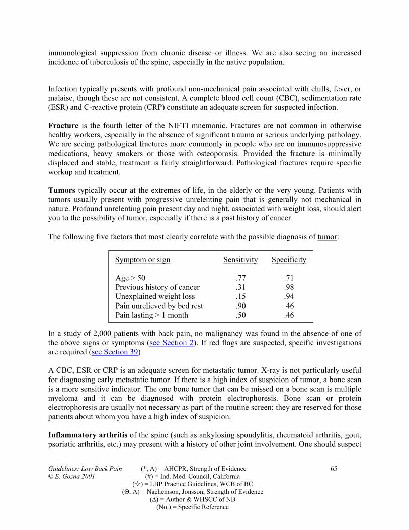
immunological suppression from chronic disease or illness. We are also seeing an increased incidence of tuberculosis of the spine, especially in the native population.
Infection typically presents with profound non-mechanical pain associated with chills, fever, or malaise, though these are not consistent. A complete blood cell count (CBC), sedimentation rate (ESR) and C-reactive protein (CRP) constitute an adequate screen for suspected infection. Fracture is the fourth letter of the NIFTI mnemonic. Fractures are not common in otherwise healthy workers, especially in the absence of significant trauma or serious underlying pathology. We are seeing pathological fractures more commonly in people who are on immunosuppressive medications, heavy smokers or those with osteoporosis. Provided the fracture is minimally displaced and stable, treatment is fairly straightforward. Pathological fractures require specific workup and treatment. Tumors typically occur at the extremes of life, in the elderly or the very young. Patients with tumors usually present with progressive unrelenting pain that is generally not mechanical in nature. Profound unrelenting pain present day and night, associated with weight loss, should alert you to the possibility of tumor, especially if there is a past history of cancer. The following five factors that most clearly correlate with the possible diagnosis of tumor:
Symptom or sign Sensitivity Specificity
Age > 50 .77 .71 Previous history of cancer .31 .98 Unexplained weight loss .15 .94 Pain unrelieved by bed rest .90 .46 Pain lasting > 1 month .50 .46
In a study of 2,000 patients with back pain, no malignancy was found in the absence of one of the above signs or symptoms (see Section 2). If red flags are suspected, specific investigations are required (see Section 39)
A CBC, ESR or CRP is an adequate screen for metastatic tumor. X-ray is not particularly useful for diagnosing early metastatic tumor. If there is a high index of suspicion of tumor, a bone scan is a more sensitive indicator. The one bone tumor that can be missed on a bone scan is multiple myeloma and it can be diagnosed with protein electrophoresis. Bone scan or protein electrophoresis are usually not necessary as part of the routine screen; they are reserved for those patients about whom you have a high index of suspicion. Inflammatory arthritis of the spine (such as ankylosing spondylitis, rheumatoid arthritis, gout, psoriatic arthritis, etc.) may present with a history of other joint involvement. One should suspect
Guidelines: Low Back Pain (*, A) = AHCPR, Strength of Evidence 65 © E. Gozna 2001 (#) = Ind. Med. Council, California ( ) = LBP Practice Guidelines, WCB of BC (Θ, A) = Nachemson, Jonsson, Strength of Evidence (∆) = Author & WHSCC of NB (No.) = Specific Reference

Guidelines: Low Back Pain (*, A) = AHCPR, Strength of Evidence 66 © E. Gozna 2001 (#) = Ind. Med. Council, California ( ) = LBP Practice Guidelines, WCB of BC (Θ, A) = Nachemson, Jonsson, Strength of Evidence (∆) = Author & WHSCC of NB (No.) = Specific Reference
ankylosing spondylitis in the young male, typically in his most productive age, who presents with intractable back pain and morning stiffness. Diminished chest expansion is one of the earliest clinical signs of ankylosing spondylitis and is usually present long before there are sacroiliac joint radiographic changes. HLA27 antigen is frequently positive with ankylosing spondylitis.
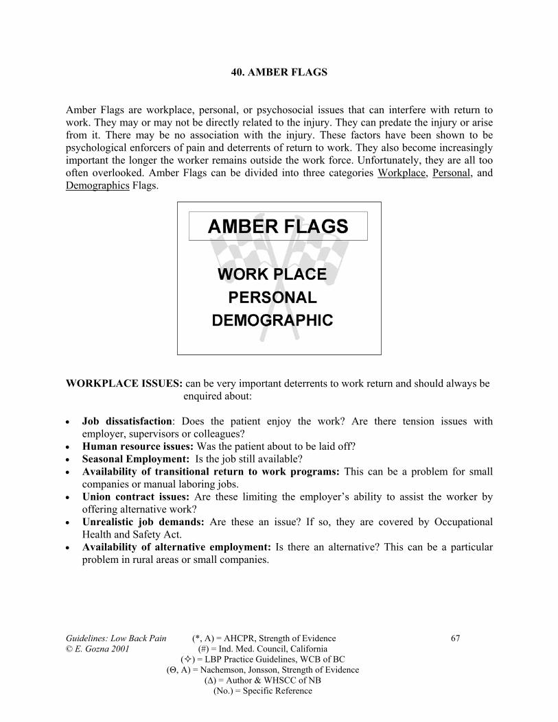
40. AMBER FLAGS Amber Flags are workplace, personal, or psychosocial issues that can interfere with return to work. They may or may not be directly related to the injury. They can predate the injury or arise from it. There may be no association with the injury. These factors have been shown to be psychological enforcers of pain and deterrents of return to work. They also become increasingly important the longer the worker remains outside the work force. Unfortunately, they are all too often overlooked. Amber Flags can be divided into three categories Workplace, Personal, and Demographics Flags.
WORKPLACE ISSUES: can be very important deterrents to work return and should always be
enquired about: • Job dissatisfaction: Does the patient enjoy the work? Are there tension issues with
employer, supervisors or colleagues? • Human resource issues: Was the patient about to be laid off? • Seasonal Employment: Is the job still available? • Availability of transitional return to work programs: This can be a problem for small
companies or manual laboring jobs. • Union contract issues: Are these limiting the employer’s ability to assist the worker by
offering alternative work? • Unrealistic job demands: Are these an issue? If so, they are covered by Occupational
Health and Safety Act. • Availability of alternative employment: Is there an alternative? This can be a particular
problem in rural areas or small companies.
Guidelines: Low Back Pain (*, A) = AHCPR, Strength of Evidence 67 © E. Gozna 2001 (#) = Ind. Med. Council, California ( ) = LBP Practice Guidelines, WCB of BC (Θ, A) = Nachemson, Jonsson, Strength of Evidence (∆) = Author & WHSCC of NB (No.) = Specific Reference

PERSONAL ISSUES: pertain to the worker’s personality traits, training and skill sets:
• History of avoidance type behavior. • Depression or somatization tendencies. • Belief that non-specific pain is harmful. • Belief that passive treatment is beneficial and active treatment harmful • Displays chronic anger and frustration over socio-economic status. • Perception of social support system as an alternative income source • Involvement in the underground economy. This is a growing problem in today’s economy. • Limited transferable skills: Can preclude finding alternative employment • Poor language skills. • Role reversal: Has spouse now become breadwinner? DEMOGAPHICS: These demographic factors statistically correlate with poor return to work
statistics: • Age > 40 • Use of narcotics beyond four weeks of date of injury correlates with poor return to work • History of excessive alcohol and drug use • Heavy smokers • Heavy users of walk-in clinics • Previous compensable injuries with prolonged disability • History of previous utilization of the welfare system ILLNESS BEHAVIOR Though not common, some patients can display behavior that appears to have no physical basis (i.e. non-organic), which should raise the suspicion of psychogenic magnification or pain exaggeration. These symptoms and findings were first described by Dr. G Waddell (1) and should always be noted as they can correlate with poor treatment outcomes (both medical and surgical). Waddell’s inappropriate symptoms:
• Pain over body of sacrum • Whole leg pain • Whole leg numbness • Whole leg giving away • Never free of pain • Intolerance of treatment • Hospital admissions for LBP
Guidelines: Low Back Pain (*, A) = AHCPR, Strength of Evidence 68 © E. Gozna 2001 (#) = Ind. Med. Council, California ( ) = LBP Practice Guidelines, WCB of BC (Θ, A) = Nachemson, Jonsson, Strength of Evidence (∆) = Author & WHSCC of NB (No.) = Specific Reference
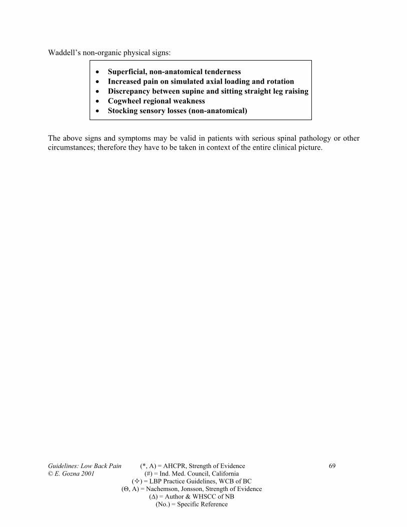
Waddell’s non-organic physical signs:
• Superficial, non-anatomical tenderness • Increased pain on simulated axial loading and rotation • Discrepancy between supine and sitting straight leg raising • Cogwheel regional weakness • Stocking sensory losses (non-anatomical)
The above signs and symptoms may be valid in patients with serious spinal pathology or other circumstances; therefore they have to be taken in context of the entire clinical picture.
Guidelines: Low Back Pain (*, A) = AHCPR, Strength of Evidence 69 © E. Gozna 2001 (#) = Ind. Med. Council, California ( ) = LBP Practice Guidelines, WCB of BC (Θ, A) = Nachemson, Jonsson, Strength of Evidence (∆) = Author & WHSCC of NB (No.) = Specific Reference

41. SIMPLE BACK PAIN AND SCIATICA PATHOMECHANICS Over 90 percent of all low back disc symptoms occur at the L4/5 and L5/S1 levels. These are the two lowest mobile segments of the spine, an area of maximal stress concentration between the highly mobile spine and the immobile fixed pelvis. This area of hypermobility is predisposed to degenerative osteoarthritis, simple back pain and occasionally sciatica. The fundamental building block of the spine is the spinal motion unit, consisting of two vertebrae separated by the intervertebral disc. The spinal unit provides a flexible conduit to support and protect the delicate neural structures that pass through the spine and nerve roots exiting to the extremities. The disc is a prestressed structure (think of a golf ball) with a spongy center called the nucleus kept under pressure by a fibrous and elastic enclosure, the annulus. Fig.1 Spinal Motion Unit: Vertebra Spinal Unit Disc
Vertebra
Nucleus Annulus
When we are young, the nucleus is a gelatinous structure and the disc is under considerable pressure. Age or damage can cause dehydration, resulting in loss of disc firmness, and producing excessive mobility. This segmental instability is felt to be the foundation of the vast majority of degenerative back problems. Segmental instability can occur as part of the normal aging process but can be accelerated by other factors such as abnormal forces (obesity, poor posture and chronic muscle imbalance) or from abnormal structure (osteoporosis, spondylolysis, tumor, etc.). Guidelines: Low Back Pain (*, A) = AHCPR, Strength of Evidence 70 © E. Gozna 2001 (#) = Ind. Med. Council, California ( ) = LBP Practice Guidelines, WCB of BC (Θ, A) = Nachemson, Jonsson, Strength of Evidence (∆) = Author & WHSCC of NB (No.) = Specific Reference

Fig 2 Segmental Instability:
Aging AbnormalForces
Abnormal Structure
Chronic disturbances of the spinal unit and segmental instability can result in either simple back pain or sciatica. It is imperative to understand the distinction between simple back pain and sciatica. SIMPLE LBP OR SCIATICA? Simple LBP is called various names (i.e. mechanical, lumbago, degenerative disc disease, etc.) but numerous studies have shown that there is no practical benefit in further subcategorizing this entity. I prefer the term “simple” (1), which highlights its usually benign nature, distinguishes it from sinister spinal problems (less than 3% of LBP) and avoids needless patient anxiety. Simple LBP accounts for > 93% of LBP and > 80 % of the cost of treatment and disability but is the one about which we have the least understanding! Though the exact sources of simple back pain are not well understood, it is generally agreed that it can emanate from a number of structures, including disc, facet joint, or the supporting ligament and myofascial structures. Simple LBP is a condition that primary physicians should feel comfortable treating in a positive, active rehabilitative environment. Rarely should specialist referral be necessary. The incidence of LBP has not changed over the past 40 years (1, Ch. 5). What has changed has been an exponential increase in chronic disability, medical certification and sickness benefits associated with LBP! Sciatica is a far less common condition accounting for less than three percent of back problems. Though less common, it is better understood and most commonly occurs when there is pressure on the exiting nerves to the legs, either as a result of disc bulging, bony or soft tissue entrapment. Guidelines: Low Back Pain (*, A) = AHCPR, Strength of Evidence 71 © E. Gozna 2001 (#) = Ind. Med. Council, California ( ) = LBP Practice Guidelines, WCB of BC (Θ, A) = Nachemson, Jonsson, Strength of Evidence (∆) = Author & WHSCC of NB (No.) = Specific Reference

Fig 3 Sources of Sciatica and Back Pain: Cauda Nerve Root
Joints Fascia Ligaments Disc
Sciatica
Simple LBP
Sciatica can present in two ways: Irritation or Impaired conduction. Nerve irritation can occur from mild extrinsic pressure from disc herniation, peripheral entrapment or even chronic disturbed posture. If the pressure on the nerve is severe enough, though, it can produce impaired conduction with resultant motor, sensory or reflex deficits NERVE ROOT IRRITATION: Nerve root irritation is manifest by three P's:
PAIN BELOW KNEE PARESTHESIAPOS BOWSTRING
3 P’S
1. Pain below the level of the knee. Simple back pain often radiates down the posterior thigh and, as a result, can be confused with sciatica; but it rarely goes below the level of the knee. In contrast sciatica is felt in the distribution of the involved nerve; that means L5 or S1 root sciatica should radiate to the ankle or foot. Whether the pain stays above or goes below the level of the knee is critical to differentiating between sciatic and simple back pain.
Guidelines: Low Back Pain (*, A) = AHCPR, Strength of Evidence 72 © E. Gozna 2001 (#) = Ind. Med. Council, California ( ) = LBP Practice Guidelines, WCB of BC (Θ, A) = Nachemson, Jonsson, Strength of Evidence (∆) = Author & WHSCC of NB (No.) = Specific Reference

Fig 4 Simple vs. Sciatic Radiation 2. Paresthesia. Pressure applied to an inflamed nerve characteristically produces “pins and needles” in the nerve’s distribution. This is what is happening when if you hit your elbow on the “funny bone;” you are feeling paresthesia in the ulnar nerve distribution to the hand. 3. Positive bowstring sign. Virtually any source of back pain will cause hamstring spasm with resultant back pain on passive straight leg raising. Therefore, painful restriction of straight leg raising is not a specific sign of nerve root irritation or tension; it can merely indicate that the patient is experiencing back pain. In contrast, a positive bowstring sign is very specific to sciatic nerve root tension. To elicit this sign, place the patient's calf on your shoulder and press over the popliteal nerve behind the flexed knee. A positive bowstring is pain experienced either in the back or down the calf. It is the single most specific clinical sign of sciatic nerve tension.
Fig 5 Positive Bowstring Sign: Guidelines: Low Back Pain (*, A) = AHCPR, Strength of Evidence 73 © E. Gozna 2001 (#) = Ind. Med. Council, California ( ) = LBP Practice Guidelines, WCB of BC (Θ, A) = Nachemson, Jonsson, Strength of Evidence (∆) = Author & WHSCC of NB (No.) = Specific Reference

IMPAIRED CONDUCTION (MSR): If for any reason a nerve is excessively compressed, it stops working properly, resulting in Motor, Sensory or Reflex disturbance. For the three nerves that are involved in sciatica, the resultant impairments are as follows: Sciatica taelicit the mnot instruc
When theor combinexaminati While mois importaneurologisame apprtreated wi
Guidelines:© E. Gozna
MOTOR SENSORY REFLEX L4 Knee Ext. Med Foot Knee L5 Ankle Ext. 1st W.S. None & EHL S1 Gastroc Lat Foot Ankle
kes two forms: irritation and impaired conduction. In medical school we are taught to otor, sensory and reflex (MSR) findings that indicate impaired conduction but we are
ted on the signs of nerve irritation (3Ps):
F O R M S O F S C IA T IC A
IR R IT A T IO N IM P A IR E D C O N D U C T IO N
P A R E S T H E S IA M O T O R
P A IN B E L O W K N E E S E N S O R Y
P O S IT IV E B O W S T R IN G R E F L E X
y talk about sciatica, clinicians should speak about nerve root irritation or impairment ations of them. If they suspect sciatica, they should carry out a careful neurological
on as a baseline for future referral.
st patients with sciatica can be treated the same way as those with simple back pain, it nt to know whether the condition is improving or worsening. For this, a thorough index cal examination is essential. Mild sciatica and simple back pain are treated with the oach, active rehabilitation. Since sciatica involves nerve function, though, it has to be th greater respect and followed more carefully.
Low Back Pain (*, A) = AHCPR, Strength of Evidence 74 2001 (#) = Ind. Med. Council, California
( ) = LBP Practice Guidelines, WCB of BC (Θ, A) = Nachemson, Jonsson, Strength of Evidence
(∆) = Author & WHSCC of NB (No.) = Specific Reference
TREATMENT: This is outlined in Medical Management (see Section 2). Acute simple back pain or mild, non-progressive sciatica should show signs of spontaneous recovery within 1 month. The cornerstone of treatment is encouragement to remain active. Having ruled out red flags (see Section 39), the physician should reassure the patient and support continued involvement in meaningful activities (including staying in the workplace if at all possible). Acute mild non-progressive sciatica and simple LBP are treated the same – reassurance and encouragement of activity (4). The practical reason for distinguishing sciatica from simple LBP is to acknowledge the importance of a careful baseline neurological assessment for suspected sciatica and the need to clinically follow its course to insure that there is no neurological progression (i.e., red flag).
Guidelines: Low Back Pain (*, A) = AHCPR, Strength of Evidence 75 © E. Gozna 2001 (#) = Ind. Med. Council, California ( ) = LBP Practice Guidelines, WCB of BC (Θ, A) = Nachemson, Jonsson, Strength of Evidence (∆) = Author & WHSCC of NB (No.) = Specific Reference

42. EXAMPLES OF POSTURAL CONTRIBUTORS TO LBP
There is strong evidence based literature to support the concept of active as opposed to passive treatment for subacute or recurrent simple LBP (* A, #, , ∆, Θ A). In recent years a great deal of effort has gone into identifying the various posture and muscle imbalance syndromes that characterize simple low back pain and to design specific treatment protocols based on identifiable syndromes. Unfortunately there are no well-controlled studies to show the relatively efficacy of these various approaches and as a result the various treatment philosophies have tended to develop anecdotally rather than based on science. Two treatment approaches about which there is general agreement are the importance of core abdominal muscle strength and the influence of the hips on LBP. These are examples of postural contributors to LBP. 1. CORE ABDOMINAL MUSCLE STRENGTH: Core abdominal weakness can be caused by obesity (stretch weakness) or deconditioning. One of the earliest concepts in managing simple LBP was recognizing the importance of the abdominal musculature is stabilizing the lumbar spine. This invokes the principle if hoop forces whereby those abdominal muscles the act circumferentially around the abdomen act like barrel hoops increasing the intra-abdominal pressure and thereby stabilizing the spine from uncontrolled movement:
Hoop Forces: Abdominal muscles that act like hoops produce greatest spinal stabilization.
The abdominal muscles are important dynamic stabilizers of the spine and although controversy exists as to be relative contribution of the various muscle groups to abdominal stability their overall role is universally accepted. Biomechanical studies suggest that the most important lumbar stabilizers (i.e., greatest influence on intra-abdominal pressure) are those abdominal muscles whose fibers are generally oriented circumferentially around the abdomen therefore producing the greatest hoop force.
Guidelines: Low Back Pain (*, A) = AHCPR, Strength of Evidence 76 © E. Gozna 2001 (#) = Ind. Med. Council, California ( ) = LBP Practice Guidelines, WCB of BC (Θ, A) = Nachemson, Jonsson, Strength of Evidence (∆) = Author & WHSCC of NB (No.) = Specific Reference
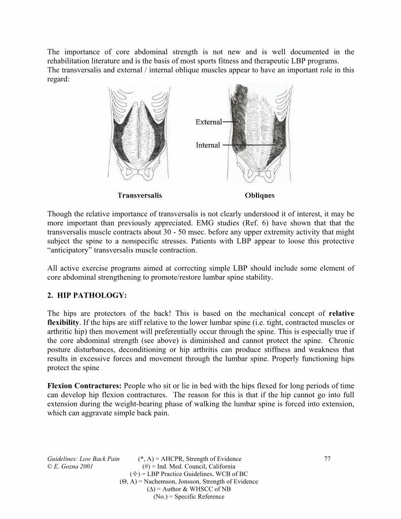
The importance of core abdominal strength is not new and is well documented in the rehabilitation literature and is the basis of most sports fitness and therapeutic LBP programs. The transversalis and external / internal oblique muscles appear to have an important role in this regard:
Though the relative importance of transversalis is not clearly understood it of interest, it may be more important than previously appreciated. EMG studies (Ref. 6) have shown that that the transversalis muscle contracts about 30 - 50 msec. before any upper extremity activity that might subject the spine to a nonspecific stresses. Patients with LBP appear to loose this protective “anticipatory” transversalis muscle contraction. All active exercise programs aimed at correcting simple LBP should include some element of core abdominal strengthening to promote/restore lumbar spine stability. 2. HIP PATHOLOGY: The hips are protectors of the back! This is based on the mechanical concept of relative flexibility. If the hips are stiff relative to the lower lumbar spine (i.e. tight, contracted muscles or arthritic hip) then movement will preferentially occur through the spine. This is especially true if the core abdominal strength (see above) is diminished and cannot protect the spine. Chronic posture disturbances, deconditioning or hip arthritis can produce stiffness and weakness that results in excessive forces and movement through the lumbar spine. Properly functioning hips protect the spine Flexion Contractures: People who sit or lie in bed with the hips flexed for long periods of time can develop hip flexion contractures. The reason for this is that if the hip cannot go into full extension during the weight-bearing phase of walking the lumbar spine is forced into extension, which can aggravate simple back pain. Guidelines: Low Back Pain (*, A) = AHCPR, Strength of Evidence 77 © E. Gozna 2001 (#) = Ind. Med. Council, California ( ) = LBP Practice Guidelines, WCB of BC (Θ, A) = Nachemson, Jonsson, Strength of Evidence (∆) = Author & WHSCC of NB (No.) = Specific Reference

Short contracted hip flexors or flexion contractures produce excessive lumbar spine extension at end of stance phase of walking:
This can be caused by either psoas or quadriceps muscle tightness. The Thomas test is used to detect hip flexion contractures and to determine which muscle is primarily involved. Thomas Test:
Typically patients with hip flexion contractures complain of LBP with walking especially going down hill. Patients with quadriceps tightness may also complain of back pain with deep knee bending activities, as this muscle is further stretched when the knees are fully bent. Tight Extensors: People who have tightness of their hip extensors (glutei and hamstrings) are often unable to flex the hip to 90 degrees. They can experience LBP when sitting in a chair
Guidelines: Low Back Pain (*, A) = AHCPR, Strength of Evidence 78 © E. Gozna 2001 (#) = Ind. Med. Council, California ( ) = LBP Practice Guidelines, WCB of BC (Θ, A) = Nachemson, Jonsson, Strength of Evidence (∆) = Author & WHSCC of NB (No.) = Specific Reference

because the absence of hip flexion to 90 degrees forces their spine into flexion. Illustration of how tight hip extensors cause lumbar flexion when sitting:
Weak External Rotators: Patients with weakness of their hip external rotator's tend to drop and rotate the pelvis when standing stationary for long periods or when ascending and descending stairs. This produces torsional and extension forces through the lumbar spine that can aggravate LBP. These same people often develop tightness of their Ileo-Tibial Bands (ITB), which affect both the back and can cause knee cap symptoms. Summary: The importance of core abdominal strength for treatment of simple LBP is universally accepted. As he hip and spine are mechanically linked, any biomechanical or arthritic condition affecting the hips can also affect the lumbar spine. The above are just a few of the examples that illustrate the necessity to make active treatment a major part of back pain management (see Section 7).
Guidelines: Low Back Pain (*, A) = AHCPR, Strength of Evidence 79 © E. Gozna 2001 (#) = Ind. Med. Council, California ( ) = LBP Practice Guidelines, WCB of BC (Θ, A) = Nachemson, Jonsson, Strength of Evidence (∆) = Author & WHSCC of NB (No.) = Specific Reference

SOURCE DOCUMENTS
1. Industrial Medical Council, Practice Parameter: Low Back Problems (Draft 4/27/94), State of California, Department of Industrial Relations, 1994.
2. Bigos S, Bowyer O, Braen G, et. al. Acute Low Back Problems in Adults. Clinical Practice
Guidelines No.14. AHCPR Publication No. 95-0642. Rockville, MD: Agency for Health Care Policy and Research, Public Health Service, U.S. Department of Health and Human Services, December 1994.
3. Spitzer, Walter O., et. al. “Report of the Quebec Task Force on Spinal Disorders, “Spine,
Volume 12, Number 7 Supplement, September 1987. 4. Clinical Standards Advisory Group. Back Pain. Chair: Professor Michael Rosen. London:
HMSO Publications, May 1994. 5. Washington State Medical Association Industrial Insurance Advisory Committee, Medical
Practice Guidelines in Washington Workers’ Compensation, 7th Draft, September 11, 1992. 6. Utilization Review and Quality Assessment Program, Review Criteria, Commonwealth of
Massachusetts, Department of Industrial Accidents, July 1, 1993. 7. The North American Spine Society’s Ad Hoc Committee on Diagnostic and Therapeutic
Procedures, “Common Diagnostic and Therapeutic Procedures of the Lumbosacral Spine”, Spine, Volume 16, Number 10, 1991.
8. Clinical Practice Guidelines for the Diagnosis and Treatment of Low Back Pain. WCB
British Columbia, October 4, 1995. 9. Neck and Back Pain: Scientific Evidence of Cause, Diagnosis and Treatment. Editors Alf L.
Nachemson, Egon Jonsson; Lippincott Williams & Wilkins, 2000
Guidelines: Low Back Pain 80 © E. Gozna 2001

SPECIFIC ARTICLES AND REFERENCES
(In order of appearance)
1. Waddell, Gordon, The Back Pain Revolution, Ed 1, 1998 Churchill Livingstone. 2. Deyo R.A., Diehl A.K., Cancer as the Cause of Back Pain: Frequency, Clinical Presentation
and Diagnostic Strategies. Journal of General Internal Medicine 1988; 3:230-238. 3. Malmivaara, Antii et al, The Treatment of Acute Low Back Pain – Bed Rest, Exercise, or
Ordinary Activity. New England Journal of Medicine 1995; 332:5; 351-355 4. Vroomen, P.C. et al, Lack of Effectiveness of Bed Rest for Sciatica. New England Journal of
Medicine 1999; 340:6; 418-423. 5. Franklin, GM et al, Outcome of Lumbar Fusions in WCB (Washington State) Patients. Spine
1994; 11:17; 1897-1904 6. Hodges, P. et al, Inefficient Muscle Stabilization of the Lumbar Spine Associated With Low
Back Pain. Spine 1996;21:2,2640-2650
Guidelines: Low Back Pain 81 © E. Gozna 2001

CMA POLICY
THE PHYSICIAN’S ROLE IN HELPING PATIENTS RETURN TO WORK AFTER AN ILLNESS OR
INJURY (UPDATE 2000) This policy addresses the role of attending physicians in assisting their patients to return to work after an illness or injury. The physician's role is to diagnose and treat the illness or injury, to advise and support the patient, to provide and communicate appropriate information to the patient and the employer and to work closely with other involved health care professionals to facilitate the patient's safe and timely return to the most productive employment possible. Carrying out this role requires physicians to understand the patient's roles in the family and the workplace. It requires physicians to recognize and support the employee-employer relationship and the primary importance of this relationship in the return to work. Finally, it requires physicians to have a good understanding of the potential roles of other health care professionals and employment personnel in assisting and promoting the return to work. The CMA recognizes the importance of a patient returning to all possible functional activities relevant to his or her life as soon as possible after an injury or illness. The purpose of this statement, however, is to address the role of the attending physician in the patient's return to work. The CMA supports a shift away from complete reliance on physician certification for work absences (Certificate of Disability 1998) to cooperation between the employee and his or her employer with the use of medical input, advice and support from the employee's attending physician and other involved health care professionals (see also Timely Return to Work Programs and the Role of the Primary Care Physician, by the Ontario Medical Association). Prolonged absence from one's normal roles, including absence from the workplace, is detrimental to a person's mental, physical and social well-being. Physicians should therefore encourage a patient's return to function and work as soon as possible after an illness or injury, provided that return to work does not endanger the patient, his or her coworkers or society. A safe and timely return to work benefits the patient and his or her family by enhancing recovery and reducing disability. Through improvement of health outcomes, a safe and timely return to work also preserves a skilled and stable workforce for employers and society and reduces demands on health and social services as well as on disability plans. •2001 Canadian Medical Association. You may, for your non-commercial use, reproduce, in whole or in part and in any form or manner, unlimited copies of CMA Policy Statements provided that credit is given to the original source. Any other use, including republishing, redistribution, storage in a retrieval system or posting on a Web site requires explicit permission from CMA. Please contact the Permissions Coordinator, Publications, CMA, 1867 Alta Vista Dr., Ottawa ON K1G 3Y6; fax 613 565-2382; [email protected].
Correspondence and requests for additional copies should be addressed to the Member Service Centre, Canadian Medical Association, 1867 Alta Vista Drive, Ottawa, ON K1G 3Y6; tel 888 855-2555 or 613 731-8610 x2307; fax 613 236-8864.
All polices of the CMA are available electronically through CMA Online (www.cma.ca).
Guidelines: Low Back Pain 82 © E. Gozna 2001

The role of physicians is to incorporate a timely return to work into the care plan for their patients. The treatment or care plan should be evidence-based, when possible, and should identify the best sequence and timing of interventions for the patient. Unnecessary waiting periods and other obstacles in the care plan should be identified. Identified obstacles should be addressed and discussed, when relevant, by those involved in the patient's return to work. Successful return to work involves primarily the employee and his or her employer and requires the assistance of the attending physician. When appropriate, patient care and outcomes may be improved through a coordinated multidisciplinary approach involving other health care professionals, including other physicians, rehabilitation specialists, nurses, physiotherapists, occupational therapists, psychologists, case managers, vocational specialists and personnel of employee assistance programs. When available, occupational health and safety services, which may involve physicians and other expert personnel, can be an invaluable resource for the attending physician and patient. The employee and the employer generally have an established relationship that is central to the return to work. Therefore, in all cases of impairment or disability, a workplace supervisor, manager or employer representative must be closely involved and must be a partner in this process. Employers increasingly recognize the value of changing the workplace to facilitate a safe and timely return to work. The employer's role is to ensure that the workplace culture supports a timely return to work, for example, by ensuring that flexible work is available. Flexible work can include modifying tasks, schedules and environmental conditions to meet the temporary or permanent needs of the patient. Employees are often unaware of their employer's ability to accommodate special needs. Direct communication by an employee with his or her employer after an illness or injury often increases the employee's perception of his or her ability to work. Therefore, with careful planning and appropriate physician input and advice to both the employee and the employer, in many cases an employee may successfully return to work before full recovery. Return to work requires that the employee's capabilities match or exceed the physical, psychological and cognitive requirements of the work offered. In some cases, it may be appropriate for the physician to advise the patient that a timely return to work can facilitate his or her recovery by assisting in restoring or improving functional capabilities. A gradual increase in activity may hasten a patient's recovery and readiness to resume work at full capacity.
Principles Concerning The Role Of The Physician In The Return-To-Work Plan • The physician should facilitate the patient's return to work by encouraging communication
between the patient and his or her employer early in treatment or rehabilitation. • The physician should be familiar with the family and community support systems available to
the patient. The physician should also be aware of the patient's general responsibilities at home and at work.
• Early in the course of treatment, the physician should discuss with the patient expected healing and recovery times as well as the positive role of an early, graduated increase in activity on physical and psychological healing.
Guidelines: Low Back Pain 83 © E. Gozna 2001
• The physician should identify and address potential obstacles to the recovery of function and return to work as soon as possible.
• The physician should be knowledgeable about and use, when appropriate, the services of a multidisciplinary team of health care professionals, who can be helpful in facilitating the patient's safe and timely return to work.
• Ultimately, the employer determines the type of work available and whether a physician's recommendations concerning an employee's return to work can be accommodated. (Under provincial and territorial human rights laws, an employer may not discriminate on the basis of disability or other illness and has legal obligations with respect to the accommodation of employees. For details, refer to the Human Rights Code in the relevant jurisdiction). In recognition of the employee-employer relationship, physicians should encourage their patients to take an active role in and to take responsibility for the return to work and to communicate directly and regularly with their employers.
• In cases of employers with occupational medical departments, the attending physician may contact the occupational physician or nurse to understand specific workplace policies, supportive in-house resources, essential job demands and possible health and safety hazards in the patient's workplace. Where occupational medical resources are available, the attending physician generally assumes a supportive or advisory medical role. For assistance with specific cases, provincial and territorial medical associations and the Occupational and Environmental Medical Association of Canada have information identifying physicians who specialize in assisting with the return to work.
• When requested by the employer, the physician, with the patient's consent (see Respecting Patient Confidentiality and Managing Medical Information), should be as specific as possible in describing the patient's work capabilities and any work accommodation required. In more complex cases, the physician should consider referring the patient to medical specialists and other appropriate health care professionals for a comprehensive, objective assessment of his or her functional capabilities and limitations and their relation to the demands of the patient's job.
• If the employer asks the physician to participate in developing a modified work plan, the physician should consider and make recommendations related to the following: o The employee's task limitations, e.g., no above-shoulder reaching with the right arm;
alternating positions between sitting and standing or walking; and maximum lifting of 10 kg from (need page 3) of text family and community support systems floor level or 20 kg from waist level. These should be specific and time-limited. When defining task limitations, the employee's functional capabilities and the demands of the job should be matched and considered.
o Schedule modifications, e.g., usual shift schedule at 4 hours per day; 1 day off, or lighter assignment in the middle of a set of shifts; and no night or rotating shifts. It should be noted when return to a normal schedule is medically appropriate.
o Environmental restrictions, e.g., avoidance of specific workplace substances or of cold environments; and no solo work in remote areas.
o Medical aids or personal protective equipment, e.g., use of a wrist strap; use of a chair with adjustable height and lumbar support; and use of a more efficient filtration mask.
Whenever possible, the physician should state whether restrictions are permanent or temporary and give an estimate of recovery time. Also, the physician should give the date when the patient's progress and his or her work restrictions will be reassessed. Guidelines: Low Back Pain 84 © E. Gozna 2001

• The employer and employee have a responsibility to provide the physician with any employment-related information that will assist him or her in giving medical advice and support. It is the employer's responsibility to provide the physician with a written job description, identifying the job risks and available work modifications, upon request.
• The physician must be aware of the risks to the patient, his or her coworkers or the public that could arise from the patient condition or drug therapy. If the medical condition of the patient and the nature of the work performed are very likely to endanger the safety of others significantly, the physician must put the public interest before that of the patient. The CMA Code of Ethics (Can Med Assoc J 1996;155:1176A-B) states that physicians must respect the patient's right to confidentiality except when this right conflicts with your responsibility to the law, or when the maintenance of confidentiality would result in a significant risk of substantial harm to others or to the patient if the patient is incompetent; in such cases, take all reasonable steps to inform the patient that confidentiality will be breached.
The CMA holds that legislation should be enacted in all jurisdictions to protect physicians from liability associated with such decisions. • If the employer and the employee cannot agree on a return-to-work plan, the employer should
contact the physician and employee to identify the minimum level of capability that can be accommodated in the workplace.
• When conflict occurs between the employer and the employee, the attending physician may wish to use, where available, the skills of an occupational physician; if these services are unavailable, the attending physician may recommend or facilitate a resolution of conflict-resolution processes be put in place to address all participants' concerns if conflicts occur.
• A physician who sees a patient for the first time concerning a long-standing condition should obtain and consult medical records on previous care before offering advice on a safe and timely return to work.
• The physician should counsel the patient on preventive strategies, when appropriate. • When the physician believes that the patient has recovered sufficiently and can return to work
safely, the patient/employee should be clearly informed of this judgment. • The patient is entitled to a copy of all return-to-work documentation that is prepared by his or
her attending physician (see the next section).
Respecting Patient Confidentiality And Managing Medical Information Medical records are confidential. In general, physicians should not give information to anyone concerning the condition of a patient or any service rendered to a patient without the patient's consent. However, there are some exceptions to this rule. For example, in some cases, provincial or territorial legislation may require physicians to provide information to workers' compensation boards without prior patient approval. Physicians should be aware of the legal requirements in their province or territory. Consistent with the general rule concerning a physician's duty to keep patient information confidential, the physician should not provide information about the patient to the patient's instance, if an employer requests information about the patient's ability to return to work, prior
Guidelines: Low Back Pain 85 © E. Gozna 2001
patient consent should be obtained. Consent should be specific rather than general. Written authorization for such disclosure is desirable. A physician's report concerning the patient's ability to return to work should be tailored to the intended audience. For example, a report directed to an employer should contain only information that the employer needs to know to assist the employee in his or her return to work (for example, the ability of the employee to perform pre-injury duties, the employee's capabilities and limitations and the extent to which the work or the workplace needs to be accommodated to meet the needs of the employee). To respect the privacy of the patient, the physician should be careful not to provide medical information that is not needed to facilitate his or her return to work. The patient has the right to examine and copy medical records that pertain to him or her, according to a recent decision of the Supreme Court of Canada (McInerney v. MacDonald [1992] 2 SCR 138). Patient access to records may be denied only if the physician reasonably believes that the patient or others will suffer substantial physical, mental or emotional harm because of information contained in the records. A physician should be aware that, if access is denied and the patient challenges the physician's decision, the onus is on the physician to justify denial of access. In regard to a patient's access to medical information obtained in an independent medical examination conducted by a physician on behalf of a third party, additional considerations may apply. Physicians should consult appropriate statements from the relevant provincial or territorial licensing body and from the Canadian Medical Protective Association for additional information and guidance. Physicians should also be aware of any relevant legislation or other legal requirements in their jurisdictions.
Billing For Return-To-Work Services Preparation of reports and documents for employers primarily for the purpose of return to work is not generally publicly insured. The patient or third party requesting the service should be billed for it. Statements from each province's medical association concerning billing should be consulted. The physician should inform the patient that there will be a charge when medical reports are requested. Conclusion The CMA believes that physicians who follow the principles outlined in this policy will support their patients and their families, their communities, employers and society. Wording and concepts in this statement were adapted from the Alberta Medical Association's statement Early Return to Work After Illness or Injury. Concepts were also taken from the Manitoba Medical Association's policy on an early return to work and the Ontario Medical Association's policy Timely Return to Work Programs and the Role of the Primary Care Physician.
Guidelines: Low Back Pain 86 © E. Gozna 2001

INDEX A oral, 28
craniosacral manipulation, 32 acetaminophen, 15, 27 CT, 8, 38, 39, 46 acupuncture, 8, 24, 35 CT- myelography, 38 acute neurological deterioration, 64 Cybex, 53 alternative work, 23, D analgesia, 27
ankylosing spondylitis, 18, 20, 65, 66 debilitation due to inactivity, 29 antidepressants, 28 decompression laminectomy, 46 arthritis, 18, 20, 65, degenerative disc, 71
depression, 13, 28, 43, 67 assessment diathermy, 30, 31 computerized, 53 disability, 16, 50, 63 ergonomic, 44
interdisciplinary, 8, 12, 50, 63 disc laboratory, 20 degeneration, 38 medical, 8, 9, 63 herniation, 10, 46, 60, 71 functional capacity, 52 discectomy, 46 psychosocial, 43 discography 54 radiographic, 18, 38 drug dependence, 27, 67
B dry needling, 35 E back braces, 37 economic factors, 67 bed rest, 4, 8, 12, 14, 15, 29, 56, 65 education, 21 biofeedback, 61, 62 electrical stimulation,30, 33 biotechnology, 45 electrodiagnostic studies, 41 bone scan, 20, 21, 41, 65 EMG, 41 braces, 36
C epidural injections, 56 ergonomic evaluation, 44 cancer, 11, 19, 20, 65 ESR, 20, 65 cauda equina syndrome, 38, 64 evaluation functional capacity, 52 CBC, 20, 65 exercise, 24, 30, 45, 53 chemonucleolysis, 60 extensions of work-conditioning, 45 chiropractic, 8, 32 F chronic low back pain, 14, 50, 62 facet joint injections, 57 chronic pain program, 50 facet syndrome, 57 chymopapain, 60 failure to return to full or modified work, 51 complications of chemonucleolysis, 60 fracture computerized testing, 53
CT, MRI, myelography, 39 congenital narrowing of the spinal canal, 48 spinal fusion for, 49 consultation
with specialist, 38, 62 x-rays in ruling out, 18 corsets, 36 functional capacity testing, 52 corticosteroids: fusion, 49
injection, 56, 57, 58
Guidelines: Low Back Pain 87 © E. Gozna 2001
due to serious spinal pathology, 4, 9, 64, 69
H heat, 24, 30
non-specific, 70 herniated disc, 71 pathologic processes, 70 hospitalization, 8 simple, 71 human engineering, 44
lumbar corsets, 36 human factors engineering, 44 lumbar manipulation, 32 I lumbar spine fusion, 48, 49 ice, 24, 30 lumbar support, 8, 36 imaging M bone scan, 40 Management medical prior to surgery, 38
acute, 4, 8, 14, 62, 75 x-ray, 18 acute recurrent, 15 imaging tests,38 subacute, 8, 16 indications chronic, 8, 17 for bone scan, 40 self-management of symptoms, 21 for discectomy, 46
manipulation, for imaging studies, 38 chiropractic, 32 for spinal fusion, 49
manual therapy, 24, 30, 32 infection, manual traction,34 x-rays in ruling out, 19 massage, 24, 30, 32 inflammatory arthritis, 20, 65 measuring motion and strength, 53 inflammatory conditions, 20, 65 medications, 27 injection metastases, 40, 64 of corticosteroids, 56 mobilization, 32, 32 of local anaesthetic, 57, 58 modalities, of opioids, 27
passive, 8, 24, 30, 32, 34, 35, 36 inserts, 37 modification insoles, 37
of activities, 22, 23 instability, 49 modified work, 4, 8, 11, 14, 16, 17, 23, 50 interdisciplinary approach, 50 MRI, 8, 15, 38, 46, 48 interdisciplinary assessment, 50 MRI with contrast, 38 interdisciplinary programs, 50 multidisciplinary approach, 50, 62 intervertebral disc, 46, 70 muscle relaxants, 8, 15, 27 J myelography, 8, 38 job dissatisfaction, 13, 43, 67 myelopathy, 41 L myopathy, 41 laboratory tests, 20 N laminectomy, 46 narcotic analgesics, 14, 27 laser treatment, 30 needle electromyography, 41 lifestyle modifications, 22 needle EMG, 41 lifts, 37 neoplasm, see Tumor ligamentous injections, 59 nerve compression, 46, 71 limb length inequality, 37 nerve conduction studies, 41 low back pain nerve entrapment, 41 causes, 70
Guidelines: Low Back Pain 88 © E. Gozna 2001

nerve root compromise, 46, 71 radionuclide imaging, 40 nerve root dysfunction, 46, 71 reassurance, 8, 14, 21, 30, 38, 52, 62 nerve root impairment, 46, 71 neurogenic claudication, 38, 48, 64 neuropathy, 41 referral
to specialist, 42, 62 non response to treatment, 50, 63 to psychologist, 43 non-organic physical signs, 69
WHSCC rehabilitation, 63 non-physical factors, 67 rest NSAIDs, 27
in bed, 4, 8, 14, 29 O in hospital, 8, 68 occult fractures, 40
S occupational rehabilitation, 45 sclerotherapy, 59 occupational therapist, 44, 50 sensory evoked potentials, 41 occupational therapy, 44, 50 SEP, 41 opioids, shoe insoles, 37 injectable, 28 shoe lifts, 37 oral, 27
orthotics, 15, 36, 37 spinal fracture, 65 osteoporosis, 18, 65 spinal fusion, 49 P spinal manipulation, 8, 32
spinal stenosis, 38, 48, 64 packs spondylolisthesis, 18, 49 hot and cold, 24 spondylolysis, 18, 70 passive treatment, 5, 8, 13, 16, 24, 25, 30,
33, 34, 35, 68 steroid, 18,56, 57, 58 strengthening exercises, 24, 30, 80 pelvic traction, 34 substance abuse, 43, 67 percutaneous discectomy, 46 support belts, 36 peripheral neuropathy, 41 surface EMG, 8, 41 phenylbutazone, 28 surgery for acute radicular low back problems, 4, 8, 10, 38, 46
physiotherapist, 25, 30, 50 physiotherapy modalities, 10
surgery for herniated discs, See above physiotherapy, 8, 24, 30 surgery for spinal stenosis, 48 pregnancy surgical intervention, 46, 48 bone scans during, 40 symptom magnification, 43, 68 progressive exercises, 24, 30 symptomatic relief, 8, 24 30, 32, 33, 35, 36 prolonged bed rest, 8, 29 symptoms of spinal stenosis, 48, 64 prolotherapy, 59 T provocation discography, 54 technetium bone scan, 40 psychological evaluation, 43
psychological factors, 43, 67 TENS, 8, 16, 30, 33 psychological involvement, 43 testing strength and ROM, 30, 53, 76 psychology, 43 thermography, 55 psychopathology, 43, 67 time contingent analgesia, 27 psychosocial factors, 43, 67 traction, 24, 34 R transcutaneous electrical nerve stimulation,
33 radiculopathy, 14, 70
Guidelines: Low Back Pain 89 © E. Gozna 2001
Guidelines: Low Back Pain 90 © E. Gozna 2001
trigger point injections, 58 tumor
CT, MRI, myelography in diagnosing, 39 x-rays in ruling out, 19, 65
U ultrasound, 24, 30, 31 unrealistic expectations, 43 unremitting back pain, 11, 19, 20, 65 V vocational rehabilitation, 50 W work capacity, 52 work cessation, 23 Work Conditioning Program, 45 work modification recommendations for, 8, 23 worksite evaluation, 44 X x-ray examination, 18, 38 x-rays, plain, 18