going beyond the surface of your retina - canon europe oct-hs100_with angio... · during scanning,...
TRANSCRIPT

Going beyond the surface of your retina

Fast, easy acquisition with incredible detail
Fast● Full OCT scan in under 2 seconds.● Full Angio scan in 3 seconds.● 70,000 A-scans per second.
Easy acquisition● Fully automated 3 click acquisition.● Easy to learn and delegate with customisable preset scan protocols. ● Convenient automated patient workflow for increased efficiency and less errors.● Real-time auto retinal tracking.● Auto Re-Scan in case of eye movement.● Easy follow up on same scan position with identical scan parameters.
● 3 μm optical resolution.● Digital resolution comparable with 1,6 μm.● Up to 13 mm wide scans with 200* times averaging.● 3D representation, with more depth.● Clear observation with SLO (scanning laser ophthalmoscope) technology.
* Requires optional OCTA2 license.
Clinical image courtesy of Tomohiro Iida, MD, PhD, Professor and Chairman
Tokyo Women’s Medical University.
Incredible detail
Clear retinal observation with SLO. Great level of detail, even the vitreous pleated structure clearly visible.

Wide FieldScan width up to 13 mm.
Example imagesIncredible detail
High definition OCT imagesUp to 200 scans* can be averaged to provide the best possible OCT image quality. Layer structure of the retina can be observed in even more detail.
Vitreous Mode Choroid Mode
10 layer recognitionThe OCT-HS100 candetermine 10 boundariesof the retina.
*requires optional OCTA2 license
● End stage choroidal neovascularization.
● Branch retinal vein occlusion.
● Full thickness macular hole.
● Central serous chorioretinopathy.
Images courtesy Skanderborg Eye Clinic, Denmark.
Enhanced Depth ImagingOptimised scanning modes, to create optimal imaging of the vitreous or choroid modes.
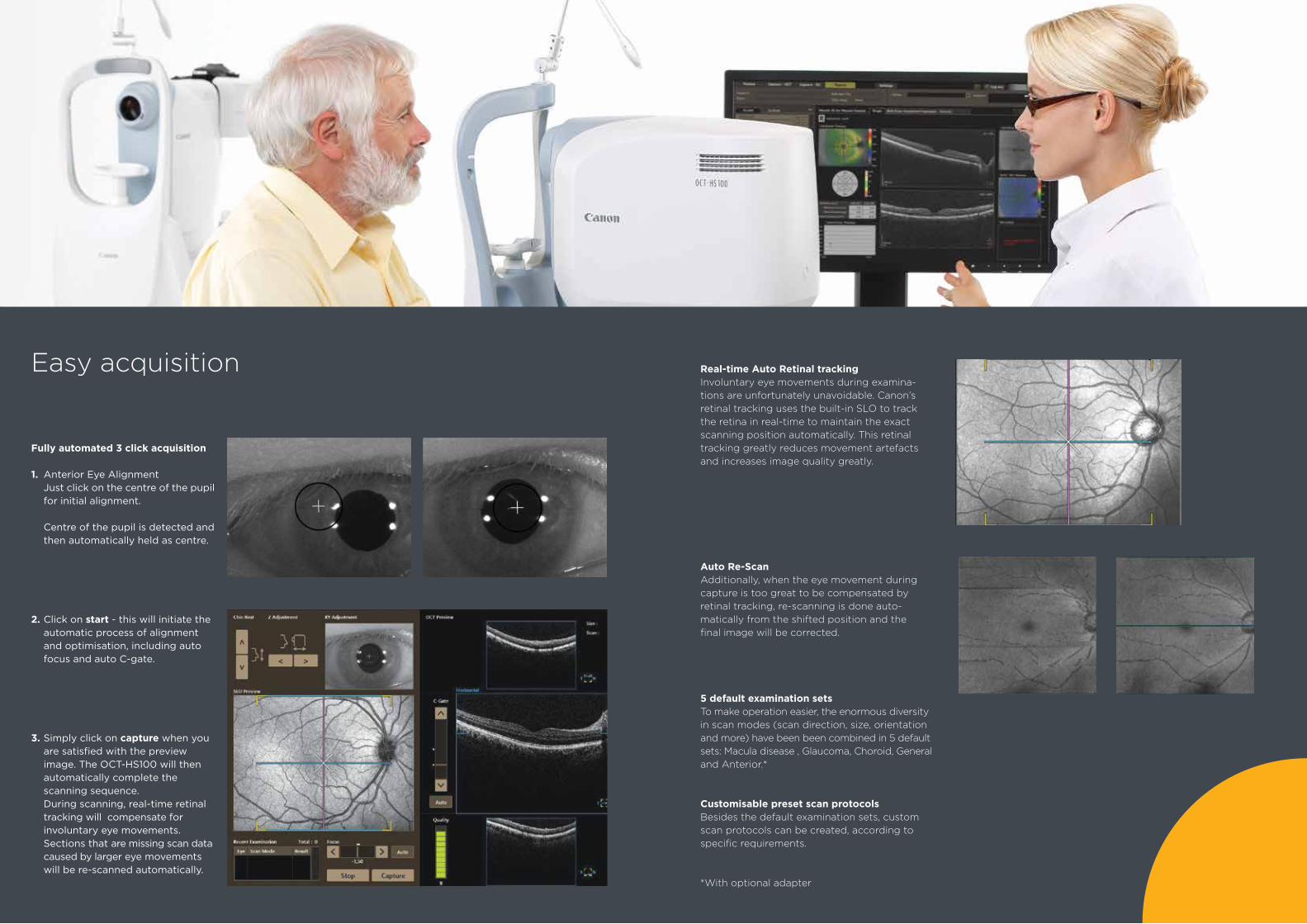
Easy acquisition
Fully automated 3 click acquisition 1. Anterior Eye Alignment Just click on the centre of the pupil for initial alignment.
Centre of the pupil is detected and then automatically held as centre.
2. Click on start - this will initiate the automatic process of alignment and optimisation, including auto focus and auto C-gate.
3. Simply click on capture when you are satisfied with the preview image. The OCT-HS100 will then automatically complete the scanning sequence. During scanning, real-time retinal tracking will compensate for involuntary eye movements. Sections that are missing scan data caused by larger eye movements will be re-scanned automatically.
Real-time Auto Retinal tracking Involuntary eye movements during examina-tions are unfortunately unavoidable. Canon’s retinal tracking uses the built-in SLO to track the retina in real-time to maintain the exact scanning position automatically. This retinal tracking greatly reduces movement artefacts and increases image quality greatly.
Auto Re-ScanAdditionally, when the eye movement during capture is too great to be compensated byretinal tracking, re-scanning is done auto-matically from the shifted position and the final image will be corrected.
5 default examination setsTo make operation easier, the enormous diversity in scan modes (scan direction, size, orientation and more) have been been combined in 5 default sets: Macula disease , Glaucoma, Choroid, General and Anterior.*
Customisable preset scan protocolsBesides the default examination sets, custom scan protocols can be created, according to specific requirements.
*With optional adapter

Combined ReportThis screen shows the analysis results comparing examina-tions of both eyes, accompanied with retinal images taken with a Canon retinal camera (optional) sharing the same database.
Versatile reporting possibilities
Macula Thickness AnalysisThis shows the tomogram image of the macula and analysis results of retinal thickness. The primary scanning direction is horizontal and priority is given to resolution in the horizontal direction.
NFL+GCL+IPL / GCL+IPL AnalysisThis shows the tomogram image from the mac-ula up to the optic disc and analysis results of retinal thickness. The primary scanning direction is vertical, and priority is given to resolution in the vertical direction.
Optic Disc AnalysisThis shows the thickness of RNFL (Retinal Nerve Fiber Layer) and analysis results ofthe optic disc shape.
Extensive Normative DatabaseComparison references available for full retinal thickness, NFL+GCL+IPL /GCL+IPL thickness and significance; RNFL thickness and significance
Single Analysis results of one eye.
BothAnalysis results comparing examinations of both eyes in the same scan mode and same size of scanning area, on the same date.
Comparison Analysis results comparing two examinations of eyes on the same side in the same scan mode, same size of scanning area, from different dates.
ProgressionAnalysis results comparing five examinations arranged in time sequence of eyes on the same side in the same scan mode, and same size of scanning area.

OCT-Angiography with Canon Angio Expert
Angio Expert Angio Expert is Canon’s angiography upgrade for the OCT-HS100.
High Definition The Angio Expert is also available in a high definition version, by simply adding the OCTA2 module to the standard OCT-A. This upgrade will provide even higher definition images and wider scans.
Easy upgradeExisting systems can be upgraded very easily without the need for hardware modification.
Detailed visualisationDetailed visualisation of the retinal blood vessels and high quality 3D display thanks to unsurpassed 3 μm optical resolution.
The corneal thickness analysis is shown as maps of corneal thickness, corneal grids, and tables.
The distance between two points, angles, and AOD (Angle Opening Distance) / TISA (Trabecular Iris Space Area) can be measured.
*with optional Anterior Segment Adaptor ASA-1
Anterior Segment Analysis*
Healthy cornea with contact lens.
OCT Angiography is image processing to depict blood vessels from OCT images. Blood vessels can be observed without using fluorescein dye.
Angio Expert 「AX」 ロゴ 決定案 2016.4.28 Visual Design Dept2.
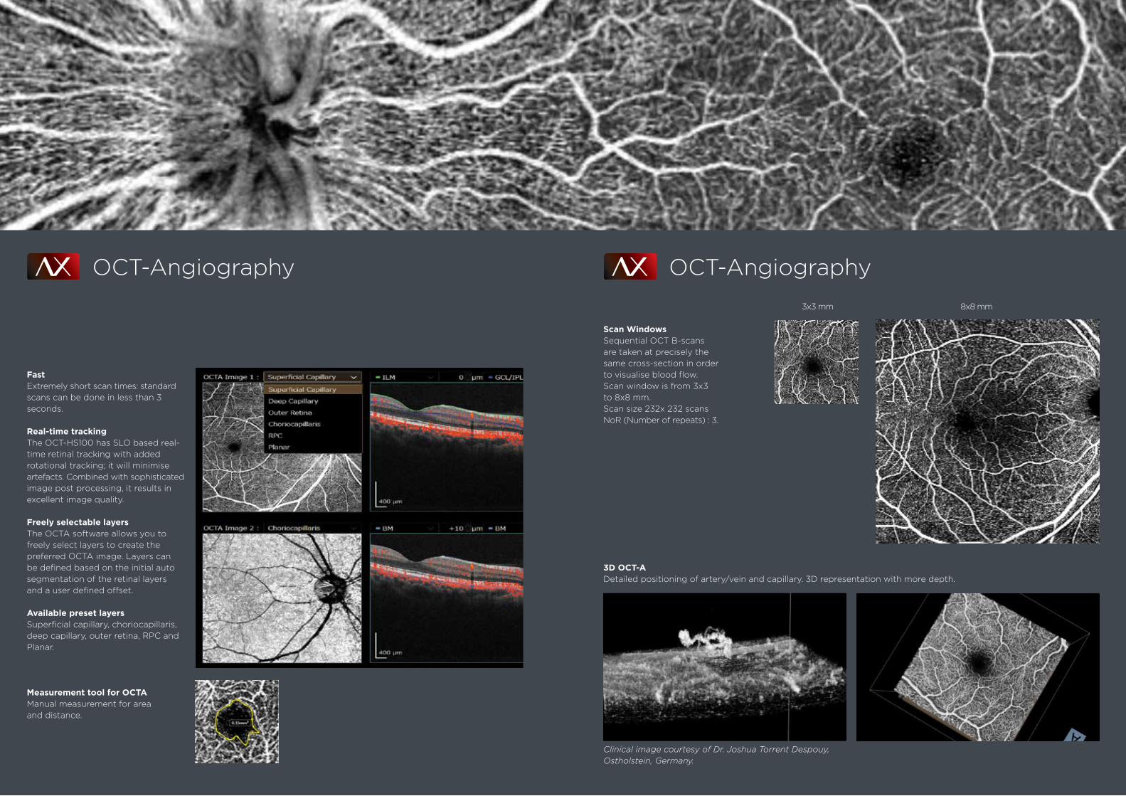
Fast Extremely short scan times: standard scans can be done in less than 3 seconds.
Real-time trackingThe OCT-HS100 has SLO based real-time retinal tracking with added rotational tracking; it will minimise artefacts. Combined with sophisticated image post processing, it results in excellent image quality.
Freely selectable layersThe OCTA software allows you to freely select layers to create thepreferred OCTA image. Layers canbe defined based on the initial auto segmentation of the retinal layers and a user defined offset.
Available preset layersSuperficial capillary, choriocapillaris, deep capillary, outer retina, RPC and Planar.
Measurement tool for OCTAManual measurement for area and distance.
OCT-Angiography
Angio Expert 「AX」 ロゴ 決定案 2016.4.28 Visual Design Dept2.
OCT-Angiography
Angio Expert 「AX」 ロゴ 決定案 2016.4.28 Visual Design Dept2.
3D OCT-ADetailed positioning of artery/vein and capillary. 3D representation with more depth.
3x3 mm 8x8 mm
Clinical image courtesy of Dr. Joshua Torrent Despouy, Ostholstein, Germany.
Scan WindowsSequential OCT B-scans are taken at precisely the same cross-section in order to visualise blood flow.Scan window is from 3x3 to 8x8 mm.Scan size 232x 232 scans NoR (Number of repeats) : 3.

Unlock the full potential of the OCT-HS100OCTA2 software module for Angio Expert - Wide field and high definition images.
Vertical Wide(232 x 696)
Large Square(696 x 696)
Medium Square(464 x 464)
Horizontal Wide(696 x 232 )
OCT-AStandard scan(232 x 232)
HD : OCTA + OCTA2
Clinical image courtesy of Tomohiro Iida, MD, PhD,Professor and Chairman, Tokyo Women’s Medical University.
Wide field OCT-A scans Wide field high quality images in a single scan:12 x 4, 10 x 10 mm.
10 x 10 mm
12 x 4 mm
Angio Expert 「AX」 ロゴ 決定案 2016.4.28 Visual Design Dept2.

Up to 200 scans can be averaged,resulting in fantastic image quality. The layer structure as well as thevitreous pleated structure can now be observed in even greater detail than ever before.
High definition scans
High definition OCT-A imageBy increasing the Number of Repeat scans from 3 to up to 10 times, the image quality will significantly improve,but with longer scan duration.
10 x 10 mm
Standard HD (OCTA2)
Cross scan 1 5 10 20 50 100 150 200
Multi Cross scan 1 5 10 20 30 50
Radial Scan 1 5 10 20 30 50
OCTA OCTA2 Scan Size 232 x 232 232 x 232 464 x 464 696 x 696 696 x 232 232 x 696
3 x 3 ● ●
4 x 4 ● ● ●
5 x 5 ● ●
6 x 6 ● ● ● ●
8 x 8 ● ● ● ●
9 x 9 ● ●
10 x 10 ● ●
9 x 3 ●
12 x 4 ●
3 x 9 ●
NoR 3 3, 4, 6, 10 3, 4, 6 3, 4 3, 4, 6 3, 4, 6
Number of averaging
Overview scan windows Courtesy of Kyoto University.

Stand alone● Capturing.● Reviewing and reporting.● Database and archive.
With viewing stations● Access the device database from other rooms. Review reports or access the study for full information.● 2 Concurrent Licenses, Install RX Viewer on as many PCs as you like! Only the actual concurrent use of the software is limited.
Server solution● Multiple modalities and viewers, storing all images on a central server.
OCT with camera● A Canon retinal Camera could be added to the system, sharing the same database.
Canon Ophthalmic Software PlatformRetinal Expert RX
The new multi modality platform for Canon retinal cameras and OCTs Designed for seamless integration with Electronic Medical Record Systems and third party software
RX CAPTURE
RX VIEWER
RX SERVER
RX server and RX viewers have to be purchased separately.
Seamless integration with patient management systems:
EMR can call RX directly via the command line interface.
RX opens on selected level: Patient, Capture or Report. Studies can be reviewed easily.
RX can call the data such as EMR and past exam data of the patient.
RX can call the other vendor’s software directly to reviewpatient record.
Extensive patient data input options ● Input data manually.● Import a list from the practice management system (CSV file).● Uses a modality worklist in a DICOM environment.
RX can be installed in a hospital’s virtual server environment (suchas VMware, Citrix and Microsoft servers) without relying on the client PC environment.
The OCT-HS100 takes up very little floor space and is flexible for use in most situations, even against a wall or in a corner.
Virtual Server
Command Line Interface
Launcher function
Very little floorspace
Retinal Expert 「RX」 ロゴ 2016.4.28 Visual Design Dept2.
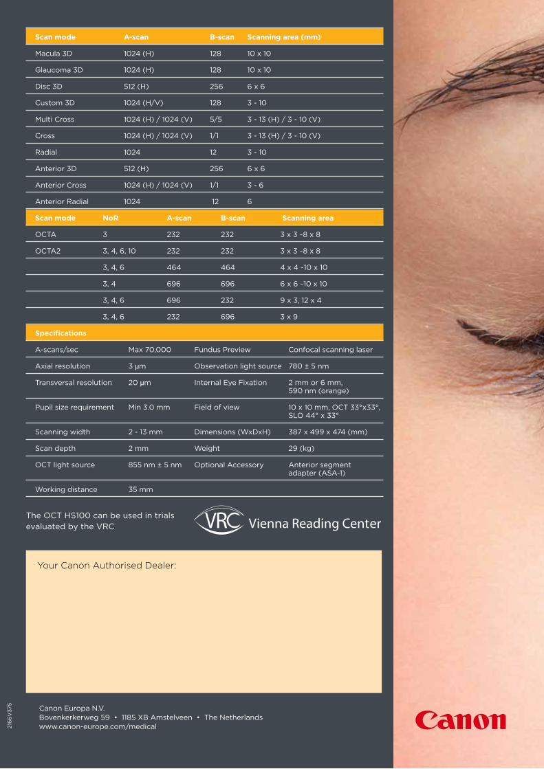
Scan mode A-scan B-scan Scanning area (mm) Macula 3D 1024 (H) 128 10 x 10 Glaucoma 3D 1024 (H) 128 10 x 10
Disc 3D 512 (H) 256 6 x 6 Custom 3D 1024 (H/V) 128 3 - 10 Multi Cross 1024 (H) / 1024 (V) 5/5 3 - 13 (H) / 3 - 10 (V) Cross 1024 (H) / 1024 (V) 1/1 3 - 13 (H) / 3 - 10 (V) Radial 1024 12 3 - 10
Anterior 3D 512 (H) 256 6 x 6
Anterior Cross 1024 (H) / 1024 (V) 1/1 3 - 6 Anterior Radial 1024 12 6 Scan mode NoR A-scan B-scan Scanning area
OCTA 3 232 232 3 x 3 ~8 x 8
OCTA2 3, 4, 6, 10 232 232 3 x 3 ~8 x 8
3, 4, 6 464 464 4 x 4 ~10 x 10 3, 4 696 696 6 x 6 ~10 x 10 3, 4, 6 696 232 9 x 3, 12 x 4 3, 4, 6 232 696 3 x 9 Specifications A-scans/sec Max 70,000 Fundus Preview Confocal scanning laser Axial resolution 3 µm Observation light source 780 ± 5 nm Transversal resolution 20 µm Internal Eye Fixation 2 mm or 6 mm, 590 nm (orange) Pupil size requirement Min 3.0 mm Field of view 10 x 10 mm, OCT 33°x33°, SLO 44° x 33° Scanning width 2 - 13 mm Dimensions (WxDxH) 387 x 499 x 474 (mm) Scan depth 2 mm Weight 29 (kg) OCT light source 855 nm ± 5 nm Optional Accessory Anterior segment adapter (ASA-1) Working distance 35 mm
Canon Europa N.V.Bovenkerkerweg 59 • 1185 XB Amstelveen • The Netherlandswww.canon-europe.com/medical21
66
V37
5
Your Canon Authorised Dealer:
The OCT HS100 can be used in trials evaluated by the VRC